kiwi first aid and veterinary care
DESCRIPTION
New Zealand Kiwi First Aid and Veterinary Care Kerri J. MorganTRANSCRIPT

Kerri J. Morgan
Kiwi first aid and veterinary care

Kiwi first aid and veterinary care
Kerri J. Morgan
Published by
Science & Technical Publishing
Department of Conservation
PO Box 10420, The Terrace
Wellington 6143, New Zealand

Cover: Okarito brown kiwi (rowi) having a computer tomography scan to diagnose a fractured
acetabulum.
Photo: B.D. Gartrell.
Individual copies of this book are printed, and it is also available from the departmental website in pdf
form. Titles are listed in our catalogue on the website, refer www.doc.govt.nz under Publications, then
Science & technical.
© Copyright April 2008, New Zealand Department of Conservation
ISBN 978–0–478–14383–6 (hardcopy)
ISBN 978–0–478–14384–3 (web PDF)
This text was prepared for publication by Science & Technical Publishing; editing and layout by Lynette
Clelland. Publication was approved by the Chief Scientist (Research, Development & Improvement
Division), Department of Conservation, Wellington, New Zealand.
In the interest of forest conservation, we support paperless electronic publishing. When printing,
recycled paper is used wherever possible.

CONTeNTS
Abstract 5
1. Introduction 6
1.1 Birds—the preservation reflex and stress 6
1.2 Legal aspects of treating protected wildlife 6
2. Assessment, first aid and stabilisation 8
2.1 Initial assessment 8
2.2 emergency stabilisation of a debilitated kiwi 12
2.3 Initial wound and fracture management 30
3. General anaesthesia 33
3.1 Important considerations in avian anaesthesia 33
3.2 Anaesthetic equipment 34
3.3 Patient monitoring 37
3.4 Anaesthetic emergencies 38
4. Diagnostics 39
4.1 evaluation of droppings 39
4.2 Haematology and biochemistry 41
4.3 Imaging 43
5. Husbandry 49
5.1 Nutrition 49
5.2 Housing 50
5.3 Transportation of kiwi 51
6. Traumatic injuries 53
6.1 Trap injuries 53
6.2 Bill injuries 54
6.3 Motor vehicle accidents 56
7. Diseases of kiwi 57
7.1 Gastrointestinal tract disorders 57
7.2 Respiratory tract disorders 61
7.3 Reproductive disorders 64
7.4 Yolk sac disorders in chicks 69
7.5 Neurological diseases 72
7.6 Nutritional diseases 74
7.7 Musculoskeletal disorders 74
7.8 Parasites 75

8. Wildlife Diagnostic Service 80
8.1 Preservation 80
8.2 Documentation 80
8.3 Packaging 81
8.4 In summary 82
9. Acknowledgements 83
10. References 83
Appendix 1
Kiwi first aid field kit 90
Appendix 2
Veterinary advice and referral centres 91
Appendix 3
Kiwi haematology and biochemistry reference ranges 93
Appendix 4
Captive diet formulation 94
Food Presentation for long-term care 94
Appendix 5
Availability of dietary products mentioned in the text 95
Appendix 6
Avian therapeutics formulary 96
Appendix 7
Wildlife submission form 102

5
© Copyright April 2008, Department of Conservation. This paper may be cited as:
Morgan, K.J. 2008: Kiwi first aid and veterinary care. Department of Conservation, Wellington. 103 p.
Kiwi first aid and veterinary care
Kerri J. Morgan
BVSc, MACVSc (Avian Health), PGDipVCS (dist.)
New Zealand Wildlife Health Centre, Institute of Veterinary, Animal and
Biomedical Sciences, Massey University, Private Bag 11 222,
Palmerston North 4442, New Zealand
email: [email protected]
A B S T R A C T
This document provides information about the treatment of sick or injured
kiwi (Apteryx spp.) for veterinarians, conservation field workers and wildlife
park staff. It incorporates basic techniques to stabilise sick or injured kiwi.
Specific diseases and common injuries that have been seen in kiwi are also
addressed. Diagnostic and treatment techniques specific to each condition
and, in some cases, specific to kiwi, are included. The information given is
not intended to be a complete reference for the veterinary treatment of kiwi,
and is subject to change as more information on diseases or injuries and their
treatment becomes available.
Keywords: kiwi, Apteryx, first aid, veterinary care, injuries, disease, parasites,
New Zealand
6
1. Introduction
1 . 1 B I R D S — T H e P R e S e R V A T I O N R e F L e x A N D S T R e S S
In many circumstances, it is immediately obvious when a bird is sick or
injured. However, in other instances it is less obvious and disease processes
may be challenging to detect.
Birds go to great lengths to hide clinical signs of illness. In the wild, sick
birds attract the attention of predators and, in flocking species, a sick bird
will be shunned by flock mates (Hume 2000). This masking of signs of illness
is known as the ‘preservation reflex’. Birds generally do not look sick until
they are in an advanced state of illness and near collapse (Cannon 1991).
Identifying that birds are sick when they are in the early stages of disease
requires detection of subtle clinical signs. Initial warning signs may include a
reduction in food intake and a drop in body weight. A bird with intermittent
clinical signs of illness is definitely unwell, and a bird with constant signs is
seriously sick.
Stress can play a significant role in the pathogenesis of avian disease. Healthy
birds can accommodate some degree of stress, but sick birds are universally
intolerant of all types of stress (Cannon 1991). Birds with subclinical problems
(i.e. diseases without detectable manifestations) often become sick when
stressed. In the wild, stressors include starvation, territorial aggression, and
physiological stress such as breeding. In captivity, stress may be induced by
malnutrition, poor hygiene and sanitation, poor handling, and overcrowding.
Furthermore, it must be remembered that captivity removes the birds’ natural
escape mechanisms (Cannon 1991). In captive birds, malnutrition caused by
an imbalanced diet is one of the most common forms of chronic stress, and
this may contribute to disease (see section 7.6).
1 . 2 L e G A L A S P e C T S O F T R e A T I N G P R O T e C T e D W I L D L I F e
The Department of Conservation (DOC) has a Captive Management
Policy (DOC 2003). Veterinarians undertaking wildlife rehabilitation
are required under this policy to obtain an authority (permit) from
DOC to hold absolutely protected wildlife. Contact your local DOC
office for more information. Contact details for DOC offices can be found on
the DOC website (www.doc.govt.nz > Publications > About DOC > Role >
Policies & plans > Captive Management Policy).
When a kiwi is brought in for treatment, there are two priorities. The first
is to administer emergency treatment and pain relief and to assess the bird’s
injuries/disease status. The second is to inform DOC.

7
According to the Captive Management Policy, DOC will:
‘Support the treatment of injured absolutely protected wildlife only in cases
where it is likely that the animal will: make a good recovery and be suitable
for release into the wild; become part of an approved Species Recovery
Programme. If the animal’s full recovery is unlikely and it cannot be placed
in an authorised programme, treatment should be withheld and the animal
euthanised.’ (DOC 2003: section 2.2.10, p. 7.)
Consult with DOC regarding the prognosis of treatment and possible options
for release of the bird or its use in the Kiwi Recovery Programme.
If further care is required after initial treatment has been completed, the kiwi
may only be transferred (after consultation with DOC) to a facility which
possesses an authority to hold absolutely protected wildlife. If/when the kiwi
is suitable for release into the wild, DOC must be consulted to determine the
appropriate location and process for release.

8
2. Assessment, first aid and stabilisation
2 . 1 I N I T I A L A S S e S S M e N T
Initial assessment of a sick kiwi involves taking a history and performing a
distance examination prior to handling the bird. This may give the assessor an
indication of the appropriateness of further handling for physical examination
and initiation of therapeutics. For example, a bird in respiratory distress may
substantially benefit from a period of pre-oxygenation and warmth prior to
further handling.
2.1.1 History
Gaining a thorough history is an important part of assessment of the avian
patient. The ability to obtain a complete history is highly dependent upon
the bird’s situation, which will vary from captivity, where birds are visually
assessed by their keepers daily, to completely wild, with very little known
history.
Important points to note
What is the age and sex of the bird?•
Is it a wild or captive bird?•
Captive birds
What is the origin of this bird (e.g. captive-raised, wild-caught, transferred •
from another captive institution)?
How many birds (in particular, other kiwi) are in contact with this bird?•
How long has the bird been in the particular kiwi house or other captive •
facility?
Have any new birds been introduced recently?•
Is the bird in contact with people?•
Is the bird housed indoors or outdoors? Obtain a description of the •
enclosure, including substrate. Is there any potential for exposure to
foreign objects (nails, screws, etc.)?
Have there been any recent changes in substrate, sources of leaf litter, etc.?•
What is the bird’s diet? Have there been any recent changes in diet? What •
is the water source for the bird?
How many birds are affected?•
When was the bird noticed to be sick?•
What are the observed abnormalities?•
What is the breeding history and current status of the bird? Is there a •
possibility the bird is gravid (carrying an egg)?
Are there any changes in the consistency, frequency or appearance of the •
bird’s droppings?
9
Are there any changes in respiration, e.g. open-mouth breathing, wheezing, •
sneezing, dyspnoea (respiratory exaggeration)?
Has the bird had any treatment to date, such as fluids, medications •
(including, but not limited to, antibiotics, antifungals, analgesics,
corticosteroids, antiparasitics)?
Does the bird have any possible access to toxic materials such as rat bait, •
snail bait, herbicides, toxic plants (e.g. karaka (Corynocarpus laevigatus)
berries)
Wild birds
Where was the bird found?•
What are the observed abnormalities?•
When was it found? If there has been a delay between the bird being •
found and the current presentation, where has it been previously?
Has it had any treatment (treatment may include fluids and medications •
including, but not limited to, antibiotics, antifungals, analgesics,
corticosteroids, antiparasitics)?
Has the bird been offered food? Has it eaten?•
Is the breeding history of this bird available?•
Has it passed any droppings? If so how did they look (volume, colour, •
consistency of faeces, urates and urine)?
2.1.2 The distance examination
A distance examination should ideally be undertaken prior to a bird’s capture
and restraint, i.e. prior to the induction of further stress. This will allow the
handler to get some idea of the severity of the disease, and how well the
bird may cope with the stress of further restraint at this stage. However, a
distance examination will not always be possible. If the bird is presented
in a transport box, observe the bird for a few moments in the box before
picking it up.
A normal bird should be alert, standing straight and even on both feet, with
barely visible respiratory efforts at rest (Hume 2000).
Points to note include:
Mental status
Is the bird bright, alert and reactive to stimuli?•
Or• is it quiet and dull?
Or• moribund?
Posture
Is the bird standing?•
If so, is it standing evenly on both legs?•
Is it using its bill for balance?•
Gait
Is the bird ambulating normally?•

10
Respiration
This should be barely visible. Abnormalities include:
Open-mouth breathing•
exaggerated respiratory effort•
Respiratory noise or click on inspiration/expiration•
Ocular or nasal discharge•
Feathering
Is there any obvious damaged or misshapen feathering?•
Are feathers fluffed up?•
Wounds
Are there any signs of injury (fresh or dry blood, cuts, open wounds, •
matted feathers, bald patches, etc.)?
Body symmetry
Are there any obviously damaged or misshapen parts of the body?•
2.1.3 Handling and restraint
Once these visual assessments have been completed, the decision whether
to handle and restrain the bird for further diagnostics and supportive therapy
should be made. A bird in respiratory distress will invariably benefit from
supplemental oxygen, warmth and rest prior to further handling.
In many cases, there will probably be an experienced kiwi handler present.
However, if not, the following description of handling kiwi by R. Jakob-Hoff
may be useful:
‘Grasp both legs above the hock joint between the thumb and middle finger
of the right hand with the index finger between the two legs. The left hand
supports the ventral and lateral body into a sitting position in the crook of the
right elbow. Direct the bird’s bill and eyes under cover of the left arm.
‘An alternative hold is often used in the field with the holder in a sitting
position. Grasp the two legs as previously described, hold the bird in an
upside-down position with the dorsal body placed on the lap of holder and
the kiwi’s head under the left arm.’ (Doneley 2006)
Taping the legs of kiwi together during handling should occur only if absolutely
necessary, and then just for short periods. Kiwi should never be transported
with legs taped. There is evidence that taping legs together causes severe
muscle damage, as indicated by marked elevations in creatine kinase in great
spotted kiwi (Apteryx haastii) that were transported with legs strapped
together (unpubl. data). If taping is necessary for weighing or transmitter
changes in the field, current recommendations are for periods no longer than
15–20 minutes. However, the impact of leg taping on kiwi physiology requires
further study. (B.D. Gartrell, Massey University, pers. comm.).
2.1.4 Physical examination
A systematic and thorough physical examination is important to ensure that
subtle abnormalities are not overshadowed by obvious injuries. Doneley
(2006) gives the following summary as a guide to examination of ostriches.

11
A similar approach can be applied to examination of kiwi. The extent to
which the described examination can be done depends upon expertise and
equipment available.
Head examination
examine the eyes, ears (caudo-lateral to the lateral canthus of the eye), bill
and nostrils (at the tip of the bill), looking for defects and discharges.
Oral cavity
examine the choana (slit on the roof of the mouth which communicates with
the upper respiratory tract), glottis (opening into the trachea), tongue, and
mucosa lining the oral cavity.
Body condition
As kiwi are flightless, it is not possible to determine the body condition
score by palpation of the pectoral muscles. Palpation of the epaxial muscles
(adjacent to the spine) and assessing the prominence of the ribs will give a
subjective indication of kiwi body condition.
Heart and respiratory system
Auscultate (preferably with a paediatric stethoscope) for abnormal cardiac
and respiratory sounds. The rigid avian lungs lie in the dorsal cranial body
wall, and are best listened to over the dorsum, halfway between the vestigial
wings. [Although less developed in kiwi than in flighted avian species
(B.D. Gartrell, Massey University, pers. comm.), the air sac system extends
throughout the entire coelomic cavity.]
Normal kiwi heart rate: 70–240 beats per minute
Normal kiwi respiratory rate:12–60 breathes per minute (Doneley 2006)
Palpate the abdomen
The firm gizzard lies in a caudoventral position, slightly to the left of the
midline.
Legs and feet
Observe the legs and feet for gross abnormalities. Limbs should be palpated
for obvious fractures and joint effusion (abnormal fluid). In chicks, tibiotarsal
alignment should be evaluated for rotational defects (see section 7.7.1).
Skin and feathers
The skin and feathers should be evaluated for external parasites. Many kiwi
carry ticks. These are often found in large numbers over the head region,
where the bird is unable to preen them off. They are also common in the
external ear canal (see section 7.8.3). Kiwi also carry lice, and it has been
observed (in birds in general) that debilitated individuals tend to have
increased numbers of lice.
The skin should also be evaluated for traumatic injuries and other lesions,
including dermatitis, which may be indicative of a vitamin B deficiency (see
section 7.6.2).
Vent
The vent should be examined for prolapse, trauma and vent soiling with
faeces and urates.
12
Neurological examination
Compared with other avian species, kiwi show minimal reliance upon vision,
as indicated by eye structure, visual field topography and reduced visual
centres in the brain (Martin et al. 2007). Functionally blind kiwi have been
found in the wild. These birds were surviving well, and for this reason blindness
should not be grounds for non-release. Instead of eyesight, kiwi have an
increased reliance upon olfactory and tactile information as an adaptation to
their nocturnal behaviour (Martin et al. 2007). However, despite the fact that
kiwi might not need good vision to survive, cranial nerve evaluation should
include assessment of the pupillary light reflex, the menace reflex and the
corneal reflex. In performing these examinations it is important to note some
differences that are particular to birds. There is no consensual light reflex,
and birds are extremely sensitive to movement of air across the surface of
their eye, which may confuse results of the menace reflex. Birds also have
some voluntary control over their pupil size because of skeletal muscles
within the iris. Anisocoria (asymmetrical pupil size) may be indicative of
cranial trauma. Other abnormalities may include strabismus (deviation of
the eye) and nystagmus (involuntary movement of the eyeballs in unison).
Other cranial nerve abnormalities may cause reductions in olfactory ability,
torticollis (head tilt), dysphagia (difficulty in swallowing), and alterations in
facial sensation.
Segmental reflexes in kiwi include the leg withdrawal and the vent reflex.
These reflexes require intact peripheral nerves without the need for an
intact spinal cord. Withdrawal of the leg when stimulated (e.g. a toe pinch)
indicates an intact peripheral nerve supply to that limb. Perception of pain
requires an intact spinal cord in addition to an intact peripheral nervous
system. Indications that a bird can perceive a painful stimulus may include
an attempt to escape, or the bird looking at the painful area in response to
the stimulus.
The vent reflex can be elicited by pinching the skin adjacent to the vent. The
vent should normally contract in response to this stimulus.
It should be noted that many reflexes and responses may be confounded
by pain, and the mechanical inability to move severely fractured limbs. The
neurological examination is most useful for initial assessment, and to evaluate
changes in neurological function over time.
2 . 2 e M e R G e N C Y S T A B I L I S A T I O N O F A D e B I L I T A T e D K I W I
The priority in the initial 24 hours after presentation of a sick or injured
kiwi is to stabilise the patient. Stabilisation involves attempting to regain
physiological homeostasis in what are often hypovolaemic, hypothermic,
malnourished and septic patients. The extent to which stabilisation can be
achieved is dependent upon the available equipment and expertise.
It may be advisable for field staff working with kiwi to carry a first aid kit
containing useful items for provision of at least warmth and fluid therapy
(Appendix 1).

13
The following text details the achievable priorities for treating sick or injured
kiwi in the field and in the veterinary hospital. It is recognised that there will
be many occasions and situations where the procedures that are achievable
will fall somewhere between these two extremes.
The goals of treatment in the first 24 hours include rehydration, provision of
warmth, stabilisation of fractures, dressing of open wounds, pain relief, and
placement of the kiwi in a warm, stress-free environment.
2.2.1 In the field
Field workers should be able to provide the debilitated kiwi with the
following:
Fluid therapy (see section 2.2.5)
Fluids may be administered orally or by subcutaneous injection.•
Warmth (see section 2.2.4)
In-the-field heat therapy may be provided with heat pads or hot water •
bottles.
Fracture and wound management (see section 2.3)
Lower limb fractures should be dressed and bandaged in order to prevent •
further injury to the limb and contamination of wounds, as well as reducing
pain associated with the injury.
Quiet, stress-free environment (see section 5.2)
The bird should be placed in a warm, quiet, dark and stress-free environment •
if there is a delay in its transportation out of the field.
2.2.2 In the veterinary clinic
In the veterinary clinic, the first priority must be to address life-threatening
dyspnoea (breathing difficulties) if present.
Provision of oxygen (see section 2.2.3)
Dyspnoeic patients will benefit from the provision of supplemental oxygen, •
either via a mask or in an oxygen tent, prior to further handling.
An upper airway obstruction requires immediate intervention by placement •
of an air sac cannula through which oxygen can be supplied.
Fluid therapy (see section 2.2.5)
Intravenous (IV) access should be prepared primarily for fluid therapy, as •
well as IV administration of analgesics and anti-infective medication. Oral
and subcutaneous fluid therapy may also be used, but these are less than
optimal in the compromised patient.
Intraosseous cannulation provides access for critical fluid and medical •
therapy in the event that intravenous access is not possible. The suggested
site in kiwi is the tibiotarsus via the tibial crest.
Warmth (see section 2.2.4)
Warming hypothermic kiwi is critical, and can be achieved by a variety •
of methods.

14
Analgesia (see section 2.2.7)
Opioids (butorphanol) may be given for analgesia. Non-steroidal anti-•
inflammatory drugs (e.g. carprofen, meloxicam) should only be used in
well-hydrated patients.
Anti-infective medications (see section 2.2.8)
Antibiotics should be started immediately if the patient is suspected to be •
septic, or has injuries that are likely to become infected.
Bandaging and wound management (see section 2.3)
The goal of initial bandaging and wound management is to prevent further •
desiccation of the wound, and to provide external stability for lower
limb fractures. This is important in preventing further tissue damage and
relieving pain associated with unstable fractures.
Initial diagnostics (see sections 4.1 and 4.2)
Ideally, a blood sample should be taken in the initial stages of treatment •
to enable investigation of haematological and biochemical parameters
before any treatment is given. At the very least, the packed cell volume
(PCV) and total plasma protein (TPP) should be assessed.
A faecal sample should also be examined to investigate the presence of •
parasites, including coccidia.
Quiet, dark, stress-free environment (see section 5.2.1)
The bird should be placed in a warm, quiet, dark and stress-free •
environment, away from foot traffic and other animals.
N.B. Corticosteroids are contraindicated in the stressed or septic avian
patient, as they may significantly suppress the hypothalamic
pituitary axis, and may result in a severe and lasting
immunosuppression.
2.2.3 Oxygen therapy
Stress resulting from handling and other procedures can be fatal in a severely
debilitated bird. Clinical signs suggestive that handling for examination is
contraindicated until the bird is stabilised include prolonged dyspnoea, and
prolonged panting and gasping for air (Harrison et al. 2006). These patients
benefit significantly from oxygen therapy prior to handling.
Prior to examination, oxygen may be administered in a chamber where the air
comprises 40–50% oxygen. An intensive care unit or incubator can be used or,
more simply, a cage covered with a plastic bag (ensure an outflow is present)
(Harrison et al. 2006). A facemask fashioned for kiwi (see section 3.2.3)
is effective for short-term treatment if an oxygen enclosure is unavailable,
but the stress resulting from handling the bird to attach the facemask may
outweigh any benefit from the extra oxygen.
Air sac cannulation is well described for other avian species. This procedure
is indicated for upper respiratory tract obstructions at the level of the
trachea or syrinx (Harrison et al. 2006), providing the bird with the ability
to inspire and expire through the caudal air sacs, bypassing the trachea. Air
sac cannulation is an emergency procedure, and involves placement of a
cannula into the caudal thoracic or abdominal air sacs (Harrison et al. 2006).

15
However, adaptations for flightlessness have resulted in a reduction in air sac
development in kiwi, making this a difficult procedure to perform in these
species (B.D. Gartrell, Massey University, pers. comm.). Several attempts
at using this procedure with kiwi have been made, with varying success,
and its use in kiwi is still at an early stage. For further information about
this procedure, contact the New Zealand Wildlife Health Centre, Massey
University (see Appendix 2 for contact details).
2.2.4 Warmth
Debilitated birds are unable to thermoregulate as effectively as healthy birds,
and are often hypothermic. Provision of warmth is indicated for most sick
and injured kiwi. Options for providing heat vary depending on the situation
and availability of equipment.
The gold standard for the provision of warmth is an intensive care unit
specifically designed for small animal use. Alternatively, a human paediatric
incubator works well. Both pieces of equipment allow thermostatic and
humidity control of the air within them. Other options include hot water
bottles, wheat sacs and heat pads, heat lamps or, simply, a heater in a small
room. All approaches have advantages and disadvantages.
Hot water bottles and heat pads are readily accessible and cheap and are
ideal for taking into the field. However, care must be taken to ensure that
birds do not get burnt from direct contact with them, and it is recommended
that heated objects such as hot water bottles be wrapped in a towel before
use. A major disadvantage of hot water bottles, heating pads and wheat bags
is that they cool down and need to be removed for reheating, which causes
disturbance and further stress to the bird.
Heat lamps are a good source of overhead heat. They can be purchased from a
pet store or lighting store. Bulbs are either ceramic (without glow) or infrared.
Heat lamps should never be left unattended, and care must be taken to not
burn the bird, especially with recumbent birds that cannot move away from
the heat source.
A heater in a small room may work well also. Ideally, the heater should have
a thermostat which can be set to regulate the temperature in the room.
The desirable ambient temperature for most sick birds is 29–30°C, with 70%
humidity (Harrison et al. 2006). However, kiwi have inherent lower body
temperatures than other avian species, and it may be more appropriate to
house them at lower temperatures (e.g. 25–26°C). Birds should be monitored
closely for signs of heat stress. Clinical signs of hyperthermia may include
open-mouth breathing and the flattening of feathers to the body. Once the bird
has reached its normal body temperature (c. 38°C), the ambient temperature
should be reduced, as kiwi do not tolerate high ambient temperatures well.
If the heat source is not under thermostat control, a maximum-minimum
thermometer should be installed in the enclosure to monitor any fluctuations
in temperature. If an ICU or incubator is unavailable, placing a small dish of
water or a moist heated towel in the bottom of the enclosure will provide
some humidity with the warm air (Harrison et al. 2006).

16
2.2.5 Fluid therapy
It should be assumed that debilitated birds will be hypovolaemic upon presentation. Hypovolaemia refers to a reduction in circulating blood flow, with a resultant reduction in hydration of body tissue. Hypovolaemia may be caused by a combination of blood loss, shock (including septic shock), and severe dehydration due to a prolonged lack of food and water intake or excess loss of body fluids.
Hypovolaemic animals require rigorous fluid therapy. Goals of the fluid therapy plan include:
Replacement of lost fluids.•
Provision of maintenance fluids to cover normal daily losses, estimated at •50 mL/kg/day.
Replacement of any ongoing abnormal losses (e.g. diarrhoea, continued •bleeding).
Assessment of dehydration and hypovolaemia
The bird’s history may assist determination of the likelihood of hypovolaemia. The likely route and duration of any fluid loss should be assessed (e.g. blood loss, diarrhoea), as well as any indications suggestive of septic shock. Fluid and food intake immediately prior to presentation should also be taken into account.
Assessment of hydration status in a bird is subjective and requires practice; however, in general, many workers assume a loss of 10% of total blood volume in a debilitated bird (Monks 1996).
Below is a guide to a more detailed subjective assessment of hypovolaemia in the avian patient (Redig 1984; Abiu-Madi & Kollias 1992; Monks 1996):
< 5% dehydrated No detectable clinical signs.
5–6% dehydrated Subtle clinical changes—subtle loss of skin elasticity.
6–8% dehydrated Moderate dehydration—skin tenting is visible over the dorsal tarsometatarsus (i.e. when the skin is pinched, it takes longer than normal to return to its normal position).
Dry mucous membranes (oral, cloacal, conjunctival).
Decreased sliding of the skin over the sternum.
10–12% dehydrated Severe dehydration—all of the above plus skin turgor, sunken eyes, thick and stringy mucous in caudal pharynx, central nervous system depression, lethargy, weakness.
12–15% dehydrated SHOCK. Death is imminent unless therapy is rapidly established.
Objective assessment of hydration status can be achieved by evaluation of haematological and biochemical parameters. Dehydration may increase the packed cell volume (PCV) by 15–30%, and the total plasma protein (TPP) by 20–40% (Lumeij 1987; Martin & Kollias 1989).
It should be remembered, however, that TPP may be low in a starving bird regardless of hydration status (Kaufman 1992). Also, PCV may be low due to any chronic disease, and this may mask any rise due to dehydration (Martin
& Kollias 1989; Hoefer 1992).

17
Initially, acute blood loss results in no change in PCV and TPP. As the time between haemorrhage and presentation increases, a reduction in PCV and
TPP becomes more likely (Forsyth et al. 1999).
Routes of administration of fluids
Regardless of the route of administration, all fluids should be warmed to body temperature (38–39oC) (Quesenberry & Hillyer 1994). Using warm fluids is particularly important with neonates and with intravenous or intraosseous administration of fluids for hypothermia or shock (Abou-Madi & Kollias 1992).
Oral fluids
Oral fluids are indicated for treatment of mild dehydration, or for daily maintenance fluids only (Quesenberry & Hillyer 1994). They are inadequate in birds with sudden or excessive fluid losses. Oral fluids should not be given to birds that are seizuring, laterally recumbent, regurgitating, in shock or have gastrointestinal stasis (Quesenberry & Hillyer 1994). For effective rehydration, oral fluids need to be readministered within 60 to 90 minutes of the first treatment (Quesenberry & Hillyer 1994).
Types of oral fluids include:
1. Oral electrolytes
Calf electrolytes, e.g. Revive™, Vytrate™, Dexolyte™.•
Human electrolytes, e.g. Gastrolyte™, Powerade™ (though not ideal •because of high levels of artificial colours and flavour).
Bird-specific electrolytes, e.g. Polyaid™ (includes nutritional support).•
2. Intravenous solutions which may be given orally
LRS, 0.9% NaCl, 2.5–5% dextrose.•
The relatively small capacity of the kiwi gastrointestinal tract limits the volume of fluids that can be administered orally. The kiwi lacks a distinct crop and has only a small proventriculus (Fergus et al. 1995). This limits the capacity for storage of food and fluids. Administration of excessive fluids may result in regurgitation and possible aspiration into the trachea. The author recommends that no more than 20 mL of oral fluids be given to an adult kiwi at any administration, and no more than 3–4 mL to a kiwi chick. This should be given slowly. To account for individual variation, it may be safer to give a smaller amount than this initially, and work up to this larger volume over subsequent administrations.
Oral fluids are administered into the distal oesophagus using either a metal avian crop tube or a silicone rubber feeding tube attached to a syringe. With the head well restrained and the neck extended, the tube should be passed beside the tongue on either the left or right side of the oral cavity, avoiding the glottis (opening to the trachea) (Fig. 1). The tube is then passed down into the oesophagus which lies on the right side of the neck. The glottis is clearly visible as an opening at the base of the tongue, identifiable by movement of the glottic cartilages as the bird breathes. When the tube is in place, the glottis should be observed to ensure that the apparatus is clear of this structure. Fluid should then be slowly syringed through the tube while the back of the oral cavity is observed for reflux. If reflux occurs, the tube should be removed and the bird’s head released to allow it to swallow and shake its head to clear the glottis.

18
Subcutaneous fluids
Subcutaneous fluids should be utilised for mild dehydration and for
maintenance fluid therapy only (Quesenberry & Hillyer 1994). This route
of administration is ineffective at treating hypovolaemia and moderate to
severe dehydration, because associated peripheral vasoconstriction reduces
the absorption of fluid in hypothermic or shocked patients (Forsyth et al.
1999).
Types of subcutaneous fluids include:
Lactated Ringers solution (LRS)•
NaCl 0.9%•
Fluids for subcutaneous fluid administration need to be isotonic (Forsyth et
al. 1999). Subcutaneous administration of 5% dextrose should be avoided
because equilibration of the extracellular fluid with a pool of electrolyte-free
solution may result in an aggravation of an electrolyte imbalance (Forsyth et
al. 1999). Sterile abscesses and local fluid accumulation may also occur at
the site of subcutaneous dextrose administration (Forsyth et al. 1999). Fluids
for subcutaneous administration must be sterile to avoid causing infection at
the site of injection.
Sites for administration of subcutaneous fluids are the:
Intrascapular region—this is the skin over the dorsum (back) of the bird, •
halfway between the vestigial wings.
Inguinal fold—this is the fold of skin cranial to (in front of) the stifle •
(knee), between the stifle and the body wall.
Fluids administered at each site should not exceed 5–10 mL/kg bodyweight,
with only one puncture hole per administration site (Quesenberry & Hillyer
1994). Use small needles (25–27 gauge); a butterfly catheter may assist
administration.
Figure 1. Intubation of a kiwi with a Cole™ endotracheal tube. The stepped portion of the
endotracheal tube will be advanced to sit against the glottis (arrowed) to form a
seal. Uncuffed endotracheal tubes are used in birds
because of the complete cartilaginous rings in their
trachea. If cuffed tubes are used, the cuff should
never be inflated, as this can cause pressure necrosis and subsequent stricture of the
trachea. Photo: K. Morgan.

19
Intraperitoneal fluids
Never administer intraperitoneal fluids to birds. This is likely to result in
fluid getting into the air sacs, and there is a risk of organ laceration (Monks
1996).
Intravenous fluids
Intravenous fluid therapy is indicated in cases of severe debilitation or severe
hypovolaemia (Harrison et al. 2006). The author recommends intravenous
fluid administration in the majority of cases of debilitated kiwi, and for all
surgical cases.
Intravenous access also allows the administration of antibiotic and analgesic
medication with minimal stress on the bird.
To minimise stress, the author recommends placement of the IV catheter
while the bird is under general anaesthesia. In kiwi, intravenous access points
are limited to the bilateral medial metatarsal veins, and the right jugular vein
as a last resort. The medial metatarsal vein is most accessible proximal to
the first phalange (medial) and courses in a caudal direction (Fig. 2). In some
cases, the right jugular vein may be useful, but it is difficult to maintain a
catheter in this position.
A 20- or 22-gauge catheter is recommended for adult kiwi, and a 24-gauge
catheter for chicks. The catheter should be securely fixed with tape, and
covered in a self-adherent dressing. extension sets are useful (Fig. 3).
Figure 2. Medial distal limb of a kiwi (with a 22-gauge catheter in place). White
lines show position of the medial metatarsal vein.
Photo: K. Morgan.
Figure 3. extension set connected to a 22-gauge
catheter in medial metatarsal vein of the right leg of a kiwi.
Photo: K. Morgan.

20
Kiwi tolerate intravenous catheters, and they can be maintained for up to
7 days with an aseptic technique. Complications of catheterisation include
thrombosis, cellulitis and loss of patency (Monks 1996).
Intravenous fluids can be given either as an intermittent bolus of fluid, or by
constant infusion in an IV drip, ideally with a fluid pump.
If a fluid pump is unavailable, it is wise to administer fluids as a bolus to
prevent accidental overhydration which may occur with a normal drip set-
up. Bolus fluid administrations should be given slowly intravenously, at a
rate of 10–25 mL/kg over 5–7 minutes. This should be repeated at 3–4 hourly
intervals for the first 12 hours, then every 8 hours for the next 48 hours.
Thereafter, IV boluses can be given twice daily for maintenance therapy
(Harrison 1986).
Intraosseous fluids
Intraosseous fluid therapy is indicated in severely debilitated birds where it is
not possible to gain intravenous access. It allows the parenteral administration
of fluids (including blood), nutritional support and glucose, analgesia,
antimicrobials and drugs for cardiovascular resuscitation. Hypertonic or
alkaline solutions should be avoided, as they are painful on administration
(Quesenberry & Hillyer 1994).
Intraosseous catheterisation is a painful procedure, and in most cases should
be done under general anaesthesia. exceptions to this include very moribund
birds that are unlikely to survive an anaesthetic.
Because they lack significant wing bones, intraosseous catheter sites in kiwi
are limited to the tibial crest (Fig. 4). In birds, the femur should be avoided for
intraosseous cannulation as this bone is pneumatic, having communications
with the air sac system (Quesenberry & Hillyer 1994).
Figure 4. Intraosseous catheterisation in the left
tibial crest of the tibiotarsus of a kiwi, using a 20-g
intravenous catheter and butterfly catheter.
Photo: J. Youl.

21
In young birds, an 18- to 22-gauge, 1.5- to 2.5-inch spinal needle is ideal for
cannulation (Quesenberry & Hillyer 1994). An 18- to 22-gauge hypodermic
needle will also suffice. In adult kiwi, a kirschner wire and chuck may be used
to drill a hole for cannulation. Aseptic preparation of the site is extremely
important to avoid introducing infection to the area. The needle/kirschner
wire should be advanced into the bone marrow via the tibial crest, passing
distally. Pressure should be applied with a slight rotating motion. Once the
cortex is penetrated, the needle/kirschner wire should slide easily, with little
resistance. If predrilling the cannulation hole with a kirschner wire, the wire
is removed and the needle inserted. Bone marrow occluding the needle lumen
should be aspirated and flushed with a small amount of heparinised saline.
The catheter should be secured with a light padded bandage. Initial fluids
should be given slowly while checking for subcutaneous swelling, which
indicates improper catheter placement (Quesenberry & Hillyer 1994).
Intraosseous fluid rates of 10 mL/h have been administered to pigeons using
an infusion pump (Lamberski & Daniel 1992). Bolus administration rates are
limited, so boluses need to be given frequently.
In pigeons, it has been demonstrated that 50% of fluids given into the ulna
enters the systemic circulation within 30 seconds (Lamberski & Daniel 1992).
Over a 2-hour period, flow into the systemic circulation approximates the
administration rate (Lamberski & Daniel 1992).
Intraosseous therapy is most successful if it is used during the first 24–48
hours of treatment for initial rehydration and shock therapy (Quesenberry
& Hillyer 1994). The catheter may be maintained for up to 72 hours without
complications if it is placed aseptically and maintained with heparin flush
every 6 hours (Otto & Crowe 1992). Clinically, after 2–3 days many birds
develop a painful response when fluids are administered through an
intraosseous catheter. This may be in response to local oedema or extravasation
of fluids around the marrow cavity (Quesenberry & Hillyer 1994).
Rates of fluid administration for dehydration
Replacement fluids
10% dehydration may be assumed in all debilitated birds (Harrison 1986).
Fluid deficits in avian species may be calculated using the following formula
(Harrison 1986):
Fluid deficit (mL) = estimated dehydration (%) × body weight (g)
Half of the required fluid should be given over the initial 12–24 hours, with
the remaining half divided over the following 48 hours.
Maintenance fluids
Maintenance fluids are estimated to be 50 mL/kg/day (Redig 1984). If the bird
is not eating or drinking on its own, fluids need to be administered either
parenterally or orally to ensure the bird is adequately hydrated. Paediatric
birds require 2–3 times the maintenance volume on a weight basis (per kg)
compared with adult birds (Bauck & Kupersmith 1991).
Ongoing losses
This includes fluids lost due to diarrhoea, polyuria, etc. If such losses are
occurring, the fluid rate should be adjusted to compensate for them.

22
example of a fluid therapy plan:
A 2.3-kg kiwi, estimated to be 10% dehydrated, with no ongoing fluid losses.
Replacement fluids: fluid deficit (mL) = 10% × 2300 g = 230 mL
Maintenance fluids: daily fluid (mL) = 50 mL × 2300 g = 115 mL/day
First 12 (–24) hours:
Fluid (mL) = replacement + maintenance fluids
= (½ × 230 mL) + 115 mL = 230 mL
Next 24 hours:
Fluid (mL) = (¼ × 230 mL) + 115 mL
= 57.5 + 115 mL = 172.5 mL
Next 24 hours:
Fluid (mL) = 172.5 mL
From here on, all of the replacement fluids have been given, and maintenance
fluids of 115 mL per day need to be administered. If the bird is eating and
drinking on its own, it may or may not require supplementation, depending
on the individual case.
Selection of fluids for intravenous rehydration
Replacement fluids
Fluid and electrolyte deficits most commonly result from inadequate water
intake, or from excessive loss of extracellular fluid due to haemorrhage or
diarrhoea. Replacement fluids are required to contain the same electrolytes
as those found in the extracellular fluid (Forsyth et al. 1999). Crystalloids are
ideal as replacement fluids. They are capable of distributing to all body fluid
compartments.
Lactated Ringer’s solution (LRS) warmed to 38–39°C is recommended for
fluid replacement and shock therapy (Quesenberry & Hillyer 1994). This
most closely approximates the extra-cellular fluid (Forsyth et al. 1999), and
is a good choice in most instances of fluid replacement. 0.9% NaCl (sodium
chloride) may also be used for fluid replacement.
Total body potassium loss occurs through a lack of intake (anorexia) or through
excessive loss (e.g. diarrhoea). Compared with other electrolytes, potassium
losses are relatively greater, so supplementation with KCl (potassium
chloride) should be considered (20 meq/L) (Forsyth et al. 1999).
Maintenance fluids
Solutions used for replacement therapy are not suitable for long-term
maintenance because of variations in electrolyte requirements (Quesenberry
& Hillyer 1994). Fluid losses occurring through the gastrointestinal tract,
urinary tract, lungs and skin contain approximately half of the sodium
and chloride concentrations found in serum and LRS (Forsyth et al. 1999).
Maintenance fluids should reflect these electrolyte concentrations by
containing much less sodium (40–60 meq/L) and more potassium (15–30
meq/L) than replacement fluids (Forsyth et al. 1999; Lichtenberger 2004).
Options for intravenous maintenance fluids include:

23
Half-strength LRS plus1. 2.5% dextrose plus 20 meq/L KCl
e.g. 500 mL LRS plus 500 mL × 5% dextrose plus 20 meq KCl
This is an adequate maintenance solution (Forsyth et al. 1999).
Lactated Ringers solution (LRS)2.
If used alone for maintenance fluid requirements, LRS can result in
hypokalaemia (low potassium) and hypernatraemia (excessive sodium)
(Forsyth et al. 1999).
3. 0.9% NaCl
This is unsuitable for maintenance fluid requirements as it is not a balanced
electrolyte solution (Forsyth et al. 1999).
Hypovolaemic shock
Rapid or extensive fluid loss causing hypovolaemic shock requires rapid
replacement of blood volume. The most common cause of hypovolaemic
shock in birds is haemorrhage (Lichtenberger 2004).
Boluses of warmed LRS may be given by slow intravenous or intraosseous
administration at 10–30 mL/kg (Harrison 1986; Lichtenberger 2004). This
amount should be administered over 5–7 minutes (Harrison 1986). There is
usually an associated transient bradycardia (Harrison 1986). Fluid overloading
rarely occurs, but may be indicated by tachypnoea (rapid breathing), cardiac
dysrhythmias, agitation and collapse (Abou-Madi & Kollias 1992).
It is difficult to determine the exact fluid requirements of birds in shock
(Quesenberry & Hillyer 1994). Thirty minutes after treatment with IV fluids,
only 25% of isotonic crystalloid fluids remain in the vascular compartment,
as the remainder of the fluid has redistributed to the interstitial fluid
compartment (Haskens 1992). Therefore, improvement in circulation may
be transient, requiring additional fluid therapy to prevent recurrence of
hypotension and vasoconstriction (Quesenberry & Hillyer 1994).
Haemodilution is the primary limitation to crystalloid therapy, making the
administration of colloids or blood necessary for effective shock therapy
(Quesenberry & Hillyer 1994).
Hypertonic saline (7.5%) can be used as an intravenous bolus at a rate of
3–4 mL/kg, followed by intravenous crystalloid solutions (Firth 1995).
Colloids have not been used to any great extent in birds (Quesenberry &
Hillyer 1994). Harrison (2006) recommends intravenous hetastarch at
a dose rate of 10–15 mL/kg every 8 hours for one to four treatments for
the treatment of hypoproteinaemia. Other reports suggest colloids should
be given at a maximal dose rate of 20 mL/kg (Monks 1996). Colloids are
large molecular weight substances that are unable to pass through capillary
membranes, thus remaining in the intravascular space. They act by drawing
fluid from the interstitium, and are thus more effective blood volume
expanders than crystalloids (Abou-Madi & Kollias 1992; Haskins 1992).
They are particularly useful at restoring circulating blood volume without
aggravating hypoproteinaemia or causing pulmonary oedema in birds with
low oncotic pressure and hypoproteinaemia (Quesenberry & Hillyer 1994).
Whole blood should be considered for severe blood loss.

24
2.2.6 Blood transfusions
Severe blood loss is much better tolerated in birds than in mammals, especially
in flighted birds (Quesenberry & Hillyer 1994). This tolerance is the result of
an increased rate of absorption of tissue fluids to replace lost blood volume,
and baroreceptor reflexes that maintain normal blood pressure (Quesenberry
& Hillyer 1994).
Indications for transfusion of whole blood include clinical signs of severe
anaemia (tachypnoea, tachycardia, weakness, pallor), a PCV of less than
15–20%, (normal: 38–54%, Appendix 3) or acute blood loss of greater than
25% of the total blood volume (Hoefer 1992). Birds with a more chronic
anaemia may be able to tolerate a lower PCV, and it is suggested that a PCV
of below 12% may be indicative of the need for transfusion in chronically
anaemic birds (B.D. Gartrell, Massey University, pers. comm.). However,
these recommendations are anecdotal, especially in kiwi, and are useful as
guidelines only.
There is little information on blood typing in avian species (Gilmour 1971),
and blood transfusion in avian species is controversial and poorly understood
(Degernes et al. 1999). In clinical practice, heterologous (between different
species) transfusions are often used because homologous (of the same
species) donors are seldom available (Degernes et al. 1999). However,
recent consultation with iwi has determined that, at the time of writing, only
homologous blood transfusions (i.e. kiwi to kiwi) are culturally acceptable,
and heterologous transfusions should not be performed (K. McInnes, DOC,
pers. comm.).
Studies of both homologous and heterologous transfusions in cockatiels
(Nymphicus hollandicus) showed no apparent transfusion reactions or other
complications during the study (Degernes et al. 1999). Single, whole-blood
transfusions have been performed on kiwi on at least three occasions (to the
author’s knowledge), without any apparent complications.
The donor kiwi should be healthy at the time of blood collection, and should
be anaesthetised for the collection procedure. A suggested maximum blood
volume for collection is between 1 and 2% of the bird’s body weight, with
the assumption that 1 g of body weight equates to 1 mL of blood. On this
basis, a 2-kg bird is able to donate between 20 mL (1%) and 40 mL (2%) of
blood. The donar bird should receive three times the volume of collected
blood of an isotonic crystalloid fluid such as LRS or 0.9% NaCl (N. Smith,
Massey University, pers. comm.). Collection of whole blood from the donor
should be done via the right jugular vein and the blood should be collected
into a citrate-filled syringe using a relatively large-bore needle or catheter
(20 gauge). A concentration of 0.14 mL CPD (citrate phosphate dextrose)
is recommended per 1 mL of blood collected (V. Walsh, Massey University,
pers. comm.). Blood should be filtered using a blood filter fitted to the syringe
(see Fig. 5), and administered to the recipient via a peripheral vein. The
medial metatarsal vein and the right jugular vein are common sites for blood
administration, but intraosseous administration can also be used. For further
information on this procedure in kiwi, contact the New Zealand Wildlife
Health Centre (see Appendix 2).

25
2.2.7 Analgesia
Not only do birds perceive and respond to noxious stimuli, but they also feel
pain (Paul-Murphy 2006). Misconceptions about the ability of birds to perceive
pain arise because of difficulties in recognising bird behaviour associated
with both acute and chronic pain (Paul-Murphy 2006). These challenges are
probably a result of the preservation reflex that birds demonstrate, which
may have evolved as a way of minimising the attention of predators (Paul-
Murphy 2006). If the bird demonstrates changes in posture, temperament or
behaviour, or if a procedure or injury involves tissue damage, it should be
assumed that the bird is in pain (Machin 2005).
Timely administration of analgesics is important, as persistent pain perception
can have a negative effect on homeostasis and healing (Wright et al. 1985;
Clyde 1994).
Pain management in birds includes the use of analgesics in combination with
non-pharmacological methods of analgesia, such as supporting or bandaging
the traumatised area. Provision of a dry, warm, quiet and non-stressful
environment is also important for pain control (Machin 2005).
There is much uncertainty amongst non-avian veterinary practitioners as to
the most appropriate analgesic therapy to use in birds (Hawkins & Machin
2004). Choices of analgesics for birds include opioids and non-steroidal anti-
inflammatory drugs.
The current recommendation for opioid analgesia in parrots is butorphanol
given at 1–3 mg/kg IM (intramuscularly) (Paul-Murphy 2006). In one study,
plasma concentrations of 2 mg/kg butorphanol in African grey parrots
(Psittacus erithacus) had a mean residence time of less than 2 hours (Paul-
Figure 5. Transfusing whole blood to a kiwi. Note the use
of a blood filter attached to the syringe.
Photo: J. Youl.
26
Murphy 2006), and there are reports suggesting that butorphanol should be
readministered every 2–4 hours (Clyde 1994; Clyde & Paul-Murphy 1999).
Other references for the frequency of medication give values that vary from
every three hours to once daily (Marx 2006).
The New Zealand Wildlife Health Centre uses a dose rate for butorphanol
of 4 mg/kg IM or IV at least twice daily in kiwi (pers. obs.). Because of
their flightlessness, kiwi lack large pectoral muscles for intramuscular
administration of therapeutics, and intramuscular injections are usually given
in the hindlimbs. To minimise muscular damage, the intravenous route is
preferred to intramuscular injection for any more than short-term parenteral
administration of therapeutics.
Pre-anaesthetic use of butorphanol will also allow a reduction in the
concentration of isoflurane needed for induction and maintenance of
anaesthesia (Paul-Murphy 2006).
Pigeons possess a higher proportion of κ receptors in their forebrains than
mammals (Hawkins & Machin 2004; Paul-Murphy 2006). For this reason, birds
do not appear to respond to µ agonists in the same manner as mammals, and
there are conflicting reports of the efficacy of other opioids in avian species
(Hawkins & Machin 2004).
Non-steroidal anti-inflammatory drugs (NSAIDs) for use in birds include
meloxicam (Metacam™) and carprofen (Rimadyl™). NSAIDs are useful for
relief of musculoskeletal and visceral pain, acute pain associated with trauma
and surgery, and chronic pain such as that associated with osteoarthritis (Paul-
Murphy 2006). These drugs are contraindicated when severe hypovolaemia,
renal or hepatic dysfunction and gastric ulceration are present (Paul-Murphy
2006). It is absolutely imperative that the kiwi is well hydrated prior to
NSAID administration. For this reason, the author does not recommend the
use of NSAIDs in the initial medical treatment of injured kiwi (i.e. during
the first 24 hours). Butorphanol should be used for analgesia until the bird
is stable, with a restored circulatory system. To reduce the risk of renal side
effects, NSAIDs should always be given in conjunction with fluid therapy.
Suggested dosage regimes for meloxicam in avian species are variable and
are, in general, anecdotal or published without reference. Frequency
of administration is generally recommended as once daily. Intravenous
administration of meloxicam in ostriches (Struthio camelus) demonstrated a
rapid half-life compared with other avian species (Hawkins 2004), and Wilson
et al. (2004) make the suggestion that the larger the bird, the shorter the
half-life. The New Zealand Wildlife Health Centre uses a regime of once-daily
administration of oral meloxicam in kiwi at a dose rate of 0.1–0.2 mg/kg, with
concurrent fluid therapy.
Carprofen may be given to birds at a dose rate of 2–10 mg/kg intravenously,
intramuscularly, subcutaneously or orally (Marx 2006). Dosing intervals are
also variable, and are reported as once to twice daily (Marx 2006).

27
Paul-Murphy (2006) advocates the use of multimodal therapy, using a
combination of both butorphanol and a NSAID. This may provide a wider
spectrum of analgesia, and usually allows the dosages of each drug to be
reduced, minimising side effects (Paul-Murphy 2006).
2.2.8 Antimicrobial therapy
The use of antimicrobial therapy in injured or sick kiwi should be considered.
In many situations, it is immediately obvious when antimicrobial medication
is warranted, e.g. possum trap injuries, open fractures, and yolk sacculitis in
chicks. Septicaemia and bacteriaemia should be considered in any bird that
is severely depressed, and prophylactic antibiotics are often used in birds
that are immunocompromised from a non-infectious disease (Quesenberry
& Hillyer 1994).
In such cases, treatment with antibiotics and, possibly, antifungals in
combination with supportive care, should begin without delay. In other
instances, the decision to start antimicrobial therapy will depend upon
results of diagnostic testing, including faecal Gram stains, and culture and
sensitivity of body fluids and tissue.
Antimicrobials should be used cautiously, as there are adverse effects
associated with their use. These include the direct toxic effects of some
antimicrobial drugs (e.g. nephrotoxicity associated with gentamicin and
amphotericin B), and inhibition of normal gastrointestinal flora, resulting
in yeast and bacterial overgrowths. It is important to consider possible side
effects and toxicities before beginning antimicrobial therapy.
Antibiotics
Parenteral (injected) antibiotics are recommended for the initial treatment of
birds that are weak, sick, debilitated or in shock (Ritchie 1991). Intravenous
administration of antibiotics gives a peak plasma concentration within
seconds, intramuscular injection takes 30 to 60 minutes to reach peak plasma
concentration, and oral administration takes between 60 and 120 minutes
(Quesenberry & Hillyer 1994).
If an IV line is set up for fluid therapy, antibiotics can be given intravenously.
This is ideal, as it saves handling and stress on the bird when further medications
are administered. Alternatively, subcutaneous or intramuscular routes can be
used. Repeated intramuscular injections are not recommended because of the
possibility of injection-site muscle necrosis (Quesenberry & Hillyer 1994).
In addition, kiwi lack pectoral muscles, requiring intramuscular injections to
be given into the hindlimbs. The renal portal blood flow system of birds may
allow passage of the drug through the kidney prior to systemic circulation.
If the drug used is renally excreted, this may mean that it is cleared prior
to distribution to target tissues (Flammer 1994). Disadvantages associated
with subcutaneous administration include the possibility of leakage from
the injection site and poor absorption (Quesenberry & Hillyer 1994). Oral
medications can be used once the bird’s condition is stable.
28
Initially, before the results of culture and sensitivity testing are available,
antibiotics should be chosen depending upon the clinical signs and history of
the bird, and any initial diagnostic results. Birds with suspected gram-negative
septicaemia should be treated with a bactericidal antibiotic effective against
the most common avian pathogens, including Escherichia coli, Enterobacter
spp., Klebsiella spp. and Pseudomonas spp. (Ritchie 1991). examples of
commonly used first-choice broad-spectrum antibiotics in avian medicine
effective against enterobacteriaceae include amoxicillin/clavulonic acid,
enrofloxacin and trimethoprim-sulfa (Quesenberry & Hillyer 1994).
Antifungals
Antifungal therapy should be instigated when fungal infections are suspected,
and may be used prophylactically during antibiotic therapy to decrease the
risk of secondary yeast overgrowths in the gastrointestinal system, especially
in immunosuppressed birds (Harrison et al. 2006).
Yeast infections confined to the gastrointestinal tract, candidiasis in particular,
can be treated with nystatin (Flammer 1994). This drug is not absorbed from
the gastrointestinal tract, and relies on contact for effect (Flammer 1994).
Itraconazole is currently the first-choice oral antifungal for treatment of
aspergillus infections in birds (Dahlhausen 2006). Concurrent nebulisation
with an antifungal (e.g. fluconazole, amphotericin B) is recommended for
treatment of fungal air sacculitis.
Dahlhausen (2006) recommends the use of amphotericin B for aspergillosis.
It may be administered intratracheally, intravenously, in sinus flushes and
via nebulisation. However, this drug should be used for short durations only,
as it is eliminated renally and there is an associated risk of nephrotoxicity
(Dahlhausen 2006). For this reason, amphotericin B should be used with
extreme caution in dehydrated birds or those suspected of having renal
disease.
Nebulisation
Nebulisation may be beneficial for birds with air sacculitis. Blood supply to
the air sacs is extremely limited (King & McLelland 1984), and parenteral
and oral antimicrobial therapeutics are thus ineffective in treatment of air
sacculitis. Nebulisation provides topical, localised treatment of the internal
air sacs and is not dependent upon absorption. In general, most parenteral
medications formulated for intravenous use (i.e. particle size less than 3 µm)
can be suspended in saline and used for nebulisation therapy (Quesenberry
& Hillyer 1994). The suggested protocol for nebulisation is for 10 to 30
minutes, two to four times daily.
Agents that are commonly used for nebulisation at the New Zealand Wildlife
Health Centre include amphotericin B and enrofloxacin. Diluted forms of the
disinfectant F-10™ are also being used by avian practitioners for nebulisation.
However, recent histopathological evidence suggests that this may cause
a chemically-induced pneumonia (B.D. Gartrell, Massey University, pers.
comm.).

29
2.2.9 Other medications
Corticosteroids
The use of corticosteroids in birds has been widely debated because of the
potential complications arising from their use (Orcutt & Flinchum 2001).
Potential side effects of corticosteroid use include immunosuppression,
adrenal suppression, delayed wound healing, and gastrointestinal ulceration
(Harrison et al. 2006). Corticosteroids are contraindicated in birds with a
history of immunosuppression or fungal disease. The author recommends
that corticosteroids are not used in routine therapy of injured or sick kiwi.
Glucose therapy
Hypoglycaemia may occur in cases of starvation or malnutrition, sepsis
or hepatic dysfunction. Intravenous dextrose alone should not be used in
dehydrated patients, and should be given in conjunction with IV fluids (e.g.
half-strength LRS and 2.5–5% dextrose). Alternatively, 25% dextrose can
be given as an intravenous bolus at a rate of 1–2 mL/kg, slowly to effect
(Harrison et al. 2006). This can be continued in fluids at 2.5–10% IV or IO
(intraosseous). Oral glucose solutions can be used in birds that are not prone
to aspiration (Harrison et al. 2006).
Nutritional support
A sick or debilitated bird should always have its hydration corrected prior to
any attempt to initiate oral gavage feeding (Harrison et al. 2006). In practice,
nutritional support is rarely provided to debilitated kiwi in the first 24–48
hours of care. Useful nutritional formulas for kiwi include tube feeding of
Hill’s a/d™ diet or Wombaroo™ Insectivore Mix, and various preparations of
a captive kiwi mince mix (see section 5.1 and Appendices 4 and 5).
An oesophagostomy feeding
tube may be placed in birds
with bill injuries (see Fig. 6).
However, it is difficult to
maintain the bird’s body
weight when feeding by this
method (see section 5.1).
Figure 6. Kiwi with a modified external fixator
stabilising multiple mandibular fractures.
An oesophagostomy tube was placed to allow feeding in the
initial phase after surgery. Photo: K. Morgan.

30
2 . 3 I N I T I A L W O U N D A N D F R A C T U R e M A N A G e M e N T
2.3.1 Wound management
During patient stabilisation, and prior to thorough wound assessment and
management, Burke et al. (2002) recommend protecting the wound with
a temporary bandage. This can be achieved by packing the wound with
moistened sterile gauze swabs or by filling the wound cavity with a water-
soluble lubricating gel (e.g. KYTM jelly).
Once the bird is in a stable condition, the wound should be thoroughly
assessed for location, extent and age (Degernes 1994). evaluation of underlying
orthopaedic injuries and the vascular and nerve supply to the tissue is
especially important (Degernes 1994; Burke et al. 2002). Development of
a greenish discolouration of traumatised skin is normal in birds. This often
occurs 2–3 days post-injury as a result of the accumulation of biliverdin
pigment following the breakdown of haemoglobin, and it may persist for
over a week (Degernes 1994). Necrotic tissue is black or blanched white
(Burke et al. 2002).
Initially, feathers surrounding the wound should be plucked or trimmed to
allow the full extent of the wound to be clearly visible. Feather plucking in
birds is extremely painful, and it should be done under general anaesthesia.
Alternatively, a water-based lubricant (KY™ jelly) can be applied to flatten-
down surrounding feathers and allow better visualisation and assessment of
the wound.
The wound should be lavaged with copious amounts of warmed sterile saline
(with or without 0.05% chlorhexidine or 0.5–1.0% povidone iodine) to remove
debris (Degernes 1994). This helps to reduce the number of bacteria present,
as well as providing tissue rehydration. It is recommended that samples for
cultures be obtained after surface contaminants have been removed from the
wound and before any antiseptics have been applied (Degernes 1994; Burke
et al. 2002).
Surgical debridement of dead and devitalised tissue under general anaesthesia
(isoflurane with oxygen) is recommended once the bird is stable (Burke et
al. 2002). The goals of wound debridement include removing as much of the
devitalised and necrotic tissue as possible until viable, vascularised tissue
can be seen (Degernes 1994).
Application of topical wound medication is indicated when treating
infected wounds (Burke et al. 2002). These medications need to be water
soluble to avoid the loss of insulation resulting from soiled feathers and to
prevent feather contamination with oily substances (Degernes 1994). Useful
topical medications include iodine, antibiotic creams, Solosite™ and silver
sulfadiazine.
Suturing of wounds is only indicated if the wound is clean and less than
8 hours old (Degernes 1994). This is very rarely the case in wildlife medicine.
Most wounds will need to be managed as open wounds and allowed to heal via
secondary intention, requiring the application of dressings and bandages.

31
Bandaging principles
The primary layer is in direct contact with the wound, and is the most critical
layer for optimal wound healing. Functions of the primary layer include
provision of a moist wound environment and debridement of the wound
(Degernes 1994). This layer should be sterile and remain in place even when
the bird moves. Adherent and non-adherent dressings can be used, depending
upon the stage of wound healing.
Adherent dressings, such as sterile saline-soaked gauze swabs, are useful
in the initial stages of wound management when there is a large amount
of necrotic debris that cannot be surgically debrided (Degernes 1994;
Burke et al. 2002). They act to absorb exudates, and contribute to wound
debridement during dressing changes. These adherent dressings need to be
changed on a daily basis, and should only be used for 2–4 days before being
replaced with a non-adherent bandage. Prolonged use of adherent dressings
will interfere with granulating epithelium. Other disadvantages of such wet-
to-dry bandages include tissue maceration and bacterial colonisation in the
moist environment (Degernes 1994).
Non-adherent bandages do not adhere to the healing wound surface (Degernes
1994). Semi-occlusive, non-adherent dressings (e.g. Melolin™) are the most
useful for avian medicine. These are indicated after tissue debridement, or
for use on clean fresh wounds that do not require debridement. They should
be changed at least every 48–72 hours.
Occlusive hydrocolloidal bandages (e.g. Duoderm™) have been used for
management of wounds in raptors (Aguilar 2004). These bandages keep the
wound surface moist, preventing the formation of a scab and increasing the
rate of re-epithelisation and wound healing. They should only be used on
non-infected wounds. Initially, dressings should be changed every 72 hours,
then replaced weekly thereafter (Aguilar 2004).
The secondary layer of the bandage consists of soft padding (e.g. Sofban™)
that acts to absorb fluids and exudates, as well as providing protection and
immobilisation of the wound.
Self-adhesive bandaging (i.e. which sticks only to itself) (e.g. Vetrap™,
Coflex™) should be used as the tertiary layer. Adhesive bandaging materials
(i.e. those with a sticky surface) should not be used, as they adhere to and
damage feathers.
2.3.2 Fracture management—external coaptation
Distal hindlimb fractures
Bandaging techniques for fracture management in kiwi are limited to fractures
of the lower hindlimb. The modified Robert Jones bandage (Fig. 7) is the
most useful and practical form of external coaptation (joining the bone edges
together).
Indications for the Robert Jones bandage include fractures to the distal third
of the tibiotarsus, fractures of the tarsometatarsus, injuries involving the hock
joint, soft tissue wounds of the tibiotarsus or tarsometatarsus, and following
orthopaedic repair of the distal two-thirds of the limb (Degernes 1994).

32
The Robert Jones bandage is contraindicated for fractures of the femur and
the proximal two-thirds of the tibiotarsus (Degernes 1994) and may cause
further tissue disruption and discomfort to the bird (pers. obs.)
A thick layer of cotton wool casting material (Sofban™) is wrapped from the
most proximal part of the leg (i.e. the part closest to the body) (Degernes
1994). The leg should be slightly flexed in a normal standing position.
Conforming gauze material is tightly applied around the padding, and a self-
adherent material is used to cover the bandage (Degernes 1994). The toes
of the bandaged limb should be monitored for swelling and discolouration if
they are not incorporated within the bandage (Degernes 1994).
Proximal hindlimb fractures
There are no effective bandaging techniques for upper tibiotarsal, femoral or
pelvic fractures in kiwi. Any attempts to bandage high up the limb results in
poor immobilisation of the stifle, and no immobilisation of the hip, and may
cause further tissue disruption and discomfort to the bird (pers. obs.). In this
instance, provision of soft bedding and confinement in a small enclosure is
indicated to minimise further trauma and pain associated with the fractures
until surgical stabilisation (if indicated) is achieved.
Figure 7. Modified Robert Jones bandage on a North Island brown kiwi with a
trapping injury to the distal hindlimb.
Photo: J Youl.

33
3. General anaesthesia
3 . 1 I M P O R T A N T C O N S I D e R A T I O N S I N A V I A N A N A e S T H e S I A
3.1.1 Air sacs and positioning
The respiratory system in birds differs from mammals in that the lungs are
small and undergo little change in volume during breathing, and birds do not
have a diaphragm (O’Malley 2005). Instead, birds possess a system of airsacs
which act as bellows to provide airflow to the rigid avian lung during both
inspiration and expiration (O’Malley 2005; edling 2006). The air sac system
of the flightless kiwi appears to be much less extensive than those of other
avian species (pers. obs).
How a bird is positioned during anaesthesia can significantly alter its
ventilation. In dorsal recumbency, the weight of the visceral organs can
compress caudal air sacs, reducing their effective volume (O’Malley 2005).
For this reason, tidal volume (the amount of gas passing through the lungs
on each breath) may be reduced by as much as half when the bird is lying on
its back (O’Malley 2005). Birds should be maintained in sternal (upright) or
lateral (side) recumbency as much as possible. If a bird must be kept in dorsal
recumbency for a period, adequate ventilation can be achieved by the use of
intermittent positive pressure ventilation (IPPV) (edling 2006).
3.1.2 Patient stabilisation
During the early stages of assessing a sick or injured kiwi, it may not be
possible to perform basic necessary procedures on a conscious bird. A brief
period of anaesthesia may be required in order to establish intravenous or
intraosseous access for fluid therapy, to address any wounds or fractures, and
to obtain a blood sample for haematology and biochemistry. This anaesthesia
should be kept as light and short as possible to allow the necessary procedures
to be carried out without compromising an already debilitated patient. Once
the bird’s body temperature and hydration have been stabilised, it will be
able to tolerate longer periods of anaesthesia. This is generally 24 hours or
more after initial presentation of the bird.
Thermal regulation
Birds are not efficient homeotherms and they rapidly lose heat when they
do not remain metabolically active (Forsyth et al. 1999). The low surface
area to volume ratio of birds facilitates heat loss, and anaesthesia reduces
a bird’s physiologic response to the reduction in body temperature (edling
2006). The use of dry anaesthetic gases further increases heat loss, as does
the removal of feathers for surgical procedures and the use of surgical skin
preparations (edling 2006).
Thermal regulation in anaethetised birds includes minimising anaesthetic
time, as well as providing an external heat source during anaesthesia. Heat
sources may be heated surgery tables (e.g. heat pads), overhead heat lamps,

34
warm towels and warm IV fluids (edling 2006). Warming the room prior
to anaesthesia will also help. Radiant heat sources are more effective than
under-bird heating because of the insulating properties of the bird’s feathers.
Insulation is necessary between the patient and the surgical table, and may
be provided with towels or plastic bubble wrap.
3.1.4 Fasting
In general, for large birds in good physical condition, removing food the
night before and water 2–3 hours prior to anaesthesia does not appear to be
harmful (Franchetti & Kilde 1978). In contrast, it has been recommended
that fasting be limited to no more than 2–3 hours in smaller bird species
because of their high metabolic rate and poor hepatic glycogen storage
(edling 2006).
In kiwi, the author recommends a fasting period of 12–24 hours prior to
anaesthesia in order to reduce the hazards associated with regurgitation.
Kiwi do not possess a crop or a large proventriculus for storage of food
(Fergus et al. 1995), and for this reason, if a bird is fasted appropriately
before anaesthesia, the risk of regurgitation and aspiration is minimal.
3.1.5 Restraint
During induction of gaseous anaesthesia, using a towel to encircle the patient
enables careful restraint of the bird while allowing the sternum freedom of
movement for respiration (edling 2006).
3.1.6 Surgical preparation
Feathers should be plucked for surgical preparation. This is an extremely
painful procedure, and should only be done under general anaesthesia.
Feathers should be pulled 1–2 at a time in the direction the shaft is growing
(usually caudally) (Cannon 2001). Cutting feathers is not recommended as
the bird will need to go through a normal moult to replace those feathers,
and this prolongs the rehabilitation process.
Skin should be prepared for surgery using a chlorhexidine or iodine scrub
and aqueous chlorhexidine (0.05%). Alcohol should not be used in birds as
it has a significant cooling effect, contributing to the risk of hypothermia
(Cannon 2001).
3 . 2 A N A e S T H e T I C e Q U I P M e N T
3.2.1 Inhalant anaesthetics
The author advocates the sole use of inhalational anaesthetic agents for the
induction and maintenance of anaesthesia in kiwi. Isoflurane is the anaesthetic
agent of choice for avian anaesthesia (Cannon 2001). Sevoflurane is also an
excellent anaesthetic agent for use in birds (edling 2006), although it is not
regularly available in New Zealand practice.

35
Halothane is no longer considered safe for use in avian species as there is
a close time interval between apnoea (cessation of unassisted breathing)
and cardiac arrest (Cannon 2001), and halothane sensitises the heart to
catecholamine-induced cardiac dysrhythmias (edling 2006). There have been
fatalities associated with use of halothane for anaesthesia in stressed birds
with high pre-existing levels of catecholamines (edling 2006).
3.2.2 Breathing circuits
Non-rebreathing circuits, such as the Ayre’s T-piece and the Bain circuit, are
indicated for use in avian species as they have the least dead space and the
lowest resistance (Forsyth et al. 1999; edling 2006).
3.2.3 Induction of anaesthesia
A mask for the induction of anaesthesia in kiwi can be fashioned from a
20-mL syringe barrel or a syringe case. A disposable latex glove can be taped
over the end of the mask opening with a small central hole for placement of
the kiwi bill (Fig. 8).
Induction techniques include a period of pre-oxygenation, after which the
concentration of gaseous anaesthetic agent is slowly increased until the
desired effect has been attained (edling 2006). This is the author’s preferred
method, especially for inexperienced avian anaesthetists, as it allows a more
gradual attainment of anaesthesia.
The oxygen flow rate should be approximately 1.5–2 L/minute, and a
period of pre-oxygenation of approximately 30 seconds is recommended.
The concentration of isoflurane should be increased in 0.5% increments,
with a period of approximately 20–30 seconds between each increase.
The vaporiser setting can then be reduced to a setting near the minimum
anaesthetic concentration (MAC) for maintenance (edling 2006). The author
finds that a light to surgical plane of anaesthesia in kiwi is usually reached at
3–4% isoflurane, and can be maintained at around 1.5–2%.
Figure 8. Induction of isoflurane anaesthesia using
a modified syringe case with a latex glove over the end for
a mask. Photo: K. Morgan.

36
3.2.4 Intubation
Intubation is usually not necessary for short (less than 10 minutes), non-
invasive procedures (edling 2006). For longer periods of anaesthesia,
intubation can be crucial.
The glottis can be easily seen in kiwi at the base of the tongue (Fig. 1). Avian
species do not possess an epiglottis, making intubation relatively easy.
The endotracheal tube should provide a good seal with the glottis but should
not fit tightly. Birds have complete cartilaginous tracheal rings, contraindicating
inflation of cuffed endotracheal tubes. Inflation of the cuff can cause pressure
necrosis and consequential stricture of the tracheal mucosa (edling 2006). Non-
cuffed avian tubes are available (Cole™ tubes). In adult kiwi, size 25–35 Cole’s
tubes are typically adequate. If a Cole tube is unavailable, normal endotracheal
tubes (sizes 2.5–3.5) may be used without inflating the cuff. Silicone tubes are
preferable to the more rigid plastic eT tubes, as they are softer and are less likely
to cause injury to the tracheal mucosa.
Once a kiwi is intubated, the endotracheal tube should be securely attached
to the lower and/or upper bill, with Sleek™ or similar tape (Fig. 9).
3.2.5 Pre-anaesthetic agents
The use of butorphanol prior to anaesthesia has been found to reduce the
concentration of isoflurane needed to maintain anaesthesia in other avian
species (edling 2006; Paul-Murphy 2006). (See section 2.2.7 for further
details.)
Figure 9. Intubated kiwi connected to anaesthetic
machine. Photo: V. Gray.

37
3 . 3 P A T I e N T M O N I T O R I N G
The most common problems associated with avian anaesthesia include
apnoea, hypoventilation, hypothermia and regurgitation (edling 2006).
Veterinary equipment, including a Doppler flow monitor and/or pulse
oximeter attached to a toe or lower leg can provide a useful audio tool for
monitoring pulse rate.
3.3.1 Respiratory system
Respiration during anaesthesia should be monitored both visually and by
auscultation. Ideally, end tidal CO2 (i.e. that expired at the end of each
breath) would also be measured via capnography (monitoring the pressure
of CO2 in respiratory gases). However, this is often not possible because of
the lack of appropriate monitoring equipment in many general veterinary
practices.
3.3.2 Cardiovascular system
The aneasthetised bird should be regularly monitored via auscultation to
observe changes in heart rate and rhythm. The heart rate should be quantified
to monitor for change. The normal kiwi heart rate has been documented as
70–240 beats per minute (Doneley 2006).
3.3.3 Central nervous system
Parameters that can be used to assess anaesthetic depth in birds include
eye reflexes, jaw tone, cloacal reflex, pedal reflex and muscle relaxation.
Muscle tone is assessed by progressive relaxation of the leg, and appropriate
anaesthesia is generally achieved once muscle tone in the limb is absent.
One study described the ideal anaesthesia level as when the patient’s eyelids
were closed, pupils mydriatic, the pupillary light reflex was delayed, the
nictitating membrane moved slowly over the entire cornea, all muscles were
relaxed and all pain reflexes were absent (Korbel et al. 1998).
Cannon (2001) gives the following guide to stages of anaesthesia (commonly
used reflexes in larger birds include the eye reflexes (corneal, palpebral) and
the interdigital (toe pinch) reflex):
Light plane of anaesthesia Reflexes present
Deep, rapid respiration
No voluntary movement
Legs flexed
Medium plane of anaesthesia Sluggish reflexes
Slow, deep, regular respiration
Legs relaxed
Deep plane of anaesthesia No reflexes
Slow, shallow respiration
Close to cardiac/respiratory arrest

38
3.3.4 Oxygenation
The colour of the mucous membrane can be monitored for signs of change,
but it is not an effective monitoring tool for patients in critical condition
(edling 2006). Pulse oximetry (using red and infrared light sources) is not
consistently accurate for measuring oxygen saturation in birds (Schmitt et
al. 1998).
3.3.5 Temperature
Without thermal support, birds under anaesthesia rapidly lose heat (edling
et al. 2006). A long, flexible, oesophageal thermometer inserted to the
level of the heart can be used to reliably monitor body temperature (edling
et al. 2006). Cloacal temperature may be monitored during anaesthesia.
However, this is less reliable than using an oesophageal thermometer, as
reported temperature can change depending on body position and cloacal
activity (edling et al. 2006). The normal body temperature of kiwi has been
documented as being lower than that of other avian species at 38°C ±1.75°C
(Farner et al. 1956).
3 . 4 A N A e S T H e T I C e M e R G e N C I e S
Table 1 lists emergency drug treatments extrapolated from those commonly
used for other anaesthetised avian species (edling 2006; Marx 2006).
TABLe 1. AVIAN eMeRGeNCY DRUGS FORMULARY.
* IV = intravenous, IO = intraosseous, IP = intraperitoneal, IT = intratracheal, IM = intramuscular.
DRUG WHAT ACTION DOeS DRUG PeRFORM? DOSAGe*
Adrenaline Adrenaline is a positive inotrope and 0.1 mg/kg (10 000IU/mL)
chronotrope. It initiates heartbeats, IO/IV/IP/IT
and increases heart rate and cardiac
output.
Atropine Atropine has parasympatholytic effects. 0.01–0.02 mg/kg IM, IV
It may correct supraventricular bradycardia
or a slow ventricular rhythm by stimulating
supraventricular pacemakers.
Doxapram HCl Doxapram HCl is a positive inotrope and 5–20 mg/kg IV/IO/IM/IT;
stimulates breathing. can also place a drop on
tongue for cardiac arrest
Isotonic crystalloids These expand blood volume and increase (See section 2.2.5)
tissue perfusion during hypotension.
Diazepam For control of seizures. 0.5–1 mg/kg IM

39
4. Diagnostics
4 . 1 e V A L U A T I O N O F D R O P P I N G S
The droppings of a bird consist of three compartments—the faecal fraction,
the white urates and the liquid urine. Gross evaluation of the colour, texture,
consistency and volume of each of these will provide information about a
bird’s appetite, and gastrointestinal, renal and hepatic functions (Harrison &
Ritchie 1994).
A reduction in the frequency of defecation and/or volume of excrement can
be an indication of decreased food intake, decreased gastrointestinal transit
time, or an obstruction. Food and water deprivation may be indicated by dry,
scant droppings (Harrison & Ritchie 1994).
4.1.1 Faeces
Normal faeces should be well formed, homogenous and brown. They have a
strong odour, and the colour may depend on the diet. Kiwi that have been
eating berries may have pigmented faeces. Normally, faeces are smooth and
present as a tight-gelled cylindrical shape. However, a stressed bird may
have stress-related polyuria indicated by an increased urinary fraction, and/
or diarrhoea.
Diarrhoea may be stress related, or it may indicate bacterial or parasitic
enteritis, or a dietary change. The physical characteristics of the faecal
fraction of a bird’s faeces may be influenced by any medications administered
(Harrison & Ritchie 1994).
Meleana has been described as looking like coffee granules, and is indicated
by dark-coloured faeces. Meleana suggests the presence of old blood. The
colouration may originate from the gastrointestinal tract, the oviduct/testes,
kidneys or the cloaca. The presence of frank blood may be associated with
coagulopathies (blood clotting problems), liver disease, cloacal pathology,
pre/post-ovulation, malnutrition, enteritis or traumatic gastritis (Harrison &
Ritchie 1994).
Faecal diagnostics
Direct wet mount
Direct wet mounts require ultra-fresh faeces (< 15 minutes old) to enable
diagnosis of motile protozoa, and are also useful for diagnosing helminths
and coccidia. A drop of faeces is placed on a (preferably warm) glass slide, a
drop of 0.9% saline is added, a coverslip is placed on top, and the wet mount
is evaluated microscopically.
Gram’s stain (or Dif-Quik)
A faecal Gram’s stain is useful for determining the type and relative number
of each microbial organism present in a faecal sample (i.e. gram positive
v. gram negative bacteria, yeasts). Dif-Quik is useful for evaluating yeasts
and cytology, but this stain does not enable gram-positive and gram-negative
bacteria to be distinguished.

40
A small amount of fresh faeces is applied to a pre-cleaned glass slide using
the wooden end of a cotton-tipped applicator. The sample is spread into a
uniform, thin film, and heat fixed prior to staining. Once stained, the slide
should be scanned under low microscopic power to determine a suitable
evaluation site; then, using oil immersion, several fields should be scanned.
Normal faecal cytology of kiwi includes mixed gram-negative and gram-
positive bacteria, with scant, non-budding yeasts. An abnormal faecal
cytology includes a low bacterial count, an increased number and percentage
of budding yeasts, and a relative overgrowth of a particular bacterial type.
evaluation of the Gram’s stain requires the clinician to determine if the
organism detected is pathologically colonising a mucosal surface. Unnecessary
antibiotic therapy instituted from an improperly evaluated Gram’s stain can
precipitate the colonisation of opportunistic pathogens (Harrison & Ritchie
1994).
Cloacal culture
The New Zealand Wildlife Health Centre advocates routine screening by
cloacal cultures for Campylobacter spp., Salmonella spp. and Yersinia spp.
during translocation of kiwi. Recent evidence suggests that Campylobacter
spp. may not be pathogenic for kiwi and many other New Zealand bird species,
and this bacteria may be able to be removed from the screening programme
(B.D. Gartrell, Massey University, pers. comm.). However, Campylobacter
can cause severe gastrointestinal disease in humans and care should be taken
to prevent human infection from birds. Escherischia coli are present in most
cloacal/faecal samples from kiwi, suggesting that (as with other omnivorous
and insectivorous avian species) this organism is part of the normal enteric
flora of the kiwi (B.D. Gartrell, Massey University, pers. comm.).
Cloacal cultures are also indicated when a bacterial enteritis is suspected.
Commonly, the gram negative enterobacteriaceae are the causative organisms
of bacterial enteritis in avian species (Gelis 2006).
Routine sterile transport culture swabs are used for insertion into the cloaca.
It is best to moisten the swab with sterile transport media or LRS before
insertion to prevent the dry swab from causing tissue damage. Transportation
media is required for transportation of the swab for culture, and it should
be sent immediately to prevent bacterial overgrowth. If there is a delay in
transportation, the swab should be refrigerated.
Faecal floatation
A faecal floatation should be performed to detect the presence of endoparasite
eggs and coccidial oocysts. These can be done in-house or at a commercial
laboratory.
4.1.2 Urates/urine
Urates are normally pasty white-yellow and slightly moist. Uric acid is
synthesised in the liver and excreted through the kidneys by glomerular
filtration as insoluble nitrogenous waste (Harrison & Ritchie 1994).
Yellow-green discolouration of the urates is indicative of haemolysis or
hepatitis, or may indicate a recent traumatic event with the breakdown
of red blood cells. Fresh droppings need to be evaluated, as urates may

41
become green-stained due to leaching of pigments from the faecal fraction.
Haematuria or bleeding from the gastrointestinal tract, cloaca, urinary tract
or genital tract may be evident as red staining of the urates or urine (Harrison
& Ritchie 1994).
Urine is usually clear, colourless and small in volume, forming the outer
portion of the excrement. It is difficult to collect urine on its own for
urinalysis due to contamination by faeces and urates. Birds often develop a
stress polyuria.
4 . 2 H A e M A T O L O G Y A N D B I O C H e M I S T R Y
A blood sample should be collected as soon as possible from injured or
sick kiwi, ideally prior to any form of treatment. This will ensure optimal
diagnostic value as well as providing a good tool for monitoring progression
of treatment.
One of the most important points to note is that kiwi whole blood lyses
rapidly in eDTA (see section 4.2.1).
4.2.1 Blood collection
A suggested protocol for blood sampling in kiwi is as follows:
Fresh smear (without anticoagulant)•
> 0.25 mL into lithium heparin microtainer for haematology•
> 0.5 mL into a second lithium heparin microtainer for biochemistry•
Blood volume
It is recommended that no more than 1% of a healthy bird’s total body weight
is taken in a blood sample (Hume 1995; Fudge 2000). For example, in a
healthy 1-kg bird, 10 ml is the maximum blood volume recommended to be
taken at any one time. In an unhealthy bird, 0.5% of the total body weight
(i.e. 5 ml from a 1-kg bird) may be safer.
Collection equipment
A fine needle (23–25 gauge) and a 1–3-mL syringe are ideal. Avian veins are
subject to collapse if a high negative pressure is applied to a large syringe.
The use of a 1-mL syringe can minimise this.
Blood sampling sites
The preferred sites for blood collection in kiwi are the medial metatarsal vein
(see Fig. 3) or the right jugular vein. The medial metatarsal vein is located
above the hock joint on the medial side of the leg. Haematoma formation at
this site is uncommon because it is substantially immobilised by surrounding
tissues (Fudge 2000). The site should be aseptically prepared with an alcohol
swab prior to venipuncture.
The right jugular vein is larger than the left, and is located in a featherless tract
along the jugular furrow. The jugular vein is located dorsal to the trachea,
which should be manipulated ventrally to allow the vein to be located. The
bird must be securely manually restrained, or under general anaesthetic.
For both sites, haematoma formation can be minimised by careful compression
of the site after venipuncture.

42
Sample containers
Because the sample sizes required for haematology and biochemistry are
small, it is recommended that micro collection tubes are used (Microtainers™,
Becton-Dickinson). These are 0.6-mL blood tubes that are available containing
lithium heparin anticoagulant (also available plain and with eDTA).
Alternatively, standard collection Vacutainers™ may be used. However,
these should be filled to at least half of their capacity to minimise the effects
of excessive anticoagulant (Hume 1995).
Anticoagulants
With kiwi blood, it is recommended that lithium heparin anticoagulant is used
for both haematology and biochemistry. Kiwi blood shows an unusual
reaction to EDTA, rapidly haemolysing after 15–30 minutes. This
reaction is similar to that of some other avian species, including ostriches
(Hume 1995). For this reason, kiwi blood should not be collected into eDTA
tubes.
Smears
Hume (1995) stated that the ideal technique for preparing avian blood slides is
the coverslip technique. This is described as follows and shown in Fig. 10:
‘A small drop of blood (without
anticoagulant) is placed near one end
of the slide. If the slide is frosted, place
the blood drop at the frosted end of the
slide. Place a coverslip on the blood,
allowing the blood to fan out. Just
before the blood reaches the end of the
slide, the two slides should be pulled
apart without lifting the coverslip. This
technique greatly reduces leucocyte
margination and the incidence of smudge
cells’ (Hume 1995).
The two-slide wedge technique, as
commonly used in mammalian medicine, is the least acceptable technique
for avian blood. This technique produces an unacceptable number of smudge
cells (ruptured white and red blood cells) as well as margination of white
blood cells (Hume 1995).
Fixing blood smears in methanol or any other fixative is not recommended.
A delay between fixation and staining can result in the reduction in quality
of leucocyte granule staining (Hume 1995).
4.2.2 Laboratory testing
Most commercial laboratories have standardised methods for analysing avian
haematology. A selection of biochemical analytes is often offered as a routine
biochemical panel for avian species. Not all of these standard panels include
uric acid, and most do not include bile acids. Both of these metabolites
should be requested whenever avian blood is sent for analysis, as they are
two of the most important biochemical parameters for birds. Alternatives for
biochemical analysis of avian blood include using a VetScan™ analyser.
Figure 10. The coverslip method for preparation of
avian blood smears.Figure: K. McInnes, DOC.

43
Biochemical artefacts may include an increase in potassium and total plasma
protein if the blood is not spun down immediately after collection (Hume
1995). This is because the erythrocytes leak potassium across their cell
membranes. Glucose should be analysed within 2 hours on whole blood.
Normal haematological parameters for North Island brown kiwi (Apteryx
australis mantelli), Okarito brown kiwi (Apteryx rowi) and the great spotted
kiwi (Apteryx haastii) can be found in Appendix 3.
4 . 3 I M A G I N G
4.3.1 Radiography
Radiographs are indicated in all cases of trauma, and they are necessary as
part of a routine diagnostic work-up of a sick bird. Survey radiographs should
include both laterolateral and ventrodorsal views of the entire bird, and areas
of particular interest can be focused on in subsequent radiographs. This
ensures that injuries that are less obvious during physical examination are
not missed.
Equipment and techniques
If possible, it is recommended that fine screens be used for radiographing
kiwi, as these provide better detail. However, if these are not available,
factors and plates used for radiography of small cats are adequate.
Mammography cassettes and film provide excellent high-detail radiographs,
and may be useful for fine detail of the extremities of kiwi, or for kiwi
chicks.
Restraint
In order to achieve diagnostic radiographs, to minimise stress on the patient,
and to prevent exposure of veterinary personnel to radiation, it is essential that
kiwi are restrained under general anaesthesia. Taking radiographs of conscious
birds places them under stress and can cause further injuries. It is also very
difficult to obtain radiographs that are diagnostically useful if bird positioning
is not appropriate. Poor positioning is the most frequently encountered factor
compromising a radiography study and hampers the interpretation of subtle
lesions in avian radiographs (McMillan 1994).
Respiratory motion is largely unaffected by appropriate planes of anaesthesia,
and motion artefact is eliminated by the use of relatively short exposure
times (0.01–0.05 seconds) (Helmer 2006).
Positioning
It is recommended that at least two views of the bird are taken—laterolateral
and ventrodorsal.
Laterolateral view Ideally, the kiwi should be positioned on the radiograph
plate so that the entire bird is in the radiographic field (Fig. 11). Both legs
should be pulled in a caudal direction, away from the abdomen, to avoid
obscuring the visceral organs. The legs should be splayed apart, with one
in front of the other, and taped to the plate using sticky tape. The cranial
(forward) leg should be marked as left or right. In order to obtain consistency
between radiographs, it is recommended that a system of bird positioning

44
is developed. At the New Zealand Wildlife Health Centre, the protocol is to
consistently put the bird into left lateral recumbency, always with the left leg
forward. When assessing appropriate positioning for the laterolateral view,
the two femoral heads should overlie one another, the legs should be pulled
caudally, and the sternum should be parallel to the film (Helmer 2006).
Ventrodorsal view In the ventrodorsal view, the bird is positioned on its
back with its legs pulled caudally and secured with tape (Fig. 12). Because of
their lack of wings, kiwi often require support to stay in dorsal recumbency.
The use of a trough for the bird to lie in, which can be fashioned from a
rolled-up towel, may assist. Alternatively, the use of foam wedges should
stop the bird from rolling laterally.
Correct positioning will be apparent, as the spine will be superimposed over
the centre of the sternum (N.B. kiwi lack a keel), and the acetabula and
femurs will be parallel (McMillan 1994).
Contrast studies
Contrast studies of the upper and lower gastrointestinal tract are often
indicated if there is suspicion of a space-occupying mass, gastrointestinal
ulceration, abnormalities in size or shape of coelomic organs, gastrointestinal
foreign bodies, alterations in gastrointestinal motility or body wall
abnormalities (Helmer 2006). Barium sulphate suspension is the most
commonly used contrast media. The amount of barium sulphate solution
to use may be calculated on the basis of 5 mL per kg of body weight (B.D.
Gartrell, Massey University, pers. comm.). The barium solution is introduced
into the bird’s stomach by gavage. The quantity recommended is much less
than that usually recommended for psittacine gastrointestinal barium studies,
and relates to the small holding capacity of the kiwi’s upper gastrointestinal
tract. Care must be taken to avoid aspiration of the contrast media, especially
in the early stages of the study when the liquid is in the upper gastrointestinal
tract.
BIPS™ (barium impregnated polyethylene spheres) may also be used for
evaluating some gastrointestinal motility. These may be a safer form of contrast
media in some instances, as there is no risk of aspiration. BIPS have been
used on one occasion (dose given 12 hours prior to radiographic examination
under anaesthesia) to detect a gizzard impaction (unpubl. data).
Normal gastrointestinal transit time for psittacine species is approximately
3 hours (Helmer 2006). However, this varies depending on species and
individual diets, and gastrointestinal transit time in kiwi is unknown. Results
of contrast studies should be interpreted in light of associated clinical signs.
4.3.2 Advanced avian imaging
Ultrasonography
Ultrasonography has limited use in birds because the ultrasound waves do
not penetrate the gas-filled air sac system, so that organs cannot be clearly
seen (Helmer 2006). Interpretation of abnormal findings is also difficult
without prior knowledge of normal structures particular to the species.
In kiwi, ultrasonography has been useful in identifying yolk sac retention in
kiwi chicks, and has been used to diagnose a partial intestinal obstruction in
an adult kiwi (unpubl. data).

45
Figure 11. Lateral radiograph of a normal adult
North Island brown kiwi. Photo: B. Gartrell.
Figure 12. Ventrodorsal radiograph of a normal adult
North Island brown kiwi. Photo: B. Gartrell.

46
Endoscopy
endoscopy has become a routine diagnostic procedure in avian medicine.
Rigid 2.7-mm scopes or small flexible scopes are of use in kiwi.
Tracheoscopy endoscopic examination of the tracheal lumen may be
indicated in cases of partial tracheal obstruction, such as those caused by
Aspergillus granulomas, changes in tracheal structure or foreign bodies
(Lierz 2006). endoscopic visualisation helps in lesion diagnosis, with the
option of subsequent mechanical debridement of the granuloma using
endoscopic instruments. However, the granuloma may be further down the
trachea (e.g. the syrinx) than can be reached by the rigid endoscope.
Gastroscopy The use of the rigid 2.7-mm or a small flexible endoscope
allows the mucosa lining the oesophagus, proventriculus and the gizzard
of the kiwi to be examined. This is often indicated as part of a diagnostic
work-up for upper gastrointestinal abnormalities, and for endoscopic-guided
removal of foreign bodies from the gizzard.
Coelioscopy Compared with flighted avian species, the air sac system in
kiwi is underdeveloped, making coelioscopic examination more difficult than
that described in many avian texts. Only small numbers of kiwi have been
examined using coelioscopy and normal statistics have not been developed
(B.D. Gartrell, Massey University, pers. comm.).
Coelioscopy is performed using the rigid 2.7-mm endoscope, and allows
examination of the serosal surface of internal organs, including the air sacs,
lungs, heart, liver, spleen, pancreas, gastrointestinal tract, gonads, and ventral
surface of the kidneys and associated ureters. It also allows biopsy of organs
using biopsy forceps within a working channel. The biopsy can be submitted
for histopathology or microbiology.
Cloacoscopy The 2.7-mm rigid scope can also be used for examination of
the cloaca and its associated structures, including rectum, ureteral openings
and uterus.
Computed tomography (CT) and magnetic resonance imaging (MRI)
Magnetic resonance imaging (MRI) is useful for imaging soft tissue structures,
including the brain, spinal cord, coelomic organs and the upper respiratory
tract (Helmer 2006). Computed tomography (CT) is best suited for evaluation
of bone and air-filled structures (Helmer 2006). Computed tomography has
been used to successfully diagnose an acetabular fracture in a kiwi that was
not visible on a plain radiograph (Figs 13, 14,& 15) (Gartrell et al. 2006).

47
Figure 14. CT images showing fracture to the left
acetabulum (arrows). Photo: B. Gartrell.
Figure 13. CT scanning of a rowi (Okarito brown kiwi)
(to diagnose an acetabular fracture).
Photo: B. Gartrell.

48
Figure 15. 3D reconstruction of images
in Fig. 14 showing a comminuted fracture of
the left acetabulum (white arrow). N.B. the floating
radiodense objects are stones within the gizzard. Photo: B. Gartrell.

49
5. Husbandry
5 . 1 N U T R I T I O N
In the wild, kiwi eat mainly invertebrates, but they will also eat some fallen
fruits and, occasionally, leaves. Their main prey are earthworms, beetle
larvae, cicadas, moths, spiders, weta, crickets and centipedes. The relative
proportion of each dietary component varies from place to place and with
season (Heather & Robertson 2000).
Captive birds are generally fed an artificial diet. The captive kiwi diet varies
slightly between kiwi houses, but is mainly made up of meat (usually ox
heart) and various fruits, vegetables and other ingredients (see Appendix 4).
A vitamin and mineral premix supplement designed for kiwi should be
added (see Appendix 5). Supplementation with invertebrates is often offered
to captive kiwi as a form of environmental enrichment. Feeding solely
invertebrates in captivity is difficult to sustain, as an adult kiwi has been
estimated to require more than 300 native earthworms a night (S. Bassett,
Kiwi encounter, Rainbow Springs, pers. comm.).
The artificial diet is by no means appropriate, given what kiwi eat in the
wild. At the time of writing, alternative captive diets which conform more
closely to the natural diet of kiwi were being investigated (C. Minson, Massey
University, pers. comm.).
Food requirements and methods of feeding a debilitated kiwi depend on the
status of the bird, and whether it is of captive or wild origin. In the hospital
situation, it will obviously be easier to initiate self-feeding in a captive kiwi
that is used to eating an artificial diet than it will in a wild kiwi that is used
to foraging for food. Very sick birds may require assisted feeding in the short
term.
5.1.1 Intensive care and short-term feeding
The first priority in dealing with an injured kiwi is to address dehydration.
A large bird such as a kiwi usually has body reserves to manage for at least
24–48 hours without food.
If the bird is not eating on its own, it may be tube-fed with a formulated
diet. Hill’s a/d™ may be used for this purpose (see Appendix 5). This needs
to be made into a slurry with warm water and gavage-fed using a crop
needle or feeding tube in the same way as oral fluids are given. The mixture
temperature should be around 38°C (i.e. body temperature). Gavage-feeding
food heated to temperatures ≥ 45°C risks oesophageal burns. The limited
capacity of the upper gastrointestinal system of the kiwi limits the volume
of tube-fed nutritional support that can be given. It is recommended that
small amounts of food are initially fed to establish the volume appropriate
for the size and status of the kiwi. Adults may be able to ingest up to 20 mL,
but chicks may only be able to take 3–5 mL. There is a risk of regurgitation
and subsequent aspiration, so care must be taken to deliver food slowly and
in small quantities. The limited volumes of nutritional support deliverable

50
by this method have been found to be insufficient at maintaining weight
of kiwi (pers. obs.) This is probably the result of the formula having to be
diluted with water to enable it to be administered by tube. Also, the stress
of repeated handling for gavage feeding is likely to have a negative effect on
the energy balance of the bird.
Kiwi with bill injuries may require that an oesophagostomy feeding tube be
installed (Fig. 6). This allows feeding to bypass the oral cavity. However, this
method is also unsuitable for maintenance of body weight, and is useful only
for short-term nutritional support.
An alternative method of feeding kiwi in intensive care situations is to hand-
feed the artificial captive diet. This can be rolled into small balls that are
placed in the caudal oral cavity of the kiwi, which stimulates swallowing.
An adult kiwi can be fed 100 g or more of this mixture at a time. Alternatively,
in the very short term (i.e. no more than a few days), strips of ox heart
may be used in a similar way. However, this diet is seriously nutritionally
imbalanced. For example, the calcium to phosphorus ratio in ox heart is
inappropriate for kiwi, and longer-term feeding of meat alone may lead to
nutritional skeletal disorders, including metabolic bone disease.
5.1.2 Medium- to long-term care
Supply of a nutritionally balanced diet is imperative for medium- to long-term
feeding of kiwi in order to avoid dietary deficiencies and imbalances. Many
kiwi, even those of wild origin, will convert to a captive diet within days to
weeks. Some remain resistant, and may require prolonged assist-feeding.
Because they are nocturnal, kiwi are inherently more active and inclined
to feed for several hours at dusk. Fresh food should be offered in the late
afternoon. An adult kiwi may eat 100–200 g of the kiwi artificial captive diet
each night. Native earthworms may be offered in soil to encourage probing
and some behavioural enrichment for the bird. Other commercially available
insects that can be offered include mealworms and wax-moth larvae. Tiger
worms (compost worms) appear to be unpalatable to kiwi.
A dish of water should be made available at all times.
5 . 2 H O U S I N G
Wild birds need to be appropriately housed to provide the most stress-free
environment possible. They must be housed away from cats and dogs and
other potential ‘predators’. examples of avoidable stressors include human
traffic, loud noise (radios, television, people talking) and excessive handling.
Birds should be placed in a quiet, dark, draught-free and warm area, and they
should be handled as infrequently as possible.
Housing requirements vary depending upon the situation and status of the
bird.

51
5.2.1 Intensive care
Very sick and debilitated birds, and those recovering from general anaesthesia,
should be housed in an intensive care situation. Ideally, this would be in an
intensive care unit or paediatric incubator with temperature and humidity
control. If this is not available, small cages with external heat sources may
be used (see section 2.2.4). In many cases (e.g. trauma), it may be necessary
to minimise bird movement by providing a small enclosure. In small hospital
cages, a towel may be placed over the front of the cage to reduce visual
stimulation and provide darkness. Similarly, incubators and intensive care
units should be covered.
5.2.2 Non-critical hospitalisation
Once the bird is stablised and no longer requiring critical care, it may be
placed in a larger container. This may be a specially-designed hospital cage,
or simply a large cardboard box (e.g. from a refrigerator or television). These
work well, as they can be used for one patient and then thrown out, and can
easily be moved to quiet, warm places, away from other animals and sources
of negative stimulation. The birds should be provided with a burrow, which
may be made from a smaller cardboard or plastic box with an entry hole cut
in the front, and turned upside down. Cardboard boxes used for burrows
can be discarded and plastic tubs can be cleaned with a disinfectant (e.g.
Virkon™, Trigene™) before use by other patients.
A shallow tray of water should also be provided—cat litter trays are ideal
for this. Food should be provided in bowls in the enclosure. Generally, kiwi
are not active during the day, and will not venture out of their burrow. In
the wild, they become active at dusk for several hours for feeding, so it
is important to provide fresh food in the late afternoon in preparation for
this. Providing live earthworms in a dish of soil and fresh, clean, leaf-litter
(with invertebrates) once or twice weekly provides an additional source of
nutrition as well as environmental enrichment for the birds.
5.2.3 Longer-term care
It is not the intention of this publication to provide information on
appropriate housing for longer-term care. It is advisable to refer to the The
Kiwi Best Practice manual: www.savethekiwi.org.nz/InformationToolkit/
Kiwi+Best+Practice (viewed January 2008) for recommendations on
husbandry.
5 . 3 T R A N S P O R T A T I O N O F K I W I
Ideally, kiwi should be transported in transport boxes specially designed for
this species. Considerations when choosing an appropriate transport box
include the provision of adequate ventilation, and room sufficient to allow
the bird to move to reposition itself, but not to move excessively, which
might worsen injuries.

52
Care must be taken to ensure that the bird’s long bill cannot protrude from
the box where it would become susceptible to trauma when the box is
moved. If the box is hinged at the top, it is important to make sure that the
bird cannot get its bill caught in the hinged lid as the box is closed.
Adequate ventilation must be provided via air vents—these can be covered
with a fine mesh (e.g. shade cloth) to prevent protrusion of the bill. The box
should be well padded with a substrate suitable for absorbing excrement.
Clean towels are an ideal covering for the box substrate.
Overheating should be prevented if there is adequate ventilation and the box
is not placed in direct sunlight. Small chicks should never be transported in
plastic containers, as this risks overheating.
Kiwi tolerate air travel if they are stabilised prior to the flight, and this method
of transporting kiwi has been carried out successfully on many occasions.
It is important to ensure that the bird to be transported is rehydrated and
appropriately treated first. Usually, it is recommended that debilitated birds
be given a stabilisation period of at least 24 hours before transportation.
Contact Air New Zealand cargo for information on air transportation of live
kiwi.
Kiwi should never be transported with their legs taped together (see section
2.1.3).
Refer to the The Kiwi Best Practice manual: www.savethekiwi.org.nz/
InformationToolkit/Kiwi+Best+Practice (viewed January 2008) for further information.
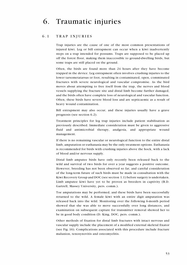
53
6. Traumatic injuries
6 . 1 T R A P I N J U R I e S
Trap injuries are the cause of one of the most common presentations of
injured kiwi. Leg or bill entrapment can occur when a kiwi inadvertently
steps on a trap intended for possums. Traps are supposed to be placed up
off the forest floor, making them inaccessible to ground-dwelling birds, but
some traps are still placed on the ground.
Often, the birds are found more than 24 hours after they have become
trapped in the device. Leg entrapment often involves crushing injuries to the
lower tarsometatarsus or foot, resulting in contaminated, open, comminuted
fractures with severe neurological and vascular compromise. As the bird
moves about attempting to free itself from the trap, the nerves and blood
vessels supplying the fracture site and distal limb become further damaged,
and the birds often have complete loss of neurological and vascular function.
Often, these birds have severe blood loss and are septicaemic as a result of
heavy wound contamination.
Bill entrapment may also occur, and these injuries usually have a grave
prognosis (see section 6.2).
Treatment principles for leg trap injuries include patient stabilisation as
previously described. Immediate consideration must be given to aggressive
fluid and antimicrobial therapy, analgesia, and appropriate wound
management.
If there is no remaining vascular or neurological function to the entire distal
limb, amputation or euthanasia may be the only treatment options. euthanasia
is recommended for birds with crushing injuries above the hock, with a lack
of blood and/or nervous supply.
Distal limb amputee birds have only recently been released back to the
wild and survival of two birds for over a year suggests a positive outcome.
However, breeding has not been observed so far, and careful consideration
of the long-term future of such birds must be made in consultation with the
Kiwi Recovery Group and DOC (see section 1.1) before surgery is undertaken.
Limb amputee kiwi have yet to be proven as breeders in captivity (B.D.
Gartrell, Massey University, pers. comm.).
Toe amputations may be performed, and these birds have been successfully
returned to the wild. A female kiwi with an entire digit amputation was
released back into the wild. Monitoring over the following 6-month period
showed that she was able to move successfully over long distances, and
examination on subsequent capture for transmitter removal showed her to
be in good body condition (D. King, DOC, pers. comm.).
Other methods of fixation for distal limb fractures with intact nervous and
vascular supply include the placement of a modified external skeletal fixator
(see Fig. 16). Complications associated with this procedure include fracture
malunion, tenosynovitis and osteomyelitis.

54
6 . 2 B I L L I N J U R I e S
Bill injuries may have a variety of causes including traps (as described
above), transportation and dog attacks. Bill tip injuries generally have a grave
prognosis, especially if the upper bill is injured. Just how important the bill
tip is to kiwi survival is becoming clear. Recent research has demonstrated
that kiwi detect their buried prey using vibration- and pressure-sensitive
mechanoreceptors embedded in pits in the bill tip. It is believed that these
specialised structures function as a detector of movement of invertebrates
during feeding (Cunningham et al. 2007). Olfaction is thought to play a
minor role in prey detection but an important role in social and territorial
behaviour. This finding correlates with clinical observations of a female great
spotted kiwi which had an upper bill tip amputation after a transport box
injury. This bird failed to feed on her own after more than 24 months in
captivity (Fig. 17) (pers. obs). euthanasia should be seriously considered in
any bird with a bill tip amputation.
An injury to the proximal upper bill, or to the lower bill, may have a slightly
more favourable prognosis than an upper bill tip injury. A sub-adult female
North Island brown kiwi injured by a muzzled dog was presented to the
New Zealand Wildlife Health Centre. It had multiple fractures of the proximal
mandible. The mandible was repaired using a modified external skeletal
fixator (Figs 18, 19). The growth plates of the mandible were affected. As
the kiwi continued to grow, it developed an undershot lower bill, which
prevented it from probing naturally for food. However, the bird is able to
eat the captive diet well, and has now been included in the captive breeding
programme.
Figure 16. Modified external fixation of an open, comminuted tarsometatarsal
fracture as a result of a trapping injury. Photo: J. Youl.

55
Figure 17. Fractured tip of the bill of a female great
spotted kiwi. The distal 10 mm of the bill was broken
off during a transportation injury.
Photo: B Gartrell.
Figure 18. Lateral radiograph of a North Island brown kiwi
with multiple mandibular fractures.
Photo: K. Morgan.
Figure 19. Ventrodorsal radiograph of kiwi in Fig. 18
after placement of a modified external skeletal fixator in the
mandible.Photo: K. Morgan.

56
6 . 3 M O T O R V e H I C L e A C C I D e N T S
Motor vehicle accidents are another common cause of injuries to kiwi, and
birds may present with limb fractures, bill injuries, pelvic and spinal fractures,
and internal injuries.
Again, treatment relies upon stabilisation of the patient, as previously
described. Once the bird’s condition has stabilised, radiographs can be carried
out. These may reveal limb fractures and other internal injuries. Pelvic and
spinal fractures are more difficult to diagnose, and often require ancillary
diagnostics such as CT scans (see section 4.3.2). Myelography (an x-ray
examination using contrast material to highlight the spinal cord) has been
described in the avian patient (Harr et al. 1997; Naeini et al. 2006). However,
it has limited use in clinical situations, because of lack of familiarity with the
technique, fear of iatrogenic injury to the spinal cord, and concern about
potentially fatal complications with the procedure (Harr et al. 1997; Naeini
et al. 2006). In birds, the fused lumbar and sacral vertebrae and pelvis (the
synsacrum) allows accessibility to the atlanto-occipital space only (Harr et al.
1997; Naeini et al. 2006). In many species, the atlanto-occipital space overlies
a large venous plexus (Harr et al. 1997) and the myelography procedure may
be fatal. MRI should be strongly considered as an alternative to myelography,
if feasible (Helmer 2006).
Due to the anatomic location of the lungs and kidneys, birds with pelvic and
spinal fractures will often have associated renal and pulmonary contusions.
The kidneys lie in a fossa within the fused synsacrum, making them susceptible
to damage during trauma to the overlying structures.

57
7. Diseases of kiwi
7 . 1 G A S T R O I N T e S T I N A L T R A C T D I S O R D e R S
7.1.1 Foreign body ingestion—traumatic gastritis
Foreign body ingestion is not an uncommon finding in captive kiwi. Metallic
foreign bodies (nails, wire, screws, etc.) may cause hardware disease,
especially in captive facilities where construction work is being undertaken
within enclosures, or where materials are brought in with leaf litter or
other substrate. The powerful contractions of the gizzard muscles can easily
force sharp objects through the muscular wall (Lumeij 1994). This causes a
reduction in ventricular contraction and insufficient digestion of food (Lumeij
1994). Commonly, the foreign body may penetrate the proventricular wall,
leading to an acute, generalised purulent
peritonitis, or to a local peritonitis with
abscess formation on the surface of the
proventriculus or gizzard, or duodenum
(Lumeij 1994). Occasionally, the foreign
object may travel through into the muscles
of the thigh. In these cases, lameness or
pathological limb fractures may occur (B.D.
Gartrell, Massey University, pers. comm.).
Clinical signs of traumatic gastritis include
anorexia, weight loss, depression and the
passing of undigested food.
Foreign body ingestion may be diagnosed by
plain and contrast radiography and gastric
endoscopy (Fig. 20). The treatment for
large, sharp foreign bodies is removal via
proventriculotomy.
To prevent captive kiwi from ingesting
metallic foreign bodies, metal detectors can
be used to screen substrate material before
it is brought into their enclosures.
7.1.2 Gastric impaction
Gizzard and proventricular impaction commonly occurs in other ratites,
typically as a result of over-consumption of substrate, including sand and
stones (Lumeij 1994). Predisposing factors include gastroenteritis.
Clinical signs of gastric impaction include anorexia, depression, weight loss
and scant faeces (Gelis 2006).
Diagnosis includes identification of a full gizzard by palpation, and
radiographs demonstrating an excess of grit and stones in the gizzard and
proventriculus. A definitive diagnosis of impaction requires the use of
contrast radiography, including barium sulphate meals and BIPS™ (barium
impregnated polyethylene spheres). Other diagnostics include endoscopy or
an exploratory laparotomy (Gelis 2006).
Figure 20. Ventrodorsal radiograph of an adult North
Island brown kiwi with a penetrating metallic gastric
foreign body. Photo: T. Kelly.

58
Treatment depends upon the severity of the impaction (Gelis 2006). Medical
therapy using psyllium seed husks (Metamucil™) may resolve the impaction
by encouraging the movement of gizzard contents. Metoclopramide may
also help stimulate small intestinal motility and thus assist in ventricular/
proventricular emptying (Gelis 2006). Proventricular flushing, as described
for ostriches (Gelis 2006), may help loosen the impaction. This should be
performed under general anaesthesia with an endotracheal tube in place to
prevent aspiration. Surgical removal of impacted material via a ventriculotomy
may sometimes be needed (Gelis 2006).
7.1.3 Candidiasis
Candidiasis (Candida albicans) in birds is also known as thrush or sour crop (Dahlhausen 2006). Candida albicans is an opportunistic yeast and is not regarded as a primary pathogen (Dahlhausen 2006). In other avian species, including pigeons, the non-budding organism is considered part of the normal gastrointestinal flora (Rupiper 1998).
When severe suppression of the normal gastrointestinal flora occurs, Candida spp. can proliferate and cause disease (Dahlhausen 2006). Predisposing factors for candidiasis include antibiotic therapy, stress, poor nutrition, poor hygiene, debilitation or immunosuppression (Dahlhausen 2006). However, infection may be spontaneous in immature animals, or may be secondary to weak or broken mucous membranes (Bauck 1994).
Candidiasis affects the mucocutaneous areas of the body and gastrointestinal mucosa; in particular, the oropharynx and oesophagus (Dahlhausen 2006). The proventriculus and gizzard may also be affected (Bauck 1994). Clinical signs of disease include white or yellow diphtheritic membranes in the oral cavity covered in adhesive mucous (Bauck 1994). Underlying mucous membranes are often inflamed. Infection of the oesophagus, proventriculur or gizzard may cause vomiting or regurgitation, depression, anorexia and poor digestion of food (Bauck 1994; Dahlhausen 2006).
Diagnosis of candidiasis includes cytological examination of affected areas with either Gram’s stain or Dif-Quik (Dahlhausen 2006). These may be smears from lesions, or crop washes or faecal samples (Dahlhausen 2006). Candidiasis is characterised by narrowly based budding yeast, oval in shape, measuring 3.5–6.0 × 6.0–10.0 µm (Dahlhausen 2006). The presence of pseudohyphae is indicative of invasive disease (Dahlhausen 2006). Sample swabs may be submitted for culture, and biopsies for histopathology, if necessary.
Treatment includes correction of predisposing factors in combination with antifungal medication. If infection is non-invasive (i.e. no presence of hyphae), oral nystatin (Mycostatin™) can be used at a dose rate of 300 000 IU/kg BID-TID (two to three times daily) for 5–10 days (Dahlhausen 2006). Nystatin is not absorbed from the gastrointestinal tract and thus relies on direct contact by oral or topical absorption. For this reason, lesions in the oral cavity require contact with the nystatin and will not respond to administration of the drug by gavage tube (Dahlhausen 2006).
Severe infections or those invading the gastrointestinal wall may be refractory (noncompliant) to nystatin treatment, and systemic antifungals are indicated. Fluconazole or ketoconazole are the systemic drugs of choice. Itraconazole may also be effective, although some Candida spp. are extremely resistant to itraconazole (Dahlhausen 2006).

59
7.1.4 Bacterial enteritis
Aerobic gram-negative bacteria, in particular those belonging to the
enterobactericeae family, are the most frequently cultured enteric pathogens
in birds (Taylor 2000). All produce endotoxins. Potentially pathogenic
isolates include Pseudomonas aeruginosa, Salmonella spp., Yersinia sp.,
Klebsiella sp. and Proteus sp. (Taylor 2000). Salmonella spp. have been
documented in two captive kiwi but not, to date, in wild kiwi (B.D. Gartrell,
Massey University, pers. comm.). Yersinia enterocolitica has been isolated
from several apparently clinically healthy kiwi, both wild and captive, during
routine health screening for translocation (C. Travers, Kiwi encounter,
Rainbow Springs, pers. comm.; pers. obs.). Escherischia coli causes bacterial
enteritis in other avian species. However, when it is present in low numbers
it may be considered to be part of the normal intestinal flora of kiwi (B.D.
Gartrell, Massey University, pers. comm.).
Gram-positive bacteria, including Clostridium spp. (Gelis 2006) also have
potential to cause enteritis in kiwi. Campylobacter spp. are frequently
cultured from clinically normal kiwi during routine health screening,
and appear to be non-pathogenic (B.D. Gartrell, Massey University, pers.
comm.).
Transmission of pathogenic enteric bacteria occurs by the faecal-oral route.
Sources of infection include contamination from other wild birds and
overgrowth of a commensal organism secondary to an immunosuppressive
disease or antibiotic administration. Infection with Pastuerella multocida is
a common sequela to predator attacks on birds, as mammalian saliva contains
pathogenic bacteria which are then ingested by birds during preening
(Quesenberry & Hillyer 1994).
Clinical signs of bacterial enteritis include lethargy, anorexia, diarrhoea
(with or without blood), cachexia (weakened body state) and emaciation
(Gelis 2006). However, signs are not restricted to the gastrointestinal tract,
as septicaemia often follows bacterial enteritis (Gerlach 1994). Joint swelling
may indicate infectious arthritis, and ocular lesions, including keratitis and
corneal ulceration, may be apparent. Multiple organ infection may occur,
including hepatitis (Bauck 1994).
Methods of diagnosing bacterial enteritis include a Gram’s stain of a faecal
smear and culture and sensitivity of a cloacal or faecal swab.
Treatment of bacterial enteritis involves supportive care, including fluids,
warmth and nutritional support. Antibiotics should be selected on the
basis of sensitivity testing and in consultation with an avian veterinarian.
The treatment of salmonellosis is controversial, as chronic infections are
refractory to treatment, and birds may become subclinical latent carriers
of the disease (Gerlach 1994). Clinically affected birds should be treated,
as should birds being translocated or in contact with humans because of
the potential for humans to catch the disease (zoonotic potential) (Gerlach
1994). Clinically healthy kiwi diagnosed with Yersinia spp. have previously
been left untreated, and remained free of clinical disease (T. Kelly, The Vet
Centre, Rotorua, pers. comm.).

60
Control of enteric bacterial pathogens consists of good hygiene practices,
and screening of birds prior to translocation. Access of wild birds to captive
kiwi enclosures should be restricted.
7.1.5 Mycobacteriosis
One case of mycobacteriosis has been reported in a captive adult female
North Island brown kiwi. Post-mortem examination showed that this bird had
classical mycobacteriosis lesions in the liver, spleen, gizzard and intestine
(Davis et al. 1984).
Diagnosis of avian mycobacteriosis can be made by demonstration of acid-fast
organisms on faecal smears (Gerlach 1994). Repeated samples must be taken
because of intermittent shedding of the bacteria (Gerlach 1994). Culture is
required for a definitive diagnosis and speciation of the organism, as non-
pathogenic mycobacteria may be transient inhabitants of the gastrointestinal
tract (Gerlach 1994). Histopathology of mycobacterial granulomas reveals a
necrotic foci containing occasional acid-fast organisms (Pollock 2006).
Birds with confirmed mycobacteriosis should be euthanased as there is no
effective treatment and there is zoonotic potential (Gerlach 1994; Pollock
2006). Control should focus on identification of affected birds through
quarantine and the use of appropriate screening techniques.
7.1.6 Cloacal prolapse
Cloacal prolapses may contain intestines, oviduct or one or both ureters
(Lumeij 1994). They may be associated with cloacal masses, neurogenic
disorders or conditions causing tenesmus (difficulty passing a stool). These
include enteritis, cloacitis, and egg binding. Clinical signs include prolapse
of the smooth, glistening pink cloacal mucosa (Lumeij 1994). examination to
rule out involvement of the ureters or uterus should be performed.
Diagnosis should include palpation of the abdomen of female kiwi for the
presence of an egg. examination of the prolapse is best performed under
general anaesthesia. Gentle manual massage of the caudal abdominal and
cloacal regions may aid in evacuation of faecal material (Lumeij 1994). The
cloaca should be examined with (ideally) a rigid endoscope or, alternatively,
a speculum and strong light source. Radiographs are useful for identifying
masses within the cloaca. A faecal wet mount as well as a Gram’s stain and
faecal culture may aid in identification of enteritis.
Supportive treatment includes fluid therapy and antibiotics for septic shock.
Tissues should be cleaned with a saline solution, covered with a sterile
lubricating jelly and replacement of the tissue should be attempted (Lumeij
1994). The prolapse may require retention sutures or a purse-string suture,
but only if there is no possibility of egg retention. Care must be taken to
ensure that stay sutures do not interfere with evacuation of the cloaca (Lumeij
1994). Chronic cases of cloacal prolapse may require a surgical cloacapexy
(not described here).

61
7.1.7 Cloacal impaction
Impaction of the cloaca has been seen in at least one kiwi chick (pers. obs.).
Cloacaliths are typically a cloacal mass of desiccated urates and faecal material,
and they may inhibit the passing of excrement, causing congestion of the
ureters and intestinal dilatation (Lumeij 1994). Renal failure and visceral gout
may occur if the ureters are blocked (Lumeij 1994). Cloacal impactions may
be associated with previous egg binding, infectious cloacitis, malnutrition,
or neurological disease of the cloaca (Bowles 2006).
Clinical signs of cloacal impaction include depression and anorexia, with
scant or no droppings. Abdominal palpation may determine a mass in the
caudal abdomen, and radiographs will show a distended cloaca filled with
faecal material.
This is an emergency requiring prompt treatment. Therapy includes fluids,
antibiotics, anti-inflammatory agents, and gentle manual removal of the
cloacolith (Gelis 2006). Manual removal involves the infusion of sterile saline
into the cloaca, in combination with manual massage to gently break up the
bulk of the impaction which can then be flushed out or manually removed.
Diagnostic procedures for the identification of underlying disease may include
cloacal culture and sensitivity, and a thorough neurological examination,
including assessment of the vent reflex.
7 . 2 R e S P I R A T O R Y T R A C T D I S O R D e R S
7.2.1 Aspergillosis
Aspergillosis has been infrequently diagnosed in kiwi, usually in captive
birds. It is most commonly caused by Aspergillus fumigatus, although
A. flavis or A. niger may occasionally be isolated (Dahlhausen 2006).
Aspergillus spp. are ubiquitous in the environment and (for captive birds)
are present at particularly high levels in damp, poorly ventilated enclosures,
and in mouldy feed or bedding (Dahlhausen 2006). Infection is generally
secondary to immunosuppression, and predisposing factors include stress,
poor ventilation, malnutrition, antibiotic therapy, concomitant disease and
respiratory irritants (Bauck 1994; Dahlhausen 2006). Very young birds are
most susceptible, although adults can also be affected.
Transmission occurs via inhalation of infective spores, and the type of disease
induced is dependent upon the source and number of spores inhaled, and the
immunecompetency of the individual bird. Disease may be local or systemic.
Typically, infection occurs within the respiratory tract, including trachea,
syrinx, lungs and air sacs. Advancement of lesions to adjacent organs and
systemically may also occur (Dahlhausen 2006).
Clinical signs of aspergillosis depend upon the site of infection. Infection
involving the lungs and air sacs is the most common presentation, and
affected birds may show depression, emaciation, open-mouth breathing,
dyspnoea, respiratory distress, wheezing and a change in vocalisation (Bauck
1994). Syringeal aspergillosis has been seen clinically in a captive kiwi (pers.
obs.). Renal aspergillosis may present as paralysis or paresis of the hindlimbs

62
due to compression of pelvic nerve roots by fungal granulomas (Greenacre
et al. 1992). Other sites for Aspergillus spp. granulomas include the heart
muscle, liver, abdominal viscera and the central nervous system (Bauck 1994;
Dahlhausen 2006).
Haematology often reveals a marked heterophilia (often 25–100 × 109/L) and
anaemia (Bauck 1994; Dahlhausen 2006). However, a normal white blood
cell count does not exclude aspergillosis. An elevation in liver enzymes and
bile acids may also be seen, in combination with biliverdinuria (Dahlhausen
2006). Radiographs may show focal densities in lungs or air sacs, a loss of air
sac wall definition, a reduction in coelomic cavity details, or asymmetrical
opacities of abdominal air sacs (Bauck 1994).
endoscopic coelioscopy or tracheal endoscopy may be indicated, and biopsy
for culture or histopathology will aid in diagnosis. Cytology of air sac washes
may also be performed. Post-mortem examination including cytology,
histopathology and culture of lesions will definitively diagnose aspergillus
infection (Bauck 1994).
Treatment is dependent upon the location and extent of disease. Itraconazole
(Sporanox™) is currently the systemic antifungal medication of choice
(10 mg/kg BID PO) (Dahlhausen 2006). Air sac aspergillosis should be
treated with nebulisation of an antifungal agent. Treatment options include
amphotericin B, enilconazole, terbinafine, clotrimazole and voriconazole.
Currently, lack of drug availability in New Zealand restricts our choices to
Amphotericin B only. Care should be taken when using amphotericin B,
as it is potentially nephrotoxic. Intratracheal flushes with amphotericin B
may be indicated for tracheal or syringeal disease (Bauck 1994; Dahlhausen
2006). Surgical debridement of air sac lesions may be required (Dahlhausen
2006). Underlying predisposing factors need to be determined and treated
accordingly.
Control of aspergillosis includes reduction of predisposing factors (e.g. stress)
(Bauck 1994). Contact with substrates with potential for mould and spore
contamination should be reduced, and damp, poorly ventilated enclosures
should be avoided (Bauck 1994). Good hygiene practices should be followed
at all times, including the feeding of mould-free products (Dahlhausen
2006).
7.2.2 Cryptococcosis
Avian cryptococcosis is a rare disease of birds, and infections may involve the
respiratory tract, digestive tract and central nervous systems (Dahlhausen
2006). Cryptococcus bacillisporus (formerly Cryptococcus neoformans var.
gatti) has been reported as causing both pulmonary and disseminated disease
in four captive kiwi (Hill et al. 1995; Malik et al. 2003). This particular
organism has an ecological association with eucalyptus trees (Dahlhausen
2006), and in one of the reported cases, eucalyptus leaves and twigs had been
spread throughout the kiwi enclosure some months prior to the development
of disease (Hill et al. 1995).
In the case reported by Hill et al. (1995), the kiwi showed clinical signs
2 hours prior to death, including lethargy, anorexia and dyspnoea. The post
mortem showed disseminated disease affecting the heart, kidneys, liver,

63
pancreas and oviducts (Hill et al. 1995). Histopathology demonstrated a
multifocal to diffuse inflammatory reaction associated with cryptococcal
organisms (Hill et al. 1995). Unfortunately, lung tissue from this bird was
not submitted for examination. In the other cases, all three kiwi died, and at
post mortem were found to have extensive granulamatous pneumonia (two
cases) or disseminated disease with involvement of the heart, kidneys and
proventriculus (one case) (Malik et al. 2003).
It has been proposed that the feeding habits of kiwi would be likely to
predispose them to initial pulmonary infection by this disease (Hill et al.
1995).
Diagnosis of cryptococcosis should be based on cytology and histopathology
in combination with culture of nasochoanal swabs or washes (Raidal & Butler
2001). Wright’s stained smears of gelatinous material obtained from swabs
often show aggregates of encapsulated yeast 6–10 µm in length within 8–12-
µm non-staining capsules (Raidal & Butler 2001). Histopathology is usually
required to confirm cryptococcosis, as it may be carried asymptomatically.
Whether or not to treat a bird with cryptococcosis needs to be considered
carefully, as there is significant zoonotic potential associated with this
infection (Dahlhausen 2006). extreme caution is necessary when handling
materials that may contain Cryptococcus spp. spores. Long-term antifungal
treatment of infected birds is required (e.g. daily for 2 months) using
amphotericin B, fluconazole or itraconazole (Bauck 1994; Dahlhausen 2006).
7.2.3 Pneumoconiosis
Pneumoconiosis is caused by inhalation of dust particles containing silica,
iron and plant material (Boardman 1995). This condition was first reported in
kiwi in 1973 (Smith et al. 1973), and is commonly found incidentally in kiwi
at post mortem, especially those in captivity. The disease is characterised
by the focal accumulation of dust-laden macrophages in bronchial walls
(NZWHC 2006). Birds most likely to be affected are those kept on dry sandy
substrates containing a large proportion of silica. The olfactory behaviour of
the kiwi and the distal placement of their nostrils on the upper bill presumably
predispose kiwi to this disease (Smith et al. 1973; Boardman 1995).
7.2.4 Aspiration pneumonia
Birds may aspirate food during tube feeding or following regurgitation
(Flammer & Clubb 1994). The lack of a crop and the small capacity of the
gizzard and proventriculus make kiwi vulnerable to regurgitation during tube
feeding. If large amounts of food are inhaled, rapid placement of an air sac
cannula is necessary to prevent asphyxiation (Flammer & Clubb 1994). The
prognosis for a bird with this disease is poor. Treatment consists of aggressive
antibiotic and antifungal therapy, preferably systemically and via nebulisation
(Flammer 1994; Flammer & Clubb 1994). Clinical signs of delayed aspiration
pneumonia from inhalation of small amounts of food include poor weight
gain, and a persistent leukopenia with or without respiratory signs (Flammer
& Clubb 1994). Radiography may assist in diagnosis.

64
7.2.5 Bacterial pneumonia and air sacculitis
Gram-negative bacteria are commonly isolated from birds with respiratory
tract infections (Tully & Harrison 1994). Gram-positive bacteria may also
be implicated (Flammer & Clubb 1994). Techniques required for diagnosing
pneumonia include auscultation, radiography and endoscopy with or without
biopsy.
To date, Chlamydophilia psittaci has not been diagnosed in kiwi (B.D.
Gartrell, Massey University, pers. comm.). A study of captive kiwi failed to
identify Mycoplasma spp. by both culture and serum agglutination tests
(Christiansen 1996).
7.2.6 Parasitic pneumonia
Migration of parasitic larvae through pulmonary parenchyma has been
demonstrated post-mortem in a single kiwi (NZWHC 2006).
7.2.7 Rhinitis
Bacterial rhinitis has been reported post-mortem in a single kiwi (NZWHC
2006). Bacteria commonly isolated from avian species with upper respiratory
tract infections include Pseudomonas spp., E. coli, Klebsiella spp.,
Pasteurella spp. and Salmonella spp. (Tully & Harrison 1994). Aspergillus
spp. granulomas and nasal cryptococcus may cause upper respiratory signs
(Dahlhausen 2006).
Clinical signs of upper respiratory tract disease include serous to mucopurulent
nasal and/or ocular discharge and sneezing (Dahlhausen 2006).
Diagnosis of rhinitis includes nasal and sinus flushing for cytology and culture.
Treatment is dependent upon aetiology, but typically includes antibiotic and/
or antifungal medication.
7 . 3 R e P R O D U C T I V e D I S O R D e R S
7.3.1 Normal reproduction in kiwi
This section was prepared by Dr B.D. Gartrell, Massey University.
The egg produced by the kiwi is the largest of any bird for its body size,
weighing approximately 400 g and comprising 20% of the mass of the adult
bird (Fig. 21) (Deeming 1991).
While the laying interval between eggs in kiwi can be 21 days or more, the
egg is not carried in the uterus for this length of time. Time from ovulation
to oviposition is 7–10 days (Jensen & Durant 2006). The bulk of this time will
be used to develop the large, energy-rich yolk follicle on the ovary (Jensen
& Durant 2006).
In the wild, kiwi eggs are incubated in a small burrow, and eggs are turned
regularly (contrary to early reports; Colbourne 2002). At approximately 80
days, incubation length is the longest for any bird (Vleck & Hoyt 1991). Mean
incubation temperature in the wild is 36.5°C at the top of the egg, while the
bottom of the egg may be up to 10°C colder (Colbourne 2002). The kiwi egg

65
has the highest energy content of any bird, with the yolk fraction comprising
65% of egg content and having an energy density of 12.4 kJ/g (wet) (Starck
& Ricklefs 1998). The yolk also has the lowest water fraction of any bird, at
61% of egg content (Jensen & Durant 2006). It has been suggested that this
is related to the extremely humid conditions of incubation (Vleck 1991). The
kiwi embryo expends only approximately 17% of the energy stored in the
egg during incubation and 48% of the yolk weight is found in the yolk sac of
the hatchling (Prinzinger & Dietz 2002). This yolk is used as the chick’s sole
source of energy and substrate for tissue production for up to 17 days after
hatching (Prinzinger & Dietz 2002).
Incubation behaviour of the parents varies amongst kiwi species (Colbourne
2002). For North Island brown kiwi (Apteryx mantelli) and little spotted
kiwi (Apteryx owenii), the male alone incubates the egg, except for the first
week (Colbourne 2002). However, in rowi/Okarito brown kiwi (formerly A.
mantelli ‘Okarito’, now A. rowi) and tokoeka (Apteryx australis), incubation
is shared by both sexes (Colbourne 2002). Clutch size ranges from 1 to 3,
with eggs being laid from 21 to 66 days apart (Marchant et al. 1991). Multiple
clutches are possible, and some North Island brown kiwi are known to lay up
to seven eggs a year (Colbourne 2002).
Chicks are precocial, hatching fully feathered and active with a weight
of around 300 g for brown kiwi (Marchant et al. 1991). No information is
available on hatching weights for great or little spotted kiwi.
Figure 21. Lateral radiograph of a gravid North Island
brown kiwi. Photo: T. Kelly.

66
7.3.2 Egg binding and dystocia
egg binding is defined as failure of an egg to pass through the oviduct at
a normal rate. Dystocia defines a condition in which the developing egg
is in the caudal oviduct and is either obstructing the cloaca, or has caused
prolapse of oviductal tissue (Joyner 1994).
Given the large relative size of kiwi eggs, problems with egg laying are less
common than could be expected. However, dystocia has been observed in
captive kiwi and, occasionally, in wild kiwi (NZWHC 2006), and occurs when
the developing egg is retained in the oviduct or cloaca.
Common causes of egg binding and dystocia in birds include (Joyner 1994;
Bowles 2006):
Oviduct muscle dysfunction secondary to excessive egg laying•
Calcium-related metabolic disease•
Malformed eggs•
Previous oviduct damage or infection•
Nutritional insufficiencies, including vitamin e and selenium deficiencies •
and malnutrition
Obesity•
Lack of exercise and poor muscle strength•
Concurrent stress•
Systemic disease•
Genetic predisposition•
Clinical signs of egg binding include depression, anorexia, dyspnoea, an
abnormally wide stance and abdominal straining (Joyner 1994). Hindlimb
paresis or paralysis may be present due to compression of pelvic nerves by the
large egg, and compression of the pelvic blood vessels and kidneys may also
occur (Bowles 2006). Sudden death may also be observed (Bowles 2006).
egg binding may be diagnosed on history and physical examination alone
(Bowles 2006). Cloacal examination and radiography may help in diagnosis if
the patient’s condition is stable enough to allow it to be carried out. Patient
stabilisation is the priority of treatment for dystocia (Joyner 1994). This
includes fluid therapy and provision of warmth. The cloaca can be lubricated
with a water-soluble lubricant (e.g. KYTM jelly), and intramuscular calcium
may be given. Analgesia and antibiotics should be considered. Supportive care
alone is often enough to allow oviposition, although the bird’s condition must
be closely monitored for deterioration, in which case further intervention
may be required (Bowles 2006).
Once the bird’s condition is stabilised, and if the oviduct is believed to be
intact and it has been determined that the utero-vaginal sphincter is open,
a parenteral prostaglandin may be given to induce oviductal contractions
(Bowles 2006). Dinoprost tromethamine (a PGF2alpha) is available in
New Zealand. This can be given at a dose rate of 0.02–0.1 mg/kg IM once only
(Bowles 2006) and may result in expulsion of the egg. Oxytocin should be used
with caution, as complications include oviductal rupture (Joyner 1994).
If medical therapies fail to elict oviposition, manual manipulation of the egg
can be attempted in conjunction with lubrication. Gentle pressure should be
applied in a persistent, caudal direction. This needs to be gentle to prevent
oviduct rupture (Joyner 1994).

67
Lack of oviposition after an inappropriate period of time, or deterioration
in the bird’s condition, may indicate the need for more aggressive therapy
such as ovocentesis. Ovocentesis involves the aspiration of the contents
of the egg with a large needle (18 gauge) (Joyner 1994). Ideally, the egg
should be manipulated with the use of a speculum so that it is observable
and tapped through the cloaca. If this is not possible, the egg should be
brought into juxtaposition to the abdominal wall so that other organs are not
damaged during a transabdominal aspiration procedure. After aspiration of
the egg contents, the egg can be gently collapsed and the shell fragments and
remaining contents of the egg will pass within several days (Joyner 1994).
There is some risk of tearing the oviduct resulting in peritonitis, but this
appears to be minor. Any shell fragments visible through the cloaca can be
gently removed. The uterus may be flushed with an iodine, chlorhexidine
or saline flush to help remove egg fragments, and this may decrease the risk
of metritis (uterine infection). A course of antibiotics should follow (Joyner
1994).
Other options for treatment of dystocia include an episiotomy if the egg is
lodged in the caudal oviduct or cloaca, especially if egg survival is critical.
A hysterotomy may be indicated or, alternatively, a hysterectomy in cases
where the uterus is ruptured or severe adhesions exist (Joyner 1994).
Hysterotomy under isoflurane general anaesthesia has been used to
successfully remove a retained egg in a captive kiwi (Figs 22, 23) (T. Kelly,
The Vet Centre, Rotorua, pers. comm.).
Post-dystocia complications include a ruptured or necrotic oviduct, peritonitis
or abdominal herniation. On at least one occasion, a captive kiwi laying its
third egg for the season presented with acute paralysis due to a synsacral
spinal fracture (pers. obs.). It is proposed that this may have been secondary
to hypocalcaemia (low calcium) with resultant osteopenia (decreased bone
density).
Figure 22. Intra-operative image of kiwi egg within the oviduct during a hysterotomy. Photo: T. Kelly.
Figure 23. Post-operative image of the bird and egg
from Fig. 22. Photo: T. Kelly.

68
7.3.3 Oviduct and cloacal prolapse
Prolapse of the oviduct and cloaca can occur secondary to ‘normal’ egg laying,
or as a sequela to dystocia (Joyner 1994). Predisposing factors for prolapse
include excessive abdominal muscle contractions, which may be exacerbated
by poor physical condition and malnutrition, abnormal or soft-shelled eggs,
obesity, salpingitis (oviduct infection) and cloacitis (inflammation of the
cloaca) (Joyner 1994; Bowles 2006).
Clinical signs of prolapse include protrusion of the oviduct through the
cloaca, often with partial prolapse of the vagina and cloaca (Bowles 2006).
In these situations, rapid aggressive therapy is necessary to prevent tissue
devitalisation and secondary infections (Joyner 1994; Bowles 2006). Tissue
should be moistened and cleaned with sterile saline solution, and replaced
using a lubricated swab and gentle pressure. Stay sutures or a purse-string
suture may be required to prevent recurrence. With immediate treatment the
prognosis is good (Joyner 1994).
If egg is present in prolapsed tissue, it must be removed before replacing
tissue in the abdomen. Surgical debridement may be required to remove the
egg if there are adhesions between the shell and the uterus (Joyner 1994).
7.3.4 Salpingitis and metritis
Salpingitis (oviduct infection) and metritis (uterine infection) in birds may
occur secondary to air sacculitis (inflammation of the air sacs), pneumonia,
liver disease or retrograde infections of the lower uterus, vagina or cloaca
(Joyner 1994; Bowles 2006). Predisposing factors include excessive abdominal
fat, oviduct impaction and egg-related peritonitis. Most frequently isolated
bacterial pathogens include E. coli, as well as Salmonella spp. and Pasteurella
spp. (Joyner 1994; Bowles 2006).
Non-specific clinical signs of salpingitis and metritis include depression,
anorexia and weight loss. Abdominal enlargement may be visible, and a
cloacal discharge may be present. An increased white blood cell count is
seen on haematology (Bowles 2006).
Treatment requires aggressive antibiotic therapy and supportive care. The
oviduct may be flushed with lactated Ringers solution (LRS). Predisposing
factors or underlying causes must be corrected also (Joyner 1994; Bowles
2006).
7.3.5 Egg-related coelomitis
Yolk-related coelomitis (previously called yolk peritonitis) and ectopic eggs
have been seen in both captive and wild kiwi (Boardman 1995; NZWHC 2006)
(Fig. 24). egg-related coelomitis may be septic or non-septic. Causes of yolk-
related coelomitis include ectopic ovulation, oviduct rupture and salpingitis
(Joyner 1994). ectopic ovulation relates to a failure of the oviductal fimbrium
to pick up the ovulating follicle with a subsequent spread of yolk material
through the abdomen (Joyner 1994). The yolk may be reabsorbed if it is free
of pathogens. However, yolk is an ideal environment for bacterial invasion, in
particular with coliform bacteria, creating a septic coelomitis (Joyner 1994).

69
Clinical signs of egg-
related septic coelomitis
include abdominal swell-
ing, respiratory distress
from air sac compression,
depression and anorexia
(Joyner 1994; Bowles
2006). Sudden death may
occur. A severe leuco-
cytosis (increased white
cell count) is seen on
haematology (Joyner
1994).
Survey radiographs may aid in diagnosis of yolk-related coelomitis.
Coelomocentesis may reveal a turbid yellow, green or brown yolky fluid, or
cheese-like masses of inspissated yolk material (Joyner 1994). Cytology may
reveal a septic or non-septic exudate, a transudate, or yolk or fat globules,
and bacteriology should be performed on coelomic fluid samples (Bowles
2006). Coelomotomy may be necessary to obtain a definitive diagnosis
(Bowles 2006).
Treatment involves supportive care with fluids, broad spectrum antibiotics
and analgesia (Joyner 1994). Coelomocentesis is not only diagnostic, but it
may also be therapeutic by relieving dyspnoea (Bowles 2006). In other avian
species, treatment of severe cases of yolk-related coelomitis may include
exploratory coelomotomy and removal of yolk material (Bowles 2006).
7.3.6 Oophoritis
Oophoritis refers to inflammation of the ovary, and it may result from
neoplastic (abnormal cell growth), mechanical or infectious causes (Bowles
2006). Clinical signs associated with oophoritis may include depression,
anorexia, chronic wasting and sudden death (Bowles 2006). Diagnosis
requires endoscopic evaluation of the ovaries, and treatment may include
supportive care and antibiotic therapy, if indicated (Joyner 1994).
7 . 4 Y O L K S A C D I S O R D e R S I N C H I C K S
The yolk sac is a diverticulum (bulge or pouch) of the intestine, situated at
the junction of the jejunum and the ileum (O’Malley 2005). The yolk sac is
normally internalised into the abdomen just prior to hatch, after which the
yolk is absorbed through the yolk stalk, providing the chick with nutrition,
minerals, fat-soluble vitamins as well as maternal antibodies (Flammer & Clubb
1994). Kiwi are precocial birds, meaning that they are born covered in feathers
and able to see, walk and feed themselves at hatching (Flammer & Clubb
1994). They have to fend for themselves from early on and thus rely entirely
on absorption of yolk to provide nutrition until they have learned to obtain
adequate food from foraging (O’Malley 2005). Once the yolk is absorbed,
usually from 10 days post hatching, only a small remnant of scar tissue should
remain (the vitelline or Meckel’s diverticulum) (O’Malley 2005).
Figure 24. ectopic yolk from a North Island brown kiwi, diagnosed at post-mortem.
Photo: B.D. Gartrell.

70
7.4.1 Yolk sacculitis and yolk sac retention
Yolk sacculitis (bacterial infection of the yolk sac), is a multifactorial problem
seen in ratite chicks usually less than one week old (Doneley 2006). Yolk sac
retention is a failure of the yolk sac to be resorbed in the absence of infection
(Doneley 2006). Yolk sacculitis and yolk sac retention have been seen in kiwi
chicks artificially incubated and reared in the Operation Nest egg programme
(C. Travers, Kiwi encounter, Rainbow Springs, pers. comm.).
In captive-raised ratites, yolk sacculitis usually arises from poor hatcher or
brooder hygiene, and chicks that have been stressed by incorrect incubator/
hatcher temperature and/or humidity are predisposed to bacterial infection
(Doneley 2006). Weak or undersized chicks are most at risk (Doneley 2006).
Transovarial and transoviductal infections may occur, as well as infection
from shell contamination (Doneley 2006). Yolk sacculitis often occurs if the
yolk sac is poorly internalised prior to hatching, and is primarily caused by
gram-negative bacteria, including E. coli, Proteus spp., and Streptococcus
fecalis (Flammer & Clubb 1994). Clostridium spp. have also been implicated
(Flammer & Clubb 1994). Clinical signs of yolk sacculitis include a thickened,
prominent, inflamed and necrotic umbilicus, and affected chicks lose weight
and become systemically ill (Stewart 1994).
Yolk sac retention in ratites is usually associated with management problems,
including incubator or hatcher problems, or chick nutrition and exercise
(Doneley 2006). Clinical signs of failure to thrive and weight loss are usually
not evident until the chick is 2–6 weeks old, and are a result of the release
of toxic substances by the autolysing yolk (Flammer & Clubb 1994; Doneley
2006). Kiwi chicks will normally lose weight for the first 10 days after hatching
and then begin to grow rapidly (C. Travers, Kiwi encounter, Rainbow Springs,
pers. comm.). Kiwi chicks with a retained yolk sac fail to thrive and any chick
which does not steadily increase in body weight should be investigated for
yolk sac retention. In ratites, the retained yolk sac feels enormous, is usually
palpable as a doughy structure, and may be 15–40% of the total body weight
(Stewart 1994). Other clinical signs of yolk sac retention include dyspnoea
and exercise intolerance resulting from air sac compression, depression,
anorexia and recumbency (Flammer & Clubb 1994).
Diagnosis of a yolk sac disorder may include radiographs showing a visibly
enlarged yolk sac, or ultrasonographic demonstration of the large yolk sac
within the chick’s abdomen (Doneley 2006). Contrast radiographs with a
barium meal are particularly helpful in diagnosing a retained yolk sac. Fine
needle aspiration of the yolk sac is contraindicated as the yolk sac wall is very
thin, and the needle puncture will cause leakage of yolk into the peritoneal
cavity, causing yolk-related peritonitis (Flammer & Clubb 1994).
Initial treatment of yolk sacculitis involves aggressive supportive care. As
antibiotics do not penetrate the yolk sac, treatment for both yolk sacculitis
and yolk sac retention involves surgical excision of the yolk sac (Doneley
2006). The technique in ostriches (from Stewart 1994) is described below
(see also Fig. 25A–F).
Pre-anaesthetic considerations should include patient stabilisation with fluid
therapy, antibiotics, and correction of hypothermia and hypoglycaemia. Pre-
surgical analgesia should be considered. Anaesthetic considerations should
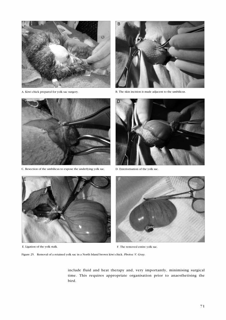
71
include fluid and heat therapy and, very importantly, minimising surgical
time. This requires appropriate organisation prior to anaesthetising the
bird.
B. The skin incision is made adjacent to the umbilicus.
B
C. Resection of the umbilicus to expose the underlying yolk sac.
C
D. exteriorisation of the yolk sac.
D
e. Ligation of the yolk stalk.
E
F. The removed entire yolk sac.
F
A. Kiwi chick prepared for yolk sac surgery.
A
Figure 25. Removal of a retained yolk sac in a North Island brown kiwi chick. Photos: V. Gray.

72
The bird should be placed in dorsal recumbency. Feathers are plucked over
the abdomen at the level of the umbilicus, and the area is prepared for
surgery. Alcohol-based surgical preps must not be used, as these cause heat
loss. Chlorhexidine aqueous solution may be used instead.
A circumferential incision should be made around the umbilicus, extending
transversely at 3 and 9 o’clock to a lateral distance required for easy removal
of the intact yolk sac. Alternatively, a longitudinal incision can be made, as
shown in Fig. 25. The body wall should be incised in a corresponding pattern,
taking care not to damage the underlying yolk sac. The yolk sac is exteriorised
by applying gentle traction on the umbilical stump. The yolk stalk should be
clamped and clipped or ligated just distal to the intestines, and the yolk sac
removed. The body wall should be closed with a monofilament absorbable
suture (e.g. Vicryl™ 5-0) in a simple continuous pattern (to minimise surgical
time).
Post-surgical antibiotics should be continued for at least 7 days. Broad
spectrum antibiotics should be initiated pending the results of culture and
sensitivity. These should be effective against gram-negative bacteria, and
first choice antibiotics may include amoxicillin/clavulonic acid, enrofloxacin
and trimethoprim-sulfa (Stewart 1994). Butorphanol analgesia should be
continued post-operatively. Chicks can be offered a small amount of food in
the evening following surgery. Most kiwi chicks will be eating within 2–3
days after the operation, but others may require assist feeding for some time
(C. Travers, Kiwi encounter, Rainbow Springs, pers. comm.).
7.4.2 External yolk sac
Yolk sac externalisation occurs if the umbilicus does not close properly by
the point of hatch and a portion of the yolk is exposed (Doneley 2006).
Causes of this include high incubation temperature, resulting in premature
hatching of the chick, poor gas exchange (as with high humidity), or egg
infections (Stewart 1994).
Treatment of mild cases involves gentle cleaning of the yolk sac with dilute
iodine, and replacement of the yolk in the abdomen. The umbilicus should
be covered with an antibiotic ointment and a light gauze dressing, and the
abdomen should be wrapped in a self-adherent bandage. (Stewart 1994;
Doneley 2006). Severe yolk sac externalisations require surgical removal as
described above. Prognosis is poor (Stewart 1994).
Broad-spectrum antibiotics are necessary, and affected chicks should be
carefully monitored for yolk sacculitis or yolk sac retention (Doneley 2006).
7 . 5 N e U R O L O G I C A L D I S e A S e S
7.5.1 Heavy metal toxicosis
Ingestion of metallic objects, particularly galvanised nails and screws, can
result in zinc, copper or lead intoxication, as well as problems associated
with the foreign body penetrating tissue (see section 7.1.4). The metal object
is degraded by grinding in the muscular gizzard, and by leaching in gastric

73
acids, with subsequent absorption of the heavy metals into the bloodstream
where they become a toxin, causing pansystemic damage (Dumonceaux &
Harrison 1994).
Non-specific clinical signs of heavy metal toxicosis include lethargy, weakness,
anorexia and emaciation. Regurgitation and diarrhoea often occur, and birds
may be polyuric. Neurological signs may include ataxia, blindness, a head
tilt, circling, paresis/paralysis, head tremors and convulsions (Dumonceaux
& Harrison 1994). Untreated, heavy metal toxicosis is often fatal.
Radiography of the gastrointestinal tract may reveal a metallic foreign body
in the gizzard. A commercial veterinary laboratory can detect the presence of
blood lead—the presence of any lead is abnormal. Serum or plasma zinc and
copper assays can also be determined. Both these are essential dietary trace
elements, and results should be compared with reference ranges. Tissue
heavy metal levels can also be determined at post mortem.
Treatment of heavy metal toxicosis requires chelation with CaeDTA (35–
50 mg/kg BID IM) (Carpenter 2005). It is recommended that affected birds
be treated for 5 days, treatment stopped for 2 days, and blood analysed again.
Treatment may need to be repeated if subsequent blood analysis indicates
that it is necessary (Carpenter 2005). As chelated metals are excreted renally,
concurrent fluid therapy is essential to prevent nephrotoxicity (Richardson
2006). Passage of small, smooth metal particles may be assisted by gavage-
feeding of psyllium seed husks (Metamucil™) in combination with oral fluids
(Dumonceaux & Harrison 1994). Larger or sharp metal objects may require
endoscopic retrieval. Often, a ventriculotomy is required, especially if the
foreign body is sharp and has penetrated the muscular wall of the gizzard
(Dumonceaux & Harrison 1994).
7.5.2 Neural larval migrans
A clinical case of neural larval migrans was seen in an adult kiwi at Auckland
Zoo, and several further cases have been identified on post-mortem in free-
living birds (Morgan et al. 2005). Neural larval migrans is generally caused
by the ingestion of ascarid larvae by an accidental host. Toxocara cati is a
possibility, as this ascarid has been identified in a kiwi from Hauturu/Little
Barrier Island (Clark & McKenzie 1982).
Clinical signs of neural larval migrans may include torticollis, ataxia,
depression and death (Bennett 1994). Diagnosis is made on demonstration
of migrating larvae within neural tissue at histopathology.
7.5.3 Karaka berry toxicosis
Karaka (Corynocarpus laevigatus) berries have been implicated as a potential
cause of neurological disease in captive kiwi (Shaw & Billing 2006). Karaka
fruits contain a toxin (karakin), and clinical signs of karakin toxicity are
weakness, hind leg paralysis and convulsions (Parton et al. 2001). Karaka
toxicosis was not definitively diagnosed as the cause of the neurological
symptoms in these cases.

74
7 . 6 N U T R I T I O N A L D I S e A S e S
7.6.1 Obesity
Obesity is a common problem in captive kiwi. Many kiwi are fed a diet
too high in calories for their sedentary lifestyle. It is recommended that
fat be removed from ox heart prior to making up the kiwi artificial diet
(see Appendix 4). Obesity can lead to reproductive problems; in particular,
predisposing females to egg binding.
7.6.2 Vitamin B responsive dermatitis
A suspected vitamin B responsive dermatitis has been seen in captive kiwi
(B.D. Gartrell, Massey University, pers. comm.). A similar abnormality has
been recorded in wild-hatched North Island brown kiwi chicks simultaneously
parasitised with Babesia, and resolved with the addition of biotin to the diet
(Doneley 2006).
Treatment of this dermatitis includes dietary alteration and appropriate
antibiotics if indicated. Supplementation of the captive diet with a vitamin B
rich formulation (i.e. kiwi premix) may prevent this disorder.
7 . 7 M U S C U L O S K e L e T A L D I S O R D e R S
7.7.1 Tibiotarsal rotation
Also called angular limb deformity, this is one of the most common and
serious limb deformities seen in ratite chicks, including kiwi chicks (Figs
26, 27) (Doneley 2006). In severe cases, tibiotarsal rotation may be such
that the chick has difficulty in walking and standing. In other ratites, this
condition most likely has a multifactorial aetiology that may include diets
high in protein and/or energy, calcium/phosphorous imbalances, leg injuries
and lack of exercise (Doneley 2006).
7.7.2 Splay leg
In kiwi chicks, splayed legs may occur when they are brooded on a slippery
surface that does not afford adequate traction (Figs 26, 27). This condition
can be corrected with hobbles if identified in its early stages, and is prevented
by the use of non-slip surfaces Doneley 2006.

75
Figure 27. Hindlimb radiograph from the chick in Fig. 26, showing rotation of the
tibiotarsus. Photo: B.D. Gartrell.
Figure 26. Bilateral tibiotarsal rotation in a North Island brown kiwi chick. Photo: C. Travers.
7 . 8 P A R A S I T e S
There have been numerous helminth and protozoal parasites described in
kiwi, mostly in scant detail (Table 2).
7.8.1 Endoparasites
Helminths
Helminths described in the kiwi include nematodes, cestodes and trematodes
(McKenna 1998). Very little work has been done to determine the relative
pathogenicity of these parasites to kiwi. Many parasites coexist with their
avian host without causing pathological changes (Greiner & Ritchie 1994).
In larger numbers, especially in immunosuppressed or immature animals,
infection has the potential to cause disease. This may manifest as anorexia,
weight loss, diarrhoea, malabsorption, intestinal obstruction, intussusception
and death (Greiner & Ritchie 1994).
Diagnosis by a faecal floatation in saturated salt solution will enable a faecal
egg count. Wet faecal smears may detect the presence of eggs without
quantification (Greiner & Ritchie 1994).
As in mammals, eosinophilia (high numbers of eosinophil white blood cells
in the blood) is occasionally reported to be associated with gastrointestinal
parasitic infections in birds. However, this association appears to be
inconsistent and it has been difficult to induce the condition experimentally
in avian species (Maxwell 1980).

76
PARASITe SPeCIeS PARASITe KIWI SPeCIeS PARASITe HAS DISTRIBUTION IN
CLASSIFICATION BeeN ISOLATeD FROM HOST SPeCIeS
Acuaria sp. Nematoda Apteryx sp.
(Orr 1994)
Ascaris apterycis Nematoda Apteryx australis mantelli Intestine
(Chahn 1884)*
Capillaria sp. Nematoda Apteryx sp.
(Clark 1983)
Cyrnea (Cyrnea) apterycis Nematoda Apteryx australis mantelli; Gizzard
(Clark & McKenzie 1982)* Apteryx sp.
Heterakis gracilicauda Nematoda Apteryx australis mantelli; Caecum
(Clark & McKenzie 1982)* Apteryx sp.
Porrocaecum ensicaudatum Nematoda Apteryx sp. Intestine
(Clark 1983)
Porrocaecum sp. Nematoda Apteryx australis mantelli Gizzard
(Clark & McKenzie 1982)*
Tetrameres sp. Nematoda Apteryx sp.
(Clark 1983)
Toxocaria cati Nematoda Apteryx australis mantelli Small intestine
(Clark & McKenzie 1982)*
Anomotaenia minuta Cestoda Apteryx australis mantelli Duodenum
(Benham 1900)*
Davainea sp. Cestoda Apteryx australis mantelli Intestine
(Reid & Williams 1975)*
Paricterotaenia apterygis Cestoda Apteryx australis mantelli Intestine
(Benham 1900)*
Rallietina sp. Cestoda Apteryx australis mantelli Intestine
(Reid & Williams 1975)*
Taenia apterycis Cestoda Apteryx australis mantelli Intestine
(Chahn 1884)*
Lyperosomum megacotylosum Trematoda Apteryx australis mantelli Small intestine
(Andrews 1977)*
Echinorhynchus sp. Acanthocephala Apteryx australis mantelli Intestine
(Benham 1900)*
Eimeria sp. Protozoa Apteryx australis mantelli; Intestine, kidneys,
(Thompson & Wright 1978; Apteryx sp. liver, pancreas, bile
Clemence 1995; Hartley 1995; duct
Morgan 2004; W.A. Charleston,
pers. comm. 2005)
Toxoplasma sp.
(Orr & Black 1996) Protozoa Apteryx owenii (little spotted kiwi) Intestine, kidneys,
liver
TABLe 2. eNDOPARASITeS IDeNTIFIeD FROM KIWI.
* References are cited from Weekes 1982.

77
Anthelmintic treatment for helminth parasites may include moxidectin,
ivermectin, fenbendazole and pyrantel/praziquantel combinations (see
Appendix 6).
The use of levamisole has been associated with a high level of mortality
in kiwi. Kiwi appear to be sensitive to levamisole toxicity at doses
that are well within the safe range for domestic poultry. Levamisole
should not be used as an anthelmintic in kiwi (Gartrell et al. 2004).
Coccidia
Coccidiosis is a naturally occurring disease in North Island brown kiwi,
having been described in both free-living and captive birds (Jakob-Hoff et al.
1999; Morgan 2004; NZWHC 2006). It was first reported in 1978 (Thompson
& Wright 1978), and both intestinal and renal coccidial forms have been
identified (Morgan 2004).
Coccidiosis, which infects primarily juvenile birds, is the main disease affecting
kiwi in captivity (Morgan 2004). Several species of coccidia, including Eimeria
spp., infect kiwi gastrointestinal and renal systems (Morgan 2004; W.A
Charleston, Massey University, pers. comm.). Hepatic, biliary and pancreatic
coccidial infections have been seen in severe infections (Clemence 1995;
Hartley 1995; Morgan 2004).
Transmission of coccidia is by the faecal-oral route. Oocysts are highly
resistant to most environmental conditions and to disinfectants, and may
survive for extended periods in the environment (Morgan 2004).
Birds infected with small numbers of coccidia may be asymptomatic. Coccidia
are commonly shed in small numbers by free-living kiwi (Jakob-Hoff et al.
1999). Higher burdens of the parasite may result in birds developing severe
clinical disease, including diarrhoea, anorexia and weight loss, and can result
in sudden death (Greiner & Ritchie 1994; Morgan 2004).
Diagnosis requires evaluation of excrement to detect oocysts shed from either
the gastrointestinal or renal tract. There is some evidence to suggest that
there is a diurnal variation in oocyst shed (pers. obs). For this reason, pooled
samples from throughout at least a full 24-hour period are recommended for
examination via a faecal floatation (collection over 3–4 days is a commonly
used technique) (pers. obs). If there are high numbers of organisms, oocysts
may be detectable on a wet faceal preparation. Oocysts of coccidia infecting
kiwi are either round or oval, diameter 12–15 µm, length 18–20 µm (unpubl.
data).
Treatment with oral toltrazuril (BaycoxTM) at 20–25 mg/kg is recommended
in birds with clinical disease attributable to coccidial infections (Carpenter
2005).
Prevention of infection requires reduction of environmental contamination
and sound hygiene practices.
7.8.2 Haemoparasites
Babesia kiwiensis and Hepatozoon kiwi have recently been diagnosed in
North Island brown kiwi (Pierce et al. 2003).

78
Babesia kiwiensis is an intracytoplasmic erythrocyte (red blood cell) parasite.
To date there has been little study of this haemoparasite; but currently, there
is no indication that B. kiwiensis is pathogenic in adult kiwi (Pierce et al.
2003). However, in kiwi chicks concomitantly debilitated by heavy burdens
of other external and internal parasites, B. kiwiensis appears to have caused
a pyrexia characteristic of babesiosis in other species (Pierce et al. 2003).
Parasitaemia (parasites present in the red blood cells) is usually less than
1% in adult carriers. The vector is likely to be the kiwi tick, Ixodes anatis
(Pierce et al. 2003).
Hepatozoon kiwi infects mononuclear leucocytes (usually monocytes, but
also lymphocytes) and has not been associated with pathology to date (Pierce
et al. 2003).
7.8.3 Ectoparasites
Many kiwi, especially those in the north of the North Island, are infected with
ticks. Ixodes anatis is the most commonly found tick species on brown kiwi,
although the cattle tick, Haemophysalis longicornis, can also infect kiwi.
Ticks are frequently found around the head region of the birds, including the
ear canals (pers. obs.). Table 3 lists ectoparasites recorded from kiwi.
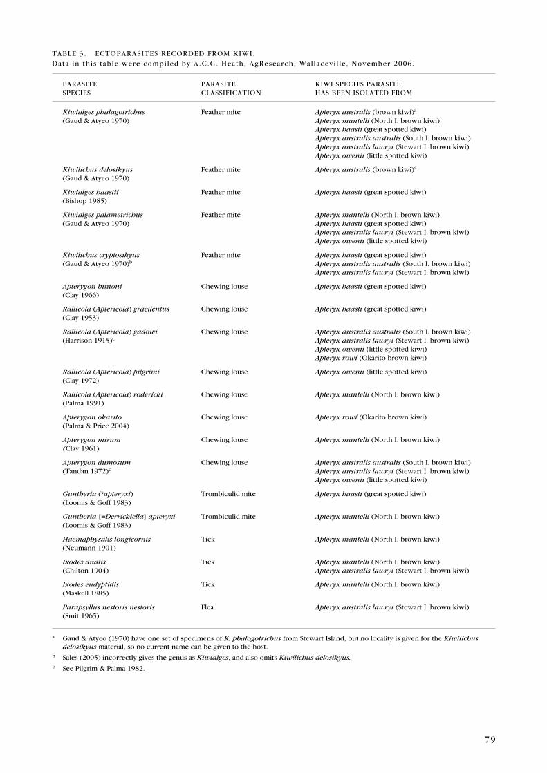
79
PARASITe PARASITe KIWI SPeCIeS PARASITeSPeCIeS CLASSIFICATION HAS BeeN ISOLATeD FROM
Kiwialges phalagotrichus Feather mite Apteryx australis (brown kiwi)a
(Gaud & Atyeo 1970) Apteryx mantelli (North I. brown kiwi) Apteryx haasti (great spotted kiwi) Apteryx australis australis (South I. brown kiwi) Apteryx australis lawryi (Stewart I. brown kiwi) Apteryx owenii (little spotted kiwi)
Kiwilichus delosikyus Feather mite Apteryx australis (brown kiwi)a
(Gaud & Atyeo 1970)
Kiwialges haastii Feather mite Apteryx haasti (great spotted kiwi)(Bishop 1985)
Kiwialges palametrichus Feather mite Apteryx mantelli (North I. brown kiwi)(Gaud & Atyeo 1970) Apteryx haasti (great spotted kiwi) Apteryx australis lawryi (Stewart I. brown kiwi) Apteryx owenii (little spotted kiwi)
Kiwilichus cryptosikyus Feather mite Apteryx haasti (great spotted kiwi)(Gaud & Atyeo 1970)b Apteryx australis australis (South I. brown kiwi) Apteryx australis lawryi (Stewart I. brown kiwi)
Apterygon hintoni Chewing louse Apteryx haasti (great spotted kiwi)(Clay 1966)
Rallicola (Aptericola) gracilentus Chewing louse Apteryx haasti (great spotted kiwi)(Clay 1953)
Rallicola (Aptericola) gadowi Chewing louse Apteryx australis australis (South I. brown kiwi)(Harrison 1915)c Apteryx australis lawryi (Stewart I. brown kiwi) Apteryx owenii (little spotted kiwi) Apteryx rowi (Okarito brown kiwi)
Rallicola (Aptericola) pilgrimi Chewing louse Apteryx owenii (little spotted kiwi)(Clay 1972)
Rallicola (Aptericola) rodericki Chewing louse Apteryx mantelli (North I. brown kiwi)(Palma 1991)
Apterygon okarito Chewing louse Apteryx rowi (Okarito brown kiwi)(Palma & Price 2004)
Apterygon mirum Chewing louse Apteryx mantelli (North I. brown kiwi)(Clay 1961)
Apterygon dumosum Chewing louse Apteryx australis australis (South I. brown kiwi)(Tandan 1972)c Apteryx australis lawryi (Stewart I. brown kiwi) Apteryx owenii (little spotted kiwi)
Guntheria (?apteryxi) Trombiculid mite Apteryx haasti (great spotted kiwi)(Loomis & Goff 1983)
Guntheria [=Derrickiella] apteryxi Trombiculid mite Apteryx mantelli (North I. brown kiwi)(Loomis & Goff 1983)
Haemaphysalis longicornis Tick Apteryx mantelli (North I. brown kiwi)(Neumann 1901)
Ixodes anatis Tick Apteryx mantelli (North I. brown kiwi)(Chilton 1904) Apteryx australis lawryi (Stewart I. brown kiwi)
Ixodes eudyptidis Tick Apteryx mantelli (North I. brown kiwi)(Maskell 1885)
Parapsyllus nestoris nestoris Flea Apteryx australis lawryi (Stewart I. brown kiwi)(Smit 1965)
TABLe 3. eCTOPARASITeS ReCORDeD FROM KIWI.
Data in this table were compi led by A.C.G. Heath, AgResearch, Wal lacevi l le , November 2006.
a Gaud & Atyeo (1970) have one set of specimens of K. phalogotrichus from Stewart Island, but no locality is given for the Kiwilichus delosikyus material, so no current name can be given to the host.
b Sales (2005) incorrectly gives the genus as Kiwialges, and also omits Kiwilichus delosikyus.c See Pilgrim & Palma 1982.

80
8. Wildlife Diagnostic Service
This section was prepared by Dr Maurice Alley, New Zealand Wildlife Health
Centre, Massey University, and is also available from http://wildlife.massey.
ac.nz.
Massey University provides a diagnostic service to the Department of
Conservation and other wildlife conservation and management institutions.
Guidelines on submission of specimens for post-mortem are detailed below.
The three important points to consider are:
Preservation•
Documentation•
Packaging•
8 . 1 P R e S e R V A T I O N
Time, heat, autolysis, microbial multiplication, maggots, and scavengers all
obscure the information that can be obtained from a dead body. Preservation
techniques to counteract these may also introduce artefacts or limit the
range of examinations which can be possible on the remains. A dead animal
should be chilled to refrigerator temperature (approximately 4°C) as soon
after death as possible, and despatched for diagnosis on the earliest available
transport. Freezing interferes with gross and microscopic examination of
tissues, and some aspects of microbiological culture and should be a last
resort if the dead body cannot be delivered within approximately 24 to 36
hours. Freezing a rotten body will not improve its diagnostic value even
if it does slightly improve its smell. Fixing bodies whole in alcohol or
formalin, and field dissection and submission of formalin-fixed tissues for
histopathology is an alternative which can be used in some circumstances—
talk to us before undertaking this. However if you are collecting material into
fixatives, remember skin contact and inhalation of formalin is hazardous; you
need to use a volume of formalin 10 times the amount of tissue you are fixing
(i.e. 100 g of tissue needs 1 L of formalin); and formalin does not penetrate
quickly enough for proper fixation for histopathology more than about 1 cm
deep into tissues.
8 . 2 D O C U M e N T A T I O N
All bodies should be sent with a wildlife submission form (see Appendix 7,
or download from http://wildlife.massey.ac.nz).
The objectives of documentation are to:
Identify the animal, tissues or specimens

81
Providing a detailed provenance may assist the diagnosis, and will also allow
a complete data entry in the wildlife disease database Huia. We want to know
the individual animal ID if it has one, the species, the geographical location,
the timepoint of collection (who, what, where, when?)
Specify what you want
Our routine practice is to try to establish a cause of death and other intercurrent
diseases when a whole body is submitted. You may want to know something
else either instead or as well as these things.
Disposition of remains and samples
Our routine practice is to retain a range of tissue samples for histopathology
and microbiology after a gross necropsy. Do you want anything else kept
or handled in a specific way? Is the body important as a natural history
specimen? We can modify our examination techniques to be in sympathy
with further uses of the remains if you let us know.
8 . 3 P A C K A G I N G
The objectives of packaging are to:
Retain and maintain the identification of the submission
A legible label in permanent ink or pencil firmly associated with the body
either attached as a tag or applied to a bag immediately containing it.
Contain the body and any leakage
Use multiple tear and puncture resistant sealed plastic bags, or plastic
containers with firmly screwed down tight-fitting lids. Surround with
enough absorbent material such as paper towel to mop up any effusions and
seepage which might be anticipated. Do not use glass. We all have a duty to
prevent contaminaton of people and equipment with potentially infectious
or hazardous substances.
Protect the samples from the transport environment (heat, water)
and damage in transport (impact and crush-proof)
A suitably sized polystyrene foam chilly bin is best, but alternatives can
include a cardboard box with newspaper and bubble-wrap protecting the
well-wrapped and bagged body. Proprietary freezer blocks, gel paks, etc.
can be improvised using 500-mL plastic PeT drink-bottles—partially fill some
with water and put them in your freezer so thay will be ready when you need
them.
The physical address for specimens to the wildlife diagnostic service is:
Attention: Maurice Alley/Brett Gartrell/Kerri Morgan
Room 8.28
Vet Tower
IVABS
Massey University
Fitzherbert Road
Palmerston North
Mark the package: Urgent, Perishable or Keep Cool, Do Not Freeze.

82
It is useful to inform us by email, phone or fax so we know to expect a parcel.
The Huia database submission form (see Appendix 7) can be faxed to 06 350
5636.
The following courier companies have been used successfully in the past:
NZ Couriers
Tranzlink
8 . 4 I N S U M M A R Y
Chill and despatch as soon as possible.•
Identify and specify what you want in the documentation.•
Contain, preserve and protect in transit by appropriate packaging.•

83
9. Acknowledgements
Special thanks to Dr Brett Gartrell, Massey University, and Richard Jakob-Hoff,
Auckland Zoo, for peer reviewing this document. Thanks also to Dr Maurice
Alley, Massey University, and Kate McInnes, Department of Conservation,
for their contributions. Jenny Youl and Vanessa Gray (Massey University),
Trevor Kelly (The Vet Centre, Rotorua) and Claire Travers (Kiwi encounter,
Rainbow Springs, Rotorua) are acknowledged for the use of their photos.
Dallas Bishop (Agresearch) and Ricardo Palma (Te Papa Tongarewa, Museum
of New Zealand) confirmed the accuracy of the ectoparasites recorded from
kiwi listed in Table 3.
10. References
Abou-Madi, N.; Kollias, G.V. (eds) 1992: Avian fluid therapy. Current veterinary therapy xI. W.B. Co,
Philadelphia.
Aguilar, R.F. 2004: The use of occlusive hydrocolloidal bandages in raptor wound management.
Pp. 135–137 in: Proceedings of the Australian Committee of the Association of Avian
Veterinarians, Kakadu.
Andrews, J.R.H. 1977: A new species of Lyperosomum (Digenea: Dicrocoeliidae) from the North
Island brown kiwi. New Zealand Journal of Zoology 4: 99–100.
Bauck, L. 1994: Mycoses. Pp. 997–1006 in Ritchie, B.W.; Harrison, G.J.; Harrison, L.R. (eds): Avian
medicine: principles and application. Wingers Publishing Inc., Lake Worth, Florida.
Bauck, L.; Kupersmith, D. 1991: Intraosseous fluids. Journal of the Association of Avian
Veterinarians 5: 74–100.
Benham, W.B. 1990: The structure of the rostellum in two new species of tapeworm, from Apteryx.
Quarterly Journal of Microscopical Science 43: 83–96.
Bennett, R.A. 1994: Neurology. Pp. 723–747 in Ritchie, B.W.; Harrison, G.J.; Harrison, L.R. (eds):
Avian medicine: principles and application. Wingers Publishing Inc., Lake Worth, Florida.
Bishop, D.M. 1985: Kiwialges haastii n.sp., a feather mite (Acari: Analgidae) from the great spotted
kiwi, Apteryx haastii Potts, 1872 (Aves: Apterygidae); with a key to species of Kiwialges,
and new host records. New Zealand Journal of Zoology 11: 233–237.
Boardman, W. 1995: Causes of mortality in North Island brown kiwi at Auckland Zoo 1960–1994.
Kokako 2(1): 11–13.
Bowles, H.L. 2006: evaluating and treating the reproductive system. Pp. 519–540 in Harrison, G.J.L.;
Lightfoot, T.L. (eds): Clinical avian medicine. Spix Publishing Inc., Florida.
Burke, H.F.; Swaim, S.F.; Amalsadvala, T. 2002: Review of wound management of raptors. Journal of
Avian Medicine and Surgery 16(3): 180–191.
Cannon, M. 2001: Anaesthesia. Pp. 19–45 in: Proceedings of the Australian Committee of the
Association of Avian Veterinarians, Hobart.
Cannon, M.J. 1991: Avian axioms. P. 1 in: proceedings of Avian Medicine Refresher Course for
veterinarians, Post Graduate Foundation in Veterinary Science, University of Sydney.
Carpenter, J.W. (ed.) 2005: exotic animal formulary. elsevier Saunders, St Louis, Missouri.

84
Chahn, J. 1884: Parasites de l’Aptérix. Comptes rendu des séances de la Société de Biologie 8(1):
770–771.
Chilton, C. 1904: A species of Ixodes parasitic on the Grey duck. Transactions of the New Zealand
Institute 36: 201–202.
Christensen, N.H. 1996: Sampling kiwis for Mycoplasma infections. New Zealand Veterinary
Journal 44: 200–201.
Clark, W.C. 1983: Nematodes of kiwis. New Zealand Veterinary Journal 10: 129.
Clark, W.C.; McKenzie, J.C. 1982: North Island kiwi Apteryx australis mantelli (Apterygiformes:
Aves): a new host for Toxocara cati (Nematoda: Ascaridoidea) in New Zealand. Journal of
Parasitology 68(1): 175–176.
Clay, T. 1953: Revisions of the genera of Mallophaga. I. The Rallicolai-complex. Proceedings of the
Zoological Society of London 123: 563–587.
Clay, T. 1961: A new genus and species of Menoponidae (Mallophaga, Insecta) from Apteryx. Annals
and Magazine of Natural History (Series 13) 3: 571–576.
Clay, T. 1966: A new species of Apterygon (Mallophaga: Menoponidae). The Entomologist 99: 292–
293.
Clay, T. 1972: The new species of Rallicola (Insecta: Phthiraptera: Ischnocera) parasitic on kiwis
(Apteryx). New Zealand Journal of Science 15: 70–76.
Clemence, M. 1995: Veterinary cases from the Kiwi House. Kokako 2(1): 15–18.
Clyde, V.L. 1994: Avian analgesia. Pp. 125–127 in: Proceedings of the American Association of Zoo
Veterinarians.
Clyde, V.L.; Paul-Murphy, J. 1999: Avian analgesia. Pp. 309–314 in Fowler, M.e.; Miller, R.e. (eds):
Zoo and wild animal medicine: current therapy 4. W.B. Saunders, Philadelphia.
Colbourne, R. 2002: Incubation behaviour and egg physiology of kiwi (Apteryx spp.) in natural
habitats. New Zealand Journal of Ecology 26(2): 129–138.
Cunningham, S.; Castro, I.; Alley, M. 2007: A new prey-detection mechanism for kiwi (Apteryx spp.)
suggests convergent evolution between paleognathous and neognathous birds. Journal of
Anatomy 211(4): 493–502.
Dahlhausen, R.D. 2006: Implications of mycoses in clinical disorders. Pp. 691–704 in Harrison, G.J.L.;
Lightfoot, T.L. (eds): Clinical avian medicine. Spix Publishing Inc., Florida.
Davis, G.B.; Watson, P.R.; Billing, A.e. 1984: Tuberculosis in a kiwi (Apteryx mantelli). New Zealand
Veterinary Journal 32(3): 30.
Deeming, D.C.; Ferguson, M.W.J (eds) 1991: egg incubation: its effects on embryonic development
in birds and reptiles. Cambridge University Press, Cambridge.
Degernes, L. 1994: Trauma medicine. Pp. 417–433 in Ritchie, B.W.; Harrison, G.J.; Harrison, L.R.
(eds): Avian medicine: principles and application. Wingers Publishing Inc., Lake Worth,
Florida.
Degernes, L.A.; Crosier, M.L.; Harrison, L.D.; Dennis, P.M.; Diaz, D.e. 1999: Autologous, homologous,
and heterologous red blood cell transfusions in cockatiels (Nymphicus hollandicus). Journal
of Avian Medicine and Surgery 13(1): 2–9.
DOC (Department of Conservation) 2003: Captive Management Policy. www.doc.govt.nz/upload/
documents/about-doc/role/policies-and-plans/protected-wildlife-policy.pdf (viewed Novem-
ber 2007).
Doneley, B. 2006: Management of captive ratites. Pp. 957–990 in Harrison, G.J.L.; Lightfoot, T.L.
(eds): Clinical avian medicine. Spix Publishing Inc., Florida.
Dumonceaux, G.; Harrison, G.J. 1994: Toxins. Pp. 1030–1052 in Ritchie, B.W.; Harrison, G.J.;
Harrison, L.R. (eds): Avian medicine: principles and application. Wingers Publishing Inc.,
Lake Worth, Florida.
edling, T.M. 2006: Updates in anaesthesia and monitoring. Pp. 747–760 in Harrison, G.J.L.; Lightfoot,
T.L. (eds): Clinical avian medicine. Spix Publishing Inc., Florida.

85
Farner, D.S.; Chivers, N.; Riney, T. 1956: Body temperatures of the North Island kiwis. Emu 56:
199–206.
Fergus, N.; Girling, S.; Kirk, e.J.; Cockram, J.F. 1995: Digestive tract of the kiwi. Kokako 2(1): 9.
Firth, A.M. 1995: Fluids, electrolytes and hypertonics. Pp. 335–337 in: Anaesthesia, emergency &
critical care. Post-graduate Foundation in Veterinary Science, University of Sydney, Sydney.
Flammer, K. 1994: Antimicrobial therapy. Pp. 434–478 in Ritchie, B.W.; Harrison, G.J.; Harrison,
L.R. (eds): Avian medicine: principles and application. Wingers Publishing Inc., Lake Worth,
Florida.
Flammer, K.; Clubb, S.L. 1994: Neonatology. Pp. 805–841 in Ritchie, B.W.; Harrison, G.J.; Harrison,
L.R. (eds): Avian medicine: principles and application. Wingers Publishing Inc., Lake Worth,
Florida.
Forsyth, S.F.; Machon, R.G.; Walsh, V.P. 1999: Veterinary anaesthesia. Pp. 214–220 in: Unpublished
BVSc notes (paper number 95.301), Massey University.
Franchetti, R.R.; Kilde, A.M. 1978: Restraint and anaesthesia. Pp. 359–364 in Fowler, M.e.; Miller, R.e.
(eds): Zoo and wild animal medicine. W.B. Saunders, Philadelphia.
Fudge, A.M. 2000: Disorders of avian erythrocytes. Chapter 4 (pp. 19–27) in: Laboratory medicine:
Avian and exotic Pets. W.B. Saunders, Philadelphia. 486 p.
Gartrell, B.D.; Morgan, K.J.; Green, C. 2006: Diagnosis of pelvic injuries using computer tomography in
a rowi kiwi. Pp. 147–153 in: proceedings of the Association of Avian Veterinarians Australian
Committee and Unusual and exotic Pet Veterinarians Combined Annual Conference,
September 2–6, Wellington.
Gaud, J.; Atyeo, W.T. 1970: Acariens Sarcoptiformes plumicoles (Analgoidea) parasites des
Apterygiformes. Acarologia 12: 402–414.
Gelis, S. 2006: evaluating and treating the gastrointestinal system. Pp. 411–440 in Harrison, G.J.L.;
Lightfoot, T.L. (eds): Clinical avian medicine. Spix Publishing Inc., Florida.
Gerlach, H. 1994: Bacteria. Pp. 949–983 in Ritchie, B.W.; Harrison, G.J.; Harrison, L.R. (eds): Avian
medicine: principles and application. Wingers Publishing Inc., Lake Worth, Florida.
Gilmour, D.G. 1971: Blood groups. Pp. 883–896 in Bell, D.J.; Freeman, B.M. (eds): Physiology and
biochemistry of the domestic fowl (Vol. 2). Academic, New York.
Greenacre, C.B.; Latimer, K.S.; Ritchie, B.W. 1992: Leg paresis in a black palm cockatoo caused by
aspergillosis. Journal of Zoo and Wildlife Medicine 23: 122–126.
Greiner, e.C.; Ritchie, B.W. 1994: Parasites. Pp. 1007–1029 in Ritchie, B.W.; Harrison, G.J.; Harrison,
L.R. (eds): Avian medicine: principles and application. Wingers Publishing Inc., Lake Worth,
Florida.
Harr, K.e.; Kollias, G.V.; Rendano, V.; Delahunta, A. 1997: A myelographic technique for avian
species.Veterinary Radiology and Ultrasound 38(3): 187–192.
Harrison, G.J. 1986: What to do until a diagnosis is made. Pp. 356–361 in Harrison, G.J.; Harrison, L.R.
(eds): Clinical avian medicine and surgery. W.B. Saunders, Philadelphia.
Harrison, G.J.; Ritchie, B.W. 1994: Making distinctions in the physical examination. Pp. 144–175 in
Ritchie, B.W.; Harrison, G.J.; Harrison, L.R. (eds): Avian medicine: applications and principle.
Wingers Publishing Inc., Lake Worth, Florida.
Harrison, G.J.L.; Lightfoot, T.L; Flinchum, G.B 2006: emergency and critical care. Pp. 212–232 in
Harrison, G.J.L.; Lightfoot, T.L. (eds): Clinical avian medicine. Spix Publishing Inc., Florida.
Harrison, L. 1915: Mallophaga from Apteryx and their significance; with a note on the genus Rallicola.
Parasitology 8: 88–100.
Hartley, W.J. 1995: Some interesting avian cases in the Taronga Zoo pathology collection. Kokako
2(1): 11.
Haskins, S.C. 1992: Management of septic shock. Journal of American Veterinary Medical
Association 20(12): 1915–1924.

86
Hawkins, M.G.M.; Machin, K.L 2004: Avian pain and analgesia. Pp. 165–174 in: Proceedings of the
2004 meeting of the Association of Avian Veterinarians, New Orleans.
Heather, B.; Robertson, H. 2000: The field guide to the birds of New Zealand. Penguin Books (NZ)
Ltd, Auckland.
Helmer, P. 2006: Advances in diagnostic imaging. Pp. 653–660 in: Harrison, G.J.L.; Lightfoot, T.L.
(eds): Clinical avian medicine. Spix Publishing Inc., Florida.
Hill, F.; Woodgyer, A.J.; Lintott, M.A. 1995: Cryptococcosis in a North Island brown kiwi (Apteryx
australis mantelli) in New Zealand. Journal of Medical and Veterinary Mycology 33:
305–309.
Hoefer, H.L. 1992: Transfusions in exotic species. Problems in Veterinary Medicine 4(4):
625–635.
Hume, A. 1995: Practical avian haematology. Pp. 163–181 in: Proceedings of the Australian Committee
of the Association of Avian Veterinarians, Dubbo.
Hume, A. 2000: What to do if someone brings in a sick bird? Pp. 3–12 in: Proceedings of the Australian
Committee of the Association of Avian Veterinarians, echuca.
Jakob-Hoff, R.; Buchan, B.; Boyland, M. 1999: Kiwi coccidian—North Island survey results. Kokako
1(6): 3–5.
Jensen, T.; Durrant, B. 2006: Assessment of reproductive status and ovulation in female brown kiwi
(Apteryx mantelli) using faecal steroids and ovarian follicle size. Zoo Biology 25: 25–34.
Joyner, K.L. 1994: Theriogenology. Pp. 723–804 in Ritchie, B.W.; Harrison, G.J.; Harrison, L.R.(eds):
Avian medicine: applications and principle. Wingers Publishing Inc., Lake Worth, Florida.
Kaufman, G.e. 1992: Avian emergencies. Pp. 723–804 in Murtaugh, R.J.; Kaplan, P.M.: Veterinary
emergency and critical care medicine. Mosby Year Book Inc., St Louis.
King, A.S.; McLelland, J. 1984: Respiratory system. Pp. 100–144 in: Birds: their structure and function.
Bailiere Tindall. London. 334 p.
Korbel, R. 1998: Verleichende untersuchungen zur inhalationsanaesthesie mit isofluran (forene) und
sevofluran (SeVOrane) bei haustauben (Columba livia Gmel., 1789, var domestica) und
vorsellung eines narkose-referenzprotokolls fuer voegel. Tierarzliche Praxix 26: 211–213.
Lamberski, N.; Daniel, G. 1992: Fluid dynamics of intraosseous fluid administration in birds. Journal
of American Veterinary Medical Association 23(1): 47–54.
Lichtenberger, M. 2004: Shock and fluid therapy for the avian veterinarian. Pp. 157–164 in:
Proceedings of the 2004 meeting of the Association of Avian Veterinarians, New Orleans.
Lierz, M. 2006: Diagnostic value of endoscopy and biopsy. Pp. 631–652 in Harrison, G.J.L.; Lightfoot,
T.L. (eds): Clinical avian medicine. Spix Publishing Inc., Florida.
Loomis, R.B.; Goff, M.L. 1983: A new species of Guntheria (Acari: Trombiculidae) from New Zealand.
Journal of Medical Entomology 20: 87–89.
Lumeij, J.T. 1987: Plasma urea, creatinine and uric acid concentrations in response to dehydration in
racing pigeons (Columba livia domestica). Avian Pathology 16: 377–382.
Lumeij, J.T. 1994: Gastroenterolgy. Pp. 482–521 in Ritchie, B.W.; Harrison, G.J.; Harrison, L.R. (eds):
Avian medicine: applications and principle. Wingers Publishing Inc., Lake Worth, Florida.
Machin, K.L. 2005: Controlling avian pain. Compendium of Continuing Education for the Practicing
Veterinarian 27: 299–309.
Malik, R.; Krockenberger, M.B.; Cross, G.; Doneley, R.; Madill, D.N.; Black, D.; McWhirter, P.;
Rozenwax, A.; Rose, K.; Alley, M.; Forshaw, D.; Russell-Brown, I.; Johnstone, C.; Martin, P.;
O’Brien, C.R.; Love, D.N. 2003: Avian cryptococcosis. Medical Mycology 41(2): 115–124.
Marchant, S.; Higgins, P.J.; Davies, S.J.J.F. 1991: The handbook of Australian, New Zealand and
Antarctic birds. Oxford University Press.
Martin, H.D.; Kollias, G.V. 1989: evaluation of water deprivation and fluid therapy in pigeons. Journal
of Zoo and Wildlife Medicine 20(2): 173–177.

87
Martin, G.R.; Wilson, K.; Wild, J.M.; Parsons, S.; Kubke, M.F.; Corfield, J. 2007: Kiwi forgo vision in
the guidance of their nocturnal activities. PLoS ONE 2(2): e198.
Marx, K.L. 2006: Therapeutic agents. Pp. 241–342 in Harrison, G.J.L.; Lightfoot, T.L. (eds): Clinical
avian medicine. Spix Publishing Inc., Florida.
Maskell, W.M. 1885: On a parasite of the penguin. Transactions of the New Zealand Institute 17:
19–20.
Maxwell, M.H. 1980: Attempted induction of an avian eosinophilia using various agents. Research in
Veterinary Science 29: 293–297.
McKenna, P. 1998: Parasites of birds in New Zealand. Pp. 5 & 18 in: Surveillance 25 (Special Issue).
McMillan, M.C. 1994: Imaging techniques. Pp. 246–326 in Ritchie, B.W.; Harrison, G.J.; Harrison,
L.R. (eds): Avian medicine: applications and principle. Wingers Publishing Inc., Lake Worth,
Florida.
Monks, D. 1996: emergency and critical care of the avian patient. Pp. 317–348 in: Proceedings of the
Australian Committee of the Association of Avian Veterinarians, Nerang.
Morgan, K.J. 2004: Coccidiosis in the North Island brown kiwi. Pp. 37–40 in: Proceedings of the
Australian Committee of the Association of Avian Veterinarians, Kakadu.
Morgan, K.J.; Alley, M.R.; Potter, J. 2005: Visceral larval migrans in North Island brown kiwi (Apteryx
mantelli). P. 66 in: Advances in exotic, zoo and wild animal medicine, proceedings of the
joint British Veterinary Zoological Society/World Association of Wildlife Veterinarians/
Zoological Society of London conference, London.
Naeini, A.T.; Dadras, H.; Naeini, B.A. 2006: Myelography in the pigeon (Columba livia). Journal of
Avian Medicine and Surgery 20(1): 27–30.
Neumann, L.G. 1901: Revision de la famille des Ixodides. 4 e memoire. Memoires. Societe Zoologique
de France, Paris 14: 249–372.
NZWHC (New Zealand Wildlife Health Centre) 2006: Huia database. New Zealand Wildlife Health
Centre, Massey University, Palmerston North.
O’Malley, B. 2005: Birds: anatomy and physiology. Pp. 97–164 in: Clinical anatomy and physiology of
exotic species. Structure and function of mammals, birds, reptiles and amphibians. elsevier
Saunders, Philadelphia. 272 p.
Orcutt, C.; Flinchum, G. 2001: Contrasting views on corticosteroid use in birds. Exotic DVM 3(5):
43.
Orr, M. 1994: Animal Health Laboratory Network—review of diagnostic cases, October–December
1994. Surveillance 22(1): 3–5.
Orr, M.; Black, A. 1996: Animal Health Laboratory Network—review of diagnostic cases, October–
December 1995. Surveillance 23(1): 3–5.
Otto, C.M.; Crowe, D.T. 1992: Intraosseous resuscitation techniques and applications. Pp. 107–
112 in Kirk, R.W.; Bonagura, J.D. (eds): Current veterinary therapy xI. W.B. Saunders,
Philadelphia.
Palma, R.L. 1991: A new species of Rallicola (Insecta: Phthiraptera: Philopteridae) from the North
Island brown kiwi. Journal of the Royal Society of New Zealand 21: 313–322.
Palma, R.L.; Price, R.D. 2004: Apterygon okarito, a new species of chewing louse (Insecta: Phthiraptera:
menoponidae) from the Okarito brown kiwi (Aves: Apterygiformes: Apterygidae).
New Zealand Journal of Zoology 31: 67–73.
Parton, K.; Bruere, A.N.; Chambers, J.P. 2001: Miscellaneous plants, shrubs and trees. P. 345 in
Veterinary clinical toxicology, 2nd edition. Publication No. 208, Foundation for Continuing
education of the New Zealand Veterinary Association and Veterinary Continuing education,
Massey University.
Paul-Murphy, J. 2006: Pain management. Pp. 233–239 in Harrison, G.J.L.; Lightfoot, T.L. (eds): Clinical
avian medicine. Spix Publishing Inc., Florida.

88
Pierce, M.A.; Jakob-Hoff, R.M.; Twentyman, C. 2003: New species of haematozoa from Apterygidae
in New Zealand. Journal of Natural History 37(15): 1797–1804.
Pilgrim, R.L.C.; Palma, R.L. 1982: A list of the chewing lice (Insecta: Mallophaga) from birds in
New Zealand. Notornis 29 (supplement): 1–32. (Also as National Museum of New Zealand
Miscellaneous Series 6.)
Pollock, C.G. 2006: Implications of Mycobacteria in clinical disorders. Pp. 681–690 in Harrison,
G.J.L.; Lightfoot, T.L. (eds): Clinical avian medicine. Spix Publishing Inc., Florida.
Prinzinger, R.; Dietz, V. 2002: Pre- and postnatal energetics of the North Island brown kiwi (Apteryx
mantelli). Comparative Biochemistry and Physiology. Part A: Molecular and Integrative
Physiology 131(4): 725–732.
Quesenberry, K.e.; Hillyer, e.V. 1994: Supportive care and emergency therapy. Pp. 382–416 in
Ritchie, B.W.; Harrison, G.J.; Harrison, L.R. (eds): Avian medicine: applications and principle.
Wingers Publishing Inc., Lake Worth, Florida.
Raidal, S.; Butler, R. 2001: Chronic rhinosinusitis and rhamphothecal destruction in a Major Mitchell’s
cockatoo (Cacatua leadbeateri) due to Cryptococcus neoformans var. gatti. Journal of
Avian Medicine and Surgery 15: 121–122.
Redig, P.T. 1984: Fluid therapy and acid-base balance in the critically ill avian patient. Pp. 59–73
in: Proceedings of the 1984 international conference on avian medicine, sponsored by the
Association of Avian Veterinarians, Toronto.
Reid, B.; Williams, G.R. 1975: The kiwi. Pp. 301–330 in Kuschel, G. (ed.): Biogeography and ecology
in New Zealand. The Hague, Junk. 680 p.
Richardson, J. 2006: Implications of toxic substances in clinical disorders. Pp. 711–719 in Harrison,
G.J.L.; Lightfoot, T.L. (eds): Clinical avian medicine. Spix Publishing Inc., Florida.
Ritchie, B.W. 1991: Avian therapeutics. Introduction to avian medicine and surgery. T2, pp. 1–18 in:
Proceedings of the Association of Avian Vets.
Robertson, H. 2006: Haematological and biochemical parameters of the great spotted kiwi Apteryx
haasti. Unpublished report, Department of Conservation, New Zealand.
Rupiper, D.J. 1998: Diseases that affect race performance of homing pigeons. Part II: bacterial, fungal
and parasitic diseases. Journal of Avian Medicine and Surgery 12: 183–148.
Sales, J. 2005: The endangered kiwi: a review. Folia Zoologica 54: 1–20.
Schmitt, P.M.; Gobel, T.; Trautvetter, e. 1998: evaluation of pulse oximetry as a monitoring method
in avian anaesthesia. Journal of Avian Medicine and Surgery 12(2): 91–99.
Shaw, S.D.; Billing, T. 2006: Karaka (Corynocarpus laevigatus) toxicosis in North Island brown
kiwi (Apteryx mantelli). Veterinary Clinics of North America. Exotic Animal Practice 9:
545–549.
Smit, F.G.A.M. 1965: Siphonaptera of New Zealand. Transactions of the New Zealand Institute 7:
1–50.
Smith, B.L.; Poole, W.S.H.; Martinovich, D. 1973: Pneumoconiosis in the captive New Zealand kiwi.
Veterinary Pathaology 10: 94–101.
Starck, J.M.; Ricklefs, R.e. 1998: Avian growth and development. evolution within the Altricial-
Precocial Spectrum. Oxford University Press, New York. 456 p.
Stewart, J. 1994: Ratites. Avian medicine: principles and application. Pp. 1284–1326 in Ritchie, B.W.;
Harrison, G.J.; Harrison, L.R. (eds): Avian medicine: applications and principle. Wingers
Publishing Inc., Lake Worth, Florida.
Tandan, B.K. 1972: The species of Apterygon (Insecta: Phthiraptera: Amblycera) parasitic on kiwi
(Apteryx). New Zealand Journal of Science 15: 52–69.
Taylor, M. 2000: Disorders of the avian digestive system. Pp. 407–416 in: Birds 2000. Post Graduate
Foundation in Veterinary Science, University of Sydney, Australia.
Thompson, e.J.; Wright, I.G.A. 1978: Coccidiosis in kiwis. New Zealand Veterinary Journal 26(6):
167.
89
Tully, T.N.; Harrison, G.J. 1994: Pneumonology. Pp. 556–581 in Ritchie, B.W.; Harrison, G.J.;
Harrison, L.R. (eds): Avian medicine: applications and principle. Wingers Publishing Inc.,
Lake Worth, Florida.
Vleck, C.M. 1991: Water economy and solute regulation of reptilian and avian embryos. Pp. 245–
259 in Ferguson, M.W.J.; Deeming, D.C. (eds): egg incubation: its effects on embryonic
development in birds and reptiles. Cambridge University Press, Cambridge.
Vleck, C. M.; Hoyt, D.F. 1991: Metabolism and energetics of reptilian and avian embryos. Pp. 285–306
in: egg incubation: its effects on embryonic development in birds and reptiles. Cambridge
University Press, Cambridge.
Weekes, P.J. 1982: Checklist of helminth parasites of birds in New Zealand. New Zealand Journal
of Zoology 9: 451–460.
Wilson, H.; Hernandez-Divers, S.; Budsberg, S; Latimer, K; Grant, K. 2004: Pharmacokinetics and use
of meloxicam in psittacine birds. Pp. 7–9 in: Proceedings of the annual conference of the
Association of Avian Veterinarians, New Orleans.
Wright, e.M.; Marcella, K.L.; Woodson, J.F. 1985: Animal pain and control. Laboratory Animal
(May/June): 20–36.

90
Appendix 1
K I W I F I R S T A I D F I e L D K I T
A first aid kit useful for birds can be constructed from a modified human first
aid kit. The following table lists suggested essential items:
Veterinarians undertaking field work could also consider taking:
Analgesia (Butorphanol)•
Antibiotics (e.g. 2.5% injectable enrofloxacin (Baytril™), Clavulox™ •
palatable drops)
equipment for an IV fluid set up•
ITeM QUANTITY (MINIMUM)
Sterile saline solution for injection (0.9% NaCl) vials (10 mL, 5 mL) 50–100 mL
Or lactated Ringer’s solution (LRS) or saline (0.9% NaCl) flexibag 500 mL
(For wound lavage, oral or subcutaneous fluids)
Melolin™ non-adherent dressings 3
Sterile gauze swabs (pkt of 5) 2
Vetrap™—5 cm width 2
Sofban™—5 cm width 2
Syringes—5 mL 3
Syringes—20 mL 1
Needles—25 gauge, 22 gauge 6
Snap-type heat pack 2
Scissors 1
Crop tubes (silicone) 1

91
Appendix 2
V e T e R I N A R Y A D V I C e A N D R e F e R R A L C e N T R e S
The following contacts are available for advice when dealing with injured or
sick kiwi, and may include the option of referral for further diagnostics and
treatment.
New Zealand Wildlife Health Centre (NZWHC)
Veterinary Teaching Hospital
Institute of Veterinary, Animal and Biomedical Sciences
Massey University
Palmerston North
Phone: (06) 350 5329 (Weekdays)
(06) 350 5955 (Weekends)
Veterinarians
Dr Brett Gartrell, BVSc, MACVSc (Avian Health), PhD
Senior Lecturer in Avian and Wildlife Health, Director NZWHC
email: [email protected]
Kerri Morgan, BVSc, MACVSc (Avian Health), PGDipVCS (dist.)
Lecturer in Avian and Wildlife Health
email: [email protected]
Facilities available through the NZWHC include radiography, CT and MRI,
endoscopy, anaesthesia and a full surgical suite. The wildlife clinic at the
NZWHC is geared towards treating threatened and endangered species on
a national scale, as well as local common native species. The NZWHC is
currently contracted to the Department of Conservation to provide the
diagnostic pathology service for endangered and threatened species (see
section 8).
Kiwi from throughout New Zealand may be referred to the NZWHC for
treatment. Sponsorship covers costs of treatment of endangered species.
Contact the NZWHC at the Massey University Veterinary Teaching hospital
(phone numbers above) for advice and to arrange transportation of kiwi to
the wildlife clinic.
New Zealand Centre for Conservation Medicine (NZCCM)
Auckland Zoo
Motions Road
Western Springs
Auckland
Phone: (09) 360 3814 or (09) 360 4704 (Weekdays)
(09) 360 3800 (Weekends)
Veterinarians
Richard Jakob-Hoff, BVMS (Hons)
Senior Veterinarian

92
email: [email protected]
John Potter, BVSc
Associate Veterinarian
email: [email protected]
Consultant Veterinarians:
Berend Westera, BVSc
Maureen Forsyth, DVM, MACVSc
The Auckland Zoo is an active participant in the Operation Nest egg
Programme, breeds North Island brown kiwi on site and has a progressive
public education and advocacy programme for kiwi.
Veterinary facilities at the NZCCM at Auckland Zoo include a fully equipped
wildlife hospital and quarantine facility. Partnerships with local veterinary
and human medical specialists enable NZCCM to utilise the most up-to-date
diagnostic and treatment options.
Staff veterinarians at NZCCM provide clinical, diagnostic, pathological and
research services for both captive and free-living wildlife. Kiwi are a major
species of interest. Technical advice is provided through the Department of
Conservation’s Wildlife Health Co-ordinator.
Other avian veterinarians
The Wildlife Society of the New Zealand Veterinary Association has a database
listing veterinarians with avian and wildlife experience. This database may be
accessed by contacting the New Zealand Wildlife Health Centre.

93
HAeMATOLOGICAL/ NORTH ISLAND OKARITO BROWN GReAT SPOTTeD
BIOCHeMICAL BROWN KIWI KIWI KIWI
VALUeS (Apteryx mantelli)a (Apteryx rowi)b n (Apteryx haasti)c n
PCV (%) 46 (38–54) 40.1 (37.1–43.1) 9 39.4 (35.3–43.5) 25
Hb (g/L)
MCHC (g/L) 250 (110–333)
WBC (×109/L) 11.6 (8.7–14.5) 15.7 (7.9–23.5) 29
Heterophils (×109/L) 6.0 (4.0–8.2) 8.7 (4.3–13.1) 29
Heterophils (%) 55.0 (45.0–65.0) 29
Lymphocytes (×109/L) 4.2 (2.5–5.9) 4.8 (1.9–7.7) 29
Lymphocytes (%) 32.0 (22.0–42.0) 29
eosinophils (×109/L) 0.18 (0.7–1.29) 0.8 (0.0–1.9) 28
eosinophils (%) 4.0 (0.0–8.0) 28
Monocytes (×109/L) 0.3 (0.1–0.5) 0.5 (0.0–1.1) 28
Monocytes (%) 2.0 (0.0–5.0) 28
Basophils (×109/L) 0.56 (0.09–1.3) 0.9 (0.4–1.4) 29
Basophils (%) 6.0 (3.0–9.0)
CK (IU/L) 521–971 758.8 (446.5–1071.1) 9
AST (IU/L) 64–138 204.2 (132.4–276.1) 9
Bile acids (µmol/L) < 35 9
Serum protein (g/L) 54–62 52.1 (47.0–57.2) 9 46 (39–53) 25
Uric acid (µmol/L) 300–380 476.6 (343.4–609.7) 9
Ca (mmol/L) 1.85–3.1 2.49 (2.45–2.54) 9
Glucose (mmol/L) 3.0–3.9
LDH (IU/L) 2380
Phosphorus (mmol/L) 2.02 (1.80–2.23) 9
Sodium (mmol/L) 147.1 (145.2–149.0) 9
a Doneley 2006.b Unpubl. data 2007.c Robertson 2006.
Appendix 3
K I W I H A e M A T O L O G Y A N D B I O C H e M I S T R Y R e F e R e N C e R A N G e S

94
Appendix 4
C A P T I V e D I e T F O R M U L A T I O N
(Courtesy of Kiwi encounter, Rainbow Springs, Rotorua)
3 kg minced ox heart (N.B. remove fat first)
2 cups rolled oats cooked with 4 cups water, allow to cool
1 cup wheat germ
2 cups cat biscuits (premium quality), soaked and mashed
2 mashed bananas
2 pieces grated fruit (e.g. kiwifruit, nectarine, peach, pear)
250 g grated veges (e.g. corn, peas, beans, silverbeet, broccoli)
Mix together. Immediately prior to feeding, add 1 g kiwi pre-mix (see
Appendix 5) per 100 g of food. Food should be presented in bowls on the
ground.
F O O D P R e S e N T A T I O N F O R L O N G - T e R M C A R e
(Courtesy of Auckland Zoo)
Food is presented in flat stainless steel trays on the ground in the late
afternoon for birds held outdoors. Food presented in tubes placed into the
ground in the morning for birds held in nocturnal exhibits. Tubes used to
encourage probing action when feeding and allow for the feeding sites to
be moved easily for enrichment. earthworms are provided daily as available
scattered throughout enclosure for the birds housed in nocturnal exhibits.
Rotten logs should be provided as often as possible. Birds should have access
to small pebbles (2–3 mm).

95
Appendix 5
A V A I L A B I L I T Y O F D I e T A R Y P R O D U C T S M e N T I O N e D I N T H e T e x T
Kiwi Vitamin and mineral Premix
Contact Tony Billing
Westshore Wildlide Reserve, Napier
Phone (06) 834 4136
email [email protected]
Hills a/d™
Available in cans from most veterinary clinics
Wombaroo Insectivore Mix
Contact: Karen Wiley
Native Bird Rescue Trust, Wellington
Phone/fax (04) 479 2936
email [email protected]

96
Therapeutic contraindications in kiwi
Levamisole Can cause death. Kiwi appear to be acutely sensitive to levamisole toxicity at doses that are well within the
safe range for domestic poultry. Levamisole should not be used as an anthelmintic in kiwia.
Organophosphates Birds are extremely sensitive to organophosphates, and these products should not be used on birds as they
may cause deathb.
Corticosteroids Corticosteroids may be contraindicated for use in birds (B.D. Gartrell, Massey University, pers. comm.)
(see section 2.2.9).
Antibiotics
AGeNT COMMeNTS PRePARATION AND SUGGeSTeD DOSAGeS PRODUCT exAMPLeS
Amikacin sulfate Least nephrotoxic of the Injectable (Amikin™). 10–15 mg/kg IV, IM, SQ bid-tid
aminoglysosides; active against (most species)c,d; Nebulisation:
gram-negative bacteria including 5–6 mg/mL in sterile water
Pseudomonas spp., and gram- or saline, × 15 min bid-tidc.
positive bacteria including
Staphylococcus spp. and Streptococcus spp. Maintain
hydration during usec. Pain on
injection; causes myositis in
ostrichesc.
Amoxicillin Broad spectrum bactericidal Oral suspension (Amoxil™ 100–150 mg/kg PO,
penicillin antibiotic, minimal paediatric drops, Ranbaxy- IV, IM bid-tid
activity for common gram- Amoxi™); (most species)c,d.
negative infections of birdsc. injectable (Moxylan™, 15–22 mg/kg PO tid
Betamox LA™). (ratites)c.
Amoxycillin/clavulanate β–Lactamase inhibitor— Oral suspension (Clavulox™ 70 mg/kg PO, IV, IM
use with allopurinol is palatable drops); bid (kiwi, pers. obs.).
contraindicatedc. tablets (Clavulox™, 10–15 mg/kg PO bid
Intramuscular injections Noroclav™); injectable (ratites)c.
can be very irritant. (Clavulox™); 125–150 mg/kg PO bid
IV powder (Augmentin™). (most species)c.
Cefotaxime Third-generation cephalosporin IV powder (Cefotaxime 75–100 mg/kg IM, IV
with broad-spectrum activity sodium). bid-tid (most species)c.
for many gram-positive and 25 mg/kg IM tid (ratites)c.
gram-negative pathogens, Nebulisation: 10 mg/ml saline
penetrates cerebrospinal fluidc. × 10–30 min bid-qidc.
Antiobiotics continued on next page
Appendix 6
A V I A N T H e R A P e U T I C S F O R M U L A R Y
Unfortunately, there are relatively few pharmacokinetic studies of medications
for use in avian species, and most dosages used in avian medicine are based
on empirical data, observations and experience. Dosages given here are
extrapolated from published data applicable to avian species other than
kiwi.

97
Antiobiotics continued from previous page
AGeNT COMMeNTS PRePARATION AND SUGGeSTeD DOSAGeS
PRODUCT exAMPLeS
Cephalexin First-generation cephalosporin; Oral suspension; 40–100 mg/kg PO, IM
active against many gram- tablets (Keflex™); tid-qid (most species)c.
positive and gram-negative Injectable (Ceporex™). 15–22 mg/kg PO tid
bacteria, including E. coli and (ratites)c.
Proteus spp., but not
Pseudomonas spp; useful for
Staphylococcus spp. dermatitisc.
Ciprofloxacin Broad-spectrum quinolonec. Oral suspension, tablets 15–40 mg/kg PO bid
(Ciproxin™). (most species)c.
10 mg/kg PO bid (ostrich
chicks)c.
3–6 mg/kg PO bid (ratites)c.
Clindamycin Lincosamide: indicated for bone, Oral suspension, capsules 50–150 mg/kg PO sid-tidc.
joint and tendon sheath infections; (Antirobe™).
may be used for up to 12 weeks
without ill effects; monitor
kidneys and liver with long-term
use as well as for yeast overgrowth.
Doxycycline Drug of choice for Oral paste, tablets 25–50 mg/kg PO sid-bid
Chlamydophilia spp. and (Vibravet™). (most species)c.
Mycoplasma spp.c (N.B. neither of 2.0–3.5 mg/kg PO bid (ratites)c.
these have previously been
identified in kiwi).
enrofloxacin Given orally, the IM formulation Tablet, injectable (Baytril™). 10–15 mg/kg IM, PO sid-bid.
(2.5%) produces therapeutic (most species)c.
plasma concentration. 5 mg/kg IM bid (ratites)c.
IM formulation is extremely Nebulisation:
painful and should not be 10 mg/ml salinec.
given repeatedly. In general,
avoid IV use in birds.c
Gentamicin Aminogylcoside—not generally Injection (Gentamax 100™, 3–5 mg/kg IM sid-bid
recommended, narrow margin Genta 50™). (most species, including
of safety, nephrotoxic c. ostriches and emus)c.
Bird should be well hydrated, 1–2 mg/kg IM tid
avoid doses higher than (ratites, use only as
2.5–5.0 mg/kg q8-12h c. last resort)c.
Nebulisation: 5 mg/mL saline
× 15 min tidc.
Metronidazole Active against most anaerobes, Tablet (Stomorgyl™); 10–50 mg/kg PO bid
also antiprotozoalc. oral suspension (Flagyl™). (most species)c.
Trimethoprim/ Broad spectrum, contraindicated Oral suspension; 20–100 mg/kg PO bid
Sulfamethoxazole with dehydration, liver disease tablets (Trisul™). (most species)c.
or bone marrow suppression. 21 mg/kg PO bid
Side effects include GI upset, (ostriches)c.
regurgitation. Resistance to
Pseudomonas spp. commonc.

98
Antifungals
AGeNT COMMeNTS PRePARATION AND SUGGeSTeD DOSAGeS
PRODUCT exAMPLeS
Amphotericin B Fungicidal, preferred IV agent for IV powder (Fungizone™); 1.5 mg/kg IV tid 3–7 days
aspergillosis. IT administration oral lozenges (Fungilin™). (most species)c. 1 mg/kg IT
for syringeal aspergilloma may bid-tid (syringeal aspergilloma,
cause tracheitis. Potentially raptors)c. Nebulisation:
nephrotoxic, maintain good 1 mg/ml sterile water or saline,
patient hydration. Resistance 15 min bid (most species)c.
may developc. 100–200 mg/kg PO tid-qidd
(for GI candidiasis, avian
gastric yeast).
Fluconazole Fungistatic. Penetrates well into Capsules (Diflucan™, 2–5 mg/kg PO sid (most
brain, CSF and eyes. Only Flucazole™). species)c.
indicated if topical treatment Syrup (Diflucan™ suspension).
(e.g. nystatin) not feasible.
effective against Candida spp.,
but may be ineffective against
aspergillosisc.
Flucytosine Fungistatic agent. May be Flucytosine capsules. 20–100 mg/kg PO bid
administered as adjunctive (most species)c.
treatment for aspergillosis, 80–100 mg/kg PO bid
but about 50% Aspergillus spp. (ratites)c.
strains are resistantc.
Itraconazole Fungistatic, indicated for Oral suspension, capsules 5–10 mg/kg PO sid-bidc.
systemic mycoses including (Sporanox™).
aspergillosis (currently the drug
of choice). Commonly used for
prophylaxis. Suspension is first
choice, if using capsules, each
granule is approximately
0.05–0.39 mg (approximately
285–290 granules/capsule but
highly variable number and drug
concentration). Method of
compounding with strong acid
and orange juice has been
reportedc. May cause anorexia.
Ketaconazole Fungistatic. Indicated for systemic Tablets (Nizoral™). 15–30 mg/kg PO sid-bid
mycoses, including aspergillosis, (most species, including
and candidiasis. Less toxic than ratites)c.
amphotericin B, more toxic than
itroconazole. Side effects include
regurgitationc.
Nystatin Drug of choice for treatment of Oral suspension (Nilstat 250 000–500 000 IU/kg PO
candidiasis, not systemically Oral™). bid (ratites)c.
absorbed across intact GI tract,
oral lesions must be treated by
direct contact with medicationc.
Voriconazole Indicated for aspergillosis, can be Tablets (Vfend™). 10 mg/kg PO bidc.
used in conjunction with
amphotericin Bc.

99
Analgesics
AGeNT COMMeNTS PRePARATION AND SUGGeSTeD DOSAGeS
PRODUCT exAMPLeS
Butorphanol Opioid agonist-antagonist. Injection (Dolorex™, 1–4 mg/kg IM, IV bid-qid
Torbugesic™). (most species)c.
Carprofen Analgesic, anti-inflammatory. Injectable, tablets (Rimadyl™). 1–2 mg/kg PO, IM, IV sid-bid
Use in conjunction with fluid (most species)c.
supplementation. 2–10 mg/kg IM, SQ,
PO sid-bid (psittacines,
passerines, raptors)c.
Meloxicam Analgesic, antiinflammatory. Oral suspension, injectable 0.1–0.2 mg/kg IM, PO sid
Use in conjunction with fluid (Metacam™). (psittacines, raptors)c.
supplementation.
Obstetric drugs
AGeNT COMMeNTS PRePARATION AND SUGGeSTeD DOSAGeS
PRODUCT exAMPLeS
Calcium gluconate Hypocalcaemia—dilute 1:1 Injection. 5–10 mg/kg slow IV to effect
with saline or sterile water for for hypocalcaemic tetany,
IM or IV injectionsc. also SQ, IM, oncec.
50–100 mg/kg IM,
slow IV, oncec.
Calcium syrup Oral suspension (Troy Calcium 25–150 mg/kg PO sid-bid
Syrup™—calcium glubionate (most species)c.
and calcium lactobionate).
Antidotes
AGeNT COMMeNTS PRePARATION AND SUGGeSTeD DOSAGeS
PRODUCT exAMPLeS
Atropine Anticholinergic agent. Rarely Injection. 0.01–0.02 mg/kg IM, IV
indicated as a preanaestheticc. (most species)c.
Calcium eDTA Preferred initial chelator for lead Injection (calcium disodium 35–50 mg/kg IM bid 5 days,
and zinc toxicity. May cause renal versenate). off 2 days, repeat prnc (as
tubular necrosis in mammals, indicated by biochemical
maintain good hydration and analysis).
monitor for PU/PD. Do not give
PO as this may increase lead
absorption from the GITc.
D-Penicillamine Preferred chelator for copper Tablet (D-Penamine™). 30–55 mg/kg PO bid
toxicity. Can be used after/in (minimum 7 days)c.
conjunction with initial chelation
with CaeDTA for lead and zinc
toxicityc.
Vitamin K1 Rodenticide toxicity. Injection, tablets (Konakion™). 0.2–2.2 mg/kg IM tid-qid
until stable, then sid PO, IM,
14–28 daysc.
Obstetric drugs continued on next page

100
Obstetric drugs continued from previous page
AGeNT COMMeNTS PRePARATION AND SUGGeSTeD DOSAGeS
PRODUCT exAMPLeS
Prostaglandin F2α Dystocia. May be helpful when Injectable (Lutalyse™). 0.02–0.1 mg/kg IM,
(Dinoprost the egg is located distally and intracloacal once onlyc.
tromethamine) the uterovaginal sphincter is
dilated. Can result in uterine
bronchoconstriction, rupture,
hypertension, deathc.
Anthelmintics
AGeNT COMMeNTS PRePARATION AND SUGGeSTeD DOSAGeS PRODUCT exAMPLeS
Fenbendazole effective against cestodes, Oral liquid (Panacur™). 20–50 mg/kg PO sid
nematodes, trematodes, Giardia for 3–5 days (most species)c.
spp. Toxicities recorded in some 15–45 mg/kg PO
speciesc. Use 2.5%. (ostriches)c.
Ivermectin Most nematodes and ecto- Sheep oral formulation 0.2 mg/kg PO, SQ, IM oncec.
parasites. Can dilute (e.g. 1:10) (Ivomec™ 0.8 g/L).
with water or saline for
immediate use, dilute with
propylene glycol for extended
usec.
Moxidectin effective against most nematodes Sheep oral formulation 0.2 mg/kg POc.
and ectoparasites. (Cydectin™ 0.1%).
Can dilute 1:10 with water.
Praziquantel effective against cestodes and Tablet (Droncit™ tablets, 5–10 mg/kg PO, repeat in
trematodesc. 50 mg). 2–4 weeks (most species)c.
N.B. can combine with
pyrantel (see below).
Pyrantel effective against intestinal Oral suspension 7 mg/kg PO (most
nematodes. (Combantrin™ children’s species, including
wormer—50mg/mL, Canex™ ostriches)c.
puppy suspension—
14.4 mg/mL.
Praziquantel/pyrantel Combine crushed 50-mg
combination praziquantel tablet with 5 mL
of 50 mg/mL pyrantel, give
1 mL/kg
Toltrazuril Coccidiosis. Oral suspension Baycox™. 20–25 mg/kg PO once (kiwi,
(50 mg/mL, 25 mg/mL). pers. obs.).

101
Key to abbreviations
IV = Intravenous
IM = Intramuscular
SQ = Subcutaneous
PO = Per os
IT = Intratracheal
SID = Once daily
BID = Twice daily
TID = Three times daily
QID = Four times daily
prn = As needed
AGeNT COMMeNTS PRePARATION AND SUGGeSTeD DOSAGeS
PRODUCT exAMPLeS
Furosemide Diuretic—overdose can cause Injectable. 0.15–2 mg/kg PO, IM sid-bid
dehydration and electrolyte Oral suspension (Lasix™), (most species)c.
abnormalities; toxicity can result tablets (Lasix™, Diurin™).
in neurological signs and deathc.
Lactulose Increases gram positive bacteria Oral syrup (Duphalac™, 150–650 mg/kg PO bid-tidc.
in GIT, reduces blood ammonia Laevolac™).
levels, exerts osmotic effect in
birds with caeca through
fermentation to acetic and lactic
acidc.
Metoclopramide Indicated for GIT motility Injection, oral suspension, 0.5–2 mg/kg PO, IM, IV
disorders, regurgitation, ileusc. tablets (Maxalon™). bid-tid (most species)c.
Miscellaneous drugs
a Gartrell, B.D.; Alley, M.R.; Mitchell, A.H. 2004: Fatal levamisole toxicosis in captive kiwi.
New Zealand Veterinary Journal 53(1): 84–86.
b Alley, M.R.; Morgan, K.J.; Robertson, C.J.R. 2005: Organophosphate toxicity in Northern Royal albatross chicks, Diomedea sanfordi.
Kokako 12(2): 19–22.
c (Cited in) Carpenter, J. 2005: exotic animal formulary. elsevier Saunders, St Louis.
d (Cited in) Marx, K.L. 2006: Therapeutic agents. Pp. 241–342 in Harrison, G.; Lightfoot, T. (eds): Clinical avian medicine. Spix Publishing,
Florida.

102
Appendix 7
W I L D L I F e S U B M I S S I O N F O R M

103
