insect molecular biology and...
TRANSCRIPT


INSECT MOLECULAR BIOLOGY AND BIOCHEMISTRY

INSECT MOLECULAR BIOLOGY AND BIOCHEMISTRYEdited byLAWRENCE I. GILBERTDepartment of BiologyUniversity of North CarolinaChapel Hill, NC
Amsterdam • Boston • Heidelberg • London • New York • Oxford Paris • San Diego • San Francisco • Singapore • Sydney • Tokyo
Academic Press is an imprint of Elsevier

Academic Press is an imprint of Elsevier32 Jamestown Road, London NW1 7BY, UK
225 Wyman Street, Waltham, MA 02451, USA525 B Street, Suite 1800, San Diego, CA 92101-4495, USA
First edition 2012
Copyright © 2012 Elsevier B.V. All Rights Reserved
No part of this publication may be reproduced, stored in a retrieval system or transmitted in any form or by any means electronic, mechanical, photocopying,
recording or otherwise without the prior written permission of the publisherPermissions may be sought directly from Elsevier’s Science & Technology Rights
Department in Oxford, UK: phone (+ 44) (0) 1865 843830; fax (+44) (0) 1865 853333; email: [email protected]. Alternatively, visit the Science and Technology Books website at
www.elsevierdirect.com/rights for further information
NoticeNo responsibility is assumed by the publisher for any injury and/or damage to persons or property as a matter of products liability, negligence or otherwise, or from any use or
operation of any methods, products, instructions or ideas contained in the material herein.Because of rapid advances in the medical sciences, in particular, independent verification of diagnoses
and drug dosages should be made
British Library Cataloguing-in-Publication DataA catalogue record for this book is available from the British Library
Library of Congress Cataloging-in-Publication DataA catalog record for this book is available from the Library of Congress
ISBN: 978-0-12-384747-8
For information on all Academic Press publications visit our website at elsevierdirect.com
Typeset by TNQ Books and Journals Pvt Ltd. www.tnq.co.in
Printed and bound in China
10 11 12 13 14 15 10 9 8 7 6 5 4 3 2 1
Preface� viiContributors� ix
� 1� �Insect�Genomics� 1Subba R. Palli, Hua Bai, and John Wigginton
� 2� �Insect�MicroRNAs:�From�Molecular�Mechanisms�to�Biological�Roles� 30Xavier Belles, Alexandre S. Cristino, Erica D. Tanaka, Mercedes Rubio, and Maria-Dolors Piulachs
� 3� �Insect�Transposable�Elements� 57Zhijian Tu
� 4� �Transposable�Elements�for�Insect�Transformation� 90Alfred M. Handler and David A. O’Brochta
� 5� �Cuticular�Proteins� 134Judith H. Willis, Nikos C. Papandreou, Vassiliki A. Iconomidou, and Stavros J. Hamodrakas
� 6� �Cuticular�Sclerotization�and�Tanning� 167Svend O. Andersen
� 7� �Chitin�Metabolism�in�Insects� 193Subbaratnam Muthukrishnan, Hans Merzendorfer, Yasuyuki Arakane, and Karl J. Kramer
� 8� �Insect�CYP�Genes�and�P450�Enzymes� 236René Feyereisen
� 9� �Lipid�Transport� 317Dick J. Van der Horst and Robert O. Ryan
�10� �Insect�Proteases� 346Michael R. Kanost and Rollie J. Clem
�11� �Biochemistry�and�Molecular�Biology�of�Digestion� 365Walter R. Terra and Clélia Ferreira
�12� �Programmed�Cell�Death�in�Insects� 419Susan E. Fahrbach, John R. Nambu, and Lawrence M. Schwartz
�13� �Regulation�of�Insect�Development�by�TGF-β�Signaling� 450Philip A. Jensen
�14� �Insect�Immunology� 480Ji Won Park and Bok Luel Lee
�15� �Molecular�and�Neural�Control�of�Insect�Circadian�Rhythms� 513Yong Zhang and Patrick Emery
Index� 553
CONTENTS
In 2005 the seven-volume series “Comprehensive Molecular Insect Science” appeared and summarized the research in many fields of insect research, including one volume on Biochemistry and Molecular Biology. That volume covered many, but not all, fields, and the newest references were from 2004, with many chapters having 2003 references as the latest in a particular field. The series did very well and chapters were cited quite frequently, although, because of the price and the inability to purchase single volumes, the set was purchased mainly by libraries. In 2010 I was approached by Academic Press to think about bringing two major fields up to date with volumes that could be purchased singly, and would therefore be available to faculty members, scientists in industry and government, postdoctoral researchers, and interested graduate students. I chose Insect Molecular Biology and Biochemistry for one volume because of the remarkable advances that have been made in those fields in the past half dozen years.
With the help of outside advisors in these fields, we decided to revise 10 chapters from the series and select five more chapters to bring the volume in line with recent advances. Of these five new chapters, two, by Subba Palli and by Xavier Belles and colleagues, are concerned with techniques and very special molecular mechanisms that influence greatly the ability of the insect to control its development and homeostasis. Another chapter, by Park and Lee, summarizes in a sophisticated but very readable way the immunology of insects, a field that has exploded in the past six years and which was noticeably absent from the Comprehensive series. The other two new chapters are by Yong Zhang and Pat Emery, who deal with circadian rhythms and behavior at the molecular genetic level, and by Philip Jensen, who reviews the role of TGF-β in insect development, again mainly at the molecular genetic level. In most cases the main protagonist is Drosophila melanogaster, but where information is available representative insects from other orders are discussed in depth. The 10 updated chapters have been revised with care, and in several cases completely rewritten. The authors are leaders in their research fields, and have worked hard to contribute chapters that they are proud of.
I was mildly surprised that, almost without exception, authors who I invited to contribute to this volume accepted the invitation, and I am as proud of this volume as any of the other 26 volumes I have edited in the past half-century. This volume is splendid, and will be of great help to senior and beginning researchers in the fields covered.
LAWRENCE I. GILBERTDepartment of Biology,
University of North Carolina, Chapel Hill
PREFACE

Svend O. AndersenThe Collstrop Foundation, The Royal Danish Academy of Sciences and Letters, Copenhagen, Denmark
Yasuyuki ArakaneDivision of Plant Biotechnology, Chonnam National University, Gwangju, South Korea
Hua BaiDepartment of Ecology and Evolutionary Biology, Brown University, Providence, RI, USA
Xavier BellesInstituto de Biología Evolutiva (CSIC-UPF), Barcelona, Spain
Rollie J. ClemDivision of Biology, Kansas State University, Manhattan, KS, USA
Alexandre S. CristinoQueensland Brain Institute, The University of Queensland, Brisbane St Lucia, Queensland, Australia
Patrick EmeryUniversity of Massachusetts Medical School, Department of Neurobiology, Worcester, MA, USA
Susan E. FahrbachDepartment of Biology, Wake Forest University, Winston-Salem, NC, USA
Clélia FerreiraUniversity of São Paulo, São Paulo, Brazil
René FeyereisenINRA Sophia Antipolis, France
Stavros J. HamodrakasDepartment of Cell Biology and Biophysics, Faculty of Biology, University of Athens, Athens, Greece
Alfred M. HandlerUSDA, ARS, Center for Medical, Agricultural, and Veterinary Entomology, Gainesville, FL, USA
Vassiliki A. IconomidouDepartment of Cell Biology and Biophysics, Faculty of Biology, University of Athens, Athens, Greece
Philip A. JensenDepartment of Biology, Rocky Mountain College, Billings, MT, USA
Michael R. KanostDepartment of Biochemistry, Kansas State University, Manhattan, KS, USA
Karl J. KramerDepartment of Biochemistry, Kansas State University, and USDA-ARS, Manhattan, KS, USA
Bok Luel LeePusan National University, Busan, Korea
Hans MerzendorferUniversity of Osnabrueck, Osnabrueck, Germany
CONTRIBUTORS

x Contributors
Subbaratnam MuthukrishnanDepartment of Biochemistry, Kansas State University, Manhattan, KS, USA
John R. NambuDepartment of Biological Sciences, Charles E. Schmidt College of Science, Florida Atlantic University, Boca Raton, FL, USA
David A. O’BrochtaUniversity of Maryland, Department of Entomology and The Institute for Bioscience and Biotechnology Research, College Park, MD, USA
Subba R. PalliDepartment of Entomology, University of Kentucky, Lexington, KY, USA
Nikos C. PapandreouDepartment of Cell Biology and Biophysics, Faculty of Biology, University of Athens, Athens, Greece
Ji Won ParkPusan National University, Busan, Korea
Maria-Dolors PiulachsInstituto de Biología Evolutiva (CSIC-UPF), Barcelona, Spain
Mercedes RubioInstituto de Biología Evolutiva (CSIC-UPF), Barcelona, Spain
Robert O. RyanChildren’s Hospital Oakland Research Institute, Oakland, CA, USA
Lawrence M. SchwartzDepartment of Biology, 221 Morrill Science Center, University of Massachusetts, Amherst, MA, USA
Erica D. TanakaInstituto de Biología Evolutiva (CSIC-UPF), Barcelona, Spain
Walter R. TerraUniversity of São Paulo, São Paulo, Brazil
Zhijian TuDepartment of Biochemistry, Virginia Tech, Blacksburg, VA, USA
Dick J. Van der HorstUtrecht University, Utrecht, The Netherlands
John WiggintonDepartment of Entomology, University of Kentucky, Lexington, KY, USA
Judith H. WillisDepartment of Cellular Biology, University of Georgia, Athens, GA, USA
Yong ZhangUniversity of Massachusetts Medical School, Department of Neurobiology, Worcester, MA, USA
2 Insect MicroRNAs: From Molecular Mechanisms to Biological Roles
Xavier Belles Instituto de Biología Evolutiva (CSIC-UPF), Barcelona, SpainAlexandre S Cristino Queensland Brain Institute, The University of Queensland, Brisbane St Lucia, Queensland, AustraliaErica D Tanaka Instituto de Biología Evolutiva (CSIC-UPF), Barcelona, SpainMercedes Rubio Instituto de Biología Evolutiva (CSIC-UPF), Barcelona, SpainMaria-Dolors Piulachs Instituto de Biología Evolutiva (CSIC-UPF), Barcelona, Spain
© 2012 Elsevier B.V. All Rights Reserved
2.1. Introduction: The Big World of Small RNAs 31 2.1.1. RNAi and siRNAs 32 2.1.2. miRNAs 32
2.2. Biogenesis of miRNAs 33 2.2.1. miRNA Processing in the Nucleus 33 2.2.2. Pre-miRNA Transport from the Nucleus to the Cytoplasm 35 2.2.3. miRNA Maturation by Dicer 35 2.2.4. Regulation of miRNA Biogenesis and Stability 35
2.3. Mechanism of Action of miRNAs 36 2.3.1. Argonaute Loading 36 2.3.2. Repression of Protein Translation 36 2.3.3. Processing Bodies and mRNA Storage 37
2.4. Identification of miRNAs in Insects 38 2.4.1. Computational Methods 38 2.4.2. High-Throughput Sequencing 39 2.4.3. miRNA Classification 40
2.5. Target Prediction 40 2.5.1. microCosm, TargetScan, and PicTar 41
Summary
MicroRNAs (miRNAs) are endogenous, ca. 22-nucleo-tide, single-strand, non-coding RNAs that regulate gene expression by acting post-transcriptionally through base-pairing between the so called “seed” sequence of the miRNA (nucleotides 2–8 at its 5′ end) and its comple-mentary seed match sequence present in the 3′ untrans-lated region of the target mRNA. Since the discovery of the first miRNAs in the 1990s, a remarkable diversity of miRNAs has been reported in various organisms, includ-ing insects, plants, viruses, and vertebrates. Moreover, computational methods have been developed to find new miRNAs as well as mRNA targets. In insects, most
miRNAs are involved in modulating a precise dosage of regulatory proteins, thus fine-tuning biological processes like cell proliferation, apoptosis and growth, oogenesis and embryogenesis, nervous system and muscle differ-entiation, metamorphosis and other morphogenetic processes, and response to biological stress. The miRNA field is still developing, and many questions remain to be solved. Technologies to determine new miRNAs and miRNA targets still need refinement. Further studies are also needed to elucidate the mechanisms regulating miRNA expression, to validate the miRNA targets in vivo, and to establish the complex networks that connect miRNAs, mRNAs, and proteins, and that govern the development and function of cells and tissues.
DOI:10.1016/B978-0-12-384747-8.10002-9
2: Insect MicroRNAs 31
2.1. Introduction: The Big World of Small RNAs
Step by step, some of the old paradigms of molecular biology have been falling away. The most significant of these is the central dogma that “one gene equals one pro-tein.” It still holds true that most information flows from DNA to proteins through intermediate RNA molecules, but today it is well known that the transcriptome is much more complex and diverse than the genome, thanks to the interplay of a variety of mechanisms. The most thoroughly studied is alternative splicing; that is, the formation of diverse mRNAs through differential splicing of the same RNA precursor, which gives rise to proteins with distinct features. Another factor accounting for transcriptome diversity in quantitative terms and in time and space is the occurrence of transcription factors, sequence-specific
DNA-binding factors that usually bind to the promoter region of target genes, thereby activating or repressing their transcription. However, to understand thoroughly the dynamics of the proteome, we have to account for the unknown mechanisms other than simply protein- coding genes and transcription factors. At least, non-coding RNAs (ncRNAs) must also be taken into account in order to have a more complete picture of what is really happen-ing in genomic regulation.
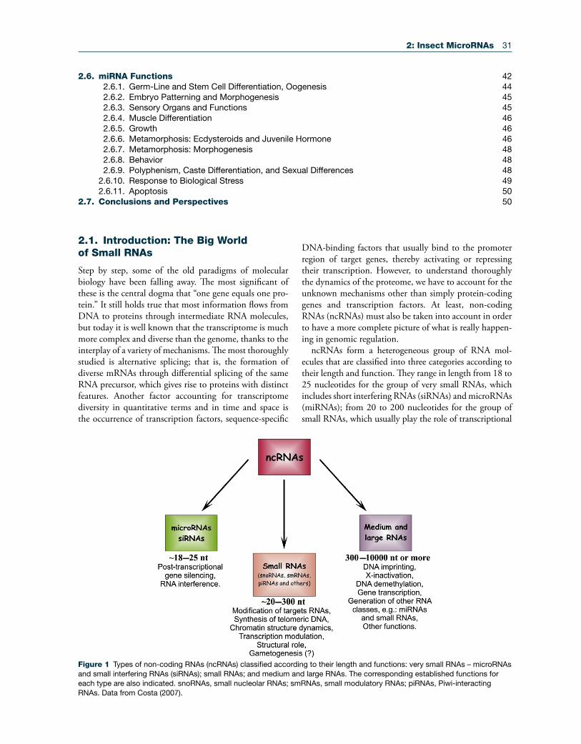
ncRNAs form a heterogeneous group of RNA mol-ecules that are classified into three categories according to their length and function. They range in length from 18 to 25 nucleotides for the group of very small RNAs, which includes short interfering RNAs (siRNAs) and microRNAs (miRNAs); from 20 to 200 nucleotides for the group of small RNAs, which usually play the role of transcriptional
Figure 1 Types of non-coding RNAs (ncRNAs) classified according to their length and functions: very small RNAs – microRNAs and small interfering RNAs (siRNAs); small RNAs; and medium and large RNAs. The corresponding established functions for each type are also indicated. snoRNAs, small nucleolar RNAs; smRNAs, small modulatory RNAs; piRNAs, Piwi-interacting RNAs. Data from Costa (2007).
2.6. miRNA Functions 422.6.1. Germ-Line and Stem Cell Differentiation, Oogenesis 442.6.2. Embryo Patterning and Morphogenesis 452.6.3. Sensory Organs and Functions 452.6.4. Muscle Differentiation 462.6.5. Growth 462.6.6. Metamorphosis: Ecdysteroids and Juvenile Hormone 462.6.7. Metamorphosis: Morphogenesis 482.6.8. Behavior 482.6.9. Polyphenism, Caste Differentiation, and Sexual Differences 48
2.6.10. Response to Biological Stress 49 2.6.11. Apoptosis 50
2.7. Conclusions and Perspectives 50
32 2: Insect MicroRNAs
and translational regulators; and, for the group of medium and large RNAs, up to (and even beyond) 10,000 nucleo-tides, which are involved in other processes, as detailed in Figure 1 (Costa, 2007). This chapter deals with the very small RNAs, and, more specifically, with miRNAs.
The history of siRNAs and miRNAs began in the late 1980s, when Jorgensen and colleagues were studying the role of chalcone synthase in the biosynthetic pathway of anthocianin in plants. Anthocianin gives a violet color to petunias, and Jorgensen’s team overexpressed chal-cone synthase in search of petunias with a deeper violet color. However, they unexpectedly obtained whitish flow-ers because the expression of chalcone synthase in these transgenic whitish petunias was some 50 times lower than in the wild type, thus suggesting that transgenic chalcone synthase had suppressed the endogenous gene (Jorgensen, 1990). Three years later, but in the field of developmen-tal biology and working on the nematode Caenorhabditis elegans, Lee and colleagues (1993) discovered two lin-4 transcripts, where the smaller, with ca. 21 nucleotides, was complementary to seven repeated sequences in the 3′ UTR of the mRNA of the heterochronic gene lin-14, which had been identified two years earlier.
These two disparate studies converged in 1998, when Fire and colleagues (1998), also working in C. elegans, dis-covered that the administration of a double-stranded RNA (dsRNA) with a strand complementary to a fragment of an endogenous mRNA can block this mRNA. This phe-nomenon is now known as RNA interference (RNAi), and its action is mediated by siRNAs of ca. 22 nucleo-tides that derive from dsRNA (Belles, 2010). A year later, while studying post-transcriptional gene silencing as a mechanism of antiviral defense, Hamilton and Baulcombe (1999) noticed the occurrence of antisense viral RNA of ca. 25 nucleotides in virus-infected plants. Hamilton and Baulcombe observed that these small RNAs were long enough to convey sequence specificity, and pointed out that they might be key determinants of the gene silenc-ing phenomenon. Further contributions showed that dsRNA-induced mRNA degradation was always mediated by RNAs of 21–23 nucleotides, thus leading researchers to investigate the endogenous source of these small RNAs. Finally, in 2001, three groups working independently (Lagos-Quintana et al., 2001; Lau et al., 2001; Lee and Ambros, 2001) described miRNAs as a novel family of small (ca. 22 nucleotides) endogenous RNAs that is diverse in sequence and temporal expression, evolutionarily wide-spread, and involved in regulating gene expression.
2.1.1. RNAi and siRNAs
The discovery of RNAi in C. elegans (Fire et al., 1998) was later extended to other animal groups, namely insects, and the basic mechanisms involved in their action on mRNAs were unveiled step by step in a few years. The biochemical
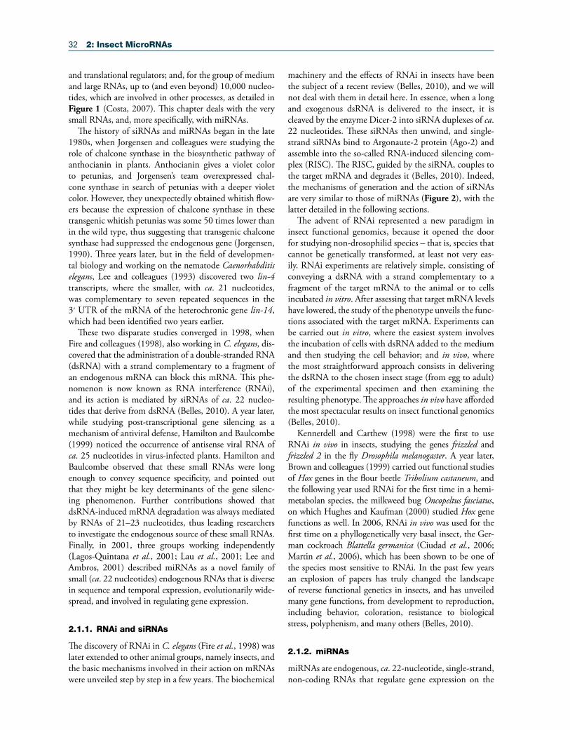
machinery and the effects of RNAi in insects have been the subject of a recent review (Belles, 2010), and we will not deal with them in detail here. In essence, when a long and exogenous dsRNA is delivered to the insect, it is cleaved by the enzyme Dicer-2 into siRNA duplexes of ca. 22 nucleotides. These siRNAs then unwind, and single-strand siRNAs bind to Argonaute-2 protein (Ago-2) and assemble into the so-called RNA-induced silencing com-plex (RISC). The RISC, guided by the siRNA, couples to the target mRNA and degrades it (Belles, 2010). Indeed, the mechanisms of generation and the action of siRNAs are very similar to those of miRNAs (Figure 2), with the latter detailed in the following sections.
The advent of RNAi represented a new paradigm in insect functional genomics, because it opened the door for studying non-drosophilid species – that is, species that cannot be genetically transformed, at least not very eas-ily. RNAi experiments are relatively simple, consisting of conveying a dsRNA with a strand complementary to a fragment of the target mRNA to the animal or to cells incubated in vitro. After assessing that target mRNA levels have lowered, the study of the phenotype unveils the func-tions associated with the target mRNA. Experiments can be carried out in vitro, where the easiest system involves the incubation of cells with dsRNA added to the medium and then studying the cell behavior; and in vivo, where the most straightforward approach consists in delivering the dsRNA to the chosen insect stage (from egg to adult) of the experimental specimen and then examining the resulting phenotype. The approaches in vivo have afforded the most spectacular results on insect functional genomics (Belles, 2010).
Kennerdell and Carthew (1998) were the first to use RNAi in vivo in insects, studying the genes frizzled and frizzled 2 in the fly Drosophila melanogaster. A year later, Brown and colleagues (1999) carried out functional studies of Hox genes in the flour beetle Tribolium castaneum, and the following year used RNAi for the first time in a hemi-metabolan species, the milkweed bug Oncopeltus fasciatus, on which Hughes and Kaufman (2000) studied Hox gene functions as well. In 2006, RNAi in vivo was used for the first time on a phyllogenetically very basal insect, the Ger-man cockroach Blattella germanica (Ciudad et al., 2006; Martin et al., 2006), which has been shown to be one of the species most sensitive to RNAi. In the past few years an explosion of papers has truly changed the landscape of reverse functional genetics in insects, and has unveiled many gene functions, from development to reproduction, including behavior, coloration, resistance to biological stress, polyphenism, and many others (Belles, 2010).
2.1.2. miRNAs
miRNAs are endogenous, ca. 22-nucleotide, single-strand, non-coding RNAs that regulate gene expression on the
2: Insect MicroRNAs 33
post-transcriptional level through base-pairing between the seed sequence of the miRNA and its complementary seed match sequence that is present in the 3′ untranslated region (UTR) of the target mRNA.
The first miRNA, lin-4, was discovered in a screen for genes required for post-embryonic development in the nematode C. elegans (Lee et al., 1993; Ambros and Horvitz, 1984). The identification of the lin-4 locus and its regulatory mechanism through the 3′ UTR of lin-14 mRNA was an interesting finding, although at that time it was almost considered to be a genetic oddity. How-ever, the discovery of another miRNA, let-7, initially in C. elegans (Reinhart et al., 2000) and later in various bila-terian species ( Pasquinelli et al., 2000), confirmed that, in the case of lin-4 and lin-14, it was not an oddity at all, but rather a new and fundamental layer of the mechanisms regulating gene expression (Lai et al., 2003; Neilson and Sharp, 2008).
The following sections will deal exclusively with miRNAs.
2.2. Biogenesis of miRNAs
miRNAs undergo molecular processing before becoming mature and ready to play their functional role. The path-way of miRNA biogenesis has many commonalities with that of siRNAs, but it is distinct in a number of ways (Fig-ure 2). miRNAs are first transcribed as part of a longer primary transcript (pri-miRNA), which folds, forming hairpin structures that correspond to miRNA precursors (pre-miRNAs). pri-miRNAs are then processed in the nucleus and transported to the cytoplasm, where they undergo final maturation (Figure 2).
2.2.1. miRNA Processing in the Nucleus
Most miRNA genes are transcribed by RNA polymerase II into pri-miRNA, although in some cases the tran-scription is mediated by RNA polymerase III (Lee et al., 2004a; Borchert et al., 2006). Usually, pri-miRNAs are several kilobases long, contain local stem-loop structures,
Figure 2 Biogenesis of miRNA and siRNA. miRNA gene is transcribed by RNA Pol II/III into a primary transcript (pri-miRNA) that is processed by Drosha/Pasha and exported to the cytoplasm by Exportin-5. In the cytoplasm, the precursor (pre-miRNA) undergoes the final step of maturation and is cleaved by Dicer-1/Loquacious into an miRNA duplex. After the miRNA duplex unwinds, the mature miRNA is maintained with Argonaute-1 protein (Ago-1) forming a RISC which will be coupled to the target mRNA and will degrade, destabilize, or translationally inhibit it, whereas the miRNA* is released and degraded. On the other hand, siRNA is formed when a long and exogenous double-strand RNA (dsRNA) is cleaved by Dicer-2/R2D2 into an siRNA duplex. Likewise with the miRNA pathway, the siRNA duplex is unwound and single-strand siRNAs are maintain with Argonaute-2 protein (Ago-2) forming a RISC which will recognize the target mRNA and degrade it.
34 2: Insect MicroRNAs
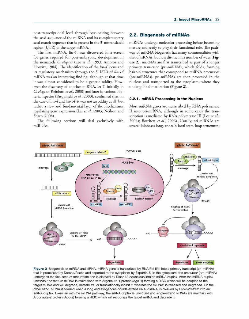
and are polyadenylated and capped, as in current mRNAs (Cai et al., 2004; Lee et al., 2004a), although the cap and the poly(A) tail are removed during miRNA processing. miRNA genes can form clusters in the genome (Behura, 2007), or can be found isolated within an intronic region of protein-coding genes, or in introns and exons of non-coding RNAs (Rodriguez et al., 2004). Moreover, pri-miRNAs can be polycistronic, thus carrying the infor-mation of more than one miRNA. In insects, the group of miR-100, let-7, and miR-125 constitutes the best studied example of polycistronic pri-miRNA. The organization of this pri-miRNA is well conserved in many species of insects, and even in vertebrates (Figure 3), although the spacer regions between miRNA precursor sequences can vary considerably in structure and length. For example, the distance between the precursor of miR-100 and that of let-7 varies from ca. 100 bp in T. castaneum to ca. 3.9 kb in Anopheles gambiae, whereas the distance between the precursor of let-7 and that of miR-125 varies within the range of 250–450 bp (Figure 3) (Behura, 2007).
pri-miRNA processing into ca. 70- to 80-nucleo-tide pre-miRNAs takes place exclusively in the nucleus by the action of the microprocessor, a protein complex of ca. 500 kDa, which in D. melanogaster is composed
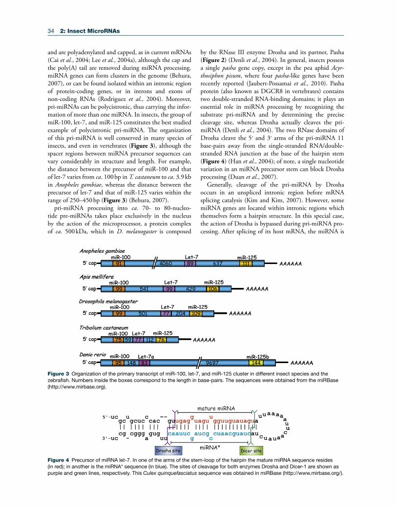
by the RNase III enzyme Drosha and its partner, Pasha (Figure 2) (Denli et al., 2004). In general, insects possess a single pasha gene copy, except in the pea aphid Acyr-thosiphon pisum, where four pasha-like genes have been recently reported (Jaubert-Possamai et al., 2010). Pasha protein (also known as DGCR8 in vertebrates) contains two double-stranded RNA-binding domains; it plays an essential role in miRNA processing by recognizing the substrate pri-miRNA and by determining the precise cleavage site, whereas Drosha actually cleaves the pri-miRNA (Denli et al., 2004). The two RNase domains of Drosha cleave the 5′ and 3′ arms of the pri-miRNA 11 base-pairs away from the single-stranded RNA/double-stranded RNA junction at the base of the hairpin stem (Figure 4) (Han et al., 2004); of note, a single nucleotide variation in an miRNA precursor stem can block Drosha processing (Duan et al., 2007).
Generally, cleavage of the pri-miRNA by Drosha occurs in an unspliced intronic region before mRNA splicing catalysis (Kim and Kim, 2007). However, some miRNA genes are located within intronic regions which themselves form a hairpin structure. In this special case, the action of Drosha is bypassed during pri-miRNA pro-cessing. After splicing of its host mRNA, the miRNA is
Figure 3 Organization of the primary transcript of miR-100, let-7, and miR-125 cluster in different insect species and the zebrafish. Numbers inside the boxes correspond to the length in base-pairs. The sequences were obtained from the miRBase (http://www.mirbase.org).
Figure 4 Precursor of miRNA let-7. In one of the arms of the stem-loop of the hairpin the mature miRNA sequence resides (in red); in another is the miRNA* sequence (in blue). The sites of cleavage for both enzymes Drosha and Dicer-1 are shown as purple and green lines, respectively. This Culex quinquefasciatus sequence was obtained in miRBase (http://www.mirbase.org/).

2: Insect MicroRNAs 35
released from the intron, then exported from the nucleus to the cytoplasm, and is finally cleaved by Dicer (see below). This class of miRNA, called mirtrons, has been described in flies, nematodes, and mammals (Berezikov et al., 2007; Okamura et al., 2007; Ruby et al., 2007a).
2.2.2. Pre-miRNA Transport from the Nucleus to the Cytoplasm
Once the pri-miRNA is processed in the nucleus, the resulting pre-miRNAs are exported to the cytoplasm by Exportin5 (EXP5) (Figure 2), which is a member of the nuclear transport receptor family, in complex with the cofactor Ran-GTP (Yi et al., 2003; Kim, 2004). EXP5 can recognize double-stranded RNA stems longer than 14 base-pairs along with a short 3′ overhang (1–8 nucleotides), which ensures the export of only those pre-miRNAs cor-rectly processed (Lund et al., 2004). Using RNAi, a num-ber of authors have demonstrated the role of EXP5 in the nucleocytoplasmic transport of pre-miRNA. Knockdown of EXP5 mRNA decreases the levels of mature miRNAs, but does not lead to an increase of pre-miRNA levels in the nucleus, which suggests that protecting pre-miRNA from digestion in the nucleus is another important role of EXP5 (Yi et al., 2003; Lund et al., 2004).
2.2.3. miRNA Maturation by Dicer
In the cytoplasm, pre-miRNAs are cleaved by Dicer near the terminal loop (Figure 4), thus resulting in the release of a ca. 22-nucleotide miRNA duplex with two nucleo-tides protruding as overhangs at each 3′-end. Dicer is an ATP-dependent multidomain enzyme of the RNase family involved in the cleavage of small double-stranded RNAs. It was identified for the first time in D. melano-gaster (Bernstein et al., 2001), and two Dicer homologs were later found in this fly, Dicer-1 and Dicer-2, which, in general, are involved in the miRNA and siRNA pathways, respectively (Lee et al., 2004b). RNAi of Dicer-1 in the last instar nymph of the cockroach B. germanica inhibits the formation of mature miRNAs and impairs the meta-morphic process (Gomez-Orte and Belles, 2009), which confirms not only the role of Dicer-1 in miRNA biogen-esis in a phyllogenetically basal insect, but also that of miRNAs in hemimetabolan metamorphosis (see below).
D. melanogaster Dicer-1 interacts with the protein Loquacious (also known as R3D1), which contains three double-stranded RNA-binding domains for pre-miRNA processing. Depletion of Loquacious results in pre-miRNA accumulation in Drosophila S2 cells, and immuno-affinity purification experiments revealed that Loquacious locates in a functional pre-miRNA processing complex along with Dicer-1, and stimulates the specific pre-miRNA process-ing activity (Saito et al., 2005). Of note, both arms of the pre-miRNA stem loop structures are imperfectly paired,
containing G : U wobble pairs and single nucleotide inser-tions (Figure 4). The imperfect base-pairing causes differ-ences in thermodynamic properties and makes one strand of the duplex less stably paired at its 5′ end. Generally, the strand with the lowest thermodynamic stability becomes the mature miRNA (guide strand), whereas the other strand (miRNA* or passenger strand) is degraded.
2.2.4. Regulation of miRNA Biogenesis and Stability
As a general principle, given that most of the miRNA genes are transcribed by RNA Polymerase II, the usual transcription factors associated with Pol II will influence their transcriptional control. A more specific modality of miRNA regulation is the process known as editing, which consists in a post-transcriptional change of RNA sequences caused by deamination of adenosine (A) to inosine (I), thus resulting in alterations in the base-pairing of the transcript. pri-miRNA transcripts modified by ADAR (adenosine deaminase acting on RNAs) have their biogenesis altered in downstream steps. These modifications of the pri-miRNA sequences may block the cleavages by Drosha and Dicer during miRNA maturation; moreover, edited mature miR-NAs can recognize other target mRNAs (Kawahara et al., 2007). Therefore, editing is a remarkable regulator of bio-genesis, and in addition increases miRNA structural diver-sity and further extends the diversity of miRNA targets.
Regulation of the miRNA biogenesis pathway also involves feedback mechanisms, like the interplay of Drosha and Pasha, which regulate each other in a circuit of negative feedback. Drosha acts by cleaving a hairpin located in the 5′ UTR of Pasha mRNA. Excess of Dro-sha decreases Pasha mRNA levels, whereas a reduction of Drosha elicits the reverse effect (Kadener et al., 2009b). Another example of feedback regulation involves human Dicer and the miRNA let-7, wherein Dicer is targeted by let-7 in sites within its coding region (Forman et al., 2008), or the reciprocal regulation showed by let-7 and the RNA-binding protein Lin-28, where let-7 suppresses Lin-28 protein synthesis whereas Lin-28 blocks let-7 maturation. Lin-28 is capable of blocking the cleavages mediated by both Drosha and Dicer; indeed, recombi-nant Lin-28 can block pri-miRNA processing, whereas knockdown of Lin-28 facilitates the expression of mature let-7 (Viswanathan et al., 2008). Lin-28 acts by induc-ing uridylation of the let-7 precursor at its 3′ end, which elicits the degradation of the uridylated pre-let-7 because Dicer fails to process hairpin RNA structures with long 3′ extensions (Heo et al., 2008).
Unlike 3′ uridylation, 3′ adenylation may have a stabiliz-ing effect on miRNAs, at least in mammals. For example, a variant of miR-122 possesses a 3′-terminal adenosine that is added by cytoplasmic poly(A) polymerase GLD-2 after unwinding of the miR-122/miR-122* duplex, and this 3′
36 2: Insect MicroRNAs
adenylation appears to prevent shortening, thus stabilizing the miRNA (Katoh et al., 2009). Apparently, adenylation and uridylation are two competing processes; it is interest-ing, in this sense, that addition of adenine residues in some small RNAs can prevent urydilation (Chen et al., 2000).
2.3. Mechanism of Action of miRNAs
The functional role of a miRNA is ultimately characterized by its effects on the expression of target genes. Currently, the regulatory mechanisms involving miRNAs are related to mRNA cleavage or translational repression by binding to complementary sites usually located on the 3′ UTR region of the mRNA (Carrington and Ambros, 2003; Lai, 2003; Ambros, 2004; Bartel, 2004). In contrast to the inhibi-tory effects, miRNAs can also stimulate the expression of target genes by upregulation of translation (Vasudevan et al., 2007; Orom et al., 2008). Moreover, miRNAs can also control cell fate by binding to heterogeneous ribonu-cleoproteins and lifting the translational repression of their target mRNAs; in this way, miRNAs act through a sort of decoy activity that interferes with the function of regulatory proteins (Beitzinger and Meister, 2010; Eiring et al., 2010). The present section, however, will emphasize the more widespread mechanisms, leading to mRNA translational repression, which start when the miRNA binds to Ago-1 protein and with the assembly of the RISC (Figure 2).
2.3.1. Argonaute Loading
The Argonaute (Ago) family can be divided into two subfamilies: the Piwi subfamily and the Ago subfamily. Piwi proteins are involved in transposon silencing, and are especially abundant in germ-line cells. Ago-subfamily proteins play key roles in post-transcriptional gene regula-tion by interacting with siRNAs (see above) and miRNAs, as detailed below.
After Dicer-1-mediated cleavage, the miRNA duplex binds to an Ago-1 protein in the RISC. To form an active RISC, the miRNA duplex has to unwind because only the mature miRNA binds to the Ago-1 protein, whereas the miRNA* is released. In human cells, miRNAs with a high degree of base-pairing in their pre-miRNA hairpin stem are initially processed by Ago-2, which cleaves the 3′ arm of the hairpin (that is, the miRNA* strand) in the middle, thus generating a nicked hairpin (Diederichs and Haber, 2007). In this case, Ago-2 acts before Dicer-1-mediated cleavage and facilitates miRNA duplex dissociation, the removal of nicked strand, and the activation of RISC. These findings elucidated the crucial role of Ago proteins not only during RISC formation, but also in relation to the mechanism that determines which of the two strands will become the survivor mature miRNA.
Identification of the target mRNA by the RISC is based on the complementarity between the mature miRNA and
the target mRNA site, and the degree of complementa-rity determines whether the target mRNA is degraded, destabilized, or translationally inhibited. Binding to Ago-1 greatly enhances miRNA stability, and although little is known about the half-life of individual miRNAs, it is clear that Ago-1 is a limiting factor for endogenous miRNA accumulation due to its protective function.
In D. melanogaster, miRNA* strands may accumulate bound to Ago-2, a protein initially thought to act exclusively in the siRNA pathway. Whether miRNA* binds to Ago-1 or to Ago-2 depends on the miRNA duplex structure, ther-modynamic stability, and the identity of first 5′-end nucleo-tide – i.e., miRNA sequences beginning with cystidine will bind to Ago-2, whereas those beginning with uridine will bind to Ago-1 (Ghildiyal et al., 2010). A number of observations indicate that some miRNA* plays a role in the regulation of gene expression. These observations include that: (1) miRNA* 5′ ends are more defined than their 3′ ends, thus suggesting that there is a seed region involved in regulatory functions (Ruby et al., 2007b; Okamura et al., 2008; Seitz et al., 2008); (2) many miRNA* sequences are evolutionarily conserved (Okamura et al., 2008); (3) in D. melanogaster (Ruby et al., 2007b) and in the basal insect B. germanica (Cristino et al., 2011), tissue concentration of some miRNA* is higher than that of the corresponding miRNA partner.
2.3.2. Repression of Protein Translation
The RISC is the key element that regulates gene expres-sion by repressing protein translation. The first step after RISC formation is the recognition of the target mRNA, mainly through the seed sequence. A number of stud-ies have demonstrated the importance not only of the seed, but also of the whole 5′ region of the miRNA dur-ing the interaction with the target mRNA. According to Brennecke and colleagues (2005), there are two categories of miRNA target sites in mRNAs. The first is called the “5′ dominant site,” and occurs when there is a near perfect base-pairing in the 5′ end of the miRNA; this category can be subdivided into “canonical” (when both 5′ and 3′ ends have strong base-pairing with the miRNA site) and “seed” (when only the 5′ region presents consistent base-pairing). The second category is called “3′ compensatory,” and occurs when base-pairing between the miRNA seed sequence and its corresponding sequence in the target mRNA is weak, and thus a stronger base-pairing in the 3′ region exerts a sort of “compensating” effect.
Initial experiments in C. elegans showed that the miRNAs lin-4 and let-7 repress their respective target mRNAs through interactions with miRNA sites in the 3′ UTR. Subsequently, many other cases of miRNA binding sites in the 3′ UTR of mRNAs were reported, leading to the presumption that this was a general rule. However, recent findings have revealed that miRNAs can
2: Insect MicroRNAs 37
repress mRNAs through sites located in the open reading frame (ORF) or in the 5′ UTR (Lee et al., 2009).
The action of RISC on target mRNAs may proceed through different mechanisms. One of them involves post-initiation repression, as shown by experiments carried out in C. elegans where lin-4 inhibits the translation of lin-14 mRNA without reducing the mRNA levels and without affecting the shifting of polysomes, thus suggesting that the inhibition of mRNA translation occurs at the elongation step (Wightman et al., 1993; Olsen and Ambros, 1999; Lee et al., 2003). Other details accounting for this mecha-nism of action have been reported, and a model has been proposed describing the inhibition of ribosome elongation, the induction of ribosome drop-off, and the facilitation of nascent polypeptides proteolysis (Fabian et al., 2009).
The second mechanism of RISC action is the accelera-tion of target mRNA destabilization, involving: (1) decap-ping of the m(7)G cap structure in the 5′ end; and/or (2) deadenylation of poly A tail during the initial step of translation (Humphreys et al., 2005). A number of reports using different experimental models have supported this second mechanism; for example, in zebrafish embryos and mammalian cells, miRNAs in the RISC accelerate mRNA deadenylation, which leads to fast mRNA decay (Figure 5) (Giraldez et al., 2006; Wu et al., 2006). In Drosophila cells both deadenylation and decapping require GW182
protein, CCR4 : NOT deadenylase, and the DCP1 : DCP2 decapping complexes. Depletion of GW182 in Drosophila cells leads to alteration of mRNA expression levels. How-ever, in Ago-1depleted cells, GW182 can still silence the expression of target mRNAs, thus indicating that GW182 acts downstream of Ago-1, and that it is a key component of the miRNA pathway (Behm-Ansmant et al., 2006a).
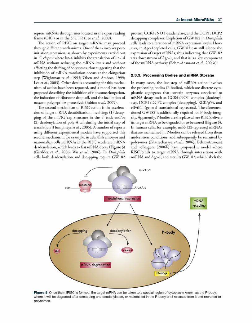
2.3.3. Processing Bodies and mRNA Storage
In many cases, the last step of miRNA action involves the processing bodies (P-bodies), which are discrete cyto-plasmic aggregates that contain enzymes associated to mRNA decay, such as CCR4 : NOT complex (deadenyl-ase), DCP1 : DCP2 complex (decapping), RCK/p54, and eIF4ET (general translational repressors). The aforemen-tioned GW182 is additionally required for P-body integ-rity. Apparently, P-bodies are the place where RISC delivers its target mRNA to be degraded or to be stored (Figure 5). In human cells, for example, miR-122-repressed mRNAs that are maintained in P-bodies can be released from them under stress conditions, and subsequently be recruited by polysomes (Bhattacharyya et al., 2006). Behm-Ansmant and colleagues (2006b) have proposed a model where RISC binds to target mRNA through interactions with miRNA and Ago-1, and recruits GW182, which labels the
Figure 5 Once the miRISC is formed, the target mRNA can be taken to a special region of cytoplasm known as the P-body, where it will be degraded after decapping and deadenylation, or maintained in the P-body until released from it and recruited to polysomes.
38 2: Insect MicroRNAs
transcript as a target for decay via deadenylation and decap-ping. Ago-1 and Ago-2 proteins have also been detected in P-bodies (Liu et al., 2005), thus suggesting that both siRNA and miRNA pathways may end in these structures. Nevertheless, this does not mean that P-bodies are crucial for the functioning of these pathways, given that disrup-tion of P-bodies after depletion of Lsm1, which is a key component of them, elicits a dispersion of Ago proteins into the cytoplasm, but does not affect siRNA and miRNA pathways (Chu and Rana, 2006).
2.4. Identification of miRNAs in Insects
Since the discovery of lin-4 and let-7 in the nematode C. elegans, a remarkable diversity of miRNAs has been reported in the genomes of various organisms, includ-ing insects, plants, viruses, and vertebrates (http:// www.mirbase.org). In insects, research on miRNAs was initially limited to D. melanogaster, but the availability of sequenced genomes from different species, as well as the development of new bioinformatic tools, has allowed the performance of systematic predictions of miRNAs in silico. Accordingly, computational methods based on the evolutionary conservation of genomic sequences and their ability to fold into stable hairpin structures have been applied to species with sequenced genomes, such as a number of nematodes, arthropods, and vertebrates (Table 1). Moreover, the development of novel techniques for directional cloning of small RNAs has led to the iden-tification of many other miRNAs (Lagos-Quintana et al., 2001; Lau et al., 2001; Lee and Ambros, 2001).
Nevertheless, the greatest progress came with the advent of high-throughput sequencing technologies and compu-tational methods. Those technologies confirmed most of the miRNA predicted in silico in species with the genome reported, made it possible to find new and unexpected
miRNAs, and contributed to the discovery de novo of miRNAs in species without the genome sequenced. Therefore, a consistent catalog of miRNAs is now avail-able not only in drosophilids, but also in a selection of species, such as the malaria mosquito (A. gambiae), the yellow fever mosquito (Aedes aegypti), the pea aphid (A. pisum), the vector of West Nile virus (Culex quin-quefasciatus), the jewel wasp (Nasonia vitripennis), the migratory locust (Locusta migratoria), the honey bee (Apis mellifera), the flour beetle (T. castaneum), the silkworm (Bombyx mori), and the German cockroach (B. germanica) (http://www.mirbase.org; http://www.ncbi.nlm.nih.gov/geo) (Griffiths-Jones, 2006). Both approaches, based on computational methods and high-throughput sequenc-ing, are discussed below.
2.4.1. Computational Methods
The most efficient computational methods for finding miRNA candidates were described in C. elegans (MiRscan) (Lim et al., 2003a) and D. melanogaster (miRseeker) (Lai et al., 2003). Both methods share conceptual similarities, such as structural and sequence similarity. MiRscan pro-duces an initial set of candidates by sliding a 110- nucleotide window across the C. elegans genome and folding those seg-ments that are filtered by the free energy and duplex length. Homologous hairpins are then identified by WU-BLAST in an additional genome which creates a reference set defining the standard features that will finally be used to score and rank all candidate hairpins. Nevertheless, MiRscan was not able to identify more than 50% of the previously known C. elegans miRNAs (Lim et al., 2003a). miRseeker was found to be more efficient at identifying genuine miRNAs in two fly species (D. melanogaster and Drosophila pseudoobscura) by taking into account the conservation across the hairpin (Lai et al., 2003). The method begins by identifying orthologous
Table 1 Algorithms Developed for miRNA Identification
Program Strategy Species group Authors/year
Grad et al. RB Nematodes Grad et al., 2003MiRScan RB Nematodes, vertebrates Lim et al., 2003a, 2003bmiRseeker RB Insects (flies) Lai et al., 2003Berezikov et al. RB Human Berezikov et al., 2005miPred RB Human Jiang et al., 2007miRAlign RB Metazoan Wang et al., 2005ProMIR HMM Human Nam et al., 2005BayesMiRNAFind NB Nematodes, mammals Yousef et al., 2006One-ClassMirnaFind SVM, NB Human, virus Yousef et al., 2008mirCoS-A SVM Mammals Sheng et al., 2007mir-abela SVM Mammals Sewer et al., 2005triplet-SVM SVM Human Xue et al., 2005RNAmicro SVM Metazoan Hertel and Stadler, 2006miPred SVM Human Ng and Mishra, 2007MiRFinder SVM Human, virus Huang et al., 2007
HMM, hidden Markov model; NB; Naive Bayes; RB, rule based; SVM, support vector machine.
2: Insect MicroRNAs 39
intergenic and intronic regions of those two fly genomes, and then folding those conserved sequences to identify and score the hairpin structures. The criteria for hairpin evalua-tion derive from a reference set of known miRNA genes of the two Drosophila species. The length of the hairpins and their minimum free energy were first evaluated, and then the distribution of divergent nucleotides was considered to score the candidates. The metrics consist in penalizing diver-gences depending on where they occur in the pre-miRNA hairpin, as the miRNA arm would tolerate less mutations than the miRNA* arm, which, by itself, would not tolerate more mutations than those observed in the loop region (Lai et al., 2003).
The establishment of guidelines for the experimental val-idation and annotation of novel miRNA candidates became obviously necessary with the increasing quantity of miRNA genes being identified in various species (Ambros et al., 2003). Thus, an initiative for organizing the information available on miRNA genes was then developed, leading to a database (miRBase, http://www.mirbase.org) where all data regarding miRNA sequences, targets, and gene nomencla-ture are deposited (Griffiths-Jones et al., 2008).
The large amount of miRNA data available in data-bases led to the development of a second generation of algorithms based on machine-learning methods. The approach consists in a learning process that identifies the most relevant characteristics and rules from a positive set of miRNA hairpins. Various machine-learning algorithms have been used for miRNA discovery (Table 1), the most common being Naïve Bayes (Yousef et al., 2006), sup-port vector machines (Yousef et al., 2008 and references therein), hidden Markov models (HMM) (Nam et al., 2005), genetic programming (Brameier and Wiuf, 2007), and random walks (Jiang et al., 2007).
All these methods contributed somehow to the identi-fication of new miRNAs, despite considerable differences in their trade-off between specificity and sensitivity. The criteria used in all of them were based on actual knowl-edge of the miRNA biogenesis, and features identified from known miRNAs conserved in at least two species. Indeed, there must be a great number of non-conserved miRNA genes still to be discovered, which may have char-acteristics and expression profiles substantially different from those of canonical miRNAs. However, the develop-ment of a new generation of sequencing technologies is changing the way of thinking about scientific approaches in all fields of biological sciences (Metzker, 2010), includ-ing the strategies to find new miRNAs in any species, even those whose genome is not sequenced yet.
2.4.2. High-Throughput Sequencing
Deep-sequencing technologies have created a new para-digm in detecting low-expression or tissue-specific miRNAs, as well as non-canonical and species-specific
ones. The most effective algorithms published so far are miRDeep (Yang et al., 2010), MIReNA (Mathelier and Carbone, 2010), and deepBase (Friedlander et al., 2008). Despite varying slightly in their workflow, their general strategy is similar, combining mapping and filtering sequences based on genome annotation, sequence and structure patterns, and properties of miRNA biogenesis.
The identification of miRNAs through deep- sequencing methods is rapidly increasing the catalogs of small RNA sequences for many species from a variety of tax onomic groups. Currently, all deep-sequencing datasets are deposited in the GEO (Gene Expression Omnibus) database at the NCBI (National Center for Biotechnology Information; http://www.ncbi.nlm.nih.gov/geo). At the date of writ-ing (January 2011), there are at least 193 studies of high- throughput sequencing of small RNAs from different eukaryotic species in the GEO database. Table 2 shows the 14 insect species in the GEO database, and the number of records for each.
Most of the 14 insect species included in Table 2 have the genome sequenced, or at least have a closely related species with an available genome (e.g., A. albopictus and C. quinquefasciatus). Two species, L. migratoria and B. germanica, have no genome sequence available, and iden-tification of miRNAs from deep-sequencing data becomes challenging because none of the methods mentioned above were designed to analyze deep-sequencing data without
Table 2 Insect Species and Number of Records Found in the GEO Database Related to Studies of miRNA Identification
Order SpeciesNumber of records
Diptera
Drosophila melanogaster 21Drosophila simulans 1Drosophila erecta 1Drosophila pseudoobscura 1Drosophila virilis 1Aedes albopictus 1Culex quinquefasciatus 1
Lepidoptera
Bombyx mori 2
Hymenoptera
Camponotus floridanus 1Harpegnathos saltator 1Apis mellifera 1
Hemiptera
Acyrthosiphon pisum 1
Orthoptera
Locusta migratoria 1
Dyctioptera
Blattella germanica 1
40 2: Insect MicroRNAs
using a genome sequence as a reference, and the diversity of small RNA types is remarkably high. However, strategies that can identify previously described miRNAs, as well as novel miRNAs on the basis of the number of reads and hairpin features, have recently been proposed (Wei et al., 2009). Genome-independent approaches for miRNA dis-covery show that we still have a poor understanding of the small RNA world and its regulatory mechanisms in the cell. For example, in the locust L. migratoria (Wei et al., 2009) and in the cockroach B. germanica (Cristino et al., 2011), sequence read numbers corresponding to miRNA*s were higher than those corresponding to the mature miRNA. Another original finding has been reported in Drosophila species (Berezikov et al., 2010), where some miRNA pre-cursors seem not to be processed by RNase III only, given that the usual one- to two-nucleotide 3′ overhang does not occur in some sequences represented by a high number of reads.
2.4.3. miRNA Classification
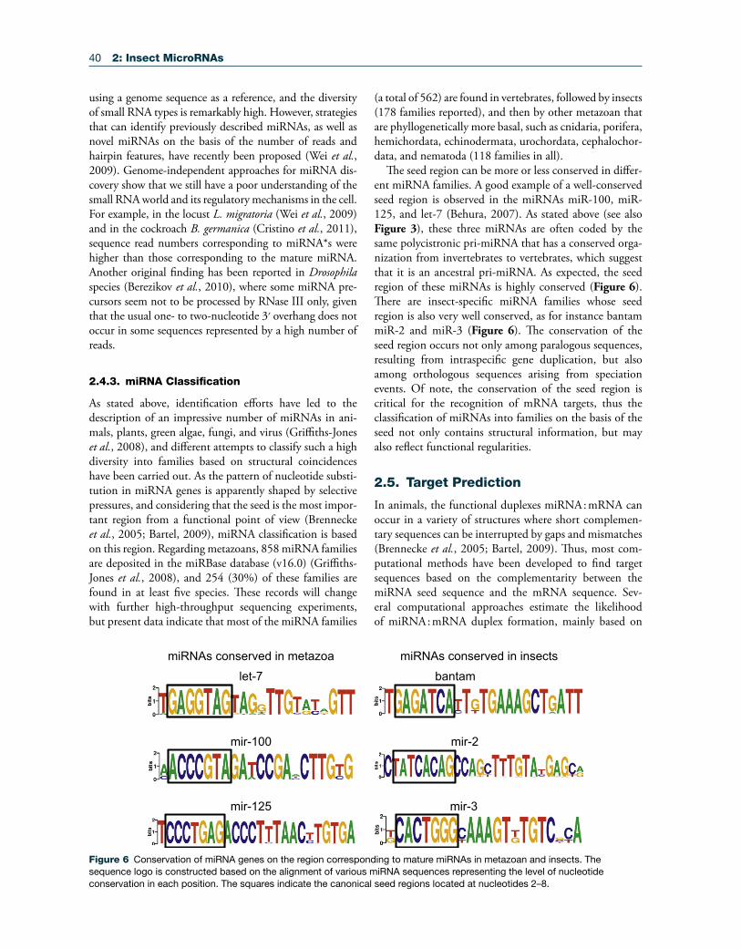
As stated above, identification efforts have led to the description of an impressive number of miRNAs in ani-mals, plants, green algae, fungi, and virus (Griffiths-Jones et al., 2008), and different attempts to classify such a high diversity into families based on structural coincidences have been carried out. As the pattern of nucleotide substi-tution in miRNA genes is apparently shaped by selective pressures, and considering that the seed is the most impor-tant region from a functional point of view (Brennecke et al., 2005; Bartel, 2009), miRNA classification is based on this region. Regarding metazoans, 858 miRNA families are deposited in the miRBase database (v16.0) (Griffiths-Jones et al., 2008), and 254 (30%) of these families are found in at least five species. These records will change with further high-throughput sequencing experiments, but present data indicate that most of the miRNA families
(a total of 562) are found in vertebrates, followed by insects (178 families reported), and then by other metazoan that are phyllogenetically more basal, such as cnidaria, porifera, hemichordata, echinodermata, urochordata, cephalochor-data, and nematoda (118 families in all).
The seed region can be more or less conserved in differ-ent miRNA families. A good example of a well-conserved seed region is observed in the miRNAs miR-100, miR-125, and let-7 (Behura, 2007). As stated above (see also Figure 3), these three miRNAs are often coded by the same polycistronic pri-miRNA that has a conserved orga-nization from invertebrates to vertebrates, which suggest that it is an ancestral pri-miRNA. As expected, the seed region of these miRNAs is highly conserved (Figure 6). There are insect-specific miRNA families whose seed region is also very well conserved, as for instance bantam miR-2 and miR-3 (Figure 6). The conservation of the seed region occurs not only among paralogous sequences, resulting from intraspecific gene duplication, but also among orthologous sequences arising from speciation events. Of note, the conservation of the seed region is critical for the recognition of mRNA targets, thus the classification of miRNAs into families on the basis of the seed not only contains structural information, but may also reflect functional regularities.
2.5. Target Prediction
In animals, the functional duplexes miRNA : mRNA can occur in a variety of structures where short complemen-tary sequences can be interrupted by gaps and mismatches (Brennecke et al., 2005; Bartel, 2009). Thus, most com-putational methods have been developed to find target sequences based on the complementarity between the miRNA seed sequence and the mRNA sequence. Sev-eral computational approaches estimate the likelihood of miRNA : mRNA duplex formation, mainly based on
mir-3
mir-2mir-100
let-7
mir-125
bantammiRNAs conserved in insectsmiRNAs conserved in metazoa
Figure 6 Conservation of miRNA genes on the region corresponding to mature miRNAs in metazoan and insects. The sequence logo is constructed based on the alignment of various miRNA sequences representing the level of nucleotide conservation in each position. The squares indicate the canonical seed regions located at nucleotides 2–8.
2: Insect MicroRNAs 41
sequence complementarity, thermodynamic stability, and evolutionary conservation of the sequence among species (Table 3). Machine learning approaches are also used for miRNA target identification. These methods usually com-bine one or more of the traditional procedures (seed com-plementarity, thermodynamic stability, and cross-species conservation) with more elaborated probabilistic models (Table 3). Also, a new generation of algorithms is inte-grating high-throughput expression data and computa-tional predictions (Huang et al., 2007; Hammell et al., 2008; van Dongen et al., 2008; Wang and El Naqa, 2008; Bandyopadhyay and Mitra, 2009; H. Liu et al., 2010; Sturm et al., 2010).
To date, miRNA target prediction has been mainly per-formed by computational approaches, and large numbers of targets have been predicted for most species with the genome sequenced (Bartel, 2009). As a general figure, pre-dictions have suggested that a single miRNA can target 200 mRNAs on average in vertebrates (Krek et al., 2005), whereas in D. melanogaster a single miRNA may regulate 54 genes on average (Grun et al., 2005).
2.5.1. microCosm, TargetScan, and PicTar
The miRBase database links miRNAs to targets using microCosm (http://www.ebi.ac.uk/enright-srv/micro cosm/), TargetScan (Lewis et al., 2005; Grimson et al., 2007; Friedman et al., 2009) and PicTar (Lewis et al., 2005;
Grimson et al., 2007; Friedman et al., 2009) prediction systems. These are therefore the most currently used, and are detailed below.
microCosm, formerly known as miRBase Targets, predicts miRNA targets in the UTR regions of animal genomes from Ensembl database (Hubbard et al., 2007; Flicek et al., 2008). It uses the miRanda algorithm to calculate a score across the miRNA vs UTR alignment (Enright et al., 2003; John et al., 2004; Betel et al., 2008); the energy for the thermodynamic stability of a miRNA : mRNA duplex is calculated by the Vienna RNA folding routines (http://www.tbi.univie.ac.at/RNA/), and the P-values are computed for all targets following the sta-tistical model implemented in RNAhybrid (Rehmsmeier et al., 2004). The Miranda algorithm (Enright et al., 2003; John et al., 2004) is basically divided into three steps. In the first step the miRNAs are aligned against the 3′ UTR sequences of the targets, allowing for G : U pairs and short indels. The method does not rely on seed matches, but increases the scaling score for complementarity at the 5′ end of the miRNA. The second step computes the ther-modynamic stability of the miRNA : mRNA duplex, and the final step reduces the false-positive rate by considering only targets with multiple sites.
TargetScan was the first algorithm that used the concept of seed matches in target prediction (Lewis et al., 2003, 2005). The method only uses miRNAs conserved across different species to scan corresponding 3′ UTR sequences.
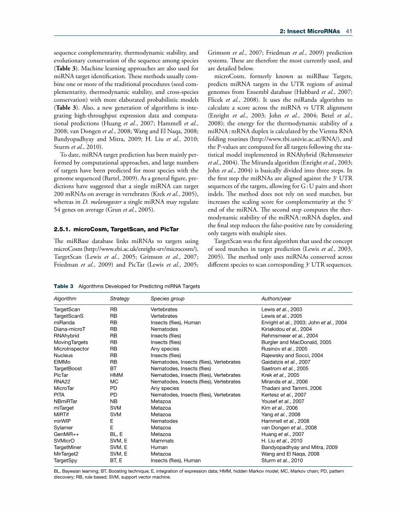
Table 3 Algorithms Developed for Predicting miRNA Targets
Algorithm Strategy Species group Authors/year
TargetScan RB Vertebrates Lewis et al., 2003TargetScanS RB Vertebrates Lewis et al., 2005miRanda RB Insects (flies), Human Enright et al., 2003; John et al., 2004Diana-microT RB Nematodes Kiriakidou et al., 2004RNAhybrid RB Insects (flies) Rehmsmeier et al., 2004MovingTargets RB Insects (flies) Burgler and MacDonald, 2005MicroInspector RB Any species Rusinov et al., 2005Nucleus RB Insects (flies) Rajewsky and Socci, 2004EIMMo RB Nematodes, Insects (flies), Vertebrates Gaidatzis et al., 2007TargetBoost BT Nematodes, Insects (flies) Saetrom et al., 2005PicTar HMM Nematodes, Insects (flies), Vertebrates Krek et al., 2005RNA22 MC Nematodes, Insects (flies), Vertebrates Miranda et al., 2006MicroTar PD Any species Thadani and Tammi, 2006PITA PD Nematodes, Insects (flies), Vertebrates Kertesz et al., 2007NBmiRTar NB Metazoa Yousef et al., 2007miTarget SVM Metazoa Kim et al., 2006MiRTif SVM Metazoa Yang et al., 2008mirWIP E Nematodes Hammell et al., 2008Sylamer E Metazoa van Dongen et al., 2008GenMiR++ BL, E Metazoa Huang et al., 2007SVMicrO SVM, E Mammals H. Liu et al., 2010TargetMiner SVM, E Human Bandyopadhyay and Mitra, 2009MirTarget2 SVM, E Metazoa Wang and El Naqa, 2008TargetSpy BT, E Insects (flies), Human Sturm et al., 2010
BL, Bayesian learning; BT, Boosting technique; E, integration of expression data; HMM, hidden Markov model; MC, Markov chain; PD, pattern discovery; RB, rule based; SVM, support vector machine.

42 2: Insect MicroRNAs
The algorithm defines the seed matches as short segments of seven nucleotides that must have a stringent comple-mentarity to the two to eight nucleotides of the mature miRNA. Then, the remaining miRNA sequence is aligned to the target site, allowing for G : U pairs; the free energy to form a secondary structure in the duplex is predicted by a folding algorithm. A Z-score is calculated on the basis of the number of matches predicted in the same target sequence and respective free energies. Finally, the Z-score is used to rank the candidate targets for each species, and each species is processed in the same way.
PicTar uses a machine learning algorithm to rank tar-get sequences using a HMM maximum likelihood score based on three main steps: (1) the seed matches must expand 7 nucleotides starting at position 1 or 2 in the 5′ end of the miRNA; (2) the minimum free energy of miRNA : mRNA duplexes is used to filter the target sites; and (3) the target sites must locate in overlapping posi-tions across the aligned corresponding 3′ UTR sequences. The target sites that pass the three-step filter are then ranked by the HMM model, which calculates the score considering all segmentations of the target sequence into target sites and background, thus allowing the algorithm to account for multiple binding sites for a single miRNA, as well as several miRNAs targeting the same mRNA.
The current target predictions available in the miRBase by microCosm, TargetScan, and PicTar have some degree of overlap and also of discrepancy that can be due to alignment artifacts, different mRNA UTR and miRNA
sequences, and intrinsic differences in the algorithms. In an attempt to provide more updated figures for the distribution of gene targets per miRNA and miRNA per gene target, we analyzed the data from target predictions available in the miRBase (Release 16; Sept 2010), com-paring D. melanogaster with Homo sapiens and C. elegans. Results show that the three methods give different aver-age numbers of miRNA-binding sites per mRNA target (19.6, 5.8, and 5.0 for MicroCosm, TargetScan, and Pic-Tar, respectively; Figure 7), as well as different numbers of mRNAs targeted by each miRNA (951, 395, and 426 for microCosm, TargetScan, and PicTar, respectively; Figure 8). The distribution of the number of miRNA-binding sites per mRNA target (Figure 7) is relatively similar among the three methods and the three species studied. Conversely, data on the number of mRNA targeted by an miRNA showed remarkable differences depending on the method, regarding not only the average values, but also and especially their pattern of distribution (Figure 8).
2.6. miRNA Functions
Insect model species can be studied through powerful genetic and genomic approaches, the paradigm being the fly D. melanogaster. Indeed, the first description of miRNA functions in insects was carried out in this species (Brennecke et al., 2003), by looking at gain-of-function mutants (Lai, 2002; Lai et al., 2005). miRNA functions
Number of miRNA binding sites predicted per target mRNA
15-2
030
-35
1-5
45-5
060
-65
75-8
090
-95 1 5 1510 20 1 5 1510 20
Figure 7 Frequency of the number of miRNA-binding sites in the 3′ UTR of target mRNAs in Homo sapiens, Drosophila melanogaster, and Caenorhabditis elegans, calculated with the three prediction methods available in miRBase: microCosm, TargetScan, and PicTar (Release 16; September 2010).

2: Insect MicroRNAs 43
are currently being demonstrated by mutating the genes coding for the miRNAs under study, overexpressing the miRNA of interest, or silencing it using specific anti-miRNAs, and then studying the resulting phenotype. Pre-dicted targets may also be validated by the above methods, including the quantification of the expression of the given target, as well as using in vitro systems with luciferase reporter target constructs, where binding of the miRNA to the target sequence is detected by luciferase activity and quantified with colorimetry.
In most cases, functions may be suggested by high-throughput sequencing comparisons in different devel-oping stages, in different organs of the same stage, or in different physiological situations. Studies of this type have been carried out in the silkworm B. mori (differences in tissue expression and in different developing stages) (Cao et al., 2008; S. Liu et al., 2010), the pea aphid A. pisum (differences in different morphs) (Legeai et al., 2010), the honey bee A. mellifera (differences between queens and workers) (Weaver et al., 2007), the migratory locust L. migratoria (differences between migratory and solitary phases) (Wei et al., 2009), and the German cockroach B. germanica (differences between metamorphic and non-metamorphic instars) (Cristino et al., 2011). Microarray analysis or detailed studies on the developmental expres-sion profiles of particular miRNAs can also suggest their
respective functions (Aravin and Tuschl, 2005; Weaver et al., 2007; He et al., 2008; Yu et al., 2008).
Silencing Dicer-1 expression by RNAi is also a use-ful approach to studying the influence of the whole set of miRNAs in a given process. This has been achieved in D. melanogaster, either in vivo, showing, for example, that Dicer-1 plays a general role in ovarian development (Jin and Xie, 2007), or in Drosophila cultured cells, where the deple-tion of Dicer-1 affected the development in both somatic and germ lineages (Lee et al., 2004b). More recently, Dicer-1 depletion by RNAi has been used in the German cockroach, B. germanica, to demonstrate the key role of miRNAs in hemimetabolan metamorphosis (see below).
Regarding the functions of particular miRNAs, the data available indicate that most of them appear to be involved in the fine-tuning of biological processes by modulating a precise dosage of regulatory proteins. Probably, they provide robustness to the whole program of gene expres-sion (Hornstein and Shomron, 2006) and resilience to environmental fluctuations, as in the case of miR-7 stud-ied by Li and colleagues (X. Li et al., 2009). However, as revealed by recent general reviews (Bushati and Cohen, 2007; Jaubert et al., 2007), information is still fragmen-tary, heavily concentrated in the D. melanogaster model, and focused on a few biological processes, as detailed in the text below and in Table 4, which summarizes cases
Number of mRNAs predicted to be targeted by a miRNA
1-50
200-
250
400-
450
600-
650
800-
850
1000
-105
012
00-1
250
1400
-145
01-
5020
0-25
040
0-45
060
0-65
080
0-85
010
00-1
050
1200
-125
014
00-1
450
1-50
200-
250
400-
450
600-
650
800-
850
1200
-125
014
00-1
450
1000
-105
0
Figure 8 Frequency of the number of mRNAs predicted to be targeted a miRNA in Homo sapiens, Drosophila melanogaster, and Caenorhabditis elegans, calculated with the three prediction methods available in miRBase: microCosm, TargetScan, and PicTar (Release 16; September 2010).
44 2: Insect MicroRNAs
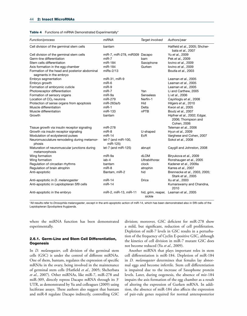
where the miRNA function has been demonstrated experimentally.
2.6.1. Germ-Line and Stem Cell Differentiation, Oogenesis
In D. melanogaster, cell division of the germinal stem cells (GSC) is under the control of different miRNAs. One of them, bantam, regulates the expression of specific mRNAs in the ovary, being involved in the maintenance of germinal stem cells (Hatfield et al., 2005; Shcherbata et al., 2007). Other miRNAs, like miR-7, miR-278 and miR-309, directly repress Dacapo mRNA through its 3′ UTR, as demonstrated by Yu and colleagues (2009) using luciferase assays. These authors also suggest that bantam and miR-8 regulate Dacapo indirectly, controlling GSC
division; moreover, GSC deficient for miR-278 show a mild, but significant, reduction of cell proliferation. Depletion of miR-7 levels in GSC results in a perturba-tion of the frequency of Cyclin E-positive GSC, although the kinetics of cell division in miR-7 mutant GSC does not become reduced (Yu et al., 2009).
Another miRNA that plays important roles in stem cell differentiation is miR-184. Depletion of miR-184 in D. melanogaster determines that females lay abnor-mal eggs and become infertile. Stem cell differentiation is impaired due to the increase of Saxophone protein levels. Later, during oogenesis, the absence of mir-184 impairs the axis formation of the egg chamber as a result of altering the expression of Gurken mRNA. In addi-tion, the absence of miR-184 also affects the expression of pair-rule genes required for normal anteroposterior
Table 4 Functions of miRNA Demonstrated Experimentally*
Function/process miRNA Target involved Authors/year
Cell division of the germinal stem cells bantam Hatfield et al., 2005; Shcher-bata et al., 2007
Cell division of the germinal stem cells miR-7, miR-278, miR309 Dacapo Yu et al., 2009Germ-line differentiation miR-7 bam Pek et al., 2009Stem cells differentiation miR-184 Saxophone Iovino et al., 2009Axis formation in the egg chamber miR-184 Gurken Iovino et al., 2009Formation of the head and posterior abdominal
segments in the embryomiRs-2/13 Boutla et al., 2003
Embryo segmentation miR-31, miR-9 Leaman et al., 2005Embryo growth miR-6 Leaman et al., 2005Formation of embryonic cuticle miR-9 Leaman et al., 2005Photoreceptor differentiation miR-7 Yan Li and Carthew, 2005Formation of sensory organs miR-9a Senseless Li et al., 2006Location of CO2 neurons miR-279 Nerfin-1 Cayirlioglu et al., 2008Protection of sense organs from apoptosis miR-263a/b Hid Hilgers et al., 2010Muscle differentiation miR-1 Delta Kwon et al., 2005Muscle differentiation miR-133 nPTB Boutz et al., 2007Growth bantam Hipfner et al., 2002; Edgar,
2006; Thompson and Cohen, 2006
Tissue growth via insulin receptor signaling miR-278 Teleman et al., 2006Growth via insulin receptor signaling miR-8 U-shaped Hyun et al., 2009Modulation of ecdysteroid pulses miR-14 EcR Varghese and Cohen, 2007Neuromusculature remodeling during metamor-
phosislet-7 (and miR-100,
miR-125)Sokol et al., 2008
Maturation of neuromuscular junctions during metamorphosis
let-7 (and miR-125) abrupt Caygill and Johnston, 2008
Wing formation miR-9a dLOM Biryukova et al., 2009Wing formation iab-4 Ultrabithorax Ronshaugen et al., 2005Regulation of circadian rhythms bantam clock Kadener et al., 2009aRegulation of brain atrophin miR-8 atrophin Karres et al., 2007Anti-apoptotic Bantam, miR-2 hid Brennecke et al., 2003, 2005;
Stark et al., 2005Anti-apoptotic in D. melanogaster miR-14 Drice Xu et al., 2003Anti-apoptotic in Lepidopteran Sf9 cells miR-14 Kumarswamy and Chandna,
2010Anti-apoptotic in the embryo miR-2, miR-13, miR-11 hid, grim, reaper,
sickleLeaman et al., 2005
*All results refer to Drosophila melanogaster, except in the anti-apoptotic action of miR-14, which has been demonstrated also in Sf9 cells of the Lepidopteran Spodoptera frugiperda.
2: Insect MicroRNAs 45
patterning and cellularization of the embryo (Iovino et al., 2009).
Finally, and also in D. melanogaster, miR-7 is involved in germ-line differentiation via maelstrom and Bag-of-marbles (Bam) gene products. Maelstrom regulates Bam via repres-sion of miR-7, by binding to the miR-7 promoter region (Pek et al., 2009); therefore, D. melanogaster mutants for maelstrom overexpress Bam, which leads to a deficient germ-line differentiation. As expected, a reduction in miR-7 expression rescues this phenotype (Pek et al., 2009)
2.6.2. Embryo Patterning and Morphogenesis
After injecting anti-miDNA-2a and anti-miDNA-13a, D. melanogaster embryos exhibited defects in the head and posterior abdominal segments, including cuticle holes and denticle belt malformations. In view of the similarity of the induced phenotypes, Boutla and colleagues (2003) concluded that these related miRNAs, miR-2a and miR-13a, act on the same target genes, together with the also related miR-2b and miR-13b, which form a functional subgroup called miRs-2/13.
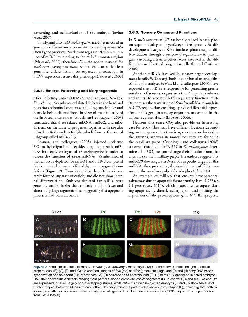
Leaman and colleagues (2005) injected antisense 2′O-methyl oligoribonucleotides targeting specific miR-NAs into early embryos of D. melanogaster in order to screen the function of these miRNAs. Results showed that embryos depleted for miR-31 and miR-9 completed development, but were affected by severe segmentation defects (Figure 9). Those injected with miR-9 antisense rarely formed any trace of cuticle, and did not show inter-nal differentiation. Embryos depleted for miR-6 were generally smaller in size than controls and had fewer and abnormally large segments, thus suggesting that apoptotic processes had been enhanced.
2.6.3. Sensory Organs and Functions
In D. melanogaster, miR-7 has been localized in early pho-toreceptors during embryonic eye development. At this developmental stage, miR-7 stimulates photoreceptor dif-ferentiation through a reciprocal regulation with yan, a gene encoding a transcription factor involved in the dif-ferentiation of retinal progenitor cells (Li and Carthew, 2005).
Another miRNA involved in sensory organ develop-ment is miR-9. Through both loss-of-function and gain-of-function analyses in vivo, Li and colleagues (2006) have reported that miR-9a is responsible for generating precise numbers of sensory organs in D. melanogaster embryos and adults. To accomplish this regulatory function, miR-9a represses the translation of Senseless mRNA through its 3′ UTR region, thus ensuring a precise differential expres-sion of this gene in sensory organ precursors and in the adjacent epithelial cells (Li et al., 2006).
Neurons that sense CO2 also provide an interesting case for study. They may have different locations depend-ing on the species. In D. melanogaster they are located in the antenna, whereas in mosquitoes they are found in the maxillary palps. Cayirlioglu and colleagues (2008) observed that loss of miR-279 in D. melanogaster deter-mines that CO2 neurons change their location from the antennae to the maxillary palps. The authors suggest that miR-279 downregulates Nerfin-1, a specific target for this miRNA, thus preventing the development of CO2 neu-rons in the maxillary palps (Cayirlioglu et al., 2008).
An example of miRNA that ensures developmental robustness during apoptotic tissue pruning is miR-263a/b (Hilgers et al., 2010), which protects sense organs dur-ing apoptosis by directly acting upon, and limiting the expression of, the pro-apoptotic gene hid. This property
Figure 9 Effects of depletion of miR-31 in Drosophila melanogaster embryos. (A) and (E) show Darkfield images of cuticle preparations; (B), (C), (F), and (G) are confocal images of Eve (red) and Ftz (green) stainings; and (D) and (H) hairy RNA in situ hybridization of blastoderm (2.5-h) embryos. (A)–(D) correspond to controls, and (E)–(H) to miR-31 antisense-injected embryos. The latter show cuticle defects ranging from partial fusion to complete loss of segments (E). In controls (B) and (C), Eve and Ftz are expressed in seven largely non-overlapping stripes, while miR-31 antisense-injected embryos (F) and (G) show fewer and weaker stripes that often bleed into each other. The hairy transcript pattern also shows fewer stripes (H), indicating that pattern formation is affected upstream of the primary pair rule genes. From Leaman and colleagues (2005), reprinted with permission from Cell (Elsevier).

46 2: Insect MicroRNAs
of some miRNAs to buffer fluctuating levels of gene activ-ity makes them well suited to serve a protective function during development (Hilgers et al., 2010).
2.6.4. Muscle Differentiation
In D. melanogaster, miR-1, which is one of the best con-served miRNAs in animals, is specifically expressed in the mesoderm during early embryogenesis, and in myo-genic precursors and muscle cells in late embryos (Sokol and Ambros, 2005). Depletion of miR-1 using genetic approaches, or by treatment with 2′O-methyl antisense oligonucleotides, resulted in lethality, which implies that miR-1 has essential functions in mesodermally derived tissues (Nguyen and Frasch, 2006).
By analyzing D. melanogaster mutants devoid of miR-1, Kwon et al. (2005) assessed the essential role of miR-1 for muscle differentiation. They showed that miR-1 reg-ulates the determination of specific cardiac and somatic muscle lineages from pluripotent progenitor cells in early embryogenesis. The Delta protein, a ligand for the Notch signaling pathway, was identified as an miR-1 tar-get in cardiac progenitor cells (Kwon et al., 2005).
Another well-conserved miRNA is miR-133, which is expressed in muscle cells together with miR-1. In D. mela-nogaster embryos, miR-133 plays a key role in controlling alternative splicing during muscle formation, and defin-ing the properties of differentiated muscle cells, through repressing the expression of the splicing factor nPTB dur-ing myoblast differentiation into myotubes (Boutz et al., 2007). The results of Boutz and colleagues not only indicate miR-133 directly downregulates a key factor during muscle development, but also establish a role for micro RNAs in the control of a developmentally dynamic splicing program.
2.6.5. Growth
In D. melanogaster, loss-of-function mutations of the bantam locus are lethal at the early pupal stage, whereas hypomorphic combinations of bantam mutant alleles give rise to adult flies that are smaller than controls (Figure 10) and that have deficiencies in fertility (Hipfner et al., 2002). Conversely, overexpression of bantam induces tis-sue overgrowth due to an increase in cell number. Bantam expression appears to be regulated by the gene Yorkie, thus controlling organ growth during development (Edgar, 2006; Thompson and Cohen, 2006).
Related to growth, and also in D. melanogaster, miR-278 has been implicated in insulin receptor (InR) signal-ing, thus contributing to regulation of the energy balance mainly by controling insulin responsiveness. Overex-pression of miR-278 promotes tissue growth in the eye and wing imaginal disks, whereas its deficiency leads to a reduction of fat body mass, which is reminiscent of the effect of impaired InR signaling in adipose tissue; the
action of miR-278 could be produced through the regu-lation of expanded gene transcripts (Teleman et al., 2006).
More recently, Hyun and colleagues (2009) have reported that miR-8 and its target, U-shaped (USH), regulate body size in D. melanogaster. miR-8 null flies are smaller in size and defective in insulin signaling in the fat body. USH inhibits PI3K activity, thus suppressing cell growth. Fat-body-specific expression and clonal analyses showed that miR-8 activates PI3K, thereby promoting fat-cell growth cell-autonomously, and enhancing organ-ismal growth non-cell-autonomously (Hyun et al., 2009).
2.6.6. Metamorphosis: Ecdysteroids and Juvenile Hormone
In insects, molting and metamorphosis are controlled by juvenile hormones and ecdysteroids, usually 20-hydroxy-ecdysone. Simultaneous expression of miR-125 and let-7 during D. melanogaster post-embryonic development is synchronized with the high titer of ecdysteroid pulses that initiate metamorphosis (Bashirullah et al., 2003; Sempere et al., 2003), which suggests that ecdysteroids might regu-late the expression of these two miRNAs.
Bashirullah and colleagues (2003), however, showed that miR-125 and let-7 expression is neither depen-dent on the Ecdysone receptor (EcR) nor inducible by 20-hydroxyecdysone in larval organs incubated in vitro. The same authors reported that the expression of both miRNAs can be induced by 20-hydroxyecdysone in Dro-sophila Kc cells, although the induction is considerably delayed with respect to what is observed in vivo (Bashirul-lah et al., 2003). The conclusion of these experiments is that the action of 20-hydroxyecdysone in Kc cells might be indirect, and that miR-125 and let-7 should be directly induced by an unknown temporal signal distinct from the well-known ecdysteroid-EcR cascade.
In a parallel paper, Sempere and colleagues (2003) followed a different approach to study the influence of ecdysteroids on the expression of miR-125, let-7, and miR-100, which are upregulated after the ecdysteroid pulse, as well as of miR-34, which is downregulated. They used the temperature-sensitive ecd1 strain that is impaired in ecdysteroid synthesis, and they showed that in ecd1 specimens blocked from pupariation by a trans-fer at 29°C, miR-125, let-7, and miR-100 were detected at much lower levels, whereas miR-34 was detected at much higher levels, compared with the wild type. Sem-pere and colleagues (2003) also studied the possible role of Broad complex, an early inducible gene in the ecdyster-oid cascade, using npr6 specimens, which lack all Broad complex factors. Results showed that miR-125, let-7, and miR-100 were detected at much lower levels (and miR-34 at much higher levels) in homozygous npr6 specimens than in npr6/+ or wild type specimens. With the same experimental approach, Sempere and colleagues (2003)
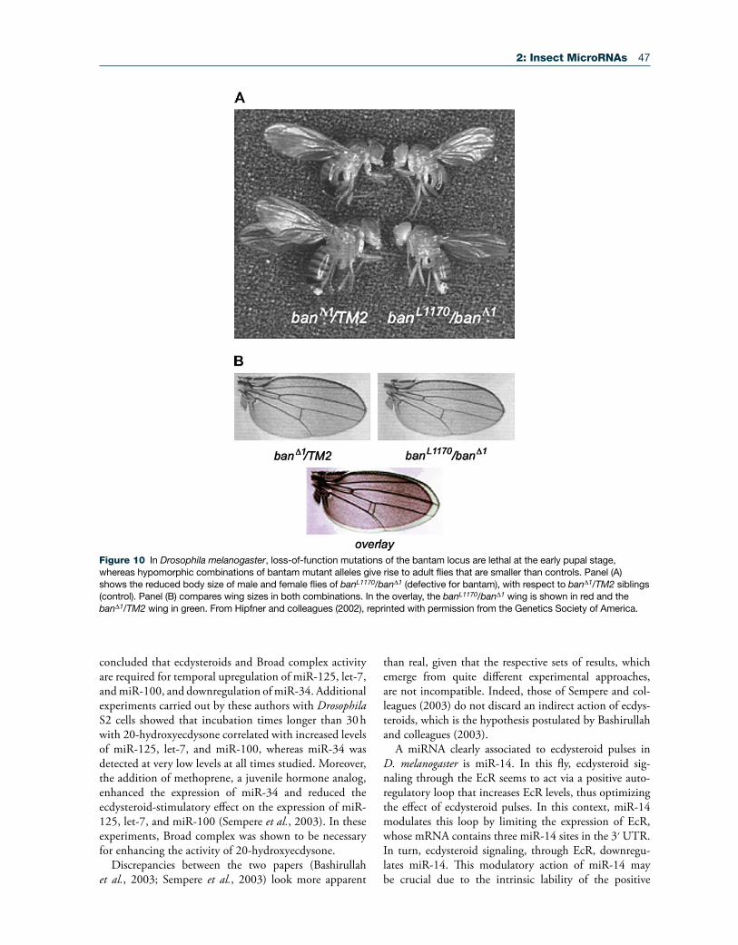
2: Insect MicroRNAs 47
concluded that ecdysteroids and Broad complex activity are required for temporal upregulation of miR-125, let-7, and miR-100, and downregulation of miR-34. Additional experiments carried out by these authors with Drosophila S2 cells showed that incubation times longer than 30 h with 20-hydroxyecdysone correlated with increased levels of miR-125, let-7, and miR-100, whereas miR-34 was detected at very low levels at all times studied. Moreover, the addition of methoprene, a juvenile hormone analog, enhanced the expression of miR-34 and reduced the ecdysteroid-stimulatory effect on the expression of miR-125, let-7, and miR-100 (Sempere et al., 2003). In these experiments, Broad complex was shown to be necessary for enhancing the activity of 20-hydroxyecdysone.
Discrepancies between the two papers (Bashirullah et al., 2003; Sempere et al., 2003) look more apparent
than real, given that the respective sets of results, which emerge from quite different experimental approaches, are not incompatible. Indeed, those of Sempere and col-leagues (2003) do not discard an indirect action of ecdys-teroids, which is the hypothesis postulated by Bashirullah and colleagues (2003).
A miRNA clearly associated to ecdysteroid pulses in D. melanogaster is miR-14. In this fly, ecdysteroid sig-naling through the EcR seems to act via a positive auto-regulatory loop that increases EcR levels, thus optimizing the effect of ecdysteroid pulses. In this context, miR-14 modulates this loop by limiting the expression of EcR, whose mRNA contains three miR-14 sites in the 3′ UTR. In turn, ecdysteroid signaling, through EcR, downregu-lates miR-14. This modulatory action of miR-14 may be crucial due to the intrinsic lability of the positive
Figure 10 In Drosophila melanogaster, loss-of-function mutations of the bantam locus are lethal at the early pupal stage, whereas hypomorphic combinations of bantam mutant alleles give rise to adult flies that are smaller than controls. Panel (A) shows the reduced body size of male and female flies of banL1170/banΔ1 (defective for bantam), with respect to banΔ1/TM2 siblings (control). Panel (B) compares wing sizes in both combinations. In the overlay, the banL1170/banΔ1 wing is shown in red and the banΔ1/TM2 wing in green. From Hipfner and colleagues (2002), reprinted with permission from the Genetics Society of America.
48 2: Insect MicroRNAs
autorregulatory loop that controls ecdysteroid signaling (Varghese and Cohen, 2007).
2.6.7. Metamorphosis: Morphogenesis
Work by Sokol and colleagues (2008) showed that the D. melanogaster let-7-Complex locus (let-7-C, compris-ing let-7, miR-100, and miR-125; see Figure 3) is mainly expressed in the pupal and adult neuromusculature. let-7-C knockout flies look morphologically normal, but dis-play defects in different adult behaviors (like flight and motility) and in fertility. Importantly, their neuromus-culature clearly shows juvenile features, which suggests that an important function of let-7-C is to ensure the appropriate remodeling of the abdominal neuromuscula-ture during the larval-to-adult transition. The study also showed that this function is carried out predominantly by let-7 alone (Sokol et al., 2008).
In a related work, Caygill and Johnston (2008) obtained a D. melanogaster mutant that lacks let-7 and miR-125 activities and shows a pleiotropic phenotype that arises during metamorphosis. These authors showed that the loss of let-7 and miR-125 results in temporal delays in the terminal cell-cycle exit in the wing, and in the maturation of neuromuscular junctions of imaginal abdominal mus-cles. The authors focused on the latter process by identify-ing the abrupt (ab) gene (which encodes a nuclear protein) as a let-7 target, and by providing evidence showing that let-7 regulates the maturation rate of abdominal neuro-muscular junctions during metamorphosis by regulating ab expression (Caygill and Johnston, 2008).
Wing morphogenesis has been studied by Biryukova and colleagues (2009), who described that miR-9a regu-lates D. melanogaster wing development through a func-tional target site in the 3′ UTR of the LIM only (dLOM) mRNA. dLMO is a transcription cofactor that directly inhibits the activity of Apterous, the factor required for the proper wing dorsal identity. Deletions of the 3′ UTR that remove the miR-9a site generate gain-of-function dLMO mutants associated with high levels of dLMO mRNA and protein. These mutants lack wing margins, a phenotype that is characteristic of null miR-9a mutants. Of note, miR-9a and dLMO are co-expressed in wing disks and interact genetically for controlling wing development; thus, the absence of miR-9a results in overexpression of dLMO, while gain-of-function miR-9a mutant sup-presses dLMO expression. The data suggest that miR-9a ensures a precise dosage of dLMO during D. melanogaster wing development (Biryukova et al., 2009).
Another miRNA involved in wing morphogenesis of D. melanogaster is iab-4. Sequence analysis suggested that iab-4 could regulate Ultrabithorax (Ubx), and expres-sion pattern studies of iab-4 and Ubx showed that they are complementary in critical developmental moments. Direct evidence for an interaction between iab-4 and Ubx
was obtained with luciferase assays. Finally, ectopic expres-sion of iab-4 miRNA in haltere disks caused a homeotic transformation of halteres to wings, which occurs when Ubx expression is reduced (Ronshaugen et al., 2005).
As stated above, RNAi experiments that reduced Dicer-1 expression in the last instar nymph of B. germanica depleted miRNA levels, and the next molt, instead of giving the adult stage, gave supernumerary nymphs. These were morpho-logically similar to the supernumerary nymphs obtained after treating the last instar nymph with juvenile hormone (Figure 11). The RNAi experiments with Dicer-1 indicate that miRNAs are crucial for hemimetabolan metamorpho-sis (Gomez-Orte and Belles, 2009).
2.6.8. Behavior
A recent paper by Kadener and colleagues (2009a) addresses the contribution of miRNAs to the regulation of circadian rhythms. The authors first knocked down the miRNA biogenesis pathway in D. melanogaster circadian tissues, which severely affected behavioral rhythms, thus indicating that miRNAs function in circadian timekeep-ing. To identify miRNA–mRNA pairs that might be important for this regulation, immunoprecipitation of Ago-1, followed by microarray analysis, led to identifica-tion of a number of mRNAs presumably under miRNA control. These included three core clock mRNAs: clock; vrille; and clockworkorange. To identify miRNAs involved in circadian timekeeping, the authors inhibited miRNA biogenesis in circadian tissues and then carried out a til-ing array analysis. Behavioral and molecular experiments showed that bantam has a role in the core circadian pace-maker, and S2 cell biochemical assays indicated that ban-tam regulates the translation of clock by targeting three sites in the clock 3′ UTR (Kadener et al., 2009a).
In a work addressed to study of the role of miR-8 in D. melanogaster, Karres and colleagues (2007) identified atrophin (also known as grunge) as a direct target of miR-8. miR-8 mutant phenotypes show high levels of apoptosis in the brain, and behavioral defects, like impaired capability for climbing, which are attributable to elevated atrophin activity. Decrease of atrophin levels in miR-8-expressing cells to below the level generated by miR-8 regulation is detrimental, which points to a sort of “tuning target” rela-tionship between them (Karres et al., 2007).
2.6.9. Polyphenism, Caste Differentiation, and Sexual Differences
Legeai and colleagues (2010) suggested that miRNA might participate in the regulation of aphid polyphenism, and studied the expression of miRNA in different female morphs of A. pisum using microarray approaches. Most (95%) of the miRNA tested (n = 149) had similar expres-sion in different morphs, but some of them, including

2: Insect MicroRNAs 49
miR*s, were differentially expressed – like let-7 and miR-100, which were upregulated in oviparae specimens, and miR2a-1, which was downregulated. The comparison between two parthenogenetic morphs gave three miR-NAs (miR-34, miR-X47, and miR-X103) and two miR*s (miR307* and miRX52*) that showed differential expres-sion. While miR307* was upregulated in virginoparae, the others were downregulated with respect to the sexu-parae morph.
Using total reads from Solexa deep sequencing, Wei and colleagues (2009) compared miRNA expression in the gregarious and solitary phases of the migratory locust, L. migratoria. In the gregarious phase, canonical miRNAs were expressed at levels between 1.5- and 2-fold higher than in the solitary phase; the most prominent differ-ences were found in miR-276, miR-125, miR-1, let-7, and miR-315. Interestingly, miR-1 is a muscle-specific miRNA and miR-315 is a potent Wingless signaling activator, at least in D. melanogaster; therefore, the dif-ferences in flying power between gregarious and solitary locusts may be related by the action of these two miRNAs. However, most of the differences concern new unanno-tated small RNAs, which are much more abundant in the solitary phase, although the functions of these miRNA candidates remain unknown.
In the honey bee A. mellifera, expression profiles of mi RNAs in workers and queens have been compared using quantitative RT-PCR, in adult body parts (head, abdo-men, thorax) as well as in the whole body of the pupal stage (Weaver et al., 2007). Results highlighted the differential expression, between queens and workers, in the abdomen,
which is probably related to the location of the ovaries and the differential fecundity of the two castes. Regarding particular miRNAs, miR-71 shows strong expression in worker pupae; comparing adult body parts, miR-71 has a higher expression in the head and thorax of adult workers, whereas expression in the abdomen is higher in the queen caste. Conversely, miR-9a is highly expressed in the thorax and abdomen of workers, while their expression levels are the highest in the thorax of workers.
In the silkworm B. mori, Liu and colleagues studied the expression of miRNA adult males and females using microarray approaches, and found that the expression of some 20 miRNAs was significantly higher in the body wall of males (S. Liu et al., 2010). This differential expres-sion was assessed for a selection of 10 miRNAs (including bantam, miR-1, miR-13a, and miR-2a) using Northern blot. Microarray analysis also revealed that the expression of 13 miRNAs was significantly higher in ovaries, whereas only 4 were differentially expressed in testes. Differences between sexes were also found in other tissues, including Malpighian tubules, head, midgut, fat body, or silk gland. However, the authors pointed out that differences in miRNA expression might be due to individual differences in the metabolic state, because the expression of some of these miRNAs is influenced by nutritional status (Cheung et al., 2009).
2.6.10. Response to Biological Stress
Larvae of the moth Lymantria dispar show differen-tiated miRNA expression after wasp parasitization
Figure 11 Inhibition of Blattella germanica metamorphosis after impairing miRNA maturation by depleting Dicer-1 expression with RNAi approaches in sixth (last) instar nymph. Dorsal and ventral view of normal sixth instar nymph (A, B), normal adult (C, D) and seventh instar supernumerary nymphoid (E, F) resulting from metamorphosis inhibition. The nymphoids resemble those obtained after treating the last instar nymph with juvenile hormone (G). Photos from Albert Masó; data from Gomez-Orte and Belles (2009).
50 2: Insect MicroRNAs
(Gundersen-Rindal and Pedroni, 2010). Microarray studies revealed that miR-1, miR-184, and miR-277 are highly upregulated in larval hemocytes, whereas miR-279 and let-7 are highly downregulated. Expression changes were assessed in hemolymph, fat body, brain, and mid-gut from infected larva, with respect to controls, using qRT-PCR. Of all the tissues analyzed from parasitized specimens, the midgut was the one that showed least miRNA activity. miR-1 was upregulated in all tissues from parasitized specimens, whereas miR-277 was the most strongly upregulated in the fat body. Expression of miR-279 was variable in the different tissues; it was remarkably upregulated in fat body but clearly downregu-lated in hemolymph, and it had a negligible expression in brain and midgut. Two human herpes virus-associated miRNAs (hcmv-miR-UL70 3p and kshv-miR-K12-3) were upregulated in hemocytes of parasitized L. dispar (Gundersen-Rindal and Pedroni, 2010). These are among many miRNAs that have been hypothesized to act as sup-pressors of the immediate and early genes that respond to a viral infection (Murphy et al., 2008).
The response of miRNAs to an infection has also been studied in mosquitoes. In A. gambiae, expression of miR-34, miR-1174, and miR-1175 decreases after Plasmodium infection, while that of miR-989 increases (Winter et al., 2007). Minor changes in miRNA expression have been observed in C. quinquefasciatus after West Nile virus infection (Skalsky et al., 2010), although miR-989 showed a 2.8-fold downregulation and miR-92 appeared somewhat upregulated. The expression of these two miR-NAs has been studied in different mosquito species, and results have shown that miR-989 expression is restricted to females, and predominantly to the ovary (Winter et al., 2007; Mead and Tu, 2008), although it was later detected in the midgut of Ae. aegypti (S. Li et al., 2009). miR-92 has been related to embryonic development in Ae. aegypti (S. Li et al., 2009) and B. mori (Liu et al., 2009). Results of deregulation of miR-989 and miR-92 suggested to Skalsky and colleagues (2010) that their targets participate in mediating flavivirus infection of the mosquito host.
2.6.11. Apoptosis
A group of D. melanogaster miRNAs including, miR-278, miR-14, bantam, and miR-2, regulate cell proliferation and apoptosis, targeting a number of pro-apoptotic genes, like hid, which is repressed by bantam and miR-2 miR-NAs (Brennecke et al., 2003, 2005; Stark et al., 2005).
One of the functions of mir-14 in D. melanogaster is suppressing cell death; therefore, loss of miR-14 is asso-ciated with a reduced lifespan, stress sensitivity, and increased levels of the apoptotic effector caspase Drice (Xu et al., 2003). The same anti-apoptotic function has been found in Lepidopteran (Spodoptera frugiperda) Sf9 cells, where miR-14 is required for constitutive cell survival
(Kumarswamy and Chandna, 2010). However, the results do not exclude that additional miRNAs might also con-tribute to regulating Lepidopteran cell survival and death.
Finally, experiments depleting miRNA functions by injection of miRNA antisense nucleic acids in early embryos, which permits systematic loss-of-function analysis in vivo, have identified the miR-2/13 family and miR-6 as controlling apoptosis during D. melanogaster embryonic development through post-transcriptional repression of the proapoptotic proteins hid, grim, reaper, and sickle (Leaman et al., 2005).
2.7. Conclusions and Perspectives
There are reasons to believe that the still-scarce data avail-able on miRNAs are just the tip of an iceberg. Nevertheless, rapidly expanding information is making it increasingly obvious that miRNAs’ contribution to the genomic out-put is not a sort of genetic oddity or “transcriptional background noise,” but a class of key post-transcriptional regulators of gene expression. Indeed, genomic regulation cannot be completely understood without incorporating the role of miRNAs, which constitute a regulatory layer that works in concert with the mRNA and protein net-work. However, the field of miRNA study is still in the development phase, and there are many aspects – the bulk of the iceberg – that require further research.
The miRNA machinery appears to be more complex than previously thought, and rapid progress is unveiling many unexpected details. An example concerning the mechanisms responsible for stabilized or reduced miRNA expression is the discovery of specific cis-acting modifica-tions and trans-acting proteins that affect miRNA half-life, which are revealing new elements that contribute to their homeostasis (Kai and Pasquinelli, 2010), and the identification of Dicer-independent miRNA biogen-esis pathways, such as those using the catalytic activity of Ago-2 (Cheloufi et al., 2010; Cifuentes et al., 2010). Therefore, recent data suggest that such mechanistic aspects will have more surprises revealed as we continue to expand our understanding of them.
Moreover, further studies are required to elucidate how miRNA genes are regulated. There are contribu-tions studying the influence of transcription factors act-ing on the promoter region of miRNA genes, like that of Pek and colleagues (2009) on the aforementioned repres-sor action of Maelstrom on the miR-7 promoter region. However, more studies in this line are needed if we wish to better understand the regulatory mechanisms medi-ated by miRNAs. Faster progress seems predictable in the field of cataloging miRNAs. The challenge is to find the unexpected, non-conserved miRNAs, and the new generation of algorithms will have to combine not only high-throughput approaches and powerful computational methods, but also expression data, genomic location, and

2: Insect MicroRNAs 51
structural and sequence features, as approached in the studies of Brennecke and Cohen (2003), Friedlander and colleagues (2008), and Mathelier and Carbone (2010).
miRNA target prediction started more than two decades ago with the serendipitous findings that emerged from miRNA target recognition (Wightman et al., 1991, 1993; Lee et al., 1993). The key principles were then applied to computational methods for miRNA target prediction (Bartel, 2009), and these methods soon allowed the pre-diction of hundreds of miRNA targets. However, compu-tational prediction of miRNA targets still relies on the few principles defined more than 20 years ago, and, arguably, this will not help to unveil novel aspects of miRNA target mechanisms. Thus, unbiased approaches to studying the interaction of miRNA and target would be valuable in order to identify new principles of miRNA-target recogni-tion, and to improve the systems for target prediction, as in the creative approach of Orom and Lund (2007).
Finally, the phase of predicting putative targets in silico following computational methods must be followed by experimental work to validate the predictions and to iden-tify targets in vivo. In this line, specific miRNA silencing will be one of the most useful approaches, and entomolo-gists and other non-biomedical researchers will benefit from the miRNA antagonists that are being designed in the context of biomedical studies in search of thera-peutic agents against human diseases (Gao and Huang, 2009). In this sense, D. melanogaster and other insect species will continue to be the favorite models, given the advantages they offer, especially straightforward manipu-lation. Validation of targets will contribute to elucidating the place and role of miRNAs in the molecular network that regulates the development and homeostasis of bio-logical processes. The networks describing the interaction of miRNAs, mRNAs, and proteins are presumably highly organized and complex, and their their study therefore represents a formidable challenge. However, it will be a worthwhile effort, because these networks are possibly the best approximation to the living world that is available with present means.
There is a fairly widespread opinion that proteins are what really matter in the functional landscape that shapes fitness, so transcript abundance is only useful as a mere proxy for the activity of the corresponding proteins (Feder and Walser, 2005). However, the expanding universe of small silencing RNAs (Ghildiyal and Zamore, 2009) and their widespread functional roles in genomic regulation show this to be yet one more old paradigm that is about to fall.
Acknowledgments
Financial support from the Ministry of Science and Inno-vation, Spain (Projects BFU2008-00484 to MDP and CGL2008-03517/BOS to XB) is gratefully acknowledged.
EDT has a post-doctoral contract from the CSIC (JAE-doc program) and MR a pre-doctoral research grant from the CSIC (JAE-pre program).Thanks are due to Stephen Cohen for providing Figure 10, and to Albert Masó for the photographs in Figure 11.
References
Ambros, V. (2004). The functions of animal microRNAs. Nature, 431, 350–355.
Ambros, V., & Horvitz, H. R. (1984). Heterochronic mutants of the nematode Caenorhabditis elegans. Science, 226, 409–416.
Ambros, V., Bartel, B., Bartel, D. P., Burge, C. B., Carrington, J. C., et al. (2003). A uniform system for microRNA annota-tion. RNA, 9, 277–279.
Aravin, A., & Tuschl, T. (2005). Identification and character-ization of small RNAs involved in RNA silencing. FEBS Lett., 579, 5830–5840.
Bandyopadhyay, S., & Mitra, R. (2009). TargetMiner: microRNA target prediction with systematic identifica-tion of tissue-specific negative examples. Bioinformatics, 25, 2625–2631.
Bartel, D. P. (2004). MicroRNAs: Genomics, biogenesis, mechanism, and function. Cell, 116, 281–297.
Bartel, D. P. (2009). MicroRNAs: Target recognition and reg-ulatory functions. Cell, 136, 215–233.
Bashirullah, A., Pasquinelli, A. E., Kiger, A. A., Perrimon, N., Ruvkun, G., et al. (2003). Coordinate regulation of small temporal RNAs at the onset of Drosophila metamorphosis. Dev. Biol., 259, 1–8.
Behm-Ansmant, I., Rehwinkel, J., Doerks, T., Stark, A., Bork, P., et al. (2006a). mRNA degradation by miRNAs and GW182 requires both CCR4:NOT deadenylase and DCP1:DCP2 decapping complexes. Genes Dev., 20, 1885–1898.
Behm-Ansmant, I., Rehwinkel, J., & Izaurralde, E. (2006b). MicroRNAs silence gene expression by repressing protein expression and/or by promoting mRNA decay. Cold Spring Harb. Symp. Quant Biol., 71, 523–530.
Behura, S. K. (2007). Insect microRNAs: Structure, function and evolution. Insect Biochem. Mol. Biol., 37, 3–9.
Beitzinger, M., & Meister, G. (2010). Preview. MicroRNAs: From decay to decoy. Cell, 140, 612–614.
Belles, X. (2010). Beyond Drosophila: RNAi in vivo and func-tional genomics in insects. Annu. Rev. Entomol., 55, 111–128.
Berezikov, E., Guryev, V., van de Belt, J., Wienholds, E., Plasterk, R. H., & Cuppen, E. (2005). Phylogenetic shadow-ing and computational identification of human microRNA genes. Cell, 120, 21–24.
Berezikov, E., Chung, W. J., Willis, J., Cuppen, E., & Lai, E. C. (2007). Mammalian mirtron genes. Mol Cell, 28, 328–336.
Berezikov, E., Liu, N., Flynt, A. S., Hodges, E., Rooks, M., et al. (2010). Evolutionary flux of canonical microRNAs and mirtrons in Drosophila. Nat. Genet., 42, 6–9, author reply 9–10.
Bernstein, E., Caudy, A. A., Hammond, S. M., & Hannon, G. J. (2001). Role for a bidentate ribonuclease in the initiation step of RNA interference. Nature, 409, 363–366.
52 2: Insect MicroRNAs
Betel, D., Wilson, M., Gabow, A., Marks, D. S., & Sander, C. (2008). The microRNA.org resource: Targets and expres-sion. Nucleic Acids Res., 36, D149–153.
Bhattacharyya, S. N., Habermacher, R., Martine, U., Closs, E. I., & Filipowicz, W. (2006). Relief of microRNA-mediated translational repression in human cells subjected to stress. Cell, 125, 1111–1124.
Biryukova, I., Asmar, J., Abdesselem, H., & Heitzler, P. (2009). Drosophila mir-9a regulates wing development via fine-tun-ing expression of the LIM only factor, dLMO. Dev. Biol., 327, 487–496.
Borchert, G. M., Lanier, W., & Davidson, B. L. (2006). RNA polymerase III transcribes human microRNAs. Nat. Struct. Mol. Biol., 13, 1097–1101.
Boutla, A., Delidakis, C., & Tabler, M. (2003). Developmental defects by antisense-mediated inactivation of micro-RNAs 2 and 13 in Drosophila and the identification of putative target genes. Nucleic Acids Res., 31, 4973–4980.
Boutz, P. L., Chawla, G., Stoilov, P., & Black, D. L. (2007). MicroRNAs regulate the expression of the alternative splic-ing factor nPTB during muscle development. Genes Dev., 21, 71–84.
Brameier, M., & Wiuf, C. (2007). Ab initio identification of human microRNAs based on structure motifs. BMC Bioin-formatics, 8, 478.
Brennecke, J., & Cohen, S. M. (2003). Towards a complete description of the microRNA complement of animal genomes. Genome Biol., 4, 228.
Brennecke, J., Hipfner, D. R., Stark, A., Russell, R. B., & Cohen, S. M. (2003). bantam encodes a developmentally regulated microRNA that controls cell proliferation and regulates the proapoptotic gene hid in Drosophila. Cell, 113, 25–36.
Brennecke, J., Stark, A., Russell, R. B., & Cohen, S. M. (2005). Principles of microRNA-target recognition. PLoS Biol., 3, e85.
Brown, S., Holtzman, S., Kaufman, T., & Denell, R. (1999). Characterization of the Tribolium Deformed ortholog and its ability to directly regulate Deformed target genes in the res-cue of a Drosophila Deformed null mutant. Dev. Genes Evol., 209, 389–398.
Burgler, C., & Macdonald, P. M. (2005). Prediction and veri-fication of microRNA targets by MovingTargets, a highly adaptable prediction method. BMC Genomics, 6, 88.
Bushati, N., & Cohen, S. M. (2007). microRNA functions. Annu. Rev. Cell. Dev. Biol., 23, 175–205.
Cai, X., Hagedorn, C. H., & Cullen, B. R. (2004). Human microRNAs are processed from capped, polyadenylated transcripts that can also function as mRNAs. RNA, 10, 1957–1966.
Cao, J., Tong, C., Wu, X., Lv, J., Yang, Z., et al. (2008). Iden-tification of conserved microRNAs in Bombyx mori (silk-worm) and regulation of fibroin L chain production by microRNAs in heterologous system. Insect Biochem. Mol. Biol., 38, 1066–1071.
Carrington, J. C., & Ambros, V. (2003). Role of microRNAs in plant and animal development. Science, 301, 336–338.
Caygill, E. E., & Johnston, L. A. (2008). Temporal regula-tion of metamorphic processes in Drosophila by the let-7 and miR-125 heterochronic microRNAs. Curr. Biol., 18, 943–950.
Cayirlioglu, P., Kadow, I. G., Zhan, X., Okamura, K., Suh, G. S., et al. (2008). Hybrid neurons in a microRNA mutant are putative evolutionary intermediates in insect CO2 sensory systems. Science, 319, 1256–1260.
Cheloufi, S., Dos Santos, C. O., Chong, M. M., & Hannon, G. J. (2010). A Dicer-independent miRNA biogenesis pathway that requires Ago catalysis. Nature, 465, 584–589.
Chen, Y., Sinha, K., Perumal, K., & Reddy, R. (2000). Effect of 3′ terminal adenylic acid residue on the uridylation of human small RNAs in vitro and in frog oocytes. RNA, 6, 1277–1288.
Cheung, L., Gustavsson, C., Norstedt, G., & Tollet-Egnell, P. (2009). Sex-different and growth hormone-regulated expres-sion of microRNA in rat liver. BMC Mol. Biol., 10, 13.
Chu, C. Y., & Rana, T. M. (2006). Translation repression in human cells by microRNA-induced gene silencing requires RCK/p54. PLoS Biol., 4, e210.
Cifuentes, D., Xue, H., Taylor, D. W., Patnode, H., Mishima, Y., et al. (2010). A novel miRNA processing pathway inde-pendent of Dicer requires Argonaute2 catalytic activity. Sci-ence, 328, 1694–1698.
Ciudad, L., Piulachs, M. D., & Belles, X. (2006). Systemic RNAi of the cockroach vitellogenin receptor results in a phenotype similar to that of the Drosophila yolkless mutant. FEBS J., 273, 325–335.
Costa, F. F. (2007). Non-coding RNAs: Lost in translation? Gene, 386, 1–10.
Cristno, A. S., Tanaka, E. D., Rubio, M., Piulachs, M.-D., & Belles, X. (2011). Deep sequencing of organ- and stage- specific microRNAs in the evolutionarily basal insect Blattella germanica (L.). PLOS One, 6(4), e19350.
Denli, A. M., Tops, B. B., Plasterk, R. H., Ketting, R. F., & Hannon, G. J. (2004). Processing of primary microRNAs by the Microprocessor complex. Nature, 432, 231–235.
Diederichs, S., & Haber, D. A. (2007). Dual role for argonautes in microRNA processing and posttranscriptional regulation of microRNA expression. Cell, 131, 1097–1108.
Duan, R., Pak, C., & Jin, P. (2007). Single nucleotide poly-morphism associated with mature miR-125a alters the pro-cessing of pri-miRNA. Hum. Mol. Genet., 16, 1124–1131.
Edgar, B. A. (2006). From cell structure to transcription: Hippo forges a new path. Cell, 124, 267–273.
Eiring, A. M., Harb, J. G., Neviani, P., Garton, C., Oaks, J. J., et al. (2010). miR-328 functions as an RNA decoy to modulate hnRNP E2 regulation of mRNA translation in leukemic blasts. Cell, 140, 652–665.
Enright, A. J., John, B., Gaul, U., Tuschl, T., Sander, C., et al. (2003). MicroRNA targets in Drosophila. Genome Biol., 5, R1.
Fabian, M. R., Mathonnet, G., Sundermeier, T., Mathys, H., Zipprich, J. T., et al. (2009). Mammalian miRNA RISC recruits CAF1 and PABP to affect PABP-dependent dead-enylation. Mol. Cell, 35, 868–880.
Feder, M. E., & Walser, J. C. (2005). The biological limitations of transcriptomics in elucidating stress and stress responses. J. Evol. Biol., 18, 901–910.
Fire, A., Xu, S., Montgomery, M. K., Kostas, S. A., Driver, S. E., et al. (1998). Potent and specific genetic interference by double-stranded RNA in Caenorhabditis elegans. Nature, 391, 806–811.
2: Insect MicroRNAs 53
Flicek, P., Aken, B. L., Beal, K., Ballester, B., Caccamo, M., et al. (2008). Ensembl 2008. Nucleic Acids Res., 36, D707–714.
Forman, J. J., Legesse-Miller, A., & Coller, H. A. (2008). A search for conserved sequences in coding regions reveals that the let-7 microRNA targets Dicer within its coding sequence. Proc. Natl. Acad. Sci. USA, 105, 14879–14884.
Friedlander, M. R., Chen, W., Adamidi, C., Maaskola, J., Eins-panier, R., et al. (2008). Discovering microRNAs from deep sequencing data using miRDeep. Nat. Biotechnol., 26, 407–415.
Friedman, R. C., Farh, K. K., Burge, C. B., & Bartel, D. P. (2009). Most mammalian mRNAs are conserved targets of microRNAs. Genome Res., 19, 92–105.
Gaidatzis, D., van Nimwegen, E., Hausser, J., & Zavolan, M. (2007). Inference of miRNA targets using evolutionary con-servation and pathway analysis. BMC Bioinformatics, 8, 69.
Gao, K., & Huang, L. (2009). Nonviral methods for siRNA delivery. Mol. Pharm., 6, 651–658.
Ghildiyal, M., & Zamore, P. D. (2009). Small silencing RNAs: An expanding universe. Nat. Rev. Genet., 10, 94–108.
Ghildiyal, M., Xu, J., Seitz, H., Weng, Z., & Zamore, P. D. (2010). Sorting of Drosophila small silencing RNAs parti-tions microRNA* strands into the RNA interference path-way. RNA, 16, 43–56.
Giraldez, A. J., Mishima, Y., Rihel, J., Grocock, R. J., Van Don-gen, S., et al. (2006). Zebrafish MiR-430 promotes deadenyl-ation and clearance of maternal mRNAs. Science, 312, 75–79.
Gomez-Orte, E., & Belles, X. (2009). MicroRNA-dependent metamorphosis in hemimetabolan insects. Proc. Natl. Acad. Sci. USA, 106, 21678–21682.
Grad, Y., Aach, J., Hayes, G. D., Reinhart, B. J., Church, G. M., Ruvkun, G., & Kim, J. (2003). Computational and experimental identification of C. elegans microRNAs. Mol. Cell., 11, 1253–1263.
Griffiths-Jones, S. (2006). miRBase: The microRNA sequence database. Methods Mol. Biol., 342, 129–138.
Griffiths-Jones, S., Saini, H. K., van Dongen, S., & Enright, A. J. (2008). miRBase: Tools for microRNA genomics. Nucleic Acids Res., 36, D154–158.
Grimson, A., Farh, K. K., Johnston, W. K., Garrett-Engele, P., Lim, L. P., et al. (2007). MicroRNA targeting specificity in mammals: Determinants beyond seed pairing. Mol. Cell, 27, 91–105.
Grun, D., Wang, Y. L., Langenberger, D., Gunsalus, K. C., & Rajewsky, N. (2005). microRNA target predictions across seven Drosophila species and comparison to mammalian tar-gets. PLoS Comput. Biol., 1, e13.
Gundersen-Rindal, D. E., & Pedroni, M. J. (2010). Larval stage Lymantria dispar microRNAs differentially expressed in response to parasitization by Glyptapanteles flavicoxis par-asitoid. Arch. Virol., 155, 787–787.
Hamilton, A. J., & Baulcombe, D. C. (1999). A species of small antisense RNA in posttranscriptional gene silencing in plants. Science, 286, 950–952.
Hammell, M., Long, D., Zhang, L., Lee, A., Carmack, C. S., et al. (2008). mirWIP: microRNA target prediction based on microRNA-containing ribonucleoprotein-enriched tran-scripts. Nat. Methods, 5, 813–819.
Han, J., Lee, Y., Yeom, K. H., Kim, Y. K., Jin, H., et al. (2004). The Drosha–DGCR8 complex in primary microRNA pro-cessing. Genes Dev., 18, 3016–3027.
Hatfield, S. D., Shcherbata, H. R., Fischer, K. A., Nakahara, K., Carthew, R. W., & Ruohola-Baker, H. (2005). Stem cell division is regulated by the microRNA pathway. Nature, 435, 974–978.
He, P. A., Nie, Z., Chen, J., Lv, Z., Sheng, Q., et al. (2008). Identification and characteristics of microRNAs from Bom-byx mori. BMC Genomics, 9, 248.
Heo, I., Joo, C., Cho, J., Ha, M., Han, J., et al. (2008). Lin28 mediates the terminal uridylation of let-7 precursor microRNA. Mol. Cell, 32, 276–284.
Hertel, J., & Stadler, P. F. (2006). Hairpins in a haystack, rec-ognizing microRNA precursors in comparative genomics data. Bioinformatics, 22, e197–202.
Hilgers, V., Bushati, N., & Cohen, S. M. (2010). Drosophila microRNAs 263a/b confer robustness during development by protecting nascent sense organs from apoptosis. PLoS Biol., 8, e1000396.
Hipfner, D. R., Weigmann, K., & Cohen, S. M. (2002). The bantam gene regulates Drosophila growth. Genetics, 161, 1527–1537.
Hornstein, E., & Shomron, N. (2006). Canalization of devel-opment by microRNAs. Nat. Genet., 38(Suppl), S20–24.
Huang, J. C., Babak, T., Corson, T. W., Chua, G., Khan, S., et al. (2007). Using expression profiling data to identify human microRNA targets. Nat. Methods, 4, 1045–1049.
Hubbard, T. J., Aken, B. L., Beal, K., Ballester, B., Caccamo, M., et al. (2007). Ensembl 2007. Nucleic Acids Res., 35, D610–617.
Hughes, C. L., & Kaufman, T. C. (2000). RNAi analysis of Deformed, proboscipedia and Sex combs reduced in the milk-weed bug Oncopeltus fasciatus: Novel roles for Hox genes in the hemipteran head. Development, 127, 3683–3694.
Humphreys, D. T., Westman, B. J., Martin, D. I., & Preiss, T. (2005). MicroRNAs control translation initiation by inhib-iting eukaryotic initiation factor 4E/cap and poly(A) tail function. Proc. Natl. Acad. Sci. USA, 102, 16961–16966.
Hyun, S., Lee, J. H., Jin, H., Nam, J., Namkoong, B., et al. (2009). Conserved MicroRNA miR-8/miR-200 and its tar-get USH/FOG2 control growth by regulating PI3K. Cell, 139, 1096–1108.
Iovino, N., Pane, A., & Gaul, U. (2009). miR-184 has multiple roles in Drosophila female germline development. Dev. Cell, 17, 123–133.
Jaubert, S., Mereau, A., Antoniewski, C., & Tagu, D. (2007). MicroRNAs in Drosophila: The magic wand to enter the Chamber of Secrets? Biochimie, 89, 1211–1220.
Jaubert-Possamai, S., Rispe, C., Tanguy, S., Gordon, K., Walsh, T., et al. (2010). Expansion of the miRNA pathway in the hemipteran insect Acyrthosiphon pisum. Mol. Biol. Evol., 27, 979–987.
Jiang, P., Wu, H., Wang, W., Ma, W., Sun, X., et al. (2007). MiPred: Classification of real and pseudo microRNA pre-cursors using random forest prediction model with com-bined features. Nucleic Acids Res., 35, W339–344.
Jin, Z., & Xie, T. (2007). Dcr-1 maintains Drosophila ovarian stem cells. Curr. Biol., 17, 539–544.
John, B., Enright, A. J., Aravin, A., Tuschl, T., Sander, C., et al. (2004). Human microRNA targets. PLoS Biol., 2, e363.
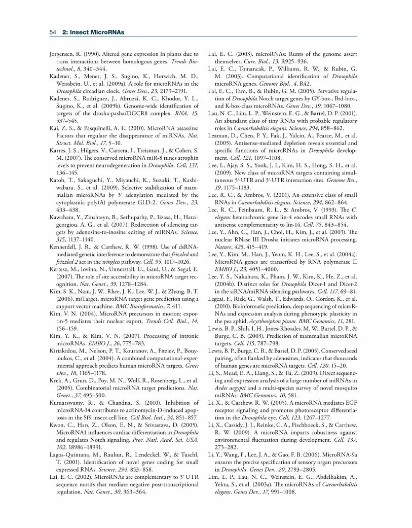
54 2: Insect MicroRNAs
Jorgensen, R. (1990). Altered gene expression in plants due to trans interactions between homologous genes. Trends Bio-technol., 8, 340–344.
Kadener, S., Menet, J. S., Sugino, K., Horwich, M. D., Weissbein, U., et al. (2009a). A role for microRNAs in the Drosophila circadian clock. Genes Dev., 23, 2179–2191.
Kadener, S., Rodriguez, J., Abruzzi, K. C., Khodor, Y. L., Sugino, K., et al. (2009b). Genome-wide identification of targets of the drosha-pasha/DGCR8 complex. RNA, 15, 537–545.
Kai, Z. S., & Pasquinelli, A. E. (2010). MicroRNA assassins: Factors that regulate the disappearance of miRNAs. Nat. Struct. Mol. Biol., 17, 5–10.
Karres, J. S., Hilgers, V., Carrera, I., Treisman, J., & Cohen, S. M. (2007). The conserved microRNA miR-8 tunes atrophin levels to prevent neurodegeneration in Drosophila. Cell, 131, 136–145.
Katoh, T., Sakaguchi, Y., Miyauchi, K., Suzuki, T., Kashi-wabara, S., et al. (2009). Selective stabilization of mam-malian microRNAs by 3′ adenylation mediated by the cytoplasmic poly(A) polymerase GLD-2. Genes Dev., 23, 433–438.
Kawahara, Y., Zinshteyn, B., Sethupathy, P., Iizasa, H., Hatzi-georgiou, A. G., et al. (2007). Redirection of silencing tar-gets by adenosine-to-inosine editing of miRNAs. Science, 315, 1137–1140.
Kennerdell, J. R., & Carthew, R. W. (1998). Use of dsRNA-mediated genetic interference to demonstrate that frizzled and frizzled 2 act in the wingless pathway. Cell, 95, 1017–1026.
Kertesz, M., Iovino, N., Unnerstall, U., Gaul, U., & Segal, E. (2007). The role of site accessibility in microRNA target rec-ognition. Nat. Genet., 39, 1278–1284.
Kim, S. K., Nam, J. W., Rhee, J. K., Lee, W. J., & Zhang, B. T. (2006). miTarget, microRNA target gene prediction using a support vector machine. BMC Bioinformatics, 7, 411.
Kim, V. N. (2004). MicroRNA precursors in motion: expor-tin-5 mediates their nuclear export. Trends Cell. Biol., 14, 156–159.
Kim, Y. K., & Kim, V. N. (2007). Processing of intronic microRNAs. EMBO J., 26, 775–783.
Kiriakidou, M., Nelson, P. T., Kouranov, A., Fitziev, P., Bouy-ioukos, C., et al. (2004). A combined computational-exper-imental approach predicts human microRNA targets. Genes Dev., 18, 1165–1178.
Krek, A., Grun, D., Poy, M. N., Wolf, R., Rosenberg, L., et al. (2005). Combinatorial microRNA target predictions. Nat. Genet., 37, 495–500.
Kumarswamy, R., & Chandna, S. (2010). Inhibition of microRNA-14 contributes to actinomycin-D-induced apop-tosis in the Sf9 insect cell line. Cell Biol. Intl., 34, 851–857.
Kwon, C., Han, Z., Olson, E. N., & Srivastava, D. (2005). MicroRNA1 influences cardiac differentiation in Drosophila and regulates Notch signaling. Proc. Natl. Acad. Sci. USA, 102, 18986–18991.
Lagos-Quintana, M., Rauhut, R., Lendeckel, W., & Tuschl, T. (2001). Identification of novel genes coding for small expressed RNAs. Science, 294, 853–858.
Lai, E. C. (2002). MicroRNAs are complementary to 3′ UTR sequence motifs that mediate negative post-transcriptional regulation. Nat. Genet., 30, 363–364.
Lai, E. C. (2003). microRNAs: Runts of the genome assert themselves. Curr. Biol., 13, R925–936.
Lai, E. C., Tomancak, P., Williams, R. W., & Rubin, G. M. (2003). Computational identification of Drosophila microRNA genes. Genome Biol., 4, R42.
Lai, E. C., Tam, B., & Rubin, G. M. (2005). Pervasive regula-tion of Drosophila Notch target genes by GY-box-, Brd-box-, and K-box-class microRNAs. Genes Dev., 19, 1067–1080.
Lau, N. C., Lim, L. P., Weinstein, E. G., & Bartel, D. P. (2001). An abundant class of tiny RNAs with probable regulatory roles in Caenorhabditis elegans. Science, 294, 858–862.
Leaman, D., Chen, P. Y., Fak, J., Yalcin, A., Pearce, M., et al. (2005). Antisense-mediated depletion reveals essential and specific functions of microRNAs in Drosophila develop-ment. Cell, 121, 1097–1108.
Lee, I., Ajay, S. S., Yook, J. I., Kim, H. S., Hong, S. H., et al. (2009). New class of microRNA targets containing simul-taneous 5′-UTR and 3′-UTR interaction sites. Genome Res., 19, 1175–1183.
Lee, R. C., & Ambros, V. (2001). An extensive class of small RNAs in Caenorhabditis elegans. Science, 294, 862–864.
Lee, R. C., Feinbaum, R. L., & Ambros, V. (1993). The C. elegans heterochronic gene lin-4 encodes small RNAs with antisense complementarity to lin-14. Cell, 75, 843–854.
Lee, Y., Ahn, C., Han, J., Choi, H., Kim, J., et al. (2003). The nuclear RNase III Drosha initiates microRNA processing. Nature, 425, 415–419.
Lee, Y., Kim, M., Han, J., Yeom, K. H., Lee, S., et al. (2004a). MicroRNA genes are transcribed by RNA polymerase II EMBO J., 23, 4051–4060.
Lee, Y. S., Nakahara, K., Pham, J. W., Kim, K., He, Z., et al. (2004b). Distinct roles for Drosophila Dicer-1 and Dicer-2 in the siRNA/miRNA silencing pathways. Cell, 117, 69–81.
Legeai, F., Rizk, G., Walsh, T., Edwards, O., Gordon, K., et al. (2010). Bioinformatic prediction, deep sequencing of microR-NAs and expression analysis during phenotypic plasticity in the pea aphid, Acyrthosiphon pisum. BMC Genomics, 11, 281.
Lewis, B. P., Shih, I. H., Jones-Rhoades, M. W., Bartel, D. P., & Burge, C. B. (2003). Prediction of mammalian microRNA targets. Cell, 115, 787–798.
Lewis, B. P., Burge, C. B., & Bartel, D. P. (2005). Conserved seed pairing, often flanked by adenosines, indicates that thousands of human genes are microRNA targets. Cell, 120, 15–20.
Li, S., Mead, E. A., Liang, S., & Tu, Z. (2009). Direct sequenc-ing and expression analysis of a large number of miRNAs in Aedes aegypti and a multi-species survey of novel mosquito miRNAs. BMC Genomics, 10, 581.
Li, X., & Carthew, R. W. (2005). A microRNA mediates EGF receptor signaling and promotes photoreceptor differentia-tion in the Drosophila eye. Cell, 123, 1267–1277.
Li, X., Cassidy, J. J., Reinke, C. A., Fischboeck, S., & Carthew, R. W. (2009). A microRNA imparts robustness against environmental fluctuation during development. Cell, 137, 273–282.
Li, Y., Wang, F., Lee, J. A., & Gao, F. B. (2006). MicroRNA-9a ensures the precise specification of sensory organ precursors in Drosophila. Genes Dev., 20, 2793–2805.
Lim, L. P., Lau, N. C., Weinstein, E. G., Abdelhakim, A., Yekta, S., et al. (2003a). The microRNAs of Caenorhabditis elegans. Genes Dev., 17, 991–1008.
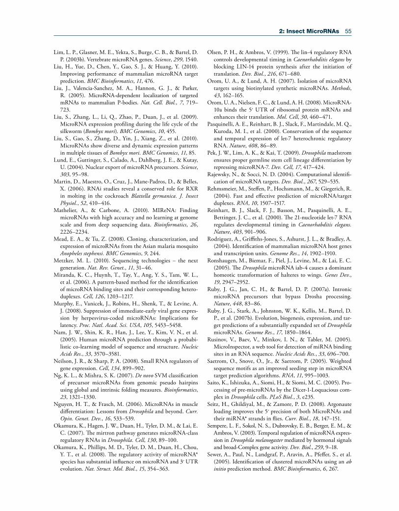
2: Insect MicroRNAs 55
Lim, L. P., Glasner, M. E., Yekta, S., Burge, C. B., & Bartel, D. P. (2003b). Vertebrate microRNA genes. Science, 299, 1540.
Liu, H., Yue, D., Chen, Y., Gao, S. J., & Huang, Y. (2010). Improving performance of mammalian microRNA target prediction. BMC Bioinformatics, 11, 476.
Liu, J., Valencia-Sanchez, M. A., Hannon, G. J., & Parker, R. (2005). MicroRNA-dependent localization of targeted mRNAs to mammalian P-bodies. Nat. Cell. Biol., 7, 719–723.
Liu, S., Zhang, L., Li, Q., Zhao, P., Duan, J., et al. (2009). MicroRNA expression profiling during the life cycle of the silkworm (Bombyx mori). BMC Genomics, 10, 455.
Liu, S., Gao, S., Zhang, D., Yin, J., Xiang, Z., et al. (2010). MicroRNAs show diverse and dynamic expression patterns in multiple tissues of Bombyx mori. BMC Genomics, 11, 85.
Lund, E., Guttinger, S., Calado, A., Dahlberg, J. E., & Kutay, U. (2004). Nuclear export of microRNA precursors. Science, 303, 95–98.
Martin, D., Maestro, O., Cruz, J., Mane-Padros, D., & Belles, X. (2006). RNAi studies reveal a conserved role for RXR in molting in the cockroach Blattella germanica. J. Insect Physiol., 52, 410–416.
Mathelier, A., & Carbone, A. (2010). MIReNA: Finding microRNAs with high accuracy and no learning at genome scale and from deep sequencing data. Bioinformatics, 26, 2226–2234.
Mead, E. A., & Tu, Z. (2008). Cloning, characterization, and expression of microRNAs from the Asian malaria mosquito Anopheles stephensi. BMC Genomics, 9, 244.
Metzker, M. L. (2010). Sequencing technologies – the next generation. Nat. Rev. Genet., 11, 31–46.
Miranda, K. C., Huynh, T., Tay, Y., Ang, Y. S., Tam, W. L., et al. (2006). A pattern-based method for the identification of microRNA binding sites and their corresponding hetero-duplexes. Cell, 126, 1203–1217.
Murphy, E., Vanicek, J., Robins, H., Shenk, T., & Levine, A. J. (2008). Suppression of immediate-early viral gene expres-sion by herpesvirus-coded microRNAs: Implications for latency. Proc. Natl. Acad. Sci. USA, 105, 5453–5458.
Nam, J. W., Shin, K. R., Han, J., Lee, Y., Kim, V. N., et al. (2005). Human microRNA prediction through a probabi-listic co-learning model of sequence and structure. Nucleic Acids Res., 33, 3570–3581.
Neilson, J. R., & Sharp, P. A. (2008). Small RNA regulators of gene expression. Cell, 134, 899–902.
Ng, K. L., & Mishra, S. K. (2007). De novo SVM classification of precursor microRNAs from genomic pseudo hairpins using global and intrinsic folding measures. Bioinformatics, 23, 1321–1330.
Nguyen, H. T., & Frasch, M. (2006). MicroRNAs in muscle differentiation: Lessons from Drosophila and beyond. Curr. Opin. Genet. Dev., 16, 533–539.
Okamura, K., Hagen, J. W., Duan, H., Tyler, D. M., & Lai, E. C. (2007). The mirtron pathway generates microRNA-class regulatory RNAs in Drosophila. Cell, 130, 89–100.
Okamura, K., Phillips, M. D., Tyler, D. M., Duan, H., Chou, Y. T., et al. (2008). The regulatory activity of microRNA* species has substantial influence on microRNA and 3′ UTR evolution. Nat. Struct. Mol. Biol., 15, 354–363.
Olsen, P. H., & Ambros, V. (1999). The lin-4 regulatory RNA controls developmental timing in Caenorhabditis elegans by blocking LIN-14 protein synthesis after the initiation of translation. Dev. Biol., 216, 671–680.
Orom, U. A., & Lund, A. H. (2007). Isolation of microRNA targets using biotinylated synthetic microRNAs. Methods, 43, 162–165.
Orom, U. A., Nielsen, F. C., & Lund, A. H. (2008). MicroRNA-10a binds the 5′ UTR of ribosomal protein mRNAs and enhances their translation. Mol. Cell, 30, 460–471.
Pasquinelli, A. E., Reinhart, B. J., Slack, F., Martindale, M. Q., Kuroda, M. I., et al. (2000). Conservation of the sequence and temporal expression of let-7 heterochronic regulatory RNA. Nature, 408, 86–89.
Pek, J. W., Lim, A. K., & Kai, T. (2009). Drosophila maelstrom ensures proper germline stem cell lineage differentiation by repressing microRNA-7. Dev. Cell, 17, 417–424.
Rajewsky, N., & Socci, N. D. (2004). Computational identifi-cation of microRNA targets. Dev. Biol., 267, 529–535.
Rehmsmeier, M., Steffen, P., Hochsmann, M., & Giegerich, R. (2004). Fast and effective prediction of microRNA/target duplexes. RNA, 10, 1507–1517.
Reinhart, B. J., Slack, F. J., Basson, M., Pasquinelli, A. E., Bettinger, J. C., et al. (2000). The 21-nucleotide let-7 RNA regulates developmental timing in Caenorhabditis elegans. Nature, 403, 901–906.
Rodriguez, A., Griffiths-Jones, S., Ashurst, J. L., & Bradley, A. (2004). Identification of mammalian microRNA host genes and transcription units. Genome Res., 14, 1902–1910.
Ronshaugen, M., Biemar, F., Piel, J., Levine, M., & Lai, E. C. (2005). The Drosophila microRNA iab-4 causes a dominant homeotic transformation of halteres to wings. Genes Dev., 19, 2947–2952.
Ruby, J. G., Jan, C. H., & Bartel, D. P. (2007a). Intronic microRNA precursors that bypass Drosha processing. Nature, 448, 83–86.
Ruby, J. G., Stark, A., Johnston, W. K., Kellis, M., Bartel, D. P., et al. (2007b). Evolution, biogenesis, expression, and tar-get predictions of a substantially expanded set of Drosophila microRNAs. Genome Res., 17, 1850–1864.
Rusinov, V., Baev, V., Minkov, I. N., & Tabler, M. (2005). MicroInspector, a web tool for detection of miRNA binding sites in an RNA sequence. Nucleic Acids Res., 33, 696–700.
Saetrom, O., Snove, O., Jr., & Saetrom, P. (2005). Weighted sequence motifs as an improved seeding step in microRNA target prediction algorithms. RNA, 11, 995–1003.
Saito, K., Ishizuka, A., Siomi, H., & Siomi, M. C. (2005). Pro-cessing of pre-microRNAs by the Dicer-1-Loquacious com-plex in Drosophila cells. PLoS Biol., 3, e235.
Seitz, H., Ghildiyal, M., & Zamore, P. D. (2008). Argonaute loading improves the 5′ precision of both MicroRNAs and their miRNA* strands in flies. Curr. Biol., 18, 147–151.
Sempere, L. F., Sokol, N. S., Dubrovsky, E. B., Berger, E. M., & Ambros, V. (2003). Temporal regulation of microRNA expres-sion in Drosophila melanogaster mediated by hormonal signals and broad-Complex gene activity. Dev. Biol., 259, 9–18.
Sewer, A., Paul, N., Landgraf, P., Aravin, A., Pfeffer, S., et al. (2005). Identification of clustered microRNAs using an ab initio prediction method. BMC Bioinformatics, 6, 267.
56 2: Insect MicroRNAs
Shcherbata, H. R., Ward, E. J., Fischer, K. A., Yu, J. Y., Reynolds, S. H., et al. (2007). Stage-specific differences in the requirements for germline stem cell maintenance in the Drosophila ovary. Cell Stem Cell, 1, 698–709.
Sheng, Y., Engstrom, P. G., & Lenhard, B. (2007). Mammalian microRNA prediction through a support vector machine model of sequence and structure. PLoS One, 2, e946.
Skalsky, R. L., Vanlandingham, D. L., Scholle, F., Higgs, S., & Cullen, B. R. (2010). Identification of microRNAs expressed in two mosquito vectors, Aedes albopictus and Culex quin-quefasciatus. BMC Genomics, 11, 119.
Sokol, N. S., & Ambros, V. (2005). Mesodermally expressed Drosophila microRNA-1 is regulated by Twist and is required in muscles during larval growth. Genes Dev., 19, 2343–2354.
Sokol, N. S., Xu, P., Jan, Y. N., & Ambros, V. (2008). Dro-sophila let-7 microRNA is required for remodeling of the neuromusculature during metamorphosis. Genes Dev., 22, 1591–1596.
Stark, A., Brennecke, J., Bushati, N., Russell, R. B., & Cohen, S. M. (2005). Animal MicroRNAs confer robustness to gene expression and have a significant impact on 3′ UTR evolu-tion. Cell, 123, 1133–1146.
Sturm, M., Hackenberg, M., Langenberger, D., & Frish-man, D. (2010). TargetSpy: A supervised machine learning approach for microRNA target prediction. BMC Bioinfor-matics, 11, 292.
Teleman, A. A., Maitra, S., & Cohen, S. M. (2006). Drosophila lacking microRNA miR-278 are defective in energy homeo-stasis. Genes Dev., 20, 417–422.
Thadani, R., & Tammi, M. T. (2006). MicroTar, predicting microRNA targets from RNA duplexes. BMC Bioinformat-ics, 7(Suppl. 5), S20.
Thompson, B. J., & Cohen, S. M. (2006). The Hippo pathway regulates the bantam microRNA to control cell proliferation and apoptosis in Drosophila. Cell, 126, 767–774.
van Dongen, S., Abreu-Goodger, C., & Enright, A. J. (2008). Detecting microRNA binding and siRNA off-target effects from expression data. Nat. Methods, 5, 1023–1025.
Varghese, J., & Cohen, S. M. (2007). microRNA miR-14 acts to modulate a positive autoregulatory loop controlling steroid hormone signaling in Drosophila. Genes Dev., 21, 2277–2282.
Vasudevan, S., Tong, Y., & Steitz, J. A. (2007). Switching from repression to activation: microRNAs can up-regulate trans-lation. Science, 318, 1931–1934.
Viswanathan, S. R., Daley, G. Q., & Gregory, R. I. (2008). Selective blockade of microRNA processing by Lin28. Sci-ence, 320, 97–100.
Wang, X., & El Naqa, I. M. (2008). Prediction of both con-served and nonconserved microRNA targets in animals. Bioinformatics, 24, 325–332.
Wang, X., Zhang, J., Li, F., Gu, J., He, T., et al. (2005). MicroRNA identification based on sequence and structure alignment. Bioinformatics, 21, 3610–3614.
Weaver, D. B., Anzola, J. M., Evans, J. D., Reid, J. G., Reese, J. T., et al. (2007). Computational and transcriptional evi-dence for microRNAs in the honey bee genome. Genome Biol., 8, R97.
Wei, Y., Chen, S., Yang, P., Ma, Z., & Kang, L. (2009). Char-acterization and comparative profiling of the small RNA transcriptomes in two phases of locust. Genome Biol., 10, R6.
Wightman, B., Burglin, T. R., Gatto, J., Arasu, P., & Ruvkun, G. (1991). Negative regulatory sequences in the lin-14 3′-untranslated region are necessary to generate a temporal switch during Caenorhabditis elegans development. Genes Dev., 5, 1813–1824.
Wightman, B., Ha, I., & Ruvkun, G. (1993). Posttranscrip-tional regulation of the heterochronic gene lin-14 by lin-4 mediates temporal pattern formation in C. elegans. Cell, 75, 855–862.
Winter, F., Edaye, S., Huttenhofer, A., & Brunel, C. (2007). Anopheles gambiae miRNAs as actors of defence reaction against Plasmodium invasion. Nucleic Acids Res., 35, 6953–6962.
Wu, L., Fan, J., & Belasco, J. G. (2006). MicroRNAs direct rapid deadenylation of mRNA. Proc. Natl. Acad. Sci. USA, 103, 4034–4039.
Xu, P., Vernooy, S. Y., Guo, M., & Hay, B. A. (2003). The Drosophila microRNA Mir-14 suppresses cell death and is required for normal fat metabolism. Curr. Biol., 13, 790–795.
Xue, C., Li, F., He, T., Liu, G. P., Li, Y., & Zhang, X. (2005). Classification of real and pseudo microRNA precursors using local structure-sequence features and support vector machine. BMC Bioinformatics, 6, 310.
Yang, J. H., Shao, P., Zhou, H., Chen, Y. Q., & Qu, L. H. (2010). deepBase: A database for deeply annotating and min-ing deep sequencing data. Nucleic Acids Res., 38, D123–130.
Yang, Y., Wang, Y. P., & Li, K. B. (2008). MiRTif, a support vector machine-based microRNA target interaction filter. BMC Bioinformatics, 9(Suppl. 12), S4.
Yi, R., Qin, Y., Macara, I. G., & Cullen, B. R. (2003). Expor-tin-5 mediates the nuclear export of pre-microRNAs and short hairpin RNAs. Genes Dev., 17, 3011–3016.
Yousef, M., Nebozhyn, M., Shatkay, H., Kanterakis, S., Showe, L. C., et al. (2006). Combining multi-species genomic data for microRNA identification using a Naive Bayes classifier. Bioinformatics, 22, 1325–1334.
Yousef, M., Jung, S., Kossenkov, A. V., Showe, L. C., & Showe, M. K. (2007). Naive Bayes for microRNA target predic-tions-machine learning for microRNA targets. Bioinformat-ics, 23, 2987–2992.
Yousef, M., Jung, S., Showe, L. C., & Showe, M. K. (2008). Learning from positive examples when the negative class is undetermined-microRNA gene identification. Algorithms Mol. Biol., 3, 2.
Yu, J. Y., Reynolds, S. H., Hatfield, S. D., Shcherbata, H. R., Fischer, K. A., et al. (2009). Dicer-1-dependent Dacapo sup-pression acts downstream of Insulin receptor in regulating cell division of Drosophila germline stem cells. Development, 136, 1497–1507.
Yu, X., Zhou, Q., Li, S. C., Luo, Q., Cai, Y., et al. (2008). The silkworm (Bombyx mori) microRNAs and their expressions in multiple developmental stages. PLoS One, 3, e2997.