insect biochemistry and molecular biology -...
TRANSCRIPT

lable at ScienceDirect
Insect Biochemistry and Molecular Biology 73 (2016) 27e37
Contents lists avai
Insect Biochemistry and Molecular Biology
journal homepage: www.elsevier .com/locate/ ibmb
Serpin-5 regulates prophenoloxidase activation and antimicrobialpeptide pathways in the silkworm, Bombyx mori
Junlan Li a, Li Ma a, Zhe Lin c, Zhen Zou c, **, Zhiqiang Lu a, b, *
a Department of Entomology, College of Plant Protection, Northwest A&F University, Yangling, Shaanxi, 712100, Chinab Key Laboratory of Plant Protection Resources and Pest Management, Ministry of Education, Northwest A&F University, Yangling, Shaanxi, 712100, Chinac State Key Laboratory of Integrated Management of Pest Insects and Rodents, Institute of Zoology, Chinese Academy of Sciences, Beijing, 100101, China
a r t i c l e i n f o
Article history:Received 18 December 2015Received in revised form30 March 2016Accepted 11 April 2016Available online 12 April 2016
Keywords:SerpinProphenoloxidaseAntimicrobial peptideSilkworm
* Corresponding author. Department of EntomologNorthwest A&F University, Yangling, Shaanxi, 712100** Corresponding author.
E-mail addresses: [email protected] (Z. Zou)(Z. Lu).
http://dx.doi.org/10.1016/j.ibmb.2016.04.0030965-1748/© 2016 Elsevier Ltd. All rights reserved.
a b s t r a c t
The prophenoloxidase (PPO) activation pathway and Toll pathway are two critical insect immune re-sponses against microbial infection. Activation of these pathways is mediated by an extracellular serineprotease cascade, which is negatively regulated by serpins. In this study, we found that the mRNAabundance of silkworm serpin-5 (BmSpn-5) increased dramatically in the fat body after bacterialinfection. The expression level of antimicrobial peptides (AMPs), gloverin-3, cecropin-D and -E decreasedin the silkworm larvae injected with recombinant BmSpn-5 protein. Meanwhile, the inhibition of beadsmelanization, systemic melanization and PPO activation by BmSpn-5 was also observed. By means ofimmunoaffinity purification and analysis by mass spectrometry, we identified that the silkworm clipdomain serine proteases BmHP6 and BmSP21 form a complex with BmSpn-5, which suggests thatBmHP6 and SP21 are the cognate proteases of BmSpn-5 and are essential in the serine protease cascadethat activates the Toll and PPO pathways. Our study provides a comprehensive characterization ofBmSpn-5 and sheds light on the multiple pathways leading to PPO activation and their regulation byserpins.
© 2016 Elsevier Ltd. All rights reserved.
1. Introduction
Insects have evolved efficient innate immune systems to defendthemselves against the invasion of pathogens and parasites(Lemaitre and Hoffmann, 2007). The production of antimicrobialpeptides (AMPs) and melanization are two key responses in thehumoral immunity. In Drosophila melanogaster, the mRNA of AMPsis transcribed through Toll or IMD (Immune Deficiency) pathwayswhen the invading bacteria or fungi are recognized as non-self.Specifically, the IMD pathway is activated by the recognition ofGram-negative bacteria, whereas the Toll pathway is activated bythe recognition of Gram-positive bacteria and fungi (Imler, 2014).Apart from NK-kB factor-mediated pathways, melanization isanother universal defense mechanism in arthropods that is cata-lyzed by phenoloxidase (PO), which is proteolytically transformed
y, College of Plant Protection,, China.
from zymogen prophenoloxidase (PPO) to its active conformation(Jiang et al., 1998; Lu et al., 2014). The activation of the Toll pathwayand PPO ismediated by an extracellular clip domain serine proteasecascade (Kambris et al., 2006; Tang et al., 2006; An et al., 2009,2013; Veillard et al., 2016; Kanost and Jiang, 2015).
This serine protease cascade is precisely regulated by serineprotease inhibitors (serpins, Spns) (Gubb et al., 2010; Kanost, 1999).Existing in all metazoans, the serpin family has a highly variableprimary structure but shares a conserved structural similarity. Mostmembers of this family are proteins between 400 and 500 aminoacids in length, with a C-terminal exposed reactive center loop(RCL) that connects b-sheet A and C. The RCL is approximately 20amino acids in length and acts as a pseudo-substrate for its cognateprotease. Following binding to the target protease, serpin is cleavedat the scissile bond (P1-P10) located in the RCL and adopts a relaxedconformation, which results in the formation of a covalent complexbetween the protease and a large part of serpin (Gettins, 2002).During this process, the RCL is inserted into b-sheet A, and dramaticconformational change of the protease causes it to lose its catalyticactivity. Serpin genes have been identified systematically in manyinsect species including Drosophila melanogaster (Reichhart, 2005),
J. Li et al. / Insect Biochemistry and Molecular Biology 73 (2016) 27e3728
Anopheles gambia (Gulley et al., 2013; Suwanchaichinda andKanost, 2009), Bombyx mori (Zou et al., 2009) and Helicoverpaarmigera (Xiong et al., 2015). In Drosophila, Spn43Ac (necrotic,CG1857) and Spn1 (Spn42Dd, CG9456) negatively regulate the Tollsignaling pathway (Fullaondo et al., 2011; Levashina et al., 1999).Spn27A (CG11331), Spn28D (CG7219) and Spn77Ba (CG6680)down-regulate the PPO cascade in Drosophila (De Gregorio et al.,2002; Ligoxygakis et al., 2002; Tang et al., 2008). In the tobaccohornworm, Manduca sexta, Spn-1J, �3, �4, �5, �6, and �7 havebeen shown to regulate PPO activation and the Toll pathway (Anand Kanost, 2010; An et al., 2011; Suwanchaichinda et al., 2013;Tong and Kanost, 2005; Zhu et al., 2003; Zou and Jiang, 2005). Inthe larval beetle Tenebrio molitor, several serpins have been iden-tified as negative regulators of the PPO and Toll pathways (Jianget al., 2009, 2011). An ortholog of M. sexta Spn-3 in Asian cornborer, Ostrinia furnacalis, plays roles in both the regulation of PPOactivation and antimicrobial peptide production (Chu et al., 2015).
In the silkworm, Bombyx mori, 34 serpin genes have beenidentified and their expression patterns in response to microbialinfection have been profiled (Zou and Jiang, 2005; Zou et al., 2009).The cDNA of silkworm Spn-2 (also known as antichymotrypsin, sw-Achy), antichymotrypsin II and Spn-9 (also known as antitrypsin,sw-AT) were cloned approximately two decades ago (Narumi et al.,1993; Sasaki, 1991; Takagi et al., 1990). However, there are nofunctional studies for these serpins. A recent study showed that asilk gland-specific serpin (Spn-16) is a cysteine protease inhibitor,rather than a serine protease inhibitor (Guo et al., 2015). Its phys-iological function in the silk gland is unclear. Bombyx mori Spn-15 isinduced by Beauveria bassiana and Micrococcus luteus and plays arole as a negative regulator in PPO activation and Toll pathways (Liuet al., 2015). In this study, we found that the expression of silkwormserpin-5 (BmSpn-5) in the fat body is remarkably induced by bac-terial infection.We used the recombinant BmSpn-5 protein to showthat it down-regulates AMPs, gloverin-3, cecropin-D and -E gen-eration and PPO activation. We further revealed that the silkwormclip domain serine proteases, BmHP6 and BmSP21, form complexeswith BmSpn-5 using immunoaffinity chromatography and massspectrometry analysis. This suggests that BmHP6 and BmSP21 arethe target proteases and participate in the serine protease cascadethat leads to activation of the Toll and PPO pathways.
2. Materials and methods
2.1. Silkworm rearing and infection with bacteria
The silkworm B. mori (Nistari strain) was reared on mulberryleaves at 27 �C in 70% RH and a photoperiod of 13:11 (light:dark).Micrococcus luteus and Yersinia pseudotuberculosis serotype O:3(YPIII) were cultured at 37 �C in LB medium overnight. The cellswere collected by centrifugation at 8000 � g. The pellet wasresuspended in 1ml sterilized 0.85% NaCl, then 40ml 70% isopropylalcohol was added and incubated for 1 h at room temperature withrotation to kill the cells. After three rounds of washing with ster-ilized 0.85% NaCl, the isopropanol-killed bacteria were resus-pended in sterilized 0.85% NaCl to OD600nm of 10. Fifty ml of thebacterial preparations were injected into the hemocoel of day-3fifth-instar silkworm.
2.2. Analysis of BmSpn-5 expression by quantitative real-time PCR
The day-3 fifth-instar larvae were injected with either sterilized0.85% NaCl (as control), isopropanol-killedM. luteus or YPIII cells. At6, 24 and 48 h post infection (hpi), fat body from 5 to 8 larvae werecollected in 500 ml TriPure Reagent (Roche, USA) and storedat�80 �C. Hemolymph from at least 15 larvae was pooled and spun
at 500 � g for 10 min at 4 �C to collect hemocytes. After discardingthe supernatant, the hemocytes were mixed with TriPure Reagentand stored at �80 �C. Total RNA was extracted from the stored fatbody and hemocytes samples following the standard protocolprovided by the producer. 30 mg total RNAwas further purified withDirect-zol™ RNA MiniPrep (Zymo Research, USA), and first strandcDNA was synthesized by Transcriptor First Strand cDNA SynthesisKit (Roche, USA). Using the prepared cDNA as a template, quanti-tative real-time PCR (qPCR) was carried out by 2 � FastStartEssential DNA GreenMaster (Roche), with 40 thermal cycles (94 �C,15 s; 55 �C, 20 s; 72 �C, 20 s) after initial denaturation at 95 �C for10 min. A melt curve (65e95 �C) was determined to confirm theamplification of specific PCR product (Bio-Rad IQ5). The Bombyxhousekeeping gene IF4A (DQ443290.1) was used as an internalcontrol to normalize the transcript level of BmSpn-5 (Wu et al.,2010). The standard curve of each gene was measured to deter-mine the amplification efficiency and r2 value. The relative quan-titative method (2�DDCt) was applied to determine the expressionlevel of BmSpn-5 (Schmittgen and Livak, 2008). Primers used forquantitative real-time PCR are listed in Table 1.
2.3. Expression and purification of recombinant BmSpn-5
50 RACE-ready cDNA was synthesized from the total RNA fromthe midgut of the fifth instar silkworm using SMARTer RACE cDNAamplification kit (Clonetech, USA) according to the providedmanual. The cDNA fragment encoding BmSpn-5 (GenBank number:AY566165.1) was amplified with gene specific primers, in which aBamHI and an XhoI restriction site were introduced (Table 1). Theamplified product was cloned in the pET32a expression vector(Novagen, USA). The expression of BmSpn-5 in the E. coli BL21 (DE3)strain was induced by 0.5 mM isopropyl-beta-D-thiogalactopyranoside (IPTG). The collected cells were resus-pended in phosphate buffered saline (PBS) and homogenized bysonication (Sonics Vibra-Cell, 130 w, 30% power). BmSpn-5 waspurified from the soluble fraction first using a Ni2þ-NTA column bya stepwise elution in PBS containing 25 mM, 50 mM, 150 mM, and500 mM imidazole. The fractions (50e150 mM imidazole) con-taining recombinant BmSpn-5 were pooled and dialyzed in Tris-HCl buffer (20 mM Tris, 50 mM NaCl, 2 mM CaCl2, pH 7.4), fol-lowed by digestionwith recombinant enterokinase (rEK) (AmoytopBiotech, China) for 5 h. The digested protein was purified on Ni2þ-NTA column again. Due to the rEK digestion, the Trx tag, His tag andS tag were cleaved from BmSpn-5, resulting in the loss of BmSpn-5affinity for the Ni2þ-NTA column. After dialysis with 50 mM Tris-HCl (pH 8.2), recombinant BmSpn-5 was further purified by Q-Sepharose Fast Flow column in 50 mM Tris-HCl (pH 8.2) with astepwise elution containing 50mM,150mM, 300mM, and 500mMNaCl. BmSpn5 was eluted in the fraction containing 150 mM and300 mM NaCl, and then desalted by dialysis. Protein concentrationwas determined using the BCA Protein Assay Kit (Beyotime, China).Purified recombinant BmSpn-5 was verified by peptide massfingerprinting (Beijing Protein Innovation). A rabbit polyclonalantiserumwas prepared against the recombinant BmSpn-5 (Cwbio,China).
2.4. SDS-PAGE and immunoblotting
Recombinant BmSpn-5 was analyzed by 12% SDS-PAGE andstained with Coomassie brilliant blue dye. For the western blot,separated proteins were electrotransferred onto a PVDF membrane(Millipore). Themembranewas subsequently blocked with 5% skimmilk powder (Merck) in TBST (0.05% Tween-20, 150 mM NaCl,50 mM Tris). Anti-His rabbit polyclonal antibody (1:5000 dilution)(Santa Cruz Biotechnology) and anti-BmSpn-5 rabbit polyclonal

Table 1Primers used in gene cloning and quantitative real-time PCR.
Primer Sequence (50 - 30)
BmSpn-5 gene cloningForward: GCGGATCCGATGTCGATTTCAGCGAGAReverse: GCTCGAGTCAATAGACAGTCGGTTTTGAABmSpn-5 quantitative RT- PCRForward: GCACCCGAGAACGAAGATReverse: GGCTAAGTCTGGCTGGAATABmIF4A quantitative RT- PCRForward: TCTGGCATCATACCTTCTACAAReverse: TCTGTGTCATCTTTTCCCTGTTBmGlov3 quantitative RT-PCRForward: GACACGAGAATGGGAGGAGReverse: AAGACCCTGGTGCCGTAABmCecD quantitative RT-PCRForward: CTCCCGGCAACTTCTTCAReverse: CGAACCCTCTGACCCATTBmCecE quantitative RT-PCRForward: ACTGTTCGACATCGCCTCTReverse: CGAATGTTCTGACCCACC
Underline showed the BamHI and XhoI restriction enzyme sites.
J. Li et al. / Insect Biochemistry and Molecular Biology 73 (2016) 27e37 29
antibody (1:10,000 dilution) were used as primary antibodies, andgoat-anti-rabbit IgG (1:10,000 dilution) (Santa Cruz Biotechnology)was used as the secondary antibody. Western blotting detection kit(Advasta) was used for detection and a chemiluminescence imag-ing system was used to document the images (Clinx Science In-struments Co., Ltd).
2.5. Melanization assay of sepharose beads in the silkwormhemolymph
Positively charged QAE Sepharose Fast Flow (QFF) chromatog-raphy beads (50e150 mm in diameter, Sigma) were used for analysisof encapsulation in silkworm hemolymph. QFF beads were stainedfully with 0.1% Congo red for 2 h and washed three times with50 mM Tris-HCl buffer (pH 8.2). The washed beads were resus-pended in 1ml of serpin-5 (300 mg/ml, in 50mM Tris-HCl, pH8.2) toapproximately 100 beads per 50 ml and incubated for 1 h at roomtemperature with gentle rotation. The serpin-5 coated beads werewashed three times with Tris-HCl buffer and resuspended in Tris-HCl buffer. Fifteen day 3 fifth-instar larvae silkworm larvae wereanesthetized on ice and 50 ml of the bead suspension was injectedinto the larval hemocoel. The larvaewere dissected 4 h later and thebeads were recovered and observed under microscope. Bovineserum albumin (BSA) coated beads were injected as a control.
2.6. Prophenoloxidase activation and phenoloxidase activity assay
Day-3 fifth-instar larvae were injected with 50 ml BSA (150 mg/ml in 50 mM Tris-HCl, pH 8.0) or 50 ml recombinant BmSpn-5(150 mg/ml in 50 mM Tris-HCl, pH 8.0). After 30 min, hemolymphwas collected and centrifuged immediately at 16,000 � g for 30 s at4 �C to remove hemocytes. 5 ml of plasma sample was added to thewells of a 96 well plate, and 5 ml of 1 mg/ml pathogen-associatedmolecular patterns (PAMPs), polysaccharides (LPS) from Porphyr-omonas gingivalis, curdlan from Alcaligenes faecalis, or peptido-glycan (PG) from E. coli or M. luteus and 100 ml of 2 mM dopaminewere added and then mixed immediately. The absorption at490 nmwas recorded every 30 s for 15 min on a microplate reader(Tecan), where the maximum slope was determined. 60 ml of thetwo groups freshly prepared plasma were added simultaneously toeach 1.5 ml Eppendorf tubes that contained 10 ml of 1mg/ml elicitorand incubated at room temperature. At 1 min, 5 min and 25 min,20 ml of each sample was taken and mixed with 20 ml of 4 � SDS
loading buffer and boiled immediately at 100 �C for 5 min. Aftercentrifugation at 5000 � g for 3 min, the samples were analyzed by8% SDS-PAGE and western blot with a mixture of anti-PPO1 andanti-PPO2 (1:5000 dilution) (gifts from Dr. Michael Strand) as theprimary antibodies.
2.7. Immunoaffinity purification of BmSpn-5-protease complexes
Anti-BmSpn-5 covalent coupled Protein A-agarose was pre-pared according to a previous study (Jedamzik and Eckmann, 2009)that used dimethyl pimelimidate (DMP) (Sigma-Aldrich) as thelinker. 2e3 ml of hemolymph from day 3 fifth instar naïve larvae orlarvae that were injected with dead M. luteus were collected into atube and 10 ml of 1% phenylthiourea (PTU) was added. Hemocyteswere then removed by centrifugation at 500 � g for 10 min at 4 �C.PMSF (final concentration of 4 mM) and protease inhibitor cocktail(Thermo) were added to prevent protease activity. For anotherhemolymph sample of M. luteus injected larvae, M. luteus-PG wasadded to a final concentration of 1mg/ml, followed by incubation atroom temperature for 20 min. M. luteus-PG was then removed bycentrifugation at 10,000 � g for 5 min at 4 �C. PMSF and proteaseinhibitor cocktail were also added in the same amount. All threegroups of samples were subsequently incubated with 50 ml ofProtein A-agarose beads for 5 h at 4 �C to remove the non-specificabsorption. After centrifugation at 12,000 � g for 1 min, the su-pernatants were added to 50 ml of anti-BmSpn-5 coupled Protein A-agarose beads and incubated overnight at 4 �C. BmSpn-5-proteasecomplexes and BmSpn-5were thenwashed and eluted as describedin the Protein A Immunoprecipitation Kit (Roche). Eluted proteinswere then separated by 4%e12% NuPAGE BIS-Tris Gels with MOPSSDS running buffer (life technology), followed by modified Blumsilver staining (Mortz et al., 2001), which is compatible with massspectrometry analysis. Anti-BmSpn-5 was used to detect BmSpn-5-protease complexes in western blots. Protein bands on the silverstained gel were cut in accordance to bands on the western blot,and then digested with trypsin (Promega). The resulting peptidemixture was pre-separated by the Eksigent nanoLC-Ultra™ 2DSystem (AB SCIEX) equipped with a C18 nanoLC trap column(100 mm � 3 cm, 3 mm, 150 Å). Then, an analytical ChromXP C18column (75 mm � 15 cm, 3 mm, 120 Å) with a spray tip was used toseparate the peptide mixture. Data acquisitionwas performed witha Triple TOF 5600 System (AB SCIEX, USA) fitted with a NanosprayIII source (AB SCIEX) and a pulled quartz tip as the emitter (NewObjectives). Tandem mass spectra were searched with MASCOTV2.3 search engine (Matrix Science Ltd.) and identifications wereconfirmed by manual evaluation.
2.8. Hemolymph antibacterial activity assay
Day 3 fifth instar larvae were anesthetized on ice for 15 min,followed by injectionwith 30 mg BSA or 30 mg recombinant BmSpn-5 in 50 ml PBS. After 30 min of incubation in a rearing incubator, thelarvae were anesthetized on ice for another 15 min. Then, the twogroups of silkwormwere then immune challenged by isopropanol-killed M. luteus and YPIII. Additionally, 50 ml 0.85% NaCl wasinjected into another BSA pre-injection group as control. Fat bodyfrom at least 5 silkworm larvae was collected 24 h after the firstinjection. The expression of AMPs was subsequently analyzed byquantitative real-time PCR (Table 1). In 24 h after the first injection,plasma samples were collected and prepared as described before(An et al., 2009). The antimicrobial activity assay of plasma wasperformed by measuring zones of growth inhibition in a thin layerof agar plates, as described previously (Hoffmann et al., 1981).M. luteus and YPIII were used as the detective strains. Moreover, 5-ml plasma samples were treated with 5 ml 2 � SDS loading buffer at

J. Li et al. / Insect Biochemistry and Molecular Biology 73 (2016) 27e3730
100 �C for 5 min and separated by electrophoresis on a 16% tricine-SDS-PAGE (Schagger, 2006) and stained with Coomassie brilliantblue. The bands were excised and digested with In-Gel TrypticDigestion Kit (Thermo). LC-MS Analysis was completed using a LTQ-XL linear ion trap mass spectrometer (Thermo Fisher Scientific).Liquid chromatography separationwas carried out on a BioBasic-18Column (150 mm � 0.075 mm, Thermo Fisher). Peptides wereeluted with a 60-min linear gradient at a flow rate of 1 ml/min from5 to 35% acetonitrile containing 0.1% formic acid. The mass spec-trometer worked in the positive nano-electrospray ionization (NSI)mode. The spray voltage was set at 2.5 kV and the ion transfercapillary temperature was 275 �C. Data were searched in the Swiss-Port Protein Sequence Database using Proteome Discoverer(version 1.1, Thermo Fisher Scientific).
2.9. Statistical analysis
Antibacterial assay, PO activity assay and qPCR experimentswere repeated independently at least three times. Graphpad Prism5.0 was used to plot the data. Student's t-test and Bonferroni's testwere used to analyze the statistical significance.
3. Results
3.1. Sequence and phylogenetic analysis of BmSpn-5
BmSpn-5 cDNA (GenBank accession number: AY566165) en-codes a polypeptide of 395 amino acid residues, with a predictedsignal peptide that consists of the first 16 residues. The calculatedmolecular weight and isoelectric point of the mature protein are42.63 kDa and 6.02, respectively. BmSpn-5 is orthologous toM. sexta serpin-5, with 79% identity in amino acid sequence.Phylogenetic analysis also revealed that BmSpn-5 is in the sameclade as Manduca Spn-4, �5, and �7 (Fig. 1A). The sequencealignment of the RCL regions indicated the same predicted P1-P10
(Arg-Ile) residues of these four serpins (Fig. 1B). The high similaritybetween these serpins suggests that they might share similar reg-ulatory functions.
3.2. Bacterial infection induces BmSpn-5 expression in the fat bodyof larval silkworm
Fat body and hemocytes are the major tissues responsible forimmune responses in insects (Kounatidis and Ligoxygakis, 2012;Lavine and Strand, 2002). We therefore examined the expressionof BmSpn-5 in hemocytes after bacterial infection. Quantitativereal-time PCR results showed that the BmSpn-5 transcriptionallevel significantly increased in the fat body during 48 h postinfection (hpi), and peaked approximately 10e20-fold at 6 hpi(Fig. 2A). In the hemocytes, mRNA abundance of BmSpn-5 was up-regulated at 48 h after bacterial infection (Fig. 2B).
3.3. Production of recombinant BmSpn-5
To investigate its physiological function, we expressed and pu-rified recombinant BmSpn-5 from E. coli. BmSpn-5 was expressedas a fusion protein with a thioredoxin (Trx) and a hexahistidine tagin the N-terminus to facilitate its correct folding and purification.The fusion protein was purified by nickel-affinity chromatographyfrom the soluble fraction. The Trx-hexahistidine tag was cut fromBmSpn-5 after digestion with enterokinase, and separated fromBmSpn-5 due to its ability to bind to the nickel column. The purifiedrecombinant BmSpn-5was verified bymass spectrometry (data notshown). Fig. 3 illustrates the induction and purification of BmSpn-5as shown in the SDS-PAGE stained with Coomassie blue (Fig. 3A)
and immunoblots using anti-His antibody (Fig. 3B) and anti-BmSpn-5 antibody (Fig. 3C).
3.4. BmSpn-5 inhibits bead melanization in the silkwormhemolymph
When foreign intruders, such as beads and parasitic wasp eggs,are introduced into the hemocoel of host insects, a melanoticcapsule will form around the beads or eggs (Michel et al., 2006;Nappi et al., 2005). Similarly, the sepharose beads injected intothe larval silkworm hemocoel were melanized by the hemolymph(as seen under stereoscopic microscope, Fig. 4A) and were alsosurrounded by a layer of hemocytes (as seen under phase contrastmicroscope, Fig. 4A). At four hours after injection, approximately 25percent of the BSA-coated beads were melanized, whereas onlyapproximately 5 percent of the BmSpn-5-coated beads weremelanized (Fig. 4B), suggesting that BmSpn-5 inhibits hemolymphmelanization in the silkworm larvae.
3.5. Bm-serpin5 inhibits systemic melanization in the larvalsilkworm
When we injected the larval silkworm with dead bacterial YPⅢcells, we found that the treated animals were inert and the bodycolor was darkened twelve hours later. The plasma samplecollected from the larvaewas dark and therewere plenty of nodulesin the hemocoel. On the contrary, if we injected the larvae withrecombinant BmSpn-5 before the injection of bacterial cells, theselarvae were still active and their plasma remained a yellow color,and there were no apparent clots found in the hemocoel (Fig. 5).These observations indicate that BmSpn-5 is able to block systemicmelanization triggered by dead bacterial cells.
3.6. BmSpn-5 inhibits PPO activation
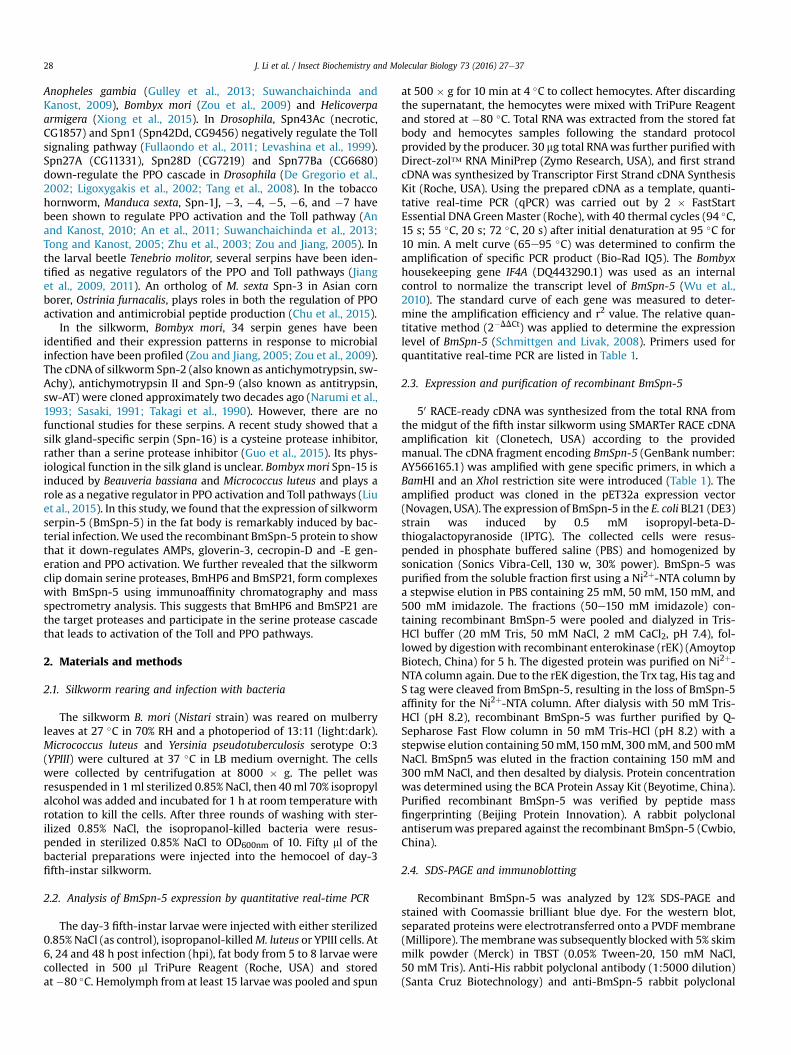
Recognition of pathogen-associated molecular patterns(PAMPs), such as LPS, PG from bacteria, and curdlan from fungi, bypattern recognition proteins (PRPs) initiates the sequential activa-tion of a serine protease cascade in the hemolymph and conse-quently results in the activation of melanization (Jiang et al., 2010).This process is under the regulation of serpins (Gubb et al., 2010;Kanost, 1999). We postulate that BmSpn-5 might regulate PPOactivation in silkworm. The larval silkworm was injected with re-combinant BmSpn-5, and shortly afterward was treated withdistinct types of PAMPs to trigger the immune response in vitro.Apart from the activation by M. luteus, pre-injection of BmSpn-5also inhibited the PO activity that resulted from the activation byPG from E. coli, LPS, and curdlan (Fig. 6A). A high molecular masscomplex formation is required for maintaining PO activity in thesilkworm (Clark and Strand, 2013). Therefore, we inspected theformation of the complex in the process of activation (Fig. 6B).When the silkworm larvae were injected with bovine serum al-bumin (BSA) and the plasma was incubated with LPS, PG, andcurdlan, we observed cleavage of PPO and the formation of the POcomplex. In contrast, in the presence of BmSpn-5, both the cleavageof PPO and the formation of the PO complex were partiallyinhibited (in the activation by curdlan and PG from M. luteus) orcompletely abolished (in the activation by LPS and PG from E. coli).These results demonstrate that BmSpn-5 down-regulates the PPOpathway when it is specifically activated by PG and LPS from Gram-negative bacteria and curdlan from fungi.
3.7. BmSpn-5 inhibits AMP generation induced by bacteria
The transcription and translation of AMPs in the fat body was

Fig. 1. Phylogenetic analysis (A) and RCL region sequences comparison (B) of BmSpn-5 and serpins of Manduca Sexta (Ms) and Drosophila melanogaster (Dm). The selectedserpin protein sequences were aligned using ClustalX. (A) The phylogram was built by neighbor-joining method. The numbers on the branches represent bootstrap values. (B)Alignment of the RCL sequences containing the scissile bond and activation sites. The P1-P10 scissile bond is indicated by an arrow. Dm_spn28Dc: NP_609172; Dm_spn77Ba:NP_730512.2; Dm_spn42Dd: ACZ94347; Dm_spn27A: AAF52420; Ms_spn1J: AAC47340; Ms_spn3: AAO21505; Ms_spn4: AAS68503; Ms_spn5: AAS68507; Ms_spn6: AAV91026;Ms_spn7: ADM86478.
Fig. 2. mRNA abundance of BmSpn-5 in fat body and hemocytes after bacterial injection. Fat body (A) and hemocyte (B) samples were collected at 12 hpi to analyze thetranscription levels of HaPGRP-A using qPCR. Error bars show mean ± SD (n ¼ 3). Significant differences between the bacterial infected samples and the NaCl injected samples areindicated with * at P < 0.05, ** at P < 0.01 and *** at P < 0.001, respectively.
Fig. 3. Expression and purification of recombinant BmSpn-5. (A) Coomassie brilliant blue stained SDS-PAGE. (B) Western blot probed with anti-His antibody. (C) Western blotprobed with anti-BmSpn-5 antibody. M, protein molecular mass markers; lane 1, total proteins from uninduced E. coli cells; lane 2, total proteins from induced E. coli cells; lane 3,soluble proteins from induced E. coli cells; lane 4, purified fusion protein before enterokinase digestion; lane 5, purified BmSpn-5.
J. Li et al. / Insect Biochemistry and Molecular Biology 73 (2016) 27e37 31
elicited by microbial infection and finally released into the hemo-lymph (Imler, 2014). We challenged larval silkworms with bacteriaM. luteus and YPIII. Remarkable antibacterial activity was detectedin the hemolymph 24 h after bacterial infection. However, pre-
injection of recombinant BmSpn-5 into the hemocoel decreasedthe antibacterial activity induced by both M. luteus and YPIII, in adosage-dependent manner when the silkworms were treated withM. luteus (Fig. 7A). Next we examined the composition in the small

Fig. 4. BmSpn-5 inhibits beads melanization in the hemolymph of larval silkworm. (A) Sepharose beads coated with BSA or recombinant BmSpn-5 were injected into thehemocoel of silkworm larvae. The beads were recovered from dissected larvae and observed under phase contrast microscope (PCM) and stereoscopic microscope (SM) 4 h postinjection. (B) The percentage of melanized beads in the hemolymph. Error bars show mean ± SEM. Significant differences between the treatment and the control are analyzed bythe student t-test.
Fig. 5. BmSpn-5 inhibits systemic melanization. These photographs show whole animals, plasma, and tissues after pre-injection of 30 mg of BSA (left panel) or BmSpn-5 (rightpanel) and following approximately 2 � 108 dead YPIII bacterial cells injection. The samples were collected and observed twelve hours after injection of bacterial cells.
J. Li et al. / Insect Biochemistry and Molecular Biology 73 (2016) 27e3732
molecule portion of challenged hemolymph in a tricine-SDS-PAGE.It appeared that proteins induced by bacterial infection werelocated at approximately 16 kDa (band 1 in Fig. 7B) and 3.5 kDa(band 2), whereas pre-injection of BmSpn-5 inhibited their in-duction in the hemolymph. LC/MS-MS analysis identified that band1 is gloverin and that band 2 corresponds to cecropin and moricin(Fig. 7C). Our semi-quantitative PCR showed that gloverin-3,cecropin-D and cecropin-E are strongly induced in the fat body byM. luteus and YPIII infection and are down-regulated by recombi-nant BmSpn-5 during M. luteus infection (data not presented).Quantitative real-time PCR confirmed that gloverin-3, cecropin-Dand cecropin-E are negatively regulated by BmSpn-5 after M. luteuschallenge; cecropin-E is also negatively regulated after YPIII chal-lenge (Fig. 7D). These results indicate that BmSpn-5 plays a role asan inhibitor in the immune response against M. luteus.
3.8. Silkworm SP21 and BmHP6 are candidates for the BmSpn-5target protease
Our results show that BmSpn-5 functions as an inhibitor to bothPPO and AMP-producing pathways. To further identify its targetprotease, we attempted to capture BmSpn-5-protease complexes.In the unchallenged plasma, we detected a ~50 kDa band corre-sponding to BmSpn-5 (lane 1 in Fig. 8A). In the challenged plasma,the intensity of the ~50 kDa band increased, and an extra lowerband was observed (lane 2 in Fig. 8A). This indicates that BmSpn-5is induced and processed by immune challenge in the plasma.When the challenged plasmawas activated by PG, we detected twoextra bands approximately 70 kDa, the expected position of serpin-protease complex (lane 3 in Fig. 8A). LC/MS-MS analysis revealedthat two clip domain serine proteases, BmHP6 (AK384444,
Fig. 6. BmSpn-5 down-regulates PPO activation. (A) The PO activity of the hemolymph is measured from larvae pre-injected with BmSpn-5 or BSA and then activated withpathogen-associated molecular patterns (PAMPs, including LPS, curdlan and PG). Error bars show mean ± SEM. Significant differences between the indicated groups are markedwith * at P < 0.05, ** at P < 0.01 and *** at P < 0.001, respectively. (B) Western blots showing PO complex formation in the hemolymph pre-injected with BmSpn-5 or BSA andactivated with LPS, curdlan, or PG. The arrowheads in C indicate the PO complex in the plasma; white and black arrows indicate PPO and PO, respectively.
J. Li et al. / Insect Biochemistry and Molecular Biology 73 (2016) 27e37 33
silkworm full-length cDNA database KAIKO_cDNA) and BmSP21(NP_001243984), are identified in complex 1 and 2, respectively(Fig. 8A and 8B). The immunoblotting and mass spectrometryanalysis showed that BmHP6 and BmSP21 could form complexeswith BmSpn-5, suggesting that these two serine proteases are thetarget proteases of BmSpn-5.
4. Discussion
Serpins are involved in the regulation of multiple physiologicaland immune signaling pathways, such as hemolymph clotting, PPOactivation, and the proteolytic activation of cytokines (Gubb et al.,2010; Kanost, 1999). In this study, we demonstrate that BmSpn-5in the silkworm negatively regulates the production of AMPs andthe PPO activation pathways. The activation of AMP synthesis andPPO pathways in insects depends on an extracellular clip domain
serine protease cascade, which is under strict regulation by serpins.BmSpn-5 forms covalent complexes with either BmSP21 or BmHP6,which implies that BmSpn-5 may inhibit the cleavage and activa-tion of the downstream proteases involved in these two pathways.
The inhibitory selectivity of serpins is largely determined by theresidues adjacent to the scissile bond. We compared the predictedcleavage sites of BmSpn-5 and serine proteases identified as thecomponents of the cascade leading to PPO and Toll pathways inBombyx, Manduca, Tenebrio and Drosophila (Fig. 8C). BombyxBAEEase and PPAE have similar activation sites (Arg-Ile and Lys-Ile,respectively) to BmSpn-5 (Arg-Ile). This suggests that BAEEase,together with PPAE, may be cleaved and activated by BmSP21 orBmHP6. The zymogen of Bombyx BAEEase was activated in thesilkworm hemolymph when PG or b-1,3-glucan was present. Itshomolog in Drosophila, sp€atzle-processing enzyme (SPE), isrequired for Toll activation and host defense (Jang et al., 2006). The

Fig. 7. The production of AMPs was down-regulated by BmSpn-5. (A) Antibacterial activity of the hemolymph determined by inhibition zone method. (B) Plasma proteinsseparated on a Tricine-SDS-PAGE and visualized by Coomassie brilliant blue stain. Note only the lower part of the gel was shown. The boxes indicate the bands of interest. (C) Themass spectrometry results of the protein bands excised from the gel in (B). (D) Antimicrobial peptide mRNA abundance in fat body determined by qPCR. Error bars show mean ± SD(n ¼ 5). Significant differences between the indicated groups are marked with * at P < 0.05, ** at P < 0.01 and *** at P < 0.001, respectively.
J. Li et al. / Insect Biochemistry and Molecular Biology 73 (2016) 27e3734
activation site of Bombyx BAEEase is similar to that of DrosophilaSPE (Jang et al., 2006), Tenebrio SPE (Kim et al., 2008) and ManducaHP8, which serves as the SPE (An et al., 2009, 2011; An and Kanost,2010) in the Toll pathway. Together with our evidence that BmSpn-5 inhibits AMP generation, we hypothesize that BAEEasemight playa critical role similar to the SPE and could be activated by BmSP21
or BmHP6 in the silkworm.In Manduca, HP8 is activated by HP6, which is the target pro-
tease of serpin-5. Manduca HP6 also cleaves PAP1, which activatesPPO (An and Kanost, 2010). Comparison of the sequences of Man-duca HP6 and BmHP6 shows that they share 66% identity of aminoacid residues and have similar activation sites, with His-Ile in HP6

Fig. 8. Capture and identification of the BmSpn-5-protease complexes. (A) Immunoaffinity purification of the BmSpn-5-protease complexes. Left, silver stained SDS-PAGE; Right,western blot probed with anti-BmSpn-5 antibody. Lane 1, unchallenged plasma; lane 2, M. luteus-challenged plasma; lane 3, M. luteus-challenged plasma activated with PG. (B)Mass and peptides matched to BmHP6 and BmSP21 in the complex 1 and complex 2, respectively. (C) Comparison of activation site sequences between BmSpn-5 and proteases fromBombyx, Drosophila, Manduca and Tenebrio. PAP, Prophenoloxidase activating protease; SPE, Sp€atzle-processing enzyme; SAE, SPE-activating enzyme. The P1-P10 scissile bond isindicated by an arrow.

J. Li et al. / Insect Biochemistry and Molecular Biology 73 (2016) 27e3736
and Tyr-Ile in BmHP6 (Fig. 8C). Tenebrio SPE is capable of cleavingPPO and pro-sp€atzle, which activates melanization and the Tollpathways. Tenebrio pro-SPE is activated by SAE (SPE-activatingenzyme), which is negatively regulated by serpin-55 (Jiang et al.,2011). BmSP21 shares 56% identity of amino acid sequence withManduca HP21 and 38% identity with Tenebrio SAE. All three ofthese proteases share similar activation sites (Leu-Ile), which sug-gests that they could be cleaved by the same protease (Fig. 8C).Manduca HP21 cleaves pro-PAP2 and pro-PAP3, resulting in theactivation of PPO (Gorman et al., 2007; Wang and Jiang, 2007).Similarly, the Bombyx PPO-activating enzyme (PPAE, KAIKO_cDNA:AK383065) has two clip domains at its amino terminus and ispredicted to be activated at Lys152-Ile153 (Satoh et al., 1999). PPAEwas identified as the ortholog of Manduca PAP3 (Tokura et al.,2014). We also identified BmPAP2 (AK386026) as the ortholog ofManduca PAP2 by using TBALSTN, the silkworm full-length cDNAdatabase. Therefore, BmSP21may also cleave and activate pro-PPAEand pro-BmPAP2, in addition to pro-BAEEase.
A recently identified ortholog of Manduca PAP1 in Bombyx(SilkDB: BGIBMGA010546) suggests that there is more than oneprotease cascade leading to the activation of PPO in the silkworm(Tokura et al., 2014), which resembles the PPO activation in Man-duca (Tong et al., 2005; An and Kanost, 2010). Another interestingobservation in our study is that inactive M. luteus induced up-regulation of gloverin-3, cecropin D and cecropin E and this up-regulation was inhibited by BmSpn-5 (Fig. 7D). However, the acti-vation of PPO by PG from M. luteus was not inhibited efficiently byBmSpn-5 (Fig. 6). This suggests that the protease cascade leading toactivation of PPO by M. luteus may bypass BmSP21 and BmHP6.Additionally, the fact that BmSP21 was present in the melanoticnodules induced by E. coli, but not in those induced by M. luteus orS. cerevisiae (Tokura et al., 2014), implies the possible existence ofmultiple PPO activation pathways in the silkworm.
Taken together, we have shown that BmSpn-5 down-regulatesthe Toll and PPO pathways by targeting BmHP6 and BmSP21 in thesilkworm. Our study provides clues for further dissection of thesilkworm Toll and PPO activation signaling.
Acknowledgements
This work was supported by NSF of China (No. 31272497,31472008, and 31401804), Strategic Priority Research Program ofCAS (No. XDB11030600), Open research Fund Program of State KeyLaboratoryof Integrated Pest Management (Chinese IPM1407). Weare grateful to Dr. Michael Strand for providing us the Bombyx moriPPO antisera. We thank Zhanfeng Zhang for his assistance inspectrometry analysis.
References
An, C., Ishibashi, J., Ragan, E.J., Jiang, H., Kanost, M.R., 2009. Functions of Manducasexta hemolymph proteinases HP6 and HP8 in two innate immune pathways.J. Biol. Chem. 284, 19716e19726.
An, C., Kanost, M.R., 2010. Manduca sexta serpin-5 regulates prophenoloxidaseactivation and the Toll signaling pathway by inhibiting hemolymph proteinaseHP6. Insect Biochem. Mol. Biol. 40, 683e689.
An, C., Ragan, E.J., Kanost, M.R., 2011. Serpin-1 splicing isoform J inhibits theproSpatzle-activating proteinase HP8 to regulate expression of antimicrobialhemolymph proteins in Manduca sexta. Dev. Comp. Immunol. 35, 135e141.
An, C., Zhang, M., Chu, Y., Zhao, Z., 2013. Serine protease MP2 activates proph-enoloxidase in the melanization immune response of Drosophila melanogaster.PLoS One 8, e79533.
Chu, Y., Zhou, F., Liu, Y., Hong, F., Wang, G., An, C., 2015. Ostrinia furnacalis serpin-3regulates melanization cascade by inhibiting a prophenoloxidase-activatingprotease. Insect Biochem. Mol. Biol. 61, 53e61.
Clark, K.D., Strand, M.R., 2013. Hemolymph melanization in the silkmoth Bombyxmori involves formation of a high molecular mass complex that metabolizestyrosine. J. Biol. Chem. 288, 14476e14487.
De Gregorio, E., Han, S.J., Lee, W.J., Baek, M.J., Osaki, T., Kawabata, S., Lee, B.L.,
Iwanaga, S., Lemaitre, B., Brey, P.T., 2002. An immune-responsive Serpin regu-lates the melanization cascade in Drosophila. Dev. Cell 3, 581e592.
Fullaondo, A., Garcia-Sanchez, S., Sanz-Parra, A., Recio, E., Lee, S.Y., Gubb, D., 2011.Spn1 regulates the GNBP3-dependent Toll signaling pathway in Drosophilamelanogaster. Mol. Cell. Biol. 31, 2960e2972.
Gettins, P.G., 2002. Serpin structure, mechanism, and function. Chem. Rev. 102,4751e4804.
Gorman, M.J., Wang, Y., Jiang, H., Kanost, M.R., 2007. Manduca sexta hemolymphproteinase 21 activates prophenoloxidase-activating proteinase 3 in an insectinnate immune response proteinase cascade. J. Biol. Chem. 282, 11742e11749.
Gubb, D., Sanz-Parra, A., Barcena, L., Troxler, L., Fullaondo, A., 2010. Protease in-hibitors and proteolytic signalling cascades in insects. Biochimie 92, 1749e1759.
Gulley, M.M., Zhang, X., Michel, K., 2013. The roles of serpins in mosquito immu-nology and physiology. J. Insect Physiol. 59, 138e147.
Guo, P.C., Dong, Z., Xiao, L., Li, T., Zhang, Y., He, H., Xia, Q., Zhao, P., 2015. Silk gland-specific proteinase inhibitor serpin16 from the Bombyx mori shows cysteineproteinase inhibitory activity. Biochem. Biophys. Res. Commun. 457, 31e36.
Hoffmann, D., Hultmark, D., Boman, H.G., 1981. Insect immunity - galleria-mellonella and other lepidoptera have cecropia-P9-like factors active againstgram-negative bacteria. Insect Biochem. 11, 537e548.
Imler, J.L., 2014. Overview of drosophila immunity: a historical perspective. Dev.Comp. Immunol. 42, 3e15.
Jang, I.H., Chosa, N., Kim, S.H., Nam, H.J., Lemaitre, B., Ochiai, M., Kambris, Z.,Brun, S., Hashimoto, C., Ashida, M., Brey, P.T., Lee, W.J., 2006. A spatzle-pro-cessing enzyme required for toll signaling activation in Drosophila innate im-munity. Dev. Cell 10, 45e55.
Jedamzik, B., Eckmann, C.R., 2009. Analysis of in vivo protein complexes by coim-munoprecipitation from Caenorhabditis elegans. Cold Spring Harb Protoc 4 pdbprot5299.
Jiang, H., Vilcinskas, A., Kanost, M.R., 2010. Immunity in Lepidopteran insects. Adv.Exp. Med. Biol. 708, 181e204.
Jiang, H.B., Wang, Y., Kanost, M.R., 1998. Pro-phenol oxidase activating proteinasefrom an insect, Manduca sexta: a bacteria-inducible protein similar toDrosophila easter. P Natl. Acad. Sci. U. S. A. 95, 12220e12225.
Jiang, R., Kim, E.H., Gong, J.H., Kwon, H.M., Kim, C.H., Ryu, K.H., Park, J.W.,Kurokawa, K., Zhang, J., Gubb, D., Lee, B.L., 2009. Three pairs of protease-serpincomplexes cooperatively regulate the insect innate immune responses. J. Biol.Chem. 284, 35652e35658.
Jiang, R., Zhang, B., Kurokawa, K., So, Y.I., Kim, E.H., Hwang, H.O., Lee, J.H.,Shiratsuchi, A., Zhang, J.H., Nakanishi, Y., Lee, H.S., Lee, B.L., 2011. 93-kDa twin-domain serine protease inhibitor (Serpin) has a regulatory function on thebeetle toll proteolytic signaling cascade. J. Biol. Chem. 286, 35087e35095.
Kambris, Z., Brun, S., Jang, I.H., Nam, H.J., Romeo, Y., Takahashi, K., Lee, W.J., Ueda, R.,Lemaitre, B., 2006. Drosophila immunity: a large-scale in vivo RNAi screenidentifies five serine proteases required for toll activation. Curr. Biol. 16,808e813.
Kanost, M.R., 1999. Serine proteinase inhibitors in arthropod immunity. Dev. Comp.Immunol. 23, 291e301.
Kanost, M.R., Jiang, H., 2015. Clip-domain serine proteases as immune factors ininsect hemolymph. Curr. Opin. Insect Sci. 11, 47e55.
Kim, C.H., Kim, S.J., Kan, H., Kwon, H.M., Roh, K.B., Jiang, R., Yang, Y., Park, J.W.,Lee, H.H., Ha, N.C., Kang, H.J., Nonaka, M., Soderhall, K., Lee, B.L., 2008. A three-step proteolytic cascade mediates the activation of the peptidoglycan-inducedtoll pathway in an insect. J. Biol. Chem. 283, 7599e7607.
Kounatidis, I., Ligoxygakis, P., 2012. Drosophila as a model system to unravel thelayers of innate immunity to infection. Open Biol. 2, 120075.
Lavine, M.D., Strand, M.R., 2002. Insect hemocytes and their role in immunity. InsectBiochem. Molec 32, 1295e1309.
Lemaitre, B., Hoffmann, J., 2007. The host defense of Drosophila melanogaster.Annu. Rev. Immunol. 25, 697e743.
Levashina, E.A., Langley, E., Green, C., Gubb, D., Ashburner, M., Hoffmann, J.A.,Reichhart, J.M., 1999. Constitutive activation of toll-mediated antifungal defensein serpin-deficient Drosophila. Science 285, 1917e1919.
Ligoxygakis, P., Pelte, N., Ji, C.Y., Leclerc, V., Duvic, B., Belvin, M., Jiang, H.B.,Hoffmann, J.A., Reichhart, J.M., 2002. A serpin mutant links Toll activation tomelanization in the host defence of Drosophila. EMBO J. 21, 6330e6337.
Liu, D.R., Wang, L., Yang, L., Qian, C., Wei, G.Q., Dai, L.S., Li, J., Zhu, B.J., Liu, C.L., 2015.Serpin-15 from Bombyx mori inhibits prophenoloxidase activation andexpression of antimicrobial peptides. Dev. Comp. Immunol. 51, 22e28.
Lu, A.R., Zhang, Q.L., Zhang, J., Yang, B., Wu, K., Xie, W., Luan, Y.X., Ling, E.J. Insectprophenoloxidase: the view beyond immunity. Front. Physiol. 5, 252.
Michel, K., Suwanchaichinda, C., Morlais, I., Lambrechts, L., Cohuet, A., Awono-Ambene, P.H., Simard, F., Fontenille, D., Kanost, M.R., Kafatos, F.C., 2006.Increased melanizing activity in Anopheles gambiae does not affect develop-ment of Plasmodium falciparum. Proc. Natl. Acad. Sci. 103, 16858e16863.
Mortz, E., Krogh, T.N., Vorum, H., Gorg, A., 2001. Improved silver staining protocolsfor high sensitivity protein identification using matrix-assisted laserdesorption/ionization-time of flight analysis. Proteomics 1, 1359e1363.
Nappi, A.J., Frey, F., Carton, Y., 2005. Drosophila serpin 27A is a likely target forimmune suppression of the blood cell-mediated melanotic encapsulationresponse. J. Insect Physiol. 51, 197e205.
Narumi, H., Hishida, T., Sasaki, T., Feng, D.F., Doolittle, R.F., 1993. Molecular-cloningof silkworm (Bombyx-Mori) Antichymotrypsin - a new member of the serpinsuperfamily of proteins from insects. Eur. J. Biochem. 214, 181e187.
Reichhart, J.M., 2005. Tip of another iceberg: Drosophila serpins. Trends Cell Biol.

J. Li et al. / Insect Biochemistry and Molecular Biology 73 (2016) 27e37 37
15, 659e665.Sasaki, T., 1991. Patchwork-structure serpins from silkworm (Bombyx-Mori) larval
hemolymph. Eur. J. Biochem. 202, 255e261.Satoh, D., Horii, A., Ochiai, M., Ashida, M., 1999. Prophenoloxidase-activating
enzyme of the silkworm, Bombyx mori e purification, characterization, andcDNA cloning. J. Biol. Chem. 274, 7441e7453.
Schagger, H., 2006. Tricine-SDS-PAGE. Nat. Protoc. 1, 16e22.Schmittgen, T.D., Livak, K.J., 2008. Analyzing real-time PCR data by the comparative
C-T method. Nat. Protoc. 3, 1101e1108.Suwanchaichinda, C., Kanost, M.R., 2009. The serpin gene family in Anopheles
gambiae. Gene 442, 47e54.Suwanchaichinda, C., Ochieng, R., Zhuang, S.F., Kanost, M.R., 2013. Manduca sexta
serpin-7, a putative regulator of hemolymph prophenoloxidase activation. In-sect Biochem. Molec 43, 555e561.
Takagi, H., Narumi, H., Nakamura, K., Sasaki, T., 1990. Amino-acid-sequence ofsilkworm (Bombyx-Mori) hemolymph antitrypsin deduced from its Cdnanucleotide-sequence e confirmation of its homology with serpins. J. Biochem.Tokyo 108, 372e378.
Tang, H., Kambris, Z., Lemaitre, B., Hashimoto, C., 2006. Two proteases defining amelanization cascade in the immune system of Drosophila. J. Biol. Chem. 281,28097e28104.
Tang, H., Kambris, Z., Lemaitre, B., Hashimoto, C., 2008. A serpin that regulatesimmune melanization in the respiratory system of drosophila. Dev. Cell 15,617e626.
Tokura, A., Fu, G.S., Sakamoto, M., Endo, H., Tanaka, S., Kikuta, S., Tabunoki, H.,Sato, R., 2014. Factors functioning in nodule melanization of insects and theirmechanisms of accumulation in nodules. J. Insect Physiol. 60, 40e49.
Tong, Y., Jiang, H., Kanost, M.R., 2005. Identification of plasma proteases inhibited bymanduca sexta serpin-4 and serpin-5 and their association with components ofthe prophenol oxidase activation pathway. J. Biol. Chem. 280, 14932e14942.
Tong, Y., Kanost, M.R., 2005. Manduca sexta serpin-4 and serpin-5 inhibit theprophenol oxidase activation pathway: cDNA cloning, protein expression, andcharacterization. J. Biol. Chem. 280, 14923e14931.
Veillard, F., Troxler, L., Reichhart, J.-M., 2016. Drosophila melanogaster clip-domainserine proteases: structure, function and regulation. Biochimie 122, 255e269.
Wang, Y., Jiang, H., 2007. Reconstitution of a branch of the Manduca sexta proph-enoloxidase activation cascade in vitro: snake-like hemolymph proteinase 21(HP21) cleaved by HP14 activates prophenol oxidase- activating proteinase-2precursor. Insect Biochem. Molec 37, 1015e1025.
Wu, S., Zhang, X., Chen, X., Cao, P., Beerntsen, B.T., Ling, E., 2010. BmToll9, anarthropod conservative toll, is likely involved in the local gut immune responsein the silkworm, Bombyx mori. Dev. Comp. Immunol. 34, 93e96.
Xiong, G.H., Xing, L.S., Lin, Z., Saha, T.T., Wang, C., Jiang, H., Zou, Z., 2015. Highthroughput profiling of the cotton bollworm Helicoverpa armigera immuno-transcriptome during the fungal and bacterial infections. BMC Genomics 16,321.
Zhu, Y., Wang, Y., Gorman, M.J., Jiang, H., Kanost, M.R., 2003. Manduca sexta serpin-3 regulates prophenoloxidase activation in response to infection by inhibitingprophenoloxidase-activating proteinases. J. Biol. Chem. 278, 46556e46564.
Zou, Z., Jiang, H., 2005. Manduca sexta serpin-6 regulates immune serine pro-teinases PAP-3 and HP8. cDNA cloning, protein expression, inhibition kinetics,and function elucidation. J. Biol. Chem. 280, 14341e14348.
Zou, Z., Picheng, Z., Weng, H., Mita, K., Jiang, H., 2009. A comparative analysis ofserpin genes in the silkworm genome. Genomics 93, 367e375.