journal of insect physiology - institut de biologia...
TRANSCRIPT

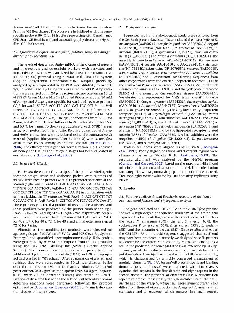
Journal of Insect Physiology 54 (2008) 1138–1147
Contents lists available at ScienceDirect
Journal of Insect Physiology
journa l homepage: www.e lsev ier .com/ locate / j insphys
Expression analysis of putative vitellogenin and lipophorin receptors in honey bee(Apis mellifera L.) queens and workers
Karina Rosa Guidugli-Lazzarini a,*, Adriana Mendes do Nascimento a, Erica Donato Tanaka a,Maria Dolors Piulachs b, Klaus Hartfelder c, Marcia Gentile Bitondi a, Zila Luz Paulino Simoes a
a Departamento de Biologia, Faculdade de Filosofia, Ciencias e Letras de Ribeirao Preto, Universidade de Sao Paulo, SP, Brazilb Department of Physiology and Molecular Biodiversity, Institute of Molecular Biology of Barcelona (CSIC), Barcelona, Spainc Departamento de Biologia Celular e Molecular e Bioagentes Patogenicos, Faculdade de Medicina de Ribeirao Preto, Universidade de Sao Paulo, Ribeirao Preto, SP, Brazil
A R T I C L E I N F O
Article history:
Received 17 January 2008
Received in revised form 18 April 2008
Accepted 21 April 2008
Keywords:
LDLR
Vitellogenin receptor
Lipophorin receptor
Honeybee
Ovary
A B S T R A C T
Two members of the low density lipoprotein receptor (LDLR) family were identified as putative orthologs
for a vitellogenin receptor (Amvgr) and a lipophorin receptor (Amlpr) in the Apis mellifera genome. Both
receptor sequences have the structural motifs characteristic of LDLR family members and show a high
degree of similarity with sequences of other insects. RT-PCR analysis of Amvgr and Amlpr expression
detected the presence of both transcripts in different tissues of adult female (ovary, fat body, midgut,
head and specifically hypopharyngeal gland), as well as in embryos. In the head RNA samples we found
two variant forms of AmLpR: a full length one and a shorter one lacking 29 amino acids in the O-linked
sugar domain. In ovaries the expression levels of the two honey bee LDLR members showed opposing
trends: whereas Amvgr expression was upregulated as the ovaries became activated, Amlpr transcript
levels gradually declined. In situ hybridization analysis performed on ovaries detected Amvgr mRNA
exclusively in germ line cells and corroborated the qPCR results showing an increase in Amvgr gene
expression concomitant with follicle growth.
� 2008 Elsevier Ltd. All rights reserved.
1. Introduction
The insect yolk precursor protein, vitellogenin, is a lipoglyco-protein synthesized by the fat body and secreted into hemolymphfrom where it is sequestered by the growing oocytes via receptor-mediated endocytosis (Raikhel and Dhadialla, 1992). In the honeybee, native vitellogenin is a monomeric 180 kDa polypeptide(Wheeler and Kawooya, 1990) which, in egg-laying queens,accounts for over 50% of total hemolymph proteins (Hartfelderand Engels, 1998). Interestingly, in this social insect vitellogeninapparently has been co-opted into additional functions since it isalso present in large amounts in the hemolymph of young workerbees, even though these are functionally sterile and do not lay eggs inthe presence of the queen, and it has even been detected in drones(Trenczek et al., 1989; Piulachs et al., 2003; Guidugli et al., 2005a).
Additional functions for vitellogenin have also been suggestedin other insects, such as the transport of carbohydrates, lipids,
* Corresponding author at: Av. Bandeirantes, 3900, 14040-901, Ribeirao Preto,
Sao Paulo, Brazil. Tel.: +55 16 3602 4332; fax: +55 16 3602 3704.
E-mail addresses: [email protected], [email protected]
(K.R. Guidugli-Lazzarini).
0022-1910/$ – see front matter � 2008 Elsevier Ltd. All rights reserved.
doi:10.1016/j.jinsphys.2008.04.021
phosphates, vitamins, metals and hormones (see Chen et al., 1997;Sappington and Raikhel, 1998), but it is in the honey bee wherevitellogenin has been co-opted into new roles integratingindividual physiology with the social environment (Amdamet al., 2006). It has become a major regulator of the lifespan ofindividual worker bees, through its anti-immunosenescenceproperties as zinc transporter and antioxidant (Amdam et al.,2005; Seehuus et al., 2006), and, in a double repressor circuitrywith juvenile hormone, its downregulation drives a worker beeinto the forager state (Amdam and Omholt, 2003; Guidugli et al.,2005b; Nelson et al., 2007). The emerging central role ofvitellogenin in the life history of the honey bee, and possibly alsoin other highly eusocial bees (Hartfelder et al., 2006), now raisesthe question on how this protein may perform such proposedsignaling functions, and a first candidate to investigate in thispathway should be the vitellogenin receptor.
Vitellogenin-specific receptors (VgRs) have been cloned andsequenced in several insects, such as Aedes aegypti (Sappingtonet al., 1996), Solenopsis invicta (Chen et al., 2004), Periplaneta
americana (Tufail and Takeda, 2005), Blattella germanica (Ciudadet al., 2006) and Leucophaea maderae (Tufail and Takeda, 2007).This group also includes the related yolk protein receptor, yolkless,of Drosophila melanogaster (Schonbaum et al., 1995). All these VgRs

K.R. Guidugli-Lazzarini et al. / Journal of Insect Physiology 54 (2008) 1138–1147 1139
are members of the superfamily of low density lipoproteinreceptors (LDLR) and their expression appears to be restricted tothe ovaries (Sappington et al., 1996; Schonbaum et al., 1995; Chenet al., 2004; Tufail and Takeda, 2005, 2007; Ciudad et al., 2006).
In accordance with the pleiotropic role of vitellogenin in Apis
mellifera, immunodetection experiments now also evidenced anextraovarian occurrence of a putative VgR. An immunoreactive205 kDa protein was detected not only in the ovaries of queens butalso in the hypopharyngeal glands of workers (Amdam et al., 2003).These glands produce the major royal jelly proteins (Albert et al.,1999; Drapeau et al., 2007). Nurse bees feed royal jelly not only tothe developing larvae, but also to the queen, allowing her tomaintain the extraordinary high rates of egg production, thusestablishing a social cycle for vitellogenin as a reproductive protein(Amdam et al., 2003).
Concomitant to the discovery of additional functions for insectvitellogenin, a dual role for lipophorin in reproduction has alsobeen demonstrated in the moth and mosquito (Kawooya et al.,1988; Sun et al., 2000). In these insects, lipophorin is responsiblefor the shuttle of lipids from the fat body to the growing oocyte,and subsequently it is incorporated into the egg yolk as a storageprotein. Like for vitellogenin, the incorporation of lipophorin alsooccurs via receptor-mediated endocytosis and the lipophorinreceptor (LpR) also belongs to the LDLR superfamily.
The members of this superfamily share common structuralcharacteristics and show a high degree of conservation in theirfunctional domains. The structural elements of LDLRs are: (1)ligand-binding domain, (2) epidermal growth factor (EGF)precursor homology domain, (3) YWXD repeats, (4) transmem-brane domain, and (5) cytoplasmic domain (for revision seeSappington and Raikhel, 1998; Rodenburg et al., 2006). Within theLDLR superfamily, VgRs and LpRs are intimately associated withoogenesis (Sappington et al., 1996; Schonbaum et al., 2000; Choand Raikhel, 2001; Seo et al., 2003; Chen et al., 2004; Tufail andTakeda, 2005, 2007; Ciudad et al., 2006, 2007).
Considering the importance of vitellogenin and lipophorin ininsect reproduction, we made use of the now available genomicinformation for the honey bee (The Honey Bee Genome SequencingConsortium, 2006) to identify putative orthologs for a vitellogeninreceptor and a lipophorin receptor, and investigated the expressionof these genes in adult queens and workers and in different tissues,with emphasis on the reproductive process.
2. Material and methods
2.1. Bees and RNA extraction
A. mellifera workers and queens were collected from colonies ofAfricanized stocks maintained in the apiary of the Department ofGenetics at the Faculty of Medicine in Ribeirao Preto, University ofSao Paulo, Brazil. Virgin queens obtained by standard queenrearing methods were either dissected immediately after emer-gence from brood cells or were introduced into queenless colony.They were collected as soon as they started to lay eggs. The ovariesfrom virgin and egg-laying queens were used for RNA extraction.
Newly emerged workers from a queenless colony were paintmarked and periodically collected to check the degree of ovaryactivation. The ovary status was assessed following establishedcriteria (Makert et al., 2006). Inactive ovaries (stage I: ovarioles areslender and oogenesis has not proceeded beyond germ cell clusterformation and follicle separation from the germarium) wereobserved in workers 1–3 day-old. Active ovaries (stage II: initialfollicle growth, where ovarioles contain mainly previtellogenic orearly vitellogenic follicles; stage III: at least some of the ovariolescontain follicles in advanced stages of vitellogenesis) were
observed in 15–20 and 20–30 day-old workers. After stageclassification, the ovaries (pools of 10–20) were used for RNAextraction. In another experiment, RNA of inactive and activeovaries was obtained from queenless workers collected at the sameage (18 days). RNA was also extracted from inactive ovaries of agroup of 18 day-old queenright workers.
RNA was extracted from the head, fat body, hypopharyngealglands, midgut and ovaries from adult workers and also fromembryos (0–6 h) to test for tissue specificity of Amvgr and Amlpr
gene expression. TRIzol reagent (Invitrogen) was used to isolatetotal RNA following manufacturer instructions. These RNA sampleswere used as templates for first-strand cDNA synthesis.
2.2. Cloning and partial sequencing of putative honey bee Amvgr
and Amlpr genes
When the assembled honey bee genome (Amel version 4.0,http://hgsc.bcm.tmc.edu/projects/honeybee/) was searched forhomologs to the yolkless gene of D. melanogaster, the predictedhoney bee gene (GB16571) was retrieved in a mutual best-hitanalysis as the best candidate for a honey bee vitellogenin receptor.This predicted gene is subsequently referred to as Amvgr,indicating its putative function as vitellogenin receptor. Similarly,a putative honey bee lipophorin receptor (Amlpr) encoding genewas represented by CG31094-PA (GenBank accession no.XP_395858.3). BLASTP searches for closely related proteinspredicted in the honey bee genome revealed an almost completeoverlap in the five sequential best matches for the predictedAmVgR and AmLpR proteins, due to shared LDL receptor domains(Supplementary Material, Table 1 and Figure 1).
To confirm that the in silico predicted genes Amvgr and Amlpr
are expressed, we designed primers to amplify correspondingfragments by RT-PCR from ovary RNA. The PCR products separatedby agarose gel electrophoresis were ligated into pGEM1-T Easy
(Promega) vector and cloned into chemically competent E. coli
DH5a cells. Dideoxy sequencing was performed on an automaticsequencer (ABI Prism 310, Applied Biosystems) using BigDyeTerminator v3.0 Cycle Sequencing Reaction (Applied Biosystems)and M13 forward and reverse primers.
2.3. Semi-quantitative expression analysis of putative honey bee
Amvgr and Amlpr
The expression of Amvgr and Amlpr in various tissues and inembryos was evaluated by semi-quantitative RT-PCR. First-strand cDNA was synthesized by reverse transcription (Super-script II, Invitrogen) using 1–2 mg of RNA. Aliquots of first-strandcDNAs were employed in PCR reactions using PCR Master Mix(Eppendorf). The primers were VgR1F: 50-ACT CAT GTT TGT GCCAAC CTG-30 and VgR2R: 50-CCT TCG ATC TGT ACC ATC CAA-30 forAmvgr, and LpRF 50-CAC TGG TCA ATC AGT TGA AG-30 and LpRR50-CTA TAA CAT AAT ACT GCT AC-30 for Amlpr. The Amvgr
fragment was amplified by the following PCR protocol: 2 min at94 8C, 30 cycles of 30 s at 94 8C, 30 s at 60 8C, 60 s at 72 8C and afinal extension step at 72 8C for 10 min. To amplify the fragmentcorresponding to the Amlpr, the thermal cycling program was:2 min at 94 8C followed by 30 cycles of 30 s at 94 8C, 30 s at 55 8C,60 s at 72 8C and a final extension step at 72 8C for 10 min. Asloading control an A. mellifera actin gene (GenBank accession no.AB023025) was amplified following the protocol described byBitondi et al. (2006).
The amplification products were analyzed by electrophoresis in1% agarose gels containing ethidium bromide. In the case of Amlpr,the PCR products were Southern blotted. A cDNA probe wasgenerated by PCR with the same primer pair and labeled with
K.R. Guidugli-Lazzarini et al. / Journal of Insect Physiology 54 (2008) 1138–11471140
fluorescein-11-dUTP using the module Gene Images RandomPriming (GE Healthcare). The blots were hybridized with this gene-specific probe at 60 8C for 16 h before processing with Gene ImagesCPD Star (GE Healthcare) and autoradiography detection (Hyper-film, GE Healthcare).
2.4. Quantitative expression analysis of putative honey bee Amvgr
and Amlpr by real-time PCR
The levels of Amvgr and Amlpr mRNA in the ovaries of queensand in queenless and queenright workers with activated andnon-activated ovaries was analyzed by a real-time quantitativeRT-PCR (qPCR) protocol using a 7500 Real Time PCR System(Applied Biosystems). First-strand cDNA samples, previouslyanalyzed by semi-quantitative RT-PCR, were diluted (1:5 or 1:10v/v) in water, and 1 ml aliquots were used for qPCR. Amplifica-tions were carried out in 20 ml reaction mixture containing 10 mlof SYBR1 Green Master Mix 2� (Applied Biosystems), and 10 mMof Amvgr and Amlpr gene-specific forward and reverse primers(VgR forward: 50-TGA ACC TTA CGA CAT TGC CCT-30 and VgRreverse: 50-TGT GAT TTT CGG TCC AAG CCC-30; LpR forward: 50-GGT CGT TCA TGT ATA TCA TCC-30 and LpR reverse 50-CGG ACAAGC ACA ACT AAG AAG-30). The qPCR conditions were 50 8C for2 min, and 95 8C for 10 min followed by 40 cycles of 95 8C for 15 s,and 60 8C for 1 min. To check reproducibility, each SYBR1 greenassay was performed in triplicate. Relative quantities of Amvgr
and Amlpr transcripts were calculated using the comparative Ctmethod (Applied Biosystems, User bulletin 2) with A. mellifera
actin mRNA levels serving as internal control (Bitondi et al.,2006). The efficacy of this gene for normalization in qPCR studieson honey bee tissues and life cycle stages has been validated inour laboratory (Lourenco et al., 2008).
2.5. In situ hybridization
For in situ detection of transcripts of the putative vitellogeninreceptor Amvgr, sense and antisense probes were synthesizedusing Amvgr specific primers with a T7 promoter sequence at the50-ends (VgR-Fow1: 50-TAA TAC GAC TCA CTA TAG GGC GAA CTC ATGTTT GTG CCA ACC TG-30; VgR-Rev1: 50-TAA TAC GAC TCA CTA TAG
GGC GAC CTT CGA TCT GTA CCA TCC AA-30) in combination withprimers lacking the T7 sequence (VgR-Fow2: 50-ACT CAT GTT TGTGCC AAC CTG-30; VgR-Rev2: 50-CCT TCG ATC TGT ACC ATC CAA-30).These primers generated a product of 653 bp. The antisense andsense products were produced by the primer combination VgR-Fow2+ VgR-Rev1 and VgR-Fow1+ VgR-Rev2, respectively. Ampli-fication conditions were: 94 8C for 2 min at 94 8C, 45 cycles of 94 8Cfor 40 s, 57 8C for 40 s, 72 8C for 40 s and a final extension step at72 8C for 7 min.
Aliquots of the amplification products were checked onagarose gels, purified (Wizard1 SV Gel and PCR Clean-Up System,Promega) and quantified spectrophotometrically. RNA probeswere generated by in vitro transcription from the T7 promoterusing the DIG RNA Labelling Kit (SP6/T7) (Roche AppliedScience). The transcription products were precipitated byaddition of 1 ml ammonium acetate (10 M) and 20 ml isopropa-nol and washed in 70% ethanol. After evaporation of any ethanolresidues they were resuspended in 50 ml hybridization buffer(50% formamide, 4� SSC, 1� Denhardt’s solution, 250 mg/mlyeast extract, 250 mg/ml salmon sperm DNA, 50 mg/ml heparin,0.1% Tween-20, 5% dextrane sulfate) and stored at �20 8C.Fixation of dissected tissue and the subsequent hybridization anddetection reactions were performed following the protocoloptimized by Osborne and Dearden (2005) for in situ hybridiza-tion studies on honey bees.
2.6. Phylogenetic analysis
Sequences used in the phylogenetic study were retrieved fromthe GenBank protein database. These included the insect VgRs of D.
melanogaster (AAB60217), Anopheles gambiae (EAA06264), A. aegypti
(AAK15810), S. invicta (AAP92450), P. americana (BAC02725), L.
maderae (BAE93218.1), B. germanica (CAJ19121), Tribolium casta-
neum (XP_968903.1) and Nasonia vitripennis (XP_001602954). Theinsect LpRs were from Galleria mellonella (ABF20542), Bombyx mori
(BAE71406.1), A. aegypti (AAQ16410 and AAK72954), D. melanoga-
ster (NP_733119.1), A. gambiae (XP_307995), L. maderae (BAE00010),B. germanica (CAL47125), Locusta migratoria (CAA03855), A. mellifera
(XP_395858.3) and T. castaneum (XP_967944). Sequences fromother ecdysozoans were the ovarian lipoprotein receptor (OLR) ofthe crustacean Penaeus semisulcatus (AAL79675.1), VgR of the tickDermacentor variabilis (AAZ31260.3), and the yolk protein receptorRME-2 of the nematode Caenorhabditis elegans (AAD56241.1).Vertebrates are represented by VgRs from Anguilla japonica
(BAB64337.1), Conger myriaster (BAB64338), Oncorhynchus mykiss
(CAD10640.1), Danio rerio (AAH47187), Xenopus laevis (AAH70552)and Gallus gallus (NP_990560); by the very low density lipoproteinreceptor (VLDLR) of Oryctolagus cuniculus (BAA01874), Rattus
norvegicus (NP_037287.1), Mus musculus (AAH13622.1) and Homo
sapiens (NP_003374.3); by the LDLR of M. musculus (CAA45759.1), R.
norvegicus (NP_786938.1), Tetraodon nigroviridis (CAF92585.1) andH. sapiens (NP_000518.1), and by the lipoprotein receptor-relatedprotein (LR8B) of G. gallus (CAA65729.1). A final addition were themegalins (=LRP2) of G. gallus (XP_422014.1), D. pseudoobscura
(EAL32723) and A. mellifera (XP_393369).Protein sequences were aligned using ClustalX (Thompson
et al., 1997). Poorly aligned positions and divergent regions wereeliminated by using Gblocks 0.91b (Castresana, 2000). Theresulting alignment was analyzed by the PHYML program(Guindon and Gascuel, 2003), based on the maximum-likelihoodprinciple in the amino acid substitution model. Four substitutionrate categories with a gamma shape parameter of 1.444 were used.Tree topologies were evaluated by 100 bootstrap replicates usingPHYML.
3. Results
3.1. Putative vitellogenin and lipophorin receptors of the honey
bee—structural features and phylogenetic analysis
The gene predicted as GB16571-PA in the A. mellifera genomeshowed a high degree of sequence similarity at the amino acidsequence level with vitellogenin receptors of other insects, such asthe wasp N. vitripennis (64%), the ant S. invicta (63%), thecockroaches P. americana (57%), B. germanica (55%), L. maderae
(55%) and the mosquito A. aegypti (55%). Since in silico analysis ofthe GB16571-PA amino acid sequence suggested that its 50-endmay have been predicted incorrectly we designed specific primersto determine the correct start codon by 50-end sequencing. As aresult, the predicted sequence (4860 bp) was extended by 312 bp.
Analysis of the deduced amino acid sequence defined thisputative VgR of A. mellifera as a member of the LDL receptor family,which is characterized by a highly conserved arrangement ofmodular elements (Fig. 1A). For AmVgR protein two ligand-bindingdomains (LBD1 and LBD2) were predicted, with four Class Acysteine-rich repeats in the first domain and eight repeats in thesecond domain. The presence of only four Class A cysteine-richrepeats resembles most closely the VgR architecture of the ant S.
invicta and of the wasp N. vitripennis. These hymenopteran VgRsdiffer from those of other insects, like A. aegypti, P. americana, B.
germanica and L. maderae, which possess five such repeats

Fig. 1. Domain structure of the putative honey bee vitellogenin receptor (AmVgR) and its position within the molecular phylogeny of LDL receptors. (A) Comparison of AmVgR
modular domains with those of other insects, S. invicta, B. germanica, A. aegypti and D. melanogaster (which is encoded by yolkless). The percentage of similarity with respect to
the A. mellifera sequence is indicated below each domain. A, Class A cysteine-rich repeats; B, Class B cysteine-rich repeats; C, cytoplasmic domain; EGF, epidermal growth
factor precursor homology domain; LBD, ligand-binding domain; O, O-linked sugar domain; SP, signal peptide; T, transmembrane domain. (B) Molecular phylogeny for VgRs
and lipophorin receptors (LpRs) within the LDLR superfamily. ClustalX alignment results were used as input for tree construction by the PHYML program, employing the
maximum-likelihood principle in the amino acid substitution model. RME-2, the C. elegans yolk protein receptor, was used as out group to root the tree. Tree topologies were
evaluated by 100 bootstrap replicates.
K.R. Guidugli-Lazzarini et al. / Journal of Insect Physiology 54 (2008) 1138–1147 1141
(Sappington et al., 1996; Tufail and Takeda, 2005, 2007; Ciudadet al., 2006). Interestingly, hymenopterans share this character offour Class A repeats with the tick D. variabilis (Mitchell et al., 2007).Each ligand-binding domain is followed by an EGF precursorhomology domain that contains two types of motifs, Class Brepeats and YWXD repeats (Fig. 1A). After the second EGFprecursor homology domain the putative AmVgR has an O-linkedsugar region (LENMNTKLIFNSSLVIYKNESIRHQNGTLI) that is alsopresent in N. vitripennis but is not found in S. invicta. Thetransmembrane domain (GIIITVLACIIIGSAYF) predicted by thePHDhtm Transmembrane Helices Prediction program (Combet
et al., 2000) is followed by a cytoplasmic domain which contains acanonical clathrin-coated pit internalization motif (FXNPXY)(Rodenburg et al., 2006), that is also present in the cytoplasmictail of the VgRs of S. invicta (Chen et al., 2004) and N. vitripennis.
The analysis of canonical motifs and domains in the putativelipophorin receptor predicted in the honey bee genome (GenBankaccession no. XP_395858.3) also revealed the typical architectureof an LDLR family member, such as: (1) Class A cysteine-richrepeats, (2) EGF precursor homology domain with Y/FWT/VDrepeats, (3) a serine and threonine-rich O-linked sugar region, (4) ahydrophobic transmembrane domain, and (5) a cytoplasmic

K.R. Guidugli-Lazzarini et al. / Journal of Insect Physiology 54 (2008) 1138–11471142
domain. Furthermore, the predicted LpR of A. mellifera has 76% and74% similarity at the amino acid sequence level with the LpRs of B.
germanica and L. maderae, 78% with L. migratoria and 75% with G.
mellonella, revealing that these receptors are conserved.This high degree of conservation of both honey bee receptors
and their respective orthologs was reflected in the molecularphylogeny of selected LDLR family members. The insect VgRs forma distinct basal clade within the LDLRs, and within the insect VgRsthe putative AmVgR groups together with the VgRs of the otherhymenopterans (Fig. 1B). The predicted honey bee AmLpR forms abasal branch in the cluster containing the insect LpRs, and thiscluster constitutes the sister group to the major branch ofvertebrate LDLRs. This coherent separation of insect and vertebrateLDLRs is only interrupted by the branch composed of megalinswhich contains members of both clades.
3.2. Expression analysis of the putative honey bee gene Amvgr using
semi-quantitative and quantitative PCR and in situ hybridization
The accumulating evidence for pleiotropic functions of vitello-genin in the context of honey bee sociobiology prompted us toinvestigate tissue-specific expression patterns of its putativereceptor by semi-quantitative RT-PCR. Using specific primers wecould detect Amvgr transcripts not only in eggs and in the femaleovaries, but also in fat body, in the head and hypopharyngealglands. A very low level of Amvgr transcripts was detected in
Fig. 2. Expression analysis of the putative honey bee vitellogenin receptor Amvgr. (A) Tiss
embryos 0–6 h), and from head (H), fat body (FB), hypopharyngeal gland (HG), midgut (M
served as loading control. C (�) is the negative control (no template), and M is the molecu
virgin and egg-laying queens; (C) Relative quantification of Amvgr mRNA in inactive ovari
ovaries of workers kept for 18 days under queenless conditions; (D) Amvgr expression in
ovaries, (II) ovaries exhibiting initial stages of follicle growth, and (III) ovaries with larg
DDCt method using a cytoplasmic actin gene as control gene. Each bar corresponds to
midgut samples (Fig. 2A). Even though the strongest expressionwas observed in worker ovaries and in early embryos sampled 0–6 h after oviposition, the relatively widespread expression of theputative honey bee Amvgr is in accordance with the suggestedpleiotropic functions of this protein.
Amvgr transcript levels were quantified in ovaries of workerskept in the presence or absence of a queen, and in ovaries ofvirgin and egg-laying queens. A drastic upregulation in Amvgr
expression was seen in the ovaries of queens that had just startedto lay eggs. Fig. 2B shows that levels of Amvgr transcriptsincreased in the ovaries of egg-laying queens in comparison tovirgin queens. A similar result was obtained for worker ovaries.In the presence of the queen, workers showed very low levels ofAmvgr expression in their ovaries (Fig. 2C), and similarly lowlevels were observed in queenless workers that had inactiveovaries (Fig. 2C and D). However, expression levels were clearlyupregulated as soon as the ovaries showed signs of activation inqueenless workers, particularly at the transition from stage I tostage II type ovaries when the amount of the mRNA increased. Asoutlined above, stage II ovaries contain previtellogenic to earlyvitellogenic follicles. There was no apparent difference in Amvgr
expression between stage II and the more advanced stage IIIovaries (Fig. 2D). These qPCR results confirmed the expectedassociation between Amvgr expression and the reproductivestatus of a female honey bee, independent of whether it is aqueen or a worker.
ue-specific expression analyzed by RT-PCR from RNA extracts of newly laid eggs (E;
G) and ovary (OV) of adult workers. Amplification of a cytoplasmic actin gene (act)
lar weight marker (100 bp ladder). (B) Amvgr transcript quantification in ovaries of
es of workers kept in the presence of a queen (queenright) and in inactive and active
queenless worker ovaries classified according to the degree of activation: (I) inactive
e vitellogenic follicles. Relative expression values were calculated according to the
a single biological sample represented as the mean � S.D. of its technical replicates.

K.R. Guidugli-Lazzarini et al. / Journal of Insect Physiology 54 (2008) 1138–1147 1143
For a global comparison of reproductive versus non-reproduc-tive females we grouped the quantitative PCR results for allfemales with active ovaries and compared them with the resultsfor females with inactive ovaries. This analysis showed a clearstatistical difference between the two groups (Mann–Whitneyrank sum test, n = 16, P < 0.001) indicating upregulation of Amvgr
expression as females become reproductive.This relationship is further supported by the results of in situ
hybridization experiments (Fig. 3). In the honey bee ovary, theovarioles are of the polytrophic meroistic type, but, in comparisonto Drosophila, their terminal filament and germarium is much
Fig. 3. Detection of Amvgr transcripts by in situ hybridization in ovarioles of honey bee q
overview of Amvgr mRNA localization along a series of follicles; (C) surface view of an ooc
are essentially void of Amvgr mRNA; (D) Amvgr expression starting in the oocytes in the
basal gradient of expression in trophic cells, apparently resulting in a gradual accumulati
observed in follicle epithelium cells. This distribution pattern is conserved throughout vit
ec, egg chamber; fec, follicle epithelial cells; g, germarium; tc, trophic chamber; pf, pr
elongated (see Fig. 3A for a schematic representation). In theseovarioles, putative AmVgR encoding mRNA was exclusivelydetected in germ line cells (oocytes and trophocytes) withexpression levels accompanying increasing follicle growth(Fig. 3B). As soon as the follicles separate from the germariumand become divided into a trophic and an egg chamber, Amvgr
expression appears to be strongly induced in the trophocytes,showing an apical to basal gradient in the trophic chamber (Fig. 3Band E). At this stage and also in the later vitellogenic stages, Amvgr
transcripts are homogeneously distributed in the cytoplasm of theoocyte and are absent in the overlaying follicle epithelial cells
ueens. (A) Drawing of a honey bee ovariole (modified from Tanaka et al., 2006); (B)
yte showing strong staining in the germline cells, whereas the follicle epithelial cells
lower germarium (arrows); (E) in previtellogenic follicles Amvgr shows an apical to
on of Amvgr mRNA in the oocyte (oocyte nucleus marked by arrow), no staining was
ellogenesis; (F and G) hybridization with sense probe to show specificity of labeling.
evitellogenic follicle; o, oocyte.

K.R. Guidugli-Lazzarini et al. / Journal of Insect Physiology 54 (2008) 1138–11471144
(Fig. 3C and E). In these vitellogenic follicles, the oocyte exhibitsstrong labeling, in accordance with the expectedly active receptor-mediated endocytosis of vitellogenin from the hemolymph(Fig. 3C). Surprisingly, we could detect Amvgr transcripts inoocytes already as soon as they become clearly distinct in the lowerportion of the germarium (Fig. 3D). The specificity of the labelingwas confirmed by hybridization with an Amvgr sense probe (Fig. 3Fand G).
3.3. Expression analysis of the putative honey bee gene Amlpr
using semi-quantitative and quantitative PCR
The transcriptional profile of the putative honey bee Amlpr genewas monitored by semi-quantitative RT-PCR. As shown in Fig. 4A,Amlpr transcripts were detected in various tissues of adult bees andalso in embryos. In RNA samples obtained from heads of adult
Fig. 4. Expression analysis of the putative honey bee lipophorin receptor Amlpr. (A) RT-P
(H), fat body (FB), hypopharyngeal gland (HG), midgut (MG) and ovary (OV) of adult work
molecular weight marker (100 bp ladder). (B) The two Amlpr splice variants (Amlpr+ an
revealed a lack of 29 amino acids in the conceptually translated AmLpR sequence. (D) Am
quantification of Amlpr mRNA in inactive ovaries of workers kept in the presence of a que
queenless conditions; (F) Amlpr expression in queenless worker ovaries classified accordi
of follicle growth, and (III) ovaries with large vitellogenic follicles. Relative expression val
control gene. Each bar corresponds to a single biological sample represented as the m
workers, but not in other tissues (analyzed in Fig. 4A), we detectedtwo amplification products, one of 468 bp detected in all othersamples, and a second smaller one of 381 bp (Fig. 4B). Sequenceanalysis of these PCR products revealed that the 381 bp fragmentlacked a stretch of 87 bp that codifies for 29 amino acid residues inthe O-linked sugar domain. Alignment of the deduced amino acidsequences of the AmLpR+ (variant form with a complete O-linkedsugar domain; GenBank accession no. DQ091184) and AmLpR�(variant form lacking the 29 amino acid residues of the O-linkedsugar domain; GenBank accession no. DQ091183) is shown inFig. 4C.
When investigating the modulation of Amlpr transcription inthe context of the reproductive cycle we observed that Amlpr
expression is high in inactive ovaries of virgin queens and becomesreduced by nearly 75% in active ovaries of egg-laying queens(Fig. 4D). Similarly, in inactive ovaries of workers, Amlpr transcript
CR detection of Amlpr transcripts in newly laid eggs (E; embryos 0–6 h), and in head
ers. Amplification of a cytoplasmic actin gene (act) served as loading control. M is the
d Amlpr�) detected in head mRNA extracts were sequenced and the alignment (C)
lpr transcript quantification in ovaries of virgin and egg-laying queens; (E) relative
en (queenright) and in inactive and active ovaries of workers kept for 18 days under
ng to the degree of activation: (I) inactive ovaries, (II) ovaries exhibiting initial stages
ues were calculated according to the DDCt method using a cytoplasmic actin gene as
ean � S.D. of its technical replicates.

K.R. Guidugli-Lazzarini et al. / Journal of Insect Physiology 54 (2008) 1138–1147 1145
levels were higher than in active ovaries (Fig. 4E). Therefore, forboth females, higher levels of Amlpr transcripts were related tonon-reproductive status. Interestingly, in the absence of the queen,the orphan workers fall into two very distinct groups in terms ofAmlpr expression in their ovaries (Fig. 4E). Workers that did notactivate their ovaries showed even higher levels, whereas inworkers with activated ovaries Amlpr expression was reduced tovery low levels. The analysis of Amlpr transcription in relation tothe degree of ovarian activity in orphan workers (Fig. 4F) showed aclear negative relationship between Amlpr expression andreproductive state, exhibiting high levels in ovaries that containedonly early follicles, followed by a gradual decline as folliclesbecame previtellogenic and vitellogenic.
Again, for a global comparison of reproductive versus non-reproductive females we grouped the quantitative PCR results forall females with active ovaries and compared them with the resultsfor females with inactive ovaries. This analysis showed a clearstatistical difference between the two groups (T-test, t = 2.231, d.f.14, P = 0.043) indicating downregulation of Amlpr expression asfemales become reproductive.
4. Discussion
In the present study we identified putative vitellogenin andlipophorin receptors in the honey bee, A. mellifera, and investigatedtheir expression with respect to tissue specificity and in thecontext of female reproduction, which is at center stage of socialityin these hymenopterans. The deduced amino acid sequence of bothpredicted receptors shows the typical domains encountered in theLDLR superfamily. Multiple alignment of members of this family ininsects, other arthropods, vertebrates and a nematode as an outgroup, revealed a series of interesting aspects in the molecularevolution of the LDLR superfamily. The putative A. mellifera VgRand LpR both fall into strongly supported clusters of theircorresponding insect receptor families. The insect VgRs are widelyseparated from the vertebrate VgRs, the latter clustering within theremainder of the vertebrate lipoprotein receptors. Interestingly,the sister group to these vertebrate lipoprotein receptors is theinsect lipophorin receptor branch, and together they form astrongly supported cluster (100% bootstrap value).
4.1. Expression pattern of the putative vitellogenin receptor in
female honey bees
In most insects, vgr expression is highly tissue-specific and vgr
transcript have exclusively been detected in the ovaries (Sapping-ton et al., 1996; Schonbaum et al., 1995; Chen et al., 2004; Tufailand Takeda, 2005, 2007; Ciudad et al., 2006). This stands incontrast with observations in vertebrates where vgr expression isnot restricted to the ovaries but was also detected in heart, liver,brain and muscle tissue, and even in male testis (Okabayashi et al.,1996; Perazzolo et al., 1999; Hiramatsu et al., 2004).
In A. mellifera, the finding of extraovarian Amvgr expressioncomes to no surprise since a putative VgR protein of 205 kDa haspreviously been reported to be present in the hypopharyngealglands of honey bee workers (Amdam et al., 2003). The detection ofAmvgr mRNA in tissues other than the ovary could, thus, be related tothe pleiotropic roles of its ligand in the social life of the bees, withvitellogenin being involved in various biological processes inaddition to reproduction, such as, the regulation of queen andworker longevity through its immunosuppressive and antioxidanteffects (Amdam et al., 2005; Seehuus et al., 2006; Corona et al., 2007),and its role in the regulation of the juvenile hormone titer in thecontext of worker behavioral development (Amdam and Omholt,2003; Guidugli et al., 2005b; Amdam et al., 2007; Nelson et al., 2007).
Amvgr expression is strongly correlated with follicle develop-ment in both female castes, and in this respect it is similar to themosquito A. aegypti. In the latter, vgr transcript levels in the ovariesrapidly increase after adult eclosion and continue to rise as theovaries become vitellogenic, reaching peak levels 24 h after a bloodmeal (Cho and Raikhel, 2001). In cockroaches on the other hand,VgR orthologs are expressed in all stages of follicle development,with highest level observed in the immature ovaries of nymphs(Ciudad et al., 2006) and in ovaries containing early previtellogenicoocytes (Tufail and Takeda, 2005, 2007).
This apparent variability in the association of vgr expressionwith the female reproductive cycle is further illustrated byexpression analysis in the ant S. invicta, where higher transcriptlevels were reported for winged virgin females than for egg-layingqueens (Chen et al., 2004). In D. melanogaster, mRNA and protein ofthe corresponding yolk protein receptor yolkless were detectedvery early during the development of the oocyte, long beforevitellogenesis begins (Schonbaum et al., 2000). For the honey bee,the current findings on the spatial and temporal dynamics of Amvgr
expression can be interpreted as correlating both with the novelfunctions of vitellogenin in the life cycle of workers, as well as withits ancient function in sustaining vitellogenic follicle growth.
In dipteran ovaries, Vg/YPR gene transcripts were localized innurse cells and in the oocyte of each follicle (Sappington et al.,1996; Schonbaum et al., 2000; Cho and Raikhel, 2001). The latterstudy also showed that a transgenic yl was transcribed exclusivelyin nurse cells and that the yl transcript subsequently accumulatesin the developing oocytes (Schonbaum et al., 2000). The gradient inAmvgr transcripts that we detected in the honey bee ovarioleswould be consistent with this observation. The situation isdifferent in the panoistic ovaries of P. americana (Tufail andTakeda, 2005) and B. germanica (Ciudad et al., 2006) where vgr
transcription starts during the early steps of oocyte differentiation,long before it would be functionally required. In P. americana, vgr
gene expression is high in immature oocytes, and this is followedby a decline in the transcript signal in more developed oocytes. Thedecrease in vgr transcript levels in mature oocytes was interpretedas a recycling of the functional receptor that was synthesizedduring the immature stages.
The in situ hybridization analysis in honey bee queen ovaries maybridge some of the above listed apparent contradictions reported forvgr expression in insect ovaries. In contrast to the dipterans, and alsocockroaches, where oogenesis is cyclic and tightly regulated byecdysteroids or juvenile hormone (for review see Raikhel et al.,2005), this is not the case in honey bees. In the polytrophic meroisticovaries of the honey bee queen, oogenesis is turned on shortly afterthe mating flight and then continues uninterruptedly allowing thequeen to produce up to 2000 eggs/day in the approximately 400ovarioles of the two ovaries (reviewed in Hartfelder and Engels,1998). The onset of follicular Amvgr expression was first detected inthe lower germarium, where the oocyte assumes a basal positionand becomes surrounded by its nurse cells in a comet-likearrangement (Tanaka and Hartfelder, 2004). Next, Amvgr transcrip-tion becomes highly activated in the trophocytes of previtellogenicfollicles in an apical to basal gradient within each trophic chamber.AmVgR encoding mRNA then accumulates in the growing oocytes,probably as a result of active transport from the trophocytes intovitellogenic oocytes. In all stages of follicle development, Amvgr
expression appears to be clearly restricted to the germ line.Unquestionably, the evidence coming from similarity and
expression analysis holds the possibility that, because of itsextraovarian expression, the gene that we annotated as Amvgr maynot be the endogenous honey bee vitellogenin receptor but arelated protein. Yet, the temporal and spatial expression patternthat we observed in the ovaries is strongly supportive.

K.R. Guidugli-Lazzarini et al. / Journal of Insect Physiology 54 (2008) 1138–11471146
4.2. Expression pattern of a putative lipophorin receptor in
female honey bees
Due to the wider functionality of lipophorins it was notsurprising to find also in the honey bee that a gene encoding aputative lipophorin receptor showed little tissue restriction. Anaspect of interest was the detection of two Amlpr mRNA isoforms inheads of honey bee workers. These variant forms of a putativehoney bee AmLpR differ in the O-linked sugar domain. Theexistence of splice variant forms of LpR was described in someinsect species and was always found related to tissue and stage-specific expression. In A. aegypti, the two LpR splice variants foundin fat body and ovary differ in their amino termini, ligand-bindingdomains and in the O-linked sugar domains (Seo et al., 2003). TheLpR of G. mellonella also has two splice variant forms, one being thefull length LpR, while the other lacks an 84 bp segment in the O-linked sugar domain (Lee et al., 2003). In B. mori, four isoforms ofthe lipophorin receptor were found, with one of these, LpR4, beingspecifically expressed in the brain and central nervous system(Gopalapillai et al., 2006). Also in B. germanica, two LpR isoformsdiffering from each other by an insertion/deletion of 24 aminoacids in the O-sugar linked domain were obtained from fat bodyand ovaries (Ciudad et al., 2007). The tissue or stage-specificexpression of LpR isoforms is clearly of interest, but withoutknowledge of the precise role of the O-linked sugar domain, theexact function of these splice variants is unclear.
With respect to the dynamics of Amlpr expression in the honeybee ovary, the general pattern is similar to that observed in G.
mellonella, where lpr transcription was detected in immaturefollicles only (Lee et al., 2003). In B. germanica, mRNA levels of bothLpR isoforms were found to increase in the fat body during the firstvitellogenic cycle, but in the ovary they decreased (Ciudad et al.,2007). This pattern resembled the expression dynamics of thevitellogenin receptor in this cockroach (Ciudad et al., 2006),suggesting that the two receptors may be coregulated. However,this does not seem to be the case in the honey bee ovary where thetranscription levels for Amvgr and Amlpr exhibit exactly oppositedirectionality.
In conclusion, our results on expression patterns of the twohoney bee LDLR members show that their regulation in thereproductive cycle appears to follow opposing trends, the putativevitellogenin receptor being upregulated as the ovaries areactivated, whereas the putative lipophorin receptor becomesdownregulated. This is the case in both castes, which otherwise arevery different in their physiologies, especially with respect to thecirculating vitellogenin titer (Hartfelder and Engels, 1998).Different from the vitellogenin titer which evolved into apleiotropic regulator of life histories in the social bees (Amdamet al., 2006; Corona et al., 2007), Amvgr expression levels in theovary, therefore, seem to reliably reflect a bee’s reproductive state.
Acknowledgments
We thank Luıs Roberto Aguiar for technical assistance in theapiary and Juliana Ramos Martins for her help in the experimentalprocedures, and we acknowledge financial support by Fundacao deAmparo a Pesquisa do Estado de Sao Paulo (FAPESP, Project Nr.2005/03926-5) and by the Ministry of Education and Science, Spain(Project BFU2005-00264).
Appendix A. Supplementary data
Supplementary data associated with this article can be found, in
the online version, at doi:10.1016/j.jinsphys.2008.04.021.
References
Albert, S., Bhattacharya, D., Klaudiny, J., Schmitzova, J., Simuth, J., 1999. The family ofmajor royal jelly proteins and its evolution. Journal of Molecular Evolution 49,290–297.
Amdam, G.V., Omholt, S.W., 2003. The hive bee to forager transition in honey beecolonies: the double repressor hypothesis. Journal of Theoretical Biology 223,451–464.
Amdam, G.V., Norberg, K., Hagen, A., Omholt, S.W., 2003. Social exploitation ofvitellogenin. Proceedings of the National Academy of Sciences of the UnitedStates of America 100, 1799–1802.
Amdam, G.V., Aase, A.L., Seehuus, S.C., Fondrk, M.K., Norberg, K., Hartfelder, K., 2005.Social reversal of immunosenescence in honey bee workers. ExperimentalGerontology 40, 939–947.
Amdam, G.V., Csondes, A., Fondrk, M.K., Page, R.E., 2006. Complex social behaviourderived from maternal reproductive traits. Nature 439, 76–78.
Amdam, G.V., Nilsen, K.A., Norberg, K., Fondrk, M.K., Hartfelder, K., 2007. Variationin endocrine signaling underlies variation in social life history. The AmericanNaturalist 170, 37–46.
Bitondi, M.M.G., Nascimento, A.M., Cunha, A.D., Guidugli, K.R., Nunes, F.M.F.,Simoes, Z.L.P., 2006. Characterization and expression of the Hex 110 geneencoding a glutamine-rich hexamerin in the honey bee, Apis mellifera. Archivesof Insect Biochemistry and Physiology 63, 57–72.
Castresana, J., 2000. Selection of conserved blocks from multiple alignments for theiruse in phylogenetic analysis. Molecular Biology and Evolution 17, 540–552.
Chen, J.S., Sappington, T.W., Raikhel, A.S., 1997. Extensive sequence conservationamong insect, nematode, and vertebrate vitellogenins reveals common ances-try. Journal of Molecular Evolution 44, 440–451.
Chen, M.E., Lewis, D.K., Keeley, L.L., Pietrantonio, P.V., 2004. cDNA cloning andtranscriptional regulation of the vitellogenin receptor from the imported fireant, Solenopsis invicta Buren (Hymenoptera: Formicidae). Insect MolecularBiology 13, 195–204.
Cho, K.-H., Raikhel, A.S., 2001. Organization and developmental expression of themosquito vitellogenin receptor gene. Insect Molecular Biology 10, 465–474.
Ciudad, L., Piulachs, M.D., Belles, X., 2006. Systemic RNAi of the cockroach vitello-genin receptor results in a phenotype similar to that of the Drosophila yolklessmutant. FEBS Journal 273, 325–335.
Ciudad, L., Belles, X., Piulachs, M.D., 2007. Structural and RNAi characterization ofthe German cockroach lipophorin receptor, and the evolutionary relationshipsof lipoprotein receptors. BMC Molecular Biology 8, 53.
Combet, C., Blanchet, C., Geourjon, C., Deleage, G., 2000. NPS@: network proteinsequence analysis. Trends in Biochemical Sciences 25, 147–150.
Corona, M., Velarde, R.A., Remolina, S., Moran-Lauter, A., Wang, Y., Hughes, K.A.,Robinson, G.E., 2007. Vitellogenin, juvenile hormone, insulin signaling, andqueen honey bee longevity. Proceedings of the National Academy of Sciences ofthe United States of America 104, 7128–7133.
Drapeau, M.D., Albert, S., Kucharski, R., Prusko, C., Maleszka, R., 2007. Evolution ofthe Yellow/Major royal jelly protein family and the emergence of social beha-vior in honey bees. Genome Research 16, 1385–1394.
Gopalapillai, R., Kadono-Okuda, K., Tsuchida, K., Yamamoto, K., Nohata, J., Ajimura,M., Mita, K., 2006. Lipophorin receptor from the silkworm, Bombyx mori: cDNAcloning, genomic structure, alternative splicing and isolation of a new isoform.Journal of Lipid Research 47, 1005–1013.
Guidugli, K.R., Piulachs, M.D., Belles, X., Lourenco, A.P., Simoes, Z.L.P., 2005a.Vitellogenin expression in queen ovaries and in larvae of both sexes of Apismellifera. Archives of Insect Biochemistry and Physiology 59, 211–218.
Guidugli, K.R., Nascimento, A.M., Amdam, G.V., Barchuk, A.R., Omholt, S., Simoes,Z.L.P., Hartfelder, K., 2005b. Vitellogenin regulates hormonal dynamics in theworker caste of a eusocial insect. FEBS Letters 579, 4961–4965.
Guindon, S., Gascuel, O., 2003. A simple, fast, and accurate algorithm to estimatelarge phylogenies by maximum likelihood. Systematic Biology 52, 696–704.
Hartfelder, K., Engels, W., 1998. Social insect polymorphism: hormonal regulation ofplasticity in development and reproduction in the honey bee. Current Topics inDevelopmental Biology 40, 45–77.
Hartfelder, K., Makert, G.R., Judice, C.C., Pereira, G.A.G., Santana, W.C., Dallacqua, R.,Bitondi, M.M.G., 2006. Physiological and genetic mechanisms underlying castedevelopment, reproduction and division of labor in stingless bees. Apidologie37, 144–163.
Hiramatsu, N., Chapman, R.W., Lindzey, J.K., Haynes, M.R., Sullivan, C.V., 2004.Molecular characterization and expression of vitellogenin receptor from whiteperch (Morone americana). Biology of Reproduction 70, 1720–1730.
Kawooya, J.K., Osir, E.O., Law, J.H., 1988. Uptake of the major hemolymph lipopro-tein and its transformation in the insect egg. Journal of Biological Chemistry263, 8740–8747.
Lee, C.S., Han, J.H., Kim, B.S., Lee, S.M., Hwang, J.S., Kang, S.W., Lee, B.H., Kim, H.R.,2003. Wax moth, Galleria mellonella, high density lipophorin receptor: alter-native splicing, tissue-specific expression, and developmental regulation. InsectBiochemistry and Molecular Biology 33, 761–771.
Lourenco, A.P., Mackert, A., Cristino, A.S., Simoes, Z.L.P., 2008. Validation of referencegenes for gene expression studies in the honey bee, Apis mellifera, by quanti-tative real-time RT-PCR. Apidologie 39, 372–385.
Makert, G.R., Paxton, R.J., Hartfelder, K., 2006. Ovariole number – a predictorof differential reproductive success among worker subfamilies in queenlesshoneybee (Apis mellifera L.) colonies. Behavioral Ecology and Sociobiology 60,815–825.

K.R. Guidugli-Lazzarini et al. / Journal of Insect Physiology 54 (2008) 1138–1147 1147
Mitchell III, R.D., Ross, E., Osgood, C., Sonenshine, D.E., Donohue, K.V., Khalil, S.M.,Thompson, D.M., Roe, R.M., 2007. Molecular characterization, tissue-specificexpression and RNAi knockdown of the first vitellogenin receptor from a tick.Insect Biochemistry and Molecular Biology 37, 375–388.
Nelson, C.M., Ihle, K.E., Fondrk, M.K., Page, R.E., Amdam, G.V., 2007. The genevitellogenin has multiple coordinating effects on social organization. PLoSBiology 5, 673–677.
Okabayashi, K., Shoji, H., Nakamura, T., Hashimoto, O., Asashima, M., Sugino, H.,1996. cDNA cloning and expression of the Xenopus laevis vitellogenin receptor.Biochemical and Biophysical Research Communications 224, 406–413.
Osborne, P., Dearden, P., 2005. Non-radioactive in-situ hybridisation to honeybeeembryos and ovaries. Apidologie 36, 113–118.
Perazzolo, L.M., Coward, K., Davail, B., Normand, E., Tyler, C.R., Pakdel, F., Schneider,W.J., Le Menn, F., 1999. Expression and localization of messenger ribonucleicacid for the vitellogenin receptor in ovarian follicles throughout oogenesis inthe rainbow trout, Oncorhynchus mykiss. Biology of Reproduction 60, 1057–1068.
Piulachs, M.D., Guidugli, K.R., Barchuk, A.R., Cruz, J., Simoes, Z.L.P., Belles, X., 2003.The vitellogenin cDNA of the honey bee, Apis mellifera: structural analysis andexpression studies. Insect Biochemistry and Molecular Biology 33, 459–465.
Raikhel, A.S., Dhadialla, T.S., 1992. Accumulation of yolk proteins in insect oocytes.Annual Review of Entomology 37, 217–251.
Raikhel, A.S., Brown, M.R., Belles, X., 2005. Hormonal control of reproductiveprocesses. In: Gilbert, L.I., Iatrou, K., Gill, S.S. (Eds.), Comprehensive InsectMolecular Science, vol. 3. Elsevier, Amsterdam, pp. 433–491.
Rodenburg, K.W., Smolenaars, M.M., Van Hoof, D., Van der Horst, D.J., 2006.Sequence analysis of the non-recurring C-terminal domains shows that insectlipoprotein receptors constitute a distinct group of LDL receptor family mem-bers. Insect Biochemistry and Molecular Biology 36, 250–263.
Sappington, T.W., Raikhel, A.S., 1998. Molecular characteristics of insect vitello-genins and vitellogenin receptors. Insect Biochemistry and Molecular Biology28, 277–300.
Sappington, T.W., Kokoza, V.A., Cho, W.L., Raikhel, A.S., 1996. Molecular character-ization of the mosquito vitellogenin receptor reveals unexpected high homol-ogy to the Drosophila yolk protein receptor. Proceedings of the NationalAcademy of Sciences of the United States of America 93, 8934–8939.
Schonbaum, C.P., Lee, S., Mahowald, A.P., 1995. The Drosophila yolkless geneencodes a vitellogenin receptor belonging to the low density lipoprotein
receptor superfamily. Proceedings of the National Academy of Sciences ofthe United States of America 92, 1485–1489.
Schonbaum, C.P., Perrino, J.J., Mahowald, A.P., 2000. Regulation of the vitellogeninreceptor during Drosophila melanogaster oogenesis. Molecular Biology of theCell 11, 511–521.
Seehuus, S.C., Norberg, K., Gimsa, U., Krekling, T., Amdam, G.V., 2006. Reproductiveprotein protects functionally sterile honey bee workers from oxidative stress.Proceedings of the National Academy of Sciences of the United States of America103, 962–967.
Seo, S.J., Cheon, H.M., Sun, J., Sappington, T.W., Raikhel, A.S., 2003. Tissue- and stage-specific expression of two lipophorin receptor variants with seven and eightligand-binding repeats in adult mosquito. Journal of Biological Chemistry 278,41954–41962.
Sun, J., Hiraoka, T., Dittmer, N.T., Cho, K.H., Raikhel, A.S., 2000. Lipophorin as a yolkprotein precursor in the mosquito, Aedes aegypti. Insect Biochemistry andMolecular Biology 30, 1161–1171.
Tanaka, E.D., Hartfelder, K., 2004. The initial stages of oogenesis and their relation todifferential fertility in the honey bee (Apis mellifera) castes. Arthropod Structure& Development 33, 431–442.
Tanaka, E.D., Schmidt Capella, I.C., Hartfelder, K., 2006. Cell death in the germline—mechanisms and consequences for reproductive plasticity in social bees. Bra-zilian Journal of Morphological Science 23, 15–26.
The Honey Bee Genome Sequencing Consortium, 2006. Insights into social insectsfrom the genome of the honey bee Apis mellifera. Nature 443, 931–949.
Thompson, J.D., Gibson, T.J., Plewniak, F., Jeanmougin, F., Higgins, D.G., 1997. TheClustalX windows interface: flexible strategies for multiple sequence alignmentaided by quality analysis tools. Nucleic Acids Research 25, 4876–4882.
Trenczek, T.A., Zillikens, A., Engels, W., 1989. Developmental patterns of vitello-genin haemolymph titre and rate of synthesis in adult drone honey bees (Apismellifera). Journal of Insect Physiology 35, 475–481.
Tufail, M., Takeda, M., 2005. Molecular cloning, characterization and regulation ofthe cockroach vitellogenin receptor during oogenesis. Insect Molecular Biology14, 389–401.
Tufail, M., Takeda, M., 2007. Molecular cloning and developmental expressionpattern of the vitellogenin receptor from the cockroach, Leucophaea maderae.Insect Biochemistry and Molecular Biology 37, 235–245.
Wheeler, D.E., Kawooya, J.K., 1990. Purification and characterization of honey beevitellogenin. Archives of Insect Biochemistry and Physiology 14, 253–267.