inhibitory effect of luteolin on estrogen biosynthesis in...
TRANSCRIPT
Inhibitory Effect of Luteolin on Estrogen Biosynthesis in HumanOvarian Granulosa Cells by Suppression of Aromatase (CYP19)Dan-feng Lu,† Li-juan Yang,†,§ Fei Wang,*,† and Guo-lin Zhang*,†
†Chengdu Institute of Biology, Chinese Academy of Sciences, Chengdu 610041, China§School of Chinese Pharmacy, Chengdu University of Traditional Chinese Medicine, Chengdu 610075, China
*S Supporting Information
ABSTRACT: Inhibition of aromatase, the key enzyme in estrogen biosynthesis, is an important strategy in the treatment ofbreast cancer. Several dietary flavonoids show aromatase inhibitory activity, but their tissue specificity and mechanism remainunclear. This study found that the dietary flavonoid luteolin potently inhibited estrogen biosynthesis in a dose- and time-dependent manner in KGN cells derived from human ovarian granulosa cells, the major source of estrogens in premenopausalwomen. Luteolin decreased aromatase mRNA and protein expression in KGN cells. Luteolin also promoted aromatase proteindegradation and inhibited estrogen biosynthesis in aromatase-expressing HEK293A cells, but had no effect on recombinantexpressed aromatase. Estrogen biosynthesis in KGN cells was inhibited with differing potencies by extracts of onion and bird chiliand by four other dietary flavonoids: kaempferol, quercetin, myricetin, and isorhamnetin. The present study suggests that luteolininhibits estrogen biosynthesis by decreasing aromatase expression and destabilizing aromatase protein, and it warrants furtherinvestigation as a potential treatment for estrogen-dependent cancers.
KEYWORDS: luteolin, aromatase, estrogen, granulosa cell
■ INTRODUCTION
Estrogen is a sex steroid hormone and plays a pivotal role in theregulation of many biological processes. Estrogen is known toinduce physiological responses in the reproductive tract,mammary tissue, and the pituitary gland and to affectnonreproductive processes such as bone formation andcardiovascular health.1 The biological actions of estrogen aremediated through nuclear and membrane estrogen receptors(ERs), which are expressed in a variety of cell types andmediate the genomic and nongenomic effects of estrogen.1
Numerous factors have been implicated in the increasedincidence of breast cancer in humans, including the Western-style diet and environmental endocrine-disrupting chemicals,which are thought to alter the production, metabolism, andaction of estrogen.2,3 Some ER antagonists such as tamoxifenand fulvestrant have been widely used to treat hormone-responsive breast cancer, but their clinical use is often limitedby side effects including vaginal bleeding, thromboembolism,and increased risk of endometrial cancer.4 Fruits and vegetablesare rich in flavonoids such as genistein. These compounds arestructurally similar to estrogen and are well-known for theircancer-preventive effects, which are partly mediated throughcompetitive inhibition of the ER. However, flavonoids alsoexhibit estrogenic activity that can enhance cell proliferation inthe uterus and the mammary gland.5,6 Thus, reduction ofestrogen levels by means of inhibition of endogenous estrogenbiosynthesis becomes another option in the prevention andtreatment of estrogen-mediated carcinogenesis.7
In humans, estrogen biosynthesis occurs at a number ofdifferent sites, with the major sites being the granulosa cells ofthe ovary in premenopausal women and stromal cells of theadipose tissue in postmenopausal women.8 In women, estrogenis also produced locally at other sites, including the brain, bone,
and breast. Estrogen biosynthesis is catalyzed by aromatase(cytochrome P450 19; CYP19), which binds the C19androgenic steroid substrate and catalyzes successive reactionsto form the phenolic A ring characteristic of estrogens.9 Inhumans, aromatase expression at various sites is regulated bytissue-specific promoters by alternative splicing mechanisms.10
In the ovary and testes, aromatase expression is controlled bypromoter II, which binds the transcription factors cAMP-response element binding protein (CREB) and steroidogenicfactor-1. Aromatase expression in the gonads is thus regulatedby gonadotropins through stimulation of cAMP generation.11
Aromatase inhibitors such as anastrozole, letrozole, andexemestane have been developed for the treatment ofhormone-dependent breast cancer in postmenopausal women,and they show superior efficacy to conventional antiestrogenreceptor drugs such as tamoxifen.12 However, the estrogendeprivation that results from inhibition of aromatase activityleads to side effects such as increased risk for osteoporosis andcardiovascular disease.13 Thus, there remains a need to discoverand develop novel aromatase inhibitors that offer greaterclinical efficacy and fewer side effects than the currentlyavailable drugs.Several natural flavonoids are known to be potent aromatase
inhibitors; this activity partly contributes to the importance ofcertain vegetables and fruits in the daily diet to decrease the riskof chronic diseases.14−16 Luteolin (3′,4′,5,7-tetrahydroxyfla-vone) is one of the most common flavonoids present in edibleand medicinal plants. Preclinical studies have shown that this
Received: May 25, 2012Revised: July 30, 2012Accepted: July 30, 2012Published: July 30, 2012
Article
pubs.acs.org/JAFC
© 2012 American Chemical Society 8411 dx.doi.org/10.1021/jf3022817 | J. Agric. Food Chem. 2012, 60, 8411−8418

compound possesses a variety of pharmacological activities,including antioxidant, anti-inflammatory, antimicrobial, andanticancer activities.17 Accumulating evidence suggests thatluteolin could be developed as an anticancer agent to sensitizesome tumor cells, such as tumor necrosis factor-relatedapoptosis-inducing ligand-sensitive or -resistant cancer cellsand hepatoma cells, for apoptosis.18,19 Luteolin has also beenproposed to be a promising adjuvant for multiple sclerosistherapy due to its additive effects with interferon-β inmodulating the immune responses of peripheral bloodmononuclear cells isolated from multiple sclerosis patients.20
Dietary sources of luteolin include celery, green peppers, birdchili, onion, parsley, cabbages, and apple skin. Although luteolinis found to inhibit estrogen biosynthesis in breast cancer MCF-7 cells, preadipocyte cells and placental choriocarcinoma JEG-3cells, its tissue specificity and mechanism of action remainpoorly understood.21−23
In this study, we investigated the effect and mechanism ofluteolin on estrogen biosynthesis in human ovarian granulosa-like KGN cells. We also compared the effects of luteolin onestrogen biosynthesis with those of other commonly consumedflavonoids and vegetables.
■ MATERIALS AND METHODSMaterials. Luteolin, quercetin, kaempferol, myricetin, and
isorhamnetin were purchased from Must Biotechnology Co., Ltd.(Chengdu, China). Testosterone, formestane, forskolin, and proteaseinhibitor cocktail were purchased from Sigma Chemical Co. (St. Louis,MO, USA). Bird chili (Capsicum frutescens) and onion (Alliumfistulosum) were purchased from a market near the institute and wereidentified by one of the authors (Prof. Guolin Zhang). The pCMV6-aromatase plasmid was purchased from OriGene (Rockville, MD,USA), and the pSV-β-galactosidase plasmid was purchased fromPromega (Beijing, China).Cell Culture and Transfection. Human ovarian granulosa-like
KGN cells (kindly supplied by Prof. Yiming Mu, Chinese PLA GeneralHospital, Beijing, China) were maintained in Dulbecco’s modifiedEagle medium/Ham’s F-12 nutrient mix (DMEM/F-12) medium(Invitrogen, Carlsbad, CA, USA) supplemented with 5% (v/v) fetalbovine serum (Gibco-Invitrogen), penicillin (100 units/mL), andstreptomycin (0.1 mg/mL) at 37 °C in a humidified 5% CO2atmosphere. Human embryonic kidney 293A (HEK293A) cells(Qbiogene, Carlsbad, CA, USA) were cultured at 37 °C in DMEMsupplemented with 10% (v/v) fetal bovine serum, penicillin (100units/mL), and streptomycin (0.1 mg/mL). For the transienttransfections, subconfluent HEK293A cells grown in 24-well plateswere cotransfected with 0.5 μg of pCMV6-aromatase plasmid and 0.3μg of pSV-β-galactosidase plasmid using Trans-EZ transfection reagentaccording to the the manufacturer’s protocol (Sunbio MedicalBiotechnology, Shanghai, China). Aromatase activity was normalizedto β-galactosidase activity to allow for differences in transfectionefficiency.Cell Proliferation Assay. KGN cells were seeded into 96-well
plates (1.0 × 10³ cells/100 μL) DMEM/F-12 medium at 37 °C in a5% CO2 humidified atmosphere. Compounds were then added to thecells, and the plates were incubated for a further 24 h. Alamar Bluereagent (10 μL/well) was added, and the fluorescence intensities weremeasured using a Verioskan Flash Multimode Reader with excitationat 544 nm and emission at 590 nm. Cytotoxicity was defined as theratio of the fluorescence intensity in test wells to that in solventcontrol wells (0.1% DMSO). The assay was conducted three times intriplicate. The IC50 value was obtained by fitting dose−response datato a four-parametric logistic nonlinear regression model usingGraphPad Prism 5.0 software (GraphPad, La Jolla, CA, USA).Cell-Based Estrogen Biosynthesis Assay. KGN cells or
transiently transfected HEK293A cells were seeded in 24-well platesand cultured overnight. The next day, the medium was replaced with
serum-free medium, and the cells were pretreated for 24 h with the testchemicals. Testosterone (10 nM) was then added to each well, and thecells were incubated for a further 24 h. At the end of this incubation,the culture supernatants were collected and stored at −20 °C. Levelsof 17β-estradiol in the supernatants were quantified using a magneticparticle-based enzyme-linked immunosorbent assay (ELISA) accord-ing to the manufacturer’s instructions (Bio-Ekon Biotechnology,Beijing, China). Optical densities (OD) were measured at 550 nmusing a Verioskan Flash Multimode Reader (Thermo Scientific,Waltham, MA, USA). Results were normalized to the total cellularprotein content and were expressed as percentage 17β-estradiolproduction compared with the control samples. Protein determinationwas carried out with a bicinchoninic acid (BCA) protein assay kit(Bestbio, Shanghai, China). For the calculation of the IC50 value ofluteolin in inhibiting estrogen biosynthesis, various concentrations ofluteolin (1 nM, 5 nM, 10 nM, 50 nM, 1 μM, 5 μM, 10 μM, 35 μM)were used to treat KGN cells for 24 h. Testosterone (10 nM) was thenadded to each well, and the cells were incubated for a further 24 h. TheIC50 value was obtained by fitting dose−response data to a four-parametric logistic nonlinear regression model using GraphPad Prism5.0 software.
Recombinant Expressed Aromatase Activity Assay. An invitro recombinant expressed aromatase activity assay was conducted asdescribed previously with minor modifications.24,25 In brief, the testcompounds (5 μL) were preincubated with an NADPH regeneratingsystem (45 μL of 2.6 mM NADP+, 7.6 mM glucose-6-phosphate, 0.8U/mL glucose-6-phosphate dehydrogenase, and 1 mg/mL albumin, in50 mM potassium phosphate, pH 7.4) for 10 min at 37 °C before 50μL of the enzyme and substrate mixture (40 pM recombinantaromatase and 0.4 μM dibenzylfluorescein in 50 mM potassiumphosphate, pH 7.4) was added. The reaction mixture was thenincubated for 2 h at 37 °C to allow the aromatase to generate theproduct and then quenched with 37.5 μL of 2 N NaOH. The mixturewas then shaken for 5 min and incubated for 2 h at 37 °C to enhancethe noise/background ratio. The fluorescence intensity was measuredat 485 nm (excitation) and 530 nm (emission). Three independentexperiments were performed in duplicate.
Western Blotting. Cells cultured in 60 mm dishes were lysed withRIPA lysis buffer (Beyotime, Haimen, China) supplemented withprotease inhibitor cocktail (Sigma). The protein concentration wasdetermined by using a BCA protein assay kit (Bestbio). Aliquots oftotal cell lysates (40 μg protein) were mixed in loading buffer, boiledfor 5 min, and subjected to SDS-PAGE (10%). Proteins were blottedonto nitrocellulose membranes. The membranes were blocked with5% bovine serum albumin and then incubated at 4 °C overnight withrabbit anti-human aromatase monoclonal antibody (1:500 dilution) orrabbit anti-human GAPDH monoclonal antibody (1:5000 dilution)(Epitomics, Burlingame, CA, USA). Membranes were then incubatedwith a horseradish peroxidase-conjugated goat anti-rabbit secondaryantibody (1:5000 dilution, Santa Cruz Biotechnology, Santa Cruz, CA,USA) and developed using an enhanced chemiluminescence detectionsystem (Amersham Bioscience, Piscataway, NJ, USA). The intensity ofeach signal was determined by a computer image analysis system(Quantity One, Bio-Rad, Hercules, CA, USA).
Quantitative Real-Time RT-PCR. Total cellular RNA was isolatedusing TRIzol reagent according to the manufacturer’s instructions(Invitrogen). The purity of extracted RNA was determined byexamining the ratio of the absorbance at 260 and 280 nm using aVerioskan Flash Multimode Reader (Thermo Scientific). The value of1.8−2.0 for A260/A280 indicates that the RNA is pure. Theconcentration of extracted RNA was determined by examining theabsorbance at 260 nm. An A260 reading of 1.0 is univalent to 40 μg/mL of RNA. Total RNA (2 μg) was reverse-transcribed usingSuperScript III rReverse Transcriptase (Invitrogen) with oligo(dT)18primers. Equal amounts of cDNA were subjected to real-timequantitative PCR with the fluorescent dye SYBR Green I using aChromo4 detection system (Bio-Rad). Reaction mixtures contained12.5 μL of 2× TransStart Top Green qPCR SuperMix (TransGenBiotech), 0.5 μL of each primer (0.2 μM), and 1 μL of templatecDNA. Sterile distilled water was added to a final volume of 25 μL.
Journal of Agricultural and Food Chemistry Article
dx.doi.org/10.1021/jf3022817 | J. Agric. Food Chem. 2012, 60, 8411−84188412

The primer pair for aromatase was 5′-ACCCTTCTGCGTCGTGTC-3′ (sense) and 5′-TCTGTGGAAATCCTGCGTCTT-3′ (antisense),and that for GAPDH was 5′-CCACCCATGGCAAATTCCATGGCA-3′ (sense) and 5′-GGTGGACCTGACCTGCCGTCTAGA-3′ (anti-sense). The thermal cycling conditions comprised an initialdenaturation step at 95 °C for 10 s, followed by 40 cycles of 95 °Cfor 5 s, 54 °C for 15 s, and 72 °C for 15 s. Standard curves wereestablished for each primer set, and both reference and target genereactions were performed for each sample. The relative quantity (n-fold) of aromatase mRNA was calculated by the Δ (ΔCt) method byusing GAPDH amplified from the same sample as a reference.Plant Extraction and HPLC Analysis. Samples of bird chili and
onion were extracted and hydrolyzed to aglycons using a methodmodified from that of Miean et al.26 Briefly, the fresh plants (500 g)were cleaned, cut into pieces, and oven-dried at 50 °C. After theaddition of 100 mL of 90% methanol containing 2 mg/mL tert-butylhydroquinoneTBHQ) and 10 mL of 6 M HCl, the extracts weremixed and refluxed at 100 °C for 2 h. The extracts were then filteredand concentrated to dryness by evaporation. The resultant flavonoidaglycons were quantified by reversed-phase HPLC (Scientific Systems,Inc., State College, PA, USA) on an Agilent C18 column (4.6 × 150mm, 5 μm) using methanol/0.4% aqueous phosphoric acid (55:45, v/v) as mobile phase (isocratic) and UV detection (360 nm) at the flowrate of 1 mL/min. The contents of luteolin and quercetin werecalculated in triplicate for each sample on the basis of the externalstandard technique, from a standard curve of peak area versusconcentration.Statistical Analysis. Statistical analyses were performed with
GraphPad Prism 5.0 software. Results are expressed as the mean ±standard deviation of individual values from three independentexperiments. Data were compared by one-way ANOVA followed by
Duncan’s multiple-range tests. P values of <0.05 were considered to bestatistically significant.
■ RESULTS
Effect of Luteolin on Estrogen Biosynthesis in HumanOvarian Granulosa-like KGN Cells. We screened a chemicallibrary consisting of 1431 natural products for small moleculemodulators of estrogen biosynthesis27 and identified luteolin, anatural flavone, as a potent inhibitor of estrogen biosynthesis inhuman granulosa-like KGN cells. The chemical structure ofluteolin is given in Figure 1A. To examine the effect of luteolinon 17β-estradiol synthesis, KGN cells were incubated for 24 hwith various concentrations of the test compounds followed bya further 24 h incubation with testosterone. As shown in Figure1B, forskolin (FSK), which is an adenylate cyclase agonist andup-regulates aromatase expression by increasing intracellularcAMP levels and activating the protein kinase A (PKA)/CREBpathway,28 significantly increased the production of 17β-estradiol, whereas formestane (FOR), an aromatase inhibitorin clinical use for the treatment of breast cancer,29 significantlyinhibited 17β-estradiol biosynthesis. These results indicatedthat the established assay is suitable for examining the effect ofbioactive compounds on estrogen biosynthesis. At 10 μM,luteolin (LUT) significantly inhibited 17β-estradiol biosyn-thesis and had a more potent effect than 50 μM formestane.Luteolin at 10 μM also potently inhibited forskolin-induced17β-estradiol biosynthesis.To examine the effect of luteolin on estrogen biosynthesis in
more detail, KGN cells were treated with luteolin at
Figure 1. Effect of luteolin on estrogen biosynthesis in KGN cells: (A) presents the chemical structure of luteolin. (B) KGN cells seeded in 24-wellplates overnight were pretreated with the test compounds for 24 h. Testosterone (10 nM) was added for a further 24 h of incubation.Concentrations of 17β-estradiol in the culture supernatants were quantified by ELISA. (C) presents the concentration−response curve of luteolin (1nM, 5 nM, 10 nM, 50 nM, 1 μM, 5 μM, 10 μM, 35 μM) for inhibition of estrogen biosynthesis in KGN cells. (D) presents the time course ofluteolin (10 μM) for the inhibition of estrogen biosynthesis in KGN cells. Cont., DMSO control; FSK, 10 μM forskolin; FOR, 50 μM formestane;LUT, 10 μM luteolin. (∗) p < 0.05, (∗∗) p < 0.01, and (∗∗∗) p < 0.001 compared with the control; (#) p < 0.05 compared with FSK-treated cells (n= 3).
Journal of Agricultural and Food Chemistry Article
dx.doi.org/10.1021/jf3022817 | J. Agric. Food Chem. 2012, 60, 8411−84188413
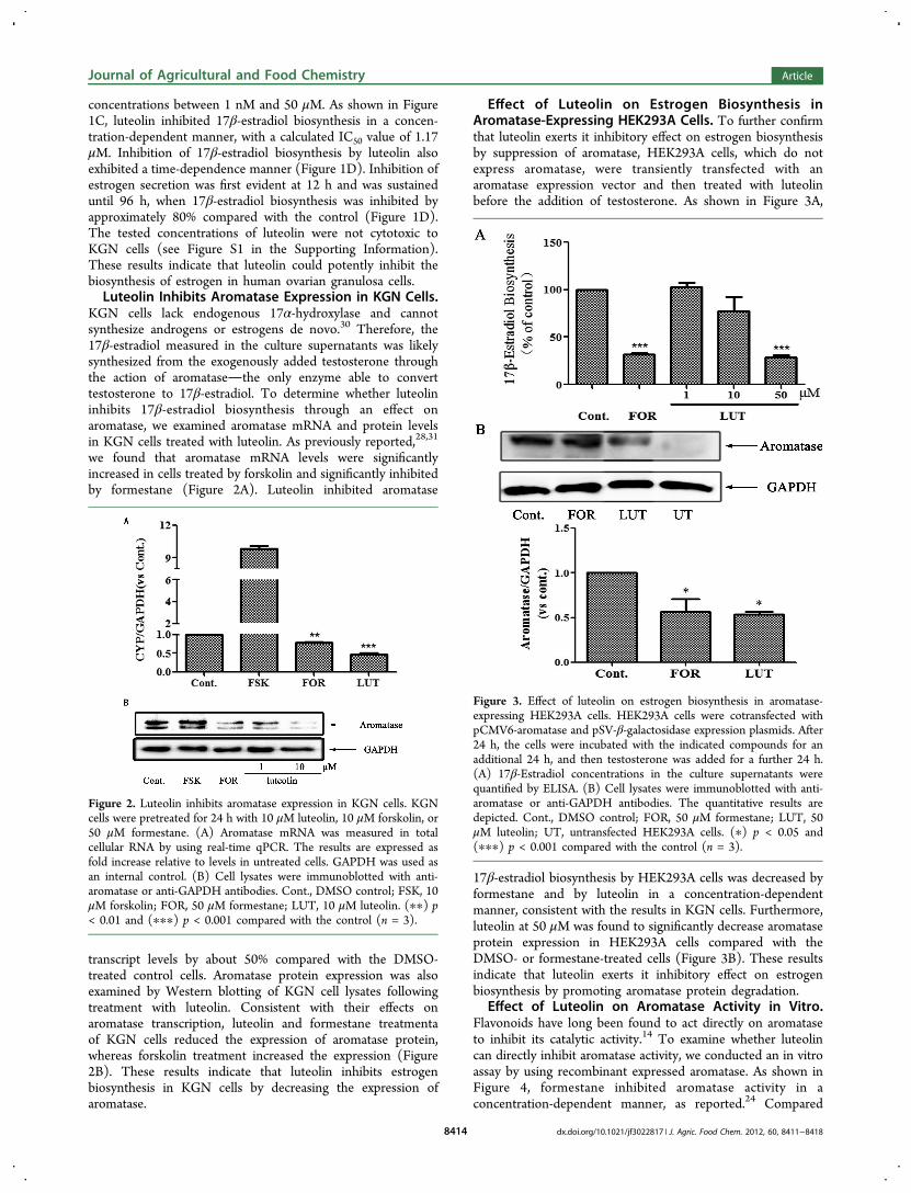
concentrations between 1 nM and 50 μM. As shown in Figure1C, luteolin inhibited 17β-estradiol biosynthesis in a concen-tration-dependent manner, with a calculated IC50 value of 1.17μM. Inhibition of 17β-estradiol biosynthesis by luteolin alsoexhibited a time-dependence manner (Figure 1D). Inhibition ofestrogen secretion was first evident at 12 h and was sustaineduntil 96 h, when 17β-estradiol biosynthesis was inhibited byapproximately 80% compared with the control (Figure 1D).The tested concentrations of luteolin were not cytotoxic toKGN cells (see Figure S1 in the Supporting Information).These results indicate that luteolin could potently inhibit thebiosynthesis of estrogen in human ovarian granulosa cells.Luteolin Inhibits Aromatase Expression in KGN Cells.
KGN cells lack endogenous 17α-hydroxylase and cannotsynthesize androgens or estrogens de novo.30 Therefore, the17β-estradiol measured in the culture supernatants was likelysynthesized from the exogenously added testosterone throughthe action of aromatasethe only enzyme able to converttestosterone to 17β-estradiol. To determine whether luteolininhibits 17β-estradiol biosynthesis through an effect onaromatase, we examined aromatase mRNA and protein levelsin KGN cells treated with luteolin. As previously reported,28,31
we found that aromatase mRNA levels were significantlyincreased in cells treated by forskolin and significantly inhibitedby formestane (Figure 2A). Luteolin inhibited aromatase
transcript levels by about 50% compared with the DMSO-treated control cells. Aromatase protein expression was alsoexamined by Western blotting of KGN cell lysates followingtreatment with luteolin. Consistent with their effects onaromatase transcription, luteolin and formestane treatmentaof KGN cells reduced the expression of aromatase protein,whereas forskolin treatment increased the expression (Figure2B). These results indicate that luteolin inhibits estrogenbiosynthesis in KGN cells by decreasing the expression ofaromatase.
Effect of Luteolin on Estrogen Biosynthesis inAromatase-Expressing HEK293A Cells. To further confirmthat luteolin exerts it inhibitory effect on estrogen biosynthesisby suppression of aromatase, HEK293A cells, which do notexpress aromatase, were transiently transfected with anaromatase expression vector and then treated with luteolinbefore the addition of testosterone. As shown in Figure 3A,
17β-estradiol biosynthesis by HEK293A cells was decreased byformestane and by luteolin in a concentration-dependentmanner, consistent with the results in KGN cells. Furthermore,luteolin at 50 μM was found to significantly decrease aromataseprotein expression in HEK293A cells compared with theDMSO- or formestane-treated cells (Figure 3B). These resultsindicate that luteolin exerts it inhibitory effect on estrogenbiosynthesis by promoting aromatase protein degradation.
Effect of Luteolin on Aromatase Activity in Vitro.Flavonoids have long been found to act directly on aromataseto inhibit its catalytic activity.14 To examine whether luteolincan directly inhibit aromatase activity, we conducted an in vitroassay by using recombinant expressed aromatase. As shown inFigure 4, formestane inhibited aromatase activity in aconcentration-dependent manner, as reported.24 Compared
Figure 2. Luteolin inhibits aromatase expression in KGN cells. KGNcells were pretreated for 24 h with 10 μM luteolin, 10 μM forskolin, or50 μM formestane. (A) Aromatase mRNA was measured in totalcellular RNA by using real-time qPCR. The results are expressed asfold increase relative to levels in untreated cells. GAPDH was used asan internal control. (B) Cell lysates were immunoblotted with anti-aromatase or anti-GAPDH antibodies. Cont., DMSO control; FSK, 10μM forskolin; FOR, 50 μM formestane; LUT, 10 μM luteolin. (∗∗) p< 0.01 and (∗∗∗) p < 0.001 compared with the control (n = 3).
Figure 3. Effect of luteolin on estrogen biosynthesis in aromatase-expressing HEK293A cells. HEK293A cells were cotransfected withpCMV6-aromatase and pSV-β-galactosidase expression plasmids. After24 h, the cells were incubated with the indicated compounds for anadditional 24 h, and then testosterone was added for a further 24 h.(A) 17β-Estradiol concentrations in the culture supernatants werequantified by ELISA. (B) Cell lysates were immunoblotted with anti-aromatase or anti-GAPDH antibodies. The quantitative results aredepicted. Cont., DMSO control; FOR, 50 μM formestane; LUT, 50μM luteolin; UT, untransfected HEK293A cells. (∗) p < 0.05 and(∗∗∗) p < 0.001 compared with the control (n = 3).
Journal of Agricultural and Food Chemistry Article
dx.doi.org/10.1021/jf3022817 | J. Agric. Food Chem. 2012, 60, 8411−84188414

with that, luteolin had no effect on aromatase activity. Theseresults indicate that luteolin does not directly inhibit aromataseactivity as other active flavonoids generally do.Effects of Dietary Flavonoids on Estrogen Biosyn-
thesis. We next examined the effects of several flavonoidsfound in commonly consumed vegetables and fruits onestrogen biosynthesis. First, KGN cells were treated withvarious concentrations of kaempferol, quercetin, myricetin, orisorhamnetin to test their effects on cell viability and todetermine the optimal concentrations for estrogen biosynthesisassays (see Figure S2 in the Supporting Information). Thechemical structures of these flavonoids are given in Figure 5A.As shown in Figure 5B, kaempferol inhibited 17β-estradiolbiosynthesis by KGN cells in a concentration-dependentmanner, with approximately 30 and 50% inhibition followingtreatment with 10 and 50 μM kaempferol, respectively.Quercetin inhibited 17β-estradiol biosynthesis by up to 75%at a high concentration (50 μM) but increased 17β-estradiolbiosynthesis slightly when added at low concentrations (1−10μM). Myricetin inhibited 17β-estradiol biosynthesis at a lowconcentration (0.1 μM) but had no effect at higherconcentrations (1−10 μM). In contrast, isorhamnetin had noeffect on 17β-estradiol biosynthesis at any concentrationexamined (1−50 μM). These results indicate that kaempferoland quercetin potently inhibit estrogen biosynthesis in KGNcells, whereas myricetin and isorhamnetin show only weak orno effects.Effects of Onion and Bird Chili Extracts on Estrogen
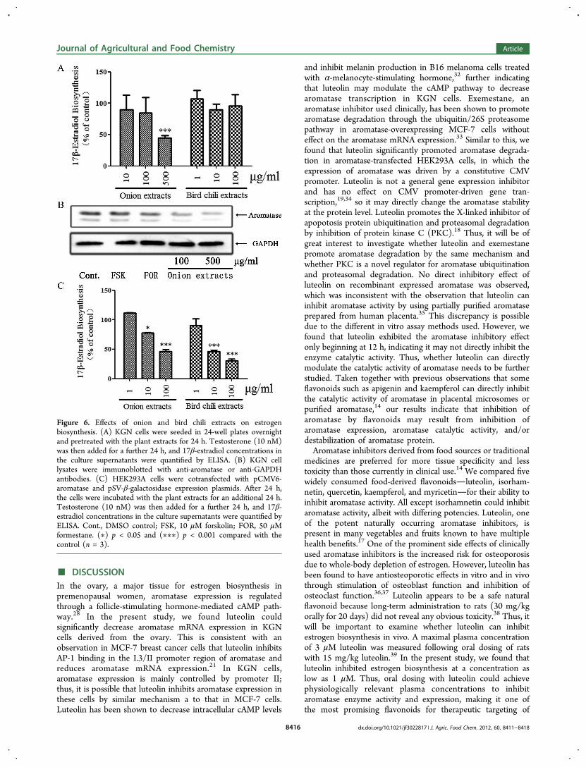
Biosynthesis. Onion and bird chili are commonly consumedvegetables that are natural sources of luteolin; therefore, wenext investigated whether extracts of onion or bird chili couldaffect estrogen biosynthesis. Quantification of quercetin andluteolin in the extracts indicated that onion contained highamounts of both quercetin and luteolin, whereas bird chilicontained relatively low levels of both compounds (Table 1).KGN cells were treated with various noncytotoxic concen-trations of the extracts (see Figure S3 in the SupportingInformation). As shown in Figure 6A, a high concentration
(500 μg/mL) of onion extract potently inhibited 17β-estradiolbiosynthesis, whereas the bird chili extract had no such effect.We then examined the effect of onion extracts on aromataseexpression in KGN cells. As shown in Figure 6B, onion extracts(100−500 μg/mL) could significantly decrease aromataseprotein expression. We also examined the effect of thevegetable extracts on estrogen biosynthesis in aromatase-expressing HEK293A cells. In contrast to the effects on KGNcells, both onion and bird chili extracts significantly inhibited17β-estradiol biosynthesis in HEK293A cells in a concen-tration-dependent manner (1−100 μg/mL) (Figure 6C). Theseresults indicate the onion and bird chili extracts can inhibitestrogen biosynthesis, and the inhibitory potency is concen-tration- and cell type-dependent.
Figure 4. Effect of luteolin on aromatase activity in vitro. An in vitroaromatase assay using recombinant expressed aromatase anddibenzylfluorescein as a substrate was conducted as described underMaterials and Methods. Various concentrations of formestane andluteolin were added into the mixtures containing recombinantexpressed aromatase and NADPH regenerating system, andfluorescence intensity induced by the aromatase-catalyzed dibenzyl-fluorescein hydrolysis was detected. Cont., DMSO control; FOR,formestane; LUT, luteolin. (∗∗∗) p < 0.001 compared with the control(n = 3).
Figure 5. Effects of dietary flavonoids on estrogen biosynthesis. (A)presents the chemical structures of isorhamnetin, kaempferol,quercetin, and myricetin. (B) KGN cells were seeded in 24-wellplates overnight and pretreated with the test compounds for 24 h.Testosterone (10 nM) was then added for a further 24 h, and 17β-estradiol concentrations in the culture supernatants were quantifiedusing ELISA. Cont., DMSO control; Lut, luteolin; Iso, isorhamnetin;Kae, kaempferol; Que, quercetin; Myr, myricetin. (∗) p < 0.05, (∗∗) p< 0.01, and (∗∗∗) p < 0.001 compared with the control (n = 3).
Table 1. Quantification of Quercetin and Luteolin in Onionand Bird Chili Extracts
content, mg/kg of dry weighta
sample scientific name quercetin luteolin
bird chili Capsicum frutescens 2.57 ± 0.05 6.40 ± 0.39onion (interior) Allium f istulosum 52.0 ± 0.19 46.1 ± 0.08
aEach value is expressed as mg/kg of dry weight of three replications± SE.
Journal of Agricultural and Food Chemistry Article
dx.doi.org/10.1021/jf3022817 | J. Agric. Food Chem. 2012, 60, 8411−84188415
■ DISCUSSIONIn the ovary, a major tissue for estrogen biosynthesis inpremenopausal women, aromatase expression is regulatedthrough a follicle-stimulating hormone-mediated cAMP path-way.28 In the present study, we found luteolin couldsignificantly decrease aromatase mRNA expression in KGNcells derived from the ovary. This is consistent with anobservation in MCF-7 breast cancer cells that luteolin inhibitsAP-1 binding in the I.3/II promoter region of aromatase andreduces aromatase mRNA expression.21 In KGN cells,aromatase expression is mainly controlled by promoter II;thus, it is possible that luteolin inhibits aromatase expression inthese cells by similar mechanism a to that in MCF-7 cells.Luteolin has been shown to decrease intracellular cAMP levels
and inhibit melanin production in B16 melanoma cells treatedwith α-melanocyte-stimulating hormone,32 further indicatingthat luteolin may modulate the cAMP pathway to decreasearomatase transcription in KGN cells. Exemestane, anaromatase inhibitor used clinically, has been shown to promotearomatase degradation through the ubiquitin/26S proteasomepathway in aromatase-overexpressing MCF-7 cells withouteffect on the aromatase mRNA expression.33 Similar to this, wefound that luteolin significantly promoted aromatase degrada-tion in aromatase-transfected HEK293A cells, in which theexpression of aromatase was driven by a constitutive CMVpromoter. Luteolin is not a general gene expression inhibitorand has no effect on CMV promoter-driven gene tran-scription,19,34 so it may directly change the aromatase stabilityat the protein level. Luteolin promotes the X-linked inhibitor ofapopotosis protein ubiquitination and proteasomal degradationby inhibition of protein kinase C (PKC).18 Thus, it will be ofgreat interest to investigate whether luteolin and exemestanepromote aromatase degradation by the same mechanism andwhether PKC is a novel regulator for aromatase ubiquitinationand proteasomal degradation. No direct inhibitory effect ofluteolin on recombinant expressed aromatase was observed,which was inconsistent with the observation that luteolin caninhibit aromatase activity by using partially purified aromataseprepared from human placenta.35 This discrepancy is possibledue to the different in vitro assay methods used. However, wefound that luteolin exhibited the aromatase inhibitory effectonly beginning at 12 h, indicating it may not directly inhibit theenzyme catalytic activity. Thus, whether luteolin can directlymodulate the catalytic activity of aromatase needs to be furtherstudied. Taken together with previous observations that someflavonoids such as apigenin and kaempferol can directly inhibitthe catalytic activity of aromatase in placental microsomes orpurified aromatase,14 our results indicate that inhibition ofaromatase by flavonoids may result from inhibition ofaromatase expression, aromatase catalytic activity, and/ordestabilization of aromatase protein.Aromatase inhibitors derived from food sources or traditional
medicines are preferred for more tissue specificity and lesstoxicity than those currently in clinical use.14 We compared fivewidely consumed food-derived flavonoidsluteolin, isorham-netin, quercetin, kaempferol, and myricetinfor their ability toinhibit aromatase activity. All except isorhamnetin could inhibitaromatase activity, albeit with differing potencies. Luteolin, oneof the potent naturally occurring aromatase inhibitors, ispresent in many vegetables and fruits known to have multiplehealth benefits.17 One of the prominent side effects of clinicallyused aromatase inhibitors is the increased risk for osteoporosisdue to whole-body depletion of estrogen. However, luteolin hasbeen found to have antiosteoporotic effects in vitro and in vivothrough stimulation of osteoblast function and inhibition ofosteoclast function.36,37 Luteolin appears to be a safe naturalflavonoid because long-term administration to rats (30 mg/kgorally for 20 days) did not reveal any obvious toxicity.38 Thus, itwill be important to examine whether luteolin can inhibitestrogen biosynthesis in vivo. A maximal plasma concentrationof 3 μM luteolin was measured following oral dosing of ratswith 15 mg/kg luteolin.39 In the present study, we found thatluteolin inhibited estrogen biosynthesis at a concentration aslow as 1 μM. Thus, oral dosing with luteolin could achievephysiologically relevant plasma concentrations to inhibitaromatase enzyme activity and expression, making it one ofthe most promising flavonoids for therapeutic targeting of
Figure 6. Effects of onion and bird chili extracts on estrogenbiosynthesis. (A) KGN cells were seeded in 24-well plates overnightand pretreated with the plant extracts for 24 h. Testosterone (10 nM)was then added for a further 24 h, and 17β-estradiol concentrations inthe culture supernatants were quantified by ELISA. (B) KGN celllysates were immunoblotted with anti-aromatase or anti-GAPDHantibodies. (C) HEK293A cells were cotransfected with pCMV6-aromatase and pSV-β-galactosidase expression plasmids. After 24 h,the cells were incubated with the plant extracts for an additional 24 h.Testosterone (10 nM) was then added for a further 24 h, and 17β-estradiol concentrations in the culture supernatants were quantified byELISA. Cont., DMSO control; FSK, 10 μM forskolin; FOR, 50 μMformestane. (∗) p < 0.05 and (∗∗∗) p < 0.001 compared with thecontrol (n = 3).
Journal of Agricultural and Food Chemistry Article
dx.doi.org/10.1021/jf3022817 | J. Agric. Food Chem. 2012, 60, 8411−84188416

aromatase. Flavonoids with potent aromatase inhibitoryactivities, such as chrysin, naringenin, and apigenin, failed toinhibit uterine growth via aromatase in vivo.40 The contra-diction was possibly due to the negative feedback effect ofestrogen depletion by increased secretion of follicle-stimulatinghormone from the hypothalamus in rats with functionalovaries.41 Thus, the ovariectomized rats with the aromatase-overexpressing MCF-7 breast tumor should be used to evaluatethe efficacy of these flavonoids, including luteolin, in theinhibition of aromatase-mediated breast tumorigenesis in vivoto obviate such negative feedback effect.Overexposure to environmental estrogenic compounds and
the Western-style diet have contributed to the increasedincidence of breast cancer.2,3 In contrast, consumption of largequantities of fruits and vegetables conveys protection against avariety of cancers, including breast cancer.16 It has been foundthat red wine, grape seed, white button mushroom, and beercan inhibit aromatase activity and modulate estrogen biosyn-thesis.42−46 Despite this, the role of fruits and vegetables inendogenous estrogen biosynthesis is still largely unknown. Inthis study, we compared two luteolin-containing vegetablesonion and bird chilifor their effects on estrogen biosynthesis.Interestingly, onion extracts significantly inhibited estrogenbiosynthesis in both KGN cells and aromatase-expressingHEK293A cells, whereas bird chili extracts inhibited estrogenbiosynthesis only in the HEK293A cells. The difference ispossibly due to the cell permeability of the compounds, theircomposition, or concentration range of flavonoids contained inthe food source. Different combinations of flavonoids may alsochange the solubility and/or accessibility of the compounds tointracellular aromatase or may act on aromatase throughdifferent mechanisms. As an example, some dietary flavonoidssuch as hesperetin can increase aromatase expression.21 Thus,the composition and concentrations of flavonoids and the celltype being tested should be considered when fruits andvegetables are compared for their effects on estrogenbiosynthesis.In conclusion, we identified luteolin, distributed widely in
consumed fruits and vegetables, as a potent inhibitor ofestrogen biosynthesis in human ovarian granulosa-like KGNcells by decreasing aromatase expression and promoting itsdegradation. We also showed that other daily consumedflavonoids, including isorhamnetin, quercetin, kaempferol, andmyricetin, inhibited estrogen biosynthesis with differingpotencies. Two widely consumed vegetables, onion and birdchili, also differed in their ability to inhibit estrogen biosynthesisin KGN cells and in aromatase-expressing HEK293A cells. Thepresent study suggests that luteolin may exert its estrogenbiosynthesis inhibitory effect through suppression of aromataseexpression and promotion of aromatase degradation, and itwarrants further investigation as a potential agent for thetreatment of breast cancer. Consumption of fruits andvegetables rich in flavonoids will be helpful for reducingendogenous estrogen levels in the prevention of estrogen-dependent disease, such as breast cancer.
■ ASSOCIATED CONTENT
*S Supporting InformationCytotoxic effects of luteolin, kaempferol, quercetin, myricetin,isorhamnetin, and plant extracts on KGN or HEK293A cells(Figures S1−S3). This material is available free of charge viathe Internet at http://pubs.acs.org.
■ AUTHOR INFORMATION
Corresponding Author*(F.W.) Phone/fax: +86 28 85256758. E-mail: [email protected]. (G.Z.) Phone/fax: +86 28 85229901. E-mail: [email protected].
FundingThis work was supported by the National Natural ScienceFoundation of China (Grants 20932007 and 30900769), theWest Light Foundation of the Chinese Academy of Sciences,and the National New Drug Innovation Major Project of China(2011ZX09307-002-02).
NotesThe authors declare no competing financial interest.
■ ABBREVIATIONS USED
ER, estrogen receptor; CREB, cAMP-response element bindingprotein; HEK293A, human embryonic kidney 293A; ELISA,enzyme-linked immunosorbent assay; BCA, bicinchoninic acid;PKA, protein kinase A; PKC, protein kinase C.
■ REFERENCES(1) Heldring, N.; Pike, A.; Andersson, S.; Matthews, J.; Cheng, G.;Hartman, J.; Tujague, M.; Strom, A.; Treuter, E.; Warner, M.;Gustafsson, J. Å. Estrogen receptors: how do they signal and what aretheir targets. Physiol. Rev. 2007, 87, 905−931.(2) Willett, W. C. Diet and breast cancer. J. Intern. Med. 2001, 249,395−411.(3) Brinbaum, L. S.; Fenton, S. E. Cancer and developmentalexposure to endocrine disruptors. Environ. Health Perspect. 2003, 111,389−394.(4) Buzdar, A. U. Advances in endocrine treatments forpostmenopausal women with metastatic and early breast cancer.Oncologist 2003, 8, 335−341.(5) Collins-Burow, B. M.; Burow, M. E.; Duong, B. N.; McLachlan, J.A. Estrogenic and antiestrogenic activities of flavonoid phytochemicalsthrough estrogen receptor binding-dependent and -independentmechanisms. Nutr. Cancer 2000, 38, 229−244.(6) Santell, R. C.; Chang, Y. C.; Nair, M. G.; Helferich, W. G. Dietarygenistein exerts estrogenic effects upon the uterus, mammary glandand the hypothalamic/pituitary axis in rats. J. Nutr. 1997, 127, 263−269.(7) Osborne, C.; Tripathy, D. Aromatase inhibitors: rationale and usein breast cancer. Annu. Rev. Med. 2005, 56, 103−116.(8) Simpson, E. R.; Rubin, G.; Clyne, C.; Robertson, K.; O’Donnell,L.; Jones, M.; Davis, S. The role of local estrogen biosynthesis in malesand females. Trends Endocrinol. Metab. 2000, 11, 184−188.(9) Simpson, E. R.; Clyne, C.; Rubin, G.; Boon, W. C.; Robertson,K.; Britt, K.; Speed, C.; Jones, M. Aromatase − a brief overview. Annu.Rev. Physiol. 2002, 64, 93−127.(10) Simpson, E. R. Aromatase: biologic relevance of tissue-specificexpression. Semin. Reprod. Med. 2004, 22, 11−23.(11) Michael, M. D.; Kilgore, M. W.; Morokashi, K.; Simpson, E. R.Ad4BB/SF1 regulates cyclic AMP-induced transcription from theproximal promoter (PII) of the human aromatase P450 (CYP19) genein the ovary. J. Biol. Chem. 1995, 270, 13561−13566.(12) Johnston, S. R.; Dowsett, M. Aromatase inhibitors for breastcancer: lessons from the laboratory. Nat. Rev. Cancer 2003, 3, 821−831.(13) Smith, I. E.; Dowsett, M. Aromatase inhibitors in breast cancer.N. Engl. J. Med. 2003, 348, 2431−2442.(14) Balunas, M. J.; Kinghorn, A. D. Natural compounds witharomatase inhibitory activity: an update. Planta Med. 2010, 76, 1087−1093.(15) Knekt, P.; Kumpulainen, J.; Jarvinen, R; Rissanen, H.;Heliovaara, M.; Reunanen, A.; Hakulinen, T.; Aromaa, A. Flavonoid
Journal of Agricultural and Food Chemistry Article
dx.doi.org/10.1021/jf3022817 | J. Agric. Food Chem. 2012, 60, 8411−84188417
intake and risk of chronic diseases. Am. J. Clin. Nutr. 2002, 76, 560−568.(16) Steinmetz, K. A.; Potter, J. D. Vegetables, fruit, and cancerprevention: a review. J. Am. Diet. Assoc. 1996, 96, 1027−1039.(17) Lopez-Lazaro, M. Distribution and biological activities of theflavonoid luteolin. Mini Rev. Med. Chem. 2009, 9, 31−59.(18) Shi, R. X.; Ong, C. N.; Shen, H. M. Protein kinase C inhibitionand X-linked inhibitor of apoptosis protein degradation contribute tothe sensitization effect of luteolin on tumor necrosis factor-relatedapoptosis-inducing ligand-induced apoptosis in cancer cells. CancerRes. 2005, 65, 7815−7823.(19) Selvendiran, K.; Koga, H.; Ueno, T.; Yoshida, T.; Maeyama, M.;Torimura, T.; Yano, H.; Kojiro, M.; Sata, M. Luteolin promotesdegradation in signal transducer and activator of transcription 3 inhuman hepatoma cells: an implication for the antitumor potential offlavonoids. Cancer Res. 2006, 66, 4826−4834.(20) Sternberg, Z.; Chadha, K.; Lieberman, A.; Drake, A.; Hojnacki,D.; Weinstock-Guttman, B.; Munschauer, F. Immunomodulatoryresponses of peripheral blood mononuclear cells from multiplesclerosis patients upon in vitro incubation with the flavonoid luteolin:additive effects of IFN-β. J. Neuroinflamm. 2009, 6, 28−35.(21) Li, F. J.; Ye, L.; Leung, L. K. Dietary flavones and flavononesdisplay differential effects on aromatase (CYP19) transcription in thebreast cancer cells MCF-7. Mol. Cell. Endocrinol. 2011, 344, 51−58.(22) Wang, C.; Makela, T.; Hase, T.; Adlercreutz, H.; Kurzer, M. S.Lignans and flavonoids inhibit aromatase enzyme in humanpreadipocytes. J. Steroid. Biochem. Mol. Biol. 1994, 50, 205−212.(23) Joshi, S. C.; Strauss, L.; Makela, S.; Santti, R. Inhibition of 17β-estradiol formation by isoflavonoids and flavonoids in cultured JEG-3cells: search for aromatase-targeting dietary compounds. J. Med. Food1999, 2, 235−238.(24) Stresser, D. M.; Turner, S. D.; McNamara, J.; Stocker, P.; Miller,V. P.; Crespi, C. L.; Patten, C. J. A high-throughput screen to identifyinhibitors of aromatase (CYP19). Anal. Biochem. 2000, 284, 427−430.(25) Maiti, A.; Cuendet, M.; Croy, V. L.; Endringer, D. C.; Pezzuto, J.M.; Cushman, M. Synthesis and biological evaluation of (±)-abyssi-none II and its analogues as aromatase inhibitors for chemopreventionof breast cancer. J. Med. Chem. 2007, 50, 2799−2806.(26) Miean, K. H.; Mohamed, S. Flavonoid (myricetin, quercetin,kaempferol, luteolin, and apigenin) content of edible tropical plants. J.Agric. Food Chem. 2001, 49, 3106−3112.(27) Tai, Z. F.; Zhang, G. L.; Wang, F. Identification of smallmolecule activators of the janus kinase/signal transducer and activatorof transcription pathway using a cell-based screen. Biol. Pharm. Bull.2012, 35, 65−71.(28) Gonzalez-Robayna, I. J.; Alliston, T. N.; Buse, P.; Firestone, G.L.; Richards, J. S. Functional and subcellular changes in the A-kinase-signaling pathway: relation to aromatase and Sgk expression during thetransition of granulosa cells to luteal cells. Mol. Endocrinol. 1999, 13,1318−1337.(29) Wiseman, L. R.; Goa, K. L. Formestane − a review of itspharmacological properties and clinical efficacy in the treatment ofpostmenopausal breast cancer. Drugs Aging 1996, 9, 292−306.(30) Nishi, Y.; Yanase, T.; Mu, Y. M.; Oba, K.; Ichino, I.; Saito, M.;Nomura, M.; Mukasa, C.; Okabe, T.; Goto, K.; Takayanagi, R.;Kashimura, Y.; Haji, M.; Nawata, H. Establishment and character-ization of a steroidogenic human granulosa-like tumor cell line, KGN,that expresses functional follicle-stimulating hormone receptor.Endocrinology 2001, 142, 437−445.(31) Cavaliere, C.; Corvigno, S.; Galgani, M.; Limite, G.; Nardone,A.; Veneziani, B. M. Combined inhibitory effect of formestane andherceptin on a subpopulation of CD44+/CD24 low breast cancer cells.Cancer Sci. 2010, 101, 1661−1669.(32) Choi, M. Y.; Song, H. S.; Hur, H. S.; Sim, S. S. Whiteningactivity of luteolin related to the inhibition of cAMP pathway in α-MSH-stimulated B16 melanoma cells. Arch. Pharm. Res. 2008, 31,1166−1171.
(33) Wang, X.; Chen, S. Aromatase destabilizer: novel action ofexemestane, a food and drug administration-approved aromataseinhibitor. Cancer Res. 2006, 66, 10281−10286.(34) Shi, R. X.; Ong, C. N.; Shen, H. M. Luteolin sensitizes tumornecrosis factor-α-induced apoptosis in human tumor cells. Oncogene2004, 23, 7712−7721.(35) Jeong, H. J.; Shin, Y. G.; Kim, II. H.; Pezzuto, J. M. Inhibition ofaromatase activity by flavonoids. Arch. Pharm. Res. 1999, 22, 309−312.(36) Choi, E. M. Modulatory effects of luteolin on osteoblasticfunction and inflammatory mediators in osteoblastic MC3T3-E1 cells.Cell Biol. Int. 2007, 31, 870−877.(37) Kim, T. H.; Jung, J. W.; Ha, B. G.; Hong, J. M.; Park, E. K.; Kim,H. J.; Kim, S. Y. The effects of luteolin on osteoclast differentiation,function in vitro and ovariectomy-induced bone loss. J. Nutr. Biochem.2011, 22, 8−15.(38) Samy, R. P.; Gopalakrishnakone, P.; Ignacimuthu, S. Anti-tumorpromoting potential of luteolin against 7,12-dimethylbenz(a)-anthracene-induced mammary tumors in rats. Chem.−Biol. Interact.2006, 164, 1−14.(39) Shimoi, K.; Okada, H.; Furugori, M.; Goda, T.; Takase, S.;Suzuki, M.; Hara, Y.; Yamamoto, H.; Kinae, N. Intestinal absorption ofluteolin and luteolin 7-O-β-glucoside in rats and humans. FEBS Lett.1998, 438, 220−224.(40) Saarinen, N.; Joshi, S. C.; Ahotupa, M.; Li, X.; Ammala, J.;Makela, S.; Santti, R. No evidence for the in vivo activity of aromatase-inhibiting flavonoids. J. Steroid Biochem. 2001, 78, 231−239.(41) Mitwally, M. F. M.; Casper, R. F. Aromatase inhibition reducesthe dose of gonadotropin ruired for controlled ovarian hyper-stimulation. J. Soc. Gynecol. Invest. 2004, 11, 406−415.(42) Eng, E. T.; Williams, D.; Mandava, U.; Kirma, N.; Tekmal, R. R.;Chen, S. Suppression of aromatase (estrogen synthetase) by red winephytochemicals. Breast Cancer Res. Treat. 2001, 67, 133−146.(43) Kijima, I.; Phung, S.; Hur, G.; Kwok, S. L.; Chen, S. Grape seedextract is an aromatase inhibitor and a suppressor of aromataseexpression. Cancer Res. 2006, 66, 5960−5967.(44) Chen, S.; Oh, S. R.; Phung, S.; Hur, G.; Ye, J. J.; Kwok, S. L.;Shrode, G. E.; Belury, M.; Adams, L. S.; Williams, D. Anti-aromataseactivity of phytochemicals in white button mushrooms (Agaricusbisporus). Cancer Res. 2006, 66, 12026−12034.(45) Monteiro, R.; Becker, H.; Azevedo, I.; Calhau, C. Effect of hop(Humulus lupulus L.) flavonoids on aromatase (estrogen synthase)activity. J. Agric. Food Chem. 2006, 54, 2938−2943.(46) Monteiro, R.; Azevedo, I.; Calhau, C. Modulation of aromataseactivity by diet polyphenolic compounds. J. Agric. Food Chem. 2006,54, 3535−3540.
Journal of Agricultural and Food Chemistry Article
dx.doi.org/10.1021/jf3022817 | J. Agric. Food Chem. 2012, 60, 8411−84188418