in vitro simultaneous measurement of refractive index and thickness of biological tissue by the low...
TRANSCRIPT

1266 IEEE TRANSACTIONS ON BIOMEDICAL ENGINEERING, VOL. 47, NO. 9, SEPTEMBER 2000
In Vitro Simultaneous Measurement of RefractiveIndex and Thickness of Biological Tissue by the Low
Coherence InterferometryMasato Ohmi*, Yasuhito Ohnishi, Koji Yoden, and Masamitsu Haruna
Abstract—We proposed and demonstratedin vitro simultaneousmeasurement of refractive index and thickness of biological tissue.The technique is based on the low coherence interferometry com-bined with precise translation stages. Refractive indices were de-termined with the accuracy of less than 1% for tissue samples ofa few hundred micron thickness, including chicken tissue, humantooth and nail. Simultaneous measurement of refractive index andthickness of multilayer tissue are also demonstrated.
Index Terms—In vitro measurement, low coherence interferom-etry, opto-mechatronics, simultaneous measurement of refractiveindex and thickness.
I. INTRODUCTION
A MAIN purpose of biomedical optics is to develop nonin-vasive measurement and diagnosis of biological tissue free
from radiology. It, however, seems that the basic knowledge andtechniques have been still insufficient for this purpose becausebiomedical optics is rather a new field. In fact, there are somereports and literatures giving us optical properties of biologicaltissue of interest [1]–[3]. Especially, the index of refraction ofbiological tissues have not been measured systematically with acertain high accuracy for different tissue samples including softtissue or skin, tooth, nail, vessel wall, etc. It is, thus, requiredto develop a precise and standard technique for measurement ofrefractive index of biological tissue.
Some experimental works have been reported for the tissueindex measurement [4]–[6]. The blood index, for instance, wasdetermined by measurement of either the numerical aperture orthe transmission of the fiber embedded in blood [4]. Anothermethod is to measure the critical angle of the total internal re-flection on the bottom of a prism tightly close to artificial mem-brane [5], [6]. These methods are limited to measurement of in-dices of liquid and soft tissue, and the measurement accuracy of1% or less was not achieved. On the other hand, more accurateindex measurement is possible by the low coherence interferom-etry. Basically, this interferometer provides the optical thickness
Manuscript received June 17, 1999; revised April 18, 2000. This workwas supported in part by a the Ministry of Education, Science, Sportsand Culture, Japan under Grant-in-Aid for Scientific Researches (B) (2)(Subjects, #11555016) and (A) (2) (Subjects, #11780620).Asterisk indicatescorresponding author.
*M. Ohmi is with the School of Allied Health Sciences, Faculty of Medicine,Osaka University 1-7, Yamada-Oka, Suita, Osaka 565-0871, Japan (e-mail:[email protected]).
Y. Ohnishi, K. Yoden, and M. Haruna are with the School of Allied HealthSciences, Faculty of Medicine, Osaka University 1-7, Yamada-Oka, Suita,Osaka 565-0871, Japan.
Publisher Item Identifier S 0018-9294(00)07999-4.
of a sample to be measured with a resolution of the co-herence length , of the light source, where and are therefractive index and the thickness of the sample, respectively.In the existing interferometers, therefore,cannot be deter-mined unless and are measured simultaneously [7]–[9]. Re-cently, the low coherence interferometry was applied toin vitrosimultaneous measurement ofand of human tissue such asdermis, muscle and adipose by Tearneyet al., in which the focallength shift from the front plane to the rear one of the sample istracked to measure and simultaneously with confocal mi-croscopy [10]. Measurement ofand of multiple layers wasalso demonstrated by Fukano and Yamaguchi [11].
In addition to the pioneer works based on the low coher-ence interferometry, as described above, we presented a possiblepractical technique for simultaneous measurement ofand , inwhich both the sample and reference mirror are placed on pre-cise translation stages and the interference light is detected insynchronization with movement of the stages [12]. The wholemeasurement system is computer-controlled to perform the au-tomatic measurement. In the case where the thickness of a trans-parent plate used as the sample is above 0.1 mm, our techniquecan provide the measurement accuracy of 0.1%, taking chro-matic dispersion of refractive index into account [13]. In mea-surement of biological tissue, the accuracy of the same ordershould be guaranteed by selective detection of the so-called bal-listic photons having less experience of scattering.
In this paper, we demonstratein vitro simultaneous measure-ment of and of biological tissue using the specific inter-ferometer system with precise translation stages developed byourselves. Refractive indices were determined with the accu-racy of less than 1% for tissue samples of a few hundred mi-cron thickness, including chicken tissue, human tooth and nail.The measurement system is described in Section II together withthe measurement principle. Measurement results are presentedin Section III classified into three subsections of soft, hard andmultilayer tissues.
II. M EASUREMENTSYSTEM AND PRINCIPLE
The low coherence interferometer is shown schematically inFig. 1. A super-luminescent diode (SLD) is used as the lightsource whose spectral width is 16 nm (FWHM) at thecenter wavelength 850 nm. The output power is typically3 mW and divided into the reference and the sample arms inthe Michelson interferometer. The measured coherence length
of the SLD is 12 m. The reference light is phase-modu-lated at 500 Hz with a mirror-tipped piezoelectric transducer
0018–9294/00$10.00 © 2000 IEEE
OHMI et al.: IN VITROSIMULTANEOUS MEASUREMENT OF REFRACTIVE INDEX AND THICKNESS OF BIOLOGICAL TISSUE 1267
Fig. 1. Low-coherence interferometer with precise translation stages. Lightfocusing on the front and rear planes of the sample and the correspondingpositions of the reference mirror are shown schematically. PZT, piezoelectrictransducer; A/D, analog to digital converter.
(PZT) placed on a translation stage 1 with a 1-m resolution,where a figure for modulation depth of the phase modulationwas 0.4. On the contrary, the sample light is focused on asample to be measured with a microscope objective. Unlikethe existing low-coherence interferometers reported so far, thesample is placed on a translation stage 2, as shown in Fig. 1.In synchronization with movement of two stages, an envelopof interferograms is heterodyne-detected by a Si photodiode(NEP ) without a pinhole. Thedetected 500-Hz signals are amplified, and the maximum signalamplitude is sorted by a sample-and-hold circuit, whose outputis converted to a 10-bit digital signal.
In simultaneous measurement ofand , the SLD light isfocused on the front plane of the sample, as shown in Fig. 1,and the reference mirror (or the stage 1) is positioned atso that there is no optical path difference between the referenceand the sample arms. Subsequently, we move the sample stage2 a distance to focus the objective upon the rear plane ofthe sample, and the stage 1 is then shifted by a distanceandre-positioned at to perform again the null optical pathdifference between two arms. We find from Snell’s law thatsatisfies
(1)
where is the numerical aperture of the focusing lens.is calibrated before measurement ofand . In (1), note that
the refractive index must be the phase index. The sum ofand equals the optical thickness of the sample that is
determined by the group index and, thus, we have
(2)
and relate to each other according to
(3)
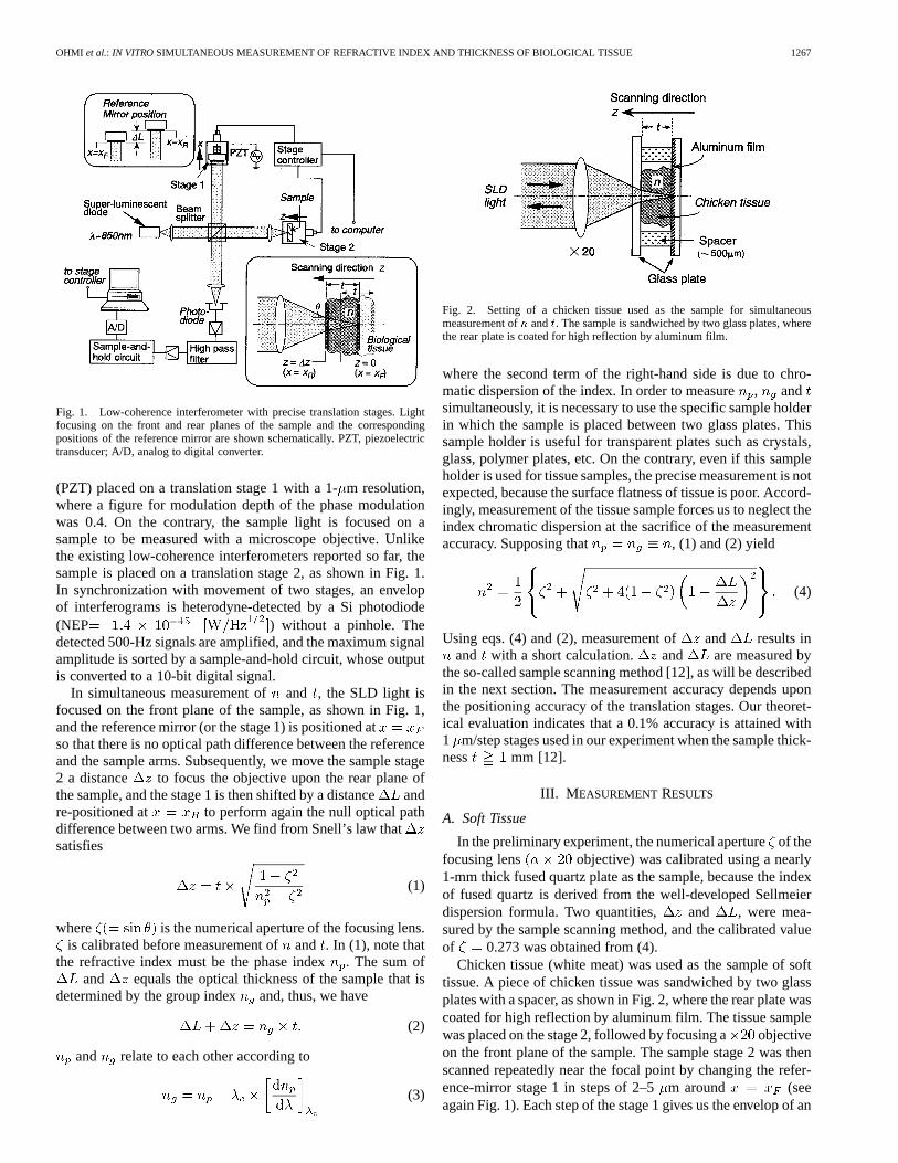
Fig. 2. Setting of a chicken tissue used as the sample for simultaneousmeasurement ofn andt. The sample is sandwiched by two glass plates, wherethe rear plate is coated for high reflection by aluminum film.
where the second term of the right-hand side is due to chro-matic dispersion of the index. In order to measure, andsimultaneously, it is necessary to use the specific sample holderin which the sample is placed between two glass plates. Thissample holder is useful for transparent plates such as crystals,glass, polymer plates, etc. On the contrary, even if this sampleholder is used for tissue samples, the precise measurement is notexpected, because the surface flatness of tissue is poor. Accord-ingly, measurement of the tissue sample forces us to neglect theindex chromatic dispersion at the sacrifice of the measurementaccuracy. Supposing that , (1) and (2) yield
(4)
Using eqs. (4) and (2), measurement of and results inand with a short calculation. and are measured by
the so-called sample scanning method [12], as will be describedin the next section. The measurement accuracy depends uponthe positioning accuracy of the translation stages. Our theoret-ical evaluation indicates that a 0.1% accuracy is attained with1 m/step stages used in our experiment when the sample thick-ness mm [12].
III. M EASUREMENTRESULTS
A. Soft Tissue
In the preliminary experiment, the numerical apertureof thefocusing lens objective) was calibrated using a nearly1-mm thick fused quartz plate as the sample, because the indexof fused quartz is derived from the well-developed Sellmeierdispersion formula. Two quantities, and , were mea-sured by the sample scanning method, and the calibrated valueof 0.273 was obtained from (4).
Chicken tissue (white meat) was used as the sample of softtissue. A piece of chicken tissue was sandwiched by two glassplates with a spacer, as shown in Fig. 2, where the rear plate wascoated for high reflection by aluminum film. The tissue samplewas placed on the stage 2, followed by focusing a objectiveon the front plane of the sample. The sample stage 2 was thenscanned repeatedly near the focal point by changing the refer-ence-mirror stage 1 in steps of 2–5m around (seeagain Fig. 1). Each step of the stage 1 gives us the envelop of an

1268 IEEE TRANSACTIONS ON BIOMEDICAL ENGINEERING, VOL. 47, NO. 9, SEPTEMBER 2000
Fig. 3. Experimental result obtained by the sample scanning method when achicken tissue was used as the sample. (a) The envelop profiles of interferogramsfor the front and rear reflection planes of the sample. (b) The envelop profileshaving the maximum peak.
Fig. 4. The envelop profiles having the maximum peak of simultaneousmeasurement ofn and t of human tooth. (a) The dentin and (b) the enamelused as the sample.
interferogram along the scanning directionof the stage 2. Thesame sample scanning was also made for the rear plane of thetissue sample. Since the sample thicknessis nearly 500 m,we can obtain sufficiently high reflection on the rear plane. Theenvelop profiles of interferograms are shown in Fig. 3(a). In the
TABLE ISIMULTANEOUS MEASUREMENT OFREFRACTIVE INDEX AND THICKNESS
OF BIOLOGICAL TISSUES
Fig. 5. Transmission microscope images and detected envelop profiles ofinterferograms of (a) a chicken bone of nearly 200�m thickness and (b) aseashell of nearly 300�m.
signal pattern for each reflection plane, the signal profile withthe maximum peak was obtained when the null optical pathdifference was performed only for light focusing upon the re-flection plane. The envelops having the maximum peak can bepicked up easily for front and rear planes, as shown in Fig. 3(b),although the envelop profiles for the rear plane are disturbed byscattering of the tissue itself, as a result, we have 375 mand m. Using (2) and (4), we obtain
1.441 and 552 m. In addition, the measured indextended to increase gradually with time due to dehydration of
chicken tissue.
B. Hard Tissue
Detection of ballistic photons is more difficult for hard tissuesuch as bone and tooth, because these are highly scatteringmedia with less water. The sample thickness, therefore, isreduced to a few hundred micron by polishing. First, enameland dentin of human tooth were separated and polished.and

OHMI et al.: IN VITROSIMULTANEOUS MEASUREMENT OF REFRACTIVE INDEX AND THICKNESS OF BIOLOGICAL TISSUE 1269
Fig. 6. Experimental result of simultaneous measurement ofn andt of a piece of crab-leg crust used as the sample of multilayer tissue.
were measured with the 1-m resolution of the translationstages, as shown in Fig. 4, as a result, refractive indices weredetermined to be 1.652 and 1.546 for enamel and dentin,respectively. The measurement results were summarized inTable I. Our method can, thus, measure clearly the indexdifference between enamel and dentin because isalmost six times as much as the measurement accuracy.
In comparison with tooth, more highly scattering tissues wereused as the sample to study influence of light scattering on theindex measurement. Experimental data are presented here fortwo typical tissue samples, chicken bone and seashell. Fig. 5shows transmission microscope images and the correspondingenvelop profiles of interferograms of a chicken bone of nearly200 m thickness and a seashell of nearly 300m. As shown intwo images, the chicken bone exhibits clearly a irregular patternof light and shade, while a regular pattern of light and shade isseen dimly on the image of seashell. This fact indicates that lightscattering is nonuniform in the chicken bone but the seashellis more crystallized tissue with uniform light scattering. Thesample scanning method provided the envelop profiles of inter-ferograms, as shown in Fig. 5, where the envelop level for therear plane is scaled up by 13.0 and 13.4 dB for the chicken boneand the seashell, respectively. On the basis of our experimentusing a variety of hard tissue samples, we found that nonuniformlight scattering in tissue produces some spurious focal points ofthe focusing lens. In fact, three distinct peaks appeared in theenvelop patterns for the rear plane of the chicken bone. In orderto distinguish a real focal point and spurious ones, the tissuesample thickness was reduced for suppression of nonuniformscattering, and thereby we found out the envelop peak corre-sponding to the real focal point, as shown in Fig. 5(a). On theother hand, the seashell gave us only one envelop peak for therear plane, because of uniform light scattering, although the re-flection was lower. The measurement results are shown again inTable I.
C. Multilayer Tissue
Usually, tissue consists of multiple layers with different com-ponents. Consider the tissue consisting oflayers where theindex and the thickness are and , respectively, in the thlayer. We also define the focusing shift from the front plane tothe rear one of theth layer and the corresponding path length as
and , respectively. By simple geometrical optics con-sideration, it is concluded that (1)–(4) are available for thethlayer only by attachment of the subscriptto , , and .Accordingly, and are determined by the envelop profilesof interferograms for the front and rear planes of theth layer.
A piece of crab-leg crust which consists of a white viscouslayer (the first layer) and semitransparent carapace (the secondlayer) was used as the sample of multilayer tissue. The mea-surement result is shown in Fig. 6, where reflection from therear plane of the second layer is 22 dB down with respect to thesample surface. Since the envelop peak is very clear for eachplane, ( , ) and ( , ) are determined easily, re-sulting in ( 1.375 , 113 m) and ( 1.411,147 m). The measurement accuracy is around 1% becauseand are measured with the 1-m resolution.
IV. DISCUSSION ANDCONCLUSION
We have presented the practical method forin vitro simul-taneous measurement of and of biological tissue by thelow coherence interferometry. In the latest measurement systemdeveloped by ourselves, the interferometer optics consists ofmierooptic components with high-resolution translation stageson an optical bench of , and the whole system iscomputer-controlled. The measurement ofand can be madeautomatically within 3 min [14], [15]. The measurement accu-racy is determined by the resolution of translation stages in-stalled in the system, whereas the coherence length of the lightsource is 12 m. When the stage resolution is 1m, the ac-curacy of 1% or less is guaranteed for the sample thicknessabove 100 m. The measurement results have been presentedfor soft, hard and multilayer tissue. Our measurement systemshould, thus, provide a practical and standard technique for theindex measurement of a variety of biological tissue.
In order to achievein vivo measurement, the focusing lensis scanned repeatedly, instead of the tissue sample, for hetero-dyne detection of the envelops of interferograms (for instance,see Fig. 1). This may be called the lens scanning method, where(1) is retained, while (2) is replaced by the expression of
. This method can provide the same measurement accuracyas the sample scanning method. In addition, the interferometeroptics will consist of a single-mode fiber directional coupler,

1270 IEEE TRANSACTIONS ON BIOMEDICAL ENGINEERING, VOL. 47, NO. 9, SEPTEMBER 2000
as used very often for the optical coherence tomography, be-cause of its compactness [16], [17]. In this scheme, however,it is rather difficult to compensate for chromatic dispersion be-tween two interference arms.
Furthermore, the index measurement technique presentedhere is applicable for distinguishing between normal anddiseased parts, leading to a novel diagnosis. In the athero-sclerosis, for instance, the index of calcified tissue increasessubstantially due to chemical and physical change of the tissue.The diagnosis of the degree of disease may require accuratemeasurement of the index change of the order of 0.001 by theuse of 0.1- m resolution translation stages.
ACKNOWLEDGMENT
The authors are grateful to T. Mitsuyama and S. Inoue, Grad-uate School of Engineering, and N. Kunizawa, Graduate Schoolof Medicine, both in Osaka University, for their helpful assis-tance and discussion in the experiment.
REFERENCES
[1] A. E. Profio, “Light transport in tissue,”Appl. Opt., vol. 28, pp.2216–2222, 1989.
[2] W. Cheong, S. A. Prahl, and A. J. Welch, “A review of the optical prop-erties of biological tissues,”IEEE J. Quantum. Electron., vol. 26, pp.2166–2185, Dec. 1990.
[3] J. M. Schmitt, A. Knuttel, and R. F. Bonner, “Measurement of opticalproperties of biological tissues by lowcoherence reflectometry,”Appl.Opt., vol. 32, pp. 6032–6042, 1993.
[4] F. P. Bolin, L. E. Preuss, R. C. Taylor, and R. J. Ference, “Refractiveindex of some mammalian tissues using a fiber optic cladding method,”Appl. Opt., vol. 28, pp. 2297–2303, 1989.
[5] H. Li and S. Xie, “Measurement method of the refractive index of bio-tissue by total internal reflection,”Appl. Opt., vol. 35, pp. 1793–1795,1996.
[6] Q. W. Song, C. Y. Ku, C. Zhang, R. B. Gross, R. R. Birge, andR. Michalak, “Modified critical angle method for measurering therefractive index of bio-optical materials and its application to bacteri-orhodopsin,”J. Opt. Soc. Amer. B, vol. 12, pp. 797–803, 1995.
[7] A. F. Fercher, K. Mengedoht, and W. Werner, “Eye-length measurementby interferometry with partially coherent light,”Opt. Lett., vol. 13, pp.186–188, 1988.
[8] C. K. Hitzen berger, “Measurement of corneal thickness by low-coher-ence interferometry,”Appl. Opt., vol. 31, pp. 6637–6642, 1992.
[9] W. Drexler, C. K. Hitzenberger, H. Sattmann, and A. F. Fercher, “Mea-surement of the thickness of fundus layers by partial coherence tomog-raphy,”Opt. Eng., vol. 34, pp. 701–710, 1995.
[10] G. J. Tearney, M. E. Brezinski, J. F. Southern, B. E. Bouma, M. R. Hee,and J. G. Fujimoto, “Determination of refractive index of highly scat-tering human tissue by optical coherence tomography,”Opt. Lett., vol.20, pp. 2258–2260, 1995.
[11] T. Fukano and I. Yamaguchi, “Simultaneous measurement of thicknessand refractive indices of multiple layers by a low-coherence confocalinterference microscope,”Opt. Lett., vol. 21, pp. 1942–1944, 1996.
[12] M. Ohmi, T. Shiraishi, H. Tajiri, and M. Haruna, “Simultaneous mea-surement of refractive index and thickness of transparent plates by lowcoherence interferometry,”Opt. Rev., vol. 4, pp. 507–515, 1997.
[13] M. Haruna, M. Ohmi, T. Mitsuyama, H. Tajiri, H. Maruyama, and M.Hashimoto, “Simultaneous measurement of the phase and group indicesand the thickness of transparent plates by low-coherence interferom-etry,” Opt. Lett., vol. 23, pp. 966–968, 1998.
[14] S. Inoue, H. Maruyama, T. Mitsuyama, M. Ohmi, K. Ihara, and M.Haruna, “A low coherence interferometer system for simultaneous mea-surement of refractive index and thickness ranging from 20�m to a fewmillimeters,” in Proc. 13th Int. Conf. Optical Fiber Sensors (OFS-13),1999, Paper Tu4-6, pp. 124–127.
[15] H. Maruyama, S. Inoue, M. Ohmi, K. Ihara, S. Nakagawa, and M.Haruna, “A practical measurement system for determination of refrac-tive index and thickness using the low coherence interferometry,”Proc.SPIE,, Int. Soc. for Optical Engineering, vol. 3740, pp. 26–29, 1999.
[16] D. Huang, E. A. Swanson, C. P. Lin, J. S. Schuman, W. G. Stinson,W. Chang, M. R. Hee, T. Flotte, K. Gregory, C. A. Puliafito, and J.G. Fujimoto, “Optical coherence tomography,”Science, vol. 254, pp.1178–1181, 1991.
[17] E. A. Swanson, J. A. Izatt, M. R. Hee, D. Huang, C. P. Lin, J. S. Schuman,C. A. Puliafito, and J. G. Fujimoto, “In vivo retinal imaging by opticalcoherence tomography,”Opt. Lett., vol. 18, pp. 1864–1866, 1993.
Masato Ohmi received the B.S. degrees in electricalengineering from Fukui University, Fukui, Japan, in1990 and M.S., and Ph.D. degrees in electromagneticenergy engineering from Osaka University, Osaka,Japan, in 1992 and 1995, respectively.
Since 1995, he has been a Research Associatein Medical Engineering Laboratory, School ofAllied Health Sciences, Faculty of Medicine,Osaka University. His present research interest is inbiomedical optics, including tissue measurement,optical tomography, etc.
Dr. Ohmi is a member of Japanese Society of Medical Electronics and Bio-logical Engineering, Japan Society of Applied Physics, and the Laser Societyof Japan.
Yasuhito Ohnishi received the B.S. degrees in alliedhealth sciences from Osaka University, Osaka Japan,in 1998. He is working toward the M.S. degree inthe Department of Allied Health Sciences, GraduateSchool of Medicine, Osaka University.
His research subject is optical imaging.
Koji Yoden received the B.S. degrees in alliedhealth sciences from Osaka University, Osaka Japan,in 1998. He is working toward the M.S. degree inthe Department of Allied Health Sciences, GraduateSchool of Medicine, Osaka University.
His research subject is tissue measurement.
Masamitsu Haruna was born in Ishikawa, Japan,in 1945. He received the B.S., M.S., and Ph.D. de-grees in electronics engineering from Osaka Univer-sity, Osaka, Japan, in 1968, 1970, and 1974, respec-tively.
In 1973, he joined the Department of Electronics,Faculty of Engineering, Osaka University, as aResearch Associate, and became an AssociateProfessor in 1988, where he was engaged in researchon integrated optics, lightwave sensing includinglaser Doppler velocimetry, laser-beam writing and
laser ablation. From 1978 to 1980, he was a Visiting Research Associate inDepartment of Electrical Engineering, McGill University, Montreal, Canada.Since 1994, he has been a Professor in Medical Engineering Laboratory,School of Allied Health Sciences, Faculty of Medicine, Osaka University.His present research interest is in biomedical optics based on optoelectronics,including tissue measurement using the low coherence interferometry, opticaltomography, tissue ablation, and diagnoses for optical biopsy, etc.
Dr. Haruna is a member of the International Society for Optical Engineering,Japanese Society of Medical Electronics and Biological Engineering, the Op-tical Society of Japan, Japan Society of Applied Physics, the Biophysical So-ciety of Japan, and the Laser Society of Japan.