hla typing, fa diagnosis and gel electrophoresis · pdf filethe results of this analysis on...
TRANSCRIPT

470 UCB Boulder, CO 80309-0470Phone: 303-492-8230, Fax: 303-492-4916,www.colorado.edu/Outreach/BSI
Sponsored by the University of Colorado at Boulder
HLA typing, FA diagnosis and Gel Electrophoresis
AcknowledgementsThe activity itself is based on information found inV. Verlinsky et al, 2001, Preimplantation Diagnosis for Fanconi Anemia Combinedwith HLA Matching, JAMA: 285, 3130-3133. andS. Grewal et al, 2004, Successful hematopoietic stem cell transplantation for Fanconianemia from an unaffected HLA-genotype-identical sibling selected usingpreimplantation genetic diagnosis, Blood: 3, 1147-1151Note: the Nash family is not identified in the above papers, so although we discussingthe case of the Nash family in today’s workshop, the information may not be that ofthe Nash family.
IntroductionIn this activity you will use a simulated form of gel electrophoresis to analyze the HLAtype and FANCC genes of one cell from several embryos that were created in vitro.This activity is based on a real medical case, real genotypes, and real embryos.
BackgroundA family wanted to have a second child who would not have FA, and who would bean HLA match for the first child. This would allow them to use the cord bloodfollowing birth as a source of cells for a bone marrow transplant for their first child.
In vitro fertilization was performed that resulted in 14 embryos to be screened. At the8-cell stage, a single cell was removed from each of the embryos. Then, two PCRreactions were performed on each sample, one to examine the alleles present at theFANCC locus, and the other to determine the HLA type of the embryo. For 3 embryos,no PCR product was obtained for one of the two reactions (doctors were unable todetermine either FA status or HLA type on the embryo because the reaction didn’twork). You will look at the results from the remaining 11 embryos.

The genotypes of members of the family are shown below.
FANCC PCRThe FANCC mutation carried by both parents in this case is the same; ivs4+4. To testfor ivs4+4, a PCR reaction was performed to amplify a portion of exon and intron 4from the FANCC gene. The primers were designed such that a ScaI site would begenerated in the product from the normal allele but not the ivs4+4 allele. FollowingPCR amplification, the PCR product was digested with ScaI and run on a gel.
JAMA 285:3130
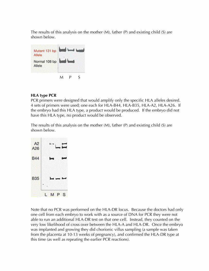
The results of this analysis on the mother (M), father (P) and existing child (S) areshown below.
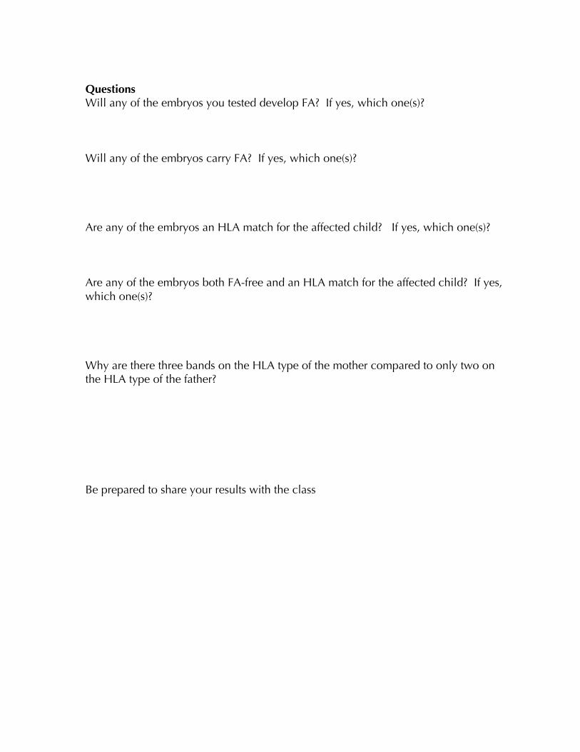
HLA type PCRPCR primers were designed that would amplify only the specific HLA alleles desired.4 sets of primers were used; one each for HLA-B44, HLA-B35, HLA-A2, HLA-A26. Ifthe embryo had this HLA type, a product would be produced. If the embryo did nothave this HLA type, no product would be observed.
The results of this analysis on the mother (M), father (P) and existing child (S) areshown below.
Note that no PCR was performed on the HLA-DR locus. Because the doctors had onlyone cell from each embryo to work with as a source of DNA for PCR they were notable to run an additional HLA-DR test on that one cell. Instead, they counted on thevery low likelihood of cross over between the HLA-A and HLA-DR. Once the embryowas implanted and growing they did chorionic villus sampling (a sample was takenfrom the placenta at 10-13 weeks of pregnancy), and confirmed the HLA-DR type atthis time (as well as repeating the earlier PCR reactions).
M P S

ProcedureYou are given samples of DNA representing several individuals:1) The mother (labeled M)2) The father (labeled D)3) The affected child (labeled AC)4) One cell from 2 or 3 embryos per gel.
As a class you will run all of the embryos to be tested (numbered 1 – 11)
You performed a PCR analysis on these samples from two lociThe FANCC locus (labeled FA)The HLA locus (labeled HLA)
♦ Set up two gels and gel boxes according to the instructions for Gel Electrophoresis.
♦ Load the gels with placing all the FA samples on one gel and all the HLA sampleson the other gel.
♦ Label your gels so that you know which sample is in each well.
♦ Run the gels at 100 volts for approximately 20 minutes.
Sketch the results from both gels below.
FA Results HLA results
Based on your results,
QuestionsWill any of the embryos you tested develop FA? If yes, which one(s)?
Will any of the embryos carry FA? If yes, which one(s)?
Are any of the embryos an HLA match for the affected child? If yes, which one(s)?
Are any of the embryos both FA-free and an HLA match for the affected child? If yes,which one(s)?
Why are there three bands on the HLA type of the mother compared to only two onthe HLA type of the father?
Be prepared to share your results with the class

Note: Embryo 2 on these gels (numbered embryo 3 in the paper and preceedingpower point presentation) was chosen by doctors as FA-free and an HLA match. Thisembryo was implanted and resulted in a healthy baby (now 6 years old). The cordblood was saved and used to successfully transplant the affected child, who is now 11.