gas6 in ards patients: determination of plasma levels...
TRANSCRIPT
1
GAS6 IN ARDS PATIENTS: DETERMINATION OF PLASMA LEVELS AND
INFLUENCE OF PEEP SETTING
Running title: Gas6 in ARDS.
Jean-Luc Diehl MD* (1,2); Nathalie Coolen MD (1); Christophe Faisy MD (1); David Osman
MD (3); Gwenaël Prat MD (4); Mustapha Sebbane MD (5); Ania Nieszkowska MD (6);
Claude Gervais MD (7); Jean-Christophe M Richard MD (8); Jack Richecoeur MD (9);
Laurent Brochard MD (10); Alain Mercat MD (11), Emmanuel Guérot MD (1); Delphine
Borgel PhD (12,13)
(1) Service de Réanimation Médicale, Hôpital Européen Georges Pompidou, 20 rue Leblanc,
75015 Paris, France; (2) INSERM UMR_S765, Faculté de Pharmacie, Université Paris
Descartes, Sorbonne Paris Cité, 4 avenue de l'Observatoire, 75006 Paris, France (3) Service
de Réanimation Médicale, CHU Bicêtre, 78 rue du Général Leclerc, 94275 Le Kremlin
Bicêtre, France ; (4) Service de Réanimation Médicale, CHU de la Cavale Blanche, boulevard
Tanguy Prigent, 29609 Brest, France (5) Anesthésie Réanimation, Hôpital Saint Eloi, 80
Avenue Augustin Fliche, 34295, Montpellier, France (6) Service de Réanimation Polyvalente,
CHU de la Pitié Salpétrière, 47 Boulevard de l’Hôpital, 75013 Paris, France (7) Service de
Réanimation Médicale, GHU Caremeau, Place du Professeur Robert Debré, 30029 Nîmes,
France (8) Service de Réanimation Médicale, CHU de Rouen, 1 rue de Germont, 76031
Rouen, France (9) Service de Réanimation Polyvalente, Hôpital de Pontoise, 6 Avenue de
l’Ile-de-France, 95303 Pontoise, France (10) Service de Soins Intensifs, Hôpitaux
Universitaires de Genève, CH-1211 Geneve Cedex 14, Suisse (11) Service de Réanimation
Médicale et de Médecine Hyperbare, CHU d’Angers, 4 rue Larrey, France (11), Service
d’Hématologie, Hôpital Européen Georges Pompidou, 20 rue Leblanc, 75015 Paris, France
(12) Université Paris-Sud, EA4531, F-92296 Châtenay-Malabry Cedex, France
* corresponding author. Tel : 33 1 56093201 ; fax : 33 1 56093202 ; mail : [email protected]
Respiratory Care
RESPIRATORY CARE Paper in Press. Published on April 09, 2013 as DOI: 10.4187/respcare.02129
Copyright (C) 2013 Daedalus Enterprises Epub ahead of print papers have been peer-reviewed and accepted for publication but are posted before being copy edited
and proofread, and as a result, may differ substantially when published in final version in the online and print editions of RESPIRATORY CARE.
2
ABSTRACT
Purpose: Growth arrest-specific protein 6 (Gas6) is a vitamin K-dependent protein expressed
by endothelial cells and leukocytes participating in cell survival, migration and proliferation
and involved in many pathological situations. The aim of our study was to assess its
implication in acute respiratory distress syndrome (ARDS) and its variation according to
positive end expiratory pressure (PEEP) setting, considering that different cyclic stresses
could alter Gas6 plasma levels.
Methods: Our patients were enrolled in the ExPress study comparing a minimal alveolar
distension (“low PEEP”) ventilatory strategy to a maximal alveolar recruitment (“high
PEEP”) strategy in ARDS. Plasma Gas 6, IL8 and VEGF levels were measured at day 0 and
day 3 by enzyme-linked immunosorbent assay in blood samples prospectively collected
during the study for a subset of 52 patients included in 8 centers during year 2005.
Results: We found that Gas6 plasma level was elevated in the whole population at day 0: 106
ng/mL (77-139), (median, IQR), with significant correlations with IL8, the Simplified Acute
Physiologic Score II and the Organ Dysfunction and Infection (ODIN) scores. Statistically
significant decreases in Gas6 and IL 8 plasma levels were observed between day 0 and day 3
in the “high PEEP” group (P=0.017); while there were no differences between day 0 and day
3 in the “low PEEP” group.
Conclusions: Gas6 plasma level is elevated in ARDS patients. The “high PEEP” strategy is
associated with a decrease in Gas6 and IL8 plasma levels at day 3, without significant
differences in day 28 mortality between the 2 groups.
Trial registration: clinicaltrials.gov Identifier: NCT00188058
Respiratory Care
RESPIRATORY CARE Paper in Press. Published on April 09, 2013 as DOI: 10.4187/respcare.02129
Copyright (C) 2013 Daedalus Enterprises Epub ahead of print papers have been peer-reviewed and accepted for publication but are posted before being copy edited
and proofread, and as a result, may differ substantially when published in final version in the online and print editions of RESPIRATORY CARE.
3
INTRODUCTION
Growth arrest-specific protein 6 (Gas6) is a vitamin K-dependant protein sharing 43% of
homology with a natural anticoagulant, protein S (1). Gas6 is expressed in various cell types,
including endothelial cells, particularly in pro-apoptotic conditions (2). Leukocytes have also
been found to release Gas6 (3-4). It is a ligand for 3 tyrosine kinase receptors (Axl, Tyro3 and
Mer) whose signaling is implicated primarily in cell survival but also in cell proliferation,
adhesion and migration (5).
Among several functions, previous studies have reported the implication of Gas6 in the
inflammatory process, particularly in the physiopathology of severe sepsis (3-4, 6-7). Indeed,
Gas6 enhances the interplay of cells implicated in the inflammatory response, endothelial
cells, leukocytes and platelets during different conditions of experimental inflammation (4).
Models of endotoxemia also suggest an important role of modulation of the immune response
(4-5, 8-11).
Given the involvement of Gas6 receptors in experimental sepsis, and the potentially major
role of leukocyte apoptosis in the pathophysiology of severe sepsis, previous clinical studies
have focused on Gas6 in septic and non-septic critical care patients (3, 6-7). As compared to
healthy subjects, levels were higher in critical care patients with one or several failing organs,
the highest values being observed in patients with severe sepsis (3). Specifically in severe
sepsis patients, a correlation was observed between the number of organ dysfunctions (as
reflected by scores such as SOFA and ODIN) reflecting the degree of tissue injury, and Gas6
plasma concentrations (3, 6).
Gas6 can be released by endothelial cells and leukocytes, which are largely implicated in
the pathophysiology of acute respiratory distress syndrome (ARDS). Since the pulmonary
vascular bed is subjected to cyclic stress in patients with ARDS, and despite the lack of in
Respiratory Care
RESPIRATORY CARE Paper in Press. Published on April 09, 2013 as DOI: 10.4187/respcare.02129
Copyright (C) 2013 Daedalus Enterprises Epub ahead of print papers have been peer-reviewed and accepted for publication but are posted before being copy edited
and proofread, and as a result, may differ substantially when published in final version in the online and print editions of RESPIRATORY CARE.
4
vitro or experimental studies evidencing that Gas6 could be modulated by such cyclic stress,
we hypothesized that the course of plasma Gas6 levels could differ according to different
ventilatory strategies. The aim of this study was to use a multicenter randomized controlled
trial therefore to compare the course of plasma Gas6 levels in patients with ARDS from the
Expiratory Pressure (ExPress) Study Group (12). Briefly, patients were randomly assigned
either to a “low PEEP” strategy (minimal alveolar distension strategy) or to a “high PEEP”
strategy (increased alveolar recruitment strategy). The “high PEEP” strategy, avoiding in part
consequences of cyclic collapse and excessive hyperinflation, could therefore be associated
with less injury and lower plasma Gas6 levels. Additionally, we measured plasma levels of
IL-8 and VEGF, as established endothelial and leukocyte markers with important implications
in the context of ARDS (13-22).
METHODS
We measured plasma levels of Gas 6, IL-8 and VEGF in a subset of ARDS patients enrolled
in the ExPress Study. The 767 enrolled patients were randomized in two groups. In the “low
PEEP” strategy group, PEEP and inspiratory plateau pressure were kept as low as possible
without falling below oxygenation targets (SpO2: 88% and/or PaO2: 55 MmHg). External
PEEP was set to maintain total PEEP (the sum of external and intrinsic PEEP) between 5 and
9 cm H2O. In the “high PEEP” strategy group, PEEP was adjusted based on airway pressure
and was kept as high as possible without increasing the maximal inspiratory plateau pressure
above 28 to 30 cm H2O. For both groups, tidal volume (VT) was set at 6 mL/kg of predicted
body weight.
Plasma Gas6, IL-8 and VEGF levels were measured in blood samples prospectively collected
during the last year (2005) of the ExPress study at day 0 and day 3 in 8 selected centers.
Indeed, the present study concerned only year 2005, as the rational originated from results
Respiratory Care
RESPIRATORY CARE Paper in Press. Published on April 09, 2013 as DOI: 10.4187/respcare.02129
Copyright (C) 2013 Daedalus Enterprises Epub ahead of print papers have been peer-reviewed and accepted for publication but are posted before being copy edited
and proofread, and as a result, may differ substantially when published in final version in the online and print editions of RESPIRATORY CARE.

5
demonstrating the implication of Gas6 in severe sepsis obtained shortly before 2005 (3).
Given these conditions, 52 patients were included in the present study: 24 in the “low PEEP”
strategy group and 28 in the “high PEEP” strategy group. The study protocol (approval
number: 2002/09, 08-Jul-2002) and the corresponding amendment (amendment # 8; 15-Feb-
2005) were approved by the ethics committee of the Angers University Hospital (Comité
Consultatif de Protection des Personnes dans la Recherche Biomédicale). Measurement of
plasma Gas6, IL-8 and VEGF levels were therefore determined by enzyme-linked
immunosorbent assays (ELISA) as previously described (23-24).
Statistical analysis
Continuous data are expressed as medians and interquartile ranges (IQR) and compared
using non parametric tests (Mann-Whitney test, Wilcoxon sign rank test or Kruskal-Wallis
test as appropriate). Results are expressed as total number (percentage) for categorical.
Comparison of categorical variables was performed by the Chi-squared test. Correlations
were assessed with the non parametric Spearman correlation test or with the non parametric
test for trend, as appropriate. A p value less than 0.05 was considered significant. Analyses
were performed using the StatView software (Abacus Concepts, Berkeley, CA).
RESULTS
Plasma Gas6, IL-8 and VEGF levels were measured at day 0 and day 3 in 52 patients from the
ExPress study (7 % of the 768 patients). The main clinical characteristics at inclusion,
including classification according to the new Berlin definition of ARDS (25), are shown in
Table 1. Characteristics at day 3 and relevant physiological and clinical end-points until day
28 are shown in Table 2.
Respiratory Care
RESPIRATORY CARE Paper in Press. Published on April 09, 2013 as DOI: 10.4187/respcare.02129
Copyright (C) 2013 Daedalus Enterprises Epub ahead of print papers have been peer-reviewed and accepted for publication but are posted before being copy edited
and proofread, and as a result, may differ substantially when published in final version in the online and print editions of RESPIRATORY CARE.
6
As expected, there was a statistically significant difference between PEEP levels at day 3: 7
cmH2O (5-8) vs 15 cmH2O (10-17), while there were no differences in PEEP levels at day 0,
nor in VT levels at days 0 and at day 3.
The main biological results at day 0 and day 3 are shown in Table 3. Gas6 plasma level was
very high in patients at the onset of ARDS: 106 ng/mL (77 - 139), with no statistically
significant difference between the “low PEEP” group and the “high PEEP” group: 121 ng/mL
(80 – 143) vs 93 ng/mL (73 – 123) respectively, P = 0.17 (Figure 1). Considering all 52
patients, there was a trend (P = 0.15) to lower Gas6 values in the less severe ARDS according
to the Berlin classification (25): mild ARDS: 92 ng/mL (80 – 96), moderate ARDS: 115
ng/mL (82-143) and severe ARDS: 127 ng/mL (67-144).
Gas6 correlated positively with the severity of disease, as assessed by the Simplified Acute
Physiologic Score II (ρ = 0.43, P = 0.002) and the Organ Dysfunction and Infection (ODIN)
score (ρ = 0.40, P = 0.007), which indicates the number of organ dysfunction (varying
between 0 and 7, including clinically evident infection as an organ dysfunction). There was no
correlation between Gas6 level at day 0 and PEEP and VT levels. There was no difference
between Gas6 levels at day 0 between septic (n = 33) and non-septic patients (n = 19).
A statistical difference between plasma Gas6 levels at day 0 and day 3 was found in the “high
PEEP” group (P = 0.017) but not in “low PEEP” group (P=0.83).
We found elevated plasma levels of IL-8 at day 0, with no statistical difference between the 2
groups. There was a statistically significant correlation between plasma levels of Gas6 and of
IL-8 at day 0 (ρ = 0.40, P = 0.006), but not at day 3. A statistical difference between plasma
IL-8 levels at day 0 and day 3 was found in the “high PEEP” group (P = 0.02) but not in “low
PEEP” group (P=0.63).
We found elevated plasma levels of VEGF at day 0, with no statistical difference between the
2 groups. There was no statistically significant correlation between plasma levels of VEGF
Respiratory Care
RESPIRATORY CARE Paper in Press. Published on April 09, 2013 as DOI: 10.4187/respcare.02129
Copyright (C) 2013 Daedalus Enterprises Epub ahead of print papers have been peer-reviewed and accepted for publication but are posted before being copy edited
and proofread, and as a result, may differ substantially when published in final version in the online and print editions of RESPIRATORY CARE.
7
and Gas6 either at day 0 or at day 3. Similarly, there was no statistically significant
correlation between plasma levels of VEGF and of IL-8 either at day 0 or at day 3. Finally,
there were no statistically differences between VEGF levels at day 0 and at day 3, either in the
“high PEEP” or in the “low PEEP” groups.
There were no differences between IL-8 and VEGF levels at day 0 between septic and non-
septic patients.
DISCUSSION
As expected, according to a previous study among more heterogeneous critically ill patients,
Gas6 plasma level was very high in patients at the onset of ARDS: 106 ng/mL (77 - 139) as
compared to reference values observed in spontaneously breathing healthy subjects: 54 ng/mL
(49-68) (3). We found also elevated plasma levels of IL-8 and VEGF at the onset of ARDS,
confirming previous series (13-15, 17,19-21). A positive correlation was found between Gas6
and IL-8 plasma levels. A significant decrease in Gas6 and IL-8 plasma levels was observed
in the “high PEEP” group, but not in the “low PEEP” group.
This is the first report of elevated Gas6 plasma levels in a homogeneous group of ARDS
patients. Obviously, over-expression of Gas6 was very likely in relation with disease severity
(as assessed by correlation with SAPS II and ODIN scores) and sepsis (33 patients in the
series). Since it was previously proposed that Gas6 originates from endothelial cells and
leukocytes (3-4), increase in Gas6 production could also in part be related to pulmonary
neutrophil infiltration and to diffuse pulmonary endothelial damage which are key features of
ARDS (13, 26). Moreover, despite the lack of surrounding experimental studies, it was
tempting to assume that mechanical ventilation, by applying cyclic stress over pulmonary
endothelial cells and by exacerbating leukocytes’ stimulation, could per se be a contributing
factor to the high Gas6 plasma levels observed at day 0 and day 3.
Respiratory Care
RESPIRATORY CARE Paper in Press. Published on April 09, 2013 as DOI: 10.4187/respcare.02129
Copyright (C) 2013 Daedalus Enterprises Epub ahead of print papers have been peer-reviewed and accepted for publication but are posted before being copy edited
and proofread, and as a result, may differ substantially when published in final version in the online and print editions of RESPIRATORY CARE.
8
As previously described in patients with severe sepsis (3,6), Gas6 correlated positively with
the severity of disease, as assessed by the Simplified Acute Physiologic Score II and the
ODIN score. The correlation values were in the same range that previous published data from
our group and from others adding external validity to our results (3,6). However, they are just
indicators of some degrees of association between the severity of the disease, as assessed by
the scores, and the Gas6 plasma levels; any causal relationship between these parameters
remaining to be investigated. We found no correlation between Gas6 level at day 0 and PEEP
and VT levels, parameters which obviously influence cyclic stress exerted on pulmonary
vasculature. However, one can observe that VT at day 0 was set at a level close to 6 mL/kg
IBW, a level known to minimize lung stress (15). Therefore, the chance to observe a
statistical correlation between Gas6 level and VT as a marker of lung stress was minimized.
There was also a rather narrow spectrum of PEEP settings at day 0, which could explain at
least in part the lack of correlation with Gas6 level at day 0. Unfortunately, we didn’t measure
in our patients transpulmonary pressures, which could have provided a better surrogate of the
stress applied on the pulmonary vasculature.
The statistical difference between plasma Gas6 levels at day 0 and day 3 could be explained,
at least in part, by a better modulation of the stress applied on the pulmonary vascular and
endothelial bed in the “high PEEP” group, mainly by preventing cyclic reopening of collapsed
pulmonary areas. Accordingly, we observed no significant variation in Gas6 level in the “low
PEEP” group, in which there was no significant differences in PEEP level between day 0 and
day 3. In contrast, we observed in the “high PEEP” group a significant decrease in Gas6 level
in parallel with an increase in PEEP level from 8 cmH2O (5-10) to 15 cmH2O (10-17). Such
differences according to ventilatory strategies have previously been reported for inflammatory
mediators and apoptosis markers both at the pulmonary and at the systemic levels (12, 15). In
Respiratory Care
RESPIRATORY CARE Paper in Press. Published on April 09, 2013 as DOI: 10.4187/respcare.02129
Copyright (C) 2013 Daedalus Enterprises Epub ahead of print papers have been peer-reviewed and accepted for publication but are posted before being copy edited
and proofread, and as a result, may differ substantially when published in final version in the online and print editions of RESPIRATORY CARE.
9
the same way, PEEP setting could have had an impact on plasma Gas6 levels in our ARDS
patients.
We found elevated plasma levels of IL-8 at day 0 in accordance with previous series of ARDS
patients (13-15, 17, 20-21). Given the implication of IL-8 as an important pro-inflammatory
mediator associated with the development of ARDS and with poor outcomes (increase in
mortality and decrease in ventilator-free days), the observed correlation with Gas6 suggest
that Gas6 could act as an important co-factor participating to exacerbated inflammation in the
context of ARDS
We found elevated plasma levels of VEGF at day 0, confirming previous results from
Azamfirei (19). However, such an elevation was not observed in another series (22).
Moreover, differential expression of VEGF at the pulmonary and plasma levels has previously
been reported (19). Finally, the implication of VEGF in the early ARDS context is not fully
established since it can promote an increase in vascular permeability but also exert a
protective vascular effect (19, 22, 27). Therefore, the lack of correlation between plasma Gas6
and VEGF levels could reflect the fact that plasma VEGF levels probably poorly reflect local
pulmonary inflammation.
In the ExPress study, the “high PEEP” strategy was associated with a clinical benefit in term
of oxygenation, higher compliance values, less ventilator-free days and less organ failure-free
days (12). In the present study, we observed in a representative subset of patients a decrease
in Gas6 plasma levels at day 3 in the “high PEEP“ group, unlike the “low PEEP” group. This
was in parallel to better oxygenation and higher compliance values at day 3, suggesting a
possible relationship between such clinical benefits and these biological findings. Results of
extensive animal experimentations from Tjwa et al (4) can also support the hypothesis of a
possible relationship between such biological findings and clinical benefits: the authors
reported that Gas6 could be involved in the inflammatory process and could enhance the
Respiratory Care
RESPIRATORY CARE Paper in Press. Published on April 09, 2013 as DOI: 10.4187/respcare.02129
Copyright (C) 2013 Daedalus Enterprises Epub ahead of print papers have been peer-reviewed and accepted for publication but are posted before being copy edited
and proofread, and as a result, may differ substantially when published in final version in the online and print editions of RESPIRATORY CARE.
10
inflammatory response in pathological conditions like sepsis or ARDS. Moreover, they
suggested that the inhibition of Gas6 might warrant further consideration as a novel strategy
for the treatment of sepsis. Importantly, such inhibition of Gas6 should not be interpreted as a
decrease to information to cell survival but rather to a return to a controlled physiological
state, therefore limiting the Gas6-induced exacerbation of inflammation.
We also found a decrease in plasma IL-8 levels between day 0 and day 3 in the “high PEEP”
group, but not in the “low PEEP” group. Considering the key role of IL-8 in ARDS, such a
result can also suggest a beneficial effect of the “high PEEP” strategy and confirms previous
series demonstrating a link between respiratory settings and IL-8 levels (13, 15).
The first limitation to our study is that we cannot distinguish between the distinct effects of
pulmonary insult and mechanical ventilation on the results of Gas6 plasma measurements
from healthy subjects, since they cannot be subjected to a short course of mechanical
ventilation. The generalization of the present results is also a critical point: measurements had
been possible only in a limited number of centers participating to the ExPress study for
technical reasons, and only during the last year of the study. The two groups were clinically
comparable and similar to the whole population of the ExPress study, but given the relatively
low number of patients, one can not formally exclude the possibility of type two errors with
regard to baseline characteristics, and especially to the repartition of the patients between the
3 ARDS categories. Another limitation to our study is that the measurements were limited to
the plasma compartment. Our hypothesis is that plasma Gas6 originates at least in part from
the pulmonary compartment; however only BAL studies performed in ARDS patients could
support it, and unfortunately such Gas6 BAL measurements were not possible planed during
the ExPress study. Another limitation of the present study is the lack of measurement of Gas6
plasma level after day 3, precluding any conclusion on the implication of Gas6 in the end-
stage ARDS. However, it’s generally admitted that Gas6 acts as an acute phase reactant (28).
Respiratory Care
RESPIRATORY CARE Paper in Press. Published on April 09, 2013 as DOI: 10.4187/respcare.02129
Copyright (C) 2013 Daedalus Enterprises Epub ahead of print papers have been peer-reviewed and accepted for publication but are posted before being copy edited
and proofread, and as a result, may differ substantially when published in final version in the online and print editions of RESPIRATORY CARE.
11
In conclusion, we found that plasma Gas6 level is elevated at the onset of ARDS and that a
“high PEEP” strategy was associated with a decrease in Gas6 plasma concentration over 3
days. Further studies are now warranted to confirm that the pulmonary compartment is a
major contributor to the high plasma values of Gas6, to extend our knowledge about the
kinetic of plasma Gas6 levels in the course of ARDS and finally to better delineate the
prognostic and pathogenetic values of Gas6 as a biomarker in ARDS.
Respiratory Care
RESPIRATORY CARE Paper in Press. Published on April 09, 2013 as DOI: 10.4187/respcare.02129
Copyright (C) 2013 Daedalus Enterprises Epub ahead of print papers have been peer-reviewed and accepted for publication but are posted before being copy edited
and proofread, and as a result, may differ substantially when published in final version in the online and print editions of RESPIRATORY CARE.
12
ACKNOWLEDGMENTS AND AUTHORS’ CONTRIBUTIONS
The authors thank Véronique Remones for her excellent technical assistance.
JLD, JCMR, LB, AM, EG and DB contributed to the design of the study. J-LD, NC, CF, DO,
GP, MS, AN, CG, EG, JR, JCMR, LB, AM collected the data. DB was responsible for
biological measurements. JLD, NC and DB wrote the manuscript. All authors read and
approved the manuscript.
FINANCIAL SUPPORT
The ExPress study was supported by the French Minister of Health (Programme Hospitalier
de Recherche Clinique).
CONFLICT OF INTEREST STATEMENT
The authors have no conflict of interest to disclose.
Respiratory Care
RESPIRATORY CARE Paper in Press. Published on April 09, 2013 as DOI: 10.4187/respcare.02129
Copyright (C) 2013 Daedalus Enterprises Epub ahead of print papers have been peer-reviewed and accepted for publication but are posted before being copy edited
and proofread, and as a result, may differ substantially when published in final version in the online and print editions of RESPIRATORY CARE.
13
REFERENCES
1. Manfioletti G, Brancolini C, Avanzi G, Schneider C. The protein encoded by a growth
arrest-specific gene (gas6) is a new member of the vitamin K-dependent proteins
related to protein S, a negative coregulator in the blood coagulation cascade. MolCell
Biol 1993; 13:4976-85.
2. Hasanbasic I, Cuerquis J, Varnum B, Blostein MD. Intracellular signaling pathways
involved in Gas6-Axl-mediated survival of endothelial cells. Am J Physiol Heart Circ
Physiol 2004; 287:H1207-13.
3. Borgel D, Clauser S, Bornstain C, Bièche I, Bissery A, Remones V, et al. Elevated
growth-arrest-specific protein 6 plasma levels in patients with severe sepsis. Crit Care
Med 2006; 34:219-22.
4. Tjwa M, Bellido-Martin L, Lin Y, Plaisance S, Bono F, Delesque-Touchard N et al.
Gas6 promotes inflammation by enhancing interactions between endothelial cells,
platelets and leukocytes. Blood 2008; 111:4096-105.
5. Camenisch TD, Koller BH, Earp HS, Matsushima GK. A novel receptor tyrosine
kinase, Mer, inhibits TNF-alpha production and lipopolysaccharide-induced endotoxic
shock. J Immunol 1999; 162:3498-503.
6. Gibot S, Massin F, Cravoisy A, Dupays R, Barraud D, Nace L, et al. Growth arrest-
specific protein 6 plasma concentrations during septic shock. Crit Care 2007; 11:R8.
doi: 10.1186/cc5158.
7. Ekman C, Linder A, Akesson P, Dahlback B. Plasma concentrations of Gas6 (growth
arrest specific protein 6) and its soluble receptor sAxl in sepsis and systemic
inflammatory responses syndromes. Crit Care 2010; 14:R158 doi: 10.1186/cc9233.
Respiratory Care
RESPIRATORY CARE Paper in Press. Published on April 09, 2013 as DOI: 10.4187/respcare.02129
Copyright (C) 2013 Daedalus Enterprises Epub ahead of print papers have been peer-reviewed and accepted for publication but are posted before being copy edited
and proofread, and as a result, may differ substantially when published in final version in the online and print editions of RESPIRATORY CARE.
14
8. Sather S, Kenyon KD, Lefkowitz JB, Liang X, Varnum BC, Henson PM, et al. A
soluble form of the Mer receptor tyrosine kinase inhibits macrophage clearance of
apoptotic cells and platelet aggregation. Blood 2007; 109:1026-33.
9. Scutera S, Fraone T, Musso T, Cappello P, Rossi S, Pierobon D, et al. Survival and
migration of human dendritic cells are regulated by an IFN-alpha-inducible Axl/Gas6
pathway. J Immunol. 2009;183:3004-13.
10. Alciato F, Sainaghi PP, Sola D, Castello L, Avanzi GC. TNF-alpha, IL-6, and IL-1
expression is inhibited by GAS6 in monocytes/macrophages. J Leukoc Biol 2010
87:869-75.
11. Feng X, Deng T, Zhang Y, Su S, Wei C, Han D. Lipopolysaccharide inhibits
macrophage phagocytosis of apoptotic neutrophils by regulating the production of
tumour necrosis factor α and growth arrest-specific gene 6. Immunology 2011;
132:287-95.
12. Mercat A, Richard JC, Vielle B, Jaber S, Osman D, Diehl JL, et al. Positive end-
expiratory pressure setting in adults with acute lung injury and acute respiratory
distress syndrome: a randomized controlled trial. JAMA 2008 299:646-55.
13. Ranieri VM, Suter PM, Tortorella C, De Tullio R, Dayer JM, Brienza A, et al. Effect
of mechanical ventilation on inflammatory mediators in patients with acute respiratory
distress syndrome: a randomized controlled trial. JAMA 1999; 282:54-61.
14. Bouros D, Alexandrakis MG, Antoniou KM, Agouridakis P, Pneumatikos I, Anevlavis
S, et al. The clinical significance of serum and broncho-alveolar lavage inflammatory
cytokines in patients at risk for acute respiratory distress syndrome. BMC Pulmonary
Medicine 2004; 4 6 doi:10.1186/1471-2466-4-6
Respiratory Care
RESPIRATORY CARE Paper in Press. Published on April 09, 2013 as DOI: 10.4187/respcare.02129
Copyright (C) 2013 Daedalus Enterprises Epub ahead of print papers have been peer-reviewed and accepted for publication but are posted before being copy edited
and proofread, and as a result, may differ substantially when published in final version in the online and print editions of RESPIRATORY CARE.
15
15. Parsons P, Eisner MD, Thompson BT, Matthay M, Ancukiewicz M, Bernard GR, et
al. Lower tidal volume ventilation and plasma cytokine markers of inflammation in
patients with acute lung injury. Crit Care Med 2005; 33:1-6.
16. Medford ARL, Ibrahim NBN, Millar AB. Vascular endothelial growth factor receptor
and coreceptor expression in human acute respiratory distress syndrome. J Crit Care
2009; 24:236-243.
17. Hildebrand F, Stuhrmann M, van Griensven M, Meier S, Hasenkamp S, Krettek C, et
al. Association of IL-8-251A/T polymorphism with incidence of acute respiratory
distress syndrome (ARDS) and IL-8 synthesis after multiple trauma. Cytokine 2007;
37: 192-199.
18. Wei-Chieh W, Chiou-Feng L, Chia-Ling C, Chang-Wen C, Yee-Shin L. Prediction of
outcome in patients with acute respiratory distress syndrome by bronchoalveolar
lavage inflammatory mediators. Exp Biol Med 2010; 235:57-65.
19. Azamfirei L, Gurzu S, Solomon R, Copotoiu R, Jung I, Tilinca M, et al. Vascular
endothelial growth factor: a possible mediator of endothelial activation in acute
respiratory distress syndrome. Minerva Anestesiol 2010: 76: 609-616.
20. Ware LB, Koyama T, Billheimer D, Wu W, Bernard GR, Thompson T, et al.
Prognostic and pathogenetic value of combining clinical and biochemical indices in
patients with acute lung injury. Chest 2010; 137:288-296.
21. Agrawal A, Zhuo H, Brady S, Levitt J, Steingrub J, Siegel MD, et al. Pathogenic and
predictive value of biomarkers in patients with ALI and lower severity of illness:
results from two trials. Am J Physiol Lung Cell Mol Physiol 2012; 303:L634-L639.
22. Wada T, Jesmin S, Gando S, Yanagida Y, Mizugaki A, Sultana SN, et al. The role of
angiogenic factors and their soluble receptors in acute lung injury (ALI)/acute
Respiratory Care
RESPIRATORY CARE Paper in Press. Published on April 09, 2013 as DOI: 10.4187/respcare.02129
Copyright (C) 2013 Daedalus Enterprises Epub ahead of print papers have been peer-reviewed and accepted for publication but are posted before being copy edited
and proofread, and as a result, may differ substantially when published in final version in the online and print editions of RESPIRATORY CARE.
16
respiratory distress syndrome (ARDS) associated with critical illness. Journal of
Inflammation 2013; 10:6.
23. Clauser S, Peyrard S, Gaussem P, Crespin M, Emmerich J, Aiach M, et al.
Development of a novel immunoassay for the assessment of plasma Gas6
concentrations and their variation with hormonal status. Clin Chem 2007; 53:1808-13.
24. Smadja DM, Borgel D, Diehl JL, Gaussem P. Vascular endothelial growth factor, as
compared with placental growth factor, is increased in severe sepsis but not in organ
failure. J Thromb Haemost 2012; 10:974-976.
25. Ferguson ND, Fan E, Camporota L, Antonelli M, Anzueto A, Beale R, et al. The
Berlin definition of ARDS: an expanded rationale, justification, and supplementary
material. Intensive Care Med 2012;38:1573-1582.
26. Orfanos SE, Mavrommati I, Korovesi I, Roussos C. Pulmonary endothelium in acute
lung injury : from basic science to the critically ill. Intens Care Med 2004; 30:1702-
14.
27. Medford ARL, Ibrahim NBN, Millar AB. Vascular endothelial growth factor receptor
and coreceptor expression in human acute respiratory distress syndrome. J Crit Care
2009, 24:236-242.
28. Hurtado B, Garcia de Frutos P. Gas6 in systemic inflammatory diseases : with and
without infection. Crit Care 2010;14:1003 doi: 10.1186/cc9263.
Respiratory Care
RESPIRATORY CARE Paper in Press. Published on April 09, 2013 as DOI: 10.4187/respcare.02129
Copyright (C) 2013 Daedalus Enterprises Epub ahead of print papers have been peer-reviewed and accepted for publication but are posted before being copy edited
and proofread, and as a result, may differ substantially when published in final version in the online and print editions of RESPIRATORY CARE.
17
Legend for Figure
Values for plasma Gas6 concentration measured by ELISA at day 0 and day 3 in the “low
PEEP” and “high PEEP” groups. A significant difference was found between Gas6 plasma
levels at day 0 and day 3 in the “high PEEP” group but not in the “low PEEP” group.
Respiratory Care
RESPIRATORY CARE Paper in Press. Published on April 09, 2013 as DOI: 10.4187/respcare.02129
Copyright (C) 2013 Daedalus Enterprises Epub ahead of print papers have been peer-reviewed and accepted for publication but are posted before being copy edited
and proofread, and as a result, may differ substantially when published in final version in the online and print editions of RESPIRATORY CARE.
1
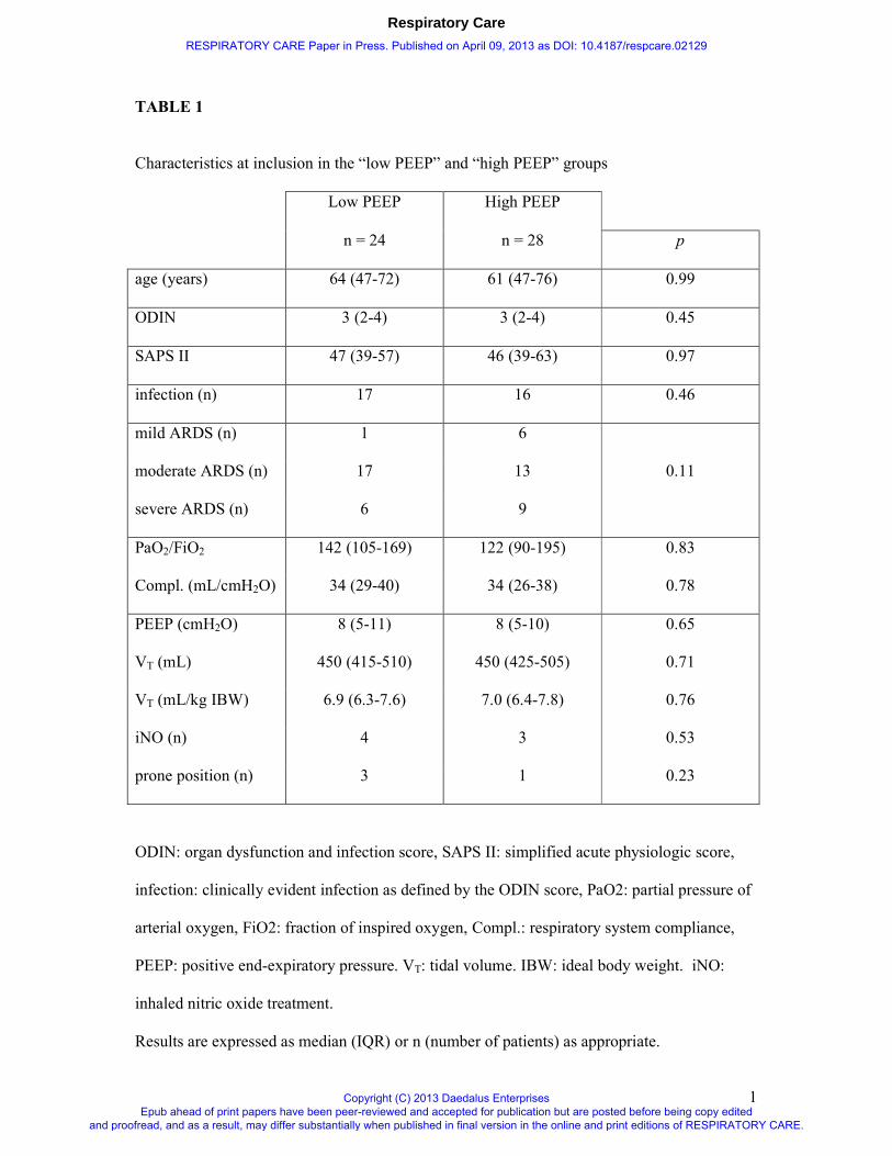
TABLE 1
Characteristics at inclusion in the “low PEEP” and “high PEEP” groups
Low PEEP High PEEP
n = 24 n = 28 p
age (years) 64 (47-72) 61 (47-76) 0.99
ODIN 3 (2-4) 3 (2-4) 0.45
SAPS II 47 (39-57) 46 (39-63) 0.97
infection (n) 17 16 0.46
mild ARDS (n) 1 6
moderate ARDS (n) 17 13 0.11
severe ARDS (n) 6 9
PaO2/FiO2 142 (105-169) 122 (90-195) 0.83
Compl. (mL/cmH2O) 34 (29-40) 34 (26-38) 0.78
PEEP (cmH2O) 8 (5-11) 8 (5-10) 0.65
VT (mL) 450 (415-510) 450 (425-505) 0.71
VT (mL/kg IBW) 6.9 (6.3-7.6) 7.0 (6.4-7.8) 0.76
iNO (n) 4 3 0.53
prone position (n) 3 1 0.23
ODIN: organ dysfunction and infection score, SAPS II: simplified acute physiologic score,
infection: clinically evident infection as defined by the ODIN score, PaO2: partial pressure of
arterial oxygen, FiO2: fraction of inspired oxygen, Compl.: respiratory system compliance,
PEEP: positive end-expiratory pressure. VT: tidal volume. IBW: ideal body weight. iNO:
inhaled nitric oxide treatment.
Results are expressed as median (IQR) or n (number of patients) as appropriate.
Respiratory Care
RESPIRATORY CARE Paper in Press. Published on April 09, 2013 as DOI: 10.4187/respcare.02129
Copyright (C) 2013 Daedalus Enterprises Epub ahead of print papers have been peer-reviewed and accepted for publication but are posted before being copy edited
and proofread, and as a result, may differ substantially when published in final version in the online and print editions of RESPIRATORY CARE.
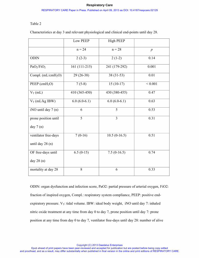
Table 2
Characteristics at day 3 and relevant physiological and clinical end-points until day 28.
Low PEEP High PEEP
n = 24 n = 28 p
ODIN 2 (2-3) 2 (1-2) 0.14
PaO2/FiO2 161 (111-215) 241 (179-292) 0.001
Compl. (mL/cmH2O) 29 (26-38) 38 (31-53) 0.01
PEEP (cmH2O) 7 (5-8) 15 (10-17) < 0.001
VT (mL) 410 (365-450) 430 (380-455) 0.47
VT (mL/kg IBW) 6.0 (6.0-6.1) 6.0 (6.0-6.1) 0.63
iNO until day 7 (n) 6 5 0.53
prone position until
day 7 (n)
5 3 0.31
ventilator free-days
until day 28 (n)
7 (0-16) 10.5 (0-16.5) 0.51
OF free-days until
day 28 (n)
6.5 (0-15) 7.5 (0-16.5) 0.74
mortality at day 28 8 6 0.33
ODIN: organ dysfunction and infection score, PaO2: partial pressure of arterial oxygen, FiO2:
fraction of inspired oxygen, Compl.: respiratory system compliance, PEEP: positive end-
expiratory pressure. VT: tidal volume. IBW: ideal body weight, iNO until day 7: inhaled
nitric oxide treatment at any time from day 0 to day 7, prone position until day 7: prone
position at any time from day 0 to day 7, ventilator free-days until day 28: number of alive
Respiratory Care
RESPIRATORY CARE Paper in Press. Published on April 09, 2013 as DOI: 10.4187/respcare.02129
Copyright (C) 2013 Daedalus Enterprises Epub ahead of print papers have been peer-reviewed and accepted for publication but are posted before being copy edited
and proofread, and as a result, may differ substantially when published in final version in the online and print editions of RESPIRATORY CARE.

ventilator free-days from day 0 to day 28, OF free-days until day 28: number of alive organ
failure free-days from day 0 to day 28.
Results expressed as median (IQR) or n (number of patients) as appropriate.
Respiratory Care
RESPIRATORY CARE Paper in Press. Published on April 09, 2013 as DOI: 10.4187/respcare.02129
Copyright (C) 2013 Daedalus Enterprises Epub ahead of print papers have been peer-reviewed and accepted for publication but are posted before being copy edited
and proofread, and as a result, may differ substantially when published in final version in the online and print editions of RESPIRATORY CARE.
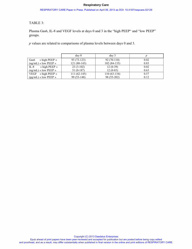
TABLE 3:
Plasma Gas6, IL-8 and VEGF levels at days 0 and 3 in the “high PEEP“ and “low PEEP”
groups.
p values are related to comparisons of plasma levels between days 0 and 3.
day 0 day 3 p
Gas6 « high PEEP » 93 (73-123) 92 (70-110) 0.02
(ng/mL) « low PEEP » 121 (80-143) 102 (84-135) 0.83
IL-8 « high PEEP » 23 (3-102) 12 (0-39) 0.02
(ng/mL) « low PEEP » 51 (0-187) 12 (0-83) 0.63
VEGF « high PEEP » 111 (62-145) 110 (63-134) 0.57
(pg/mL) « low PEEP » 99 (53-148) 98 (55-202) 0.12
Respiratory Care
RESPIRATORY CARE Paper in Press. Published on April 09, 2013 as DOI: 10.4187/respcare.02129
Copyright (C) 2013 Daedalus Enterprises Epub ahead of print papers have been peer-reviewed and accepted for publication but are posted before being copy edited
and proofread, and as a result, may differ substantially when published in final version in the online and print editions of RESPIRATORY CARE.
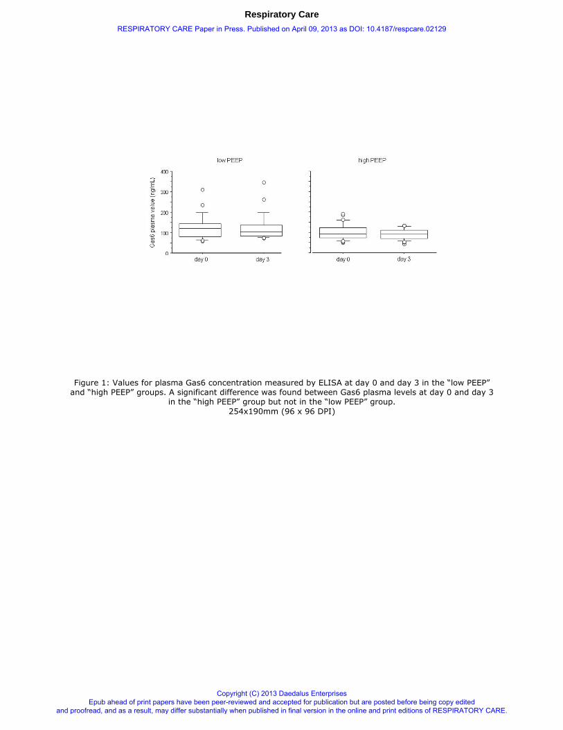
Figure 1: Values for plasma Gas6 concentration measured by ELISA at day 0 and day 3 in the “low PEEP” and “high PEEP” groups. A significant difference was found between Gas6 plasma levels at day 0 and day 3
in the “high PEEP” group but not in the “low PEEP” group.
254x190mm (96 x 96 DPI)
Respiratory Care
RESPIRATORY CARE Paper in Press. Published on April 09, 2013 as DOI: 10.4187/respcare.02129
Copyright (C) 2013 Daedalus Enterprises Epub ahead of print papers have been peer-reviewed and accepted for publication but are posted before being copy edited
and proofread, and as a result, may differ substantially when published in final version in the online and print editions of RESPIRATORY CARE.