a comprehensive review of prone position in...
TRANSCRIPT
A Comprehensive Review of Prone Position in ARDS
Richard H Kallet MSc RRT FAARC
IntroductionEffects of PP on Chest MechanicsVentilation/Perfusion RelationshipDistribution of Ventilation in PPPulmonary Perfusion in PPAirway Closure and Ventilation/Perfusion Matching in Experimental
ARDS and PEEP During PPEffects of PP on Oxygenation in ARDSPrevalence of Positive Oxygenation ResponseMagnitude of Oxygenation ResponseTemporal and Etiological Aspects of Oxygenation ResponseAlveolar Ventilation and Recruitment During PP in ARDSEffects of PP on Airway Secretion Clearance and
Ventilator-Associated PneumoniaEffects of PP on Alveolar Fluid ClearanceHemodynamic Effects of PPHemodynamic Effects of Intra-Abdominal Pressure During PPEffects of PP on Right Heart Function in ARDSEffects of PP in Acute Brain InjuryEffects of PP on Ventilator-Induced Lung Injury in
Experimental and Clinical ARDSRandomized Controlled Trials of PPMeta-Analysis of Randomized Controlled TrialsAdverse Events Associated With PPIncorporating PP into Bundled CareHigh-Level PEEP and Recruitment ManeuverInhaled VasodilatorsExtracorporeal Membrane OxygenationPractical Considerations: Indications, PEEP Strategy, Deescalating
TherapyManual Versus Automated PPIncorporating Continuous Rotational Therapy With PPSummary
Prone position (PP) has been used since the 1970s to treat severe hypoxemia in patients with ARDSbecause of its effectiveness at improving gas exchange. Compared with the supine position (SP),placing patients in PP effects a more even tidal volume distribution, in part, by reversing thevertical pleural pressure gradient, which becomes more negative in the dorsal regions. PP alsoimproves resting lung volume in the dorsocaudal regions by reducing the superimposed pressure ofboth the heart and the abdomen. In contrast, pulmonary perfusion remains preferentially distrib-uted to the dorsal lung regions, thus improving overall alveolar ventilation/perfusion relationships.
1660 RESPIRATORY CARE • NOVEMBER 2015 VOL 60 NO 11
Moreover, the larger tissue mass suspended from a wider dorsal chest wall effects a more homo-geneous distribution of pleural pressures throughout the lung that reduces abnormal strain andstress development. This is believed to ameliorate the severity or development of ventilator-inducedlung injury and may partly explain why PP reduces mortality in severe ARDS. Over 40 years ofclinical trials have consistently reported improved oxygenation in approximately 70% of subjectswith ARDS. Early initiation of PP is more likely to improve oxygenation than initiation during thesubacute phase. Maximal oxygenation improvement occurs over a wide time frame ranging fromseveral hours to several days. Meta-analyses of randomized controlled trials suggest that PP pro-vides a survival advantage only in patients with relatively severe ARDS (PaO2
/FIO2<150 mm Hg).
Moreover, survival is enhanced when patients are managed with a smaller tidal volume(<8 mL/kg), higher PEEP (10–13 cm H2O), and longer duration of PP sessions (>10–12 h/session).Combining adjunctive therapies (high PEEP, recruitment maneuvers, and inhaled vasodilators)with PP has an additive effect in improving oxygenation and may be particularly helpful in stabi-lizing gas exchange in very severe ARDS. Key words: ARDS; acute lung injury; prone position;transpulmonary pressure; ventilation/perfusion ratios; recruitment maneuvers; ventilator-induced lunginjury [Respir Care 2015;60(11):1660–1687. © 2015 Daedalus Enterprises]
Introduction
Prone position (PP) has been used since the 1970s totreat severe hypoxemia in patients with ARDS.1-3 Mellins1
observed that in advanced cystic fibrosis, children spon-taneously position themselves on their hands and knees toimprove ventilation. A concurrent study reported that pas-sive mechanical ventilation in the supine position (SP)resulted in ventilation distributed primarily to nondepen-dent lung regions where perfusion was reduced.4 Based onthe recognition that acute respiratory failure is associatedwith diminished functional residual capacity (FRC) andthat SP enhances dependent airway closure, Bryan5 sug-gested that PP might recruit and stabilize dependent lungsegments. Two subsequent small case studies of ARDSreported mean PaO2
increased by 47 and 53 mm Hg whenpositioning was changed from SP to PP.2,3
Despite numerous observational and randomized con-trolled trials (RCTs) demonstrating the effectiveness of PPin improving oxygenation, this did not translate into im-
proved outcomes. However, the recent publication of thelandmark PROSEVA study6 and consistent results of nu-merous meta-analyses of RCTs7-13 now demonstrate a clearmortality reduction when PP is applied early and for pro-longed time periods in subjects with severe ARDS. Thisrepresents the most significant advance in the managementof ARDS since the seminal ARDS Net trial14 of low tidalvolume (VT) ventilation over a decade ago. Because PP islikely to become commonplace in the management of se-vere ARDS, this comprehensive review is intended to serveboth as a resource for clinicians wishing to gain a betterunderstanding of the physiologic principles upon whichPP is based and as a detailed examination of its effects ongas exchange, hemodynamics, ventilator-induced lung in-jury (VILI), and outcomes based upon 40 years of clinicalevidence.
This review first focuses on the physiologic principlesexplaining how patient positioning impacts lung and chestmechanics and alveolar ventilation/perfusion (VA/Q) rela-tionships. It also incorporates recent advances in our un-derstanding of pulmonary physiology. Afterward, this pa-per systematically reviews the results of clinical trials onPP from 1974 to 2014 on gas exchange, hemodynamics,VILI, associated adverse effects, and the impact of PP onother supportive therapies.
Effects of PP on Chest Mechanics
The theoretical underpinning of PP is based on a modelof the chest and abdominal compartments consisting oforgans with profoundly different densities separated by athin membrane (ie, the diaphragm).5 Differences in organdensities are further magnified by disparities in their re-spective volumes with an abdominal compartment of 10 Lcompared with a thoracic volume of 5 L.15 The abdominal
Mr Kallet is affiliated with Respiratory Care Services, Department ofAnesthesia, University of California, San Francisco at San FranciscoGeneral Hospital, San Francisco, California.
Mr Kallet presented a version of this paper at the 30th New HorizonsSymposium at the AARC Congress 2014, held December 9–12, 2014 inLas Vegas, Nevada.
Supplementary material related to this paper is available at http://www.rcjournal.com.
Correspondence: Richard H Kallet MSc RRT FAARC, Respiratory CareServices, San Francisco General Hospital, NH:GA-2, 1001 Potrero Av-enue, San Francisco, CA 94110. E-mail: [email protected].
DOI: 10.4187/respcare.04271
PRONE POSITION IN ARDS
RESPIRATORY CARE • NOVEMBER 2015 VOL 60 NO 11 1661

cavity has 2 rigid walls (anchored by the pelvis and thespine) and 2 flexible walls (its ventral surface and thediaphragm). Consequentially, both pleural pressure (PPL)and intra-abdominal pressure (IAP) change with body po-sition and the resulting distortion of the flexible portionsof the abdominal wall (Fig. 1). This in turn influences theshape and position of the diaphragm.18
In SP, the hydrostatic pressures in the abdominal com-partment exceed those in the chest cavity by a factor of 5(ie, 1.0 vs 0.2 cm H2O/cm height, respectively).4 More-over, the vertical IAP gradient strongly influences the ver-tical PPL gradient in SP.19 Disparities in hydrostatic pres-sures between these compartments are magnified furtherwith obesity and morbid obesity, wherein IAP increasesfrom a normal of 5–7 mm Hg (7–9 cm H2O) to 11–16 mm Hg, respectively (14–16 cm H2O).20 ARDS asso-ciated with intra-abdominal sepsis or trauma frequentlyis associated with abdominal compartment syndrome,
where IAP is extraordinarily elevated (�25 mm Hg or34 cm H2O).21 Under all conditions, the highest IAP ismeasured in the dorsal regions and is transmitted to thepleural space, thus acting to compress the dorsocaudalregions of the lung.
The response of respiratory system compliance (CRS) toPP is variable and complex. A number of observationalstudies reported that CRS was unaltered or modestly de-creased when turning from SP to PP in both surgical sub-jects22,23 and those with ARDS.24-30 Other studies havereported improved CRS upon being placed in PP,31,32 afteran extended period of PP,24 or once subjects were returnedto SP.33 There is also evidence suggesting that patientswith extrapulmonary sources of ARDS (ARDSexp) may bemore likely to exhibit decreased CRS when placed in PP,26
possibly attributable to an accentuation of the characteris-tically decreased chest wall compliance (CCW) found inthat condition.34
Fig. 1. Schematic representation of vertical pleural pressure (Ppl) distribution from the apex to the bases in the upright position and theirrespective diminishment in the recumbent supine and prone position. Numeric values for pleural pressure and hydrostatic intra-abdominalpressure (Pabd) are based on values provided by Bryan,5 whereas those in the upright position are based upon values provided by West.16
The actual values are subject to debate due to measurement techniques and the impact of lung disease. Agostoni,17 for example, estimatedslightly lower ventral-dorsal values in the recumbent supine and prone position (�4 to 0 cm H2O, respectively). Moreover, he cited evidencethat increased lung density in respiratory disease may enhance the vertical gradient.
PRONE POSITION IN ARDS
1662 RESPIRATORY CARE • NOVEMBER 2015 VOL 60 NO 11

PP generally reduces CCW, but the magnitude of impactdepends in part upon the use and type of padding supports,mattress type, and baseline abdominal girth and rigidity. Incontrast, change in lung compliance (CL) is dependentupon the degree of lung recruitment achieved. As an ex-ample, Lee et al35 reported that those who responded to PPwith improved oxygenation also had a corresponding im-provement in CL and CRS despite a significant drop in CCW
(from 172 to 124 mL/cm H2O), whereas nonrespondershad no change in either CL or CCW. Pelosi et al36 reporteda similar reduction in CCW (from 205 to 147 mL/cm H2O)but without improvement in CL.
Blanch et al31 observed that responders to PP were char-acterized by improvements in both CRS and oxygenationsuggestive of lung recruitment. Both Pelosi et al36 andGuerin et al37 assessed lung recruitment during PP bymeasuring changes in end-expiratory lung volume (EELV).Guerin et al37 discovered evidence of recruitment in lessthan half of the subjects, yet lung recruitment correlatedwith oxygenation. In contrast, Pelosi et al36 found no cor-relation between these variables. Improved oxygenationoften occurs without corresponding improvements in CRS,suggesting that improved VA/Q with PP may be the pri-mary mechanism in many cases.
During passive ventilation in SP, the thoracic dimen-sions diminish with a corresponding decrease in FRC ofapproximately 17% (0.5 L), whereas the abdominal vol-ume increases.15 As a result, the dorsal aspect of the dia-phragm shifts cephalad, resulting in lower regional restinglung volume and CL. During passive mechanical ventila-tion with a physiologic VT (5–6 mL/kg), inspired volumeis preferentially distributed to the nondependent, ventrallung regions.4 For example, rib cage expansion duringpassive mechanical ventilation increases from 40 to 72%of total chest expansion compared with spontaneous breath-ing in the SP.38
PP was originally used in clinical practice for surgicalprocedures. It was there that problems with ventilation andhemodynamic instability (from compression of the abdom-inal portion of the inferior vena cava) were attributed torestricted movement of the chest and abdomen.39 The so-lution was to support the upper chest and pelvic regionwith padding to facilitate unrestricted inspiratory abdom-inal movement, thus improving caudal thoracic expansionand ventilation distribution. Using thoracic and abdominalsupports in obese patients (mean body mass index of 35kg/m2) undergoing elective surgery, Pelosi et al22 reporteda 20% increase in CL with a 20% decrease in CCW and a220% increase in mean FRC. In contrast, the same inves-tigators found that in nonobese subjects (mean body massindex of 23 kg/m2) undergoing elective surgery, an im-provement in FRC of 150% was not accompanied by animprovement in either CL or CCW.23 Both studies used atraditional VT strategy (12 and 10 mL/kg ideal body weight,
respectively) not typically used for patients with acuterespiratory failure.
It is noteworthy that the particular method used for sup-porting the chest may be equally as important as bodyhabitus. Paradoxically, chest padding can worsen compli-ance if the entire chest is supported lengthwise, such thatthe anterior abdominal wall also is compressed.39 In pa-tients with ARDS, the need for padding support during PPis not considered an important factor. For example, Albertet al40 found that PP did not appreciably alter regionaldiaphragmatic movement in experimental ARDS. Chiu-mello et al41 found that CCW paradoxically was lower insubjects when chest and abdominal supports were usedduring PP (102 mL/cm H2O vs 158 mL/cm H2O) becauseof increased contact pressure at the supported sites(29 cm H2O vs 17 cm H2O with and without paddingsupport, respectively). However, CL was higher when sup-ports were used (102 mL/cm H2O vs 93 mL/cm H2O).These offsetting changes in CL versus CCW, with or with-out the use of chest and abdominal supports, had no effecton either pulmonary gas exchange or hemodynamics. How-ever, others have cautioned that allowing for free abdom-inal motion may yet be an important consideration in pa-tients with baseline hemodynamic instability, particularlyif this occurs in the context of intra-abdominal hyperten-sion.20
One of the most detailed and interesting studies on chestmechanics during PP compared the effects of PP withlow-level PEEP on mechanical heterogeneity in pneumo-nia-induced ARDS. In this patient population, Vieillard-Baron et al42 reported that PP was comparable with low-level external PEEP in eliminating intrinsic PEEP. PP alsoreduced airway resistance, the expiratory time constant,and the trapped volume of a functionally noncommunicat-ing slow compartment of the lungs. In consequence, CRS
increased, and PaCO2decreased. None of the subjects in
this study had a history of obstructive lung disease.
Ventilation/Perfusion Relationship
The architectures of both the airways and pulmonaryblood vessels share small asymmetries in their respectivebranching angles and diameters. These asymmetries growin magnitude with each succeeding generation, resulting inheterogeneous distribution in ventilation and perfusion bothin horizontal and vertical dimensions.43 Of particular im-portance is that only 1–25% of pulmonary perfusion ismediated by gravitational forces compared with the influ-ence of vascular architecture.44 This has been referred toas the Slinky effect,44 in which the lung is analogous to adeformable spring that distorts under its own weight (eg,more spring coils gather at the base of a vertically orientedspring). Therefore, lung tissue density and, hence, pulmo-nary vasculature are greater in the dorsum when lying
PRONE POSITION IN ARDS
RESPIRATORY CARE • NOVEMBER 2015 VOL 60 NO 11 1663
supine and in the bases when upright. Gravitational changeswith posture also impact regional resting alveolar size be-cause of gravity’s impact on the regional trans-pulmonarypressure gradient (alveolar � pleural pressure). In SP, trans-pulmonary pressure decreases (ie, becomes more positive)from the ventral to dorsal lung, resulting in smaller alve-olar size but also the potential for larger inspiratory vol-ume changes during tidal ventilation.45
Distribution of Ventilation in PP
During spontaneous ventilation, diaphragmatic contrac-tion causes a relatively greater PPL gradient that favorsventilation distribution in the dorsocaudal regions, thusmaximizing VA/Q matching. However, during passive me-chanical ventilation (particularly with a physiologic VT),VA/Q matching becomes less effective as VT is distributedprimarily to the ventral lung.4 This is explained by the ribcage component of the chest wall, which is nonhomoge-neous in its displacement. The ventral chest wall has alarger freedom of movement compared with the dorsalchest wall, so that the tendency toward nonhomogeneousdisplacement is accentuated in SP.36 From a historical per-spective, this aspect of chest mechanics also explains theinitial adoption of supranormal VT during the early de-cades of mechanical ventilation practice.46-48
To these issues is added the superimposed compressiveforces of the heart upon the lungs (particularly the left) inSP.49 Moreover, the compressive forces acting upon thelungs assume much greater importance in the presence ofpulmonary or systemic inflammation as well as trauma.The superimposed pressures of the overlying edematouslung and chest wall (including the abdominal component)collapse the densely vascularized dorsocaudal lung tis-sue.50
The clinical relevance of managing patients in PP ver-sus SP is based, in part, upon lung tissue distribution withinthe chest cavity. A substantially greater proportion of lungtissue is oriented toward the dorsum of the chest. In SP,approximately 20% of lung tissue is oriented in the ventralplane, compared with 50% in the dorsal plane (below thelevel of the heart).51 Although tissue mass redistributestoward the ventral portion of the chest cavity in PP,52
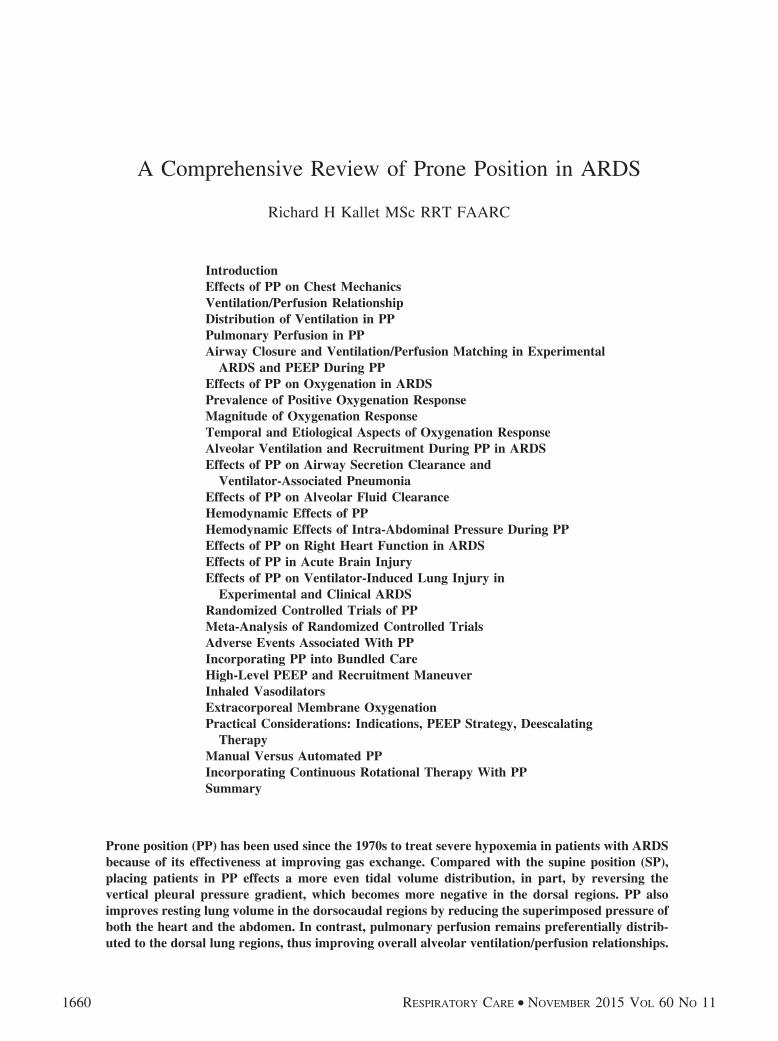
relatively less tissue mass is exposed to compressive forces.Moreover, passive mechanical ventilation in PP causes amuch greater dorsocaudal diaphragmatic displacement, re-sulting in great regional volume change (Fig. 2).53,54 Fur-thermore, during PP, the heart rests almost completelyupon the sternum and decompresses the left lower lobe aswell as a portion of the right lower lobe.49
The gravitational effects on pulmonary gas distributionare determined by spatial distribution in alveolar sizes atthe commencement of inspiration that, in turn, are dictatedby gravitational differences in PPL gradients (with the small-
est fractional gas content found in the lung bases).43 Inaddition, when inspiration begins from residual volumecompared with FRC, little volume change occurs in thedependent lung because of airway closure (this mimicsthe mechanical conditions present in severe ARDS).43 Asthe lung inflates, alveolar size distribution becomes moreuniform.
Interestingly, studies of ventilation distribution in PPcould not detect the effects of a gravitational PPL gradient,because alveolar tissue density was more evenly distrib-uted compared with SP. Moreover, in normal subjectsbreathing either spontaneously or under anesthesia (ie, pas-sive mechanical ventilation) in PP, regional lung volumedistribution at FRC was not influenced by vertical gradi-ents.55 A reduced, more homogeneous PPL gradient (andhence more homogeneous transpulmonary pressure gradi-ent) with PP induces a more evenly distributed tissue stress.This is because the greater mass of dorsal lung tissue issuspended along a relatively larger horizontal dorsal chest
Fig. 2. Lateral chest radiograph of a dog with a fixed, externalplumb bob to demonstrate marked improvement in dorsocaudalaeration in the prone position (top) versus the supine position(bottom). From Reference 53, with permission.
PRONE POSITION IN ARDS
1664 RESPIRATORY CARE • NOVEMBER 2015 VOL 60 NO 11

wall. Specifically, in PP, the ratio of the transverse tho-racic diameter to the sternovertebral height is 3:1.56
A useful analogy likens the lung to a triangular-shapedspring alternatively suspended from its apex (analogous toSP) and from its base (analogous to PP) (Fig. 3).57 Thecombined effects of gravity and the greater tissue masssuspended from a larger dorsal chest wall area producemore equal stress distribution throughout the lung, result-ing in more uniform alveolar size (Fig. 3).
Pulmonary Perfusion in PP
In concert with these changes in lung volume distribu-tion, several studies found that pulmonary perfusion isrelatively unaffected by gravitational forces in PP and ap-pears to be more evenly distributed despite a continuedbias favoring the dorsal lung.52,58,59 In part, this is becausethe vertical impact of gravity on pulmonary perfusion be-tween SP and PP is estimated to account for between 4 and7%, between 4 and 13%, and up to 21–41% of distributiondifferences, depending upon methodology.43,60
Several plausible explanations have been offered to ex-plain the persistence of dorsal perfusion bias irrespective
of body position. Pulmonary vascular endothelium pro-duction of nitric oxide is substantially greater in the dorsalversus the ventral lung.61 Also, because more lung tissueresides in the dorsal plane, there is more pulmonary vas-culature available and a higher capacitance to accommo-date cardiac output compared with the ventral lung, re-gardless of gravitational forces. Therefore, regionalpulmonary vascular resistance would remain lower in thedorsal lung. Moreover, due to fractal geometry, geographicregions containing high conductance vessels tend to ap-proximate one another, whereas a negative correlation invascular size distribution exists between lung regions re-mote from one another.62 This suggests that ventral lungtissue possesses smaller caliber vasculature and hence in-creased resistance.
Airway Closure and VA/Q Matching in ExperimentalARDS and PEEP During PP
Under normal physiologic conditions in dogs, the rela-tive effects of PP have been a slightly improved PaO2
withno change in PaCO2
but increased VA/Q matching from0.83 to 0.94.63 Similar findings have been reported in pigs
Fig. 3. Schematic representation of strain-stress distribution and its impact on alveolar size distribution between the supine and proneposition. The Slinky effect of a triangular-shaped spring suspended from its apex (supine position) causes higher strain and larger variationin the distribution of alveolar sizes due to the effects of gravity and a steeper stress production during mechanical inspiration in the upperlung regions. In contrast, suspending the spring by its base across a wider surface area (prone position) produces a more even strain andmore homogeneous distribution of alveolar size that lessens inhomogeneity in stress development throughout the lungs during mechanicalinspiration.
PRONE POSITION IN ARDS
RESPIRATORY CARE • NOVEMBER 2015 VOL 60 NO 11 1665

with VA/Q increasing from 0.72 to 0.82 between SP andPP.64
When ARDS is induced in dogs, changing from SP toPP causes major improvements in PaO2
attributed both tothe effects of decreased intrapulmonary shunt (by approx-imately 50%) and decreasing regional VA/Q heterogene-ity.40,65 The primary location for this effect was found inthe dorsal lung. Wiener et al66 reported that the apparentventrodorsal gravitational gradient affecting regional pul-monary perfusion in SP disappears in PP because the ma-jority of perfusion continues to be preferentially distrib-uted to dorsal lung regions.
Despite this fact, injury-induced extravascular lung wa-ter remains evenly distributed throughout the lungs. Pul-monary edema increases PPL in the dorsocaudal areas, caus-ing a drop in regional transpulmonary pressure below theregional critical closing pressure in SP.64 Therefore, in PP,given the same magnitude of lung edema in the dorsocau-dal lung regions, the regional PPL decreases (becomes morenegative), favoring the likelihood of recruitment and par-ticipation in tidal ventilation in lung regions with a higherfraction of pulmonary perfusion.
Another important consideration is that the positionalimpact of PPL gradients becomes magnified during volumeoverload, whereby the vertical PPL gradient increases sig-nificantly in SP (from 0.53 to 0.71 cm H2O/cm) comparedwith PP (from 0.17 to 0.27 cm H2O/cm). The clinicalimplication is that aggressive fluid resuscitation (as wouldoccur during resuscitation in sepsis or trauma-inducedARDS) has a greater impact on promoting dependent air-way closure in SP than in PP.67
Severe hypervolemia causes intra-abdominal fluid col-lection, resulting in abdominal distention. Elevated IAPdecreases CCW and markedly increases mean PPL in thedorsocaudal region from �0.2 to �4.2 cm H2O).68 More-over, in the presence of ARDS, elevated IAP is transmittedto the thoracic cavity, simultaneously raising pulmonaryarterial pressure and compressing the thoracic veins. Thispotentiates pulmonary edema formation by aggravatingtranscapillary fluid filtration while also impairing alveolarfluid clearance.69 Studying the impact of increased IAP onPPL and VA/Q between SP and PP, Mure et al70 discoveredthat PP increased oxygenation primarily by decreasingVA/Q heterogeneity compared with SP, the impact beingmore salient in the presence of abdominal distention.
Adding to the complexity of VA/Q mismatching in ARDSis emerging evidence that the intensity of hypoxic pulmo-nary vasoconstriction is heterogeneous in its distribu-tion.71-73 In brief, hypoxic pulmonary vasoconstriction var-ies by lung regions. More intense vasoconstriction occursin the ventrocranial lung regions, whereas the weakestresponse (and therefore the largest relative increase in pul-monary perfusion) occurs in the dorsocaudal regions. Thereis evidence of intrinsically higher endothelial expression
of nitric oxide in these regions, which may modulate theresponse to endothelin-1 (a potent vasoconstrictor releasedduring hypoxia). There also may be uneven distribution ofvascular smooth muscle arrangements and densities be-tween lung regions that modify the compensatory responseto hypoxia.72
Moreover, when regional hypoxia becomes severe (ie,alveolar PO2
�50 mm Hg), hypoxic pulmonary vasocon-striction fails as a compensatory mechanism.73 The poten-tial clinical relevance is that PP would compensate for theweaker vasoconstrictor response by facilitating alveolarrecruitment and increasing alveolar ventilation in the dor-sacaudal lung regions, thereby eliminating, or at least ame-liorating, the stimulus for pulmonary vasoconstriction.
Finally, the effects of PEEP on pulmonary perfusiondiffer considerably between SP and PP. Under normalphysiologic conditions, the application of high-level PEEP(20 cm H2O) in SP exaggerates the gravitational forcesinfluencing lung perfusion by redistributing pulmonary per-fusion away from the upper and middle zones to dorsallung regions. This creates areas of very high VA/Q andincreased VD/VT, partly caused by deterioration in cardiacoutput.74 To avoid this confounding effect, others haveapplied low-level PEEP (5 cm H2O) in sheep with normallungs and found that pulmonary perfusion heterogeneityincreased only modestly (15%) in SP.75
In response to both moderate and high-level PEEP, mark-edly different patterns of pulmonary perfusion distribution(both in the vertical and horizontal planes) were foundbetween SP and PP. In PP at 0 and 10 cm H2O PEEP,perfusion to the nondependent (dorsal) lung was unaltered,and it actually increased at a PEEP of 20 cm H2O.76 Lessperfusion heterogeneity within horizontal planes also wasfound in PP at all levels of PEEP. Decreased heterogeneityin pulmonary perfusion distribution with PEEP during PPhas been reported by others.77 Moreover, whereas the par-adigm of zonal (gravitational) effects on pulmonary per-fusion distribution explained the results found in responseto PEEP during SP, this effect was absent during PP. Thisfinding again reinforces the idea that anatomic and/or phys-iologic differences in the pulmonary vasculature primarilydetermine pulmonary perfusion distribution in PP.
More homogeneous transpulmonary pressure distribu-tion in PP probably results in more uniform lung expan-sion when PEEP is applied and therefore may cause neg-ligible redistribution of pulmonary perfusion.75 Suchspeculation was supported by a study on ventilation dis-tribution between SP and PP with the application of PEEP.Greater heterogeneities in ventilation distribution in totalas well as within isogravitational planes were increased inSP at both 0 and 10 cm H2O PEEP. In contrast, gravita-tional heterogeneity was less in PP, and the application of10 cm H2O PEEP resulted in even more uniform ventila-tion distribution.78
PRONE POSITION IN ARDS
1666 RESPIRATORY CARE • NOVEMBER 2015 VOL 60 NO 11
In another experimental ARDS study, application ofmoderate-level PEEP (10 cm H2O) resulted in significantalveolar recruitment in both SP and PP.79 PEEP caused aredistribution of both ventilation and perfusion toward thedependent lung in both SP and PP. However in SP, PEEP-induced alveolar recruitment occurred only in the dorsallung, whereas applying PEEP during PP affected alveolarrecruitment diffusely down the dorsoventral aspects of thelung.
Effects of PP on Oxygenation in ARDS
Since 1974, at least 40 observational studies havereported that most subjects with ARDS placed in PPexhibit mild to dramatic improvements in oxygen-ation.2,3,24,26,27-33,35-37,42,56,80-105 Improvements occur re-gardless of lung injury categorization (pulmonary ARDS[ARDSp] or ARDSexp),26,56,84,86,87,91,102 specific etiology(pneumonia,42 aspiration,90 following extensive sur-gery,29 trauma,33 acute brain injury,100,101 presence ofmorbid obesity,105 or severe burns and inhalational in-juries94), or severity of lung injury.33,36,85 Significantimprovement in oxygenation also has been reported inpediatric ARDS,106-109 neonates with or without respi-ratory distress syndrome,110-112 subjects with othercauses of hypoxemia,87 and cardiothoracic113,114 andother surgical subjects23 as well as in those with hydro-static pulmonary edema97 and COPD.115
The most detailed clinical study of PP on gas exchangein ARDS, either from direct (pulmonary) injury (ARDSp)or ARDSexp from indirect blood-borne sources, reportedthat the predominant defect in SP was uneven alveolarVA/Q matching.56 When these subjects were placed in PP,VA/Q mismatching decreased from 44 to 34%. These im-provements, however, were essentially lost upon returningto SP.
PP may be most effective in improving oxygenationwhen initiated early (eg, �3 d) during the exudative phase,when congestive and compressive atelectasis are predom-inant features,29,31 as opposed to the intermediate phase ofARDS (eg, �1 week), when fibrosis and Type II cellhyperplasia are more prevalent.116 For example, initiatingPP within 3 d of ARDS onset resulted in substantial im-provement in PaO2
/FIO2compared with initiation at approx-
imately 5 d.97 The same study found that subjects withhydrostatic pulmonary edema had a similar oxygenationresponse to early ARDS, whereas those with pulmonaryfibrosis (similar to the intermediate phase of ARDS) showedno improvement. Nonetheless, several studies have reportedsignificant improvements in oxygenation even when PPwas initiated at a mean of 6 –11 d after ARDS on-set.24,27,30,33,82,86,87
Prevalence of Positive Oxygenation Response
The proportion of subjects whose oxygenation improveswith PP varies according to arbitrary study criteria, includ-ing cut-off values deemed relevant for oxygenation im-provement as well as the time point chosen for assessingthe response. It is also determined by the number of sub-jects sampled. Among the 31 observational studies re-viewed, between 54 and 100% exhibited improved oxy-genation. Twenty percent of studies reported a positiveoxygenation response in �70% of subjects,35,56,80,81,85,94
whereas 47% reported a positive response in 70–85% ofsubjects,3,30,32,37,82,84,86,92,97,102,105,108,115 and 33% of stud-ies found improved oxygenation in approximately �90%of subjects.24,27-29,33,83,87-89,99,100 Most studies have usedcut-off values of 10–20 mm Hg improvement in eitherPaO2
or PaO2/FIO2
32,36,56,85,95,10 or a 10–20% increase inPaO2
/FIO2as clinically important.31,83,86,91,97,105
Magnitude of Oxygenation Response
The actual mean and ranges of oxygenation im-provement with PP have been more impressive than theminimum cut-off values used to assess efficacy. Inobservational studies that reported PaO2
, the average in-crease ranged from 23 to 78 mm Hg, or an im-provement of 34–62%.2,3,24,56,80,81,101 Of the observationalstudies reviewed that measured PaO2
/FIO2, the mean in-
crease ranged from 21 to 161 mm Hg (19–168% improve-ment).26,27,29-33,36,37,82,84-97,42,103,105,108 The magnitude ofimprovement in PaO2
/FIO2among morbidly obese subjects
versus nonobese subjects with ARDS was found to besignificantly greater (104 mm Hg vs 61 mm Hg, respec-tively, P � .04) and was also observed in a larger propor-tion of subjects (77% vs 50%, respectively, P � .044).105
Similar positive responses also have been reported insubjects without ARDS, where the mean PaO2
/FIO2increase
was from 39 –192 mm Hg (24 –267% improve-ment).23,87,97,113,115 Among the few interventional PP stud-ies that presented tabular data, similar improvements inPaO2
(28–37 mm Hg) and PaO2/FIO2
(60–75 mm Hg) weredocumented.117-119 Only the pediatric study by Curleyet al120 reported modest improvements in mean PaO2
/FIO2
of 30 mm Hg during early ARDS.
Temporal and Etiological Aspectsof Oxygenation Response
The time required for oxygenation to improve during PPis highly variable. Regardless of ARDS classification, thetypical response is rapid initial improvement in oxygen-ation (ie, � 30 min), followed by a more gradual increaseover an extended, variable time frame. In both ARDSp andARDSexp, Papazian et al86 reported that 73% of responders
PRONE POSITION IN ARDS
RESPIRATORY CARE • NOVEMBER 2015 VOL 60 NO 11 1667
to PP exhibited a fast (1 h) improvement in PaO2/FIO2
,whereas 27% were characterized as slow responders, re-quiring 6 h. In ARDSp, Langer et al80 reported that PaO2
increased from 70 to 90 mm Hg at 30 min and reached112 mm Hg at 2 h. Pappert et al56 reported similar results,with PaO2
/FIO2improving from 98 to 136 mm Hg at 30 min
to 146 mm Hg at 2 h among subjects with ARDSp orARDSexp.
Steady oxygenation improvements over a prolonged timecourse have been a consistent feature of many studies. Intrauma-associated ARDS, Fridrich et al24 found an imme-diate increase in PaO2
/FIO2with continued improvement
over 20 h. Likewise, in ARDS associated with severe burns,Hale et al94 observed that mean PaO2
/FIO2increased almost
immediately after placement in PP (from 87 to 133 mm Hg)and improved steadily until reaching a peak of 236 mm Hgat 36 h. Reutershan et al92 described both subjects whoseoxygenation plateaued early (2–4 h) and those with con-tinual improvement over 8 h. Stocker et al27 observed sub-jects who required up to 24 h before oxygenation improved.
Patients with ARDSp and ARDSexp may respond dif-ferently in terms of intensity and time course. Lim et al26
observed that subjects with ARDSp required 2 h of PP tosubstantially improve PaO2
/FIO2(37%), whereas those with
ARDSexp had significant improvement at 30 min (45%)but showed no further improvement at 2 h (46%). In sub-jects with ARDSp, Jolliet et al85 found no improvement inPaO2
/FIO2beyond 30 min, whereas L’Her et al89 reported a
progressive increase in PaO2/FIO2
at 1, 4, and 12 h. Charronet al93 reported that �15 h in PP was required to achievea significant increase in PaO2
/FIO2in subjects with severe
ARDSp. Subjects with either ARDS or ARDSexp studiedby McAuley et al88 had progressive improvements in PaO2
/FIO2over 20 h. More importantly, an increasing number
of subjects became responders during this time period from73% (1 h) to 100% (20 h).
Two studies have explored the impact of extraordinarilyprolonged PP sessions on oxygenation (�48 h). Nakoset al97 found that PaO2
/FIO2continuously improved from
130 (30 min) to 218 mm Hg at 48 h. Moreover, theyobserved that late responders tended to be either subjectswith ARDSp or those in the late phase of the syndrome.Finally, Romero et al98 maintained PP for 55 h and alsoreported similar, continued improvement throughout inPaO2
/FIO2(92 mm Hg vs 227 mm Hg) in subjects with
ARDSp.Other temporal aspects of oxygenation in PP are repro-
ducible improvement upon repeated PP, progressive andsustained improvement over time (particularly upon re-turn to SP), and whether initial failure to improve gasexchange represents definitive therapeutic failure. There isevidence suggesting that in both patients with ARDSp andthose with ARDSexp, initial nonresponders to PP may be-come responders on subsequent attempts.3,33 In subjects
with ARDS related to trauma and high-risk surgery, Jo-hannigman et al29 described a consistently high rate (86%)of significantly improved oxygenation over 6 d of PP ther-apy. Likewise, Fridrich et al24 reported that approximatelyhalf of their subjects with trauma-related ARDS were slowresponders who required �1 week of extended PP ses-sions (�20 h). However, on average, PP was initiated inthese subjects at approximately 1 week of mechanical ven-tilation. Easby et al90 observed a trend toward ongoingimprovements in oxygenation with repeated PP sessions insubjects with ARDSp. Stocker et al27 found that oxygen-ation improved progressively until a plateau was reachedafter the third trial. Similarly, Charron et al121 reported that84% of subjects required �3 PP sessions of 18-h durationto stabilize oxygenation.
Many observational studies reported sustained improve-ments in oxygenation once subjects are returned toSP.2,24,32,35,36,80,86,87, 89,94,98,113 This has been referred to as“tissue memory,” but more precisely the mechanism istissue hysteresis as described by pressure-volume curvecharacteristics of the chest.122 However, many of theseobservations were time-limited (eg, 1–2 h after return tosupine position), so that progressive loss of FRC and ox-ygenation, particularly in patients with anasarca and/orhighly elevated IAP, is likely to occur over a more ex-tended time period.
Alveolar Ventilation and RecruitmentDuring PP in ARDS
The effects of PP on PaCO2have been inconsistent in
clinical studies. The majority of observational studies re-ported no improvement in PaCO2
,23,24,27,29,31,35,56,80-82,85,91,103
and sometimes increased PaCO2during PP.3,56 This finding
is puzzling because gas exchange improvements with PP,through better VA/Q matching and reduced intrapulmo-nary shunt from recruitment, are both mechanisms asso-ciated with improved physiologic dead-space fraction(VD/VT) in ARDS.123 In some studies, PP increased PaCO2
because decreased CCW reduced VT and minute ventilationin subjects managed with pressure control ventilation.56
Nevertheless, a substantial number of studies have re-ported decreased PaCO2
with PP.28,93,98,42,104,115,124-126 Ro-mero et al98 found an initial decrement in PaCO2
after 30 minin PP (from 54 to 45 mm Hg) that continued to improveover 55 h (39 mm Hg). Protti et al104 studied subjectsventilated with either volume or pressure control ventila-tion. They attributed decreased PaCO2
either to improvedCL from alveolar recruitment (which increased VT deliv-ery during pressure control ventilation) or to an overallreduction in alveolar dead-space during volume controlventilation. Another mechanism that may sometimes ex-plain drastic reductions in PaCO2
with PP is the reduction inpulmonary hypertension and associated intracardiac shunt-
PRONE POSITION IN ARDS
1668 RESPIRATORY CARE • NOVEMBER 2015 VOL 60 NO 11

ing in patients with ARDS and cor pulmonale (see effectsof PP on right heart function).125
Recently, several investigators93,95,104,126 have classifiedsubjects treated with PP as either PaO2
or PaCO2responders.
Protti et al104 observed that improved PaCO2rather than
PaO2was associated with the magnitude of lung injury and
recruitment potential. What distinguished PaCO2respond-
ers from nonresponders was the amount of non-aeratedtissue. Indirectly, PaCO2
response signified the magnitudeof pulmonary edema and compressive forces acting on thedorsal lung as well as the recruitment potential of PP.Charron et al93 found that subjects categorized as PaCO2
responders after 15 h of PP (decreased PaCO2� 2 mm Hg)
had decreased alveolar dead-space and increased CRS aswell as increased PaO2
/FIO2.
In contrast, classification according to PaO2response
was not associated with changes in either CO2-related vari-ables or CRS. The maximum decrease in PaCO2
occurredafter 9 h in PP compared with 15 h for maximum improve-ment in PaO2
/FIO2. Lee et al,95 however, reported that only
PaO2response to PP was associated with 28-d mortality in
subjects with severe ARDS. Although a similar trend wasobserved between PaCO2
responders versus nonresponders,it was not statistically significant.
In a study examining the effects of PP following a re-cruitment maneuver between subjects with diffuse versuslobar ARDS, Galiatsou et al.124 found that PP caused asignificant decrease in PaCO2
only in those with lobar ARDS,and, consistent with the findings of Charron et al,93 thepositive PaCO2
response also was associated with the larg-est improvement in PaO2
/FIO2and CRS. Using CT imaging,
Galiatsou et al.124 found that PP increased recruitment ofthe non-aerated dorsal lung tissue while decreasing over-inflated lung tissue in the ventral and middle lungs.124 Insubjects with diffuse ARDS, PP decreased the amount ofnon-aerated lung but did not reduce the amount of over-inflated lung. This suggests that a positive PaCO2
responseto PP may signify both the magnitude of non-aerated lungtissue and its recruitability but also more even gas distri-bution and reduced overinflation.
PP also may decrease PaCO2by reducing heterogeneous
lung emptying and intrinsic PEEP. Vieillard-Baron et al42
discovered that applying low-level PEEP (6 cm H2O) andPP in severe ARDS decreased mean airway resistance by24%, reduced the expiratory time constant by 23%, in-creased CRS by 22%, and decreased mean PaCO2
by 5 mm Hg.These changes coincided with a 55% reduction in the “slowcompartment” representing gas trapping in areas of thelung that “poorly communicate” during tidal ventilationand probably signify areas that are overinflated and there-fore at risk of stretch-related lung injury.
Finally, a post hoc analysis126 of 225 subjects enrolledin a multi-center RCT of PP reported that PaCO2
responders(PaCO2
decrease �1 mm Hg) had improved 28-d survival,
whereas a positive PaO2response (PaO2
/FIO2increase
�20 mm Hg) was not associated with outcome. PaCO2
responders had a mean decrease of 6 mm Hg and a mor-tality of 35%, whereas PaCO2
nonresponders had a meanincrease of 6 mm Hg and a mortality of 52%. Moreover,when all subjects randomized to PP were examined, therewas no net change in mean PaCO2
(0.4 � 8.2 mm Hg)d e s p i t e a d r a s t i c i m p r o v e m e n t i n P a O 2
/ F I O 2
(70 � 70 mm Hg).This finding helps to explain the sample-level lack of
PaCO2response in small observational studies of PP. A
relatively equal distribution between PaCO2responders and
nonresponders would negate cohort-level changes in PaCO2
despite an overall improvement in oxygenation. The au-thors attributed the improved PaCO2
to increased recruit-ment of non-aerated lung that exceeded the negative ef-fects of decreased CCW. This suggests that CO2 (which isapproximately 20 times more diffusible than O2 acrosstissues) is more perfusion-sensitive and therefore a bettermarker of lung recruitment in ARDS123 and thus may in-directly signify a reduced risk for VILI.
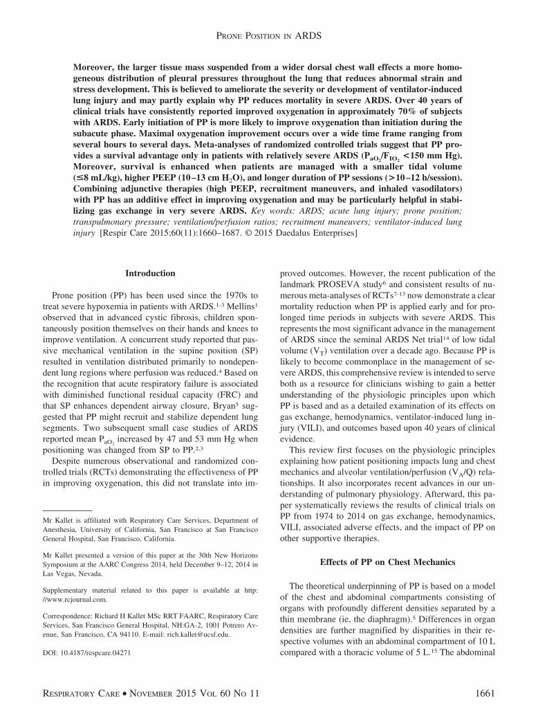
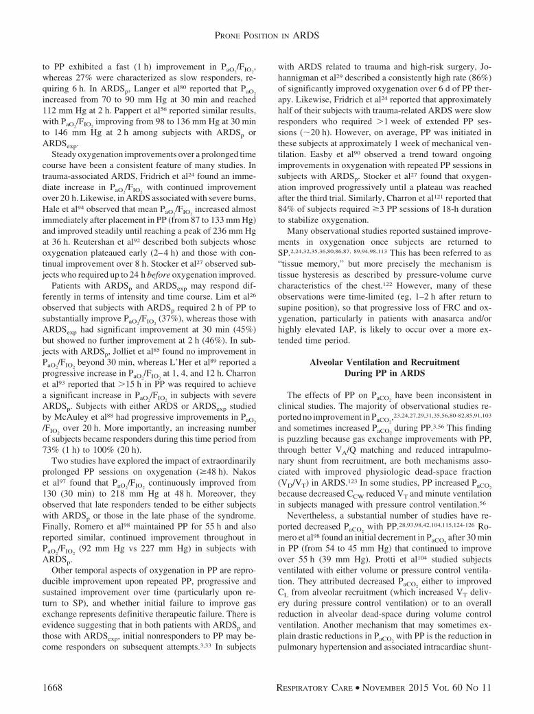
In summary, several mechanisms are probably respon-sible for the improved gas exchange when patients withARDS are placed in PP. These include changes in dia-phragm position and/or tidal diaphragmatic motion, lesscardiac-induced compression of the lungs, and increasedtrans-pulmonary pressure in the dorsal caudal lung facili-tating alveolar recruitment (Fig. 4). The more homoge-neous distribution of trans-pulmonary pressure results inmore uniformity of alveolar size and regional end-expira-tory lung volume. As a consequence, both regional com-pliance and therefore VT distribution should also becomemore uniform. Furthermore, these effects on lung volumealso reduce pulmonary vascular resistance heterogeneityby reducing local hypoxic pulmonary vasoconstriction andincreasing overall VA/Q matching.
Effects of PP on Airway Secretion Clearance andVentilator-Associated Pneumonia
PP probably improves gas exchange by enhancing se-cretion drainage. Most patients with ARDS respond to PPwith increased oxygenation without noticeable secretiondrainage.127 However, several studies2,80,82,87,114,115 re-ported copious secretion mobilization in some subjectsupon turning to PP that may improve oxygenation andalveolar ventilation as well as potentially reducing the in-cidence of ventilator-associated pneumonia (VAP).
It has been speculated PP might reduce the incidence ofVAP by several mechanisms. Prolonged immobilization inthe supine position has long been associated with “hypo-static pneumonia” arising from atelectasis and retainedsecretions, primarily in posterior and superior segments ofthe lower lobes.128 This is because the trachea and main
PRONE POSITION IN ARDS
RESPIRATORY CARE • NOVEMBER 2015 VOL 60 NO 11 1669
bronchi are oriented toward the dorsum, favoring gravita-tional flow and subsequent pooling of secretions deeperinto the lungs.129 Therefore, PP would promote forwarddrainage toward the central airways to facilitate secretionclearance. PP is thought to limit both gastroesophagealreflux and the pooling of oropharyngeal secretions abovethe endotracheal tube cuff, thus limiting microaspiration ofinfected secretions into the lower respiratory tract.129 How-ever, other studies have found that PP is associated with ahigher incidence of increased gastric residual volumes,which is a risk factor for aspiration pneumonia.130
Several RCTs have examined the impact of PP on theincidence of VAP as a secondary outcome with mixedresults. In the only RCT in which the incidence of VAPwas the primary outcome, acute brain-injured subjects whoreceived intermittent PP (4 h/d) showed a strong trendtoward a lower incidence of VAP compared with SP withhead of bed elevation maintained at 20 degrees, (20% vs38%, respectively P � .14).131 In trauma-associated ARDS,Voggenreiter et al132 found a significantly lower preva-lence of VAP compared with SP (62% vs 89% respec-tively, P � .05). Among subjects with various causes ofhypoxemic acute respiratory failure, Guerin et al118 alsofound a significant reduction in both the prevalence (20.6%vs 24.1%) and incidence density (1.66/100 d vs 2.14/100 d)
of VAP in subjects randomized to the PP treatment arm. Ina prematurely terminated RCT in which the majority ofsubjects had ARDSp, Mancebo et al133 found no differencein the prevalence of VAP between PP (18.4%) and SP(15%, P � .65). In addition, a 9-year, retrospective, multi-center study of PP analyzed �2,400 subjects with aPaO2
/FIO2�300 mm Hg and reported that PP had no effect
on the incidence of VAP.134 Therefore, the preponderanceof higher-level evidence suggests that PP may reduce theincidence of VAP in a variety of patients with acute re-spiratory failure. The data also suggest that as a precau-tion, patients placed in PP should be managed in the re-verse-Trendelenburg position to further reduce the chancesof gastroesophageal reflux and aspiration risk.
Effects of PP on Alveolar Fluid Clearance
Enhanced clearance of extravascular lung water also hasbeen reported with PP and may partly explain why somepatients exhibit improved oxygenation only after prolongedperiods in that position.88 Enhanced fluid clearance alsohas been observed in patients with ARDS who are PaO2
responders to a recruitment maneuver.135 Alveolar recruit-ment might increase pulmonary edema clearance by si-multaneously recruiting aquaporins in the alveolar walls as
Fig. 4. Differences in the distribution of lung densities in a patient with ARDS on a computed tomography scan between supine position (top)and prone position (bottom). A: Image taken at end expiration in the supine position. B: Image taken at end inspiration in the supine position.Images C and D were taken from the same lung volumes in the prone position. Note the improved aeration in the dorsal lungs both at endexpiration and end inspiration in the prone position compared with the supine position. From Reference 116, with permission.
PRONE POSITION IN ARDS
1670 RESPIRATORY CARE • NOVEMBER 2015 VOL 60 NO 11
well as increasing total alveolar surface area available forfluid reabsorption.135 In pediatric subjects with ARDS,increased oxygenation was associated with a two-thirdsreduction in positive fluid balance during the PP period.109
Extravascular lung water and alveolar protein measure-ment during PP have not been studied. This might be anunderappreciated aspect of both gas exchange and lungmechanics improvement with PP in ARDS.
Hemodynamic Effects of PP
Hypotension associated with PP during surgery occursfrequently and is attributed to decreased venous returnfrom the combined effects of intravascular fluid depletionand elevated IAP that compresses the inferior venacava.39,136 However, in hemodynamically stable subjectsPP does not cause significant changes in systemic bloodpressure or cardiac index.24,29,31,33,35,36,80-82,84-86,88,91,105 Inaddition, most studies found either no differenc-es24,31,33,35,36,80-82,85 or small increases in central venouspressure, pulmonary arterial pressure, pulmonary arterialocclusion pressures, or pulmonary vascular resistance dur-ing PP.29,91,113 This suggests that the increased vascularpressures may reflect the combined effects of reduced CCW
and positive-pressure ventilation during PP.137 Notably,clinical studies have enrolled only subjects with stablehemodynamic function, so that a thorough hemodynamicassessment is important before utilizing PP. For example,patients with a history of myocardial infarction or isch-emic heart disease are known to be susceptible to systolicand diastolic dysfunction when placed in PP.138
Hemodynamic Effects of IAP During PP
Increased IAP during PP has been a particular concernand is influenced by the type of support surface used.Michelet et al139 reported that air-cushioned mattresses (vsfoam mattresses) prevented an increase in IAP and pre-served hepatic perfusion but had no impact on hemody-namics, extravascular lung water, or pulmonary gas ex-change. Hering et al140 found that air-cushioned mattresseswithout abdominal support caused IAP to increase by2 mm Hg, coinciding with a small increase in cardiacindex that maintained adequate renal function.
These results were reproduced in a subsequent study byHering et al,141 wherein the same 2-mm Hg increase inIAP did not affect intrathoracic blood volume, hepaticfunction, or gastric mucosal perfusion. The authors con-cluded that impaired hemodynamic function may be moreprevalent in patients with inadequate intravascular volumestatus.140,141 Similar findings using an air-cushioned mat-tress without abdominal support were reported by Mate-jovic et al,142 wherein PP caused no change in hemody-namics, abdominal perfusion pressure, hepatosplanchnic
perfusion, hepatic function, or urine output. PP produced asignificant rise in IAP (�3 mm Hg) in only 18% of sub-jects. However, none of the subjects in these studies hadintra-abdominal hypertension (�15 mm Hg) so that theseresults cannot be generalized to patients with highly ele-vated IAP.
Effects of PP on Right Heart Function in ARDS
Acute pulmonary hypertension is a common feature ofARDS and has multiple sources, including hypoxemia,hypercapnia, acidosis, and pulmonary vascular obstructionfrom interstitial edema and disseminated arterial and mi-crovascular embolization.143-146 Acute cor pulmonale oc-curs in 22–25% of patients with ARDS and increases to50% in those with a PaO2
/FIO2�100 mm Hg.147 Acute cor
pulmonale has been independently associated with bothhigh driving pressures during mechanical ventilation andinfectious causes of ARDS; the presence of acute cor pul-monale increases mortality from 36 to 60%.148
In a patient with severe ARDS, Legras et al125 reportedthat PP caused an instantaneous near cessation of shuntingacross the foramen ovale that coincided with decreasedpulmonary vascular pressures. PP increased PaO2
/FIO2from
59 to 278 mm Hg, and PaCO2decreased from 54 to 39 mm Hg.
Dramatic decreases in pulmonary vascular resistance in-dex (from 514 to 234 dynes�s/cm5/m2) and right ventric-ular dimensions in ARDS have been reported with PP,coinciding with increased PaO2
/FIO2.28 Similarly, in severe
ARDS with documented acute cor pulmonale, PP substan-tially increased PaO2
/FIO2and decreased PaCO2
, which co-incided with decreased right ventricular size and increasedcardiac index.149
Approximately 19% of patients with ARDS have a mod-erate to large patent foremen ovale coinciding with eitherincreased right ventricular size or acute cor pulmonale thatis exacerbated by PEEP.150 The prevalence of both acutecor pulmonale and patent foremen ovale in ARDS andtheir association with increased mortality might partiallyexplain the mortality benefit recently reported in severeARDS.
Effects of PP in Acute Brain Injury
Two studies examined how PP affects neurovascularfunction and brain tissue oxygenation in subjects with acutebrain injury and ARDS or pneumonia. Reinprecht et al100
used PP sessions of 16 h in subjects with subarachnoidhemorrhage and ARDS. Although intracranial pressure(ICP) was moderately increased (from 10 � 4 to16 � 4 mm Hg) and cerebral perfusion pressure decreased(from 74 � 8 to 67 � 7 mm Hg), PaO2
/FIO2markedly
increased (range of 37–183 mm Hg) in 88% of subjects.This resulted in a significant increase in brain tissue oxy-
PRONE POSITION IN ARDS
RESPIRATORY CARE • NOVEMBER 2015 VOL 60 NO 11 1671

gen tension (from 27 � 4 to 35 � 5 mm Hg). Similarfindings of improved PaO2
and modestly increased ICP(from 12 � 6 to 15 � 4 mm Hg) with PP were reported byNekludov et al101 in subjects with acute brain injury andpneumonia. Mean arterial pressure increased markedlycompared with ICP, so that cerebral perfusion pressureincreased from 66 � 7 to 73 � 8 mm Hg, along with a24% increase in PaO2
.Importantly, these studies enrolled subjects in whom
ICP and coronary perfusion pressure were well controlledwith vasopressors, mannitol, and ventricular drainage. De-spite these therapies, Reinprecht et al100 found that sub-jects placed in PP had a higher occurrence of ICP�20 mm Hg (18% vs 2% in SP) and cerebral perfusionpressure �60 mm Hg (22% vs 8%). However, PP reducedthe incidence of brain tissue oxygen tension �20 mm Hgfrom 33% in SP to 10%. Using PP in patients with acutebrain injury and ARDS should be approached with cautionand restricted to those in whom baseline ICP can be main-tained �20 mm Hg and who are hemodynamically stable.
Effects of PP on VILI in Experimentaland Clinical ARDS
In healthy animals, the dependent, nondependent, andmiddle lung regions possess differing stress-strain charac-teristics under both static and dynamic conditions due tothe effects of gravity, regional traction, and regional com-pression. Under static conditions in SP, the lungs are in-herently inhomogeneous. Nondependent regions exhibit asteep stress-strain curve during inflation, whereas the stress-strain curve rises more gradually in dependent regions.During PP, regional differences in stress-strain ratios be-tween lung regions are more uniform, and within eachregion, stress rises more gradually relative to strain.151
Several experimental models of ARDS, both with andwithout VILI, have examined whether PP confers a lung-protective benefit compared with SP.152-156 Broccard et al152
combined oleic acid with high-stretch injury, producingsimilar degrees of pulmonary edema formation throughoutthe lung regardless of position. However, histologic injurywas less severe and less extensive in dependent lung re-gions with PP. In a high-stretch low-PEEP VILI model,53
PP resulted in milder, more homogeneously distributedinjury. Pulmonary edema and histologic injury were ap-proximately 30% less than with SP. In a similar study,Nakos et al155 also reported more evenly distributed his-topathological changes throughout the lung, and lung in-jury was approximately 50% less than with SP.
PP also delays the onset of VILI by 50–80%.153,154 Thiswas attributable to more even lung density distribution inthe dorsoventral axis. The lungs appeared slightly shorterand wider with a corresponding 28% reduction in esti-mated lung strain, consistent with more homogeneous pleu-
ral pressure distribution in PP.154 Santana et al156 foundthat PP resulted in decreased lung elastance and viscoelas-tance associated with a more homogeneous distribution ofalveolar air/tissue ratios.
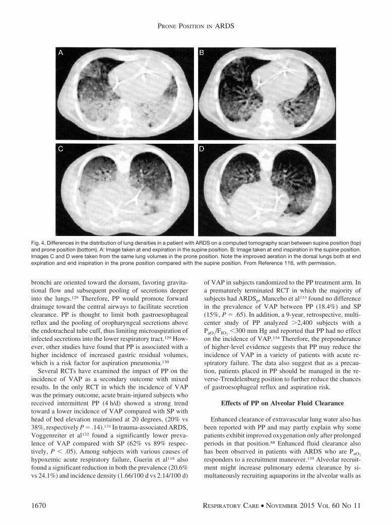
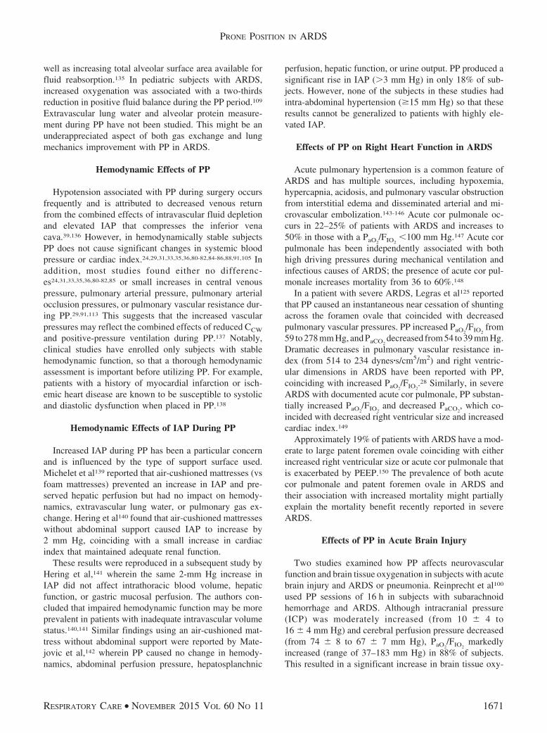
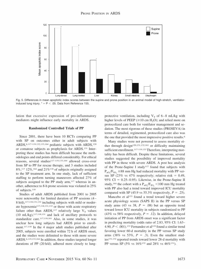
One of the most consequential findings was made byNakos et al,155 who measured the apoptopic index in thelungs, liver, kidneys, small intestine, and diaphragm. Afteronly 90 min of exposure to a high-stretch low-PEEP VILImodel, animals in SP had significantly greater apoptosisscores in the lungs, liver, and diaphragm, with trends to-ward higher scores in the other organs (Fig. 5). Apoptosis(“programmed cell death”) is caused by signaling mole-cules released within or between organs. This may partlyexplain why mortality in ARDS results from multiorgansystem failure rather than refractory hypoxemia. Pulmo-nary translocation and dissemination of these signalingmolecules (as well as bacteria and pro-inflammatory me-diators) to the systemic circulation is believed to be theresponsible mechanism.157,158
Only 2 clinical studies have compared pro-inflamma-tory mediator expression between SP and PP. Papazianet al159 discovered that PP reduced both neutrophil levelsand pro-inflammatory mediator levels of IL-1�, IL-6, andIL-8 in bronchoalveolar lavage fluid compared with SP.Of particular interest is IL-8 (a chemo-attractant cytokinefor neutrophils), which is secreted by the alveolar epithe-lium and macrophages in response to excessive stretch.Lung injury is perpetuated from neutrophil infiltration andsubsequent proteolytic enzyme release.160 This underscoresthe importance of PP in ameliorating VILI by effectingmore homogeneous pleural pressure gradients and moreuniform regional EELV, VT distribution, and strain-stressrelationships throughout the lungs.
Chan et al161 examined cytokine profiles in ARDS be-tween PP and SP and their impact upon early mortality at2 time points. Twenty-two subjects with community-ac-quired pneumonia-associated ARDS were randomized toPP or SP with both groups receiving lung-protective ven-tilation. Plasma IL-6 levels were measured at baseline,24 h, and 72 h. Baseline IL-6 levels were higher in the PPversus SP arms (396 � 31 pg/mL vs 323 � 50 pg/mL,respectively) but steadily declined over 72 h with PP(196 � 47 pg/mL) compared with SP (278 � 53 pg/mL).IL-6 levels predicted mortality at 14 d for all subjects(18% in PP arm; 27% in SP). IL-6 levels in community-acquired pneumonia are directly associated with the dis-ease severity.162 PP may reduce VILI risk through im-proved oxygenation that decreases exposure to hyperoxia,a powerful stimulant for IL-6 expression.163 Although 28-dmortality was not different between treatment arms (36%),early mortality might be impacted through this mecha-nism. Interestingly, IL-6 levels typically peak at approxi-mately 10 d of exposure to hyperoxia,163 inviting specu-
PRONE POSITION IN ARDS
1672 RESPIRATORY CARE • NOVEMBER 2015 VOL 60 NO 11
lation that excessive expression of pro-inflammatorymediators might influence early mortality in ARDS.
Randomized Controlled Trials of PP
Since 2001, there have been 10 RCTs comparing PPwith SP on outcomes either in adult subjects withARDS,6,117-119,132,133,164 pediatric subjects with ARDS,120
or comatose subjects as prophylaxis for ARDS.131 Inter-preting these studies has been difficult because the meth-odologies and end points differed considerably. For ethicalreasons, several studies117-119,133,164 allowed cross-overfrom SP to PP for rescue therapy, and 3 studies included8%,117 12%,164 and 21%118 of subjects originally assignedto the SP treatment arm. In one study, lack of sufficientstaffing to perform turning maneuvers affected 27% ofsubjects assigned to the PP study arm,117 whereas in an-other, adherence to 8-h prone sessions was violated in 25%of subjects.118
Studies of adult ARDS published from 2001 to 2005were noteworthy for limited duration of PP sessions (4–8 h/d),117,118,131,132 including subjects with mild or moder-ate hypoxemia117,118,131,132 or those with acute respiratoryfailure other than ARDS,117 relatively large VT values(10 mL/kg),117,118,131 and lack of ancillary protocols tostandardize care.117,118,161 Also, in some studies, it wasunclear how long subjects had ARDS prior to enroll-ment.117,118 In the 4 major adult studies published after2005, subjects were enrolled within 72 h of ARDS onset,and the studies were delimited to those with more severeARDS.6,119,133,164 In addition, these studies targeted longerdurations of PP (20 h/d); adhered more closely to lung-
protective ventilation, including VT of 6–8 mL/kg withhigher levels of PEEP (�10 cm H2O); and relied more onprotocolized care both for ventilator management and se-dation. The most rigorous of these studies (PROSEVA) interms of detailed, regimented, protocolized care also wasthe one that provided the most impressive positive results.6
Many studies were not powered to assess mortality ei-ther through design120,131,132,161 or difficulty maintainingsufficient enrollment.117-119,133 Therefore, interpreting mor-tality has been difficult. Despite these limitations, severalstudies suggested the possibility of improved mortalitywith PP in those with severe ARDS. A post hoc analysisof the Prone-Supine I study117 found that subjects withPaO2
/FIO2�88 mm Hg had reduced mortality with PP ver-
sus SP (23% vs 47% respectively; relative risk � 0.49,95% CI � 0.25–0.95). Likewise, in the Prone-Supine IIstudy,164 the cohort with a PaO2
/FIO2�100 mm Hg treated
with PP also had a trend toward improved ICU mortalitycompared with SP (45.9 vs 55.3% respectively, P � .25).
Mancebo et al133 found a trend toward higher severeacute physiology scores (SAPS II) in the PP versus SPstudy arms (43 vs 38, P � .08) but an opposite trendtoward lower ICU mortality in subjects randomized to PP(43% vs 58% respectively, P � .12). In addition, delayedinitiation of PP from ARDS onset was a significant factorin predicting mortality (odds ratio of 2.83, 95% CI: 1.63–4.90, P � .001).133 Fernandez et al119 found a similar trendfavoring lower 60-d mortality in the PP versus SP studyarms (38% vs 53%, P � .3). Even the smallest stud-ies131,132 reported trends toward lower 28-d mortality withPP versus SP (5% vs 16%132 and 28% vs 46%131).
Fig. 5. Differences in mean apoptotic index scores between the supine and prone position in an animal model of high-stretch, ventilator-induced lung injury. * � P � .05. Data from Reference 155.
PRONE POSITION IN ARDS
RESPIRATORY CARE • NOVEMBER 2015 VOL 60 NO 11 1673
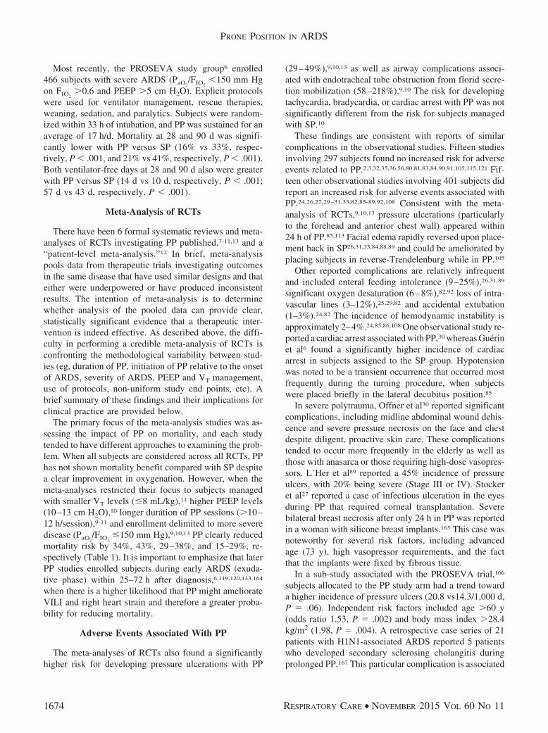
Most recently, the PROSEVA study group6 enrolled466 subjects with severe ARDS (PaO2
/FIO2�150 mm Hg
on FIO2�0.6 and PEEP �5 cm H2O). Explicit protocols
were used for ventilator management, rescue therapies,weaning, sedation, and paralytics. Subjects were random-ized within 33 h of intubation, and PP was sustained for anaverage of 17 h/d. Mortality at 28 and 90 d was signifi-cantly lower with PP versus SP (16% vs 33%, respec-tively, P � .001, and 21% vs 41%, respectively, P � .001).Both ventilator-free days at 28 and 90 d also were greaterwith PP versus SP (14 d vs 10 d, respectively, P � .001;57 d vs 43 d, respectively, P � .001).
Meta-Analysis of RCTs
There have been 6 formal systematic reviews and meta-analyses of RCTs investigating PP published,7-11,13 and a“patient-level meta-analysis.”12 In brief, meta-analysispools data from therapeutic trials investigating outcomesin the same disease that have used similar designs and thateither were underpowered or have produced inconsistentresults. The intention of meta-analysis is to determinewhether analysis of the pooled data can provide clear,statistically significant evidence that a therapeutic inter-vention is indeed effective. As described above, the diffi-culty in performing a credible meta-analysis of RCTs isconfronting the methodological variability between stud-ies (eg, duration of PP, initiation of PP relative to the onsetof ARDS, severity of ARDS, PEEP and VT management,use of protocols, non-uniform study end points, etc). Abrief summary of these findings and their implications forclinical practice are provided below.
The primary focus of the meta-analysis studies was as-sessing the impact of PP on mortality, and each studytended to have different approaches to examining the prob-lem. When all subjects are considered across all RCTs, PPhas not shown mortality benefit compared with SP despitea clear improvement in oxygenation. However, when themeta-analyses restricted their focus to subjects managedwith smaller VT levels (�8 mL/kg),11 higher PEEP levels(10–13 cm H2O),10 longer duration of PP sessions (�10–12 h/session),9-11 and enrollment delimited to more severedisease (PaO2
/FIO2�150 mm Hg),9,10,13 PP clearly reduced
mortality risk by 34%, 43%, 29–38%, and 15–29%, re-spectively (Table 1). It is important to emphasize that laterPP studies enrolled subjects during early ARDS (exuda-tive phase) within 25–72 h after diagnosis,6,119,120,133,164
when there is a higher likelihood that PP might ameliorateVILI and right heart strain and therefore a greater proba-bility for reducing mortality.
Adverse Events Associated With PP
The meta-analyses of RCTs also found a significantlyhigher risk for developing pressure ulcerations with PP
(29–49%),9,10,13 as well as airway complications associ-ated with endotracheal tube obstruction from florid secre-tion mobilization (58–218%).9,10 The risk for developingtachycardia, bradycardia, or cardiac arrest with PP was notsignificantly different from the risk for subjects managedwith SP.10
These findings are consistent with reports of similarcomplications in the observational studies. Fifteen studiesinvolving 297 subjects found no increased risk for adverseevents related to PP.2,3,32,35,36,56,80,81,83,84,90,91,105,115,121 Fif-teen other observational studies involving 401 subjects didreport an increased risk for adverse events associated withPP.24,26,27,29 –31,33,82,85-89,92,108 Consistent with the meta-analysis of RCTs,9,10,13 pressure ulcerations (particularlyto the forehead and anterior chest wall) appeared within24 h of PP.85,113 Facial edema rapidly reversed upon place-ment back in SP26,31,33,84,88,89 and could be ameliorated byplacing subjects in reverse-Trendelenburg while in PP.105
Other reported complications are relatively infrequentand included enteral feeding intolerance (9–25%),26,31,89
significant oxygen desaturation (6–8%),82,92 loss of intra-vascular lines (3–12%),25,29,82 and accidental extubation(1–3%).24,82 The incidence of hemodynamic instability isapproximately 2–4%.24,85,86,108 One observational study re-ported a cardiac arrest associated with PP,30 whereas Guérinet al6 found a significantly higher incidence of cardiacarrest in subjects assigned to the SP group. Hypotensionwas noted to be a transient occurrence that occurred mostfrequently during the turning procedure, when subjectswere placed briefly in the lateral decubitus position.85
In severe polytrauma, Offner et al30 reported significantcomplications, including midline abdominal wound dehis-cence and severe pressure necrosis on the face and chestdespite diligent, proactive skin care. These complicationstended to occur more frequently in the elderly as well asthose with anasarca or those requiring high-dose vasopres-sors. L’Her et al89 reported a 45% incidence of pressureulcers, with 20% being severe (Stage III or IV). Stockeret al27 reported a case of infectious ulceration in the eyesduring PP that required corneal transplantation. Severebilateral breast necrosis after only 24 h in PP was reportedin a woman with silicone breast implants.165 This case wasnoteworthy for several risk factors, including advancedage (73 y), high vasopressor requirements, and the factthat the implants were fixed by fibrous tissue.
In a sub-study associated with the PROSEVA trial,166
subjects allocated to the PP study arm had a trend towarda higher incidence of pressure ulcers (20.8 vs14.3/1,000 d,P � .06). Independent risk factors included age �60 y(odds ratio 1.53, P � .002) and body mass index �28.4kg/m2 (1.98, P � .004). A retrospective case series of 21patients with H1N1-associated ARDS reported 5 patientswho developed secondary sclerosing cholangitis duringprolonged PP.167 This particular complication is associated
PRONE POSITION IN ARDS
1674 RESPIRATORY CARE • NOVEMBER 2015 VOL 60 NO 11
Table 1. Meta-Analysis of Randomized Clinical Trials of Prone Positioning in ARDS
Study Trials N Primary Findings: PP vs SP OR (95% CI) or WMD, P
Abroug et al7 5 1,372 Mortality: 34.9% vs 35.5% 0.97 (0.77–1.22)VAP 0.77 (0.57–1.04), P � .09Airway compromise: 10.5% vs 10.4% 1.01 (0.71–1.43), P � .95Pressure ulcers/facial edema: 41% vs 34% 1.35 (1.08–1.69), P � .0071PaO2
/FIO225 mm Hg (15–35), P � .001
Alsaghir et al8 4 1,271 90-d all mortality 0.99 (0.77–1.79)Early 1PaO2
/FIO252 mm Hg (7–96)
Intermediate 1PaO2/FIO2
44 mm Hg (14–74)Late 1PaO2
/FIO225 mm Hg (15–34)
Total ventilator days �0.42 (�1.56 to 0.72)VAP 0.78% (0.40–1.51)
Sud et al13 10 1,867Mortality: all subjects 0.97 (0.88–1.07)
7 495 Cohort: PaO2/FIO2
� 100 mm Hg* 0.85 (0.74–0.98), P � .027 852 Cohort: PaO2
/FIO2� 100 mm Hg* 1.04 (0.89–1.22), P � .60
Oxygenation: 1PaO2/FIO2
: 27–39%8 1,066 VAP 0.81 (0.67–1.00), P � .058 1,588 Total ventilator days �0.7 (–2.01 to 0.62), P � .307 1,279 Pressure ulcers 1.29 (1.16–1.44), P � .0017 1,351 ETT obstruction 1.58 (1.24–2.01), P � .0018 886 CT dislodgement 3.14 (1.02–9.69)
Lee et al9 11 2,246Mortality: overall 41.5% vs 46.2% 0.77 (0.59–0.99), P � .039PP �10 h/session 1.04 (0.80–1.36), P � .76PP �10 h/session 0.62 (0.48–0.79), P � .001ALI/AHRF 1.02 (0.76–1.36), P � .92PaO2
/FIO2� 150 mm Hg 0.72 (0.55–0.95), P � .02
Pressure ulcers 1.49 (1.18–1.89), P � .001Airway emergencies† 1.55 (1.10–2.17), P � .01ETT obstruction (as sole factor) 2.16 (1.53–3.05), P � .001VAP 0.76 (0.44–1.33), P � .34Tachycardia/bradycardia 1.08 (0.78–1.50), P � .64Cardiac arrest 0.74 (0.47–1.17), P � .20Pneumothoraces 0.77 (0.46–1.30), P � .33
Hu et al10 9 2,2421,600 Mortality: 90-d all ARDS: PaO2
/FIO2� 300 mm Hg 0.85 (0.62–1.18), P � .33
508 28–30-d when PaO2/FIO2
�100 mm Hg 0.71 (0.57–0.89), P � .003521 28–30-d when PaO2
/FIO2: 101–200 mm Hg 0.72 (0.39–1.34), P � .30
506 90-d with PEEP 10–13 cm H2O 0.57 (0.43–0.75), P � .0011,094 90-d with PEEP �10 cm H2O 1.04 (0.92–1.18), P � .561,067 28–30-d mortality PP �12 h/session 0.73 (0.54–0.99), P � .041,095 28–30-d mortality PP �12 h/session 1.04 (0.89–1.22), P � .60
Beitler et al11 7 2,119Mortality: overall 0.83 (0.68–1.02), P � .07VT �8 mL/kg PBW 0.66 (0.50–0.86), P � .002VT �8 mL/kg PBW 1.00 (0.88–1.13), P � .9521 mL/kg PBW in baseline VT 2RR by 16.7% (6.1–28.3), P � .001PP �12 h/session 0.71 (0.56–0.90), P � .004PP �12 h/session 1.05 (0.92–1.19), P � .47
* Estimate excludes subjects with acute hypoxemic respiratory failure not caused by ARDS enrolled into some studies.90
† Airway emergencies included accidental extubation, endotracheal tube migration, and endotracheal tube obstruction.PP � prone positionSP � supine positionOR � odds ratioWMD � weighted mean differenceVAP � ventilator-associated pneumoniaETT � endotracheal tubeCT � chest tubeALI � acute lung injury (ie, PaO2/FIO2 �200 mm Hg)AHRF � acute hypoxemic respiratory failure (non-ARDS)PBW � predicted body weight
PRONE POSITION IN ARDS
RESPIRATORY CARE • NOVEMBER 2015 VOL 60 NO 11 1675

with high PEEP and vasopressor therapy, resulting in bil-iary duct ischemia from hypoperfusion of the peribiliaryvascular plexis. All 21 subjects had severe ARDS requir-ing PEEP �15 cm H2O and PP sessions of 12 h/d. The 5subjects who developed severe cholangitis were morbidlyobese (body mass index of 37 � 6 kg/m2) and requireddaily PP for several weeks.
Incorporating PP Into Bundled Care
PP improves oxygenation over a variable time coursethat may reach a plateau after 12–24 h,26,79,88,89 but insome cases, improvement continues for 30–55 h97,98 Thishas been attributed to the interplay between several fac-tors, including increased IAP and abnormal CCW, com-pression atelectasis, and persistent small airway closuredespite high levels of PEEP (ie, 14 cm H2O).168 Airwayclosure is both asynchronous in onset and unevenly dis-tributed, particularly in dependent lung regions that delayin opening, close quickly, and have a lower EELV.169
Effective lung recruitment requires a combination ofpressure and time, with time mostly reflecting the influ-ence of airway closure and stress-relaxation (tissue yield-ing) characteristics of the lung and chest wall.170 Reopen-ing collapsed small airways is a dynamic process with avariable time course.171 To recruit collapsed small air-ways, applied airway pressure must overcome the surfacetension, viscosity, and film thickness of the airway liningfluid, which in turn is influenced by airway radius, axialwall traction exerted by the surrounding alveoli, and thepresence of functional surfactant.171,172 Increased liningfluid surface tension requires higher airway pressures toopen collapsed airways, whereas increased lining fluid vis-cosity increases the time necessary to open sequentiallycollapsed airways.172
High-Level PEEP and Recruitment Maneuver
In a subset of patients with particularly severe ARDS,PP by itself may be insufficient to stabilize gas exchangeand minimize the risk of VILI. Developing a multifacetedapproach that incorporates high-level PEEP, recruitmentmaneuvers, and inhaled vasodilators may enhance the ef-fectiveness of PP and expedite gas exchange stabilization.This may reduce the likelihood for adverse effects asso-ciated with prolonged exposure to toxic levels of oxygen,high PEEP, and PP. Several prospective studies have com-pared the impact of PP on the effectiveness of PEEP91 andrecruitment maneuvers124,173-175 to improve pulmonary gasexchange and mechanics in severe ARDS.
Gainnier et al91 demonstrated that PEEP and PP have anadditive effect on lung recruitment but also are mediatedby lung injury characteristics: diffusely distributed injuryversus restricted injury either to the dependent regions
(lobar) or otherwise localized (patchy). Subjects with lo-calized infiltrates showed relatively modest improvementin PaO2
/FIO2when PEEP was adjusted stepwise from 0 to
15 cm H2O either in SP (�110–130 mm Hg) or PP (�160–220 mm Hg). In contrast, subjects with diffuse injuryresponded to the same PEEP adjustments by increasingPaO2
/FIO2from 60 to 160 mm Hg (SP) and from 140 to
240 mm Hg (PP). This suggests that in severe ARDSwith localized lung injury, PP is likely to be more ef-fective than PEEP, whereas those with diffuse injurymay receive additional benefit from a further trial ofincreasing PEEP.
Various forms of recruitment maneuvers have been testedwith PP, including periodic sighs,173 extended high-pres-sure post-inspiratory pauses,175 brief periods of either pres-sure control ventilation with super-PEEP,124 or high-levelCPAP.174 Using spiral CT imaging, Galiatsou et al124 com-pared the impact of PP on augmenting a recruitment ma-neuver done in SP. In agreement with the previous studieson PP and PEEP,91 Galiatsou et al124 reported that both PPand recruitment maneuvers have an additive effect on im-proving oxygenation. However, in contrast to the findingsof Gainnier et al,91 PP was most effective in augmenting arecruitment maneuver in subjects with lobar infiltratesrather than those with diffuse infiltrates. Galiatsou et al124
found that a recruitment maneuver followed by PP im-proved lung aeration and reduced the risk of VILI. PP wasmore effective than a recruitment maneuver performed inSP in recruiting non-aerated dorsal lung and reversed over-inflation of the ventral lung.
Comparing a recruitment maneuver done in SP withthose done at 1 and 6 h after PP, Rival et al175 reported thatthe response to combining a recruitment maneuver withPP was more pronounced in subjects with ARDSp. ThePaO2
/FIO2increased from 115 to 128 mm Hg after the first
recruitment maneuver in SP and increased to 230 mm Hgafter the final recruitment maneuver in PP. In ARDSexp,PaO2
/FIO2increased from 102 to 107 mm Hg after the re-
cruitment maneuver in SP to 154 mm Hg after the finalrecruitment maneuver in PP. Mean PaCO2
also decreasedby 2–4 mm Hg after each recruitment maneuver with atotal decrease of approximately 7 mm Hg from baseline tothe final recruitment maneuver in PP. Most relevant wasthe fact that oxygenation improvements following a re-cruitment maneuver were transitory when done in SP butwere sustained in PP.
In early ARDSexp, Oczenski et al174 found greater im-provement in PaO2
/FIO2(60%) following a recruitment ma-
neuver when analyzing only those subjects who were alsoresponsive to PP. In these subjects, mean PaO2
/FIO2im-
proved from 147 mm Hg in SP to 225 mm Hg after 6 h inPP and then increased to 368 mm Hg just after the recruit-ment maneuver, an improvement that was sustained over3 h in PP. Both PaCO2
and CRS also improved over 3 h
PRONE POSITION IN ARDS
1676 RESPIRATORY CARE • NOVEMBER 2015 VOL 60 NO 11

following the recruitment maneuver in PP. In another studyof early ARDS, Pelosi et al173 reported that adding sighbreaths to PP in order to achieve plateau pressures (Pplat)of 45 cm H2O increased mean PaO2
more than sighs usedin SP (37 � 17 mm Hg vs 16 � 11 mm Hg, respectively).Sighs increased EELV by 17% and decreased lung elas-tance by 17% when applied in PP but had only a minorimpact when applied during SP. Moreover, the combina-tion of sigh breaths and PP increased EELV over baselinemeasurements in SP by 35%.
To date, only one RCT has reported the impact of com-bining a recruitment maneuver with PP on outcomes. In arecent study of 116 subjects with severe ARDS, Zhouet al176 reported decreased 28-d mortality, duration of me-chanical ventilation, and ICU stay in those managed witha recruitment maneuver and PP compared with controls.Unfortunately, only the study abstract is available in Eng-lish, so that a thorough examination of the methodologyand results is not currently available. In summary, a lim-ited number of observational studies using various meth-ods for alveolar recruitment consistently demonstrate thatincorporating a recruitment maneuver with PP enhancesoxygenation and should be considered when PP itself doesnot improve oxygenation.
Inhaled Vasodilators
Inhaled vasodilators, such as nitric oxide (INO),aerosolized prostacyclin, or alprostadil, are used to im-prove oxygenation in severe ARDS.177 These agents workas selective vasodilators enhancing perfusion of ventilatedalveoli and thus improving VA/Q. Six observational stud-ies involving 85 subjects examined the combined effectsof INO and PP on gas exchange and hemodynamics inARDS.178-182 In 5 of these studies, the combined therapieshad an additive effect in improving oxygenation in sub-jects with early, severe ARDS of various etiologies. Forexample, mean PaO2
or PaO2/FIO2
increased an average of29% with INO and 47% with PP alone, but when usedtogether, oxygenation improved by an average of88%.25,179-182 Furthermore, combining these therapiessubstantially increased the prevalence of responders overeither therapy alone from 54% (INO) and 76% (PP) to92% for both.25,179,180,182
Some studies found that PP modestly enhanced the ef-fects of INO in reducing pulmonary vascular resistance25
or mean pulmonary arterial pressure,182 whereas others didnot.179-181 An important caveat in interpreting these studiesis not only the small number of subjects enrolled (n � 14)but also the fact that both the INO dosage tested (1–13ppm) and PP time (30 min to 6 h) varied considerably.Although combining these therapies further enhances ox-ygenation and responder rate, the optimal combination ofdose and timing remains unknown.
Investigation of bundled therapy for severe ARDS is inits nascent stage, and the potential impact on patient out-comes is unknown. However, several meta-analyses oftherapeutic trials involving lung-protective ventilation inconjunction with PP,13 higher PEEP,183 neuromuscularblocking agent,184 and recruitment maneuvers185 suggestthat these individual therapies improve outcomes in themost severe cohort of ARDS subjects. Since 2006, wehave instituted a bundled approach using lung-protectiveventilation with PP, aerosolized prostacyclin, and a vari-ation of a previously described recruitment maneuver (pres-sure control ventilation with super-PEEP)186 in dire cir-cumstances of refractory ARDS. Two cases of thistechnique are summarized for illustrative purposes (Figs. 6and 7).
We have only found it necessary to utilize these extraor-dinary measures infrequently under immanently life-threatening scenarios, wherein gas exchange could not bestabilized despite maximal therapies in SP. In general, thecommon features of these cases have been severe ARDScomplicated by unusually low CCW from morbid obesity,abdominal compartment syndrome, or severe anasarca. Ourexperience has been similar to that described by Barbaset al186 in that a sustained recruitment maneuver in PP overseveral hours sometimes is necessary to stabilize gas ex-change. Performing a recruitment maneuver in PP is pref-erable because the decreased CCW and more equal distri-bution of pleural pressure gradients reduce lung stress andthe likelihood of barotrauma. However, it should be em-phasized that in most cases of severe ARDS, we havefound that PEEP levels between 16 and 24 cm H2O in PPare sufficient to stabilize gas exchange.
Extracorporeal Membrane Oxygenation
Recently, PP has been used as an adjunctive therapywith extracorporeal membrane oxygenation (ECMO) inpatients with severe ARDS187-194 All studies were retro-spective in nature and consisted of case reports or caseseries. PP was used because of severe hypoxemia despitemaximal ECMO and/or ventilatory support (eg,PEEP � 20 cm H2O), an inability to wean from ECMO, oran inability to treat atelectasis. PP typically was initiated8–10 d into the course of ARDS.189-191 As in other studies,PP improved oxygenation that was sustained when sub-jects were returned to SP. In one case, PP induced pro-nounced secretion drainage with marked improvement inpulmonary function that expedited weaning from ECMO.189
The duration of PP sessions varied widely from 4 to24 h. Sometimes, a modified PP maneuver of 135° wasused to protect the intravascular catheter from compres-sion or dislodgement.187,194 No catheter dislocations oc-curred during the turning procedures. Two additional ret-rospective studies of subjects with presumed ARDS who
PRONE POSITION IN ARDS
RESPIRATORY CARE • NOVEMBER 2015 VOL 60 NO 11 1677
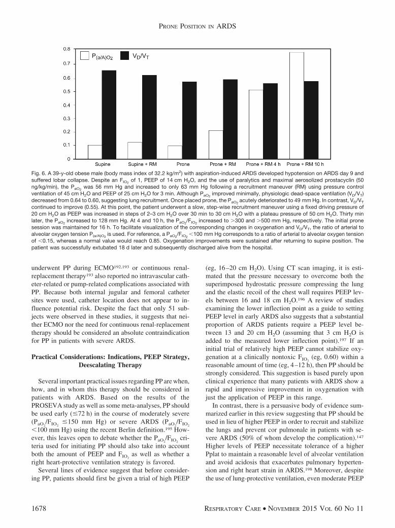
underwent PP during ECMO192,193 or continuous renal-replacement therapy193 also reported no intravascular cath-eter-related or pump-related complications associated withPP. Because both internal jugular and femoral cathetersites were used, catheter location does not appear to in-fluence potential risk. Despite the fact that only 51 sub-jects were observed in these studies, it suggests that nei-ther ECMO nor the need for continuous renal-replacementtherapy should be considered an absolute contraindicationfor PP in patients with severe ARDS.
Practical Considerations: Indications, PEEP Strategy,Deescalating Therapy
Several important practical issues regarding PP are when,how, and in whom this therapy should be considered inpatients with ARDS. Based on the results of thePROSEVA study as well as some meta-analyses, PP shouldbe used early (�72 h) in the course of moderately severe(PaO2
/FIO2�150 mm Hg) or severe ARDS (PaO2
/FIO2
�100 mm Hg) using the recent Berlin definition.195 How-ever, this leaves open to debate whether the PaO2
/FIO2cri-
teria used for initiating PP should also take into accountboth the amount of PEEP and FIO2
as well as whether aright heart-protective ventilation strategy is favored.
Several lines of evidence suggest that before consider-ing PP, patients should first be given a trial of high PEEP
(eg, 16–20 cm H2O). Using CT scan imaging, it is esti-mated that the pressure necessary to overcome both thesuperimposed hydrostatic pressure compressing the lungand the elastic recoil of the chest wall requires PEEP lev-els between 16 and 18 cm H2O.196 A review of studiesexamining the lower inflection point as a guide to settingPEEP level in early ARDS also suggests that a substantialproportion of ARDS patients require a PEEP level be-tween 13 and 20 cm H2O (assuming that 3 cm H2O isadded to the measured lower inflection point).197 If aninitial trial of relatively high PEEP cannot stabilize oxy-genation at a clinically nontoxic FIO2
(eg, 0.60) within areasonable amount of time (eg, 4–12 h), then PP should bestrongly considered. This suggestion is based purely uponclinical experience that many patients with ARDS show arapid and impressive improvement in oxygenation withjust the application of PEEP in this range.
In contrast, there is a persuasive body of evidence sum-marized earlier in this review suggesting that PP should beused in lieu of higher PEEP in order to recruit and stabilizethe lungs and prevent cor pulmonale in patients with se-vere ARDS (50% of whom develop the complication).147
Higher levels of PEEP necessitate tolerance of a higherPplat to maintain a reasonable level of alveolar ventilationand avoid acidosis that exacerbates pulmonary hyperten-sion and right heart strain in ARDS.198 Moreover, despitethe use of lung-protective ventilation, even moderate PEEP
Fig. 6. A 39-y-old obese male (body mass index of 32.2 kg/m2) with aspiration-induced ARDS developed hypotension on ARDS day 9 andsuffered lobar collapse. Despite an FIO2
of 1, PEEP of 14 cm H2O, and the use of paralytics and maximal aerosolized prostacyclin (50ng/kg/min), the PaO2
was 56 mm Hg and increased to only 63 mm Hg following a recruitment maneuver (RM) using pressure controlventilation of 45 cm H2O and PEEP of 25 cm H2O for 3 min. Although PaO2
improved minimally, physiologic dead-space ventilation (VD/VT)decreased from 0.64 to 0.60, suggesting lung recruitment. Once placed prone, the PaO2
acutely deteriorated to 49 mm Hg. In contrast, VD/VT
continued to improve (0.55). At this point, the patient underwent a slow, step-wise recruitment maneuver using a fixed driving pressure of20 cm H2O as PEEP was increased in steps of 2–3 cm H2O over 30 min to 30 cm H2O with a plateau pressure of 50 cm H2O. Thirty minlater, the PaO2
increased to 128 mm Hg. At 4 and 10 h, the PaO2/FIO2
increased to �300 and �500 mm Hg, respectively. The initial pronesession was maintained for 16 h. To facilitate visualization of the corresponding changes in oxygenation and VD/VT, the ratio of arterial toalveolar oxygen tension P(a/A)O2
is used. For reference, a PaO2/FIO2
�100 mm Hg corresponds to a ratio of arterial to alveolar oxygen tensionof �0.15, whereas a normal value would reach 0.85. Oxygenation improvements were sustained after returning to supine position. Thepatient was successfully extubated 18 d later and subsequently discharged alive from the hospital.
PRONE POSITION IN ARDS
1678 RESPIRATORY CARE • NOVEMBER 2015 VOL 60 NO 11
levels (eg, 13 cm H2O) impair RV function,199 which isexacerbated further by the presence of acute respiratoryacidosis.200 In a right heart-protective approach, the targetsare a Pplat � 27 cm H2O and an elastic driving pressure(Pplat � PEEP) �17 cm H2O.147 This would necessitaterestricting PEEP to approximately 10–12 cm H2O. There-fore, adhering to a right heart-protective ventilation strat-egy virtually necessitates incorporating PP into the man-agement strategy for moderately severe and severe ARDS.
In clinical practice, deescalating PP therapy will largelybe dictated by patient response, priorities related to oxy-genation interpreted within the context of PEEP and FIO2
goals, and considerations of preventing VILI and cor pul-monale. Among the later RCTs that used PP sessions av-eraging 17–20 h, the number of days PP was required tomeet predetermined discontinuation criteria averaged 4, 8,and 10 d6,133,164 In the study by Voggenreiter et al,132 PPsessions averaged 11 h, and subjects required an averageof 7 d before discontinuation.
Various criteria have been used to test discontinuationof PP that may explain the wide variability between stud-ies in the required number of PP sessions. The PROSEVAstudy6 required a PaO2
/FIO2�150 mm Hg on a FIO2
�0.6and PEEP �10 cm H2O, which would translate to aPaO2
of 90 mm Hg. Fernandez et al119 appeared to haverequired a stable PaO2
/FIO2�250 mm Hg on a FIO2
of 0.5and PEEP �8 cm H2O for 12 h before attempting todiscontinue PP. Similarly, Voggenreiter et al132 mandated
a stable PaO2/FIO2
�300 mm Hg for 48 h in both PP and SPbefore discontinuing PP.
Currently, there is no way of discerning whether anyparticular criterion for discontinuing PP is optimal. Rely-ing upon reversal of PaO2
/FIO2criteria for ARDS (ie,
PaO2/FIO2
� 300 mmHg) appears to be influenced by val-ues used to signify full lung recruitment, which has beendefined as achieving a value of PaO2
� PaCO2�400 mm Hg
(� 5%) on an FIO2of 1.191 However, in patients with ARDSp
or in the subacute phase, there may be limited recruitmentwith PP, so that achieving PaO2
/FIO2�300 may not be a
realistic expectation. The obvious advantage of thePROSEVA criterion is that it allows an aggressive chal-lenge to test discontinuation of PP when oxygenation isreasonably stable. This in turn may facilitate a reduction inthe duration of mechanical ventilation (albeit indirectly)but may increase the risk of gas exchange instability. Incontrast, requiring greater lung recruitment and stabiliza-tion before discontinuing PP may result in fewer inci-dences of hypoxemic relapses but may needlessly prolongthe duration of mechanical ventilation.
Manual Versus Automated PP
Manual PP maneuvers are relatively easy to performquickly with appropriate staffing.201 Therefore, definedroles are required for each clinician with a clear, detaileddescription of all preparatory and procedural steps that can
Fig. 7. A 39-y-old pregnant female (body mass index of 36.2 kg/m2) in her third trimester who developed ARDS secondary to H1N1 influenzapneumonia underwent emergency cesarean section in the ICU for severe hypoxemia (SpO2
of 74%) with loss of fetal heart rate. She wassuccessfully managed in SP on PEEP of 16 cm H2O until ARDS d 3, when her PaO2
fell to 48 mm Hg on an FIO2of 1 and PEEP of 22 cm H2O
(SP1�22). An hour after her first prone session, her oxygenation improved substantially (PP-1H�22) until she was returned to supine 5 hlater for wound care. Her oxygenation again deteriorated despite an FIO2
of 1 and PEEP of 22 cm H2O (SP2�22). Returning her to proneresulted in transient improvement in oxygenation over the following 16 h despite increasing PEEP to 24 cm H2O (PP2�24; PP-2–8H�24).A prolonged super-PEEP recruitment maneuver as described previously (Fig 6) was implemented with a plateau pressure (Pplat) of55 cm H2O, PEEP of 36 cm H2O, VT of 7–8 mL/kg, and breathing frequency of 30 breaths/min. Within 30 min, there was a markedimprovement in PaO2
/FIO2(PP�36) that slowly increased to �200 mm Hg at 10 h (PP-10�36) and �300 mm Hg 4 h later (PP-14H�36).
Despite continued episodes of derecruitment when turned to supine, the patient was managed successfully in prone at lower levels of PEEP(15 cm H2O) and without the use of paralytics over the next several days. Unfortunately, 10 d later, the patient developed severe corpulmonale secondary to a massive pulmonary embolus and died shortly thereafter.
PRONE POSITION IN ARDS
RESPIRATORY CARE • NOVEMBER 2015 VOL 60 NO 11 1679
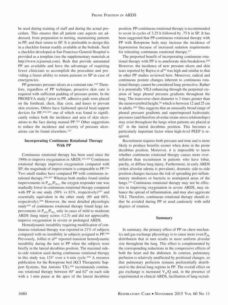
be used during training of staff and during the actual pro-cedure. This ensures that all patient care aspects are ad-dressed, from preparation to turning, maintaining patientsin PP, and their return to SP. It is preferable to design thisin a checklist format readily available at the bedside. Sucha checklist developed at San Francisco General Hospital isprovided as a template (see the supplementary materials athttp://www.rcjournal.com). Beds that provide automatedPP are available and have the advantage of requiringfewer clinicians to accomplish the procedure and pro-viding a faster ability to return patients to SP in case ofemergencies.
PP generates pressure ulcers at a constant rate.166 There-fore, regardless of PP technique, proactive skin care isrequired with sufficient padding of pressure points. In thePROSEVA study,6 prior to PP, adhesive pads were placedon the forehead, chest, iliac crest, and knees to preventskin erosions. Others have fashioned special head supportdevices for PP,202,203 one of which was found to signifi-cantly reduce both the incidence and area of skin ulcer-ations to the face during manual PP.202 Other suggestionsto reduce the incidence and severity of pressure ulcer-ations can be found elsewhere.127
Incorporating Continuous Rotational TherapyWith PP
Continuous rotational therapy has been used since the1990s to improve oxygenation in ARDS.204,205 Continuousrotational therapy improves oxygenation compared withSP; the magnitude of improvement is comparable to PP.204
Two small studies have compared PP with continuous ro-tational therapy,206,207 Whereas both studies found similarimprovements in PaO2
/FIO2, the oxygen responder rate was
markedly lower in continuous rotational therapy comparedwith PP in one study (50% vs 83%, respectively)207 andessentially equivalent in the other study (69 and 68%,respectively).206 However, the most detailed physiologicstudy205 of continuous rotational therapy found large im-provements in PaO2
/FIO2only in cases of mild to moderate
ARDS (lung injury scores �2.5) and did not appreciablyimprove oxygenation in severe or prolonged ARDS.
Hemodynamic instability requiring modification of con-tinuous rotational therapy was reported in 21% of subjectscompared with no instability in subjects assigned to PP.206
Previously, Jolliet et al85 reported transient hemodynamicinstability during the turn to PP when the subjects werebriefly in the lateral decubitus position. The maximal side-to-side rotation used during continuous rotational therapyin this study was 124° over a 4-min cycle.206 A resourcepublication for the Rotoprone bed (KCI Therapeutic Sup-port Systems, San Antonio TX),208 recommends continu-ous rotational therapy between 40o and 62o on each sidewith a 1-min pause at the apex of the lateral decubitus
position. PP-continuous rotational therapy is recommendedto occur in cycles of 3.25 h followed by .75 h in SP. It hasbeen suggested that PP-continuous rotational therapy withPP with Rotoprone beds may increase the incidence ofhypotension because of increased sedation requirementsfor tolerating continuous rotational therapy.57
The purported benefit of incorporating continuous rota-tional therapy with PP is to ameliorate skin breakdown.208
However, the incidence of new pressure ulcers and skintears reported by Bajwa et al99 was high and similar to thatin other PP studies reviewed here. Moreover, radical andcontinuous posture changes inherent to continuous rota-tional therapy cannot be considered lung-protective. Ratherit is potentially VILI-enhancing through the perpetual cre-ation of large pleural pressure gradients throughout thelung. The transverse chest diameter is 3 times greater thanthe sternovertebral height,56 which is between 12 and 25 cmin adults.209 This suggests that an unusually broad range ofpleural pressure gradients and superimposed hydrostaticpressures (and therefore alveolar strain-stress relationships)may exist throughout the lungs when patients are placed at62° in the lateral decubitus position. This becomes aparticularly important factor when high-level PEEP is re-quired.
Recruitment requires both pressure and time and is morelikely to produce benefits sooner when done in the pronedecubitus position. Moreover, it is impossible to knowwhether continuous rotational therapy causes more over-inflation than recruitment in patients who have lobar,patchy, or diffuse lung injury. Furthermore, in early ARDS(when alveolar edema is prevalent), dramatic side-to-sideposition changes increase the risk of spreading pro-inflam-matory mediators or bacteria to noninjured areas of thelungs.210 Continuous rotational therapy may not be effec-tive in improving oxygenation in severe ARDS, may en-hance the spread of inflammation, and may also aggravateVILI. Therefore, continuous rotational therapy should ei-ther be avoided during PP or used cautiously with milddegrees of rotation.
Summary
In summary, the primary effect of PP on chest mechan-ics and gas exchange physiology is to cause more even PPL
distribution that in turn results in more uniform alveolarsize throughout the lung. This effect is complemented bythe corresponding reductions in the compressive effects ofboth the heart and the abdomen. In contrast, pulmonaryperfusion is relatively unaffected by positional changes, sothat pulmonary perfusion remains preferentially distrib-uted to the dorsal lung regions in PP. The overall effect ongas exchange is increased VA/Q and, in the presence ofexperimental or clinical ARDS, facilitation of lung recruit-
PRONE POSITION IN ARDS
1680 RESPIRATORY CARE • NOVEMBER 2015 VOL 60 NO 11

ment by lowering the threshold for recruiting collapsedairways and alveoli.
The preponderance of evidence demonstrates that mostpatients with ARDS (�70%) managed in PP have im-proved oxygenation regardless of etiology or timing ofinitiation. It appears that early intervention with PP ismore likely to be effective, but improved oxygenation fre-quently has been reported when PP is used during thesubacute phase. The time course for maximal improve-ment in oxygenation is variable from several hours to sev-eral days. However, most studies suggest that PP sessionsshould be sustained for no less than 10–12 h and ideallyfor 16–20 h. The impact of PP on improving alveolar ven-tilation is less consistent and may be explained by recentevidence that there are both PaO2
and PaCO2responders to
PP, reflecting the extent and distribution of lung injury.That many studies reported no impact on PaCO2
may be dueto a balancing of these 2 subgroups.
Meta-analyses of RCTs demonstrate that PP provides asurvival advantage in subjects with relatively severe ARDS(PaO2
/FIO2� 150 mm Hg). This reflects the impact of PP
on either delaying the onset or reducing the severity VILI.Reduction in hypoxic pulmonary vasoconstriction and vas-cular resistance also may reduce the likelihood of devel-oping cor pulmonale that is believed to increase mortalityrisk in ARDS. Combining other adjunctive therapies withPP appears to have an additive effect in improving oxy-genation, thus providing clinicians with an array of strat-egies to stabilize gas exchange.
REFERENCES
1. Mellins RB. Pulmonary physiotherapy in the pediatric age group.Am Rev Respir Dis 1974;110(6):137-142.
2. Piehl MA, Brown RS. Use of extreme position changes in acuterespiratory failure. Crit Care Med 1976;4(1):13-14.
3. Douglas WW, Rehder K, Beynen FM, Sessler AD, Marsh HM.Improved oxygenation in patients with acute respiratory failure: theprone position. Am Rev Respir Dis 1977;115(4):559-566.
4. Froese AB, Bryan AC. Effects of anesthesia and paralysis on dia-phragmatic mechanics in man. Anesthesiology 1974;41(3):242-255.
5. Bryan AC. Comments of a devil’s advocate. Am Rev Respir Dis1974;110(6 Pt 2):143-144.
6. Guerin C, Reignier J, Richard JC, Beuret P, Gacouin A, Boulain T,et al. PROSEVA Study Group: prone positioning in severe acuterespiratory distress syndrome. N Engl J Med 2013;368(23):2159-2168.
7. Abroug F, Ouanes-Besbes L, Elatrous S, Brochard L. The effect ofprone positioning in acute respiratory distress syndrome or acutelung injury: a meta-analysis: areas of uncertainty and recommen-dations for research. Intensive Care Med 2008;34(6):1002-1011.
8. Alsaghir AH, Martin CM. Effect of prone positioning in patientswith acute respiratory distress syndrome: a meta-analysis. Crit CareMed 2008;36(2):603-609.
9. Lee JM, Bae W, Lee YJ, Cho YJ. The efficacy and safety of pronepositional ventilation in acute respiratory distress syndrome: up-dated study-level meta-analysis of 11 randomized controlled trials.Crit Care Med 2014;42(5):1252-1262.
10. Hu SL, He HL, Pan C, Liu AR, Liu SQ, Liu L, et al. The effect ofprone positioning on mortality in patients with acute respiratorydistress syndrome: a meta-analysis of randomized controlled trials.Crit Care 2014;18(3):R109.
11. Beitler JR, Shaefi S, Montesi SB, Devlin A, Loring SH, Talmor D,Malhotra A. Prone positioning reduces mortality from acute respi-ratory distress syndrome in the low tidal volume era: a meta-anal-ysis. Intensive Care Med 2014;40(3):332-341.
12. Gattinoni L, Carlesso E, Taccone P, Polli F, Guerin C, Mancebo J.Prone positioning improves survival in severe ARDS: a pathophys-iologic review and individual patient meta-analysis. Minerva Anes-tesiol 2010;76(6):448-454.
13. Sud S, Friedrich JO, Taccone P, Polli F, Adhikari NKJ, Latini R,et al. Prone ventilation reduces mortality in patients with acuterespiratory failure and severe hypoxemia: systematic review andmeta-analysis. Intensive Care Med 2010;36(4):585-599.
14. NHLBI Acute Respiratory Distress Syndrome Network. Ventila-tion with lower tidal volumes as a compared with traditional tidalvolumes for acute respiratory distress syndrome. N Engl J Med2000;342(18):1301-1308.
15. Hedenstierna G, Strandberg A, Brismar B, Lundquist H, SvenssonL, Tokics L. Functional residual capacity, thoracoabdominal di-mensions, and central blood volume during general anesthesia withmuscle paralysis and mechanical ventilation. Anesthesiology 1985;62(3):247-254.
16. West JB. Respiratory physiology: the essentials. 9th edition. Phil-adelphia: Lippincott Williams & Wilkins; 2012:105.
17. Agostoni E. Mechanics of the pleural space. Physiol Rev 1972;52(1):57-128.
18. Froese AB. Gravity, the belly and the diaphragm: you can’t ignorephysics. Anesthesiology 2006;104(1):193-196.
19. Agostoni E, D’Angelo E, Bonanni MV. The effect of the abdomenon the vertical gradient of pleural surface pressure. Respir Physiol1970;8(3):332-346.
20. De Keulenaer BL, De Waele JJ, Powell B, Malbrain MLNG. Whatis normal intra-abdominal pressure and how is it affected by pso-tioning, body mass and positive end-expiratory pressure? IntensiveCare Med 2009;35(6):969-976.
21. Malbrain MLNG. Abdominal pressure in the critically ill: measure-ment and clinical relevance. Intensive Care Med 1999;25(12):1453-1458.
22. Pelosi P, Croci M, Calappi E, Mulazzi D, Cerisara M, Vercesi P,Vicardi P, Gattinoni L. Prone positioning improves pulmonary func-tion in obese patients during general anesthesia. Anesth Analg 1996;83(3):578-583.
23. Pelosi P, Croci M, Calappi E, Cerisara M, Mulazzi D, Vicardi P,Gattinoni L. The prone positioning during general anesthesia min-imally affects respiratory mechanics while improving functionalresidual capacity and increasing oxygen tension. Anesth Analg 1995;80(5):955-960.
24. Fridrich P, Krafft P, Hochleuthner H, Mauritz W. The effects oflong-term prone positioning in patients with trauma-induced adultrespiratory distress syndrome. Anesth Analg 1996;83(6):1206-1211.
25. Johannigman JA, Davis K Jr, Miller SL, Campbell RS, LuchetteFA, Frame SB, Branson RD. Prone positioning and inhaled nitricoxide: syngergistic therapies for acute respiratory distress syndrome.J Trauma 2001;50(4):589-595; discussion 595-596.
26. Lim CM, Kim EK, Lee JS, Shim TS, Lee SD, Koh Y, et al. Com-parison of the response to the prone position between pulmonaryand extrapulmonary acute respiratory distress syndrome. IntensiveCare Med 2001;27(3):477-485.
27. Stocker R, Neff T, Stein S, Ecknauer E, Trentz O, Russi E. Pronepositioning and low-volume pressure-limited ventilation improve
PRONE POSITION IN ARDS
RESPIRATORY CARE • NOVEMBER 2015 VOL 60 NO 11 1681
survival in patients with severe ARDS. Chest 1997;111(4):1008-1017.
28. Jozwiak M, Teboul JL, Anguel N, Persichini R, Silva S, Chemla D,et al. Beneficial hemodynamic effects of prone positioning in pa-tients with acute respiratory distress syndrome. Am J Respir CritCare Med 2013;188(12):1428-1433.
29. Johannigman JA, Davis K Jr, Miller SL, Campbell RS, LuchetteFA, Frame SB, Branson RD. Prone positioning for acute respiratorydistress syndrome in the surgical intensive care unit: Who when andhow long? Surgery 2000;128(4):708-716.
30. Offner PJ, Haenel JB, Moore EE, Biffl WL, Franciose RJ, BurchJM. Complications of prone ventilation in patients with multisys-tem trauma with fulminant acute respiratory distress syndrome.J Trauma 2000;48(2):224-228.
31. Blanch L, Mancebo J, Perez M, Martinez M, Mas A, Betbese AJ,et. al. Short-term effects of prone position in critically ill patientswith acute respiratory distress syndrome. Intensive Care Med 1997;23(10):1033-1039.
32. Servillo G, Roupie E, De Robertis E, Rossano F, Brochard L,Lemaire F, Tufano R. Effect of ventilation in the decubitus positionon respiratory mechanics in adult respiratory distress syndrome.Intensive Care Med 1997;23(12):1219-1224.
33. Voggenreiter G, Neudeck F, Aufmkolk M, Fassbinder J, Hirche H,Obertacke U, Schmit-Neuerburg KP. Intermittent prone positioningin the treatment of severe and moderate post traumatic lung injury.Crit Care Med 1999;27(11):2375-2382.
34. Kallet RH, Katz JA. Respiratory system mechanics in acute respi-ratory distress syndrome. Respir Care Clin N Am 2003;9(3):297-319.
35. Lee DL, Chiang HT, Lin SL, Ger LP, Kun MH, Huang YC. Prone-position ventilation induces sustained improvement in oxygenationin patients with acute respiratory distress syndrome who have alarge shunt. Crit Care Med 2002;30(7):1446-1452.
36. Pelosi P, Tubiolo D, Mascheroni D, Vicardi P, Crotti S, Valenza F,Gattinoni L. Effects of the prone position on respiratory mechanicsand gas exchange during acute lung injury. Am J Respir Crit CareMed 1998;157(2):387-393.
37. Guerin C, Badet M, Rosselli S, Heyer L, Sab JM, Langevin B, et al.Effects of prone position on alveolar recruitment and oxygenationin acute lung injury. Intensive Care Med 1999;25(11):1222-1230.
38. Grimby G, Hedenstierna G, Lofstrom B. Chest wall mechanicsduring artificial ventilation. J Appl Physiol 1975;38(4):576-580.
39. Palmon SC, Kirsch JR, Depper JA, Toung TJK. The effect of theprone position on pulmonary mechanics is frame-dependent. AnesthAnalg 1998;87(5):1175-1180.
40. Albert RK, Leasa D, Sanderson M, Robertson HT, Hlastala MP.The prone position improves oxygenation and reduces shunt inoleic acid-induced acute lung injury. Am Rev Respir Dis 1987;135(3):628-633.
41. Chiumello D, Cressoni M, Racagni M, Landi L, Li Bassi G, PolliF, et al. Effects of thoro-pelvic supports during prone positioning inpatients with acute lung injury/acute respiratory distress syndrome:a physiologic study. Crit Care 2006;10(3):R87.
42. Vieillard-Baron A, Rabiller A, Chergui K, Peyrouset O, Page B,Beauchet A, Jardin F. Prone position improves mechanics and al-veolar ventilation in acute respiratory distress syndrome. IntensiveCare Med 2005;31(2):220-226.
43. Glenny RW, Robertson HT. Spatial distribution of ventilation andperfusion: mechanisms and regulation. Compr Physiol 2011;1(1):375-395.
44. Hopkins SR, Henderson AC, Levin DL, Yamada K, Arai T, BuxtonRB, Prisk GK. Vertical gradients in regional lung density and per-fusion in the supine human lung: the Slinky effect. J Appl Physiol2007;103(1):240-248.
45. Kaneko K, Milic-Emili J, Dolovich MB, Dawson A, Bates DV.Regional distribution of ventilation and perfusion as a function ofbody position. J Appl Physiol 1966;21(3):767-777.
46. Bendixen HH, Hedley-Whyte J, Laver MB. Impaired oxygenationin surgical patients during general anesthesia with controlled ven-tilation. N Engl J Med 1963;269(19):991-996.
47. Pontoppidan H, Hedley-Whyte J, Bendixen HH, Laver MB, Rad-ford EP Jr. Ventilation and oxygen requirements during prolongedartificial ventilation in patients with respiratory failure. N EnglJ Med 1965;273(8):401-409.
48. Visick WD, Fairley HB, Hickey RF. The effects of tidal volumeand end-expiratory pressure on pulmonary gas exchange duringanesthesia. Anesthesiology 1973;39(3):285-290.
49. Albert RK, Hubmayr RD. The prone position eliminates compres-sion of the lungs by the heart. Am J Respir Crit Care Med 2000;161(5):1660-1665.
50. Gattinoni L, Pesenti A, Carlesso E. Body position changes redis-tribute lung computed tomographic density in patients with acuterespiratory failure: impact and clinical fallout through the following20 years. Intensive Care Med 2013;39(11):1909-1915.
51. Gattinoni L, Pelosi P, Vitale G, Pesenti A, D’Andrea L, MascheroniD. Body position changes redistribute lung computed-tomographicdensity in patients with acute respiratory failure. Anesthesiology1991;74(1):15-23.
52. Petersson J, Rohdin M, Sanchez-Crespo A, Nyren S, Jacobsson H,Larsson SA, et al. Posture primarily affects lung tissue distributionwith minor effect on blood flow and ventilation. Respir PhysiolNeurobiol 2007;156(3):293-303.
53. Broccard A, Shapiro RS, Schmitz LL, Adams AB, Nahum A, MariniJJ. Prone positioning attenuates and redistributes ventilator-inducedlung injury in dogs. Crit Care Med 2000;28(2):295-303.
54. Krayer S, Rehder K, Vettermann J, Didier EP, Ritman E. Positionand motion of the human diaphragm during anesthesia-paralysis.Anesthesiology 1989;70(6):891-898.
55. Rehder K, Knopp TJ, Sessler AD. Regional intrapulmonary gasdistribution in awake and anesthetized-paralyzed prone man. J ApplPhysiol 1978;45(4):528-535.
56. Pappert D, Rossaint R, Slama K, Gruning T, Falke KJ. Influence ofpositioning on ventilation-perfusion relationships in severe adultrespiratory distress syndrome. Chest 1994;106(5):1511-1516.
57. Benson AB, Albert RK. Prone positioning for acute respiratorydistress syndrome. Clin Chest Med 2014;35(4):743-752.
58. Nyren S, Mure M, Jacobsson H, Larsson SA, Lindahl SGE. Pul-monary perfusion is more uniform in the prone than in the supineposition: scintigraphy in healthy humans. J Appl Physiol 1999;86(4):1135-1141.
59. Nyren S, Radell P, Lindahl SGE, Mure M, Petersson J, Larsson SA,et al. Lung ventilation and perfusion in prone and supine postureswith reference to anesthetized and mechanically ventilated healthyvolunteers. Anesthesiology 2010;112(3):682-687.
60. Glenny RW, Robertson HT. Determinants of pulmonary blood flowdistribution. Compr Physiol 2011;1(1):39-59.
61. Rimeika D, Nyren S, Wiklund NP, Koskela LR, Tørring A, Gustafs-son LE, et al. Regulation of regional lung perfusion by nitric oxide.Am J Respir Crit Care Med 2004;170(4):450-455.
62. Hlastala MP, Glenny RW. Vascular structure determines pulmo-nary blood flow distribution. News Physiol Sci 1999;14:182-186.
63. Beck KC, Vettermann J, Rehder K. Gas exchange in dogs in theprone and supine positions. J Appl Physiol 1992;72(6):2292-2297.
64. Mure M, Domino KB, Lindahl SGE, Hlastala MP, Altemeier WA,Glenny RW. Regional ventilation-perfusion distribution is moreuniform in the prone position. J Appl Physiol 2000;88(3):1076-1083.
PRONE POSITION IN ARDS
1682 RESPIRATORY CARE • NOVEMBER 2015 VOL 60 NO 11

65. Lamm WJE, Graham MM, Albert RK. Mechanism by which theprone position improves oxygenation in acute lung injury. Am JRespir Crit Care Med 1994;150(1):184-193.
66. Wiener CM, Kirk W, Albert RK. Prone position reverses gravita-tional distribution of perfusion in dog lungs with oleic acid-inducedinjury. J Appl Physiol 1990;68(4):1386-1392.
67. Mutoh T, Guest RJ, Lamm WJE, Albert RK. Prone position altersthe effect of volume overload on regional pleural pressures andimproves hypoxemia in pigs in vivo. Am Rev Respir Dis 1992;146(2):300-306.
68. Mutoh T, Lamm WJE, Embree LJ, Hildebrandt J, Albert RK. Vol-ume infusion produces abdominal distension, lung compression,and chest wall stiffening in pigs. J Appl Physiol 1992;72(2):575-582.
69. Quintel M, Pelosi P, Caironi P, Meinhardt JP, Luecke T, HermannP, et al. An increase of abdominal pressure increases pulmonaryedema in oleic acid-induced lung injury. Am J Respir Crit CareMed 2004;169(4):534-541.
70. Mure M, Glenny RW, Domino KB, Hlastala MP. Pulmonary gasexchange improves in the prone position with abdominal disten-sion. Am J Respir Crit Care Med 1998;157(6):1785-1790.
71. Lamm WJE, Starr IR, Neradilek B, Polissar NL, Glenny RW,Hlastala MP. Hypoxic pulmonary vasoconstriction is heteroge-neously distributed in the prone dog. Respir Physiol Neurobiol2004;144(2):281-294
72. Hlastala MP, Lamm WJE, Karp A, Polissar NL, Starr IR, GlennyRW. Spacial distribution of hypoxic pulmonary vasoconstriction inthe supine pig. J Appl Physiol 2004;96(5):1589-1599.
73. Starr IR, Lamm WJE, Neradilek B, Polissar N, Glenny RW, HlastalaMP. Regional hypoxic pulmonary vasoconstriction in prone pigs.J Appl Physiol 2005;99(1):363-370.
74. Hedenstierna G, White FC, Mazzone R, Wagner PD. Redistributionof pulmonary blood flow in the dog with PEEP ventilation. J ApplPhysiol 1979;46(2):278-287.
75. Walther SM, Domino KB, Glenny RW, Hlastala MP. Positive end-expiratory pressure redistributes perfusion to dependent lung re-gions in supine but not in prone lambs. Crit Care Med 1999;27(1):37-45.
76. Walther SM, Johansson MJ, Flatebø T, Nicolaysen A, NicolaysenG. Marked differences between prone and supine sheep in effect ofPEEP on perfusion distribution in zone II lung. J Appl Physiol2005;99(3):909-914.
77. Richard JC, Decailliot F, Janier M, Annat G, Guerin C. Effects ofpositive end-expiratory pressure and body position on pulmonaryblood flow redistribution in mechanically ventilated normal pigs.Chest 2002;122(3):998-1005.
78. Johansson MJ, Wiklund A, Flatebø T, Nicolaysen A, Nicolaysen G,Walther SM. Positive end-expiratory pressure affects regional dis-tribution of ventilation differently in prone and supine sheep. CritCare Med 2004;32(10):2039-2044.
79. Richard JC, Bregeon F, Costes N, Bars DLE, Tourvieille C, LavenneF, et al. Effects of prone position and positive end-expiratory pres-sure on lung perfusion and ventilation. Crit Care Med 2008;36(8):2373-2380.
80. Langer M, Mascheroni D, Marcolin R, Gattinoni L. The proneposition in ARDS patients: a clinical study. Chest 1988;94(1):103-107.
81. Vollman KM, Bander JJ. Improved oxygenation utilizing a pronepositioner with acute respiratory distress syndrome. Intensive CareMed 1996;22(10):1105-1111.
82. Chatte G, Sab JM, Dubois JM, Sirodot M, Gaussorgues P, RobertD. Prone position in mechanically ventilated patients with severeacute respiratory failure. Am J Respir Crit Care Med 1997;155(2):473-478.
83. Mure M, Martling CR, Lindahl SGE. Dramatic effect on oxygen-ation in patients with severe acute lung insufficiency treated in theprone position. Crit Care Med 1997;25(9):1539-1544.
84. Flaatten H, Aardal S, Hevrøy O. Improved oxygenation using theprone position in patients with ARDS. Acta Anaesthesiol Scand1998;42(3):329-334.
85. Jolliet P, Bulpa P, Chevrolet JC. Effect of the prone position on gasexchange and hemodynamics in severe acute respiratory distresssyndrome. Crit Care Med 1998;26(12):1977-1985.
86. Papazian L, Paladini MH, Bregeon F, Huiart L, Thirion X, Saux P,et al. Is a short trial of prone positioning sufficient to predict theimprovement in oxygenation in patients with acute respiratory dis-tress syndrome? Intensive Care Med 2001;27(6):1044-1049.
87. Venet C, Guyomarc’h S, Migeot C, Bertrand M, Gery P, Page D,et al. The oxygenation variations related to prone positioning duringmechanical ventilation: a clinical comparison between ARDS andnon-ARDS hypoxemic patients. Intensive Care Med 2001;27(8):1352-1359.
88. McAuley DF, Giles S, Fichter H, Perkins GD, Gao F. What is theoptimal duration of ventilation in the prone position in acute lunginjury and acute respiratory distress syndrome? Intensive Care Med2002;28(4):414-418.
89. L’Her E, Renault A, Oger E, Robaux MA, Boles JM. A prospectivesurvey of early 12-h prone positioning effects in patients with acuterespiratory distress syndrome. Intensive Care Med 2002;28(5):570-575.
90. Easby J, Abraham BK, Bonner SM, Graham S. Prone ventilationfollowing witnessed pulmonary aspiration: the effect on oxygen-ation. Intensive Care Med 2003;29(12):2303-2306.
91. Gainnier M, Michelet P, Thirion X, Arnal JM, Sainty JM, PapazianL. Prone position and positive end-expiratory pressure in respira-tory distress syndrome. Crit Care Med 2003;31(12):2719-2726.
92. Reutershan J, Schmitt A, Dietz K, Unertl K, Fretschner R. Alveolarrecruitment during prone position: times matters. Clin Sci 2006;110(6):655-663.
93. Charron C, Repesse X, Bouferrache K, Bodson L, Castro S, Page B,et al. PaCO2
and alveolar dead space are more relevant than PaO2/FIO2
ratio in monitoring the respiratory response to prone position inARDS patients: a physiological study. Crit Care 2011;15(4):R175.
94. Hale DF, Cannon JW, Batchinsky AI, Cancio LC, Aden JK, WhiteCE, et al. Prone positioning improves oxygenation in adult burnpatients with severe acute respiratory distress syndrome. J TraumaAcute Care Surg 2012;72(6):1634-1639.
95. Lee K, Kim MY, Yoo JW, Hong SB, Lim CM, Koh Y. Clinicalmeaning of early oxygenation improvement in severe acute respi-ratory distress syndrome under prolonged prone positioning. Ko-rean J Intern Med 2010;25(1):58-65.
96. Vieillard-Baron A, Charron C, Caille V, Belliard G, Page B, JardinF. Prone positioning unloads the right ventricle in severe ARDS.Chest 2007;132(5):1440-1446.
97. Nakos G, Tsangaris I, Kostanti E, Nathanail C, Lachana A, Kou-louras V, Kastani D. Effect of the prone position with hydrostaticpulmonary edema compared with patients with acute respiratorydistress syndrome and pulmonary fibrosis. Am J Respir Crit CareMed 2000;161(2):360-368.
98. Romero CM, Cornejo RA, Galvez LR, Llanos OP, Tobar EA,Berasaın MA, et al. Extended prone position ventilation in severeacute respiratory distress syndrome: a pilot feasibility study. J CritCare 2009;24(1):81-88.
99. Bajwa AA, Arasi L, Canabal JM, Kramer DJ. Automated pronepositioning and axial rotation in critically ill, nontrauma patientswith acute respiratory distress syndrome (ARDS). J Intensive CareMed 2010;25(2):121-125.
PRONE POSITION IN ARDS
RESPIRATORY CARE • NOVEMBER 2015 VOL 60 NO 11 1683

100. Reinprecht A, Greher M, Wolfsberger S, Dietrich W, Illievich UM,Gruber A. Prone position in subarachnoid hemorrhage patients withacute respiratory distress syndrome: effects on cerebral tissue ox-ygenation and intracranial pressure. Crit Care Med 2003;31(6):1831-1838.
101. Nekludov M, Bellander BM, Mure M. Oxygenation and cerebralperfusion pressure improved in the prone position. Acta Anaesthe-siol Scand 2006;50(8):932-936.
102. Varpula T, Jousela I, Niemi R, Takkunen O, Pettila V. Combinedeffects of prone positioning and airway pressure release ventilationon gas exchange in patients with acute lung injury. Acta Anaesthe-siol Scand 2003;47(5):516-524.
103. Cornejo RA, Dıaz JC, Tobar EA, Bruhn AR, Ramos CA, GonzalesRA, et al. Effect of prone positioning on lung protection in patientswith acute respiratory distress syndrome. Am J Respir Crit CareMed 2013;188(4):440-448.
104. Protti A, Chiumello D, Cressoni M, Carlesso E, Mietto C, Berto V,et al. Relationship between gas exchange response to prone positionand lung recruitability during acute respiratory failure. IntensiveCare Med 2009;35(6):1011-1017.
105. De Jong A, Molinari N, Sebbane M, Prades A, Futier E, Jung B,et al. Feasibility and effectiveness of prone position in morbidlyobese patients with ARDS. Chest 2013;143(6):1554-1561.
106. Curley MA, Thompson JE, Arnold JH. The effect of early andrepeated prone positioning in pediatric patients with acute lunginjury. Chest 2000;118(1):156-163.
107. Relvas MS, Silver PC, Sagy M. Prone positioning of pediatricpatients with ARDS results in improvement in oxygenation if main-tained for �12 h daily. Chest 2003;124(1):269-274.
108. Casado-Flores J, Martınez de Azagra A, Ruiz-Lopez MJ, Ruiz M,Serrano A. Pediatric ARDS: effect of supine-prone postural changeson oxygenation. Intensive Care Med 2002;28(12):1792-1976.
109. Kornecki A, Frndova H, Coates AL, Shemie SD. 4A randomizedtrial of prolonged prone positioning in children with acute respira-tory failure. Chest 2001;119(1):211-218.
110. Martin RJ, Herrell N, Rubin D, Fanaroff A. Effect of supine andprone positions on arterial oxygen tension in the preterm infant.Pediatrics 1979;63(4):528-531.
111. Wagaman MJ, Shutack JG, Moomjian AS, Schwartz JG, ShafferTH, Fox WW. Improved oxygenation and lung compliance withprone positioning of neonates. J Pediatr 1979;94(5):787-791.
112. Eghbalian F. A comparison of supine and prone positioning onimproving arterial oxygenation in premature neonates. J NeonatalPerinatal Med 2014;7(4):273-277.
113. Brussel T, Hachenberg T, Roos N, Lemzem H, Konertz W, LawinP. Mechanical ventilation in the prone position for acute respiratoryfailure after cardiac surgery. J Cardiothorac Vasc Anesth 1993;7(5):541-546.
114. Watanabe I, Fujihara H, Sato K, Honda T, Ohashi S, Endoh H, et al.Beneficial effect of a prone position for patients with hypoxemiaafter transthoracic esophagectomy. Crit Care Med 2002;30(8):1799-1802.
115. Reignier J, Lejeune O, Renard B, Fiancette M, Lebert C, BontempsF, et al. Short-terms effects of prone position in chronic obstructivepulmonary disease patients with severe acute and hypercapnic re-spiratory failure. Intensive Care Med 2005;31(8):1128-1131.
116. Pelosi P, Brazzi L, Gattinoni L. Prone position in acute respiratorydistress syndrome. Eur Respir J 2002;20(4):1017-1028.
117. Gattinoni L, Tognoni G, Pesenti A, Taccone P, Mascheroni D,Labarta V, et al. Effect of prone positioning on the survival ofpatients with acute respiratory distress syndrome. N Engl J Med2001;345(8):568-573.
118. Guerin C, Gaillard S, Lemasson S, Ayzac L, Girard R, Beuret P,et al. Effects of systematic prone positioning in hypoxemic acute
respiratory failure: a randomized controlled trial. JAMA 2004;292(19):2379-2387.
119. Fernandez R, Trenchs X, Klamburg J, Castedo J, Serrano JM, BessoG, et al. Prone positioning in acute respiratory distress syndrome: amulticenter randomized clinical trial. Intensive Care Med 2008;34(8):1487-1491.
120. Curley MA, Hibberd PL, Fineman LD, Wypij D, Shih MC, Thomp-son JE, et al. Effect of prone positioning on clinical outcomes inchildren with acute lung injury: a randomized controlled trial. JAMA2005;294(2):229-237.
121. Charron C, Bouferrache K, Caille V, Castro S, Aegerter P, Page B,et al. Routine prone positioning in patients with severe ARDS:feasibility and impact on prognosis. Intensive Care Med 2011;37(5):785-790.
122. Kallet RH. Pressure-volume curves in the management of acuterespiratory distress syndrome. Respir Care Clin N Am 2003;9(3):321-341.
123. Kallet RH. Measuring dead-space in acute lung injury. MinervaAnestesiol 2012;78(11):1297-1305.
124. Galiatsou E, Kostanti E, Svarna E, Kitsakos A, Koulouras V, Efremi-dis SC, Nakos G. Prone position augments recruitment and preventsalveolar overinflation in acute lung injury. Am J Respir Crit CareMed 2006;174(2):187-197.
125. Legras A, Dequin PF, Hazouard E, Doucet O, Tranquart F, PerrotinD. Right-to-left shunt in ARDS: dramatic improvement in proneposition. Intensive Care Med 1999;25(4):412-414.
126. Gattinoni L, Vagginelli F, Carlesso E, Taccone P, Conte V, Chiu-mello D, et al. Decrease in PaCO2
with prone position is predictiveof improved outcome in acute respiratory distress syndrome. CritCare Med 2003;31(12):2727-2733.
127. Piedalue F, Albert RK. Prone positioning in acute respiratory dis-tress syndrome. Respir Care Clin N Am 2003;9(4):495-509.
128. Mackenzie CF. Anatomy, physiology and pathology of the proneposition and postual drainage. Crit Care Med 2001;29(5):1084-1085.
129. Beuret P. Prone position for the prevention of lung infection. Min-erva Anestesiol. 2002;68(4):266-268.
130. Mentec H, Dupont H, Bocchetti M, Cani P, Ponche F, Bleichner G.Upper digestive intolerance during enteral nutrition in critically illpatients: frequency, risk factors, and complications. Crit Care Med2001;29(10):1955-1961.
131. Beuret P, Carton MJ, Nourdine K, Kaaki M, Tramoni G, DucreuxJC. Prone position as a prevention of lung injury in comatose pa-tients: a prospective, randomized, controlled study. Intensive CareMed 2002;28(5):564-569.
132. Voggenreiter G, Aufmkolk M, Stiletto RJ, Baacke MG, WaydhasC, Ose C, et al. Prone position improves oxygenation in post-traumatic lung injury: a prospective randomized trial. J Trauma2005;59(2):333-341; discussion 341-343.
133. Mancebo J, Fernandez R, Blanch L, Rialp G, Gordo F, Ferrer M,et al. A multicenter trial of prolonged prone ventilation in severeacute respiratory distress syndrome. Am J Respir Crit Care Med2006;173(11):1233-1239.
134. Mounier R, Adrie C, Francais A, Garrouste-Orgeas M, Cheval C,Allaouchiche B, et al. Study of prone positioning to reduce venti-lator-associated pneumonia in hypoxaemic patients. Eur Respir J2010;35(4):795-804.
135. Constantin JM, Cayot-Constantin S, Roszyk L, Futier E, Sapin V,Dastugue B, et al. Response to recruitment maneuver influences netalveolar fluid clearance in acute respiratory distress syndrome. An-esthesiology 2007;106(5):944-951.
136. Knight DJW, Mahajan RP. Patient positioning in anesthesia. ContEd Anaesth Crit Care Pain 2004;4(5):160-163.
PRONE POSITION IN ARDS
1684 RESPIRATORY CARE • NOVEMBER 2015 VOL 60 NO 11

137. Kallet RH, Katz JA, Pittet JF, Ghermey J, Siobal M, Alonso JA,Marks JD. Measuring intraesophageal pressure to assess transmuralpulmonary arterial occlusion pressure in patients with clinical acutelung injury: a case series report and review. Respir Care 2000;45(9):1072-1084.
138. Shimizu M, Fujii H, Yamawake N, Nishizaki M. Cardiac functionchanges with switching from the supine to prone position: analysisby quantitative semiconductor gated single photon emission com-puted tomography. J Nucl Cardiol 2015;22(2):301-307.
139. Michelet P, Roch A, Gainnier M, Sainty JM, Auffray JP, PapazianL. Influence of support on intra-abdominal pressure, hepatic kinet-ics of indocyanine green and extravascular lung water during pronepositioning in patients with ARDS: a randomized crossover study.Crit Care 2005;9(3):R251-R257.
140. Hering R, Wrigge H, Vorwerk R, Brensing KA, Schroder S, Zin-serling J, et al. The effects of prone positioning on intraabdominalpressure and cardiovascular and renal function in patients withacute lung injury. Anesth Analg 2001;92(5):1226-31.
141. Hering R, Vorwerk R, Wrigge H, Zinserling J, Schroder S, vonSpiegel T, et al. Prone positioning, systemic hemodynamics, he-patic indocyanine green kinetics, and gastric intramucosal energybalance in patients with acute lung injury. Intensive Care Med2002;28(1):53-58.
142. Matejovic M, Rokyta R Jr, Radermacher P, Krouzecky A, SramekV, Novak I. Effect of prone position on hepato-splanchnic hemo-dynamics in acute lung injury. Intensive Care Med 2002;28(12):1750-1755.
143. Moloney ED, Evans TW. Pathophysiology and pharmacologicaltreatment of pulmonary hypertension in acute respiratory distresssyndrome. Eur Respir J 2003;21(4):720-727.
144. Marhong J, Fan E. Carbon dioxide in the critically-ill: too much ortoo little of a good thing? Respir Care 2014;59(10):1597-1605.
145. Curley GF, Laffey JG. Acidosis in the critically ill: balancing risksand benefits to optimize outcome. Crit Care 2014;18(2):129.
146. Tomashefski JF Jr, Davies P, Boggis C, Greene R, Zapol WM, ReidLM. The pulmonary vascular leisions of the adult respiratory dis-tress syndrome. Am J Pathol 1983;112(1):112-126.
147. Vieillard-Baron A, Price LC, Matthay MA. Acute cor pulmonale inARDS. Intensive Care Med 2013;39(10):1836-1838.
148. Boissier F, Katsahian S, Razazi K, Thille AW, Roche-Campo F,Leon R, et al. Prevalence and prognosis of cor pulmonale duringprotective ventilation for acute respiratory distress syndrome. In-tensive Care Med 2013;39(10):1725-1733.
149. Vieillard-Baron A, Charron C, Caille V, Belliard G, Page B, JardinF. Prone positioning unloads the right ventricle in severe ARDS.Chest 2007;132(5):1440-1146.
150. Mekontso Dessap A, Boissier F, Leon R, Carreira S, Campo FR,Lemaire F, Brochard L. Prevalance and prognosis of shunting acrosspatent foremen ovale during acute respiratory distress syndrome.Crit Care Med 2010;38(9):1786-1792.
151. Perchiazzi G, Rylander C, Vena A, Derosa S, Polieri D, Fiore T,et al. Lung stress and strain as a function of posture and ventilatorymode. J Appl Physiol 2011;110(5):1374-1383.
152. Broccard AF, Shapiro RS, Schmitz LL, Ravenscraft SA, MariniJJ. Influence of prone position on the extent and distribution of lunginjury in a high tidal volume oleic acid model of acute respiratorydistress syndrome. Crit Care Med 1997;25(1):16-27.
153. Nishimura M, Honda O, Tomiyama N, Johkoh T, Kagawa K, NishidaT. Body position does not influence the location of ventilator-induced lung injury. 2000;26(11):1664-1669.
154. Valenza F, Guglielmi M, Maffioletti M, Tedesco C, Maccagni P,Fossali T, et al. Prone position delays the progression of ventilator-induced lung injury in rats: does lung strain distribution play a role?Crit Care Med 2005;33(2):361-367.
155. Nakos G, Batistatou A, Galiatsou E, Konstanti E, Koulouras V,Kanavaros P, et al. Lung and end organ injury due to mechanicalventilation in animals: comparison between prone and supine po-sitions. Crit Care 2006;10(1):R38-R39.
156. Santana MCE, Garcia CSNB, Xisto DG, Nagato LKS, LassanceRM, Prota LFM, et al. Prone position prevents regional alveolarhyperinflation and mechanical stress and strain in mild experimen-tal acute lung injury. Respir Physiol Neurobiol 2009;167(2):181-188.
157. Villar J, Blanco J, Zhang H, Slutsky AS. Ventilator-induced lunginjury and sepsis: two sides of the same coin? Minerva Anestesiol2011;77(6):647-653.
158. Vincent JL, Zambon M. Why do patients who have acute lunginjury/acute respiratory distress syndrome die from multiorgan dys-function syndrome? Implications for management. Clin Chest Med2006;27(4):725-731; abstract x-xi.
159. Papazian L, Gainnier M, Marin V, Donati S, Arnal JM, Demory D,et al. Comparison of prone positioning and high-frequency oscilla-tory ventilation in patients with acute respiratory distress syndrome.Crit Care Med 2005;33(10):2162-2171.
160. Kotani M, Kotani T, Ishizaka A, Fujishima S, Koh H, Tasaka S,et al. Neutrophil depletion attenuates interleukin-8 production inmild-overstretch ventilated normal rabbit lung. Crit Care Med 2004;32(2):514-519.
161. Chan MC, Hsu JY, Liu HH, Lee YL, Pong SC, Change LY, et al.Effects of prone position on inflammatory markers in patients withARDS due to community-acquired pneumonia. J Formos Med As-soc 2007;106(9):708-716.
162. Antunes G, Evans SA, Lordan JL, Frew AJ. Systemic cytokinelevels in community-acquired pneumonia and their association withdisease severity. Eur Respir J 2002;20(4):990-995.
163. Bhandari V. Developmental differences in the role of interleukinsin hyperoxic lung injury in animal models. Front Biosci 2002;7(12):d1624-d1633.
164. Taccone P, Pesenti A, Latini R, Polli F, Vagginelli F, Mietto C,et al. Prone positioning in patients with moderate and severe acuterespiratory distress syndrome. JAMA 2009;302(18):1977-1984.
165. Burdet L, Liaudet L, Schaller MD, Broccard AF. Bilateral breastnecrosis after prone position ventilation. Intensive Care Med 2001;27(8):1435.
166. Girard R, Baboi L, Ayzac L, Richard JC, Guerin C, Proseva StudyGroup. The impact of patient positioning on pressure ulcers inpatients with severe ARDS: results from a multicentre randomizedcontrolled trial on prone positioning. Intensive Care Med 2014;40(3):397-403.
167. Weig T, Schubert MI, Gruener N, Dolch ME, Frey L, Miller J, et al.Abdominal obesity and prolonged prone positioning increase riskof developing sclerosing cholangitis in critically ill patients withinfluenza A-associated ARDS. Eur J Med Res 2012;17:30.
168. Pelosi P, Cadringher P, Bottino N, Panigada M, Carrieri F, Riva E,et al. Sigh in acute respiratory distress syndrome. Am J Respir CritCare Med 1999;159(3):872-880.
169. Bindslev L, Hedenstierna G, Santesson J, Norlander O, Gram I.Airway closure during anesthesia and its prevention by positive endexpiratory pressure. Acta Anaesth Scand 1980;24(3):199-205.
170. Katz JA, Ozanne GM, Zinn SE, Fairley HB. Time course andmechanisms of lung-volume increase with PEEP in acute pulmo-nary failure. Anesthesiology 1981;54(1):9-16.
171. Naureckas ET, Dawson CA, Gerber BS, Gaver DP 3rd, Gerber HL,Linehan JH, et al. Airway reopening pressure inisolated rat lungs.J Appl Physiol 1994;76(3):1372-1377.
172. Gaver DP 3rd, Samsel RW, Solway J. Effects of surface tensionand viscosity on airway reopening. J Appl Physiol 1990;69(1):74-85.
PRONE POSITION IN ARDS
RESPIRATORY CARE • NOVEMBER 2015 VOL 60 NO 11 1685

173. Pelosi P, Bottino N, Chiumello D, Caironi P, Panigada M, Gam-beroni C, et al. Sigh in supine and prone position during acuterespiratory distress syndrome. Am J Respir Crit Care Med 2003;167(4):521-527.
174. Oczenski W, Hormann C, Keller C, Lorenzi N, Kepka A, SchwarzS, Fitzgerald RD. Recruitment maneuvers during prone positioningin patients with acute respiratory distress syndrome. Crit Care Med2005;33(1):54-61.
175. Rival G, Patry C, Floret N, Navellou JC, Belle E, Capellier G.Prone position and recruitment manoevre: the combined effect im-proves oxygenation. Crit Care 2011;15(3):R125.
176. Zhou X, Liu D, Long Y, Zhang Q, Cui N, He H, et al. The effectof prone position combined with recruitment maneuvers on out-comes in patients with severe acute respiratory distress syndrome.Zhonghua Nei Ke Za Zhi 2014;53(6):437-441.
177. Kallet RH. Adjunctive therapies during mechanical ventilation: air-way clearance techniques, therapeutic aerosols and gases. RespirCare 2013;58(6):1053-1073.
178. Jolliet P, Bulpa P, Ritz M, Ricou B, Lopez J, Chevrolet JC.Additive beneficial effects of the prone position, nitric oxide, andalmitrine bismesylate on gas exchange and oxygen transport inacute respiratory distress syndrome. Crit Care Med 1997;25(5):786-794.
179. Papazian L, Bregeon F, Gaillat F, Thirion X, Gainnier M, GregoireR, et al. Respective and combined effects of prone position andinhaled nitric oxide in patients with acute respiratory distress syn-drome. Am J Respir Crit Care Med 1998;157(2):580-585.
180. Martinez M, Diaz E, Joseph D, Villagra A, Mas A, Fernandez R,Blanch L. Improvement in oxygenation by prone position and nitricoxide in patients with acute respiratory distress syndrome. IntensiveCare Med 1999;25(1):29-36.
181. Borelli M, Lampati L, Vascotto E, Fumagalli R, Pesenti A. Hemo-dynamic and gas exchange response to inhaled nitric oxide andprone positioning in acute respiratory distress syndrome. Crit CareMed 2000;28(8):2707-2712.
182. Rialp G, Betbese AJ, Perez-Marquez M, Mancebo J. Short-termeffects of inhaled nitric oxide and prone positioning in pulmonaryand extrapulmonary acute respiratory distress syndrome. Am J Re-spir Crit Care Med 2001;164(2):243-249.
183. Briel M, Meade M, Mercat A, Brower RG, Talmor D, Walter SD,et al. Higher vs lower positive end-expiratory pressure in patientswith acute lung injury and acute respiratory distress syndrome.JAMA 2010;303(9):865-873.
184. Alhazzani W, Alshahrani M, Jaeschke R, Forel JM, Papazian L,Sevransky J, Meade MO. Neuromuscular blocking agents in acuterespiratory distress syndrome: a systematic review and meta-analysis of randomized controlled trials. Crit Care 2013;17(2):R43.
185. Suzumura EA, Figueiro M, Normilio-Silva K, Laranjeira L, Ol-iveira C, Buehler AM, et al. Effect of alveolar recruitment maneu-vers on clinical outcomes in patients with acute respiratory distresssyndrome: a systematic review and meta-analysis. Intensive CareMed 2014;40(9):1227-1240.
186. Barbas CSV, de Matos GF, Pincelli MP, da Rosa Borges E, An-tunes T, de Barros JM, et al. Mechanical ventilation in acute respi-ratory failure: recruitment and high positive end-expiratory pres-sure are necessary. Curr Opin Crit Care 2005;11(1):18-28.
187. Litmathe J, Sucker C, Easo J, Wigger L, Dapunt O. Prone andECMO: a contradiction per se? Perfusion 2012;27(1):78-82.
188. Otterspoor LC, Smit FH, van Laar TJ, Kesecioglu J, van Dijk D.Prolonged use of extracorporeal membrane oxygenation combinedwith prone positioning in patients with acute respiratory distresssyndrome and invasive aspergillosis. Perfusion 2012;27(4):335-337.
189. Kimmoun A, Guerci P, Bridey C, Ducrocq N, Vanhuyse F, Levy B.Prone positioning use to hasten veno-venous ECMO weaning inARDS. Intensive Care Med 2013;39(10):1877-1879.
190. Kipping V, Weber-Carstens S, Lojewski C, Feldmann P, RydlewskiA, Boemke W, et al. Prone position during ECMO is safe andimproves oxygenation. Int J Artif Organs 2013;36(11):821-832.
191. Guervilly C, Hraiech S, Gariboldi V, Xeridat F, Dizer S, Toesca R,et al. Prone positioning during veno-venous extracorporeal mem-brane oxygenation for severe acute respiratory distress syndrome inadults. Minerva Anestesiol 2014;80(3):307-313.
192. Masuda Y, Tatsumi H, Imaizumi H, Gotoh K, Yoshida S, ChiharaS, et al. Effect of prone positioning on cannula function and im-paired oxygenation during extracorporeal circulation. J ArtifOrgans 2014;17(1):106-109.
193. Goettler CE, Pryor JP, Hoey BA, Phillips JK, Balas MC, ShapiroMB. Prone positioning does not affect cannula function duringextracorporeal membrane oxygenation or continuous renal replace-ment therapy. Crit Care 2002;6(5):452-455.
194. Kredel M, Bischof L, Wurmb TE, Roewer N, Muellenbach RM.Combination of positioning therapy and venovenous extracorporealmembrane oxygenation in ARDS patients. Perfusion 2014;29(2):171-177.
195. ARDS Definition Taskforce, Ranieri VM, Rubenfeld GD, Thomp-son BT, Ferguson ND, Caldwell E, et al. Acute respiratory dis-tress syndrome: the Berlin definition. JAMA 2012;307(23): 2526-2533.
196. Cressoni M, Chiumello D, Carlesso E, Chiurazzi C, Amini M,Brioni M, et al. Compressive forces and computed tomography-derived positive end-expiratory pressure in acute respiratory dis-tress syndrome. Anesthesiology 2014;121(3):572-581.
197. Kallet RH, Branson RD. Do the NIH ARDS clinical trials networkPEEP/FIO2
tables provide the best evidence based guide to balancePEEP and FIO2
settings in adults? Respir Care 2007;52(4):461-475;discussion 475-477.
198. Lheritier G, Legras A, Caille A, Lherm T, Mathonnet A, Frat JP,et al. Prevalence and prognostic value of acute cor pulmonale andpatent foramen ovale in ventilated patients with early acute respi-ratory distress syndrome: a multicenter study. Intensive Care Med2013;39(10):1734-1742.
199. Schmitt JM, Vieillard-Baron A, Augarde R, Prin S, Page B, JardinF. Positive end-expiratory pressure titration in acute respiratorydistress syndrome patients: impact on right ventricular outflow im-pedance evaluated by pulmonary artery Doppler flow velocity mea-surements. Crit Care Med 2001;29(6):1154-1158.
200. Mekontso Dessap A, Charron C, Devaquet J, Aboab J, Jardin F,Brochard L, Vieillard-Baron A. Impact of acute hypercapnia andaugmented positive end-expiratory pressure on right ventricle func-tion in severe acute respiratory distress syndrome. Intensive CareMed 2009;35(11):1850-1858.
201. Prone Tutorial. https://www.youtube.com/watch?v�Hd5o4ldp3c0.Accessed June 19, 2015.
202. Prebio M, Katz-Papatheophilou E, Heindl W, Gelbmann H, Bur-ghuber OC. Reduction of pressure sores during prone positioningof ventilated intenseive care patients by the prone-head supportsystem: a pilot study. Wien Klin Wochenschr 2005;117(3):98-105.
203. Wenk M, Popping D, Henning M, Wenk M, Liljenqvist U, Moll-mann M. Creation of a head and face protection device for childrenundergoing procedures in prone position. Pediatr Anesth 2009;19(6):587-592.
204. Pape HC, Regel G, Borgmann W, Sturm JA, Tscherne H. The effectof kinetic positioning on lung function and pulmonary haemody-namics in posttraumatic ARDS: a clinical study. Injury 1994;25(1):51-57.
PRONE POSITION IN ARDS
1686 RESPIRATORY CARE • NOVEMBER 2015 VOL 60 NO 11
205. Bein T, Reber A, Metz C, Jauch KW, Hedenstierna G. Acute effectsof continuous rotational therapy on ventilation-perfusion inequalityin lung injury. Intensive Care Med 1998;24(2):132-137.
206. Calaf Tost C, Alvarez Garcıa P. Lateralization as an alternative tostatic prone decubitus in patients with ARDS. Enferm Intensiva2006;17(1):12-18.
207. Staudinger T, Kofler J, Mullner M, Locker GJ, Laczika K, KnappS, et al. Comparison of prone position and continuous rotation ofpatients with acute respiratory distress syndrome: results of a pilotstudy. Crit Care Med 2001;29(1):51-56.
208. KCI Therapeutic Support Systems. KCI Rotoprone therapy system:frequently asked questions. San Antonio: KCI USA; 2008. pp 1-5.
209. Gattinoni L, D’Andrea L, Pelosi P, Vitale G, Pesenti A, Fum-agalli R. Regional effects and mechanism of positive end-expi-ratory pressure in early adult respiratory distress syndrome.JAMA 1993;269(16): 2122-2127. Erratum in: JAMA 1993;270(15):1814.
210. Graf J, Marini JJ. Do airway secretions play an underappreciatedrole in acute respiratory distress syndrome? Curr Opin Crit Care2008;14(1):44-49.
PRONE POSITION IN ARDS
RESPIRATORY CARE • NOVEMBER 2015 VOL 60 NO 11 1687