extracts with anti-chikungunya virus activity file1. introduction chikungunya fever is caused by an...
TRANSCRIPT

LC-MS2-Based Dereplication of Euphorbia
Extracts with Anti-Chikungunya Virus Activity
Louis-Félix Nothias-Scagliaa,b, Vincent Dumontetb, Johan Neytsc, Fanny Roussib, Jean Costaa, Pieter
Leyssenc, Marc Litaudonb, Julien Paolinia,*
a Laboratoire de Chimie de Produits Naturels, UMR CNRS SPE 6134, University of Corsica, 20250,
Corte, France
b Institut de Chimie des Substances Naturelles ICSN-CNRS UPR 2301, Univ. Paris-Sud, 1 avenue de
la terrasse, Labex CEBA, 91198, Gif-sur-Yvette, France
c
Laboratory for Virology and Experimental Chemotherapy, Rega Institute for Medical Research, KU
Leuven, Minderbroedersstraat 10, B-3000 Leuven, Belgium.
* Corresponding author: Tel:/fax: + 33 4 95 45 01 97
E-mail adress: [email protected]
Chemical compounds studied in this article
12-O-tetradecanoylphorbol-13-acetate (PubChem CID:27924), 12-deoxyphorbol-13-acetate (PubChem CID:45217), phorbol-12,13-didecanoate (PubChem CID:71308620), ingenol-3-mebutate (PubChem CID:6918670), ingenol-3,20-dibenzoate (PubChem: 442043), resiniferatoxin (PubChem CID:5702546)

A B S T R A C T
Recently, diterpenoids from various Euphorbiaceae species, such as phorbol esters, have been shown
to be potent inhibitors of chikungunya virus (CHIKV) replication. To discover new natural inhibitors
of chikungunya virus (CHIKV) replication, forty-five extracts prepared from various plant parts of 11
Euphorbiaceae species (Euphorbia and Mercuralis genus) were evaluated for antiviral effect against
CHIKV in a virus-cell-based assay. All EtOAc extracts from Euphorbia species exhibited potent and
selective anti-CHIKV activity, latex extracts proved to be the most potent inhibitors. A LC-MS2-based
dereplication strategy was established for the detection in the active extracts of known substances
displaying potent antiviral activity such as 12-O-tetradecanoylphorbol-13-acetate (TPA) (13), phorbol-
12,13-didecanoate (11) and prostratin (21), and twenty-four other commercially available diterpenoids
of tigliane-, ingenane- and daphnane-type. This approach allowed the identification of three
compounds in Euphorbia extracts: ingenol-3-mebutate in E. peplus, 13-O-isobutyryl-12-
deoxyphorbol-20-acetate in E. segetalis ssp. pinea and ingenol-3,20-dibenzoate in E. peplus and E.
pithyusa ssp. pithyusa. Known potent inhibitors of CHIKV replication such as phobol esters were not
identified in the Euphorbia extracts. Thus, the present study suggested that untargeted diterpene esters
are responsible for the antiviral properties of the Euphorbia extracts.
Keywords:
Euphorbia extracts, Diterpene esters, LC-MS2, Antiviral activity, Chikungunya virus

1. Introduction
Chikungunya fever is caused by an arthropod-borne virus that is associated with massive epidemics
and severe morbidity (virus-induced arthralgia, fever, myalgia and rashes). Worldwide expansion of
the mosquito vectors Aedes aegypti and A. albopictus is responsible for the spread of chikungunya
virus (CHIKV) from Africa and the Indian subcontinent to Southeast Asia, around the Indian Ocean,
and more recently to the Caribbean islands, Central and South America [1–3]. Currently, no antiviral
drugs or vaccines are available for the treatment or prevention of CHIKV infection [4]. Recent
scientific reviews have highlighted issues and latest developments in the search for new therapeutic
solutions [5,6].
In an effort to identify novel inhibitors of CHIKV replication, Euphorbiaceae species have been
selected and investigated by means of bioassay-guided purification, which resulted in the isolation of
daphnane- and tigliane-type esters with anti-CHIKV activity [7,8]. In particular, tigliane-type esters
such as 12-O-tetradecanoylphorbol-13-acetate (TPA) (13), phorbol-12,13-didecanoate (11) and
prostratin (21) were found to be potent and selective inhibitors of CHIKV and HIV replication in vitro
[9,10] However, TPA along with other phorbol esters are known to possess pro-inflammatory and
tumor-promoting activities [11–14] through broad activation of PKCs (Protein kinase C isoenzymes)
[15,16].
The genus Euphorbia is the largest genus in the family Euphorbiaceae with over 2.000 species,
ranging from prostrate annuals, cactus-like succulents to tall trees [17]. In Europe, this genus is
represented by more than hundred species that mainly belong to the subgenus (subg.) Esula Pers.,
which vary from annual herbs to small shrubs growing in the wild in Mediterranean region [18,19]. In
Corsica, the Euphorbia genus consists of 33 taxa, of which 22 are endemic to Euro-Mediterranean
areas [20]. Spurges (Euphorbia spp.) produce an irritant milky-white sap (latex), which acts as a
chemical defense barrier when the plant is wounded. The toxicity of the latex to the skin, mucosae and
eyes has been known since ancient time [21] and is still a common cause of gardening mishaps [22].
According to Greek and Roman literature, medical utilization of spurges included treatment of
cancerous conditions, relieve of chronic pain and as drastic purgative [21,23]. In Corsica, an
ethnobotanical study revealed that latex of Euphorbia spp. was traditionally used as vesicant agent to
remove warts [24]. In Sardinia and in Central Italy, ethnobotanical studies reported that E. characias
and E. rigida were used as fish poison to flush out the eels which other while would suffocate [25,26].
Diterpenoids isolated from Euphorbia species represent a unique group of structurally diverse
compounds that possess remarkable biological activities [27–31], such as potent antiviral activity
against human immunodeficiency virus (HIV) for tigliane-[32], ingenane- [28,33,34] and macrocyclic-
type esters [35–37], or powerful P-glycoprotein modulation activity for macrocyclic diterpene esters
[38,39].
The recent launch of Picato® (ingenol-3-mebutate), a broad PKC modulator isolated from E. peplus
for the treatment of precancerous skin condition (actinic keratosis), highlights the therapeutic potential
of diterpene esters [40]. In addition, EBC-46, a tigliane-type diterpene ester, was able to induce
regression and ultimate cure of diverse tumors following a single intra-lesional injection in a pre-
clinical model for cancer [41].
Several LC-MS based methods targeting commercial and isolated compounds were developed in
order to monitor diterpene esters of tigliane [42,43], ingenane [42,44–46], lathyrane [46–48] and
daphnane-types [49,50].
In the present study, 45 extracts from different plant parts of eleven Euphorbiaceae (including ten
Euphorbia and one Mercurialis species) were evaluated for selective inhibition of CHIKV in a virus-
cell-based assay. In a dereplication perspective, a targeted LC-MS2-based method was developed for
the detection of 27 diterpene esters possessing antiviral properties (anti-CHIKV and/or anti-HIV) [10],
or known to possess PKC modulation ability, pro-inflammatory and tumour-promotion activities. The
bioactivity guided-fractionation procedure performed on an EtOAc extract of E. amygdaloides ssp.
semiperfoliata was performed following the work presented herein [51].

2. Experimental
2.1. Plant material
The samples of various Euphorbiaceae were collected on different locations in Corsica Island
(France) during July and August 2011 (Table 1). The botanical identification of the species has been
established by Louis-Félix Nothias-Scaglia. A voucher specimen for each species has been deposited
at the Herbarium of the CPN laboratory at the University of Corsica (Corte).
2.2. Extract preparation
Harvested plants were air-dried for a period of three weeks at ambient temperature. The vegetal
parts were powdered using a blade miller (PX-MCF 90D Kinematica). All the samples were extracted
with 3 x 100 mL of ethyl acetate (EtOAc) and subsequently, with 3 x 100 mL of methanol (MeOH)
using an automatic solvent extractor (ASE 200). For each solvent, a maceration was carried out during
15 min at 40 °C and 34 bar. The solution was evaporated to dryness in vacuo yielding dried extract.
The latex were collected into EtOH after making cuts on stalks. A whitish precipitate was removed
from the latex by cotton filtration. Filtrates were evaporated to dryness in vacuo yielding crude extract.
The laths were partitioned by liquid-liquid extraction with EtOAc and water. For some latex, an
emulsion was observed during liquid-liquid partition and thus, the solution was evaporated to dryness
using a rotary evaporator and solid-liquid extractions were performed using EtOAc. All extracts were
stored at 4°C until analysis. Solvents and others chemicals were purchased from VWR (France). The
samples used for LC-MS2 analysis were prepared by dissolving the extracts in MeOH at 2.5 mg/mL,
and then filtered on 0.2 µm PTFE filter.
2.3. MS2 and LC-MS2 analysis
2.3.1. Chemicals and standard compounds
Solvents and reagents used for sample preparation and chromatography were LC-MS grade:
acetonitrile (ACN), methanol (MeOH), and ammonium acetate (NH4AcO) were obtained from Fisher
Scientific (Illkirch, FR). Deionized water was purified by Milli-Q water Millipore (Bedford, USA)
purification system. All reference compounds 1-24 and 26-27 (98% purity by LC) were purchased
from Santa Cruz Biotechnology Inc (Heidelberg, Germany) except ingenol-3-angelate 25 (98% purity
by LC), which was purchased from Coger SAS (Paris, France). Solutions of reference compounds
were prepared by dissolving each component in MeOH at 1 mg/mL and then filtered on 0.2 µm PTFE
filter. Flow injection analysis (FIA) were performed with reference solutions at a concentration of 0.1
mg/mL in 8:2 ACN/H2O + 0,1 % NH4AcO.
2.3.2. Analysis by flow injection analysis MS (FIA)
Experiments were performed using a 3200 QTRAP AB Sciex (Framingham, MA, USA) linear triple
quadrupole mass spectrometer fitted with ESI Turbo VTM ion source operating in positive mode. High
purity nitrogen was used both as nebulizer and turbo gas. The ESI source parameters used for FIA
were set as follow: CUR (curtain gas), 10 psi; CAD (collision gas), high; GS1 (nebulizer gas), 20 psi;
GS2 (heater gas), 0 psi; IS (ion spray voltage), 5000 V; temperature (150°C). The software used for
data acquisition and data analysis was Analyst version 1.5.1 (Framingham, MA, USA).
For each reference compounds, a relevant transition of pseudo-molecular ion was selected using the
automated component optimization function of the Analyst software. The instrumental parameters
were also optimized in direct infusion (flow rate : 10 µL/min) to achieve maximum signal/noise (S/N).
2.3.3. LC-MS2 analysis
The LC system consists of a Flexar LC PerkinElmer (Waltham, MA, USA) made up of two Flexar
FX-10 LC pump, a Flexar solvent manager, a 275-Flexar autosampler, and a Flexar LC PE200 column
oven. LC separations were performed on a 100×2.1 mm i.d., RP 18, 3 µm, LUNA 3U column
(Phenomenex) and the column temperature was set at 25°C. The injected sample volume was 10 µL
using an injection loop of 15 µL in partial loop mode. The mobile phase consisted in milliQ water
(solvent A) and ACN (solvent B) each containing 0.1% (v/v) NH4AcO buffer. During LC analysis, the
flow rate was set at 700 µL/min and equilibration of the column was perform by a 50% A-50% B
elution (5 min); elution was carried out with the following steps: 50 % A-50% B for 1 min, followed
by a linear gradient of 50-75% B during 16 min; increased from 75% B to 100% in 4 min; and 100%
B during 10 min. The ESI source parameters were optimized to achieve maximum detection of
diterpene esters and the following parameters were used for LC-MSn analysis: CUR (25 psi); CAD
(high); GS1 (45 psi); GS2 (40 psi); IS (5000 V); temperature (500°C). MS2 spectra were acquired by
an MS2 scan with the following parameters: Q1 resolution (unit), Q3 resolution (unit); DP
(declustering potential) 70 V; EP (entrance potential) 10 V; CE (collision energy) 35 V and CES (± 15
V). To achieve maximum sensitivity, data acquisition was performed by scanning specific precusor-
to-product transition of each standard compound in the multiple reaction monitoring (MRM), followed
by automatic acquisition of MS2 spectrum in EPI (Enhanced Product Ion) mode. MS2-EPI mass

spectra were recorded in the range of m/z 50-1000 at 4000 Da/s. Retention times, MRM transitions
and multiple MS2 spectra of each standard compound were recorded into Analyst software spectral
database. Several mixtures of standard compounds were analyzed by LC-MS2, detection and
identification of reference solutions was allowed down to 0.10 ng/mL (i.e. 0.1 ng, injected). At lower
concentration, MS2 spectra did not permit unambiguous compounds annotation.
2.3.4. Untargeted LC-MS2
Analyses were performed by using ion trap full scan MS through EMS (Enhanced Mass
Spectrometry) followed by an MS2 (Enhanced Product Ion) scan triggered by IDA (Information
Dependent Acquisition). MS Range used for EMS and MS2 experiments were m/z 100-1000. IDA
properties were set to select 1 to 2 peaks above 30.000 counts, and with an exclusion rule after 10
occurrences for 30 sec with dynamic background subtraction.
2.3.5. Targeted LC-MS2
Detection of standard compounds was performed by using MRM mode followed by an MS2 scan
(MRM-MS2) triggered by IDA filter. For this purpose, compound-specific parameters of all reference
compounds were optimized using the automated compound optimization function of Analyst software
by flow injection analysis (FIA) into the source: DP, EP, CE, CXP (collision cell exit potential) (Table
2). Retention time of reference compounds could be determined by untargeted LC-MS2 analysis of
reference sample in chromatographic conditions described above. MS2 spectra observed by FIA and
MRM-MS2 were recorded in Analyst spectral library. MRM-MS2 parameters were set as follow, for
MRM experiment: detection window (180 sec), Q1 resolution (unit), Q3 resolution (low), target scan
time (2.3 sec); MS2 scans were acquired at m/z 100-1.000. IDA properties were set to select 1 to 2
peaks above 300 counts with an exclusion filter after 10 occurrences for 30 sec with dynamic
background subtraction. All reference standards compounds could be detected in MRM-MS2 by
injection of 10 µL at 10.0 ng/mL with a S/N (signal/noise ratio) above 25. Compound identification
was allowed by comparison of retention time, observation of characteristic transition (S/N > 10) and
by matching MS2 spectrum of the reference compounds using Analyst library.
2.4. CHIKV virus-cell-based antiviral assay
Throughout the experiments, Vero (African green monkey kidney) cells were used. Chikungunya
virus (Indian Ocean strain 899), kindly provided by C. Drosten (Institute of Virology, University of
Bonn, Germany), was used. Serial dilutions of the test compounds, as well as the reference

compounds, chloroquine, were prepared in 100 µL of assay medium [MEM Rega3 (cat. no. 19993013;
Invitrogen), 2% FCS (Integro), 5 mL of 200 mM L-glutamine, and 5 mL of 7.5% sodium bicarbonate],
added to empty wells of a 96-well microtiter plate (Falcon, BD). Subsequently, 50 µL of a 4× virus
dilution in assay medium was added, followed by 50 µL of a cell suspension. This suspension, with a
cell density of 25.000 cells/50 µL, was prepared from a Vero cell line subcultured in cell growth
medium (MEM Rega3, supplemented with 10% FCS, 5 mL of L-glutamine, and 5 mL of sodium
bicarbonate) at a ratio of 1:4 and grown for seven days in 150 cm2 tissue culture flasks (Techno Plastic
Products). The assay plates were returned to the incubator for 6−7 days (37 °C, 5% CO2, 95−99%
relative humidity), a time at which maximal virus-induced cell death or cytopathic effect (CPE) is
observed in untreated, infected controls.
Subsequently, the assay medium was aspirated, replaced with 75 µL of a 5% MTS (Promega)
solution in phenol red-free medium, and incubated for 1.5 h. Absorbance was measured at a
wavelength of 498 nm (Safire2, Tecan), with the optical densities (OD values) reaching 0.6−0.8 for
the untreated, uninfected controls. Raw data were converted to percentages of controls, and the EC50
(50% effective concentration, or concentration calculated to inhibit virus-induced cell death by 50%)
and CC50 (50% antimetabolic concentration, or concentration that is calculated to inhibit the overall
cell metabolism by 50%) values were derived from the dose−response curves. All assay conditions
producing an antiviral effect that exceeded 50% were checked microscopically for signs of a
cytopathic effect or adverse effects on the host cell (i.e., altered cell or monolayer morphology). A
sample was considered to elicit a selective antiviral effect on virus replication only when, following
microscopic quality control, at least at one concentration no CPE or any adverse effect was observed
(image resembling untreated, uninfected cells). Multiple, independent experiments were performed.
The antiviral experiments have been performed in a biosafety screening facility that has been validated
for handling of chikungunya virus as well as the manipulation of molecules of unknown chemical
safety risk. All studies have been performed by trained staff.
3. Results and discussion
3.1. Evaluation of Euphorbia extracts for selective inhibition of CHIKV replication
A total of 45 extracts were prepared from different parts of eleven Euphorbiaceae species growing
wild on Corsica Island (Table 1). These extracts were evaluated for selective anti-CHIKV activity in a
virus-cell-based assay (Table 2 and Table 3). As shown in Table 2, EtOAc extracts of latex from nine
Euphorbia species exhibited a significant and selective antiviral effect on CHIKV replication (EC50s <

2.1 µg/ml and SI > 31). Whereas, beside the extract prepared from E. dendroides latex, no aqueous
extracts showed any potent anti-CHIKV activity (EC50 > 100 µg/ml).
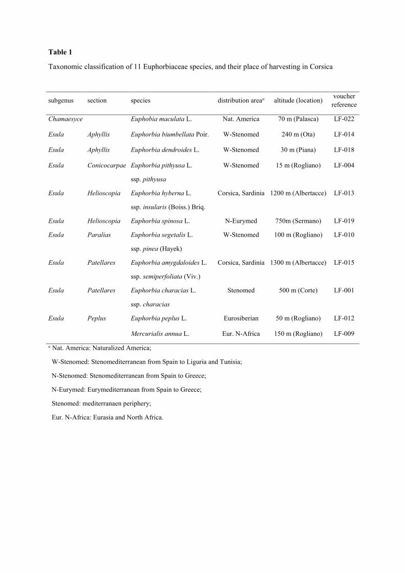
Table 1
Taxonomic classification of 11 Euphorbiaceae species, and their place of harvesting in Corsica
subgenus section species distribution areaa altitude (location) voucher
reference
Chamaesyce Euphobia maculata L. Nat. America 70 m (Palasca) LF-022
Esula Aphyllis Euphorbia biumbellata Poir. W-Stenomed 240 m (Ota) LF-014
Esula Aphyllis Euphorbia dendroides L. W-Stenomed 30 m (Piana) LF-018
Esula Conicocarpae Euphorbia pithyusa L.
ssp. pithyusa
W-Stenomed 15 m (Rogliano) LF-004
Esula Helioscopia Euphorbia hyberna L.
ssp. insularis (Boiss.) Briq.
Corsica, Sardinia 1200 m (Albertacce) LF-013
Esula Helioscopia Euphorbia spinosa L. N-Eurymed 750m (Sermano) LF-019
Esula Paralias Euphorbia segetalis L.
ssp. pinea (Hayek)
W-Stenomed 100 m (Rogliano) LF-010
Esula Patellares Euphorbia amygdaloides L.
ssp. semiperfoliata (Viv.)
Corsica, Sardinia 1300 m (Albertacce) LF-015
Esula Patellares Euphorbia characias L.
ssp. characias
Stenomed 500 m (Corte) LF-001
Esula Peplus Euphorbia peplus L. Eurosiberian 50 m (Rogliano) LF-012
Mercurialis annua L. Eur. N-Africa 150 m (Rogliano) LF-009
a Nat. America: Naturalized America;
W-Stenomed: Stenomediterranean from Spain to Liguria and Tunisia;
N-Stenomed: Stenomediterranean from Spain to Greece;
N-Eurymed: Eurymediterranean from Spain to Greece;
Stenomed: mediterranaen periphery;
Eur. N-Africa: Eurasia and North Africa.
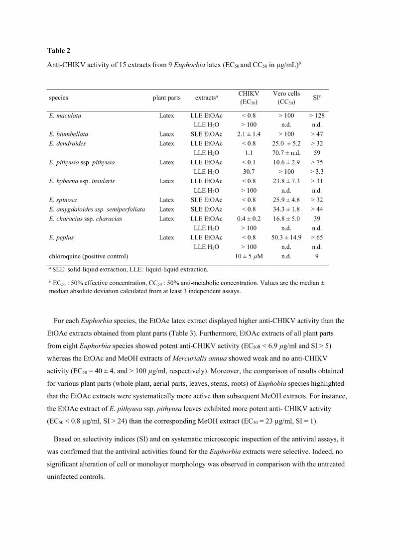
Table 2
Anti-CHIKV activity of 15 extracts from 9 Euphorbia latex (EC50 and CC50 in µg/mL)b
species plant parts extractsa CHIKV (EC50)
Vero cells (CC50)
SIc
E. maculata Latex LLE EtOAc < 0.8 > 100 > 128
LLE H2O > 100 n.d. n.d.
E. biumbellata Latex SLE EtOAc 2.1 ± 1.4 > 100 > 47
E. dendroides Latex LLE EtOAc < 0.8 25.0 ± 5.2 > 32
LLE H2O 1.1 70.7 ± n.d. 59
E. pithyusa ssp. pithyusa Latex LLE EtOAc < 0.1 10.6 ± 2.9 > 75
LLE H2O 30.7 > 100 > 3.3
E. hyberna ssp. insularis Latex LLE EtOAc < 0.8 23.8 ± 7.3 > 31
LLE H2O > 100 n.d. n.d.
E. spinosa Latex SLE EtOAc < 0.8 25.9 ± 4.8 > 32
E. amygdaloides ssp. semiperfoliata Latex SLE EtOAc < 0.8 34.3 ± 1.8 > 44
E. characias ssp. characias Latex LLE EtOAc 0.4 ± 0.2 16.8 ± 5.0 39
LLE H2O > 100 n.d. n.d.
E. peplus Latex LLE EtOAc < 0.8 50.3 ± 14.9 > 65
LLE H2O > 100 n.d. n.d.
chloroquine (positive control) 10 ± 5 µM n.d. 9
a SLE: solid-liquid extraction, LLE: liquid-liquid extraction.
b EC50 : 50% effective concentration, CC50 : 50% anti-metabolic concentration. Values are the median ± median absolute deviation calculated from at least 3 independent assays.
For each Euphorbia species, the EtOAc latex extract displayed higher anti-CHIKV activity than the
EtOAc extracts obtained from plant parts (Table 3). Furthermore, EtOAc extracts of all plant parts
from eight Euphorbia species showed potent anti-CHIKV activity (EC50s < 6.9 µg/ml and SI > 5)
whereas the EtOAc and MeOH extracts of Mercurialis annua showed weak and no anti-CHIKV
activity (EC50 = 40 ± 4, and > 100 µg/ml, respectively). Moreover, the comparison of results obtained
for various plant parts (whole plant, aerial parts, leaves, stems, roots) of Euphobia species highlighted
that the EtOAc extracts were systematically more active than subsequent MeOH extracts. For instance,
the EtOAc extract of E. pithyusa ssp. pithyusa leaves exhibited more potent anti- CHIKV activity
(EC50 < 0.8 µg/ml, SI > 24) than the corresponding MeOH extract (EC50 = 23 µg/ml, SI = 1).
Based on selectivity indices (SI) and on systematic microscopic inspection of the antiviral assays, it
was confirmed that the antiviral activities found for the Euphorbia extracts were selective. Indeed, no
significant alteration of cell or monolayer morphology was observed in comparison with the untreated
uninfected controls.

Table 3
Anti-CHIKV activity of 30 MeOH and EtOAc extracts of 8 Euphorbia species and Mercurialis annua (EC50 and CC50 in µg/mL)c
species plant parts extractsa yield (%)b CHIKV (EC50)
Vero cells (CC50)
SId
E. biumbellata Whole plant ASE EtOAc 4.4 4.1 > 100 > 47 ASE MeOH 6.7 > 100 n.d. n.d. E. pithyusa ssp. pithyusa Leaves ASE EtOAc 6.0 < 0.8 18.8 ± 1.2 > 24 ASE MeOH 11.0 23.0 23.3 ± 2.8 1 Stems ASE EtOAc 4.6 < 0.8 15.2 ± 5.9 > 20 ASE MeOH 9.0 4.5 8.5 ± 0.4 2 Roots ASE EtOAc 4.0 < 0.8 11.5 ± 1.6 > 15 ASE MeOH 8.0 < 0.8 21.0 ± 4.5 > 27 E. hyberna ssp. insularis Aerial parts ASE EtOAc 3.9 1.0 35.5 ± 3.1 36.0 E. spinosa Leaves ASE EtOAc 1.0 4.8 30.9 ± 2.2 6.4 Stems ASE EtOAc 5.0 3.4 23.2 ± 8.2 6.8 ASE MeOH 8.7 20.3 33.0 ± 2.4 1.6 Roots ASE EtOAc 7.0 < 0.8 30.5 ± 2.4 > 39.1 ASE MeOH 4.8 2.3 54.1 ± 18.0 23.5 E. segetalis ssp. pinea Aerial parts ASE EtOAc 7.0 3.7 35.6 ± 1.6 9.6 ASE MeOH 9.0 35.9 89.1 ± 5.1 2.5 Stems ASE EtOAc 3.6 3.5 25.8 ± 3.5 7.5 ASE MeOH 5.5 57.0 > 100 > 1.7 Roots ASE EtOAc 4.3 1.8 30.6 ± 0.2 17.3 ASE MeOH 5.9 29.7 > 100 > 3.4 E. amygdaloides ssp semiperfoliata Whole plant ASE EtOAc 5.2 < 0.8 30.5 ± 2.4 > 39.1
ASE MeOH 8.0 30.6 > 100 > 3.3 E. characias ssp. characias Leaves ASE EtOAc 6.3 6.9 34.0 ± 0.8 5.0 ASE MeOH 8.3 8.3 70.3 ± 3.4 8.5 Stems ASE EtOAc 5.0 2.9 32.1 ± 0.3 11.2 ASE MeOH 3.4 8.3 70.7 ± 8.5 8.5 E. peplus Whole plant ASE EtOAc 7.4 4.3 63.1 ± 7.0 14.7 ASE MeOH 7.3 30.0 > 100 > 3.3 Mercurialis annua Whole plant ASE EtOAc 3.1% 40 ± 4 89.1 ± 5.4 2 ASE MeOH 6.7% > 100 n.d. - chloroquine (positive control) 10 ± 5 µM n.d. 9 a SLE: solid-liquid extraction, LLE: liquid-liquid extraction, ASE: automatic solvent extraction.
b based on the dry weight of vegetal material (%); n.d.: not determined.
c EC50 : 50% effective concentration, CC50 : 50% anti-metabolic concentration. Values are the median ± median absolute deviation calculated from at least 3 independent assays.
d SI : selectivity index (SI calculated as CC50Vero/EC50 CHIKV).
Taking into account of these results, it is likely that the anti-CHIKV activities of Euphorbia extracts
are due to specific secondary metabolites of this genus. Previous studies have showed that Euphorbia
extracts can exhibit antiviral [52–56], but never against CHIKV replication. Diterpenoids of tigliane-
[32], ingenane- [28,33,34] and macrocyclic-type esters [35–37] isolated from various Euphorbia
species, were found to possess potent and selective anti-HIV activities. Regarding anti-CHIKV
activity of diterpenes from Euphorbiaceae, phorbol esters are the most potent inhibitors of CHIKV
replication [10] but are also known for their pro-inflammatory and tumour promoter activities [13,12].

3.2. Analysis of Euphobia extracts using targeted LC-MS2 method
In order to perform a dereplication strategy on Euphorbia extracts with potent anti-CHIKV activity,
a LC-MS2-based method had been developed to detect 27 commercially available natural diterpenoids
(1-27) belonging to tigliane- (phorbol and deoxyphorbol derivatives), ingenane- and daphnane-types
(Figure 1). For each standards, MS/MS optimized parameters (precursor-to-product transition
monitored, declustering potential DP, entrance potential EP, collision cell entrance potential CEP,
collision energy CE, collision cell exit potential CXP), and retention time in LC, had been determined
and are summarized in Table 4. Main ions observed in MS2 spectrum are also included in Table 4. The
fragmentation behavior of diterpene esters were consistent with previous data reported in the literature
[42,43,46,47], Indeed, in ESI positive ion mode, diterpene esters form pseudo-molecular ions, which
undergo neutral loss of their acyl chain(s) under collision-induced dissociation (CID), producing the
corresponding high-abundance fragment ions.
The identification of diterpenoids in plant extracts was established by comparison with reference
compounds: MRM transition at specific retention time and MS2 spectrum of the precursor ion to those
recorded in the Analyst software spectral library. In addition, to avoid artefactual detection, an
untargeted LC-MS2 (MRM-MS2) analysis was systematically carried out to confirm that the supposed
targeted pseudo-molecular ion was not formed from a parent compound with a higher molecular
weight.
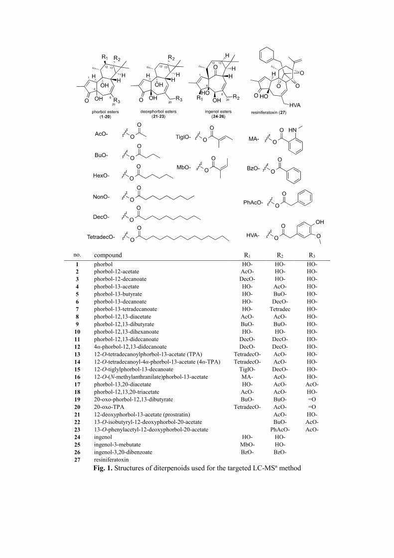
no. compound R1 R2 R3
1 phorbol HO- HO- HO- 2 phorbol-12-acetate AcO- HO- HO- 3 phorbol-12-decanoate DecO- HO- HO- 4 phorbol-13-acetate HO- AcO- HO- 5 phorbol-13-butyrate HO- BuO- HO- 6 phorbol-13-decanoate HO- DecO- HO- 7 phorbol-13-tetradecanoate HO- Tetradec
O- HO-
8 phorbol-12,13-diacetate AcO- AcO- HO- 9 phorbol-12,13-dibutyrate BuO- BuO- HO- 10 phorbol-12,13-dihexanoate HO- HO- HO- 11 phorbol-12,13-didecanoate DecO- DecO- HO- 12 4α-phorbol-12,13-didecanoate DecO- DecO- HO- 13 12-O-tetradecanoylphorbol-13-acetate (TPA) TetradecO- AcO- HO- 14 12-O-tetradecanoyl-4α-phorbol-13-acetate (4α-TPA) TetradecO- AcO- HO- 15 12-O-tiglylphorbol-13-decanoate TiglO- DecO- HO- 16 12-O-(N-methylanthranilate)phorbol-13-acetate MA- AcO- HO- 17 phorbol-13,20-diacetate HO- AcO- AcO- 18 phorbol-12,13,20-triacetate AcO- AcO- HO- 19 20-oxo-phorbol-12,13-dibutyrate BuO- BuO- =O 20 20-oxo-TPA TetradecO- AcO- =O 21 12-deoxyphorbol-13-acetate (prostratin) AcO- HO- 22 13-O-isobutyryl-12-deoxyphorbol-20-acetate BuO- AcO- 23 13-O-phenylacetyl-12-deoxyphorbol-20-acetate PhAcO- AcO- 24 ingenol HO- HO- 25 ingenol-3-mebutate MbO- HO- 26 ingenol-3,20-dibenzoate BzO- BzO- 27 resiniferatoxin
Fig. 1. Structures of diterpenoids used for the targeted LC-MSn method

Table 4
LC-MSn parameters for reference standards in ESI positive ion mode
cpd Rt (min)
Q1 Mass (m/z)b
Q3 Mass (m/z)c
DP (V)
EP (V)
CEP (V)
CE (V)
CXP (V)
Most abundant ions (m/z) observed in MS2 spectrum
1 1.4 387.2 369.3 156 11.5 18 21 6 369, 351, 333, 315, 311, 293 2 2.0 429.2 369.2 56 8.0 20 21 6 369, 351, 333, 329, 315, 311, 293 3 13.9 541.3 369.2 86 4.5 30 25 6 369, 351, 333, 329, 315, 311, 293 4 2.0 429.2 369.2 71 8.0 30 21 6 369, 351, 333, 329, 315, 311, 293 5 2.6 457.2 369.2 236 10 24 25 6 369, 351, 333, 329, 315, 311, 293 6 12.9 541.3 369.2 86 4.5 20 27 6 369, 351, 333, 329, 315, 311, 293 7 21.5 597.3 369.2 231 4.5 62 29 6 369, 351, 333, 329, 315, 311, 293 8 3.1 471.2 411.2 266 9.5 24 23 6 411, 351, 333, 329, 315, 311, 293 9 9.4 527.3 439.3 71 5.0 24 25 6 439, 421, 351, 333, 329, 315, 311, 293 10 17.7 583.3 467.2 301 6.5 30 25 6 467, 351, 333, 329, 315, 311, 293 11 28.5 695.5 523.3 91 9.0 50 29 8 523, 351, 333, 329, 315, 311, 293 12 29.0 695.5 523.3 211 10.5 24 45 6 523, 351, 333, 329, 315, 311, 293 13 23.5 639.4 411.2 71 8.5 44 29 6 579, 411, 351, 333, 329, 315, 311, 293 14 24.4 639.4 411.2 71 8.5 44 29 6 579, 411, 351, 333, 329, 315, 311, 293 15 22.2 623.4 523.3 206 9.0 30 27 6 523, 451, 351, 333, 329, 315, 311, 293 16 9.3 562.2 411.2 301 5.0 24 25 6 411, 502, 351, 333, 329, 315, 311, 293 17 2.8 471.2 411.2 266 9.5 24 23 6 411, 351, 333, 329, 315, 311, 293 18 5.8 513.2 453.2 66 4.5 20 25 6 453, 393, 365, 333, 315, 311, 293 19 3.5 525.2 437.2 71 6.0 20 27 8 437, 349, 331, 309, 291 20 24.0 637.3 409.2 71 8.0 27 27 6 577, 409, 349, 331, 309, 291 21 3.3 413.2 353.2 156 8.0 24 21 6 353, 335, 317, 313, 307, 295 22 11.3 483.2 395.2 71 7.0 22 25 6 335, 317, 313, 307, 295 23 11.7 531.2 395.2 116 8.0 26 27 6 335, 317, 313, 307, 295 24 3.0 371.1 353.2 261 10.0 28 23 6 353, 335, 317, 313, 307, 295, 285 25 12.4 453.2 353.2 126 7.5 20 23 6 353, 335, 317, 313, 307, 295, 285 26 18.2 579.4 457.2 96 9.0 28 27 6 457, 335, 317, 313, 307, 295, 285 27 16.3 651.3 515.2 266 10.5 30 33 6 515, 469, 441, 333

LC-MS2 analysis of the EtOAc Euphorbia extracts allowed the identification of three diterpenes
(Table 5). The results indicated that ingenol-3-mebutate (25) was present in E. peplus extracts (latex
and whole plant). Indeed, the identification of compound 25 was allowed by the detection of a
transition m/z 453→ 335 at Rt 12.4 min and by comparison with MS2 spectra recorded in our spectral
library (Fig. S1). Using the same methodology, ingenol-3,20-dibenzoate (26) was detected by
observation of a transisiton m/z 579→457 at Rt 18.3 min in E. peplus and E. segetalis ssp. pinea
extracts (Fig S2-S3). Compound 26 was found in latex extracts as well as in other plant parts of the
two species. Furthermore, 13-O-isobutyryl-12-deoxyphorbol-20-acetate (22) was detected in roots and
aerial parts extracts of E. segetalis ssp. pinea (Fig S4).
Table 5
Reference standard diterpene esters detected by targeted LC-MS (MRM-MS2) (see Fig. S1-S4 for MS2 spectra)
extracts (EtOAc)
plant partsa
compound transition
(m/z)
Rt (min)
[M+Na]+ (m/z)
ions observed in MS2 spectrum
S/N
E. peplus
Lat, WP
ingenol-3,20-dibenzoate 26 579.4/457.2 18.3 579.21 457.2, 335.2 188
ingenol-3-mebutate 25 453.2/353.2 12.4 453.22 335.2, 295.2 82
E. pithyusa
ssp. pithyusa
Lat, OPP
13-O-isobutyryl-
12-deoxyphorbol-20-acetate 22
483.2/395.2 11.2 483.24 423.4, 395.2, 335.2
85
E. segetalis ssp. pinea
R, AP ingenol-3,20-dibenzoate 26 579.4/457.2 18.4 579.21 457.2, 335.2 145
a Lat : latex, WP : whole plant, OPP : other plant parts, R : roots, AP : aerial parts
The present results are in agreeement with the previously reported isolation of ingenol-3-mebutate
(25) in E. peplus extract [57], and the isolation of other ingenane-type esters in E. peplus and in E.
segetalis extracts [58,59]. The detection of 13-O-isobutyryl-12-deoxyphorbol-20-acetate (22) is also
reinforced by the isolation of deoxyphorbol diesters in E. pithyusa ssp. pithyusa [60]. However, one
must keep in mind that even if comparison with MS2 spectrum and Rt obtain in the same conditions is
considered sufficient to confidently identified compound in LC-MS2 [61], it cannot be exclude that the
ion detected is an isomer.
Beside detection of these three compounds, no other standard compound could be detected in the
Euphorbia extracts, including the highly potent anti-CHIKV phorbol esters 11, 13, and 21. These
results are consistent with (i) previous phytochemical investigation of these species (Table S1) and (ii)
the fact that, while tigliane-type diterpene are commonly found in Euphorbia spp. [27,31], occurrence

of phorbol derivatives stricto sensu was rare in this genus [13,62,63]. Indeed, most of phorbol esters
had been found in genus Croton [64], Jatropha [65] and Sapium [66].
From the dereplication perspective, taking into account the IC50 values for inhibition of CHIKV
replication of 13-O-isobutyryl-12-deoxyphorbol-20-acetate (22), and ingenol-3,20-dibenzoate (26),
(EC50 = 0.7 ± 0.1 µM, SI = 5.0, and EC50 = 1.2 ± 0.1 µM, SI = 6.4, respectively), it can be concluded
that the presence of compound 22 in E. pithyusa ssp. pithyusa, and compound 26 in E. peplus and E.
segetalis ssp. pinea extracts should partially explain their potent anti-CHIKV activity. However, it is
likely that other diterpene esters may contribute to the anti-CHIKV activities of these extracts. For
instance, it has been demonstrated that jatrophane esters isolated from E. amygdaloides ssp.
semiperfoliata exhibited significant anti-CHIKV activities [51].
It should be noted that the diterpenes 22, 25, and 26 are potent and selective inhibitors of HIV-1 and
HIV-2 at the nanomolar scale [10]. Thus, it is likely that the EtOAc extracts of E. peplus, E. segetalis
ssp. pinea and E. pithyusa ssp. pithyusa possess strong anti-HIV activities.
4. Conclusion
In conclusion, several EtOAc and MeOH extracts of Euphorbia species exhibited potent and
selective inhibitory activities of CHIKV replication. A LC-MS2 based method was used to detect the
possible presence in the biologically active extracts of diterpene esters endowed with antiviral
activities. The results of the LC-MS2 analysis indicated the presence of 13-O-isobutyryl-12-
deoxyphorbol-20-acetate (22) in E. pithyusa ssp. pithyusa extracts, ingenol-3-mebutate (25) in E.
peplus extracts, ingenol-3,20-dibenzoate (26) in E. peplus and E. segetalis ssp. pinea extracts. Potent
inhibitors of CHIKV replication such as phorbol-12,13-didecanoate (11), TPA (13) and prostratin (21)
were not detected in the Euphorbia extracts. Thus, the present results suggested that untargeted
diterpene esters are responsible of the anti-CHIKV activity of Euphorbia extracts.
Acknowledgments
We are grateful to Dr. M-J Battesti (University of Corsica) for her precious advises and to Laurène
Peter and Naym Ben Amara for their help during collection of vegetal material. This work has also
benefited from an “Investissement d’Avenir” grant managed by Agence Nationale de la Recherche
(CEBA, ref. ANR-10-LABX-25-01). We would like to acknowledge Stijn Delmotte, Caroline Collard,
Nick Verstraeten and Charlotte Vanderheydt for their excellent technical assistance in the acquisition
of the antiviral data.

References
[1] Thiberville S-D, Moyen N, Dupuis-Maguiraga L, Nougairede A, Gould EA, Roques P, et al. Chikungunya fever: Epidemiology, clinical syndrome, pathogenesis and therapy. Antiviral Res 2013;99:345–70. doi:10.1016/j.antiviral.2013.06.009.
[2] Morrison TE. Reemergence of Chikungunya Virus. J Virol 2014;88:11644–7. doi:10.1128/JVI.01432-14.
[3] Powers A. Chikungunya virus outbreak expansion and microevolutionary events affecting epidemiology and epidemic potential. Res Rep Trop Med 2015;6:11–9. doi:10.2147/RRTM.S53698.
[4] Singh P, Chhabra M, Mittal V, Sharma P, Rizvi MA, Chauhan L, et al. Current research and clinical trials for a vaccine against Chikungunya virus. Vaccine Dev Ther 2013:35–46. doi:10.2147/VDT.S25513.
[5] Kaur P, Chu JJH. Chikungunya virus: an update on antiviral development and challenges. Drug Discov Today 2013;18:969–83. doi:10.1016/j.drudis.2013.05.002.
[6] Rashad AA, Mahalingam S, Keller PA. Chikungunya Virus: Emerging Targets and New Opportunities for Medicinal Chemistry. J Med Chem 2013;4:1147–66. doi:10.1021/jm400460d.
[7] Allard P-M, Leyssen P, Martin M-T, Bourjot M, Dumontet V, Eydoux C, et al. Antiviral chlorinated daphnane diterpenoid orthoesters from the bark and wood of Trigonostemon cherrieri. Phytochemistry 2012;84:160–8. doi:10.1016/j.phytochem.2012.07.023.
[8] Corlay N, Delang L, Girard-Valenciennes E, Neyts J, Clerc P, Smadja J, et al. Tigliane diterpenes from Croton mauritianus as inhibitors of chikungunya virus replication. Fitoterapia 2014;97:87–91. doi:10.1016/j.fitote.2014.05.015.
[9] Bourjot M, Delang L, Nguyen VH, Neyts J, Guéritte F, Leyssen P, et al. Prostratin and 12-O-Tetradecanoylphorbol 13-Acetate Are Potent and Selective Inhibitors of Chikungunya Virus Replication. J Nat Prod 2012;75:2183–7. doi:10.1021/np300637t.
[10] Nothias-Scaglia L-F, Pannecouque C, Renucci F, Delang L, Neyts J, Roussi F, et al. Antiviral Activity of Diterpene Esters on Chikungunya Virus and HIV Replication. J Nat Prod 2015:in press.
[11] Evans FJ, Schmidt RJ. An assay procedure for the comparative irritancy testing of esters in the tigliane and daphnane series. Inflammation 1979;3:215–23. doi:10.1007/BF00914178.
[12] Opferkuch HJ, Hecker E. On the active principles of the spurge family (Euphorbiaceae). J Cancer Res Clin Oncol 1982;103:255–68. doi:10.1007/BF00409701.
[13] Evans FJ, Taylor SE. Pro-inflammatory, Tumor-promoting and Anti-tumour diterpenes of the plants of families Euphorbiaceae and Thymelaeaceae. Fortschritte Chem Org Naturstoffe 1983;44:1–99.
[14] Furstenberger G, Hecker E. On the active principles of the spurge family (Euphorbiaceae). XI.[1] The skin irritant and tumor promoting diterpene esters of Euphorbia tirucalli L. originating from South Africa. Z Naturforsch C 1985;40:631–46.
[15] Saraiva L, Fresco P, Pinto E, Gonçalves J. Characterization of phorbol esters activity on individual mammalian protein kinase C isoforms, using the yeast phenotypic assay. Eur J Pharmacol 2004;491:101–10. doi:10.1016/j.ejphar.2004.03.035.
[16] Das J, Rahman GM. C1 Domains: Structure and Ligand-Binding Properties. Chem Rev 2014;114:12108–31. doi:10.1021/cr300481j.

[17] Webster GL. Classification of the Euphorbiaceae. Ann Mo Bot Gard 1994;81:33–144. doi:10.2307/2399908.
[18] Smith AR, Turin TG, Burges NA, Valentine DH. Euphorbia L. Flora Eur., vol. 2, Cambridge University Press; 1968, p. 213–26.
[19] Frajman B, Schönswetter P. Giants and dwarfs: Molecular phylogenies reveal multiple origins of annual spurges within Euphorbia subg. Esula. Mol Phylogenet Evol 2011;61:413–24. doi:10.1016/j.ympev.2011.06.011.
[20] Jeanmonod D, Gamisans J. Flora Corsica. vol. 1. Edisud 921. Aix-En-Provence: 2007.
[21] Dioscorides Pedanius. De Materia Medica. Johannesburg, South Africa: IBIDIS; 2000.
[22] Eke T, Al-Husainy S, Raynor MK. The spectrum of ocular inflammation caused by Euphorbia plant sap. Arch Ophthalmol 2000;118:13–6.
[23] Appendino G, Szallasi A. Euphorbium: modern research on its active principle, resiniferatoxin, revives an ancient medicine. Life Sci 1997;60:681–96.
[24] Conrad M. Les plantes sauvages dans la vie quotidienne des Corses: essai d’ethnobotanique. Société des sciences historiques et naturelles de la Corse. Bastia: 1973.
[25] Bruni A, Ballero M, Poli F. Quantitative ethnopharmacological study of the Campidano Valley and Urzulei district, Sardinia, Italy. J Ethnopharmacol 1997;57:97–124. doi:10.1016/S0378-8741(97)00055-X.
[26] Leto C, Tuttolomondo T, La Bella S, Licata M. Ethnobotanical study in the Madonie Regional Park (Central Sicily, Italy)—Medicinal use of wild shrub and herbaceous plant species. J Ethnopharmacol 2013;146:90–112. doi:10.1016/j.jep.2012.11.042.
[27] Shi Q-W, Su X-H, Kiyota H. Chemical and Pharmacological Research of the Plants in Genus Euphorbia. Chem Rev 2008;108:4295–327. doi:10.1021/cr078350s.
[28] Abreu CM, Price SL, Shirk EN, Cunha RD, Pianowski LF, Clements JE, et al. Dual Role of Novel Ingenol Derivatives from Euphorbia tirucalli in HIV Replication: Inhibition of De Novo Infection and Activation of Viral LTR. PLoS ONE 2014;9. doi:10.1371/journal.pone.0097257.
[29] Ferreira AMVD, Carvalho MJM, Silva AMS, Carvalho LHM, Majumdar DK, Govil JN, et al. Diterpenes of the Family Euphorbiaceae. Recente Prog. Med. Plants, vol. 9, Texas: Studium Press LLC; 2005, p. 15–118.
[30] Mwine JT, Van Damme P. Why do Euphorbiaceae tick as medicinal plants? A review of Euphorbiaceae family and its medicinal. J Med Plants Res 2011;5:652–62.
[31] Vasas A, Hohmann J. Euphorbia Diterpenes: Isolation, Structure, Biological Activity, and Synthesis (2008–2012). Chem Rev 2014;114:8579–612. doi:10.1021/cr400541j.
[32] Pan L-L, Fang P-L, Zhang X-J, Ni W, Li L, Yang L-M, et al. Tigliane-Type Diterpenoid Glycosides from Euphorbia fischeriana. J Nat Prod 2011;74:1508–12. doi:10.1021/np200058c.
[33] Fujiwara M, Ijichi K, Tokuhisa K, Katsuura K, Shigeta S, Konno K, et al. Mechanism of selective inhibition of human immunodeficiency virus by ingenol triacetate. Antimicrob Agents Chemother 1996;40:271.
[34] Hong K-J, Lee HS, Kim Y, Kim SS. Ingenol Protects Human T Cells From HIV-1 Infection. Osong Public Health Res Perspect 2011;2:109–14. doi:10.1016/j.phrp.2011.07.001.

[35] Bedoya LM, Márquez N, Martínez N, Gutiérrez-Eisman S, Álvarez A, Calzado MA, et al. SJ23B, a jatrophane diterpene activates classical PKCs and displays strong activity against HIV in vitro. Biochem Pharmacol 2009;77:965–78. doi: 10.1016/j.bcp.2008.11.025.
[36] Avila L, Perez M, Sanchez-Duffhues G, Hernández-Galán R, Muñoz E, Cabezas F, et al. Effects of diterpenes from latex of Euphorbia lactea and Euphorbia laurifolia on human immunodeficiency virus type 1 reactivation. Phytochemistry 2010;71:243–8. doi:10.1016/j.phytochem.2009.10.005.
[37] Tian Y, Xu W, Zhu C, Lin S, Guo Y, Shi J. Diterpenoids with Diverse Skeletons from the Roots of Euphorbia micractina. J Nat Prod 2013;76:1039–46. doi:10.1021/np400029d.
[38] Corea G, Di Pietro A, Dumontet C, Fattorusso E, Lanzotti V. Jatrophane diterpenes from Euphorbia spp. as modulators of multidrug resistance in cancer therapy. Phytochem Rev 2009;8:431–47. doi:10.1007/s11101-009-9126-8.
[39] Vasas A, Rédei D, Csupor D, Molnár J, Hohmann J. Diterpenes from European Euphorbia Species Serving as Prototypes for Natural-Product-Based Drug Discovery. Eur J Org Chem 2012;27:5115–30. doi:10.1002/ejoc.201200733.
[40] Ogbourne SM, Parsons PG. The value of nature’s natural product library for the discovery of New Chemical Entities: The discovery of ingenol mebutate. Fitoterapia 2014;98:36–44. doi:10.1016/j.fitote.2014.07.002.
[41] Boyle GM, D’Souza MMA, Pierce CJ, Adams RA, Cantor AS, Johns JP, et al. Intra-Lesional Injection of the Novel PKC Activator EBC-46 Rapidly Ablates Tumors in Mouse Models. PLoS ONE 2014;9:e108887. doi:10.1371/journal.pone.0108887.
[42] Vogg G, Mattes E, Rothenburger J, Hertkorn N, Stefan Achatz, Sandermann Jr. H. Tumor promoting diterpenes from Euphorbia leuconeura L. Phytochemistry 1999;51:289–95. doi:doi: 10.1016/S0031-9422(99)00016-3.
[43] Tang Q, Su Z, Han Z, Ma X, Xu D, Liang Y, et al. LC–MS method for detecting prostratin in plant extracts and identification of a high-yielding population of Euphorbia fischeriana. Phytochem Lett 2012;5:214–8. doi:10.1016/j.phytol.2011.12.011.
[44] Hou J-J, Wu W-Y, Liang J, Yang Z, Long H-L, Cai L-Y, et al. A single, multi-faceted, enhanced strategy to quantify the chromatographically diverse constituents in the roots of Euphorbia kansui. J Pharm Biomed Anal 2014;88:321–30. doi:10.1016/j.jpba.2013.08.049.
[45] Jarret M, Katsamas J, Cawkill K, Henderson L, Welburn P. A pharmacokinetic study using a highly sensitive and specific method to determine systemic exposure of ingenol mebutate and its two main metabolites, PEP015 and PEP025 in human whole blood after topical administration. J Am Acad Dermatol 2013;68:AB156. doi:10.1016/j.jaad.2012.12.646.
[46] Bicchi C, Appendino G, Cordero C, Rubiolo P, Ortelli D, Veuthey J-L. HPLC-UV and HPLC-positive-ESI-MS analysis of the diterpenoid fraction from caper spurge (Euphorbia lathyris) seed oil. Phytochem Anal 2001;12:255–62. doi:10.1002/pca.592.
[47] Hou X-R, Wan L-L, Zhan Z-J, Li C-P, Shan W-G. Analysis and determination of diterpenoids in unprocessed and processed Euphorbia lathyris seeds by HPLC–ESI-MS. J Pharm Anal 2011;1:197–202. doi:doi: 10.1016/j.jpha.2011.06.003.
[48] Meng X, Zhao X, Long Z, Yuan Y, Zhuang H, Bi K, et al. A sensitive liquid chromatography–mass spectrometry method for simultaneous determination of three diterpenoid esters from Euphorbia lathyris L. in rat plasma. J Pharm Biomed Anal 2013;72:299–305. doi:10.1016/j.jpba.2012.09.005.

[49] Chow S, Fletcher MT, Mckenzie RA. Analysis of Daphnane Orthoesters in Poisonous Australian Pimelea Species by Liquid Chromatography−Tandem Mass Spectrometry. J Agric Food Chem 2011;58:7482–7. doi: 10.1021/jf101752r.
[50] Zhang S, Zhang F, Li X, Dong W, Wen L, Wang S. Evaluation of Daphne genkwa diterpenes: Fingerprint and quantitative analysis by high performance liquid chromatography. Phytochem Anal 2007;18:91–7. doi:10.1002/pca.953.
[51] Nothias-Scaglia L-F, Retailleau P, Paolini J, Pannecouque C, Neyts J, Dumontet V, et al. Jatrophane Diterpenes as Inhibitors of Chikungunya Virus Replication: Structure–Activity Relationship and Discovery of a Potent Lead. J Nat Prod 2014;77:1505–12. doi:10.1021/np500271u.
[52] Vlietinck A, Van Hoof L, Totte J, Lasure A, Berghe DV, Rwangabo P, et al. Screening of hundred Rwandese medicinal plants for antimicrobial and antiviral properties. J Ethnopharmacol 1995;46:31–47.
[53] Semple SJ, Reynolds GD, O’Leary MC, Flower RLP. Screening of Australian medicinal plants for antiviral activity. J Ethnopharmacol 1998;60:163–72. doi:10.1016/S0378-8741(97)00152-9.
[54] Ramezani M, Behravan J, Arab M, Farzad SA. Antiviral Activity of Euphorbia helioscopia Extract. J Biol Sci 2008;8:809–13. doi:10.3923/jbs.2008.809.813.
[55] Behravan J, Salmasi Z, Ramezani M, Sabeti NZ. Euphorbia microsciadia percolation and Soxhlet extracts exhibit antiviral activity. Clin Biochem 2011;44:S325. doi:10.1016/j.clinbiochem.2011.08.801.
[56] Forero JE, Avila L, Taborda N, Tabares P, López A, Torres F, et al. In vitro anti-influenza screening of several Euphorbiaceae species: Structure of a bioactive Cyanoglucoside from Codiaeum variegatum. Phytochemistry 2008;69:2815–9. doi:10.1016/j.phytochem.2008.09.003.
[57] Hohmann J, Evanics F, Berta L, Bartók T. Diterpenoids from Euphorbia peplus. Planta Med 2000;66:291–4. doi:10.1055/s-2000-8568.
[58] Jakupovic J, Jeske F, Morgenstern T, Tsichritzis F, Marco JA, Berendsohn W. Diterpenes from Euphorbia segetalis. Phytochemistry 1998;47:1583–600. doi: 10.1016/S0031-9422(97)00830-3.
[59] Jakupovic J, Morgenstern T, Bittner M, Silva M. Diterpenes from Euphorbia peplus. Phytochemistry 1998;47:1601–9. doi:10.1016/S0031-9422(97)00831-5.
[60] Appendino G, Belloro E, Tron GC, Jakupovic J, Ballero M. Diterpenoids from Euphorbia pithyusa subsp. cupanii. J Nat Prod 1999;62:1399–404. doi: 10.1021/np990209u.
[61] Dunn WB, Erban A, Weber RJM, Creek DJ, Brown M, Breitling R, et al. Mass appeal: metabolite identification in mass spectrometry-focused untargeted metabolomics. Metabolomics 2012;9:44–66. doi:10.1007/s11306-012-0434-4.
[62] Evans FJ, Kinghorn AD. A comparative phytochemical study of the diterpenes of some species of the genera Euphorbia and Elaeophorbia (Euphorbiaceae). Bot J Linn Soc 1977;74:23–35. doi:10.1111/j.1095-8339.1977.tb01163.x.
[63] Fürstenberger G, Hecker E. New highly irritant Euphorbia factors from latex of Euphorbia tirucalli L. Experientia 1977;33:986–8. doi:10.1007/BF01945920.
[64] Zhang X-L, Wang L, Li F, Yu K, Wang M-K. Cytotoxic Phorbol Esters of Croton tiglium. J Nat Prod 2013;76:858–64. doi:10.1021/np300832n.
[65] Devappa R, Makkar H, Becker K. Jatropha Diterpenes: a Review. J Am Oil Chem Soc

2011;88:301–22.
[66] Al Muqarrabun LMR, Ahmat N, Aris SRS. A review of the medicinal uses, phytochemistry and pharmacology of the genus Sapium. J Ethnopharmacol 2014;155:9–20. doi:10.1016/j.jep.2014.05.028.