human ifitm3 restricts chikungunya virus and mayaro virus
TRANSCRIPT
Research Article
Human IFITM3 restricts chikungunya virus and Mayarovirus infection and is susceptible to virus-mediatedcounteractionSergej Franz1,2, Fabian Pott3,4 , Thomas Zillinger5 , Christiane Schüler3,4 , Sandra Dapa1, Carlo Fischer3 ,Vania Passos1, Saskia Stenzel3,4, Fangfang Chen6 , Katinka Dohner7, Gunther Hartmann3, Beate Sodeik7,8 ,Frank Pessler6, Graham Simmons2, Jan Felix Drexler3, Christine Goffinet1,3,4
Interferon-induced transmembrane (IFITM) proteins restrict mem-brane fusion and virion internalization of several envelopedviruses. The role of IFITM proteins during alphaviral infection ofhuman cells and viral counteraction strategies are insufficientlyunderstood. Here, we characterized the impact of human IFITMson the entry and spread of chikungunya virus and Mayaro virusand provide first evidence for a CHIKV-mediated antagonism ofIFITMs. IFITM1, 2, and 3 restricted infection at the level of alphavirusglycoprotein-mediated entry, both in the context of direct infectionand cell-to-cell transmission. Relocalization of normally endo-somal IFITM3 to the plasma membrane resulted in loss of antiviralactivity. rs12252-C, a naturally occurring variant of IFITM3 that mayassociate with severe influenza in humans, restricted CHIKV, MAYV,and influenza A virus infection as efficiently as wild-type IFITM3.Antivirally active IFITM variants displayed reduced cell surfacelevels in CHIKV-infected cells involving a posttranscriptional pro-cess mediated by one or several nonstructural protein(s) of CHIKV.Finally, IFITM3-imposed reduction of specific infectivity of nascentparticles provides a rationale for the necessity of a virus-encodedcounteraction strategy against this restriction factor.
DOI 10.26508/lsa.202000909 | Received 16 September 2020 | Revised 21 May2021 | Accepted 21 May 2021 | Published online 2 June 2021
Introduction
Infection of humans by mosquito-transmitted chikungunya virus(CHIKV) and Mayaro virus (MAYV) causes, in most of the infectedindividuals, an acute febrile illness accompanied by symmetricjoint pain and inflammation. In a subset of infected individuals of
varying size depending on the outbreak, long-term morbiditymanifesting as chronic arthritis with debilitating pain has beenreported for both members of the Semliki Forest Virus complex andis causing growing medical concern and socioeconomical loss inaffected countries. Although likely being endemic in East Africasince several centuries, chikungunya virus outbreaks of increasingfrequency have occurred worldwide in the past 15 yr. MAYV iscausing increasing attention in the neotropics. Neither approvedvaccines nor antivirals are available against either alphavirus in-fection. Despite increasing importance of emerging arthritogenicalphaviruses for the human population, the biology of their rep-lication cycle and their interplay with host proteins only begin to beelucidated. Specifically, the entry process of CHIKV and MAYV isgreatly facilitated in the presence of target cell–expressed adhe-sion molecule MXRA8 (Zhang et al, 2018), and FHL1 serves as acofactor for CHIKV, but not MAYV, RNA replication (Meertens et al,2019).
Interferon-induced transmembrane (IFITM) proteins are broadlyactive against numerous enveloped RNA viruses, including HIV-1 (Luet al, 2011), West Nile virus (Gorman et al, 2016), Zika virus (Savidiset al, 2016), and influenza A virus (IAV) (Brass et al, 2009), as well asenveloped DNA viruses (Li et al, 2018, 2019a). Among the five humanIFITM genes, only IFITM1, IFITM2, and IFITM3 have antiviral propertiesby restricting virus entry (Bailey et al, 2014). IFITM genes encodesmall transmembrane proteins of debated topology (Liao et al,2019). IFITM2 and IFITM3 predominantly localize in endosomalmembranes, whereas IFITM1 resides on the cell surface (Chesarinoet al, 2014; Weston et al, 2014; Narayana et al, 2015; Compton et al,2016). The mechanisms by which IFITM proteins inhibit viral in-fections appear to involve interference with fusion of viral andcellular membranes, resulting in virions trapped at the hemi-fusion
1Institute of Experimental Virology, TWINCORE Centre for Experimental and Clinical Infection Research, a Joint Venture Between the Hannover Medical School (MHH) andthe Helmholtz Centre for Infection Research (HZI), Hannover, Germany 2Vitalant Research Institute, San Francisco, CA, USA 3Charite–Universitatsmedizin Berlin,Corporate Member of Freie Universitat Berlin and Humboldt-Universitat zu Berlin, Institute of Virology, Berlin, Germany 4Berlin Institute of Health atCharite–Universitatsmedizin Berlin, Berlin, Germany 5Institute of Clinical Chemistry and Clinical Pharmacology, University Hospital, Venusberg-Campus 1, Bonn,Germany 6Research Group Biomarkers for Infectious Diseases, TWINCORE, Centre for Experimental and Clinical Infection Research, a Joint Venture Between the HanoverMedical School (MHH) and the Helmholtz Centre for Infection Research (HZI), Hanover, Germany 7Institute of Virology, Hannover Medical School, Hanover, Germany8Cluster of Excellence RESIST (EXC 2155), Hannover Medical School, Hannover, Germany
Correspondence: [email protected]
© 2021 Franz et al. https://doi.org/10.26508/lsa.202000909 vol 4 | no 7 | e202000909 1 of 15
on 9 February, 2022life-science-alliance.org Downloaded from http://doi.org/10.26508/lsa.202000909Published Online: 2 June, 2021 | Supp Info:
stage (Li et al, 2013; Desai et al, 2014). IFITM3 retains viral particles inlate endosomes and targets them for lysosomal degradation(Feeley et al, 2011; Spence et al, 2019; Suddala et al, 2019). Experi-mental retargeting of IFITM3 to the cell surface, which can be in-duced by disruption of its Yxxθ type endocytosis motif throughintroduction of the Y20A mutation (Williams et al, 2014) or deletionof the 21 N-terminal amino acids (Weidner et al, 2010; Jia et al, 2012),nullifies its activity against many viruses which enter by endocy-tosis, including IAV (John et al, 2013).
The potential importance of human IFITM3 protein as antiviralfactor has been addressed in clinical observation studies of in-fluenza A. Specifically, the single-nucleotide polymorphism (SNP)rs12252-C allele might associate with increased influenza A mor-tality and morbidity (Everitt et al, 2012; Williams et al, 2014; Pan et al,2017), although other cohorts failed to display this association (Millset al, 2014; Lopez-Rodriguez et al, 2016; Randolph et al, 2017; Carteret al, 2018; David et al, 2018). A debated mechanistic working modelbased on the idea that the genetic variation induces alteration of asplice acceptor site, resulting in expression of a truncated IFITM3protein which lost its antiviral activity (Everitt et al, 2012).
The impact of human IFITM proteins on alphaviral infectionremains poorly elucidated, and no information on the anti-alphavirus ability of the SNP rs12252-C allele is available. In Musmusculus, a non-natural host of alphaviruses from the SemlikiForest Virus complex, IFITM3 shares 65% homology in amino acidsequence with the human ortholog and inhibits multiple arthri-togenic alphaviruses, including CHIKV, and encephalitogenicalphaviruses in vivo (Poddar et al, 2016). Heterologously expressedhuman IFITM3, and to a lesser extent IFITM2 restrict Sindbis virusand Semliki Forest virus through inhibition of fusion of viral andcellular membranes (Weston et al, 2016). Furthermore, humanIFITM1, 2, and 3 emerged as potential inhibitors of CHIKV infection ina high-throughput ISG overexpression screen (Schoggins et al,2011), but their anti-alphaviral properties have not been charac-terized in the context of CHIKV and MAYV infection.
Here, we addressed the activity of human IFITM1, 2, and 3 as well asof naturally occurring and experimental human IFITM3 variantsagainst CHIKV and MAYV. All three IFITM proteins restricted infectionat the level of alphaviral glycoprotein-mediated entry. Experimentallyinduced relocalization of normally endosomal IFITM3 to the plasmamembrane resulted in the loss of its antiviral activity despite robustexpression. The rs12252-C allele restricted CHIKV, MAYV, and IAV asefficiently as wild-type IFITM3. Finally, steady-state IFITM3 expressionlevels were markedly reduced at the posttranscriptional level inproductively infected cells, suggesting the existence of a so far un-appreciated virus-mediated counteraction strategy of IFITM factorsmediated by expression of one or several nonstructural proteins.Antagonism of IFITMs may contribute to mitigation of the potentialability of IFITM3 to negatively imprint nascent virions.
Results
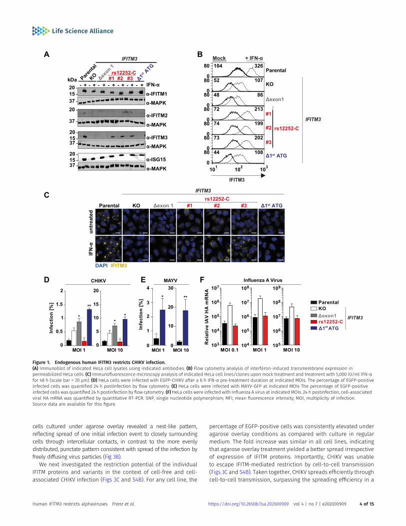
Endogenous human IFITM3 restricts CHIKV infection
To investigate the anti-CHIKV activities of human IFITM proteins, weapplied a CRISPR/Cas9-assisted approach to edit the IFITM3 gene in
CHIKV-susceptible, IFITM3-expressing HeLa cells (Fig S1). Specifi-cally, we functionally ablated the IFITM3 gene by introducing aframeshift after nucleotide 84 (KO) or by deleting a large part ofexon 1 of IFITM3 (Δexon1) in both alleles. In addition, we introduceda T-to-C transition at position 89 in both IFITM3 alleles to expressthe minor C allele of the SNP rs12252 (rs12252-C), which has beensuggested to associate with severe IAV infections (Everitt et al, 2012;Zhang et al, 2013; Pan et al, 2017). Furthermore, we deleted a regionof 31 base-pairs encompassing the primary ATG codon (Δ1st ATG)(Fig S1). In all clones, Sanger sequencing confirmed that IFITM3(Table 1), but not the highly homologous IFITM2 gene (data notshown) had been edited. Immunoblotting showed that although allcell lines and clones clearly up-regulated expression of IFITM1 andthe prototypic ISG product ISG15 upon IFN stimulation, IFITM3protein detection was abrogated in HeLa cells encoding IFITM3 KOand IFITM3 Δexon1 (Fig 1A). In addition, no signal was detectable forIFITM3 (Δ1st ATG), arguing against expression of a truncated proteinand rather for absence of expression (Fig 1A). In contrast, threeclones expressing the rs12252 T-to-C variant displayed detectableIFN-induced IFITM3 expression, although slightly lower than inparental cells. Importantly, the immunoblot provided no evidencefor expression of a truncated IFITM3 protein in rs12252-C cells, butrather displayed a band of equal molecular weight as the one fromwild-type IFITM3 (Fig 1A). Detection of up-regulated IFITM2 proteinfailed despite the confirmed specificity of the IFITM2-targetingantibody (see Fig 2A). Flow cytometry analysis of IFITM3 proteinexpression in permeabilized cells paralleled the results of theimmunoblot analysis, with IFITM3 in rs12252-C cells being slightlyless abundant at baseline and upon IFN stimulation (Fig 1B). Finally,immunofluorescence microscopy confirmed expression of IFITM3protein in parental and in rs12252-C cells, but not in the other celllines (Fig 1C).
We then inoculated the individual cell lines with CHIKV. Throughoutthe study, we used the Indian Ocean lineage strain LR2006 harboringan EGFP reporter at the 59endof the viral genome, hereafter referred toas EGFP-CHIKV. Absence of IFITM3 expression in the KO, Δexon1, andΔ1st ATG cells was accompanied by diminished effectivity of the IFN-induced antiviral program, whereas IFITM3 expression in parental andrs12252-C cells impaired EGFP-CHIKV infection to similar levels (Figs 1Dand S2). Similarly, the susceptibility of Δ1st ATG cells to EGFP-MAYV (Li etal, 2019b) infection was markedly enhanced (Figs 1E and S2). Finally,parental cells and rs12252-C cells were equally restrictive to IAV in-fection, as opposed to KO cells (Fig 1F). In conclusion, we show thatendogenous IFITM3 restricts CHIKV and MAYV infection. Furthermore,the naturally occurring variant of IFITM3, rs12252-C, drove expression ofan IFITM3 protein that displays a similar anti-CHIKV and anti-IAVpotency as the major IFITM3 allele.
Ectopic expression of human IFITM-HA proteins restrictsalphaviral infection by interfering with glycoprotein-mediatedentry
To gain mechanistic insight into the differential antiviral potency ofIFITM proteins and variants, we stably transduced HEK293T cells,which lack endogenous IFITM protein expression, with C-terminallyHA-tagged IFITM1, 2, or 3 (Brass et al, 2009). In addition, we gen-erated individual cell lines expressing the mutant proteins
Human IFITM3 restricts alphaviruses Franz et al. https://doi.org/10.26508/lsa.202000909 vol 4 | no 7 | e202000909 2 of 15
IFITM3(Y20A)-HA and IFITM3(Δ1-21)-HA, which localize to the plasmamembrane (Weidner et al, 2010; Feeley et al, 2011; Jia et al, 2012;Williams et al, 2014). Finally, we overexpressed the IFITM3 rs12252-Cvariant (IFITM3[rs12252-C]-HA). Appropriate protein expression wasconfirmed by immunoblot analysis using both an anti-HA antibodyand primary antibodies targeting the individual authentic IFITMproteins (Fig 2A). We confirmed similar expression levels of allproteins and variants by flow cytometry (Fig 2B) and immunoflu-orescence microscopy (Fig 2C) using an anti-HA antibody in intactand in permeabilized cells. Thereby, we confirmed the previouslyreported, predominant cell surface localization of IFITM3(Y20A)-HAand IFITM3(Δ1-21)-HA (Perreira et al, 2013). IFITM3(rs12252-C)-HA didnot express a shorter variant and the protein localized predomi-nantly intracellularly, as observed in HeLa cells (Fig 1). To assess theantiviral capacity of the IFITM-HA proteins, we infected the cell lineswith EGFP-CHIKV and determined the amount of EGFP-positive cellsat 24 h postinfection. Over a wide range of MOIs, IFITM1-HA andIFITM3-HA, and to a lesser extent IFITM2-HA, displayed antiviralactivity against CHIKV (Figs 2D and S3A and B). IFITM3(rs12252-C)-HArestricted EGFP-CHIKV infection at a similar efficiency as IFITM3-HA.In contrast, IFITM3(Y20A)-HA and IFITM3(Δ1-21)-HA failed to impairinfection (Figs 2D and S3A and B). We observed a similar pattern ofinhibition at the level of infectivity released in the culture super-natant of EGFP-CHIKV- and MAYV-infected cells, as judged byplaque assays (Fig 2E). To monitor whether inhibition of CHIKVinfection was related to the established ability of IFITMs to interferewith enveloped virus entry, we quantified the cells’ susceptibility totransduction by lentiviral pseudoparticles decorated with heter-ologous viral glycoproteins and expressing a luciferase cassette (Fig2F). In line with previous reports (Brass et al, 2009), IFITM proteinexpression did not reduce the susceptibility of cells to transductionby particles pseudotyped with glycoproteins of murine leukemiavirus. In contrast, expression of IFITM2-HA and IFITM3-HA, but notIFITM1-HA, reduced the cells’ susceptibility to transduction by Ebolavirus glycoprotein-pseudotyped particles, as reported before(Brass et al, 2009; Wrensch et al, 2015). Transduction of cells byparticles incorporating CHIKV glycoproteins was impaired byIFITM1-HA and IFITM3-HA and, to a lesser extent, by IFITM2-HA. The
A226V mutation in CHIKV E1 that enabled adaptation of the virus tothe alternative vector Aedes albopictus (Tsetsarkin et al, 2007) didnot overtly modulate susceptibility to IFITM protein-mediated re-striction (Fig 2F). CHIKV glycoproteins from sublineages 1–4, whichare derived from the S27 strain (Tsetsarkin et al, 2014), sharedsusceptibility to IFITM-mediated inhibition, although to slightlydifferent extents (Fig 2F). Collectively, these data establish thatIFITM proteins restrict CHIKV and MAYV, and that inhibition is di-rected against entry mediated by glycoproteins of different CHIKVsublineages. In addition, antiviral activity is only exerted by IFITM3variants with reported endosomal localization.
Cell-to-cell transmission of and direct infection by CHIKVdiffer in efficiency, but share susceptibility to IFITM-mediatedrestriction
Cell-to-cell transmission of virions can antagonize or evade theIFN-induced antiviral state, including restriction by some (Richardsonet al, 2008; Vendrame et al, 2009; Jolly et al, 2010), but not all(Puigdomenech et al, 2013) antiviral factors. Expression of IFITM3renders target cells less susceptible to infection by cell-free HIV-1particles, but cell-to-cell transmission of HIV-1 partially escapes thisantiviral effect (Compton et al, 2014). We thus addressed the relativeability of cell-associated and cell-free transmission modes in sup-porting CHIKV spread, and their relative susceptibility to IFITMprotein-mediated restriction. As a surrogate system for cell-to-celltransmission, we subjected infected cells with an agarose overlay,which favors transfer of virions between neighboring cells and im-pairs infection by freely diffusing virus particles. In this set-up, theinitial infection is seeded by cell-free virus, whereas subsequentrounds of infection aremediated by cell-associated virus and cell-to-cell spread. For comparison, infected cells were cultured in regulargrowth medium without agarose. EGFP-CHIKV spread was enhancedunder agarose overlay conditions, with to twofold higher percentagesof EGFP-positive cells as compared to cells cultured in regular medium(Fig 3A). Of note, in cells under agarose, an EGFP-encoding HSV-1 spreadat identical or even lower efficiencies than in regular medium (Figs 3Aand S4A). Fluorescence microscopy analysis of EGFP-CHIKV-infected
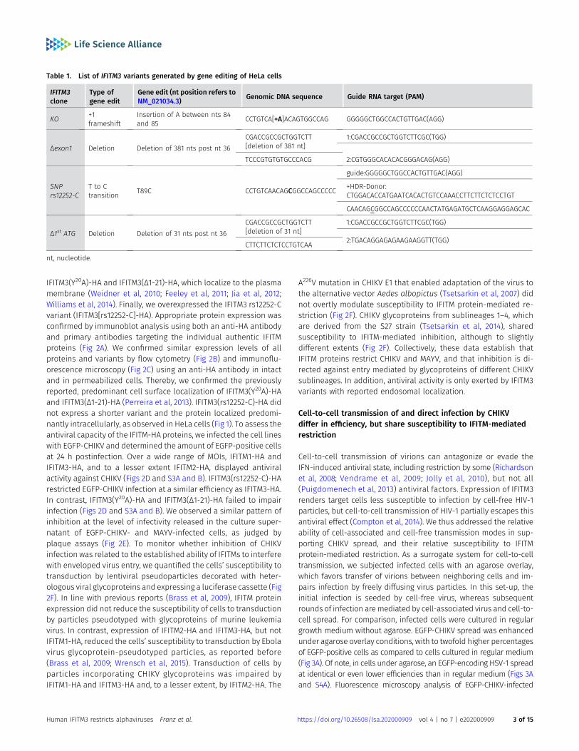
Table 1. List of IFITM3 variants generated by gene editing of HeLa cells
IFITM3clone
Type ofgene edit
Gene edit (nt position refers toNM_021034.3) Genomic DNA sequence Guide RNA target (PAM)
KO +1frameshift
Insertion of A between nts 84and 85 CCTGTCA[+A]ACAGTGGCCAG GGGGGCTGGCCACTGTTGAC(AGG)
Δexon1 Deletion Deletion of 381 nts post nt 36CGACCGCCGCTGGTCTT[deletion of 381 nt]
1:CGACCGCCGCTGGTCTTCGC(TGG)
TCCCGTGTGTGCCCACG 2:CGTGGGCACACACGGGACAG(AGG)
SNPrs12252-C
T to Ctransition T89C CCTGTCAACAGCGGCCAGCCCCC
guide:GGGGGCTGGCCACTGTTGAC(AGG)
+HDR-Donor:CTGGACACCATGAATCACACTGTCCAAACCTTCTTCTCTCCTGT
CAACAGCGGCCAGCCCCCCAACTATGAGATGCTCAAGGAGGAGCAC
Δ1st ATG Deletion Deletion of 31 nts post nt 36CGACCGCCGCTGGTCTT[deletion of 31 nt]
1:CGACCGCCGCTGGTCTTCGC(TGG)
2:TGACAGGAGAGAAGAAGGTT(TGG)CTTCTTCTCTCCTGTCAA
nt, nucleotide.
Human IFITM3 restricts alphaviruses Franz et al. https://doi.org/10.26508/lsa.202000909 vol 4 | no 7 | e202000909 3 of 15
cells cultured under agarose overlay revealed a nest-like pattern,reflecting spread of one initial infection event to closely surroundingcells through intercellular contacts, in contrast to the more evenlydistributed, punctate pattern consistent with spread of the infection byfreely diffusing virus particles (Fig 3B).
We next investigated the restriction potential of the individualIFITM proteins and variants in the context of cell-free and cell-associated CHIKV infection (Figs 3C and S4B). For any cell line, the
percentage of EGFP-positive cells was consistently elevated underagarose overlay conditions as compared with culture in regularmedium. The fold increase was similar in all cell lines, indicatingthat agarose overlay treatment yielded a better spread irrespectiveof expression of IFITM proteins. Importantly, CHIKV was unableto escape IFITM-mediated restriction by cell-to-cell transmission(Figs 3C and S4B). Taken together, CHIKV spreads efficiently throughcell-to-cell transmission, surpassing the spreading efficiency in a
Figure 1. Endogenous human IFITM3 restricts CHIKV infection.(A) Immunoblot of indicated HeLa cell lysates using indicated antibodies. (B) Flow cytometry analysis of interferon-induced transmembrane expression inpermeabilized HeLa cells. (C) Immunofluorescence microscopy analysis of indicated HeLa cell lines/clones upon mock treatment and treatment with 5,000 IU/ml IFN-αfor 48 h (scale bar = 20 µm). (D) HeLa cells were infected with EGFP-CHIKV after a 6 h IFN-α pre-treatment duration at indicated MOIs. The percentage of EGFP-positiveinfected cells was quantified 24 h postinfection by flow cytometry. (E) HeLa cells were infected with MAYV-GFP at indicated MOIs The percentage of EGFP-positiveinfected cells was quantified 24 h postinfection by flow cytometry. (F) HeLa cells were infected with Influenza A virus at indicated MOIs. 24 h postinfection, cell-associatedviral HA mRNA was quantified by quantitative RT-PCR. SNP, single nucleotide polymorphism; MFI, mean fluorescence intensity; MOI, multiplicity of infection.Source data are available for this figure.
Human IFITM3 restricts alphaviruses Franz et al. https://doi.org/10.26508/lsa.202000909 vol 4 | no 7 | e202000909 4 of 15
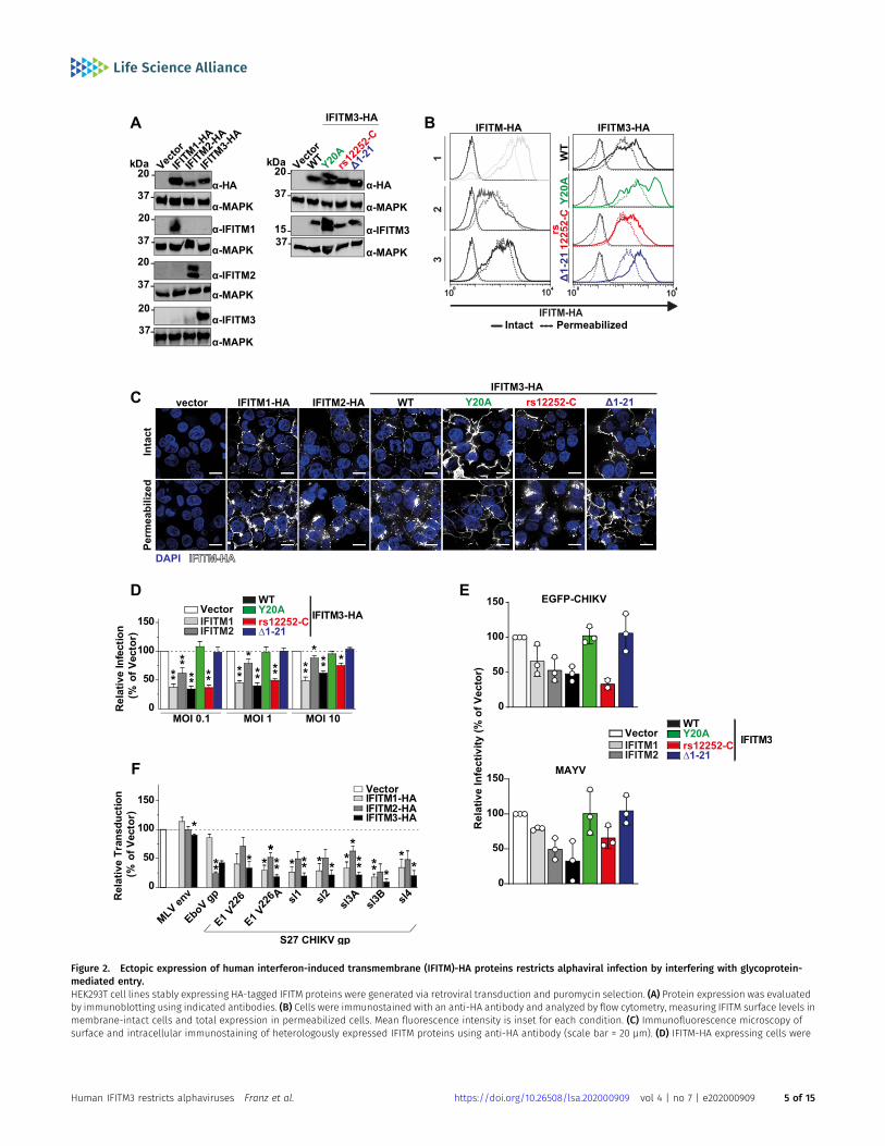
Figure 2. Ectopic expression of human interferon-induced transmembrane (IFITM)-HA proteins restricts alphaviral infection by interfering with glycoprotein-mediated entry.HEK293T cell lines stably expressing HA-tagged IFITM proteins were generated via retroviral transduction and puromycin selection. (A) Protein expression was evaluatedby immunoblotting using indicated antibodies. (B) Cells were immunostained with an anti-HA antibody and analyzed by flow cytometry, measuring IFITM surface levels inmembrane-intact cells and total expression in permeabilized cells. Mean fluorescence intensity is inset for each condition. (C) Immunofluorescence microscopy ofsurface and intracellular immunostaining of heterologously expressed IFITM proteins using anti-HA antibody (scale bar = 20 µm). (D) IFITM-HA expressing cells were
Human IFITM3 restricts alphaviruses Franz et al. https://doi.org/10.26508/lsa.202000909 vol 4 | no 7 | e202000909 5 of 15
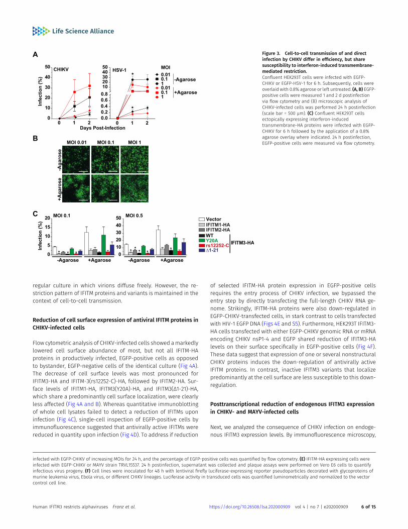
regular culture in which virions diffuse freely. However, the re-striction pattern of IFITM proteins and variants is maintained in thecontext of cell-to-cell transmission.
Reduction of cell surface expression of antiviral IFITM proteins inCHIKV-infected cells
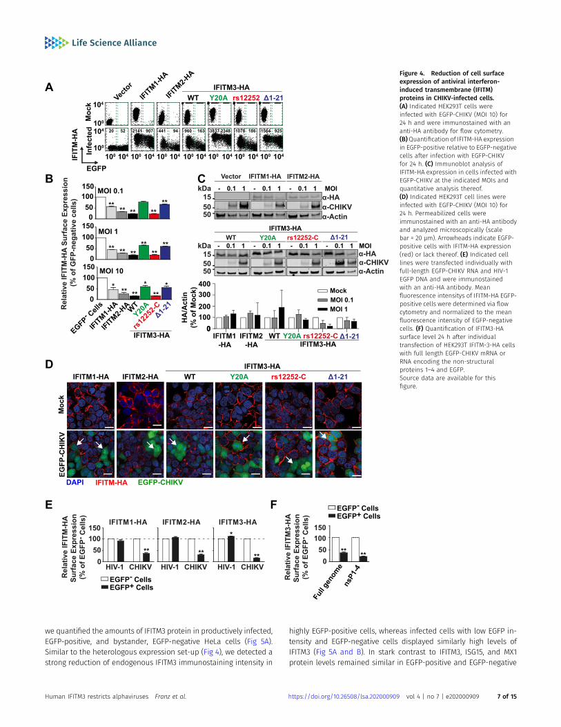
Flow cytometric analysis of CHIKV-infected cells showed amarkedlylowered cell surface abundance of most, but not all IFITM-HAproteins in productively infected, EGFP-positive cells as opposedto bystander, EGFP-negative cells of the identical culture (Fig 4A).The decrease of cell surface levels was most pronounced forIFITM3-HA and IFITM-3(rs12252-C)-HA, followed by IFITM2-HA. Sur-face levels of IFITM1-HA, IFITM3(Y20A)-HA, and IFITM3(Δ1-21)-HA,which share a predominantly cell surface localization, were clearlyless affected (Fig 4A and B). Whereas quantitative immunoblottingof whole cell lysates failed to detect a reduction of IFITMs uponinfection (Fig 4C), single-cell inspection of EGFP-positive cells byimmunofluorescence suggested that antivirally active IFITMs werereduced in quantity upon infection (Fig 4D). To address if reduction
of selected IFITM-HA protein expression in EGFP-positive cellsrequires the entry process of CHIKV infection, we bypassed theentry step by directly transfecting the full-length CHIKV RNA ge-nome. Strikingly, IFITM-HA proteins were also down-regulated inEGFP-CHIKV-transfected cells, in stark contrast to cells transfectedwith HIV-1 EGFP DNA (Figs 4E and S5). Furthermore, HEK293T IFITM3-HA cells transfected with either EGFP-CHIKV genomic RNA or mRNAencoding CHIKV nsP1-4 and EGFP shared reduction of IFITM3-HAlevels on their surface specifically in EGFP-positive cells (Fig 4F).These data suggest that expression of one or several nonstructuralCHIKV proteins induces the down-regulation of antivirally activeIFITM proteins. In contrast, inactive IFITM3 variants that localizepredominantly at the cell surface are less susceptible to this down-regulation.
Posttranscriptional reduction of endogenous IFITM3 expressionin CHIKV- and MAYV-infected cells
Next, we analyzed the consequence of CHIKV infection on endoge-nous IFITM3 expression levels. By immunofluorescence microscopy,
Figure 3. Cell-to-cell transmission of and directinfection by CHIKV differ in efficiency, but sharesusceptibility to interferon-induced transmembrane-mediated restriction.Confluent HEK293T cells were infected with EGFP-CHIKV or EGFP-HSV-1 for 6 h. Subsequently, cells wereoverlaid with 0.8% agarose or left untreated. (A, B) EGFP-positive cells were measured 1 and 2 d postinfectionvia flow cytometry and (B) microscopic analysis ofCHIKV-infected cells was performed 24 h postinfection(scale bar = 500 µm). (C) Confluent HEK293T cellsectopically expressing interferon-inducedtransmembrane-HA proteins were infected with EGFP-CHIKV for 6 h followed by the application of a 0.8%agarose overlay where indicated. 24 h postinfection,EGFP-positive cells were measured via flow cytometry.
infected with EGFP-CHIKV of increasing MOIs for 24 h, and the percentage of EGFP-positive cells was quantified by flow cytometry. (E) IFITM-HA expressing cells wereinfected with EGFP-CHIKV or MAYV strain TRVL15537. 24 h postinfection, supernatant was collected and plaque assays were performed on Vero E6 cells to quantifyinfectious virus progeny. (F) Cell lines were inoculated for 48 h with lentiviral firefly luciferase-expressing reporter pseudoparticles decorated with glycoproteins ofmurine leukemia virus, Ebola virus, or different CHIKV lineages. Luciferase activity in transduced cells was quantified luminometrically and normalized to the vectorcontrol cell line.
Human IFITM3 restricts alphaviruses Franz et al. https://doi.org/10.26508/lsa.202000909 vol 4 | no 7 | e202000909 6 of 15
we quantified the amounts of IFITM3 protein in productively infected,EGFP-positive, and bystander, EGFP-negative HeLa cells (Fig 5A).Similar to the heterologous expression set-up (Fig 4), we detected astrong reduction of endogenous IFITM3 immunostaining intensity in
highly EGFP-positive cells, whereas infected cells with low EGFP in-tensity and EGFP-negative cells displayed similarly high levels ofIFITM3 (Fig 5A and B). In stark contrast to IFITM3, ISG15, and MX1protein levels remained similar in EGFP-positive and EGFP-negative
Figure 4. Reduction of cell surfaceexpression of antiviral interferon-induced transmembrane (IFITM)proteins in CHIKV-infected cells.(A) Indicated HEK293T cells wereinfected with EGFP-CHIKV (MOI 10) for24 h and were immunostained with ananti-HA antibody for flow cytometry.(B)Quantification of IFITM-HA expressionin EGFP-positive relative to EGFP-negativecells after infection with EGFP-CHIKVfor 24 h. (C) Immunoblot analysis ofIFITM-HA expression in cells infected withEGFP-CHIKV at the indicated MOIs andquantitative analysis thereof.(D) Indicated HEK293T cell lines wereinfected with EGFP-CHIKV (MOI 10) for24 h. Permeabilized cells wereimmunostained with an anti-HA antibodyand analyzed microscopically (scalebar = 20 µm). Arrowheads indicate EGFP-positive cells with IFITM-HA expression(red) or lack thereof. (E) Indicated celllines were transfected individually withfull-length EGFP-CHIKV RNA and HIV-1EGFP DNA and were immunostainedwith an anti-HA antibody. Meanfluorescence intensitys of IFITM-HA EGFP-positive cells were determined via flowcytometry and normalized to the meanfluorescence intensity of EGFP-negativecells. (F) Quantification of IFITM3-HAsurface level 24 h after individualtransfection of HEK293T IFITM-3-HA cellswith full length EGFP-CHIKV mRNA orRNA encoding the non-structuralproteins 1–4 and EGFP.Source data are available for thisfigure.
Human IFITM3 restricts alphaviruses Franz et al. https://doi.org/10.26508/lsa.202000909 vol 4 | no 7 | e202000909 7 of 15
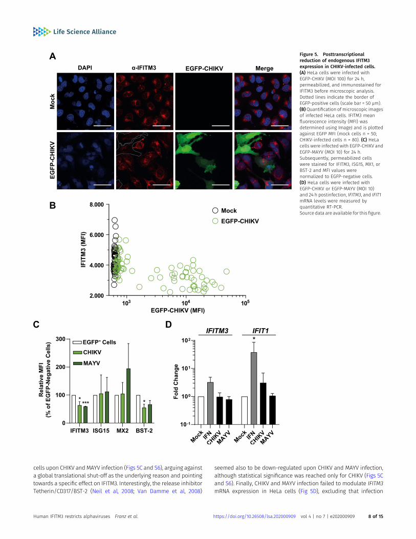
cells upon CHIKV and MAYV infection (Figs 5C and S6), arguing againsta global translational shut-off as the underlying reason and pointingtowards a specific effect on IFITM3. Interestingly, the release inhibitorTetherin/CD317/BST-2 (Neil et al, 2008; Van Damme et al, 2008)
seemed also to be down-regulated upon CHIKV and MAYV infection,although statistical significance was reached only for CHIKV (Figs 5Cand S6). Finally, CHIKV and MAYV infection failed to modulate IFITM3mRNA expression in HeLa cells (Fig 5D), excluding that infection
Figure 5. Posttranscriptionalreduction of endogenous IFITM3expression in CHIKV-infected cells.(A) HeLa cells were infected withEGFP-CHIKV (MOI 100) for 24 h,permeabilized, and immunostained forIFITM3 before microscopic analysis.Dotted lines indicate the border ofEGFP-positive cells (scale bar = 50 µm).(B)Quantification of microscopic imagesof infected HeLa cells. IFITM3 meanfluorescence intensity (MFI) wasdetermined using ImageJ and is plottedagainst EGFP MFI (mock cells n = 50;CHIKV-infected cells n = 80). (C) HeLacells were infected with EGFP-CHIKV andEGFP-MAYV (MOI 10) for 24 h.Subsequently, permeabilized cellswere stained for IFITM3, ISG15, MX1, orBST-2 and MFI values werenormalized to EGFP-negative cells.(D) HeLa cells were infected withEGFP-CHIKV or EGFP-MAYV (MOI 10)and 24 h postinfection, IFITM3, and IFIT1mRNA levels were measured byquantitative RT-PCR.Source data are available for this figure.
Human IFITM3 restricts alphaviruses Franz et al. https://doi.org/10.26508/lsa.202000909 vol 4 | no 7 | e202000909 8 of 15
modulates transcription of the IFITM3 gene, and suggesting aninfection-induced modulation of IFITM3 protein steady-state levels.IFN-α treatment boosted IFITM3 mRNA levels, as expected. Further-more, another prototypic ISG, IFIT1, was clearly induced by both IFN-αtreatment and CHIKV infection (Fig 5D). Conclusively, these datasupport a posttranscriptional modulation of IFITM3 expression inCHIKV- and MAYV-infected cells, potentially representing a virus-mediated counteraction strategy of IFITM restriction.
Reduction of particle infectivity through expression ofendogenous IFITM3 in alphavirus-producing cells
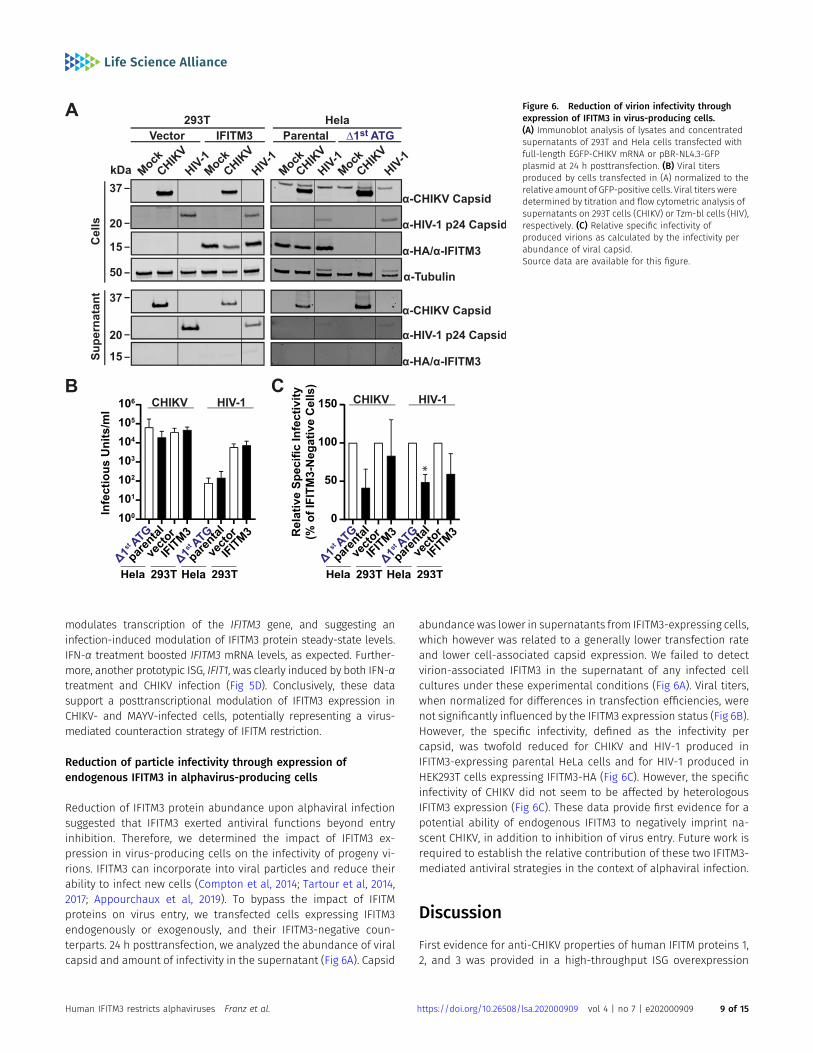
Reduction of IFITM3 protein abundance upon alphaviral infectionsuggested that IFITM3 exerted antiviral functions beyond entryinhibition. Therefore, we determined the impact of IFITM3 ex-pression in virus-producing cells on the infectivity of progeny vi-rions. IFITM3 can incorporate into viral particles and reduce theirability to infect new cells (Compton et al, 2014; Tartour et al, 2014,2017; Appourchaux et al, 2019). To bypass the impact of IFITMproteins on virus entry, we transfected cells expressing IFITM3endogenously or exogenously, and their IFITM3-negative coun-terparts. 24 h posttransfection, we analyzed the abundance of viralcapsid and amount of infectivity in the supernatant (Fig 6A). Capsid
abundance was lower in supernatants from IFITM3-expressing cells,which however was related to a generally lower transfection rateand lower cell-associated capsid expression. We failed to detectvirion-associated IFITM3 in the supernatant of any infected cellcultures under these experimental conditions (Fig 6A). Viral titers,when normalized for differences in transfection efficiencies, werenot significantly influenced by the IFITM3 expression status (Fig 6B).However, the specific infectivity, defined as the infectivity percapsid, was twofold reduced for CHIKV and HIV-1 produced inIFITM3-expressing parental HeLa cells and for HIV-1 produced inHEK293T cells expressing IFITM3-HA (Fig 6C). However, the specificinfectivity of CHIKV did not seem to be affected by heterologousIFITM3 expression (Fig 6C). These data provide first evidence for apotential ability of endogenous IFITM3 to negatively imprint na-scent CHIKV, in addition to inhibition of virus entry. Future work isrequired to establish the relative contribution of these two IFITM3-mediated antiviral strategies in the context of alphaviral infection.
Discussion
First evidence for anti-CHIKV properties of human IFITM proteins 1,2, and 3 was provided in a high-throughput ISG overexpression
Figure 6. Reduction of virion infectivity throughexpression of IFITM3 in virus-producing cells.(A) Immunoblot analysis of lysates and concentratedsupernatants of 293T and Hela cells transfected withfull-length EGFP-CHIKV mRNA or pBR-NL4.3-GFPplasmid at 24 h posttransfection. (B) Viral titersproduced by cells transfected in (A) normalized to therelative amount of GFP-positive cells. Viral titers weredetermined by titration and flow cytometric analysis ofsupernatants on 293T cells (CHIKV) or Tzm-bl cells (HIV),respectively. (C) Relative specific infectivity ofproduced virions as calculated by the infectivity perabundance of viral capsid.Source data are available for this figure.
Human IFITM3 restricts alphaviruses Franz et al. https://doi.org/10.26508/lsa.202000909 vol 4 | no 7 | e202000909 9 of 15
screen in STAT-deficient human fibroblasts (Schoggins et al, 2011).However, the pattern of IFITM-mediated CHIKV inhibition and po-tential CHIKV-mediated counteraction strategies remain obscure.By applying a two-armed approach that included investigation ofendogenous IFITM3 and variations thereof, and of heterologouslyexpressed, epitope-tagged IFITM proteins and mutants, we estab-lished and characterized the antiviral activity of IFITM proteinsagainst CHIKV and MAYV.
Our study centered on the investigation of IFITM3’s role onalphaviral infection. Expression of endogenous and heterologousIFITM3 rendered HeLa and HEK293T cells less susceptible to CHIKVinfection, respectively. In the endogenous expression context,frameshift insertion in the 59 part of the IFITM3 gene, deletion of alarge part of the first exon of the IFITM3 gene or of a smaller, 31-basepair region comprising the first, canonical ATG, resulted in ablationof IFITM3 expression and antiviral activity. Similarly, cells expressinga frameshift in the 59 part of the first exon of the IFITM3 gene wereunable to restrict IAV infection and lost the ability to restrict CHIKV.
It has been hypothesized that the T-to-C substitution in the SNPrs-12252, located in the first exon of the IFITM3 gene, alters a spliceacceptor site, resulting in a truncated IFITM3 protein lacking its first21 amino acids and exerting reduced antiviral activity (Everitt et al,2012). However, this working model did not substantiate becauseneither the predicted alternatively spliced mRNA (Randolph et al,2017; Makvandi-Nejad et al, 2018) nor a truncated IFITM3 protein(Makvandi-Nejad et al, 2018) has been detected in cells homozy-gously expressing rs12252-C. In line with these negative results, wedetect a protein of normal size both in cells homozygouslyexpressing the SNP and in cells overexpressing an IFITM3-encodingconstruct carrying the T-to-C transition. However, our anti-IFITM3immunoblotting technique is clearly able to detect the smallermolecular weight of IFITM3(Δ1-21)-HA, excluding a technical in-ability to detect marginally smaller IFITM3 proteins in general.Whereas IFITM3(Δ1-21)-HA localized to the cell surface and lacksantiviral activity, subcellular localization and antiviral phenotype ofthe rs12252-C variant remained indistinguishable from the wild-type IFITM3 in terms of anti-CHIKV, anti-MAYV, and anti-IAV activity,molecular weight and subcellular localization. However, IFITM-3rs12252-C seemed to be expressed to slightly lower levels than wild-type IFITM3, without impacting its antiviral efficacy. Together, weconclude that if any, specific functional properties of the rs12252-Callele remain to be discovered andmay not directly be implicated incell-intrinsic immunity.
Experimental redirection of IFITM3 to the cell surface by dis-rupting the sorting motif YxxV (Jia et al, 2012, 2014) through in-troduction of the Y20A or Δ1-21 mutations resulted in a loss of itsanti-CHIKV and anti-MAYV activity. Analogous findings have beenobtained by others for SINV and SFV (Weston et al, 2016), and otherenveloped viruses which invade cells via receptor-mediated en-docytosis (John et al, 2013; Jia et al, 2014). In addition, heterologousassays revealed an anti-CHIKV and anti-MAYV activity of IFITM1 thatequaled that of IFITM3. This observation contrasts reports by othersfor Sindbis virus and Semliki Forest virus (Weston et al, 2016) but isin accordance with results obtained in a study that screened theantiviral potential of several ISGs against CHIKV (Schoggins et al,2011). Heterologous expression of genes can cause aberrant sub-cellular localization and/or nonphysiological expression levels.
Therefore, results obtained by heterologous expression need to beinterpreted with caution. Importantly, levels of IFITM protein ex-pression obtained by heterologous expression of IFITM were similarto levels induced by IFN treatment of HeLa cells (data not shown).Future studies are warranted to corroborate the contribution ofIFITM1 to alphavirus restriction.
Restriction was directed against CHIKV E2/E1 glycoprotein-mediated entry, was maintained in the context of CHIKV glyco-proteins expressing the vector switch-enabling mutation A226V inCHIKV E1 and displayed a significant breadth because glycoproteinsof several CHIKV S27 strain sublineages were sensitive to IFITMprotein-mediated restriction.
Most of the cell culture studies on CHIKV are conducted usingcell-free, purified virus. An early report published in 1970 suggestedthat CHIKV spreads via cell-to-cell contacts when free virions areimmunologically neutralized (Hahon & Zimmerman, 1970). Alongthis line, intercellular transmission of CHIKV was reported morerecently to be less sensitive to antibody-mediated neutralization(Lee et al, 2011). In the present study, we applied a semi-solid,agarose-containing overlay to infected cultures to determine thecontribution of cell-free virus versus intercellular transmission, asperformed by others for hepatitis C virus (Timpe et al, 2008), ve-sicular stomatitis virus and murine leukemia virus (Liberatore et al,2017). Interestingly, CHIKV tended to spread more efficiently underagarose overlay, as opposed to HSV-1. The exact mode of inter-cellular transmission of CHIKV in different cell types will be animportant future object of investigation and might contribute tounderstanding alphavirus persistence in vivo. The relative re-striction potential of individual IFITM proteins and variants wasidentical in the cell-free and cell-associated transmission set-ups.As a contrasting example, HIV-1 is able to overcome IFITM3-mediated restriction via cell-to-cell spread; however, only whenIFITM3 is expressed solely on target cells (Compton et al, 2014).Remarkably, IFITM proteins display a second layer of antiviral ac-tivity, which consists in diminishing the infectivity and fusogenicityof enveloped virus particles produced in IFITM protein–expressingcells (Compton et al, 2014; Tartour et al, 2014). This process is often,but not always, accompanied by incorporation of IFITM proteinsinto the membrane of secreted virions (Appourchaux et al, 2019).Interestingly, our identification of a reduced particle infectivity inIFITM3-expressing HeLa cells argues for the ability of IFITM3 tonegatively imprint CHIKV and MAYV. Interestingly, this phenotypewas absent in HEK293T cells heterologously expressing IFITM3,suggesting differences depending on the expression context andquantities, which have already been reported in the context ofother viral infections (Bozzo et al, 2020 Preprint).
CHIKV has evolved several strategies to evade or antagonize cell-intrinsic immunity, facilitating successful replication and spread inits host. The multifunctional nonstructural protein nsP2 of CHIKV,besides proteolytically processing the nonstructural polyproteinprecursor (Rausalu et al, 2016), inhibits IFN-induced nucleartranslocation of STAT1 in Vero cell lines (Fros et al, 2010). Fur-thermore, it degrades the Rpb1 subunit of the RNA polymerase II inBHK-21 and NIH3T3, but not mosquito cells (Akhrymuk et al, 2012). Inaddition, translational shutoff has been observed in someCHIKV-infected cell lines (White et al, 2011). Here, in two differentcellular systems, we obtained no evidence for a broad
Human IFITM3 restricts alphaviruses Franz et al. https://doi.org/10.26508/lsa.202000909 vol 4 | no 7 | e202000909 10 of 15
transcriptional or translational shutoff indicating some cell line orcell type specificity. On the contrary, productive CHIKV infectionassociates with reduced IFITM3 protein levels, and this reductionappears to operate at the posttranscriptional level. In the HEK293T-based heterologous expression system, only antivirally active,endosomally located (WT; rs-12252-C) but not inactive, plasmamembrane–resident (Y20A; Δ1-21) IFITM3 proteins were reduced inquantity in CHIKV-infected cells. This specificity was observeddespite all IFITM-HA proteins being heterologously expressedunder the control of the identical CMV immediate early promoter,which is unlikely to be targeted by CHIKV evasion strategies. InCHIKV-infected HeLa cells, endogenous IFITM3 protein levels werereduced in the absence of a detectable net decrease of IFITM3mRNA, again pointing towards a specific counteraction strategydirected against the IFITM3 protein. Interestingly, IFITM3 degra-dation has been reported to occur through ubiquitination of ahighly conserved PPxY motif that overlaps with the aforementionedYxxV endocytosis motif by the E3 ubiquitin ligase NEDD4 (Chesarinoet al, 2015). With Y20 representing both a critical phosphorylationsite required for IFITM3 internalization and part of a ubiquitinationmotif important for degradation of the protein (Yount et al, 2012;Chesarino et al, 2014), it is tempting to speculate that a nonstructuralprotein of CHIKV promotes, either directly or indirectly, endocytosisand/or ubiquitination-dependent degradation of IFITM3, a processthat has yet to be studied in detail.
Materials and Methods
Cell lines
BHK-21, HeLa (CCL-2; ATCC), HEK293T (CRL-3216; ATCC), Vero E6 cells(a kind gift from C Drosten, Charite Berlin), and TZM-bl cells (ob-tained from the NIH AIDS Reagent Program) were cultured in DMEMwith 10% FBS, 100 µg/ml streptomycin, and 2 mM L-glutamine. Foran agarose-overlay, pre-warmed DMEM with 2% FBS mixed withliquid SeaPlaque Agarose (Lonza) to a final concentration of 0.8%agarose was added to cells 2 h postinfection. For plaque assays,Vero E6 cells were overlayed with 2.5% Avicel (Merck) after 1 h ofvirus inoculation. HEK293T cells expressing vector or IFITM-HAproteins were generated via retroviral transduction and subse-quent puromycin selection. Interferon stimulation was performedusing indicated concentrations of Roferon (Interferon-α2a; Roche).
Gene editing
IFITM3-edited HeLa clones were generated by electroporation ofpMAX-CRISPR plasmids encoding EF1α promotor-driven Cas9-2A-EGFP and U6 promoter-driven chimeric gRNAs via the NeonTransfection System (Thermo Fisher Scientific), settings were asrecommended by the manufacturer (Neon cell line database, Helacells): 5 × 10⁶ cells/ml, 1,005 V, 35 ms pulse width, two pulses. 50 µg/ml of each plasmid was used, for generation of rs12252 pointmutation via HDR, 5 µM ssDNA repair template (Integrated DNATechnologies) was added. After electroporation, EGFP-expressingcells were FACS-sorted and single cell clones were obtained by
limiting dilution. For gRNA target sequences and HDR-templatesequence see Table 1. PCR-amplified gene loci of individual cloneswere genotyped by Sanger sequencing (SeqLab) using the followingprimers. IFITM3 locus forward: TTTGTTCCGCCCTCATCTGG; IFITM3 lo-cus reverse1 (KO, rs12252. Δ1st ATG): CACCCTCTGAGCATTCCCTG, IFITM3locus reverse2 (Δexon1): GTGCCAGTCTGGAAGGTGAA, IFITM2 locusforward: CCCTGGCCAGCTCTGCA and IFITM2 locus reverse: CCCCTGGATTTCCGCCAG.
Plasmids, retro-, and lentiviral vectors
Individual pQCXIP constructs encoding IFITM1-HA, IFITM2-HA, andIFITM-3-HA were obtained by Stephen Elledge (Brass et al, 2009).pQCXIP-IFITM3-HA rs12252, Δ1-21, and Y20A were generated bycloning corresponding gblocks (Integrated DNA Technologies) intopQXCIP using the NotI and AgeI restriction sites. Retroviral particleswere generated by lipofection of HEK293T cells with pQCXIP-IFITM-HA constructs, and plasmids encoding MLV gag pol (Bartosch et al,2003) and pCMV-VSV-G (Stewart et al, 2003). For production oflentiviral pseudotypes expressing luciferase, HEK293T cells werelipofected with pCSII-EF-luciferase (Agarwal et al, 2006), pCMVDR8.91 (Zufferey et al, 1997) and a plasmid encoding indicated viralglycoprotein, MLV gp and Ebola gp (Chan et al, 2000). CHIKV gly-coprotein mutations were introduced into the pIRES2-EGFP-CHIKVE3-E1 (Weber et al, 2017), S27 isolate-based plasmid via site-directedmutagenesis using the QuikChange II Site-Directed Mutagenesis Kit(Agilent Technologies). The following mutations were introduced:E1(A226V); E1(A226V/M269V/), E2(K252Q): sl1; E1(K211N/A226V), E2(V222I): sl2;E1(A226V/M269V), E3(S18F): sl3A; E1(A226V/M269V), E2(R198Q), E3(S18F): sl3B;E1(A226V/M269V), E2(L210Q): sl4. pCHIKrep1 EGFP, encoding CHIKV non-structural proteins 1–4 was kindly provided by Gorben Pijlman (Froset al, 2010).
Viruses
EGFP-CHIKV and EGFP-MAYV were produced by electroporation of invitro transcribed RNA derived from the molecular clones pCHIK-LR2006-OPY-59EGFP (Tsetsarkin et al, 2007) and pACYC-MAYV-EGFP(Li et al, 2019b), respectively, into BHK-21 cells. 2 d later, supernatantwas filtered, and viral titers were determined by titration onHEK293T cells. In vitro transcribed full-length CHIKV mRNA andmRNA encoding single viral proteins was transfected into targetcells with the TransIT mRNA kit (Mirus). MAYV (strain TRVL15537) waspassaged on Vero cells. HSV-1 stocks were prepared as previouslydescribed (Grosche et al, 2019). Briefly, almost confluent BHK cellswere infected at anMOI of 0.01 PFU/cell for 3 d with the BAC-derivedstrain HSV1(17+)Lox-pMCMVGFP, which expresses GFP under thecontrol of the major immediate-early promoter of murine cyto-megalovirus (Snijder et al, 2012). The culture medium was collected,cells and debris were sedimented, and HSV-1 particles were har-vested by high-speed centrifugation. The resulting virus pelletswere resuspended in MNT buffer. Single-use stocks were aliquotedand kept at −80°C. IAV (strain A/PuertoRico/8/34 H1N1) was gen-erated by an eight-plasmid rescue system kindly provided byRichard Webby (St. Jude Children’s Research Hospital), usingtransfection of HEK293T cells and subsequent infection of MDCKcells to generate viral progeny (Hoffmann et al, 2002). HIV clone
Human IFITM3 restricts alphaviruses Franz et al. https://doi.org/10.26508/lsa.202000909 vol 4 | no 7 | e202000909 11 of 15
pBR-NL4.3-EGFP (Wildum et al, 2006) was transfected into targetcells using Lipofectamine 2000 (Thermo Fisher Scientific) and viraltiters were determined by titration on Tzm-bl cells.
Immunoblotting
Cells were lysed with M-PER Mammalian Protein Extraction Reagent(Pierce) and processed according to the recommended protocol.The lysate was mixed with Laemmli buffer and boiled for 10 min at95°C. Proteins were run on a 10% SDS–PAGE and immobilized on anitrocellulose membrane (GE Healthcare) using the Trans-BlotTurbo system (Bio-Rad). Blocked membranes were incubatedwith the following antibodies: mouse anti-MAPK (clone D2, 1:1,000;Santa Cruz Technologies), rabbit anti-Tubulin (2144, 1:1,000; CellSignaling Technologies), mouse anti-HA (clone 16B12, 1:1,000;BioLegend), mouse anti-IFITM1 (clone 5B5E2, 1:5,000; Proteintech),mouse anti-IFITM2 (clone 3D5F7, 1:5,000; Proteintech), rabbit anti-IFITM3 (Cat. no. AP1153a, 1:500; Abcepta), rabbit anti-CHIKV antise-rum (1:10,000; IBT Bioservices), or mouse anti-HIV-1 p24 (1:1,000;ExBio). Secondary antibodies conjugated to Alexa 680/800 fluo-rescent dyes were used for detection and quantification by OdysseyInfrared Imaging System (LI-COR Biosciences).
Flow cytometry
Cells were fixed with 4% PFA (Carl Roth) and permeabilized in 0.1%Triton X-100 (Thermo Fisher Scientific) in PBS before immuno-staining, if not stated otherwise. Cells were immunostained with thefollowing antibodies: rabbit anti-IFITM3 (Cat. no. AP1153a, 1:500;Abcepta), mouse anti-ISG15 (clone F-9, 1:500; Santa Cruz Technol-ogies), rabbit anti-MX2 (sc-166412, 1:500; Santa Cruz Technologies),mouse anti-BST2 BV421 (566382, 1:40; BD Biosciences) and mouseanti-HA (clone 16B12, 1:1,400; BioLegend). Secondary antibodiesconjugated to Alexa Fluor 488 or 647 (1:1,500; Invitrogen) were usedfor detection. Flow cytometry analysis was performed using FACSCalibur, FACS Celesta, or FACS Accuri with BD CellQuest Pro 4.0.2Software (BD Pharmingen) and FlowJo V10 Software (FlowJo).
Immunofluorescence microscopy
Cells were grown in µ-slide eight wells (Ibidi). Cells were fixed with4% PFA and permeabilized with 0.5% Triton X-100 in PBS. Immu-nostaining was performed with primary antibodies for 1 h at roomtemperature for HA (1:1,000; BioLegend) or rabbit anti-IFITM3 (Cat.no. AP1153a, 1:500; Abcepta) and secondary antibodies conjugatedto Alexa Fluor 488 and 647 (1:1,000; Invitrogen) for 1 h at roomtemperature. Nuclear DNA was stained with 2.5 µg/ml DAPI (Invi-trogen) for 5 min at room temperature. Microscopic analysis wasperformed using a Nikon Ti-E microscope equipped with a Yoko-gawa CSU-X1 spinning-disc and an Andor DU-888 camera. ImageJwas used to prepare microscopy images and for quantification ofsignal intensity of the immunostaining.
Quantitative RT-PCR
Cells were lysed and RNA extracted using the Promega Maxwell 16with the LEV simplyRNA tissue. cDNA was prepared using dNTPs
(Thermo Fisher Scientific), random hexamers (Jena Bioscience) andM-MuLV reverse transcriptase (NEB) with buffer. Quantification ofrelative cellular mRNA levels was performed with the 7500 FastReal-Time PCR System (Applied Biosystems) or the LightCycler 480PCR System (Roche) in technical triplicates using Taq-Man PCRtechnology with the following Taqman probes and primers (ThermoFisher Scientific): human IFITM3 (assay ID Hs03057129_s1), IFIT1(assay ID Hs01911452_s1), and RNASEP (#4316849). Influenza virusRNA replication was assessed by quantifying HA mRNA levels withforward primer CAGATGCAGACACAATATGT and reverse primer TAGTGGGGCTATTCCTTTTA. Relative expression was calculated with theΔΔCT method, using RNASEP or GAPDH mRNA as reference.
Data presentation and statistical analysis
If not otherwise stated, bars and symbols show the arithmetic meanof indicated amount of repetitions. Error bars indicate SD from atleast three or SEM from the indicated amount of individual ex-periments. Statistical analysis was performed with GraphPad Prism8.3.0 using two-tailed unpaired t tests. P-values < 0.05 were con-sidered significant (*), <0.01 very significant (**), <0.001 extremelysignificant (***); n.s., not significant (≥0.05).
Supplementary Information
Supplementary Information is available at https://doi.org/10.26508/lsa.202000909.
Acknowledgements
We thank Oliver Dittrich-Breiholz and the Transcriptomics Facility fromHanover Medical School, and Victor Tarabykin for granting access to the StepOne Plus Real Time PCR System and the ABI7500 Real Time PCR System,respectively. We thank Hildegard Schilling for technical support with cultureand typing of gene-edited cell lines. We thank Bo Zhang for providing theMAYV-GFP clone. We thank the NIH AIDS Research and Reference ReagentProgram for providing essential reagents. We thank Thomas Pietschmannand Christian Drosten for constant support. This work was supported byfunding from the Deutsche Forschungsgemeinschaft (DFG) for Germany’sExcellence Strategy, EXC 2155, project number 390874280 and SFB 900–158989968(TPC2) awarded to B Sodeik; by grant GO2153/3-1 to C Goffinet within DFGGerman/African Cooperation Projects in Infectiology, and by DFG grantGO2153/6-1 to C Goffinet, by the Impulse and Networking Fund of theHelmholtz Association through the HGF-EU partnering grant PIE-008 to CGoffinet, and funding of the Helmholtz Center for Infection Research (HZI) andof Berlin Institute of Health (BIH) to C Goffinet.
Author Contributions
S Franz: investigation.F Pott: investigation.T Zillinger: investigation.C Schüler: investigation.S Dapa: investigation.C Fischer: investigation.V Passos: investigation.S Stenzel: investigation.
Human IFITM3 restricts alphaviruses Franz et al. https://doi.org/10.26508/lsa.202000909 vol 4 | no 7 | e202000909 12 of 15
F Chen: investigation.K Dohner: investigation.G Hartmann: resources.B Sodeik: resources.F Pessler: resources.G Simmons: resources.JF Drexler: resources.C Goffinet: conceptualization.
Conflict of Interest Statement
The authors declare that they have no conflict of interest.
References
Agarwal S, Nikolai B, Yamaguchi T, Lech P, Somia NV (2006) Construction anduse of retroviral vectors encoding the toxic gene barnase.Mol Ther 14:555–563. doi:10.1016/j.ymthe.2006.03.025
Akhrymuk I, Kulemzin SV, Frolova EI (2012) Evasion of the innate immuneresponse: The old world alphavirus nsp2 protein induces rapiddegradation of rpb1, a catalytic subunit of rna polymerase ii. J Virol 86:7180–7191. doi:10.1128/jvi.00541-12
Appourchaux R, Delpeuch M, Zhong L, Burlaud-Gaillard J, Tartour K, Savidis G,Brass A, Etienne L, Roingeard P, Cimarelli A (2019) Functional mappingof regions involved in the negative imprinting of virion particleinfectivity and in target cell protection by interferon-inducedtransmembrane protein 3 against hiv-1. J Virol 93: e01716–e01718.doi:10.1128/JVI.01716-18
Bailey CC, Zhong G, Huang IC, Farzan M (2014) Ifitm-family proteins: The cell’sfirst line of antiviral defense. Annu Rev Virol 1: 261–283. doi:10.1146/annurev-virology-031413-085537
Bartosch B, Dubuisson J, Cosset FL (2003) Infectious hepatitis c virus pseudo-particles containing functional e1-e2 envelope protein complexes. JExp Med 197: 633–642. doi:10.1084/jem.20021756
Bozzo CP, Nchioua R, Volcic M, Krüger J, Heller S, Stürzel CM, Kmiec D,Conzelmann C, Müller J, Zech F, et al (2020) Ifitm proteins promotesars-cov-2 infection and are targets for virus inhibition. BioRxivdoi:10.1101/2020.08.18.255935(Preprint posted December 28, 2020).
Brass AL, Huang IC, Benita Y, John SP, Krishnan MN, Feeley EM, Ryan BJ, WeyerJL, van der Weyden L, Fikrig E, et al (2009) The ifitm proteins mediatecellular resistance to influenza a h1n1 virus, west nile virus, anddengue virus. Cell 139: 1243–1254. doi:10.1016/j.cell.2009.12.017
Carter TC, Hebbring SJ, Liu J, Mosley JD, Shaffer CM, Ivacic LC, Kopitzke S,Stefanski EL, Strenn R, Sundaram ME, et al (2018) Pilot screening studyof targeted genetic polymorphisms for association with seasonalinfluenza hospital admission. J Med Virol 90: 436–446. doi:10.1002/jmv.24975
Chan SY, Speck RF, Ma MC, Goldsmith MA (2000) Distinct mechanisms of entryby envelope glycoproteins of marburg and ebola (zaire) viruses. J Virol74: 4933–4937. doi:10.1128/jvi.74.10.4933-4937.2000
Chesarino NM, McMichael TM, Hach JC, Yount JS (2014) Phosphorylation of theantiviral protein interferon-inducible transmembrane protein 3(ifitm3) dually regulates its endocytosis and ubiquitination. J BiolChem 289: 11986–11992. doi:10.1074/jbc.M114.557694
Chesarino NM, McMichael TM, Yount JS (2015) E3 ubiquitin ligase nedd4promotes influenza virus infection by decreasing levels of theantiviral protein ifitm3. PLoS Pathog 11: e1005095. doi:10.1371/journal.ppat.1005095
Compton AA, Bruel T, Porrot F, Mallet A, Sachse M, Euvrard M, Liang C,Casartelli N, Schwartz O (2014) Ifitm proteins incorporated into hiv-1
virions impair viral fusion and spread. Cell Host Microbe 16: 736–747.doi:10.1016/j.chom.2014.11.001
Compton AA, Roy N, Porrot F, Billet A, Casartelli N, Yount JS, Liang C, SchwartzO (2016) Natural mutations in ifitm3 modulate post-translationalregulation and toggle antiviral specificity. EMBO Rep 17: 1657–1671.doi:10.15252/embr.201642771
David S, Correia V, Antunes L, Faria R, Ferrão J, Faustino P, Nunes B, Maltez F,Lavinha J, Rebelo de Andrade H (2018) Population genetics of ifitm3 inPortugal and central africa reveals a potential modifier of influenzaseverity. Immunogenetics 70: 169–177. doi:10.1007/s00251-017-1026-2
Desai TM, Marin M, Chin CR, Savidis G, Brass AL, Melikyan GB (2014) Ifitm3restricts influenza a virus entry by blocking the formation of fusionpores following virus-endosome hemifusion. PLoS Pathog 10:e1004048. doi:10.1371/journal.ppat.1004048
Everitt AR, Clare S, Pertel T, John SP, Wash RS, Smith SE, Chin CR, Feeley EM,Sims JS, Adams DJ, et al (2012) Ifitm3 restricts the morbidity andmortality associated with influenza. Nature 484: 519–523. doi:10.1038/nature10921
Feeley EM, Sims JS, John SP, Chin CR, Pertel T, Chen LM, Gaiha GD, Ryan BJ,Donis RO, Elledge SJ, et al (2011) Ifitm3 inhibits influenza a virusinfection by preventing cytosolic entry. PLOS Pathog 7: e1002337.doi:10.1371/journal.ppat.1002337
Fros JJ, Liu WJ, Prow NA, Geertsema C, Ligtenberg M, Vanlandingham DL,Schnettler E, Vlak JM, Suhrbier A, Khromykh AA, et al (2010)Chikungunya virus nonstructural protein 2 inhibits type i/iiinterferon-stimulated jak-stat signaling. J Virol 84: 10877–10887.doi:10.1128/jvi.00949-10
Gorman MJ, Poddar S, Farzan M, Diamond MS (2016) The interferon-stimulated gene ifitm3 restricts west nile virus infection andpathogenesis. J Virol 90: 8212–8225. doi:10.1128/jvi.00581-16
Grosche L, Dohner K, Duthorn A, Hickford-Martinez A, Steinkasserer A, SodeikB (2019) Herpes simplex virus type 1 propagation, titration and single-step growth curves. Bio Protoc 9: e3441. doi:10.21769/BioProtoc.3441
Hahon N, Zimmerman WD (1970) Chikungunya virus infection of cellmonolayers by cell-to-cell and extracellular transmission. ApplMicrobiol 19: 389–391. doi:10.1128/am.19.2.389-391.1970
Hoffmann E, Krauss S, Perez D, Webby R, Webster RG (2002) Eight-plasmidsystem for rapid generation of influenza virus vaccines. Vaccine 20:3165–3170. doi:10.1016/s0264-410x(02)00268-2
Jia R, Pan Q, Ding S, Rong L, Liu SL, Geng Y, Qiao W, Liang C (2012) The n-terminal region of ifitm3 modulates its antiviral activity by regulatingifitm3 cellular localization. J Virol 86: 13697–13707. doi:10.1128/jvi.01828-12
Jia R, Xu F, Qian J, Yao Y, Miao C, Zheng YM, Liu SL, Guo F, Geng Y, Qiao W, et al(2014) Identification of an endocytic signal essential for the antiviralaction of ifitm3. Cell Microbiol 16: 1080–1093. doi:10.1111/cmi.12262
John SP, Chin CR, Perreira JM, Feeley EM, Aker AM, Savidis G, Smith SE, Elia AE,Everitt AR, Vora M, et al (2013) The cd225 domain of ifitm3 is requiredfor both ifitm protein association and inhibition of influenza a virusand dengue virus replication. J Virol 87: 7837–7852. doi:10.1128/JVI.00481-13
Jolly C, Booth NJ, Neil SJ (2010) Cell-cell spread of human immunodeficiencyvirus type 1 overcomes tetherin/bst-2-mediated restriction in t cells. JVirol 84: 12185–12199. doi:10.1128/JVI.01447-10
Lee CY, Kam YW, Fric J, Malleret B, Koh EG, Prakash C, Huang W, Lee WW, Lin C,Lin RT, et al (2011) Chikungunya virus neutralization antigens anddirect cell-to-cell transmission are revealed by human antibody-escape mutants. PLoS Pathog 7: e1002390. doi:10.1371/journal.ppat.1002390
Li C, Du S, Tian M, Wang Y, Bai J, Tan P, Liu W, Yin R, Wang M, Jiang Y, et al (2018)The host restriction factor interferon-inducible transmembraneprotein 3 inhibits vaccinia virus infection. Front Immunol 9: 228.doi:10.3389/fimmu.2018.00228
Human IFITM3 restricts alphaviruses Franz et al. https://doi.org/10.26508/lsa.202000909 vol 4 | no 7 | e202000909 13 of 15
Li C, Zheng H, Wang Y, Dong W, Liu Y, Zhang L, Zhang Y (2019a) Antiviral role ofifitm proteins in classical swine fever virus infection. Viruses 11: 126.doi:10.3390/v11020126
Li K, Markosyan RM, Zheng YM, Golfetto O, Bungart B, Li M, Ding S, He Y, Liang C,Lee JC, et al (2013) Ifitm proteins restrict viral membrane hemifusion.PLoS Pathog 9: e1003124. doi:10.1371/journal.ppat.1003124
Li X, Zhang H, Zhang Y, Li J, Wang Z, Deng C, Jardim ACG, Terzian ACB, NogueiraML, Zhang B (2019b) Development of a rapid antiviral screening assaybased on egfp reporter virus of mayaro virus. Antiviral Res 168: 82–90.doi:10.1016/j.antiviral.2019.05.013
Liao Y, Goraya MU, Yuan X, Zhang B, Chiu SH, Chen JL (2019) Functionalinvolvement of interferon-inducible transmembrane proteins inantiviral immunity. Front Microbiol 10: 1097. doi:10.3389/fmicb.2019.01097
Liberatore RA, Mastrocola EJ, Powell C, Bieniasz PD (2017) Tetherin inhibitscell-free virus dissemination and retards murine leukemia viruspathogenesis. J Virol 91: e02286-16. doi:10.1128/JVI.02286-16
Lopez-Rodriguez M, Herrera-Ramos E, Sole-Violan J, Ruiz-Hernandez JJ,Borderias L, Horcajada JP, Lerma-Chippirraz E, Rajas O, Briones M,Perez-Gonzalez MC, et al (2016) Ifitm3 and severe influenza virusinfection. No evidence of genetic association. Eur J Clin MicrobiolInfect Dis 35: 1811–1817. doi:10.1007/s10096-016-2732-7
Lu J, Pan Q, Rong L, He W, Liu SL, Liang C (2011) The ifitm proteins inhibit hiv-1infection. J Virol 85: 2126–2137. doi:10.1128/jvi.01531-10
Makvandi-Nejad S, Laurenson-Schafer H, Wang L, Wellington D, Zhao Y, Jin B,Qin L, Kite K, Moghadam HK, Song C, et al (2018) Lack of truncatedifitm3 transcripts in cells homozygous for the rs12252-c variant that isassociated with severe influenza infection. J Infect Dis 217: 257–262.doi:10.1093/infdis/jix512
Meertens L, Hafirassou ML, Couderc T, Bonnet-Madin L, Kril V, Kummerer BM,Labeau A, Brugier A, Simon-Loriere E, Burlaud-Gaillard J, et al (2019)Fhl1 is a major host factor for chikungunya virus infection. Nature 574:259–263. doi:10.1038/s41586-019-1578-4
Mills TC, Rautanen A, Elliott KS, Parks T, Naranbhai V, Ieven MM, Butler CC,Little P, Verheij T, Garrard CS, et al (2014) Ifitm3 and susceptibility torespiratory viral infections in the community. J Infect Dis 209:1028–1031. doi:10.1093/infdis/jit468
Narayana SK, Helbig KJ, McCartney EM, Eyre NS, Bull RA, Eltahla A, Lloyd AR,Beard MR (2015) The interferon-induced transmembrane proteins,ifitm1, ifitm2, and ifitm3 inhibit hepatitis c virus entry. J Biol Chem 290:25946–25959. doi:10.1074/jbc.M115.657346
Neil SJ, Zang T, Bieniasz PD (2008) Tetherin inhibits retrovirus release and isantagonized by hiv-1 vpu. Nature 451: 425–430. doi:10.1038/nature06553
Pan Y, Yang P, Dong T, Zhang Y, Shi W, Peng X, Cui S, Zhang D, Lu G, Liu Y, et al(2017) Ifitm3 rs12252-c variant increases potential risk for severeinfluenza virus infection in Chinese population. Front Cell InfectMicrobiol 7: 294. doi:10.3389/fcimb.2017.00294
Perreira JM, Chin CR, Feeley EM, Brass AL (2013) Ifitms restrict the replicationof multiple pathogenic viruses. J Mol Biol 425: 4937–4955. doi:10.1016/j.jmb.2013.09.024
Poddar S, Hyde JL, Gorman MJ, Farzan M, Diamond MS (2016) The interferon-stimulated gene ifitm3 restricts infection and pathogenesis ofarthritogenic and encephalitic alphaviruses. J Virol 90: 8780–8794.doi:10.1128/jvi.00655-16
Puigdomenech I, Casartelli N, Porrot F, Schwartz O (2013) Samhd1 restrictshiv-1 cell-to-cell transmission and limits immune detection inmonocyte-derived dendritic cells. J Virol 87: 2846–2856. doi:10.1128/JVI.02514-12
Randolph AG, Yip WK, Allen EK, Rosenberger CM, Agan AA, Ash SA, Zhang Y,Bhangale TR, Finkelstein D, Cvijanovich NZ, et al (2017) Evaluation ofifitm3 rs12252 association with severe pediatric influenza infection. JInfect Dis 216: 14–21. doi:10.1093/infdis/jix242
Rausalu K, Utt A, Quirin T, Varghese FS, Zusinaite E, Das PK, Ahola T, Merits A(2016) Chikungunya virus infectivity, rna replication and non-structural polyprotein processing depend on the nsp2 protease’sactive site cysteine residue. Sci Rep 6: 37124. doi:10.1038/srep37124
Richardson MW, Carroll RG, Stremlau M, Korokhov N, Humeau LM, Silvestri G,Sodroski J, Riley JL (2008) Mode of transmission affects the sensitivityof human immunodeficiency virus type 1 to restriction by rhesustrim5alpha. J Virol 82: 11117–11128. doi:10.1128/JVI.01046-08
Savidis G, Perreira JM, Portmann JM, Meraner P, Guo Z, Green S, Brass AL (2016)The ifitms inhibit zika virus replication. Cell Rep 15: 2323–2330.doi:10.1016/j.celrep.2016.05.074
Schoggins JW, Wilson SJ, Panis M, Murphy MY, Jones CT, Bieniasz P, Rice CM(2011) A diverse range of gene products are effectors of the type iinterferon antiviral response. Nature 472: 481–485. doi:10.1038/nature09907
Snijder B, Sacher R, Ramo P, Liberali P, Mench K, Wolfrum N, Burleigh L, ScottCC, Verheije MH, Mercer J, et al (2012) Single-cell analysis of populationcontext advances rnai screening at multiple levels. Mol Syst Biol 8:579. doi:10.1038/msb.2012.9
Spence JS, He R, Hoffmann HH, Das T, Thinon E, Rice CM, Peng T, Chandran K,Hang HC (2019) Ifitm3 directly engages and shuttles incoming virusparticles to lysosomes. Nat Chem Biol 15: 259–268. doi:10.1038/s41589-018-0213-2
Stewart SA, Dykxhoorn DM, Palliser D, Mizuno H, Yu EY, An DS, Sabatini DM,Chen IS, Hahn WC, Sharp PA, et al (2003) Lentivirus-delivered stablegene silencing by rnai in primary cells. RNA 9: 493–501. doi:10.1261/rna.2192803
Suddala KC, Lee CC, Meraner P, Marin M, Markosyan RM, Desai TM, Cohen FS,Brass AL, Melikyan GB (2019) Interferon-induced transmembraneprotein 3 blocks fusion of sensitive but not resistant viruses bypartitioning into virus-carrying endosomes. PLoS Pathog 15: e1007532.doi:10.1371/journal.ppat.1007532
Tartour K, Appourchaux R, Gaillard J, Nguyen XN, Durand S, Turpin J,Beaumont E, Roch E, Berger G, Mahieux R, et al (2014) Ifitm proteins areincorporated onto hiv-1 virion particles and negatively imprint theirinfectivity. Retrovirology 11: 103. doi:10.1186/s12977-014-0103-y
Tartour K, Nguyen XN, Appourchaux R, Assil S, Barateau V, Bloyet LM, BurlaudGaillard J, Confort MP, Escudero-Perez B, Gruffat H, et al (2017)Interference with the production of infectious viral particles andbimodal inhibition of replication are broadly conserved antiviralproperties of ifitms. PLoS Pathog 13: e1006610. doi:10.1371/journal.ppat.1006610
Timpe JM, Stamataki Z, Jennings A, Hu K, Farquhar MJ, Harris HJ, Schwarz A,Desombere I, Roels GL, Balfe P, et al (2008) Hepatitis c virus cell-celltransmission in hepatoma cells in the presence of neutralizingantibodies. Hepatology 47: 17–24. doi:10.1002/hep.21959
Tsetsarkin KA, Chen R, Yun R, Rossi SL, Plante KS, Guerbois M, Forrester N,Perng GC, Sreekumar E, Leal G, et al (2014) Multi-peaked adaptivelandscape for chikungunya virus evolution predicts continued fitnessoptimization in aedes albopictus mosquitoes. Nat Commun 5: 4084.doi:10.1038/ncomms5084
Tsetsarkin KA, Vanlandingham DL, McGee CE, Higgs S (2007) A single mutationin chikungunya virus affects vector specificity and epidemic potential.PLoS Pathog 3: e201. doi:10.1371/journal.ppat.0030201
Van Damme N, Goff D, Katsura C, Jorgenson RL, Mitchell R, Johnson MC,Stephens EB, Guatelli J (2008) The interferon-induced protein bst-2restricts hiv-1 release and is downregulated from the cell surface bythe viral vpu protein. Cell Host Microbe 3: 245–252. doi:10.1016/j.chom.2008.03.001
Vendrame D, Sourisseau M, Perrin V, Schwartz O, Mammano F (2009) Partialinhibition of human immunodeficiency virus replication by type iinterferons: Impact of cell-to-cell viral transfer. J Virol 83: 10527–10537.doi:10.1128/JVI.01235-09
Human IFITM3 restricts alphaviruses Franz et al. https://doi.org/10.26508/lsa.202000909 vol 4 | no 7 | e202000909 14 of 15

Weber C, Berberich E, von Rhein C, Henss L, Hildt E, Schnierle BS (2017)Identification of functional determinants in the chikungunya virus e2protein. PLoS Negl Trop Dis 11: e0005318. doi:10.1371/journal.pntd.0005318
Weidner JM, Jiang D, Pan XB, Chang J, Block TM, Guo JT (2010) Interferon-induced cell membrane proteins, ifitm3 and tetherin, inhibit vesicularstomatitis virus infection via distinct mechanisms. J Virol 84:12646–12657. doi:10.1128/JVI.01328-10
Weston S, Czieso S, White IJ, Smith SE, Kellam P, Marsh M (2014) A membranetopology model for human interferon inducible transmembraneprotein 1. PLoS One 9: e104341. doi:10.1371/journal.pone.0104341
Weston S, Czieso S, White IJ, Smith SE, Wash RS, Diaz-Soria C, Kellam P, MarshM (2016) Alphavirus restriction by ifitm proteins. Traffic 17: 997–1013.doi:10.1111/tra.12416
White LK, Sali T, Alvarado D, Gatti E, Pierre P, Streblow D, Defilippis VR (2011)Chikungunya virus induces ips-1-dependent innate immuneactivation and protein kinase r-independent translational shutoff. JVirol 85: 606–620. doi:10.1128/jvi.00767-10
Wildum S, Schindler M, Munch J, Kirchhoff F (2006) Contribution of vpu, env,and nef to cd4 down-modulation and resistance of humanimmunodeficiency virus type 1-infected t cells to superinfection. JVirol 80: 8047–8059. doi:10.1128/JVI.00252-06
Williams DE, Wu WL, Grotefend CR, Radic V, Chung C, Chung YH, Farzan M,Huang IC (2014) Ifitm3 polymorphism rs12252-c restricts influenza aviruses. PLoS One 9: e110096. doi:10.1371/journal.pone.0110096
Wrensch F, Karsten CB, Gnirß K, Hoffmann M, Lu K, Takada A, Winkler M,Simmons G, Pohlmann S (2015) Interferon-induced transmembraneprotein-mediated inhibition of host cell entry of ebolaviruses. J InfectDis 212: S210–S218. doi:10.1093/infdis/jiv255
Yount JS, Karssemeijer RA, Hang HC (2012) S-palmitoylation andubiquitination differentially regulate interferon-inducedtransmembrane protein 3 (ifitm3)-mediated resistance to influenzavirus. J Biol Chem 287: 19631–19641. doi:10.1074/jbc.M112.362095
Zhang R, Kim AS, Fox JM, Nair S, Basore K, Klimstra WB, Rimkunas R, Fong RH,Lin H, Poddar S, et al (2018) Mxra8 is a receptor for multiplearthritogenic alphaviruses. Nature 557: 570–574. doi:10.1038/s41586-018-0121-3
Zhang YH, Zhao Y, Li N, Peng YC, Giannoulatou E, Jin RH, Yan HP, Wu H, Liu JH,Liu N, et al (2013) Interferon-induced transmembrane protein-3genetic variant rs12252-c is associated with severe influenza inChinese individuals. Nat Commun 4: 1418. doi:10.1038/ncomms2433
Zufferey R, Nagy D, Mandel RJ, Naldini L, Trono D (1997) Multiply attenuatedlentiviral vector achieves efficient gene delivery in vivo. NatBiotechnol 15: 871–875. doi:10.1038/nbt0997-871
License: This article is available under a CreativeCommons License (Attribution 4.0 International, asdescribed at https://creativecommons.org/licenses/by/4.0/).
Human IFITM3 restricts alphaviruses Franz et al. https://doi.org/10.26508/lsa.202000909 vol 4 | no 7 | e202000909 15 of 15