exposure to leptospira icterohaemorrhagiae in inner-city
TRANSCRIPT
Exposure to Leptospira icterohaemorrhagiae in Inner-City and Suburban Children:
A Serologic ComparisonRaymond Y. Demers, MD, MPH, Alex Thiermann, DVM, PhD,
Paul Demers, and Robert Frank, MDDetroit, Michigan
This study explores the exposure of urban children to the spirochete Leptospira icterohaemorrhagiae. This organism is carried by 90 percent of the rats in Detroit. It is felt that these rats are a potential vehicle for childhood exposure to this organism.
Strain-specific tests were performed comparing antibody levels in sera of inner-city (exposed) and suburban (unexposed) children. Study and control groups, numbering 124 and 113, respectively, showed significant serologic differences, with urban children having higher antibody titers. The findings are indicative of differential exposure rates and suggest that actual cases of leptospirosis may be present, yet undiagnosed.
Leptospirosis is the most widespread of the contemporary zoonoses. It was First described as a clinical syndrome in 1812 by Larrey,' and in 1886 Weil2 called attention to the characteristic hepatic and renal involvement. Although Weil s disease,
From the Department of Family Medicine, Wayne State University, Detroit, Michigan, the Lepto Unit at the National Animal Disease Center in Ames, Iowa, and the Department of Internal Medicine, Wayne State University, Detroit, Michigan. Requests for reprints should be afjdressed to Dr. Raymond Y. Demers, Department of Faml|Y ^e d cme Wayne State University, 4201 St. Antoine, 4-J, Detroit, Ml 48201.
as the ailment became known, was typically a debilitating one, including hepatic and renal failure with high fatality rates, the scope of its clinical presentation is now known to be much broader. Patients with leptospirosis commonly exhibit fever, myalgia, headache, nausea and vomiting, jaundice, and meningismus lasting 7 to 14 days. In its mildest form, it may be clinically similar to viral influenza. The symptoms and signs of leptospirosis are nonspecific, and it has been speculated that many cases go undiagnosed unless there exists a high index of suspicion.3
Leptospirosis is considered to be a relatively rare disease. During a five-year period from 1974 to 1979, 498 cases were reported for an incidence
© 1983 Appleton-Century-Crofts
THE JOURNAL OF FAMILY PRACTICE, VOL. 17, NO. 6: 1007-1011, 19831007
LEPTOSPIROSIS
of 0.05 cases per 100,000 people per year. Only 38 cases were reported in children younger than 10 years of age.4 Instances in which investigators have looked for the disease have revealed some unexpected results. Molner,5 while serving as the director of the Detroit Health Department, uncovered 74 cases of confirmed leptospirosis between 1937 and 1945, a significant number when compared with a total of 153 cases reported in the entire US literature during the same period. A study by Wong et al6 at the St. Louis Children’s Hospital revealed nine confirmed cases in that institution alone during a 54-month period. (A presumptive diagnosis was made for an additional 25 children.) These two studies would seem to lend considerable support to the claim that the number of cases of confirmed leptospirosis may largely reflect efforts to look for them.
Leptospirosis is passed to humans primarily through contact with the urine of infected animals. There are over 100 known serologic strains of the organism, and many animals such as rats, cattle, swine, dogs, squirrels, and mice are known to be carriers of the disease. Leptospirosis has long been considered an occupational disease, with workers in agriculture, meat packing, and allied industries considered to be at risk. Although occupational exposure still represents a major source of the disease, a Centers for Disease Control report published in 1979 stated that “ in the period of 1974 to 1978, only 104 (29%) of the 360 leptospirosis patients whose occupation or source of infection was reported were infected as the result of their occupation.”4 The report went on to say that in 112 cases involving children and students, the infection was due to avocational activities. Among the most common nonoccupational sources of leptospirosis are household pets, contaminated water, and rats, which may act as healthy carriers of the disease.
The role of rats as carriers of L icterohaemor- rhagiae was first described in 1917 by Ido et al7 in Japan, with similar findings reported by Noguchi8 in the United States. In 1977 Thiermann9 studied Detroit's Norway rat (Rattus norvegicus) population and revealed over 90 percent of the adult rats to be carriers of pathogenic leptospires. Relatively robust organisms, the leptospires can remain viable in rat urine and excrement in the soil for months at a time during the warmer seasons. The
Rodent Control Program of the Detroit Health D partment reported that during 1980 in the City if Detroit 17 percent of the housing was infested with ra ts .10 Therefore, one can speculate that viable and potentially infectious leptospiral organisms are found in many regions of the Detroit environ ment. This possibility, coupled with the natural play habits of preschool children, sets up a potential exposure to the disease in this age group. Soil some of which may contain these organisms, frequently comes into contact with the mouths of preschool children. Since ingestion is a common mode of contracting the disease, one can further speculate that children in this environment may be a previously undetected population at risk.
The purpose of the study reported here was to determine whether antibodies against Leptospira icterohaemorrhagiae found in children aged 1 through 6 years are more common in inner-city children than in suburban children.
A strong and common interest of enhancing the health of urban children facilitated the cooperation of the Wayne State University Department of Family Medicine and Detroit Health Department in undertaking this study.
MethodsTwo groups of sera were obtained and tested for
the study. Both groups of sera were drawn for purposes other than this study and would normally have been discarded. The first group (n = 124), henceforth referred to as the study group, was obtained from the lead-screening program of the Detroit Department of Public Health. Blood from this program was collected by health department personnel and was tested for its lead content within 24 hours. Serum in excess of that needed for the lead testing was subsequently removed and frozen at negative 20° C until serologic testing was performed. The subject’s age, census tract, sex, and day on which the blood was drawn were recorded for each specimen. Only children aged 1 through 6 years were included in the study group.
The second group of sera (n = 113), henceforth referred to as the comparison group, was obtained
1008 THE JOURNAL OF FAMILY PRACTICE, VOL. 17, NO. 6, 1983
LEPTOSPIROSIS
from two Detroit area hospitals. The sera were collected from patients undergoing elective surgery, such as hernia operations, from patients attending specialty clinics, such as hematology, and from patients having blood drawn as a requirement for paternity suit litigation. Sera were drawn by hospital personnel after the necessary tests for which the blood was originally drawn had been performed. The sera were then frozen as above until serologic testing was performed. The age, sex, city of residence, and date when blood was drawn were recorded for each specimen. The sampling frame of the comparison group was as follows: aged 1 through 6 years residing in a suburb of Detroit and not suffering any immunosuppressive disorder or taking any immunoreactive medication such as corticosteroids.
All sera were sent to the National Animal Disease Center in Ames, la, for testing by one of the investigators (AT). Testing was performed without knowledge of the origin (city or suburban) of the sera. The microscopic agglutination test (MAT) was conducted as described by Galton et al11 and modified by Cole et al.12 The test is considered highly sensitive and specific, with serologic reactions remaining positive for roughly one year after exposure.13
Live five-day-old cultures of L icterohaemor- rhagiae were used as antigens. Twofold dilutions of serum in phosphate-buffered saline (pH 7.2) were tested, and the agglutinations were read under darkfield microscopy after two hours of room- temperature incubation. The end titer was determined as the highest serum dilution showing agglutination of at least 50 percent of the cells.
The results were returned to Wayne State University, where together with demographic data, they were entered into the computer for analysis.
ResultsThe mean ages of the study and comparison
groups were 3.2 years and 2.6 years, respectively {t = 2.96, 2-tail probability = .003). Sera from male subjects made up 52.8 percent of the study group sera and 53 percent of the comparison group
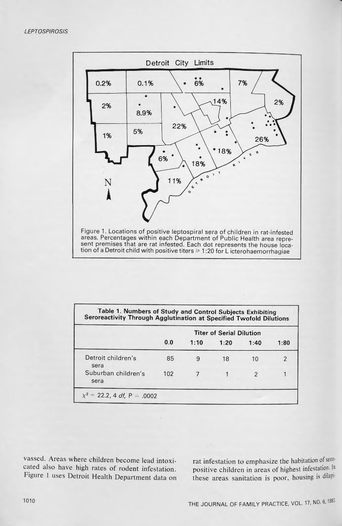
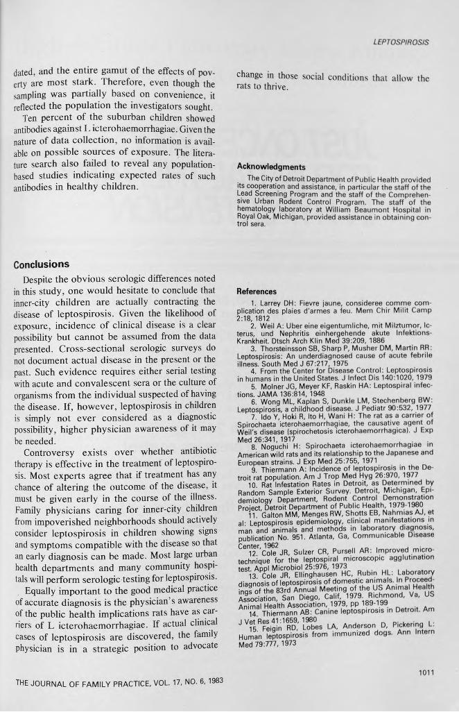
sera. Study group subjects generally resided in the most impoverished “core” areas of the City of Detroit, whereas comparison group subjects generally lived in affluent suburban areas. Children with positive sera tended to reside in areas of major rat infestation as regularly determined by the Rodent Control Program of the Detroit Health Department (Figure 1).
Seropositivity differed significantly between the study and comparison groups. Study group children exhibited a significantly higher number of positive titers against L icterohaemorrhagiae at all dilutions. Thirty-one percent of the study group sera contained antibodies against L icterohaemorrhagiae compared with 10 percent in the comparison group. Table 1 summarizes the serologic findings.
Since the study children were significantly older than the controls, age was considered as a confounding variable. A Mantel-Haenszel test for equality of risks across categories was performed, thus controlling for age. The difference in serologic titers remained highly significant (x2 = 12.84, P = .001). Seasonal variation of seropositivity, another potential confounder, was not a factor, since all sera were collected during autumn.
DiscussionThe above findings show significant titer differ
ences between urban and suburban children against the strain of leptospirosis carried by Detroit rats. Since the strain carried by urban rats is almost exclusively L icterohaemorrhagiae, the hypothesis that urban children are being exposed to leptospirosis through contact with rat urine and feces is supported. It is also known that rats transmit the organism to dogs, so that urban dogs might be an intermediary to this exposure."1"
The sampling of blood already taken through the lead-screening program was based on both convenience and logic. Children participating in the health department lead-screening program are selected through door-to-door canvassing of economically deprived neighborhoods. The age and condition of housing along with family income levels determine which neighborhoods are can-
1009THE JOURNAL OF FAMILY PRACTICE, VOL. 17, NO. 6, 1983
LEPTOSPIROSIS
Detroit City Limits
7%
1 2%•
8.9%• \
1 1% 5%22%
M • \f 6% * V% y •
N
A
11%
,14% 2%
26%
18%
18%
Figure 1. Locations of positive leptospiral sera of children in rat-infested areas. Percentages within each Department of Public Health area represent premises that are rat infested. Each dot represents the house location of a Detroit child with positive titers & 1:20 for L icterohaemorrhagiae
Table 1. Numbers of Study and Control Subjects Exhibiting Seroreactivity Through Agglutination at Specified Twofold Dilutions
Titer of Serial Dilution0.0 1:10 1:20 1:40 1:80
Detroit children's 85 9 18 10 2sera
Suburban children's 102 7 1 2 1sera
X2 = 22.2, 4 df, P = .0002
vassed. Areas where children become lead intoxicated also have high rates of rodent infestation. Figure 1 uses Detroit Health Department data on
1010
rat infestation to emphasize the habitation of seropositive children in areas of highest infestation. In these areas sanitation is poor, housing is dilapi-
THE JOURNAL OF FAMILY PRACTICE, VOL. 17, NO. 6,1983
LEPTOSPIROSIS
dated, and the entire gamut of the effects of poverty are most stark. Therefore, even though the sampling was partially based on convenience, it reflected the population the investigators sought.
Ten percent of the suburban children showed antibodies against L icterohaemorrhagiae. Given the nature of data collection, no information is available on possible sources of exposure. The literature search also failed to reveal any population- based studies indicating expected rates of such antibodies in healthy children.
change in those social conditions that allow the rats to thrive.
AcknowledgmentsThe City of Detroit Department of Public Health provided
its cooperation and assistance, in particular the staff of the Lead Screening Program and the staff of the Comprehensive Urban Rodent Control Program. The staff of the hematology laboratory at William Beaumont Hospital in Royal Oak, Michigan, provided assistance in obtaining control sera.
ConclusionsDespite the obvious serologic differences noted
in this study, one would hesitate to conclude that inner-city children are actually contracting the disease of leptospirosis. Given the likelihood of exposure, incidence of clinical disease is a clear possibility but cannot be assumed from the data presented. Cross-sectional serologic surveys do not document actual disease in the present or the past. Such evidence requires either serial testing with acute and convalescent sera or the culture of organisms from the individual suspected of having the disease. If, however, leptospirosis in children is simply not ever considered as a diagnostic possibility, higher physician awareness of it may be needed.
Controversy exists over whether antibiotic therapy is effective in the treatment of leptospirosis. Most experts agree that if treatment has any chance of altering the outcome of the disease, it must be given early in the course of the illness. Family physicians caring for inner-city children from impoverished neighborhoods should actively consider leptospirosis in children showing signs and symptoms compatible with the disease so that an early diagnosis can be made. Most large urban health departments and many community hospitals will perform serologic testing for leptospirosis.
Equally important to the good medical practice of accurate diagnosis is the physician’s awareness of the public health implications rats have as carriers of L icterohaemorrhagiae. If actual clinical cases of leptospirosis are discovered, the family physician is in a strategic position to advocate
References1. Larrey DH: Fievre jaune, consideree comme com
plication des plaies d'armes a ieu. Mem Chir Milit Camp 2:18, 1812
2. Weil A: Uber eine eigentumliche, mit Milztumor, Icterus, und Nephritis einhergehende akute Infektions- Krankheit. Dtsch Arch Klin Med 39:209, 1886
3. Thorsteinsson SB, Sharp P, Musher DM, Martin RR: Leptospirosis: An underdiagnosed cause of acute febrile illness. South Med J 67:217, 1975
4. From the Center for Disease Control: Leptospirosis in humans in the United States. J Infect Dis 140:1020, 1979
5. Molner JG, Meyer KF, Raskin HA: Leptospiral infections. JAMA 136:814, 1948
6. Wong ML, Kaplan S, Dunkle LM, Stechenberg BW: Leptospirosis, a childhood disease. J Pediatr 90:532, 1977
7. Ido Y, Hoki R, Ito H, Wani H: The rat as a carrier of Spirochaeta icterohaemorrhagiae, the causative agent of Weil's disease (spirochetosis icterohaemorrhagica). J ExpMed 26:341, 1917
8. Noguchi H: Spirochaeta icterohaemorrhagiae in American wild rats and its relationship to the Japanese and European strains. J Exp Med 25:755, 1971
9. Thiermann A: Incidence of leptospirosis in the Detroit rat population. Am J Trap Med Hyg 26:970, 1977
10 Rat Infestation Rates in Detroit, as Determined by Random Sample Exterior Survey. Detroit, Michigan, Epi- demioloqv Department, Rodent Control Demonstration Project, Detroit Department of Public Health, 1979-1980
11. Galton MM, Menges RW, Shotts EB, Nahmias AJ, et aL Leptospirosis epidemiology, clinical manifestations in man and animals and methods in laboratory diagnosis, publication No. 951. Atlanta, Ga, Communicable Disease
Cote JR, Sulzer CR, Pursell AR: Improved microtechnique for the leptospiral microscopic agglutination test. Appl Microbiol 25:976, 1973
13. Cole JR, Ellinghausen HC, Rubin HL: Laboratory diaonosis of leptospirosis of domestic animals. In Proceed- fnqs of the 83rd Annual Meeting of the US Animal Health Association, San Diego, Calif 1979 Richmond, Va, US Animal Health Association, 1979, pp 189-199
14 Thiermann AB: Canine leptospirosis in Detroit. AmI Vet Res 41:1659, 1980 „ _. . .
15 Feiqin RD, Lobes LA, Anderson D, Pickering L. Human leptospirosis from immunized dogs. Ann Intern Med 79:777, 1973
THE JOURNAL OF FAMILY PRACTICE, VOL. 17, NO. 6, 19831011