compatibility cylinder fragments amphibian … and nutritional dependence of mouse egg cylinder...
TRANSCRIPT
Compatibility and nutritional dependenceof mouse egg cylinder fragments associatedwith amphibian embryonic tissues
F. D. BARBIERI, N. CASTRO D. C. MICELI
Departamento de Bio%gia del Desarrollo,Instituto superior de lnvestigaciones biológicas,Consejo nae/ona/ de lnvestigaciones c/enf/7/cas y técnicasand Universidad f)ac/ona/ de 7’t/CM/nan, Chacabuco Ø1,4000 Sa! Miguel de Tucumán, Argentina.
Summary. Embryonic and extraembryonic fragments of mouse egg cylinders at the earlyprimitive-streak stage have been apposed to early gastrula cells of the toad, Bufo arena-rum, using two techniques. One lot of fragments was implanted into the blastocoel ; thesecond lot was cultured in vitro in association with undetermined epiblast excised from theventral half of the blastocoel roof. All samples were cultured up to 3 days in Barth’s solu-tion, optimized for the amphibian embryo and not permitting isolated mouse egg cylinderfragments to survive.
Small grafts, completely surrounded by Bufo cells, had an apparently normal aspectand frequent mitotic figures. Large grafts, attached to the host embryo, were in closecontact with the endoblast and mesoblast cells, but the ectoblast layer showed no ten-dency to cover the egg cylinder fragment. No differentiation was detected in mouse
embryo fragments, probably because their development was retarded. The cells in themost proximal region of these fragments, i.e. near the host tissues, had a normal aspectand showed proliferative activity. On the other hand, cells in the most distal region werein advanced involution. The presence of the graft did not appear to impair normal differen-tiation of the host cells.
Introduction.
There is much evidence indicating that graft exchanges between embryos ofdifferent vertebrate species are viable throughout most of the embryonic period.These combinations have been widely used as a valuable experimental approachto the analysis of early development (see Ebert, 1959 ; Owen, 1959). Transplan-tations between anuran and urodelean species and grafts of mammalian tissuesin the chick embryo showed no incompatibility (see Ebert, 1959). Similar resultswere obtained when assaying the association of embryonic organs from thechick and the mouse in vitro (Wolff, 1954). There is general consensus that thehost does not destroy the graft because it lacks immunological reactivity during
early development. However, since it is known that cell surface components playan important role in morphogenesis and differentiation, the apparent lack of spe-cificity with which cells of different species adhere to each other is striking. Thishas been tentatively explained by assuming that some determinants persist onthe cell surface of different species during the course of evolution. On the basisof this hypothesis, the viability of the chick-mouse combination might be under-stood as additional evidence of the evolution of these species from a reptilianstem (Burdick and Steinberg, 1969). The attachment between the xenogeneicembryo fragments has also been tentatively ascribed to the structural similaritiesof protein molecules throughout the zoological scale (Wolff, 1954 ; Garrod andNicol, 1981).
In the present investigation, fragments of mouse and toad embryos wereassociated in order to determine (a) whether the apposed embryonic fragmentsof vertebrates not linked by a common ancestry, as amphibians and mammals,would attach to one another, and (b) whether the cells of the quasi cleidoic
amphibian embryo (which only requires water and oxygen from the environment)could fulfill the nutritional requirements of the mammalian embryo which recei-ves its nutritional supply from the maternal organism.
Material and methods.
Embryos. ― Female Swiss mice were mated overnight, and the day onwhich the vaginal plug was found was designated as day 0. Embryos in the earlyprimitive-streak stage (6-7 days old) (Witschi stages 11-12, according to New,1966) were isolated from pregnant uteri and put in sterile Tyrode’s solution.
The toad, Bufo arenarum, was induced to ovulate by an injection of a pitui-tary suspension. The oocytes were removed from the ovisacs and inseminatedwith a sperm suspension prepared by mincing testes in 10 % amphibian Ringersolution. The eggs were allowed to develop in the same saline to the requiredstage.
Dissection. ― Mouse and toad embryos were operated with tungsten need-les and hair loops.
After the jelly coats of the amphibian embryo had been dissolved with a1 % thioglycolic acid solution neutralized with NaOH, the fertilization membranewas removed with sharpened forceps. The mouse embryos were dissected in a
Petri dish containing sterile Tyrode’s solution. In some experiments, 0.1 % ofalbumin was added to the medium to make the eggs less sticky and thus facili-tate manipulation during dissection. After Reichert’s membrane was removed, theegg cylinders were cut transversely at the junction of the embryonic and
extraembryonic parts. Fragments of different sizes (about 20-20OPm) were thenarbitrarily cut from the embryonic and extraembryonic regions and either (a)
implanted into the blastocoel of late blastulae or early gastrulae of Bufo
(stages 9-10, according to Del Conte and Sirlin, 1951) or (b) associated with theepiblast isolated from the ventral side of the blastocoel roof, according to the
sandwich procedure of Holtfreter (1933). The recipient embryos were about1.5 mm in diameter and the epiblast pieces were about 0.5 x 0.5 mm square.Operations were carried out in Barth’s solution (Barth and Barth, 1959) contai-
ning 50 pg/ml of penicillin and 30 pg/ml of dihydrostreptomycin.
Embryo culture and histo%gy. - Grafted amphibian embryos and explantswere cultured up to 3 days at 25-27 °C in Barth’s solution containing the antibio-tics indicated above. At the end of culture, all material was fixed in Smith’s solu-tion and the serial sections were stained with hematoxylin and eosin.
Results.
As the aim of the present work was to investigate the adhesiveness betweenmouse and toad embryonic cells as well as the ability of the amphibian embryosto support the development of mouse embryo fragments, the experiments werecarried out according to the following design. As stated in Material and
methods, the culture conditions were optimized for the amphibian embryo, notthe mammalian embryo and most of the mouse embryo fragments were cut largein order to prevent them from passively attaching as a result of being covered bythe proliferating tissues of the host embryo or the associated epiblast.
As expected, mouse embryo fragments did not survive when isolated in
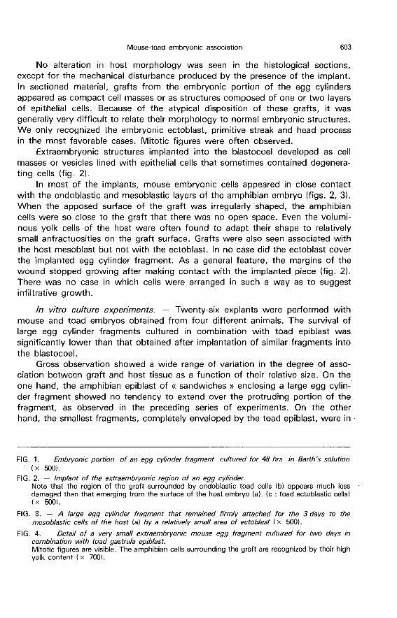
Barth’s solution. In fact, the cells of these specimens exhibited intense cytoplas-mic eosinophilia and vacuolization, and their nuclei were pycnotic (fig. 1). Themore superficial cells were also found to dissociate to some extent and to remainstuck by a mucous material.
Transplantation experiments. ― We performed 71 grafts in seven series of
experiments, each conducted with embryos derived from different animals. Wefound that the cells of Bufo embryos could support the development of mouseembryo fragments during the 3 days of the study, as shown by gross andmicroscopic observations of the implanted specimens. About 10 % of the graftsremained closely attached to the host, although they were just apposed to theamphibian tissues (fig. 3).
The small specimens of mouse embryo that were completely enveloped bytoad tissues remained healthy ; they were indistinguishable from normal eggcylinders fixed in utero, and mitotic figures were always present. Further evi-dence that the host tissues were supporting was provided by the aspect of thegrafts partially exposed to the surrounding medium. In fact, while the proximalzone of these grafts had a normal aspect, clear signs of degeneration were pre-sent in their most distal portions (figs. 2, 3).
Twenty-four hours after the operation, toad embryos implanted with a pieceof egg cylinder were at the closed neural tube stage (stage 16). The woundswere still open because of the large size of the grafts, but no sign of cytolysiswas found. When the toad embryos reached the stage of muscular responseafter 3 days (stage 18), the wounds were much more reduced but not completelyclosed because the grafts were still attached to the host. They had a normalappearance under the stereomicroscope both before and after fixation.

No alteration in host morphology was seen in the histological sections,except for the mechanical disturbance produced by the presence of the implant.In sectioned material, grafts from the embryonic portion of the egg cylindersappeared as compact cell masses or as structures composed of one or two layersof epithelial cells. Because of the atypical disposition of these grafts, it was
generally very difficult to relate their morphology to normal embryonic structures.We only recognized the embryonic ectoblast, primitive streak and head processin the most favorable cases. Mitotic figures were often observed.
Extraembryonic structures implanted into the blastocoel developed as cell
masses or vesicles lined with epithelial cells that sometimes contained degenera-ting cells (fig. 2).
In most of the implants, mouse embryonic cells appeared in close contactwith the endoblastic and mesoblastic layers of the amphibian embryo (figs. 2, 3).When the apposed surface of the graft was irregularly shaped, the amphibiancells were so close to the graft that there was no open space. Even the volumi-nous yolk cells of the host were often found to adapt their shape to relativelysmall anfractuosities on the graft surface. Grafts were also seen associated withthe host mesoblast but not with the ectoblast. In no case did the ectoblast coverthe implanted egg cylinder fragment. As a general feature, the margins of thewound stopped growing after making contact with the implanted piece (fig. 2).There was no case in which cells were arranged in such a way as to suggestinfiltrative growth.
In vitro culture experimenis. - Twenty-six explants were performed withmouse and toad embryos obtained from four different animals. The survival oflarge egg cylinder fragments cultured in combination with toad epiblast wassignificantly lower than that obtained after implantation of similar fragments intothe blastocoel.
Gross observation showed a wide range of variation in the degree of asso-ciation between graft and host tissue as a function of their relative size. On theone hand, the amphibian epiblast of « sandwiches » enclosing a large egg cylin-der fragment showed no tendency to extend over the protruding portion of thefragment, as observed in the preceding series of experiments. On the otherhand, the smallest fragments, completely enveloped by the toad epiblast, were in
close contact with cells forming the central mass of the explant but not with themore superficial pigmented cells (fig. 4).
Bufo cells contacting mouse cells in the explants exhibited no differencecompared to those located far away from the mouse embryo fragment or tothose of control epiblast pieces cultured without the associated egg cylinderfragment ; they formed an atypical compact, unorganized tissue covered by cilia-ted and interspersed secretory cells and melanoblasts.
Discussion.
The results of implanting fragments of mouse egg cylinders at the earlyprimitive-streak stage to early gastrulae of the toad, Bufo arenarum, or of asso-ciating them with an explant of undetermined epiblast of the same species, showthat there was no apparent incompatibility for at least 3 days after the operation.No evidence of direct damage to the graft by toxic substances was found, asreported for embryonic grafts between anurans and urodeles (Eakin and Harris,1945). Furthermore, the grafts attached to the amphibian host with apparentlyvery close cell-cell adhesion. The low percentage of large grafts that attached is
probably the result of mechanical problems arising before the cells of the hostand the graft adhered. It is possible that the cells formed junctions, as suggestedby the fact that, when combined, cells of chick corneal epithelium and mouseepidermis were found to make stable desmosomes (Overton, 1977).
In this first assay of embryonic cell adhesion between a cold and a warm-blooded species, we decided that, in order to minimize alterations of the cell sur-face structure, it was preferable to test the association of embryo fragmentsrather than the aggregation of dissociated cells. As a matter of fact, we still donot know to what extent the artifactual alteration of cell surface molecules
during the dissociation process may account for the different results on variousmouse-chick combinations. Thus, while several cell associations seemed to indi-cate that there is no sorting-out according to species origin (Moscona, 1957,1960, 1961, 1962 ; Garber and Moscona, 1964 ; Garber et al., 1968 ; Burdick,1972), this conclusion has been questioned by later experiments (Burdick andSteinberg, 1969) ; even sorting-out followed by cell intermixing has been repor-ted for combinations of mouse and chick limb mesoblast (Burdick, 1970). It hasbeen generally concluded that selective adhesive behavior is probably less influen-ced by species difference than by tissue difference (Garrod and Nicol, 19811. ).
Despite the intimate contact established between mouse and toad cells, nopropensity to infiltrative growth was noted. This contrasts with the association invitro of embryonic organs of mouse and chick carried out by Wolff (1954) ; thelimits of this association tended to disappear due to the migration of connectivecells into the alien tissues and the formation of chimeric epithelial structures.
The present study also suggests that, in addition to cell compatibility, themammalian embryo fragments seemed to be nutritionally dependent on the
amphibian embryonic cells. This was inferred from the fact that the aspect of the
cells of fragments held in amphibian saline without a serum component appearedto be normal, as they remained attached to the host embryonic tissues and, in
particular, as the large fragments seemed to be normal only in the vicinity oftoad cells. In the light of this assumption, the presence of mitotic figures mightbe interpreted as an expression of proliferative activity, although the presentresults do not exclude the possibility that these figures could represent a processinitiated prior to grafting that remained in an arrested state during the experi-ments. It is obvious that some nutrients must have been released from the host
cells ; otherwise, the mouse embryonic cells could not have multiplied in a
serum-free hypothonic saline medium (see New, 1966, 1978 ; Skreb and Svajger,1973 ; Hsu, 1979).
The lack of apparent differentiation in the sectioned material may be ascri-bed to a slower rate of graft development. It should be noted in this respect thatthere is a delay in mouse embryonic development, even when the embryos aretransplanted to extrauterine sites of the same species (Fawcett et al., 1947), andthat the specimens of the present experiments were kept at room temperature.On the other hand, the cells of the host embryo were found to differentiate nor-mally without any apreciable reaction.
Recu en aocit 1982.Accepte en janvier 1983.
Acknowledgements. ― We wish to thank Mr. Roberto Ordonez and Dr. Raymond F. Lau-rent for their assistance in preparing this paper, Mr. Jos6 Greco and Mr. Enrique Dozetosfor technical assistance and Mr. Hugo Gomez for typing the text. F.D.B. and D.C.M. aremembers of the Career Investigator of the CONICET, and N.C. was a CONICET fellowwhile this work was carried out.
Résumé. Compatibilité et dépendance nutritionnelle de fragments de cylindre de /’ceufde Souris associés à des tissus embryonnaires d Amphibien.
Des fragments embryonnaires et extraembryonnaires du cylindre d’ceuf de souris austade de la lignée primitive ont été associés avec des cellules de la jeune gastrula de Bufoarenarum suivant deux procédés différents. Une série a été implantée dans le blastocele.Une seconde série a été cultivée in vitro en combinaison avec de l’épiblaste indéterminéprélevé du côté ventral du toit du blastocèle. Tous les échantillons ont été cultivés pen-dant trois jours dans la solution de Barth, optimisée pour l’embryon d’amphibien, et nepermettant pas la survie des fragments isolés de l’oeuf de souris. Les greffons de petitetaille se présentaient entièrement entourés par les cellules de Bufo, avec un aspect appa-remment normal et de fréquentes figures mitotiques. Les greffons de grande taille étaientbien fixés a l’hôte en contact intime avec ses cellules endoblastiques et mésoblastiques.D’autre part, le feuillet ectoblastique de l’embryon d’amphibien n’a montré aucune ten-dance à couvrir le fragment du cylindre oeuf. Les fragments d’embryon de souris ne mon-traient pas de différenciation probablement à cause d’un retard dans leur développement.Les cellules localisées dans la région proximale du greffon, proche des tissus de l’hôte,avaient une structure d’apparence normale et présentaient des mitoses. Au contraire, lescellules qui se trouvaient dans la région distale montraient un état de régression avancée.La présence du greffon ne semblait pas affecter la différenciation normale des cellules del’hôte.

References
BARTH L. G., BARTH L. J., 1959. Differentiation of cells of the Rana pipiens gastrula in
unconditioned medium. J. Embryol. exp. Morph., 7, 210-222.BURDICK M. L., 1970. Cell sorting-out according to species in aggregates containing mouse
and chick embryonic limb mesoblast cells. J. exp. Zool., 75, 357-368.BURDICK M. L., 1972. Differences in the morphogenetic properties of mouse and chick
embryonic liver cells. J. exp. Zool., 180, 117-125.BURDICK M. L., STEINBERG M. S., 1969. Embryonic cell adhesiveness : Do species differences
exist among warm-blooded vertebrates ? Proc. nat . Acad. Sci. USA, 63, 1169-1173.DEL CONTE, E., SIRLIN J. L., 1951. Serie tipo de los primeros estadios embrionarios en
Bufo arenarum. Acta zool. lilloana, 12, 495-499.EAKIN R. M., HARRIS M., 1945. Incompatibility between amphibian hosts and xenoplastic
grafts as related to host age. J. exp. Zool., 98, 35-64.EBERT J. D., 1959. The acquisition of biological specificity, 619-693. In BRACHET J.,
MIRSKY A., The cell, Vol. 1. Acad. Press, New York.FAWCETT D. W., WISLOCKI G. B., WALDO C. M., 1947. The development of mouse
ova in the anterior chamber of the eye and in the abdominal cavity. Am. J. Anat., 81,413-443.
GARBER B. B., MOSCONA A. A., 1964. Aggregation in vivo of dissociated cells. 1. Reconstruc-
tion of skin in the choriallantoic membrane from suspensions of embryonic chick and mouseskin cells. J. exp. Zool., 155, 179-202.
GARBER B. B., KOLLAR E. J., MOSCONA A. A., 1968. Aggregation in vivo of dissociated
cells. III. Effects of state of differentiation of cells on feather development in hybrid aggrega-tes of embryonic mouse and chick cells. J. exp. Zool., 168, 455-472.
GARROD D. R., NICOL A., 1981. Cell behaviour and molecular mechanisms of cell-cell adhesion.
Biol. Rev., 56, 199-240.HOLTFRErER J., 1933. Der Einfluss von Wirtsalter und Verschiedenen Organbezirken auf die Diffe-
renzierung von angelagerten Gastrulaektoderm. W. Roux’s Arch. EntwMech. Org., 127, 619-775.
HSU Y., 1979. In vitro development of individually cultured whole mouse embryos from blastocystto early somite stage. Dev. Biol., 68, 453-461.
MOSCONA A. A., 1957. The development in vitro of chimeric aggregates of dissociated embryonicchick and mouse cells. Proc. nat. Acad. Sci. USA, 43, 184-194.
MOSCONA A. A., 1960. Patterns and mechanisms of tissue reconstruction from dissociated cells,45-70. In RUDNICK D., Developing cell systems and their control. Ronald Press, New York.
MOSCONA A. A., 1961. Rotation-mediated histogenetic aggregation of dissociated cells : a quanti-fiable approach to cell interactions in vitro. Exp. Cell Res., 22, 455-475.
MOSCONA A. A., 1962. Analysis of cell recombination in experimental synthesis of tissues
in vitro. J. cell. comp. Physiol. (suppl. 1) 60, 65-80.NEW D. A. T., 1966. The culture of vertebrate embryos. Logos Press, London.NEW D. A. T., 1978. Whole-embryo culture and the study of mammalian embryos during
organogenesis. Biol. Rev., 53, 81-122.OVERTON J., 1977. Formation of junctions and cell sorting in aggregates of chick and mouse
cells. Dev. Bio/., 55, 103-116.OWEN R. D., 1959. Genetic aspects of tissue transplantation and tolerance. J. Med. Educ.,
34, 366-383.SKREB N., SVAJGER A., 1973. Histogenetic capacity of rat and mouse embryonic shields
cultivated in vitro. W. Roux’ Arch., 173, 228-234.WOLFF E., 1954. Potentialit6s et affinites des tissus, révélées par la culture in vitro d’organes
en associations h6t6rog6nes et x6noplastiques. Bull. Soc. Zool. France, 79, 357-368.