clostridium perfringens as foodborne pathogen in broiler
TRANSCRIPT

animals
Review
Clostridium perfringens as Foodborne Pathogen inBroiler Production: Pathophysiology and PotentialStrategies for Controlling Necrotic Enteritis
Zuamí Villagrán-de la Mora 1 , María Esther Macías-Rodríguez 2 , Jenny Arratia-Quijada 3,Yesica Sughey Gonzalez-Torres 1 , Karla Nuño 3,* and Angélica Villarruel-López 2,*
1 Departamento de Ciencias de la Salud, Centro Universitario de Los Altos, Universidad de Guadalajara,Av. Rafael Casillas Aceves 1200, Tepatitlán de Morelos 47620, Mexico;[email protected] (Z.V.-d.l.M.); [email protected] (Y.S.G.-T.)
2 Departamento de Farmacobiología, Centro Universitario de Ciencias Exactas e Ingenierías, Universidad deGuadalajara, Blvd. Gral. Marcelino García Barragán 1421, Olímpica 44430, Guadalajara, Mexico;[email protected]
3 Departamento de Ciencias Biomédicas, Centro Universitario de Tonalá, Universidad de Guadalajara,Nuevo Perif. Ote. 555, Ejido San José, Tateposco 45425, Tonalá, Mexico; [email protected]
* Correspondence: [email protected] (K.N.); [email protected] (A.V.-L.)
Received: 4 September 2020; Accepted: 17 September 2020; Published: 22 September 2020 �����������������
Simple Summary: Clostridium perfringens (Cp.) is an important microorganism from a clinical,food and veterinary point of view. In humans, it is the causal agent of foodborne diseases, commonlyassociated with the consumption of chicken meat, while, in broilers, it causes clinical or subclinicalnecrotic enteritis. Cp. has the ability to synthesize toxins, bacteriocins, and enzymes of differentnature, which modify the anatomical structure of the intestinal mucosa, enterocytes, and the cellularmatrix altering the physiological activities of the gastrointestinal tract, resulting in gastrointestinaldisorders, diarrhea, and if it is not attended, death, resulting in significant economic losses for thepoultry industry. Food additives such as probiotics, prebiotics, synbiotics, essential oils, organic acids,and enzymes have been presented as alternatives to mitigate the incidence of necrotic enteritis (NE)in broilers, by improving the overall intestinal health and producing healthy birds for consumption.It is imperative to conduct further research on alternatives and efficient products to modulate theintestinal microbiota, and to know the role they play in the immune system, complementing thecurrent demand, economic gain, and keeping the ecology.
Abstract: Clostridium perfringens (Cp.) is the cause of human foodborne desease. Meat and poultryproducts are identified as the main source of infection for humans. Cp. can be found in poultrylitter, feces, soil, dust, and healthy birds’ intestinal contents. Cp. strains are known to secrete over20 identified toxins and enzymes that could potentially be the principal virulence factors, capableof degrading mucin, affecting enterocytes, and the small intestine epithelium, involved in necroticenteritis (NE) pathophysiology, also leading to immunological responses, microbiota modificationand anatomical changes. Different environmental and dietary factors can determine the colonizationof this microorganism. It has been observed that the incidence of Cp-associated to NE in broilers hasincreased in countries that have stopped using antibiotic growth promoters. Since the banning ofsuch antibiotic growth promoters, several strategies for Cp. control have been proposed, includingdietary modifications, probiotics, prebiotics, synbiotics, phytogenics, organic acids, and vaccines.However, there are aspects of the pathology that still need to be clarified to establish better actions tocontrol and prevention. This paper reviews the current knowledge about Cp. as foodborne pathogen,the pathophysiology of NE, and recent findings on potential strategies for its control.
Animals 2020, 10, 1718; doi:10.3390/ani10091718 www.mdpi.com/journal/animals

Animals 2020, 10, 1718 2 of 28
Keywords: C. perfringens; necrotic enteritis; pathophysiology; control strategies
1. Introduction
Clostridium perfringens is a Gram-positive, anaerobic, nonmotile rod that forms subterminal spores.The size of the bacillus on the environment where is found, for example, in culture media for sporulationbased on starch the bacillus is long. Meanwhile, in media rich in glucose the bacillus is short. Vegetativecells are relatively cold resistant, and their spores are heat resistant [1,2]. C. perfringens can hydrolyzegelatin and reducing nitrates to nitrites; in sulphite media, it generates black colonies due to sulphitereduction. A characteristic test is the lactose fermentation produced by this microorganism, known asstormy lactose fermentation in milk due to the large amount of gas it generates [3,4]. This bacterium candevelop under microaerophilic conditions due to its ability to produce high amounts of the enzymesuperoxide dismutase [5,6]. Its ability to form spores allows it to be ubiquitous and can be found in theenvironment [7,8].
Beef and poultry, as well as other meat products, are the most important vehicles for thismicroorganism [9–11], although it has also been recovered from vegetables [12] and spices [13].Butler et al. (2015) [14] described the transmission of C. perfringens through water by contact withanimals and transmission from person to person. Considered a natural inhabitant of the gastrointestinaltract, the main source of contamination towards meat is fecal matter [15].
According to data reported by the CDC (2019) [16], C. perfringes is one of the five pathogens thatmost frequently cause foodborne illnesses in the United States, ranking second among the etiologicalagents identified, and, in Australia, it is considered one of the bacteria causing outbreaks [17].
The consumption of chicken meat is important worldwide and a 13% increase in its production isestimated for the year 2027 (OECD-FAO, 2017). In animal production, approximately 70% of the totalcost is attributable to the feed. The diets for farm animals contain antibiotics or growth promoters thatseek to improve the productive parameters on the farm; however, there is a tendency to use them lessfrequently, seeking to replace them with what is currently known as sustainable animal diets [18].
It is important to mention that some pathogens that cause disease in chickens can be transmittedto humans through their consumption. Salmonella, Campylobacter jejuni, and C. perfringens are the moststudied so far. C. perfringens is the cause of subclinical necrotic enteritis in broilers, producing toxinsand is the cause of disease in humans [9,19].
2. C. perfringens as a Foodborne Pathogen
Clostridium perfringens can produce a large amount of toxins (Table 1). Toxinotypes of C. perfringenscause different diseases in both humans and animals, ranging from subclinical manifestations toserious, life-threatening diseases (Table 2) [20].
Table 1. Types of Clostridium perfringens according to the toxins produced and the genes that encodethe toxins.
Toxins
Type Alpha (α) Beta (β) Epsilon (ε) Iota (ι) CPE NetB(plc o cpa) * (cpb) * (etx) * (iap y ibp) * (cpe) * (netB) *
A + − − − − −
B + + + − − −
C + + − − +/− −
D + − + − +/− −
E + − − + +/− −
F + − − − + −
G + − − − − +
*Gene for each toxin. Taken from Rood et al., 2018 [21].

Animals 2020, 10, 1718 3 of 28
These diseases are mediated by one or more C. perfringens toxins [21,22]. Enteric infections inhumans and animals have been shown to be associated with C. perfringens type C [23,24], while theother type of toxins have been confirmed to cause disease in humans or animals, but not both (Table 2).Of the seven C. perfringens toxin types described, type A is the most frequently identified strain [12,25].However, type F is the one that causes food-related poisoning in humans [21,26].
Table 2. Toxigenic types of Clostridium perfringens and their association with diseases in humansand animals.
Type ofToxin Main Toxin Diseases that Cause
Aα
Wound infection in humans (gas gangrene or clostridial myonecrosis),necrotic enteritis in birds, ulcerative abomasitis, mild necrotizingenteritis in piglets, and endotoxemia in South American camelids.
α, CPE Food poisoning in humans, non-food gastrointestinal diseases inhumans, and diarrhea in animals such as dogs, pigs, and foals.
α, β2 Gastrointestinal disease in swine.B α, β, ε Dysentery and hemorrhagic enteritis in lambs and kids.
Cα, β Necrotizing enteritis in humans, enteritis in dogs, chickens, and South
American camelids.α, β, β2 Gastrointestinal disease in swine.
D α, ε Enterotoxemia in sheep and goats (pulpy kidney disease).E α, ι Enterotoxemia in rabbits, dogs, cattle, and sheep.F α, CPE Human food poisoning and non-food associated diarrhea.G α, NetB Subclinical necrotic enteritis in chickens.
Bruce et al., 2006; Kiu& Hall, 2018 and Rood et al., 2018 [20,21,27].
The diversity of toxins produced by C. perfringens has allowed it to be the cause of various diseasesin humans and animals. In humans, it is associated with diseases related to food consumption that hasbeen prepared or preserved in inadequate hygienic conditions [17,28]. This type of illness is usuallycharacterized by watery diarrhea and abdominal pain, without fever or vomiting, and the symptomsdisappear after 12 to 24 hours [29]. Non-food associated diarrhea due to C. perfringens has also beendescribed, which usually occurs after a treatment with broad-spectrum antibiotics, and it is common inolder adults. It is worth mentioning that this type of diarrhea usually last longer than those associatedwith contaminated food [30]. Another symptom is necrotic enteritis (NE) caused by C. perfringens typeC [31]. Myonecrosis due to C. perfringens (also known as gas gangrene) is another condition that canoccur in people because of wound infection, generating significant pain, gas accumulation at the site ofinfection and extensive muscle necrosis, which can put people’s life at risk [32,33].
The toxin of this bacterium also affects some animal species. For example, in broilers the toxincauses necrotic enteritis, which could lead economic losses. The role of the necrotic enteritis B-like toxin(NetB) present in G strains causes NE, which is more frequent in chickens fed wheat or barley-baseddiets than in those fed with corn [34,35], due to the difference in clostridia proliferation in the dietsresulting in a higher number of bacteria in the intestine, as well as a lack of fluidity and digestion,generating an increase in the incidence of NE in chickens and increasing the viscosity of the intestinalcontents, mucus production, and growing bacteria [35,36].
3. Necrotic Enteritis Pathophysiology
Clostridium perfringens is a bacterium found in the gastrointestinal tract of broilers and is acquiredfrom environmental sources such as water, food, or any part of the farm producing these birds, beingpart of their microbiota [37]. However, a high enumeration number of this microorganism and thepresence of toxins in some strains can cause different types of pathologies, among them necroticenteritis (NE). It is important to mention that an elevated enumeration of C. perfringens by itself is notthe cause of NE but must be accompanied by one or more predisposing factors to develop clinical signsand lesions of the pathology. Enumerations of 0 to 105 CFU/g of C. perfringens have been observed
Animals 2020, 10, 1718 4 of 28
in the intestine of healthy chickens, while animals with NE report enumerations of 106–108 CFU/g,besides the presence of bacteriocins, adhesins, proteolytic enzymes, collagenolytic enzymes, necrotictoxin enteritis B-like (NetB) and tpeL [38,39].
Currently, the NetB toxin is considered the determining factor inducing NE in birds [40,41].This 33-kDa toxin is a member of the family of pore-forming toxins with a beta barrel structureencoded by the netB gene located in an 85 kb plasmid. The toxin production is stimulated when theC. perfringens concentration is higher than 109 CFU/g and the bird has low food bioavailability, has ahigh consumption of polysaccharides, dysbiosis or has suffered intestinal damage. This damage couldbe caused by coccidial pathogens of the Eimeria species, and their colonization causes the release ofplasma proteins to the gastrointestinal tract lumen, including more than 11 amino acids, growth factors,and vitamins; they will supply the growth substrate for C. perfringens [42–44].
When C. perfringens enters the gastrointestinal tract of the bird and encounters a favorableenvironment, it secretes adhesins and proteolytic enzymes that exert their action on the intestinalmucosa and the surface of the intestinal epithelial membranes, due to their composition. The intestinalmucosa contains mucin binding sites for bacterial adhesins and O-glycosylated glycoproteins that willbe degraded by chitinases to provide energy substrates for bacteria. At the same time, C. perfringenscan secrete the bacteriocin perforin, which will inhibit other strains of Clostridium, allowing it to havegreater bioavailability of nutrients and damage the intestinal mucosa [43–48].
Besides colonization and degradation of the intestinal mucosa, the NetB toxin will generate poresto access the enterocytes, and at the same time, adhesins and enzymes capable of degrading collagenof the cell matrix are secreted, which together will allow for the colonization and will determinethe NE appearance. NetB toxin production is positively regulated by the VirR/VirS two-componentphosphorelay system and by the Agr-type quorum sensing system, the latter being responsiblefor mediating the regulation of genes involved in phospholipid metabolism and adherence [49,50].In addition, the phosphorelay system regulates the production of sialidases or neuraminidases withthe capacity to hydrolyze the α-glucosidic bond of terminal sialic acid in host glycoproteins andglycolipids, to produce free sialic acid that can be used as a carbon source [45,51], nitrogen, amino acidsand energy, as it is metabolized to fructose 6-P by the pathogenic microorganism. They also participatein bacterial adhesion by modifying the epithelial surface and exposing receptors on the enterocytemembrane. Subsequently, C. perfringens adheres to extracellular matrix compounds such as type III,IV and V collagen, fibrinogen and vitronectin, to later secrete collagenolytic enzymes and hydrolyzethem. Adhesion to the extracellular matrix occurs through the fimbrial adhesins of NetB-positivestrains [43,44,46,48]. The primary changes occur in the basolateral membrane of the enterocytes, tofinally produce necrosis at the level of the mucosa as a result of the destruction of the lamina propria,interruption of intercellular junctions and changes in the extracellular matrix, thus leading to cellulardeath [45,46].
The netB gene along with 36 additional genes, including those that code for two glycohydrolases,two leukocidins, chitinases, an internalin-like protein, a metalloprotease, and several adhesin-likeproteins, is located in a plasmid of approximately 85 kb that encodes the pathogenicity loci (NELoc-1,42 kb), which has been specifically harbored by bird isolates with NE. The high conservation degree ofthe sequence of this and other identified plasmids (NELoc-2 and NELoc-3) suggests that these comefrom a recent evolutionary event through conjugative transfer. In accordance with these findings, it isassumed that various virulence factors participate in NE development, whose genes are grouped inpathogenicity loci, some of which are harbored in plasmids [52].
The structural analysis of NetB shows that the interaction domain and binding of the proteinwith membrane lipids is rich in aromatic amino acids, being essential amino acids R230 and W287,and structurally differs from other proteins of the hemolysin family, substantial for oligomerization ofresidue S254, suggesting that NetB has a different binding mechanism to membrane receptors [45];according to some experiments, it is suggested that it binds to membrane cholesterol [53]. Once NetBis secreted in a soluble monomeric form, it binds to the cell surface through the RIM domain and

Animals 2020, 10, 1718 5 of 28
subsequently oligomerizes, producing a pre-heptameric pore in the lipid bilayer. This oligomerizationprocess induces conformational changes in the protein to generate a barrel structure with antiparallelβ-sheets and forms a mushroom-like transmembrane pore with a subsequent alteration of membranepermeability [53,54]. The heptameric pore formed at the plasma membrane level by the NetB toxinhas an internal diameter of 26 Å, with a hydrophilic nature, which favors the destabilization of theion flow by allowing the exit of K+ ions and the entry of Ca2+, Na+ and Cl- (showing preference forcations), producing osmotic cell lysis [22,41,55]. As intracellular calcium increases, the cascade ofevents for necrosis programming is influenced by the activation of calpain and cathepsin secretionfrom lysosomes. In addition, an alteration in the mitochondrial activity is observed with an increase inreactive oxygen species and a decrease in ATP [56]. Free radicals can accumulate in the mitochondriaand uncouple the proteins of the mitochondrial inner membrane, leading to a decrease in ATP levels,with losing the integrity of the intercellular junctions in the gastrointestinal epithelium, increasing thepermeability of the mucosa and, finally, cell death (Figure 1) [56,57].
Animals 2020, 10, x 5 of 29
subsequently oligomerizes, producing a pre-heptameric pore in the lipid bilayer. This oligomerization process induces conformational changes in the protein to generate a barrel structure with antiparallel β-sheets and forms a mushroom-like transmembrane pore with a subsequent alteration of membrane permeability [53,54]. The heptameric pore formed at the plasma membrane level by the NetB toxin has an internal diameter of 26 Å, with a hydrophilic nature, which favors the destabilization of the ion flow by allowing the exit of K+ ions and the entry of Ca2+, Na+ and Cl- (showing preference for cations), producing osmotic cell lysis [22,41,55]. As intracellular calcium increases, the cascade of events for necrosis programming is influenced by the activation of calpain and cathepsin secretion from lysosomes. In addition, an alteration in the mitochondrial activity is observed with an increase in reactive oxygen species and a decrease in ATP [56]. Free radicals can accumulate in the mitochondria and uncouple the proteins of the mitochondrial inner membrane, leading to a decrease in ATP levels, with losing the integrity of the intercellular junctions in the gastrointestinal epithelium, increasing the permeability of the mucosa and, finally, cell death (Figure 1) [56,57].
Figure 1. Pathophysiology of necrotic enteritis caused by necrotic enteritis B-like toxin (NetB)-positive Clostridium perfringens. Created with BioRender.com
In the conserved region of the plasmid NELoc-1 that codes for the NetB toxin, there is also the gene that codes for the zinc metalloprotease, ZmpA, which, together with another metalloprotease, ZmpB, has been implicated in NE in chickens. Such proteins have high binding affinity for the mucin glycoprotein, a constituent of the mucosa of the gastrointestinal epithelium. Such metalloproteases participate in the development of NE, since the zmpA gene has been identified in isolated strains of birds with the disease, although the zmpB gene was still identified in isolates of birds in the absence
Figure 1. Pathophysiology of necrotic enteritis caused by necrotic enteritis B-like toxin (NetB)-positiveClostridium perfringens. Created with BioRender.com
In the conserved region of the plasmid NELoc-1 that codes for the NetB toxin, there is also thegene that codes for the zinc metalloprotease, ZmpA, which, together with another metalloprotease,ZmpB, has been implicated in NE in chickens. Such proteins have high binding affinity for the mucinglycoprotein, a constituent of the mucosa of the gastrointestinal epithelium. Such metalloproteasesparticipate in the development of NE, since the zmpA gene has been identified in isolated strains ofbirds with the disease, although the zmpB gene was still identified in isolates of birds in the absence ofthe disease, the lack of one or both genes generate strains with reduced virulence, which is why theyare presumed to participate together in pathogenesis’s development [47].
Likewise, it has been observed that the expression of the tpeL gene occurs during sporulation andthe TpeL toxin is secreted to promote the adhesion of C. perfringens type A in epithelial cell cultures [58];

Animals 2020, 10, 1718 6 of 28
disease and mortality are induced more rapidly in birds infected with TpeL-producing strains, whichcould potentiate the effect of other toxins like NetB [59]. The binding and entry of this toxin into theenterocyte is mediated by the endocytic receptor Lpr1 [60].
3.1. Clinical Alterations
Chicken’s intestinal health is determined by the balance between anatomical components andtheir physiological activities. However, the development and maturation of the gastrointestinal tractis generated through the bird’s life, as well as the exposure to different environmental variables,which will establish the morphological changes and the specific activities of each intestinal segment.Thus, the mucosa and intestinal villi with their microvilli are necessary for the adequate absorptionof nutrients and the establishment of the intestinal microbiota, while the gut-associated lymphoidtissue (GALT), together with the mucosa and microbiota, provides an immune complex that willwork as a gastric defense mechanism [61–63]. Therefore, an aggression to any of these components,especially in the first weeks of life, could trigger an alteration in the integrity of the intestinal epithelium,in the bioavailability and absorption of nutrients, promoting a bacterial dysbiosis and an intestinalinflammatory process.
Due to the above, the chicks, being immunologically and physiologically immature, are moresusceptible to be infected by C. perfringens (NetB positive), either clinically or subclinically. The clinicalphase of disease caused by C. perfringens infection is called NE and is characterized by a suddenincrease in mortality of up to 50% of the population [44,64]. The main characteristic is necrosis at theintestinal level, whose clinical signs include depression, dehydration, drowsiness, diarrhea, and adecreased food consumption. Likewise, lesions are observed throughout the gastrointestinal tractmainly in the jejunum, ileum, expanding to the cecum and duodenum, with a thin, dilated wall andwith the presence of gas. The mucosa is gray-brown or yellow-green, and, occasionally, lesions occurin other organs like the cecum, liver, and kidney [42,56,64].
At the microscopic level, in the early stages of the disease, a hyperemic lamina propria is observed,with infiltration of heterophiles, lymphocytes, and plasmatic cells, edematous areas and structuralalterations. Likewise, villi flattening, and the congestion of blood vessels are observed in the laminapropria and submucosa. Subsequently, there is necrosis in the mucosa and villi, a pseudomembranewith a tissue fragment, fibrin presence with cellular adhesions to the gastric mucosa, where there arebacterial conglomerates. In the later stages of the disease, blood vessels, liver and kidney are affected,accompanied by red cell changes and necrosis in follicular lymphocytes [42,44,56,64].
While subclinical pathology has several non-specific signs, such as poor digestion, low weightgain, increased feed conversion ratio, and an increased risk of mortality, lesser-grade histopathologicallesions can be observed in the intestinal tract, including ulcers, bile duct hyperplasia, and inflammation.The chronic subclinical disease process allows bacteria to reach the bile duct and bloodstream, therefore,the pathogen can be found in the liver [42,65,66]. Because the subclinical process does not haveexacerbated clinical manifestations and high mortality, many birds do not receive treatment, whichleads to severe economic losses.
3.2. Immune System Activity
Part of the clinical picture of the disease is due to the action of the immune system against theaggression exerted by the virulence factors of C. perfringens. The first step is observed in the intestinalmucosa, whose degradation allows access to nutrients and pathogen colonization; therefore, bacterialaccumulations are observed in this segment and a decrease in the thickness of the intestinal mucosa.
Subsequently, the formation of the transmembrane pore in the enterocyte and the alteration inthe extracellular matrix by the collagenolytic enzymes of C. perfringens will affect the tight junctionsand their components, as well as the binding proteins claudins, occludins, molecules of junctionaladhesion molecules (JAMs), coxsackie virus and adenovirus (CAR) receptors, and tricellulins, untilthe damage compromises the integrity of the lamina propria [42,45,67]. This is accompanied by the

Animals 2020, 10, 1718 7 of 28
activation of the mucosal immune response to increase epithelial permeability. Thus, pro-inflammatorycytokines, such as TNF, IL-1-, and LIGHT (tumor necrosis factor superfamily member 14), promotethe dysfunction of the barrier generated by tight junctions by inhibiting the transcription of bindingproteins and inducing the redistribution of occludins, ZO-1 and claudins-1 through the dynamicsof the cytoskeleton. Additionally, cytokines promote the transcription of MLCK kinase (MyosinLight-chain kinase), which activates myosin II by phosphorylation, which leads to the reorganizationof tight junction proteins and even promotes endocytosis of the binding complex from the apicalzone of the enterocyte, thus altering paracellular permeability [57]. The loss of the tight junctionintegrity results in a leaky gut, altering the passage of solutes in the transmembrane, affecting thecytoskeleton and function of the enterocyte, or giving way to microorganisms or their components,such as lipopolysaccharides to circulation (endotoxemia). This compromises the epithelial function,the structure of the apical and basolateral barrier of the enterocyte, causing diarrhea besides activatingthe gastric immune system, resulting in local and, later, systemic inflammation [67,68].
During the inflammatory process, there is activation of CD4, Th1 and Th17 lymphocytes, whoseinflammatory cytokines promote the recruitment of heterophiles, monocytes, and lymphocytes, aswell as their translocation and migration to the damaged site. Therefore, its accumulation resultsin an inflammatory process accompanied by the flattening of villi and hyperplasia of the crypts,which, together with the degradation of the gastric mucosa and alteration of the enterocyte, leadsto a decrease in the absorption surface and diarrhea [56,67,69]. Villi flattening decreases the site ofabsorption of macro and micronutrients that impact on the health status of the bird, which can presentmal absorption, malnutrition, food deficiencies and pathologies related to nutrition such as anemia,low weight and low feeding efficiency [62,70].
The effect on the enterocyte and tight junctions, caused by the NetB toxin and collagenolyticenzymes, is enhanced by the presence of other inflammation mediators, since these exert their actionon vascular permeability, which causes overexpression of oxygen reactive species and capillarycongestion, leading to edema and the possible presence of hemorrhages, and necrosis [42,63,71].The increase in vascular permeability at the injury site caused by inflammatory mediators suchas histamine, leukotrienes, prostaglandins, among others, stimulating endothelial cells to expressadhesion molecules in the basement membrane, which allow the anchoring of heterophiles and platelets.These cells are exposed to inflammatory mediators and are activated to release oxidant molecules andproteases (elastase), in addition to cytokines like TNF and IL-1β, which damage the endothelium andmicrovasculature by increasing the inflammatory response recruiting more leukocyte cells [72].
Heterophiles transmigration between endothelial cells disrupts inter-endothelial junctions, andin conjunction with the reorganization of tight junction proteins promoted by TNF and IL-1β, thereis a considerable increase in vascular leakage [57,72]. Macrophages located in the lamina propria,the submucosa, and the intestinal lymphoid organs are among the first cells of the epithelium torespond to infection. Activated macrophages produce cytokines TNF, IL-8 and IL-1β; in addition, theycan produce nitric oxide, which has a vasodilator and antimicrobial effect. IL-8 promotes the attractionof lymphocytes, the activation and degranulation of heterophils, in response to mediators such ascyclooxygenase-2 and 5-lipoxygenase that produce potent vasoactive and pro-inflammatory effects byactivating endothelial cells, neutrophils, and platelets [72].
The extracellular matrix of an inflamed tissue is composed of fibronectin, fibrinogen,and vitronectin, which are deposited in the tissues as a result of plasma extravasation and byprotein synthesis, from stromal cells, in response to the activation of the inflammatory mediators andadhesion of the heterophiles, which, when degranulated, release proteases with fibrinolytic activity,with the consequent deposition of fibrin in the injured tissue. For their part, activated mast cells releasehistamine, 5-hydroxytryptamine, proteases, heparin, cytokines and other inflammatory mediators fromtheir granules, which increase vascular permeability, generate vasodilation, alter intestinal motility,promote epithelial cell secretion, with the consequent increase in transit, and the expulsion of intestinalcontent [72].

Animals 2020, 10, 1718 8 of 28
The loss of mucosa, the flattening of villi and the alteration of intestinal permeability generatea change in the site of action and the available nutrients of the intestinal microbiota, and with it analteration in the bacterial communities and their metabolic and immunological effect [73]. Dysbiosis ismainly seen in bacterial groups such as Ruminococcus, Clostridium, and Lactobacillus. The first is found ina higher percentage in the cecum with a metabolic activity that includes the production of butyric acid.Gharib-Naseri et al. [38] reported that low Ruminococcus enumerations have been observed in chickenswith NE, which could cause a decrease in the main energy metabolite of the intestinal epithelium,butyrate, which decreases blood flow that is linked to nutrients absorption, reducing cell proliferation,mucin production, as well as the defense mechanisms and anti-inflammatory activity of IL10 [63].
On the other hand, it has been observed that in chickens that have been challenged, Lactobacillusenumerations, particularly in cecum, are higher compared to controls. This could be due to disturbancesin the bioavailability of nutrients in the models, the increase due to the recovery of the chickens afterNE or due to the over-influx of the ileum to the cecum due to the microbial challenge and that isrelated to a greater amount of acid lactic in blind. On the other hand, it has been reported that inanimal models that were infected with C. perfringens, said pathogen displaces or inhibits the nativemicrobiota, particularly the Clostridial community, whose proportion in healthy birds is represented byClostridium proponicum, Clostridium leptum, and Ruminococcusbromii. Competition between clostridialsprobably allows bacteria such as lactobacilli to increase their enumerations and the overpopulation ofother less dominant species [38,74,75].
4. Detection Mechanisms
4.1. Histopathological Detection
Evaluating the damage of the disease has been carried out through biological models. To do this,the presence of colonies of Gram-positive bacilli is observed, and a score has been proposed that allowsa semi-quantitative evaluation, whose criteria include the observation of macroscopic and microscopicdamage to the epithelium, mucosa, and reliability of the intestine and gas accumulation [70].
After histopathological evaluation, it is common to find necrotic enteritis (NE) lesions in theproximal region of the jejunum (between the distal end of the duodenum and Meckel’s diverticulum),anywhere in the small intestine, as well as in the cecum and/or the colorectal region (Table 3).
Table 3. Lesions found on histopathological examination.
Lesion Characteristics
NecrosisMucous discolored, thick, coarse granular texture, moderately firm and
adherent or smooth, and moist. Areas of intensely eosinophilic villicovered with clostridia delimited by heterophilic infiltrate with fibrin.
UlcersSunken fossae with rough and reddened exposed surface, crater-like,thinning and detachment of mucosa, shiny appearance. Presence of
re-epithelialized ulcers on the serous surface.Hemorrhage Limited in the margin of the lesions or in the intestinal lumen.
Thin and flaccid intestinal wall
Detachment of large areas of mucosa that accumulate in the intestinallumen, loss of smooth muscle tone, presence of discoloration producedby thick dark green bile at the duodenum and proximal jejunum. Smelly
gas build-up.
Cooper et al., 2013; Smyth, 2016 [64,66].
Occasionally, multifocal coagulative necrosis lesions can be found in the liver and bile ducts,with the presence of exudative fibrin and Gram-positive bacilli; the tissue appears thickened and withgranulomatous inflammation [64]. The gross lesions that occur in the NE are recorded according to ascale of tissue damage (Table 4).

Animals 2020, 10, 1718 9 of 28
Table 4. Scale of gross lesions in the small intestine with chronic enteritis.
Number Lesions
0 No apparent injuries.
1 Thin or brittle wall.Congested intestinal mucosa.
2 Focal necrosis or ulceration (1 to 5 lesions).3 Coalescent multifocal areas of necrosis (6 to 15 lesion targets).4 Extent of severe necrosis (more than 16 lesion targets.5 Necrosis patches 2–3 cm long (variable amount).6 Extensive diffuse necrosis (variable amount).
Keyburn et al., 2006; Shojadoost et al., 2012; Yang et al., 2019 [76–78].
Birds that die from NE undergo a rapid decomposition, the intestine begins an autolysis process,which makes the histopathological analysis difficult; thus, the diagnosis requires further evaluation.
4.2. Immunological Detection
Through an ELISA-type immunosorbent assay, it has been possible to detect the presence ofhigh levels of CPA toxin in intestinal samples from chickens with NE and/or serum anti-CPA [64] oranti-NetB [79] antibodies, the latter reflecting a clinical or subclinical picture of the disease. It is worthmentioning that toxins can be degraded by proteases, or be produced after death, so their detection isnot conclusive for NE.
Recently, a method has been developed to detect the levels of IL-10 in serum of infected chickensand intestinal epithelial cells stimulated with C. perfringens, with an ELISA to capture antigens by mousemonoclonal antibodies against chicken IL-10, representing a useful tool to monitor the disease [80].
4.3. Molecular Detection
C. perfringens detection in samples from the gastrointestinal tract of chickens can be performedby quantitative real-time PCR using a fluorogenic assay, with a hydrolysis probe (5′ nuclease) forthe detection and quantification of specific 16S rDNA sequences for C. perfringens obtained fromthe gastrointestinal contents of chickens [81]. The pathogenic strains can be detected through theidentification of genes coding for the relevant toxins in the netB and tpeL pathogenesis of NE inisolated clinical samples, with a specific multiplex PCR, thus allowing for more efficient sampling anddiagnosis [43,82].
Although the detection of pathogenic strains of C. perfringens may be simple, the diagnosis ofNE as such is not possible in a timely manner, since it is feasible until the bird has died. Among thestrategies used for raising healthy animals is the use of various methods of disease control throughthe incorporation of compounds in the diet that contribute to modulating their nutrition. The dietsignificantly affects the intestinal microbiota of broilers and is responsible for regulating importantaspects such as immune and metabolic response.
5. Control
The intestinal microbiota of broilers constitutes a crucial factor in modulating the immuneresponse and productive efficiency. However, its composition is affected by the diet suppliedand the incorporation of food additives (antibiotics or other growth promoters) to improve theproductive parameters on the farm, causing alterations that favor the development of pathogens suchas C. perfringens. Currently, there is a trend to replace the use of antibiotics with what is known assustainable animal diets [18].
The basic strategies used to control necrotic enteritis (NE) in broilers are the reduction of pathogensand modification of diets and/or feed additives [82]. The first strategy usually involves establishingbiosecurity and sanitation protocols on farms. For its part, the nutritional approach includes the use

Animals 2020, 10, 1718 10 of 28
of probiotics, prebiotics, symbiotics, phytogens, organic acids, and dietary modifications, which arediscussed below.
5.1. Probiotics
The incorporation of probiotics in diets has been considered as a promising alternative to the useof antibiotics and growth promoters. Probiotics have been defined as "live microorganisms which,when administered in adequate amounts, can confer benefits to the health of the host" [83].
The benefits attributed to the probiotics incorporated in broilers diets are diverse andinclude: (1) modulation in the composition of the intestinal microbiota through the productionof pathogen growth inhibitory metabolites; (2) improving food efficiency conversion and, therefore,a significant increase in production performance, in addition to showing improvement in meat quality;(3) stimulation of the immune system, increasing the levels of immunoglobulins in serum, specificallyIgG (or IgY) and IgA and the secretion of IgA in mucous membranes (sIgA), while reducing the severityof pro-inflammatory processes, and (4) contribution to the improvement in the safety of raw meatsdestined for human consumption by competitive exclusion mechanisms and/or by neutralization oftoxins [84–89].
Probiotics are widely used microorganisms to deal with specific diseases such as avian subclinicalNE. The efficacy of probiotics belonging to the genera Bacillus, Lactobacillus, Enterococcus, Bifidobacteria,and Saccharomyces has been evaluated both in vivo and in vitro [90]. However, in in vivo tests usingstrains of the Bacillus, Lactobacillus, and Enterococcus genera, their beneficial effects have been describedin greater depth (Table 5). One of these studies performed a meta-analysis that included independenttrials carried out in different countries simultaneously, demonstrating in large-scale evaluations that thesupplementation of probiotics like B. subtilis DSM32315 significantly improves productive parametersand decreases the histological damage caused by C. perfringens [91].
The composition of the microbiome associated with broilers has been correlated with improvedproduction efficiency, alluding to the fact that the use of probiotics represents a viable alternative toavoid the use of antibiotics in diets [92]. It has been suggested that probiotics may beneficially affectthe structure of the host gut microbiota, consequently improving the growth and survival of farmorganisms [86]. The main effects described in animals whose diets were supplemented with probioticsare related to an increase in the enumerations of Lactobacillus, Bifidobacterium, and Butyricicoccus anda decrease in Escherichia coli, C. perfringens, and Staphylococci. Stanley et al. (2016) [27] identifieda significant correlation between the presence of Faecalibacterium prausnitzii, feed conversion andmetabolizable energy in broilers ceca microbiota, while the genus Lactobacillus was correlated with a highlevel of feed intake and a low feed conversion [93]. In other studies, the efficacy of L. johnsonii FI9785,a producer of a heterologous endolysin, was observed in vitro and in vivo reducing C. perfringens as away to improve the safety of chicken meat for human consumption [94,95] (Table 6).

Animals 2020, 10, 1718 11 of 28
Table 5. Probiotics used in the treatment of necrotic enteritis caused by Clostridium perfringens in an avian model.
Genus Strain Results Reference
Bacillus
B. amyloliquefaciens H57Improvement of feed conversion.
Lower score in intestinal lesions caused by C. perfringens.Structural protection of villi at the mucosal level (improves intestinal integrity).
[96]
B. coagulans
Improvement of intestinal morphology and cecum and liver damage decreases (P < 0.01).Expression increase of fowlicidin-2, an antimicrobial peptide described in chickens.
Increased levels of sIgA and alkaline-phosphatase activity in jejunum.Increase in the expression levels of lysozyme in the jejunum.
Inhibition of growth, colonization, and invasion by C. perfringens.
[97]
B. licheniformis H2 Normalization of disorders in the microbiota caused by infection with C. perfringens. [98]
Significant suppression of the negative effects on weight gain, decrease in feedconsumption, and feed conversion rate (P < 0.05).
Increase in the villis height: depth ratio of the crypts in the ileum (P < 0.05).Increase in the activity of antioxidant enzymes and intestinal capacity in ileum, serum,
and liver (P < 0.05).Increase in the concentration of Bcl-2 protein in the liver.
[99]
B. subtilis DSM32315
Meta-analysis carried out in three different countries and five independent trials.Significantly improves weight gain and feed conversion.
Decreases mortality.Lower injury score.
[91]
B. subtilis PB6 Lower score of intestinal lesions.Increase in the concentration of propionic acid in cecum. [100]
A mix of 6-probiotic strains, 4 Bacillussubtilis (CPB 011, CPB 029, HP 1.6, and D
014) and 2 Bacillus velezensis (CBP 020and CPB 035)
Significantly improves feed conversion.Increase in villus height (P < 0.0001) and in the ratio of villi height: crypt depth
(P < 0.0004) in duodenum and jejunum.[101]
Lactobacillus L. johnsonii BS15 Significant increase in the levels of IgG (or IgY) and IgA in serum after 21 days.Beneficial effects on subpopulations of T lymphocytes in peripheral blood. [102]
L. plantarum 1.2567
Significant decrease in the loss of epithelial cells and lymphocyte infiltration, showing aneffect of attenuation of the inflammatory response.
Significant reduction in intestinal injury scores.Improvement in weight gain.
Improvement in the structure of microvilli.
[103]

Animals 2020, 10, 1718 12 of 28
Table 5. Cont.
Genus Strain Results Reference
L. fermentum 1.2029
Attenuation of the inflammatory damage causing distortion in the crypt architecture,infiltration of granulocytes in the lamina propria and subepithelial and hyperplasia in the
lamina propria.Modulation in the expression levels of interferon γ, interleukin IL-10 and the Toll-like
receptor 2 receptor.Decrease in the percentage of injury incidence, intestinal injury score and injury severity.
[104]
L. acidophilus CGMCC 1.1878 and L.fermentum CGMCC 1.2029
In in vitro assays, both strains degraded C. perfringens α-toxin at 2 and 4 h of incubation.The pretreatment of C. perfringens with L. acidophilus significantly decreased (P < 0.05) the
percentage of adhesion of the pathogen to chicken intestinal epithelial cells.The relative expression levels of interleukins 6, 8 and 1β, inducible nitric oxide synthase
and tumor necrosis factor α (TNF-α) were under-expressed in cells treated withLactobacillus strains.
[105]
L. plantarum R1.0320
Increase in the villus height: crypt depth ratio.Greater expression of MUC2 and a decrease in the expression of TNF-α in the mucosa of
the ileum.Significant increase in the levels of IgA and IgG (or IgY) (on the 3rd day of
administration) and IgM (on the 10th day of administration).
[106]
Enterococcus E. faecium NCIMB 11181
Significant improvement in weight gain.Lower rate of intestinal lesions, histopathological inflammation, and apoptosis in
intestinal cells.Overexpression of the gene encoding Claudin-1 that promotes epithelial cell attachment.Promote a balance in the intestinal immune response by modulating the expression of pro
and anti-inflammatory cytokines, growth factors, heat shock proteins, and negativeregulators of signaling mediated by Toll-like receptors.
Modulation of the intestinal microbiota.
[107]
Animals 2020, 10, 1718 13 of 28
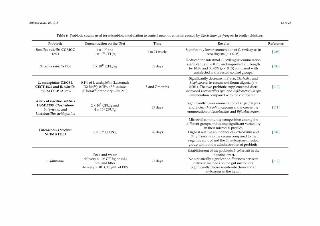
Table 6. Probiotic strains used for microbiota modulation to control necrotic enteritis caused by Clostridium perfringens in broiler chickens.
Probiotic Concentration on the Diet Time Results Reference
Bacillus subtilis CGMCC1.921
1 × 10⁷ and1 × 108 CFU/g
1 to 24 weeks Significantly lower enumeration of C. perfringens inceca digesta (p < 0.05). [108]
Bacillus subtilis PB6 5 × 10¹¹ CFU/kg 35 days
Reduced the intestinal C. perfringens enumerationsignificantly (p < 0.05) and improved villi length
by 10.88 and 30.46% (p < 0.05) compared withuninfected and infected control groups.
[109]
L. acidophilus D2/CSLCECT 4529 and B. subtilis
PB6 ATCC-PTA 6737
0.1% of L. acidophilus (LactomaltD2 Bio®); 0.05% of B. subtilis
(Clostat® brand dry—740210)5 and 7 months
Significantly decrease in E. coli, Clostridia, andStaphylococci in cecum and ileum digesta (p <0.001). The two probiotic-supplemented diets,
increased Lactobacillus spp. and Bifidobacterium spp.enumeration compared with the control diet.
[110]
A mix of Bacillus subtilisDSM17299, Clostridium
butyricum, andLactobacillus acidophilus
2 × 10² CFU/g and4 × 102 CFU/g
35 daysSignificantly lower enumeration of C. perfringens
and Escherichia coli in caecum and increase theenumeration of Lactobacillus and Bifidobacterium.
[111]
Enterococcus faeciumNCIMB 11181 1 × 10⁶ CFU/kg 26 days
Microbial community composition among thedifferent groups, indicating significant variability
in their microbial profiles.Highest relative abundance of Lactobacillus and
Butyricicoccus in the cecum compared to thenegative control and the C. perfringens-infectedgroup without the administration of probiotic.
[107]
L. johnsonii
Feed and waterdelivery > 106 CFU/g or mL;
oral and litterdelivery > 108 CFU/mL of PBS
21 days
Establishment of the probiotic L. johnsonii in theintestinal tract.
No statistically significant differences betweendelivery methods on the gut microbiota.
Significantly decrease enterobacteria and C.perfringens in the ileum.
[112]

Animals 2020, 10, 1718 14 of 28
In addition to the above, the concept of competitive exclusion has been described, which was bornfrom the work carried out by Rantala and Nurmi in 1973, who proposed the inclusion of bacteria isolatedfrom adult chickens to prevent Salmonella Infantis colonization [113]. This term raises the possibility of“implanting a healthy microbiota” in the first days of the animal’s life, and thus preventing colonizationby pathogens [114]. For this, commercial products (Aviguard®, BROILACT®, PoultryStar®, MSC™)have been suggested with an effect against C. perfringens, which causes NE in broilers [114]. However,only MSC™ was evaluated to decrease the enterotoxin produced by C. perfringens, suggesting itsusefulness against the incidence of NE in chickens and reducing the risk of disease in humans [115].
Due to the above, the possibility of an early programming to modulate the intestinal microbiotahas been considered as a potentially useful strategy to improve health, well-being and productivity inbroilers through probiotics, by reducing pathogen enumerations in the gastrointestinal tract of chickensand, with it, the risk of contamination to contribute to the safety of raw meats [116,117].
5.2. Prebiotics
The International Scientific Association for Probiotics and Prebiotics defines prebiotics as“a substrate that is selectively used by host microorganisms, conferring health benefits”; whenadministered orally, these are called dietary prebiotics [118].
A good prebiotic should meet the following characteristics: 1) resist exposure to gastric acid,it should not be hydrolyzed or absorbed in the upper part of the gastrointestinal tract; 2) serve as aselective source of nutrients that support growth and/or metabolic activity of beneficial host membersof the gut microbiota, and 3) induce luminal responses or other systemic physiological responses thatbenefit the host in some way [119]. Thus, the compounds that meet these characteristics are indigestibleoligosaccharides or polysaccharides [120], also named refined functional carbohydrates [118], such asmannan-oligosaccharides (derived from the cell walls of Saccharomyces cerevisiae), β-glucans (derivedfrom cell walls of fungi or yeasts), galacto-oligosaccharides and fructo-oligosaccharides like inulin,levan and branched groups (extracted from different plants, hydrolyzed from polysaccharides orproduced by microorganisms) [121], being inulin and fructooligosaccharides the most used in thepoultry industry [122], with a degree of polymerization of two to twenty monomers [123].
The use of prebiotics in poultry production systems is based on the fact that they are able toimprove the intestinal epithelium (longer villi and shallower crypts) [124] and feed conversion andefficiency [125] through the synthesis of metabolites from their fermentation, such as short-chain fattyacids [126], mainly acetate, propionate, and butyrate, which are absorbed directly from the hindgut andused as an energy source in tissues [127], which in turn promote weight gain and performance [125].
Moreover, they improve the mineral absorption, specially Ca and P, when administered at a rateof 10 g/kg of feed, which in turn impacts bone mineralization in broilers [120], promoting a symbiosisin the intestinal microbiota, increase intestinal colonization of lactic acid bacteria and are capable ofinhibiting intestinal colonization of pathogens, thereby restricting the amount of toxic metabolitesgenerated by them (ammonia, indoles, phenols, and thiols) [128]. They also reduce the intensity andtime of histopathological conditions caused by C. perfringens in the jejunum and duodenum [124,126].
The effects obtained are dependent on the quantity, type and origin of the administered prebiotic,as well as on the characteristics of the birds (breed, sex, age) and the environment (hygiene, housemaintenance, environmental stress, temperature) [120,129].
5.3. Synbiotics
The term synbiotic was used for the first time in 1995 by Gibson and Roberfroid when referring to“a mixture of probiotics and prebiotics that can beneficially affect the host by improving the survivaland implantation in the gastrointestinal tract of live microorganisms supplemented in the diet, by aselective stimulation of the growth and/or activation of the metabolism of one or a limited number ofhealth-promoting bacteria, and therefore, improving the well-being of the host” [130].

Animals 2020, 10, 1718 15 of 28
The main reason for using a symbiotic is that the probiotic without the prebiotic will haveless chance of surviving in the gastrointestinal tract, as it will show less tolerance to temperature,oxygen, and low pH. In addition to the above, the administration of a synbiotic improves the survivalof the probiotic during its passage through the upper gastrointestinal tract [122,131]. Among thebenefits of using synbiotics are: (1) raising the levels of lactobacilli and bifidobacteria, as well as thebalance of the intestinal microbiota; (2) improving immunomodulation; and (3) preventing bacterialtranslocation [132]. In broilers, dietary supplementation with synbiotic products has been reported tosignificantly improve body weight, average daily weight gain, feed efficiency and percentage of bodymass yield compared to the controls or chickens fed only with probiotics [133].
There are some commercial synbiotic products intended for the chicken meat industry; among themare: Biomin®IMBO (ME BIOMIN GmbH) made up of Enterococcus faecium and fructooligosaccharides(FOS), and PoultryStar® (ME BIOMIN GmbH), which includes a mixture of Bifidobacterium animalis,Enterococcus faecium, Lactobacillus reuteri, L. salivarius, Pediococcus acidilactici and inulin, and Synbioticpoultry (Vetafarm) containing L. acidophilus, L. casei, L. salivarius, L. plantarum, L. rhamnosus, L. brevis,Bifidobacterium bifidum, B. lactis, Streptococcus thermophilus and inulin [134].
Synbiotics have been evaluated in the poultry industry to eliminate or decrease intestinal countsof specific pathogens such as Campylobacter jejunio. Supplementation of a mixture of Bifidobacteriumlongum subsp. longum PCB133 and xylooligosaccharides demonstrated their efficacy in reducing thepathogen through the alteration of the intestinal microbiota when it is developing [135].
Few studies have addressed the use of synbiotics as a strategy to decrease the severity of necroticenteritis (NE) caused by C. perfringens. Among the most important results, a consistent impact hasbeen observed in the reduction of the pathogen enumerations and the severity of the histopathologicaldamage at the intestinal level, in the intestinal damage score and in mortality percentages (Table 7).On the other hand, these studies describe an increase in weight gain, in the enumeration of lactic acidbacteria at the intestinal level, and in the number of specific antibodies at the mucosa level in broilers(Table 7) [136,137].
The results of studies conducted with synbiotics in chickens remain controversial. Some researchershave highlighted the efficacy that they have on the significant reduction of pathogens such asEscherichia coli in the cecum content when used combined, strains from the group of lactic acidbacteria and yeasts [136]. For their part, Mookiah et al. [137] did not observe a synergistic effectwhen combining probiotics with prebiotics (11 Lactobacillus strains and isomalto oligosaccharides) indetermining microbial populations in cecum or volatile and non-volatile fatty acid concentrationsin broilers.
Studying the effect that different synbiotics have on pathogens of sanitary importance such asC. perfringens and/or its toxins, and their impact on the safety of meat for human consumption, is asubject that still needs to be explored in more detail.

Animals 2020, 10, 1718 16 of 28
Table 7. Effect of the use of symbiotics on health, production parameters and the elimination of Clostridium perfringens in broilers.
Synbiotic Composition Dose Time Results Reference
Enterococcus faecium + FOS +phycophytic substances 1 kg/ton of feed 3 weeks
Decrease in mortality rate.Significant improvement (p < 0.05) in the intestinal lesion score.
Absence and reduction of histopathological alterations.Significant decrease (p < 0.05) in the counts of C. perfringens inintestine and cecum, from day 3 to day 21, all this between the
control group and the infected group fed with the synbiotic.
[138]
Saccharomyces cerevisiae,Enterococcus faecium, and Bacillus
spp. (Avi-Lution®)1 and 2 g/Kg of feed 42 days
Significant increase in weight gain.Decrease in the percentage of mortality and in cumulative
mortality at day 28 and 42 (both levels ofsynbiotic supplementation).
No effect on intestinal lesions was observed.
[139]
Synbiotic mix Kurago Biotek, 1 mLcontains (7 log UFC/g of
Lactobacillus rhamnosus HN001,Pediococcus acidilactici MA18/5Mand 4.5% Agave tequilana fructans)
50 µL/day 39 and 42 daysIncrease in lactic acid bacteria enumerations in the duodenum.
Improvement in intestinal morphology (higher villi andshallow crypts) in the duodenal mucosa.
[75]
L. reuteri, E. faecium, B. animalis,and P. acidilactici con FOS. 0.05% 21 and 42 days
Significant difference in the height of the jejunal villi (p < 0.05)on day 28 and 42.
Significant weight gain (p < 0.01) (at 21 and 42 days ofthe experiment).
Significant decrease in C. perfringens enumerations from day 28to day 42.
Increasing the number of specific antibodies (IgA) at the level ofthe ceca mucosa.
[140]

Animals 2020, 10, 1718 17 of 28
5.4. Phytogenics
Phytogenic additives are components and biologically active substances extracted from plants,such as oleoresins, tannins, saponins, flavonoids and alkaloids, with a positive effect on growth andanimal health [141].
Phytogenics increase antimicrobial activity, have antiviral, antioxidant, and anti-inflammatoryproperties, stimulating the endocrine and immune system. They promote a higher metabolic andimmune status in chickens, as well as greater well-being. Several plant-derived compounds have beenshown to have beneficial effects on the gut environment and gut microbiota. Its action mechanismis based on altering the permeability of the membrane of microorganisms, causing the leakage ofintracellular material. It is difficult to identify its active principle, because there is a variation ingrowth conditions, climate, harvest, and manufacture, as well as in the biological factors of each plantspecies [142,143]. Table 8 shows a list of the plants most used for the control of C. perfringens in broilers,as well as a description of the effects caused by the phytogenics tested.
Table 8. Phytogenics used in the control of Clostridium perfringens in broilers.
Product Species Results Reference
Anise essential oil Pimpinella anisum
Promotes intestinal development (longer villiand shallow crypts).
Decreased intensity of intestinal lesionsassociated with necrotic enteritis.
[144]
Benzophenanthridine(alkaloids) Chelidonium majus
Improves productive efficiency parameters.Reduces intestinal lesions and mortality
associated with necrotic enteritis.[145]
Oregano essential oil Origanum vulgare
Increase in the body weight and breast weightat 42 d and promotes the cell proliferation in
duodenum (P = 0.001) and jejunum (P = 0.012).Significantly decrease in the Clostridium counts.Decrease of gut lesions caused by C. perfringens
and improved villus height to crypt depth,improvement of feed conversion efficiency.
Increase of serum antibody titers and tendencyto elevate occludin mRNA expression at thesame time that linearly inhibited the mRNA
expression of TLR-2 and tumor necroticfactor-α in the ileum.
[146–148]
Carvacrol Origanum vulgare
Improved health (longer villi and shallowcrypts) and function of the intestinal barrier.
They promote intestinal colonizationby Bifidobacterium.
Antimicrobial activity against C. perfringens andreduction of intestinal lesions associated with
necrotic enteritis.
[141,142,149,150]
Curcumin Curcuma Longa Decreases C. perfringens enumerations inintestinal contents. [143]
Piperine Piper nigrum Decreases C. perfringens enumerations inintestinal contents. [143]
Protopine (alkaloids) Eschscholzia californicaFumaria officinalis
Improves productive efficiency parameters.Reduces intestinal lesions and mortality
associated with necrotic enteritis.[145]
Tannins Castanea sativaInhibits the growth of C. perfringens in vitro and
in vivo, without affecting food consumptionand weight gain.
[151]
Thymol Thymus vulgaris
Improved health (longer villi and shallowcrypts) and function of the intestinal barrier.
Promotes intestinal colonizationby Bifidobacterium.
Antimicrobial activity against C. perfringens.
[142,149,150]
Sanguinarin Chelidonium majusImproves productive efficiency parameters.
Reduces intestinal lesions and mortalityassociated with necrotic enteritis.
[145]

Animals 2020, 10, 1718 18 of 28
5.5. Organic Acids
Organic acids and their corresponding salts or esters are widely used as a feed additive in poultryproduction. They can vary considerably in their functionality due to the number of carbon atoms andit they are aliphatic or aromatic. They are natural constituents of animal or plant tissues or products ofmicrobial fermentation [143].
Carboxylic acids with an aliphatic chain or fatty acids are classified into short chain fatty acids(SCFAs, 1–5 carbon atoms; C1–C5) and medium chain fatty acids (SCFA, 6–12 carbon atoms; C6–C12).The suggested effects of organic acids are antibacterial activity through pH regulation, changes inthe composition of the microbiota, immunomodulatory action, and stimulation of the intestinalmucosa [133,143].
Organic acids have been used as inhibitors of enteric pathogens or as antimicrobials.Their mechanism of action can be by non-dissociation, where, by penetrating the bacterial cellwall, they alter their normal physiology and generate a change in their internal pH, with a dissociationbetween H+ and anions, which leads to energy consumption that puts the growth of the bacteria at risk,even causing death. In addition, they promote changes in the microbiota, have an immunomodulatoryaction and stimulate the intestinal mucosa. The effects in broilers depend on the base of the organicacid product, dose and type [143,152].
Capric-caprylic, caproic, and lauric acids are associated with improved intestinal histomorphologyand decreased stool C. perfringens enumerations [149]. On the other hand, hexanoic, benzoic and butyricacids are associated with an improvement in intestinal histomorphology, a decrease in C. perfringenscounts in the liver and cecum content, and a decrease in the frequency and intensity of intestinal lesionsassociated with NE [142]. Sodium lauryl lactylate acid has also been reported to prevent and inhibitintestinal colonization by C. perfringens [153].
5.6. Dietary Modifications and Enzymes
The nutritional content and feed presentation significantly affect the development of NE inbroilers. Dietary management is a promising strategy for its control [151]. In this sense, differentstrategies are considered, among which are dietary restriction, modification of the content and sourceof macronutrients, [154,155] and the addition of enzymes to the diet [156].
Food restriction is applied in poultry, to control the growth rate and prevent metabolic disorders.Its protective effect against NE could be attributed to the stimulation of the immune system, the influenceof the endocrine system, a decrease in pH and the viscosity of the intestinal content, promoted by foodrestriction [157].
With regard to modifications in the content and source of macronutrients, it has been shownthat the level and source of dietary protein have a direct effect on the concentration of C. perfringensin broilers [155]; this is how those diets high in protein from fish increase the risk of developingNE, for which reason the use of other sources such as soy is currently promoted [158]. Regardingcarbohydrates, the administration of whole grains is a frequent practice in poultry rearing, since it isassociated with the improvement in productive performance and the general and intestinal healthof the birds [159]. The use of whole grains to control NE is based on mechanical stimulation of thegizzard, pH reduction and viscosity of the intestinal content, which together create an unfavorableenvironment for the proliferation of C. perfringens [160].
In broiler production, the use of enzymes derived from microorganisms (fungi and bacteria)through traditional submerged liquid fermentation or solid-state fermentation is common [161].Enzymes such as proteases, glucanase, manase, cellulase, amylase, phytase and xylanase areadded to the feed to overcome the negative effects of non-starch polysaccharides and increasethe digestibility, management and absorption of nutrients for poultry [157,162,163]. Furthermore,the proliferation of C. perfringens in the gastrointestinal tract of broilers [150,157,161,164], which isachieved by reducing the viscosity of the gastric and intestinal contents (cellulase [164], phytase [161],glucanase [150,157], mannanase [150] and xylase [162,165,166]), promotes intestinal colonization of

Animals 2020, 10, 1718 19 of 28
lactic acid bacteria (glucanase [150,157], mannanase [150] and xylase [162,165,166]) and even improvesphysical characteristics such as longer villi and fewer deep crypts and functional characteristics suchas gut permeability (glucanase [150,157] and xylase [162,165,166]).
As has been reviewed, there are currently different alternatives to the use of growth-promotingantibiotics to control NE; however, these alternatives remain questioned in their efficacy [152].Although the bacterial microbiota associated with broilers has shown variations in the structureof their communities with regarding control strategies and the influence of pathogenic bacteria,more comparable studies of farm chicken microbiomes are required that consider individual variabilityand variations between samples of cecal, jejunal or associated mucosal content [167] to deepen andcompare the information.
6. Conclusions
Clostridium perfringens is an important microorganism in the clinical, food and veterinary areas.The diversity of toxins produced by this microorganism not only makes it a risk to human health,but also to animal health. In the latter, the problem is that it causes subclinical diseases that generategreat losses, particularly in the poultry industry, because C. perfringens is capable of producing varioustoxins and bacteriocins, some of which have already been identified and characterized. However,other pathogenicity factors cannot be discarded.
Currently, the infection produced in broilers, known as necrotic enteritis (NE), associated withthis microorganism, has become a problem to maintaining the health of birds, affecting reproductionand conservation, and the supply for human consumption, due to the fact that the disease occurssubclinically and a diagnosis cannot be made in a timely manner, generating significant economiclosses for the producer.
Chicken meat is the most consumed animal protein and enough supply for consumers requiresmass production strategies, exacerbating the problem of by infections by pathogens such as C. perfringens.Due to this, there is a need to find economical, environmentally friendly and efficient alternatives inthe modulation of the intestinal microbiota, which contribute to the efficient production of broilerchicken to meet current and future demand.
The use of various food additives based on probiotics, prebiotics, symbiotics, essential oils,organic acids and enzymes have been presented as various alternatives to mitigate the incidence of NE,achieving an improvement in the general intestinal health of birds, with the opportunity to producehealthy birds for consumption.
Perspectives: It is imperative to carry out more research on alternative and efficient productsfor the modulation of the intestinal microbiota, in addition to the role they play in the immunesystem, where consistent positive effects are needed to fulfill the current demand, while keeping a safeenvironment. It is also important to establish standardized protocols that consider individual andinter-sample variability, and consider the utility of molecular detection mechanisms and epigeneticmodifications underlying treatment with alternative products such as essential oils and organic acidswhere research has not yet been clarified.
Author Contributions: Conceptualization, Z.V.-d.l.M., K.N. and A.V.-L.; formal analysis, Z.V.-d.l.M., K.N.and A.V.-L.; investigation, Z.V.-d.l.M., M.E.M.-R., J.A.-Q., Y.S.G.-T., K.N. and A.V.-L.; writing—original draftpreparation, Z.V.-d. l.M., M.E.M.-R., J.A.-Q., Y.S.G.-T., K.N. and A.V.-L.; writing—review and editing, Z.V.-d.l.M.,K.N. and A.V.-L.; visualization, K.N. and A.V.-L.; supervision, K.N. and A.V.-L.; project administration, A.V.-L.;funding, A.V.-L. All authors have read and agreed to the published version of the manuscript.
Funding: This research received no external funding.
Conflicts of Interest: The authors declare no conflict of interest.
Animals 2020, 10, 1718 20 of 28
References
1. Sarker, M.R.; Shivers, R.P.; Sparks, S.G.; Juneja, V.K.; McClane, B.A. Comparative experiments to examine theeffects of heating on vegetative cells and spores of Clostridium perfringens isolates carrying plasmid genesversus chromosomal enterotoxin genes. Appl. Environ. Microbiol. 2000, 66, 3234–3240. [CrossRef] [PubMed]
2. Juneja, V.K.; Novak, J.S.; Labbe, R.J. Clostridium perfringens. In Pathogens and Toxins in Foods: Challenges andInterventions; Juneja, V.K., Sofos, J.N., Eds.; ASM Press: Washington, DC, USA, 2010; Volume 34, pp. 53–70.
3. Mcclane, B.A.; Uzal, F.A.; Fernandez, M.; Lyerly, D.; Wilkins, T. The Enterotoxic Clostridia. In TheProkaryotes. Bacteria: Firmicutes, Cyanobacteria; Dworkin, M., Falkow, S., Rosenberg, E., Schleifer, K.-H.,Stackebrandt, E., Eds.; Springer Science: Singapore, 2006; p. 1186, ISBN 978-0-387-25494-4.
4. Al-Khaldi, S. Clostridium perfringens, phytohaemagglutinin (kidney bean lectin),Yersinia species. In Bad BugBook. Foodborne Pathogenic Microorganisms and Natural Toxins; Lampel, K.A., Al-Khaldi, S., Cahill, S.M., Eds.;International Medical Publishing: McLean, VA, USA, 2012; ISBN 9780323401814.
5. Geissmann, T.A.; Teuber, M.; Meile, L. Transcriptional analysis of the rubrerythrin and superoxide dismutasegenes of Clostridium perfringens. J. Bacteriol. 1999, 181, 7136–7139. [CrossRef] [PubMed]
6. Jean, D.; Briolat, V.; Reysset, G. Oxidative stress response in Clostridium perfringens. Microbiology 2004, 150,1649–1659. [CrossRef] [PubMed]
7. Johansson, A.; Aspan, A.; Bagge, E.; Båverud, V.; Engström, B.E.; Johansson, K.E. Genetic diversity ofClostridium perfringens type A isolates from animals, food poisoning outbreaks and sludge. BMC Microbiol.2006, 6, 1–12. [CrossRef] [PubMed]
8. Saito, M. Production of enterotoxin by Clostridium perfringens derived from humans, animals, foods, and thenatural environment in Japan. J. Food Prot. 1990, 53, 115–118. [CrossRef] [PubMed]
9. Grass, J.; Gould, L.H.; Mahon, B. Epidemiology of Foodborne Disease Outbreaks Caused by Clostridiumperfringens, United States, 1998–2010. Foodborne Pathog. Dis. 2013, 10, 131–136. [CrossRef]
10. Mellou, K.; Kyritsi, M.; Chrysostomou, A.; Sideroglou, T.; Georgakopoulou, T.; Hadjichristodoulou, C.Clostridium perfringens foodborne outbreak during an athletic event in northern Greece, June 2019. Int. J.Environ. Res. Public Health 2019, 16, 3967. [CrossRef]
11. Monma, C.; Hatakeyama, K.; Obata, H.; Yokoyama, K.; Konishi, N.; Itoh, T.; Kai, A. Four foodborne diseaseoutbreaks caused by a new type of enterotoxin-producing Clostridium perfringens. J. Clin. Microbiol. 2015, 53,859–867. [CrossRef]
12. Shaltout, A.; Zakaria, M.; Nabil, E. Detection and typing of Clostridium perfringens in some retail chickenmeat products. Benha Vet. Med. J. 2017, 33, 283–291. [CrossRef]
13. Lee, C.A.; Labbé, R. Distribution of enterotoxin- and epsilon-positive Clostridium perfringens spores in U.S.retail spices. J. Food Prot. 2018, 81, 394–399. [CrossRef]
14. Butler, A.J.; Thomas, M.K.; Pintar, K.D.M. Expert elicitation as a means to attribute 28 entericpathogens to foodborne, waterborne, animal contact, and person-to-person transmission routes in Canada.Foodborne Pathog. Dis. 2015, 12, 335–344. [CrossRef] [PubMed]
15. Fohler, S.; Klein, G.; Hoedemaker, M.; Scheu, T.; Seyboldt, C.; Campe, A.; Jensen, K.C.; Abdulmawjood, A.Diversity of Clostridium perfringens toxin-genotypes from dairy farms. BMC Microbiol. 2016, 16, 1–7.[CrossRef] [PubMed]
16. Centers for Disease Control and Prevention (CDC) Surveillance for Foodborne Disease Outbreaks UnitedStates, 2017: Annual Report. U.S. Department of Health and Human Services: Atlanta, GA, USA, 2019;Volume 62, pp. 1–10.
17. May, F.J.; Polkinghorne, B.G.; Fearnley, E.J. Epidemiology of bacterial toxin—mediated foodbornegastroenteritis outbreaks in Australia, 2001 to 2013. CDI 2016, 40, 460–469.
18. Makkar, H.P.S.; Ankers, P. Towards sustainable animal diets: A survey-based study. Anim. Feed Sci. Technol.2014, 198, 309–322. [CrossRef]
19. Kay, S.; Edwards, J.; Brown, J.; Dixon, R. Galleria mellonella infection model identifies both high and lowlethality of Clostridium perfringens toxigenic strains and their response to antimicrobials. Front. Microbiol.2019, 10, 1–11. [CrossRef]
20. Kiu, R.; Hall, L.J. An update on the human and animal enteric pathogen Clostridium perfringens. Emerg. MicrobesInfect. 2018, 7. [CrossRef]

Animals 2020, 10, 1718 21 of 28
21. Rood, J.I.; Adams, V.; Lacey, J.; Lyras, D.; Mcclane, B.A.; Stephen, B.; Moore, R.J.; Popoff, M.R.; Sarker, M.R.;Songer, J.G.; et al. Expansion of the Clostridium perfringens toxin-based typing scheme. Anaerobe 2018, 5–10.[CrossRef]
22. Uzal, F.A.; Freedman, J.C.; Shrestha, A.; Theoret, J.R.; Garcia, J.; Awad, M.M.; Adams, V.; Moore, R.J.;Rood, J.I.; Mcclane, B.A. Towards an understanding of the role of Clostridium perfringens toxins in human andanimal disease. Future Microbiol. 2014, 9, 361–377. [CrossRef]
23. Uzal, F.A.; Navarro, M.A.; Li, J.J.; Freedman, J.C.; Shrestha, A.; McClane, B.A. Comparative pathogenesis ofenteric clostridial infections in humans and animals. Anaerobe 2018, 53, 11–20. [CrossRef]
24. Sayeed, S.; Uzal, F.A.; Fisher, D.J.; Saputo, J.; Vidal, J.E.; Chen, Y.; Gupta, P.; Rood, J.I.; McClane, B.A. Betatoxin is essential for the intestinal virulence of Clostridium perfringens type C disease isolate CN3685 in arabbit ileal loop model. Mol. Microbiol. 2008, 67, 15–30. [CrossRef]
25. Ghoneim, N.H.; Hamza, D.A. Epidemiological studies on Clostridium perfringens food poisoning in retailfoods. Rev. Sci. Tech. Off. Int. Epiz 2017, 36, 1025–1032. [CrossRef] [PubMed]
26. Shrestha, A.; Uzal, F.A.; McClane, B.A. Enterotoxic Clostridia: Clostridium perfringens Enteric Diseases.Gram-Positive Pathog. 2018, 6, 977–990. [CrossRef]
27. Miyamoto, K.; Li, J.; McClane, B.A. Enterotoxigenic Clostridium perfringens: Detection and identification.Microbes Environ. 2012, 27, 343–349. [CrossRef] [PubMed]
28. Villarruel-López, A.; Ruíz-Quezada, S.L.; Castro-Rosas, J.; Gomez-Aldapa, C.A.; Olea-Rodríguez, M.A.;Nuño, K.; Navarro-Hidalgo, V.; Torres-Vitela, M.R. Behavior and inactivation of enterotoxin-positiveClostridium perfringens in pork picadillo and tamales filled with pork picadillo under different cooking,storage, and reheating conditions. J. Food Prot. 2016, 79, 741–747. [CrossRef] [PubMed]
29. DuPont, H.L. Bacterial Diarrhea. N. Engl. J. Med. 2009, 361, 1560–1569. [CrossRef]30. Wong, S.; Santullo, P.; O’driscoll, J.; Jamous, A.; Hirani, S.P.; Saif, M. Use of antibiotic and prevalence of
antibiotic-associated diarrhoea in-patients with spinal cord injuries: A UK national spinal injury centreexperience. Spinal Cord 2017, 55, 583–587. [CrossRef]
31. Gui, L.; Subramony, C.; Fratkin, J.; Hughson, M.D. Fatal enteritis necroticans (Pigbel) in a diabetic adult.Mod. Pathol. 2002, 15, 66–70. [CrossRef]
32. Stevens, D.L.; Bryant, A.E. Necrotizing soft-tissue infections. N. Engl. J. Med. 2017, 377, 2253–2265. [CrossRef]33. Zúñiga-Pereira, A.M.; Santamaría, C.; Gutierrez, J.M.; Alape-Girón, A.; Flores-Díaz, M. Deficient skeletal
muscle regeneration after injury induced by a Clostridium perfringens strain associated with gas gangrene.Infect. Immun. 2019, 87, 1–21. [CrossRef]
34. Zahoor, I.; Ghayas, A.; Basheer, A. Genetics and genomics of susceptibility and immune response to necroticenteritis in chicken: A review. Mol. Biol. Rep. 2018, 45, 31–37. [CrossRef]
35. Annett, C.B.; Viste, J.R.; Chirino-Trejo, M.; Classen, H.L.; Middleton, D.M.; Simko, E. Necrotic enteritis: Effectof barley, wheat and corn diets on proliferation of Clostridium perfringens type A. Avian Pathol. 2002, 31,598–601. [CrossRef] [PubMed]
36. Mejia, D.B.; Peñuela, -S.L.M.; Sanmiguel, R.A. El gran impacto de Clostridium perfringens en aves de corral.Pubvet 2018, 12, 1–9. [CrossRef]
37. Van Immerseel, F.; De Buck, J.; Pasmans, F.; Huyghebaert, G.; Haesebrouck, F.; Ducatelle, R. Clostridiumperfringens in poultry: An emerging threat for animal and public health. Avian Pathol. 2004, 33, 537–549.[CrossRef] [PubMed]
38. Gharib-Naseri, K.; Kheravii, S.K.; Keerqin, C.; Morgan, N.; Swick, R.A.; Choct, M.; Wu, S.B. Two differentClostridium perfringens strains produce different levels of necrotic enteritis in broiler chickens. Poult. Sci. 2019,98, 6422–6432. [CrossRef] [PubMed]
39. Timbermont, L.; Haesebrouck, F.; Ducatelle, R.; Van Immerseel, F. Necrotic enteritis in broilers: An updatedreview on the pathogenesis. Avian Pathol. 2011, 40, 341–347. [CrossRef]
40. Timbermont, L.; Lanckriet, A.; Dewulf, J.; Nollet, N.; Schwarzer, K.; Haesebrouck, F.; Ducatelle, R.; vanImmerseel, F. Control of clostridium perfringens-induced necrotic enteritis in broilers by target-releasedbutyric acid, fatty acids and essential oils. Avian Pathol. 2010, 39, 117–121. [CrossRef]
41. Rood, J.I.; Keyburn, A.L.; Moore, R.J. NetB and necrotic enteritis: The hole movable story. Avian Pathol. 2016,45, 295–301. [CrossRef]
42. Van Immerseel, F.; Rood, J.I.; Moore, R.J.; Titball, R.W. Rethinking our understanding of the pathogenesis ofnecrotic enteritis in chickens. Trends Microbiol. 2009, 17, 32–36. [CrossRef]

Animals 2020, 10, 1718 22 of 28
43. Moore, R.J. Necrotic enteritis predisposing factors in broiler chickens. Avian Pathol. 2016, 45, 275–281.[CrossRef]
44. Timbermont, L.; De Smet, L.; Van Nieuwerburgh, F.; Parreira, V.R.; Van Driessche, G.; Haesebrouck, F.;Ducatelle, R.; Prescott, J.; Deforce, D.; Devreese, B.; et al. Perfrin, a novel bacteriocin associated with netBpositive Clostridium perfringens strains from broilers with necrotic enteritis. Vet. Res. 2014, 45, 1–10.[CrossRef]
45. Flores-Díaz, M.; Barquero-Calvo, E.; Ramírez, M.; Alape-Girón, A. Role of Clostridium perfringens Toxins inNecrotic Enteritis in Poultry. Microb. Toxins 2016, 1–16. [CrossRef]
46. Prescott, J.F.; Parreira, V.R.; Mehdizadeh Gohari, I.; Lepp, D.; Gong, J. The pathogenesis of necrotic enteritis inchickens: What we know and what we need to know: A review. Avian Pathol. 2016, 45, 288–294. [CrossRef][PubMed]
47. Razmyar, J.; Peighambari, S.M.; Zamani, A.H. Detection of a Newly Described Bacteriocin, Perfrin, amongClostridium perfringens Isolates from Healthy and Diseased Ostriches and Broiler Chickens in Iran. Avian Dis.2017, 61, 387–390. [CrossRef] [PubMed]
48. Wade, B.; Keyburn, A.L.; Seemann, T.; Rood, J.I.; Moore, R.J. Binding of Clostridium perfringens to collagencorrelates with the ability to cause necrotic enteritis in chickens. Vet. Microbiol. 2015, 180, 299–303. [CrossRef][PubMed]
49. Cheung, J.K.; Keyburn, A.L.; Carter, G.P.; Lanckriet, A.L.; Van Immerseel, F.; Moore, R.J.; Rood, J.I. The VirSRtwo-component signal transduction system regulates NetB toxin production in Clostridium perfringens.Infect. Immun. 2010, 78, 3064–3072. [CrossRef] [PubMed]
50. Yu, Q.; Lepp, D.; Gohari, I.M.; Wu, T.; Zhou, H.; Yin, X.; Yu, H.; Prescott, J.F.; Nie, S.P.; Xie, M.Y.; et al.The Agr-like quorum sensing system is required for pathogenesis of necrotic enteritis caused by Clostridiumperfringens in poultry. Infect. Immun. 2017, 85, 1–17. [CrossRef]
51. Wang, Y.H. Sialidases From Clostridium perfringens and Their Inhibitors. Front. Cell. Infect. Microbiol. 2020, 9,1–11. [CrossRef]
52. Lepp, D.; Roxas, B.; Parreira, V.R.; Marri, P.R.; Rosey, E.L.; Gong, J.; Songer, J.G.; Vedantam, G.; Prescott, J.F.Identification of novel pathogenicity loci in Clostridium perfringens strains that cause Avian necrotic enteritis.PLoS ONE 2010, 5. [CrossRef]
53. Savva, C.G.; Da Costa, S.P.F.; Bokori-Brown, M.; Naylor, C.E.; Cole, A.R.; Moss, D.S.; Titball, R.W.; Basak, A.K.Molecular architecture and functional analysis of NetB, a pore-forming toxin from Clostridium perfringens.J. Biol. Chem. 2013, 288, 3512–3522. [CrossRef]
54. Popoff, M.R.; Bouvet, P. Clostridial toxins. Future Microbiol. 2009, 4, 1021–1064. [CrossRef]55. Navarro, M.A.; McClane, B.A.; Uzal, F.A. Mechanisms of action and cell death associated with Clostridium
perfringens toxins. Toxins 2018, 10, 212. [CrossRef] [PubMed]56. Paiva, D.; McElroy, A. Necrotic enteritis: Applications for the poultry industry. J. Appl. Poult. Res. 2014, 23,
557–566. [CrossRef]57. Edelblum, K.L.; Turner, J.R. The Tight Junction in Inflammatory Disease: Communication Breakdown.
Curr. Opin. Pharmacol. 2009, 9, 715–720. [CrossRef] [PubMed]58. Llanco, L.A.; Nakano, V.; Moraes, C.T.P.d.; Piazza, R.M.F.; Avila-Campos, M.J. Adhesion and invasion
of Clostridium perfringens type A into epithelial cells. Brazilian J. Microbiol. 2017, 48, 764–768. [CrossRef][PubMed]
59. Coursodon, C.F.; Glock, R.D.; Moore, K.L.; Cooper, K.K.; Songer, J.G. TpeL-producing strains of Clostridiumperfringens type A are highly virulent for broiler chicks. Anaerobe 2012, 18, 117–121. [CrossRef] [PubMed]
60. Schorch, B.; Song, S.; Van Diemen, F.R.; Bock, H.H.; May, P.; Herz, J.; Brummelkamp, T.R.; Papatheodorou, P.;Aktories, K. LRP1 is a receptor for Clostridium perfringens TpeL toxin indicating a two-receptor model ofclostridial glycosylating toxins. Proc. Natl. Acad. Sci. USA 2014, 111, 6431–6436. [CrossRef] [PubMed]
61. Alshamy, Z.; Richardson, K.C.; Hünigen, H.; Hafez, H.M.; Plendl, J.; Al Masri, S. Comparison of thegastrointestinal tract of a dual-purpose to a broiler chicken line: A qualitative and quantitative macroscopicand microscopic study. PLoS ONE 2018, 13, e0204921. [CrossRef]
62. Laledashti, M.A.; Saki, A.A.; Rafati, A.A.; Abdolmaleki, M. Effect of in-ovo feeding of iron nanoparticlesand methionine hydroxy analogue on broilers chickens small intestinal characteristics. Acta Sci. Anim. Sci.2020, 42. [CrossRef]
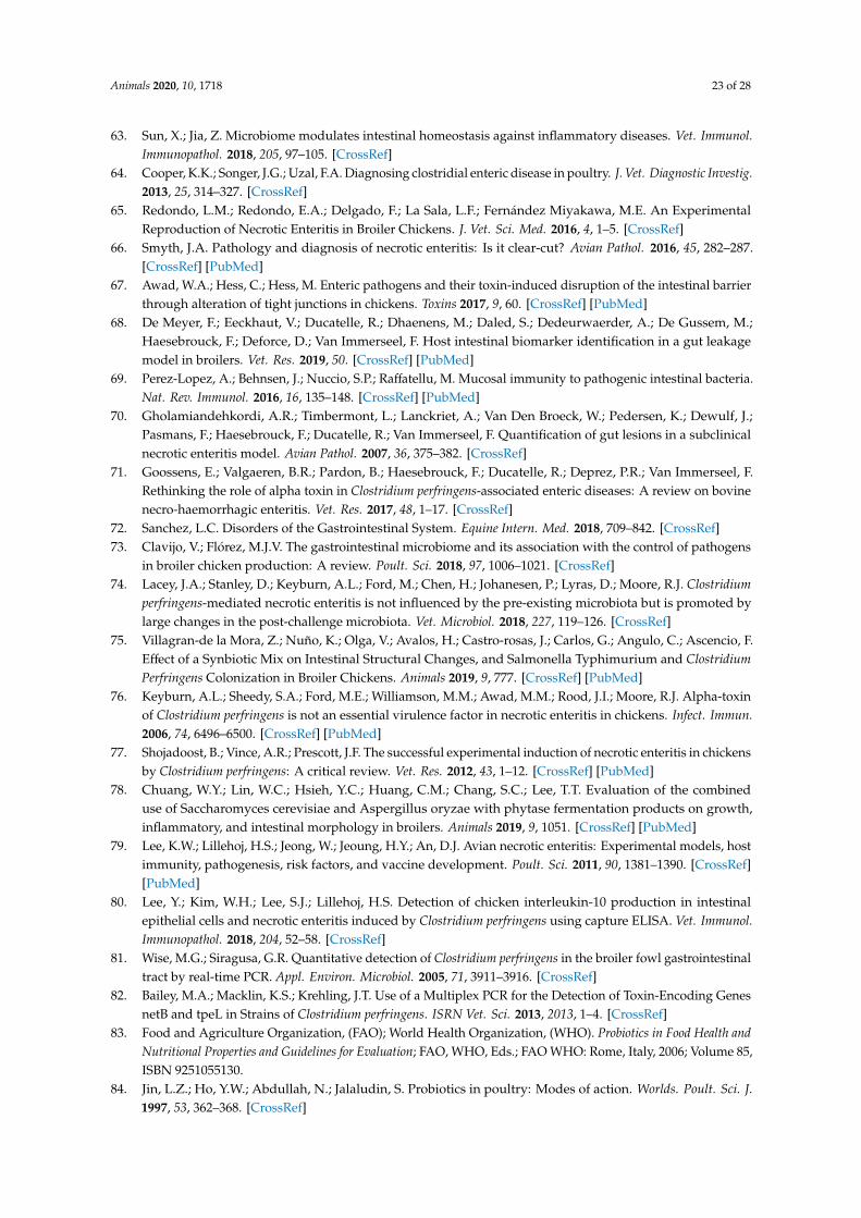
Animals 2020, 10, 1718 23 of 28
63. Sun, X.; Jia, Z. Microbiome modulates intestinal homeostasis against inflammatory diseases. Vet. Immunol.Immunopathol. 2018, 205, 97–105. [CrossRef]
64. Cooper, K.K.; Songer, J.G.; Uzal, F.A. Diagnosing clostridial enteric disease in poultry. J. Vet. Diagnostic Investig.2013, 25, 314–327. [CrossRef]
65. Redondo, L.M.; Redondo, E.A.; Delgado, F.; La Sala, L.F.; Fernández Miyakawa, M.E. An ExperimentalReproduction of Necrotic Enteritis in Broiler Chickens. J. Vet. Sci. Med. 2016, 4, 1–5. [CrossRef]
66. Smyth, J.A. Pathology and diagnosis of necrotic enteritis: Is it clear-cut? Avian Pathol. 2016, 45, 282–287.[CrossRef] [PubMed]
67. Awad, W.A.; Hess, C.; Hess, M. Enteric pathogens and their toxin-induced disruption of the intestinal barrierthrough alteration of tight junctions in chickens. Toxins 2017, 9, 60. [CrossRef] [PubMed]
68. De Meyer, F.; Eeckhaut, V.; Ducatelle, R.; Dhaenens, M.; Daled, S.; Dedeurwaerder, A.; De Gussem, M.;Haesebrouck, F.; Deforce, D.; Van Immerseel, F. Host intestinal biomarker identification in a gut leakagemodel in broilers. Vet. Res. 2019, 50. [CrossRef] [PubMed]
69. Perez-Lopez, A.; Behnsen, J.; Nuccio, S.P.; Raffatellu, M. Mucosal immunity to pathogenic intestinal bacteria.Nat. Rev. Immunol. 2016, 16, 135–148. [CrossRef] [PubMed]
70. Gholamiandehkordi, A.R.; Timbermont, L.; Lanckriet, A.; Van Den Broeck, W.; Pedersen, K.; Dewulf, J.;Pasmans, F.; Haesebrouck, F.; Ducatelle, R.; Van Immerseel, F. Quantification of gut lesions in a subclinicalnecrotic enteritis model. Avian Pathol. 2007, 36, 375–382. [CrossRef]
71. Goossens, E.; Valgaeren, B.R.; Pardon, B.; Haesebrouck, F.; Ducatelle, R.; Deprez, P.R.; Van Immerseel, F.Rethinking the role of alpha toxin in Clostridium perfringens-associated enteric diseases: A review on bovinenecro-haemorrhagic enteritis. Vet. Res. 2017, 48, 1–17. [CrossRef]
72. Sanchez, L.C. Disorders of the Gastrointestinal System. Equine Intern. Med. 2018, 709–842. [CrossRef]73. Clavijo, V.; Flórez, M.J.V. The gastrointestinal microbiome and its association with the control of pathogens
in broiler chicken production: A review. Poult. Sci. 2018, 97, 1006–1021. [CrossRef]74. Lacey, J.A.; Stanley, D.; Keyburn, A.L.; Ford, M.; Chen, H.; Johanesen, P.; Lyras, D.; Moore, R.J. Clostridium
perfringens-mediated necrotic enteritis is not influenced by the pre-existing microbiota but is promoted bylarge changes in the post-challenge microbiota. Vet. Microbiol. 2018, 227, 119–126. [CrossRef]
75. Villagran-de la Mora, Z.; Nuño, K.; Olga, V.; Avalos, H.; Castro-rosas, J.; Carlos, G.; Angulo, C.; Ascencio, F.Effect of a Synbiotic Mix on Intestinal Structural Changes, and Salmonella Typhimurium and ClostridiumPerfringens Colonization in Broiler Chickens. Animals 2019, 9, 777. [CrossRef] [PubMed]
76. Keyburn, A.L.; Sheedy, S.A.; Ford, M.E.; Williamson, M.M.; Awad, M.M.; Rood, J.I.; Moore, R.J. Alpha-toxinof Clostridium perfringens is not an essential virulence factor in necrotic enteritis in chickens. Infect. Immun.2006, 74, 6496–6500. [CrossRef] [PubMed]
77. Shojadoost, B.; Vince, A.R.; Prescott, J.F. The successful experimental induction of necrotic enteritis in chickensby Clostridium perfringens: A critical review. Vet. Res. 2012, 43, 1–12. [CrossRef] [PubMed]
78. Chuang, W.Y.; Lin, W.C.; Hsieh, Y.C.; Huang, C.M.; Chang, S.C.; Lee, T.T. Evaluation of the combineduse of Saccharomyces cerevisiae and Aspergillus oryzae with phytase fermentation products on growth,inflammatory, and intestinal morphology in broilers. Animals 2019, 9, 1051. [CrossRef] [PubMed]
79. Lee, K.W.; Lillehoj, H.S.; Jeong, W.; Jeoung, H.Y.; An, D.J. Avian necrotic enteritis: Experimental models, hostimmunity, pathogenesis, risk factors, and vaccine development. Poult. Sci. 2011, 90, 1381–1390. [CrossRef][PubMed]
80. Lee, Y.; Kim, W.H.; Lee, S.J.; Lillehoj, H.S. Detection of chicken interleukin-10 production in intestinalepithelial cells and necrotic enteritis induced by Clostridium perfringens using capture ELISA. Vet. Immunol.Immunopathol. 2018, 204, 52–58. [CrossRef]
81. Wise, M.G.; Siragusa, G.R. Quantitative detection of Clostridium perfringens in the broiler fowl gastrointestinaltract by real-time PCR. Appl. Environ. Microbiol. 2005, 71, 3911–3916. [CrossRef]
82. Bailey, M.A.; Macklin, K.S.; Krehling, J.T. Use of a Multiplex PCR for the Detection of Toxin-Encoding GenesnetB and tpeL in Strains of Clostridium perfringens. ISRN Vet. Sci. 2013, 2013, 1–4. [CrossRef]
83. Food and Agriculture Organization, (FAO); World Health Organization, (WHO). Probiotics in Food Health andNutritional Properties and Guidelines for Evaluation; FAO, WHO, Eds.; FAO WHO: Rome, Italy, 2006; Volume 85,ISBN 9251055130.
84. Jin, L.Z.; Ho, Y.W.; Abdullah, N.; Jalaludin, S. Probiotics in poultry: Modes of action. Worlds. Poult. Sci. J.1997, 53, 362–368. [CrossRef]

Animals 2020, 10, 1718 24 of 28
85. Park, Y.H.; Hamidon, F.; Rajangan, C.; Soh, K.P.; Gan, C.Y.; Lim, T.S.; Abdullah, W.N.W.; Liong, M.T.Application of probiotics for the production of safe and high-quality poultry meat. Korean J. Food Sci.Anim. Resour. 2016, 36, 567–576. [CrossRef]
86. Ma, T.; Suzuki, Y.; Guan, L.L. Dissect the mode of action of probiotics in affecting host-microbial interactionsand immunity in food producing animals. Vet. Immunol. Immunopathol. 2018, 205, 35–48. [CrossRef][PubMed]
87. Azad, M.A.K.; Sarker, M.; Li, T.; Yin, J. Probiotic Species in the Modulation of Gut Microbiota: An Overview.Biomed Res. Int. 2018, 2018. [CrossRef]
88. Popova, T. Effect of probiotics in poultry for improving meat quality. Curr. Opin. Food Sci. 2017, 14, 72–77.[CrossRef]
89. Carlander, D.; Stålberg, J.; Larsson, A. Chicken antibodies: A clinical chemistry perspective. Ups. J. Med. Sci.1999, 104, 179–189. [CrossRef] [PubMed]
90. Khalique, A.; Zeng, D.; Shoaib, M.; Wang, H.; Qing, X.; Rajput, D.S.; Pan, K.; Ni, X. Probiotics mitigatingsubclinical necrotic enteritis (SNE) as potential alternatives to antibiotics in poultry. AMB Express 2020, 10.[CrossRef] [PubMed]
91. Menconi, A.; Sokale, A.O.; Mendoza, S.M.; Whelan, R.; Doranalli, K. Effect of Bacillus subtilis DSM 32315under different Necrotic Enteritis models in broiler chickens: A meta-analysis of 5 independent researchtrials. Avian Dis. 2020. [CrossRef]
92. Johnson, T.J.; Youmans, B.P.; Noll, S.; Cardona, C.; Evans, N.P.; Peter Karnezos, T.; Ngunjiri, J.M.; Abundo, M.C.;Lee, C.W. A consistent and predictable commercial broiler chicken bacterial microbiota in antibiotic-freeproduction displays strong correlations with performance. Appl. Environ. Microbiol. 2018, 84, 1–18. [CrossRef]
93. Stanley, D.; Hughes, R.J.; Geier, M.S.; Moore, R.J. Bacteria within the gastrointestinal tract microbiotacorrelated with improved growth and feed conversion: Challenges presented for the identification ofperformance enhancing probiotic bacteria. Front. Microbiol. 2016, 7, 1–13. [CrossRef]
94. La Ragione, R.M.; Narbad, A.; Gasson, M.J.; Woodward, M.J. In vivo characterization of Lactobacillusjohnsonii FI9785 for use as a defined competitive exclusion agent against bacterial pathogens in poultry.Lett. Appl. Microbiol. 2004, 38, 197–205. [CrossRef]
95. Gervasi, T.; Lo Curto, R.; Minniti, E.; Narbad, A.; Mayer, M.J. Application of Lactobacillus johnsonii expressingphage endolysin for control of Clostridium perfringens. Lett. Appl. Microbiol. 2014, 59, 355–361. [CrossRef]
96. Kim, C.H.; Shin, K.S.; Woo, K.C.; Paik, I.K. Effect of Dietary Oligosaccharides on the Performance, IntestinalMicroflora and Serum Immunoglobulin Contents in Laying Hens. Korean J. Poult. Sci. 2009, 36, 125–131.[CrossRef]
97. Wu, Y.; Shao, Y.; Song, B.; Zhen, W.; Wang, Z.; Guo, Y.; Shahid, M.S.; Nie, W. Effects of Bacilluscoagulans supplementation on the growth performance and gut health of broiler chickens with Clostridiumperfringens-induced necrotic enteritis. J. Anim. Sci. Biotechnol. 2018, 9, 1–14. [CrossRef] [PubMed]
98. Xu, S.; Lin, Y.; Zeng, D.; Zhou, M.; Zeng, Y.; Wang, H.; Zhou, Y.; Zhu, H.; Pan, K.; Jing, B.; et al. Bacilluslicheniformis normalize the ileum microbiota of chickens infected with necrotic enteritis. Sci. Rep. 2018, 8,1–10. [CrossRef] [PubMed]
99. Zhao, Y.; Zeng, D.; Wang, H.; Qing, X.; Sun, N.; Xin, J.; Luo, M.; Khalique, A.; Pan, K.; Shu, G.; et al. DietaryProbiotic Bacillus licheniformis H2 Enhanced Growth Performance, Morphology of Small Intestine andLiver, and Antioxidant Capacity of Broiler Chickens Against Clostridium perfringens–Induced SubclinicalNecrotic Enteritis. Probiotics Antimicrob. Proteins 2019. [CrossRef] [PubMed]
100. Aljumaah, M.R.; Alkhulaifi, M.M.; Abudabos, A.M.; Aljumaah, R.S.; Alsaleh, A.N.; Stanley, D. Bacillussubtilis PB6 based probiotic supplementation plays a role in the recovery after the necrotic enteritis challenge.PLoS ONE 2020, 15, e0232781. [CrossRef] [PubMed]
101. Ramlucken, U.; Ramchuran, S.O.; Moonsamy, G.; Lalloo, R.; Thantsha, M.S.; Jansen van Rensburg, C. A novelBacillus based multi-strain probiotic improves growth performance and intestinal properties of Clostridiumperfringens challenged broilers. Poult. Sci. 2020, 99, 331–341. [CrossRef]
102. Wang, H.; Ni, X.; Qing, X.; Liu, L.; Xin, J.; Luo, M.; Khalique, A.; Dan, Y.; Pan, K.; Jing, B.; et al. Probioticlactobacillus johnsonii BS15 improves blood parameters related to immunity in broilers experimentallyinfected with subclinical necrotic enteritis. Front. Microbiol. 2018, 9, 1–12. [CrossRef]
Animals 2020, 10, 1718 25 of 28
103. Cao, L.; Wu, X.H.; Bai, Y.L.; Wu, X.Y.; Gu, S.B. Anti-inflammatory and antioxidant activities of probioticpowder containing Lactobacillus plantarum 1.2567 in necrotic enteritis model of broiler chickens. Livest. Sci.2019, 223, 157–163. [CrossRef]
104. Cao, L.; Yang, X.J.; Li, Z.J.; Sun, F.F.; Wu, X.H.; Yao, J.H. Reduced lesions in chickens with Clostridiumperfringens-induced necrotic enteritis by Lactobacillus fermentum 1.2029. Poult. Sci. 2012, 91, 3065–3071.[CrossRef]
105. Guo, S.; Liu, D.; Zhang, B.; Li, Z.; Li, Y.; Ding, B.; Guo, Y. Two Lactobacillus species inhibit the growth andα-toxin production of Clostridium perfringens and induced proinflammatory factors in chicken intestinalepithelial cells in vitro. Front. Microbiol. 2017, 8, 1–12. [CrossRef]
106. Xu, T.; Chen, Y.; Yu, L.; Wang, J.; Huang, M.; Zhu, N. Effects of Lactobacillus plantarum on intestinal integrityand immune responses of egg-laying chickens infected with Clostridium perfringens under the free-range orthe specific pathogen free environment. BMC Vet. Res. 2020, 16, 1–12. [CrossRef] [PubMed]
107. Wu, Y.; Zhen, W.; Geng, Y.; Wang, Z.; Guo, Y. Pretreatment with probiotic Enterococcus faecium NCIMB11181 ameliorates necrotic enteritis-induced intestinal barrier injury in broiler chickens. Sci. Rep. 2019, 9,1–17. [CrossRef] [PubMed]
108. Guo, J.R.; Dong, X.F.; Liu, S.; Tong, J.M. Effects of long-term Bacillus subtilis CGMCC 1.921 supplementationon performance, egg quality, and fecal and cecal microbiota of laying hens. Poult. Sci. 2017, 96, 1280–1289.[CrossRef] [PubMed]
109. Jayaraman, S.; Thangavel, G.; Kurian, H.; Mani, R.; Mukkalil, R.; Chirakkal, H. Bacillus subtilis PB6improves intestinal health of broiler chickens challenged with Clostridium perfringens-induced necroticenteritis. Poult. Sci. 2013, 92, 370–374. [CrossRef]
110. Forte, C.; Acuti, G.; Manuali, E.; Casagrande Proietti, P.; Pavone, S.; Trabalza-Marinucci, M.; Moscati, L.;Onofri, A.; Lorenzetti, C.; Franciosini, M.P. Effects of two different probiotics on microflora, morphology, andmorphometry of gut in organic laying hens. Poult. Sci. 2016, 95, 2528–2535. [CrossRef] [PubMed]
111. Hossain, M.M.; Begum, M.; Kim, I.H. Effect of Bacillus subtilis, Clostridium butyricum and Lactobacillusacidophilus endospores on growth performance, nutrient digestibility, meat quality, relative organ weight,microbial shedding and excreta noxious gas emission in broilers. Vet. Med. 2015, 60, 77–86. [CrossRef]
112. Olnood, C.G.; Beski, S.S.M.; Iji, P.A.; Choct, M. Delivery routes for probiotics: Effects on broiler performance,intestinal morphology and gut microflora. Anim. Nutr. 2015, 1, 192–202. [CrossRef]
113. Rantala, M.; Nurmi, E. Prevention of the growth of salmonella infantis in chicks by the flora of the alimentarytract of chickens. Br. Poult. Sci. 1973, 14, 627–630. [CrossRef]
114. Caly, D.L.; D’Inca, R.; Auclair, E.; Drider, D. Alternatives to antibiotics to prevent necrotic enteritis in broilerchickens: A microbiologist’s perspective. Front. Microbiol. 2015, 6, 1336. [CrossRef]
115. Craven, A.S.E.; Stern, N.J.; Cox, N.A.; Bailey, J.S.; Berrang, M.; Craven, S.E.; Stern, N.J.; Cox, N.A.; Bailey, J.S.;Berrang, M. Cecal Carriage of Clostridium perfringens in Broiler Chickens Given Mucosal Starter Culture.Avian Dis. 2018, 43, 484–490. [CrossRef]
116. Rubio, L.A. Possibilities of early life programming in broiler chickens via intestinal microbiota modulation.Poult. Sci. 2019, 98, 695–706. [CrossRef] [PubMed]
117. Ghareeb, K.; Awad, W.A.; Mohnl, M.; Porta, R.; Biarnés, M.; Böhm, J.; Schatzmayr, G. Evaluating the efficacyof an avian-specific probiotic to reduce the colonization of Campylobacter jejuni in broiler chickens. Poult. Sci.2012, 91, 1825–1832. [CrossRef] [PubMed]
118. Froebel, L.K.; Jalukar, S.; Lavergne, T.A.; Lee, J.T.; Duong, T. Administration of dietary prebiotics improvesgrowth performance and reduces pathogen colonization in broiler chickens. Poult. Sci. 2019, 98, 6668–6676.[CrossRef] [PubMed]
119. Kim, S.A.; Jang, M.J.; Kim, S.Y.; Yang, Y.; Pavlidis, H.O.; Ricke, S.C. Potential for prebiotics as feed additivesto limit foodborne Campylobacter establishment in the poultry gastrointestinal tract. Front. Microbiol. 2019,10, 1–12. [CrossRef]
120. Bucław, M. Inulin in poultry production. Worlds. Poult. Sci. J. 2017, 73, 301–308. [CrossRef]121. Teng, P.; Kim, W.K. Review: Roles of Prebiotics in Intestinal Ecosystem of Broilers. Front. Vet. Sci. 2018, 5,
1–18. [CrossRef]122. Ghasemi, H.A.; Shivazad, M.; Mirzapour Rezaei, S.S.; Torshizi, M.A.K. Effect of synbiotic supplementation
and dietary fat sources on broiler performance, serum lipids, muscle fatty acid profile and meat quality.Br. Poult. Sci. 2015, 57. [CrossRef]

Animals 2020, 10, 1718 26 of 28
123. Huyghebaert, G.; Ducatelle, R.; Van Immerseel, F. Van An update on alternatives to antimicrobial growthpromoters for broilers. Vet. J. 2011, 187, 182–188. [CrossRef]
124. Hashim, M.M.; Arsenault, R.J.; Byrd, J.A.; Kogut, M.H.; Al-Ajeeli, M.; Bailey, C.A. Influence of differentyeast cell wall preparations and their components on performance and immune and metabolic pathways inClostridium perfringens-challenged broiler chicks. Poult. Sci. 2018, 97, 203–210. [CrossRef]
125. Guaragni, A.; Boiago, M.M.; Bottari, N.B.; Morsch, V.M.; Lopes, T.F.; Schafer da Silva, A. Feed supplementationwith inulin on broiler performance and meat quality challenged with Clostridium perfringens: Infection andprebiotic impacts. Microb. Pathog. 2020, 139, 103889. [CrossRef]
126. Ricke, S.C.; Lee, S.I.; Kim, S.A.; Park, S.H.; Shi, Z. Prebiotics and the poultry gastrointestinal tract microbiome.Poult. Sci. 2020, 99, 670–677. [CrossRef] [PubMed]
127. Ajuwon, K.M. Toward a better understanding of mechanisms of probiotics and prebiotics action in poultryspecies. J. Appl. Poult. Res. 2016, 25, 277–283. [CrossRef]
128. Kheravii, S.K.; Swick, R.A.; Choct, M.; Wu, S.B. Effect of oat hulls as a free choice feeding on broilerperformance, short chain fatty acids and microflora under a mild necrotic enteritis challenge. Anim. Nutr.2018, 4, 65–72. [CrossRef] [PubMed]
129. Mtei, A.W.; Abdollahi, M.R.; Schreurs, N.; Girish, C.K.; Ravindran, V. Dietary inclusion of fibrous ingredientsand bird type influence apparent ileal digestibility of nutrients and energy utilization. Poult. Sci. 2019, 98,6702–6712. [CrossRef] [PubMed]
130. Gibson, G.R.; Roberfroid, M.B. Dietary modulation of the human colonic microbiota: Introducing the conceptof prebiotics. J. Nutr. 1995, 125, 1401–1412. [CrossRef]
131. Peña, A.S. Flora intestinal, probióticos, prebióticos, simbióticos y alimentos novedosos. Rev. Esp. Enferm. Dig.2007, 99, 653–658. [CrossRef]
132. Pandey, K.R.; Naik, S.R.; Vakil, B.V. Probiotics, prebiotics and synbiotics—A review. J. Food Sci. Technol. 2015,52, 7577–7587. [CrossRef]
133. Gadde, U.; Kim, W.H.; Oh, S.T.; Lillehoj, H.S. Alternatives to antibiotics for maximizing growth performanceand feed efficiency in poultry: A review. Anim. Heal. Res. Rev. 2017, 18, 26–45. [CrossRef]
134. Markowiak, P.; Slizewska, K. The role of probiotics, prebiotics and synbiotics in animal nutrition. Gut Pathog.2018, 10, 1–20. [CrossRef]
135. Baffoni, L.; Gaggìa, F.; Garofolo, G.; Di Serafino, G.; Buglione, E.; Di Giannatale, E.; Di Gioia, D. Evidence ofCampylobacter jejuni reduction in broilers with early synbiotic administration. Int. J. Food Microbiol. 2017,251, 41–47. [CrossRef]
136. Koc, F.; Samli, H.; Okur, A.; Ozduven, M.; Akyurek, H.; Senkoylu, N. Effects of Saccharomyces cerevisiaeand/or mannanoligosaccharide on performance, blood parameters and intestinal microbiota of broiler chicks.Bulg. J. Agric. Sci. 2010, 16, 643–650.
137. Mookiah, S.; Sieo, C.C.; Ramasamy, K.; Abdullah, N.; Ho, Y.W. Effects of dietary prebiotics, probiotic andsynbiotics on performance, caecal bacterial populations and caecal fermentation concentrations of broilerchickens. J. Sci. Food Agric. 2014, 94, 341–348. [CrossRef] [PubMed]
138. Abd El-Ghany, W.A. Comparative evaluation on the effect of coccidiostate and synbiotic preparations onprevention of Clostridium perfringens in broiler chickens. Glob. Vet. 2010, 5, 324–333.
139. Krueger, L.A.; Spangler, D.A.; Vandermyde, D.R.; Sims, M.D.; Ayangbile, G.A. Avi-Lution® supplementedat 1.0 or 2.0 g/kg in feed improves the growth performance of broiler chickens during challenge withbacitracin-resistant Clostridium perfringens. Poult. Sci. 2017, 96, 2595–2600. [CrossRef] [PubMed]
140. Shanmugasundaram, R.; Markazi, A.; Mortada, M.; Ng, T.T.; Applegate, T.J.; Bielke, L.R.; Syed, B.;Pender, C.M.; Curry, S.; Murugesan, G.R.; et al. Research Note: Effect of synbiotic supplementation on caecalClostridium perfringens load in broiler chickens with different necrotic enteritis challenge models. Poult. Sci.2020, 99, 2452–2458. [CrossRef]
141. Yang, C.; Chowdhury, M.A.K.; Hou, Y.; Gong, J. Phytogenic compounds as alternatives to in-feed antibiotics:Potentials and challenges in application. Pathogens 2015, 4, 137–156. [CrossRef]
142. Pham, V.H.; Kan, L.; Huang, J.; Geng, Y.; Zhen, W.; Guo, Y.; Abbas, W.; Wang, Z. Dietary encapsulatedessential oils and organic acids mixture improves gut health in broiler chickens challenged with necroticenteritis. J. Anim. Sci. Biotechnol. 2020, 11, 1–18. [CrossRef]

Animals 2020, 10, 1718 27 of 28
143. Granstad, S.; Kristoffersen, A.B.; Benestad, S.L.; Sjurseth, S.K.; David, B.; Sørensen, L.; Fjermedal, A.;Edvardsen, D.H.; Sanson, G.; Løvland, A.; et al. Effect of feed additives as alternatives to in-feed antimicrobialson production performance and intestinal Clostridium perfringens counts in broiler chickens. Animals 2020,10, 240. [CrossRef]
144. Abudabos, A.M. Use of a competitive exclusion product (Aviguard) to prevent Clostridium perfringenscolonization in broiler chicken under induced challenge. Pak. J. Zool. 2013, 45, 371–376.
145. Hussein, E.O.S.; Ahmed, S.H.; Abudabos, A.M.; Aljumaah, M.R.; Alkhlulaifi, M.M.; Nassan, M.A.;Suliman, G.M.; Naiel, M.A.E.; Swelum, A.A. Effect of antibiotic, phytobiotic and probiotic supplementationon growth, blood indices and intestine health in broiler chicks challenged with Clostridium perfringens.Animals 2020, 10, 507. [CrossRef]
146. Tzora, A.; Giannenas, I.; Karamoutsios, A.; Papaioannou, N.; Papanastasiou, D.; Bonos, E.; Skoufos, S.;Bartzanas, T.; Skoufos, I. Effects of oregano, attapulgite, benzoic acid and their blend on chicken performance,intestinal microbiology and intestinal morphology. J. Poult. Sci. 2017, 54, 218–227. [CrossRef] [PubMed]
147. Kirkpinar, F.; Ünlü, H.B.; Özdemir, G. Effects of oregano and garlic essential oils on performance, carcase,organ and blood characteristics and intestinal microflora of broilers. Livest. Sci. 2011, 137, 219–225. [CrossRef]
148. Du, E.; Wang, W.; Gan, L.; Li, Z.; Guo, S.; Guo, Y. Effects of thymol and carvacrol supplementation onintestinal integrity and immune responses of broiler chickens challenged with Clostridium perfringens.J. Anim. Sci. Biotechnol. 2016, 7, 1–10. [CrossRef] [PubMed]
149. Abdelli, N.; Pérez, J.F.; Vilarrasa, E.; Luna, I.C.; Melo-Duran, D.; D’angelo, M.; Solà-Oriol, D. Targeted-releaseorganic acids and essential oils improve performance and digestive function in broilers under a necroticenteritis challenge. Animals 2020, 10, 259. [CrossRef] [PubMed]
150. Sun, Q.; Liu, D.; Guo, S.; Chen, Y.; Guo, Y. Effects of dietary essential oil and enzyme supplementation ongrowth performance and gut health of broilers challenged by Clostridium perfringens. Anim. Feed Sci. Technol.2015, 207, 234–244. [CrossRef]
151. Mannelli, F.; Minieri, S.; Tosi, G.; Secci, G.; Daghio, M.; Massi, P.; Fiorentini, L.; Galigani, I.; Lancini, S.;Rapaccini, S.; et al. Effect of chestnut tannins and short chain fatty acids as anti-microbials and as feedingsupplements in broilers rearing and meat quality. Animals 2019, 9, 659. [CrossRef]
152. Dahiya, J.P.; Wilkie, D.C.; Van Kessel, A.G.; Drew, M.D. Potential strategies for controlling necrotic enteritisin broiler chickens in post-antibiotic era. Anim. Feed Sci. Technol. 2006, 129, 60–88. [CrossRef]
153. Lensing, M.; van der Klis, J.D.; Fabri, T.; Cazemier, A.; Else, A.J. Efficacy of a lactylate on productionperformance and intestinal health of broilers during a subclinical Clostridium perfringens infection. Poult. Sci.2010, 89, 2401–2409. [CrossRef]
154. Drew, M.D.; Syed, N.A.; Goldade, B.G.; Laarveld, B.; Van Kessel, A.G. Effects of dietary protein source andlevel on intestinal populations of Clostridium perfringens in broiler chickens. Poult. Sci. 2004, 83, 414–420.[CrossRef]
155. Kumar, S.; Adhikari, P.; Oakley, B.; Kim, W.K. Changes in cecum microbial community in response to totalsulfur amino acid (TSAA: DL-methionine) in antibiotic-free and supplemented poultry birds. Poult. Sci.2019, 98, 5809–5819. [CrossRef]
156. Sieo, C.C.; Abdullah, N.; Tan, W.S.; Ho, Y.W. Influence of β-glucanase-producing lactobacillus strains onintestinal characteristics and feed passage rate of broiler chickens. Poult. Sci. 2005, 84, 734–741. [CrossRef]
157. Tsiouris, V.; Georgopoulou, I.; Batzios, C.; Pappaioannou, N.; Ducatelle, R.; Fortomaris, P. Temporary feedrestriction partially protects broilers from necrotic enteritis. Avian Pathol. 2014, 43, 139–145. [CrossRef][PubMed]
158. Wilkie, D.C.; Van Kessel, A.G.; White, L.J.; Laarveld, B.; Drew, M.D. Dietary amino acids affect intestinalClostridium perfringens populations in broiler chickens. Can. J. Anim. Sci. 2005, 85, 185–193. [CrossRef]
159. Singh, Y.; Amerah, A.M.; Ravindran, V. Whole grain feeding: Methodologies and effects on performance,digestive tract development and nutrient utilisation of poultry. Anim. Feed Sci. Technol. 2014, 190, 1–18.[CrossRef]
160. Tsiouris, V. Poultry management: A useful tool for the control of necrotic enteritis in poultry. Avian Pathol.2016, 45, 323–325. [CrossRef]
161. Kiarie, E.; Romero, L.F.; Nyachoti, C.M. The role of added feed enzymes in promoting gut health in swineand poultry. Nutr. Res. Rev. 2013, 26, 71–88. [CrossRef]

Animals 2020, 10, 1718 28 of 28
162. Barekatain, M.R.; Antipatis, C.; Rodgers, N.; Walkden-Brown, S.W.; Iji, P.A.; Choct, M. Evaluation of highdietary inclusion of distillers dried grains with solubles and supplementation of protease and xylanase in thediets of broiler chickens under necrotic enteritis challenge. Poult. Sci. 2013, 92, 1579–1594. [CrossRef]
163. Wu, Y.B.; Ravindran, V. Influence of whole wheat inclusion and xylanase supplementation on the performance,digestive tract measurements and carcass characteristics of broiler chickens. Anim. Feed Sci. Technol. 2004,116, 129–139. [CrossRef]
164. Latorre, J.D.; Hernandez-Velasco, X.; Kuttappan, V.A.; Wolfenden, R.E.; Vicente, J.L.; Wolfenden, A.D.;Bielke, L.R.; Prado-Rebolledo, O.F.; Morales, E.; Hargis, B.M.; et al. Selection of Bacillus spp. for cellulaseand xylanase production as direct-fed microbials to reduce digesta viscosity and Clostridium perfringensproliferation using an in vitro digestive model in different poultry diets. Front. Vet. Sci. 2015, 2, 1–8.[CrossRef]
165. Choct, M.; Sinlae, M.; Al-Jassim, R.A.M.; Pettersson, D. Effects of xylanase supplementation on between-birdvariation in energy metabolism and the number of Clostridium perfringens in broilers fed a wheat-based diet.Aust. J. Agric. Res. 2006, 57, 1017–1021. [CrossRef]
166. Wang, G.; Wang, Y.; Yu, L.; Jiang, Y.; Liu, J.; Cheng, Z. New pathogenetic characters of reticuloendotheliosisvirus isolated from Chinese partridge in specific-pathogen-free chickens. Microb. Pathog. 2012, 53, 57–63.[CrossRef] [PubMed]
167. Borda-Molina, D.; Seifert, J.; Camarinha-Silva, A. Current Perspectives of the Chicken Gastrointestinal Tractand Its Microbiome. Comput. Struct. Biotechnol. J. 2018, 16, 131–139. [CrossRef] [PubMed]
© 2020 by the authors. Licensee MDPI, Basel, Switzerland. This article is an open accessarticle distributed under the terms and conditions of the Creative Commons Attribution(CC BY) license (http://creativecommons.org/licenses/by/4.0/).