biomechanics of aqueous humor outflow resistance*...aqueous humor enters one of approximately 30...
TRANSCRIPT

Biomechanics of Aqueous Humor Outflow Resistance*M Johnson, Northwestern University, Evanston, IL, USAE R Tamm, University of Regensburg, Regensburg, Germany
ã 2010 Elsevier Ltd. All rights reserved.
Glossary
Conventional aqueous outflow pathway –
Comprised of the trabecular meshwork, the
juxtacanalicular connective tissue, the endothelial
lining of Schlemm’s canal, Schlemm’s canal itself,
the collecting channels, and aqueous veins.
Flow resistance – The flow resistance (R=DP/Q) of
a tissue is the ratio between the pressure drop across
that tissue (DP) and the flow rate generated by that
pressure drop (Q).
Giant vacuoles – The outpouchings of the inner-wall
endothelium of Schlemm’s canal into its lumen. They
are caused by the pressure drop across inner-wall
endothelial cells.
Hydraulic conductivity (Lp) – A measure of the
ease with which a fluid passes through a tissue:
Lp=Q/A/DP where A is the cross-sectional area of the
tissue facing flow.
Inner-wall region – Comprised of the inner-wall
endothelium of Schlemm’s canal, its basement
membrane, and the adjacent juxtacanalicular tissue.
Laser trabeculoplasty – The surgical treatment of
glaucoma, in which a laser beam is focused on the
trabecular meshwork making tiny, evenly spaced
burns.
More than 135 years ago, in 1873, Leber recognized thatthe elevated pressure characteristic of primary open-angle glaucoma arises due to an increased resistance tothe outflow of aqueous humor out of the eye. However, aconclusive determination of where in the outflow path-ways this elevated outflow resistance is generated hasbeen elusive. Surprisingly, the locus of aqueous humoroutflow resistance in the normal eye has also not beenunequivocally determined.
In 1921, Seidel, using light microscopy, stated ‘‘that theinnerwall of Schlemm’s canal stand in open communicationwith the anterior chamber, and that the aqueous humordirectly washes around the inner wall endothelium ofSchlemm’s canal and is only separated from the lumenof Schlemm’s canal by a thin, outer membrane.’’ Our viewis little different today. The locus of outflow resistance, bothin the normal eye and the glaucomatous eye, is thought to
arise either in the endothelial lining of Schlemm’s canal,or very near to this location. In this article, current view-points on where that flow resistance might be generatedare reviewed.
There are a number of excellent review articles (seethe section titled ‘Further reading’) that describe thedetailed morphology and physiology of the aqueous out-flow pathway. Here, we first review the evidence leadingto the conclusion that the region surrounding the inner-wall endothelium of Schlemm’s canal generates the bulkof aqueous outflow resistance. We then turn our attentionto the aspect of the outflow nearest to the endotheliallining of Schlemm’s canal, and examine the transportcharacteristics of those structures.
The bulk of the aqueous humor flows out of the ante-rior chamber of the eye through the conventional aqueousoutflow pathway comprised of the trabecular meshwork(TM), the juxtacanalicular connective tissue, the endo-thelial lining of Schlemm’s canal, Schlemm’s canal itself,the collecting channels, and aqueous veins, and, thenfinally, drains into the episcleral venous system, rejoiningthe venous system. A second unconventional outflowpathway exists, but carries less than 10% of the totalflow in the adult human eye, and thus does not signifi-cantly contribute to the dynamics of aqueous humoroutflow in the normal eye. This pathway is important,however, for the understanding of the mechanism ofaction of prostaglandins in the treatment of glaucoma.
Regions of Low Outflow Resistance
In this section, those aspects of the conventional aqueoushumor outflow pathway that are generally agreed to havesmall or negligible outflow resistance are examined.These include the TM, Schlemm’s canal, and the collec-tor channels and aqueous veins.
The TM is made up of the uveal meshwork, corneoscl-eral meshwork, and the juxtacanalicular connective tissue( JCT). The former two regions are highly porous struc-tures with numerous openings that range in size from25 to 75 mm in the proximal regions of the uveal mesh-work to 2–15 mm in the deeper aspects of the corneoscl-eral meshwork. In 1958, McEwen used Poiseuille’s law toshow that a single pore that is 100 mm long (the thicknessof the TM) and 20 mm in diameter could carry the entireaqueous humor flow with a pressure drop of 5mmHg, andthus concluded that there was negligible flow resistance in
*Adapted from Johnson, M. (2006). What controls aqueous humouroutflow resistance? Experimental Eye Research 82: 545–557.
173

this region. In 1963, Grant provided experimental supportfor this conclusion by cutting through the proximalaspects of the meshwork of enucleated human eyes andfound no effect on outflow resistance. The JCT is dis-cussed in the next section.
Schlemm’s canal and its endothelium, shown inFigure 1,exist between the JCTand the sclera.
While the canal is open at low intraocular pressures(IOPs), Johnstone and Grant, in 1973, showed that theTM expands and Schlemm’s canal collapses as the IOP isincreased. While the size of the canal at low IOP is muchtoo large to generate a significant outflow resistance, thecollapse of Schlemm’s canal at higher IOP has led some tospeculate that this might be a cause of primary open-angleglaucoma. However, in 1983, Johnson and Kamm pointedout that outflow resistance, when measured at high IOP innonglaucomatous human eyes, is not nearly as high as thatof a glaucomatous eye. Collapse of the canal would makea glaucomatous condition worse, but it could not causethe glaucoma.
After traveling circumferentially in Schlemm’s canal, theaqueous humor enters one of approximately 30 collecting
channels that connect Schlemm’s canal with the aqueousveins. The collector channels and aqueous veins have dia-meters that are tens of microns across. The use of Poiseuil-le’s law leads to the conclusion that these vessels shouldhave negligible flow resistance.
The experimental support for this conclusion is mixed.In 1992, Mapea and Bill measured pressures in Schlemm’scanal of primate eyes and found that the pressure therewas little different from episcleral venous pressure. This isin agreement with the theoretical calculations. However, a360� trabeculotomy, which would be expected to elimi-nate all flow resistance proximal to the collector channelsand aqueous veins, leaves substantial flow resistanceremaining. These disparate experimental results regard-ing the flow resistance of the collector channels andaqueous veins have not been reconciled, perhaps due tothe fact that while a significant fraction of normal aqueoushumor outflow resistance may be generated by the col-lector channels and/or aqueous veins, these vessels do notappear to be responsible for the elevated outflow resis-tance characteristic of the glaucomatous eye. Severalobservations lead to this conclusion. First, trabeculotomywas shown to eliminate nearly all elevated glaucomatousflow resistance in eight glaucomatous eyes. This resultindicates that in primary open-angle glaucoma, the out-flow obstruction is proximal to the collector channels andaqueous veins. Further support for this conclusion can befound in the success of laser trabeculoplasty (LTP) inreducing outflow resistance in glaucomatous eyes. Whileit is not known precisely where in the outflow pathwayLTP acts, recent evidence suggests that the site of action isin the TM, and it seems very unlikely that LTP has asignificant effect on the outflow resistance of the collectorchannels and/or aqueous veins.
The conventional wisdom now is that little, if any,significant flow resistance in the normal eye is found inthe uveal meshwork, corneoscleral meshwork, Schlemm’scanal, or the collector channels and aqueous veins. Fur-thermore, while some increased resistance might be foundin these structures in the glaucomatous eye, they are notresponsible for the bulk of the elevated outflow resistancecharacteristic of the glaucomatous eye. All evidencesindicate that the bulk of normal aqueous humor outflowresistance resides in the region surrounding the inner wallof Schlemm’s canal, and this is also the region where theelevated flow resistance characteristic of primary open-angle glaucoma likely arises.
The Inner-Wall Region of Schlemm’sCanal
We use the term inner-wall region to refer to the inner-wall endothelium of Schlemm’s canal, its basement mem-brane, and the adjacent JCT. Figure 2 shows a
(a)
(b)
Figure 1 Light micrographs of meridional sections of the
anterior chamber angle (a) and the trabecular meshwork (b). The
dotted line in (a) marks the boundary between filtering andnonfiltering trabecular meshwork. SC, Schlemm’s canal; TM,
trabecular meshwork; SS, scleral spur; Ir, iris; CB, ciliary body; PC,
posterior chamber; AC, anterior chamber; JCT, juxtacanaliculartissue; CSTM, corneoscleral trabecular meshwork; UVTM, uveal
trabecular meshwork. Magnification: 100 mm (a), 50 mm (b).
Adapted from Tamm, E. R. (2009). The trabecular meshwork
outflow pathways. Functional morphology and surgical aspects.In: Shaarawy, T. M., Sherwood, M. B., Hitchings, R. A., and
Crowston, J. G. (eds.) Glaucoma, vol. II, pp. 31–44. Philadelphia,
PA: Saunders/Elsevier, with permission from Elsevier.
174 Biomechanics of Aqueous Humor Outflow Resistance
transmission electron micrograph (TEM) of the tissuesthat neighbors Schlemm’s canal on its upstream side.
The endothelial lining of the inner wall of Schlemm’scanal is typical in some ways of other vascular linings. Thecells have their long axis parallel to the canal (in thedirection of flow through the canal) with a length of40–100 mm and a width of 5–15 mm. They are attachedto one another with tight junctions.
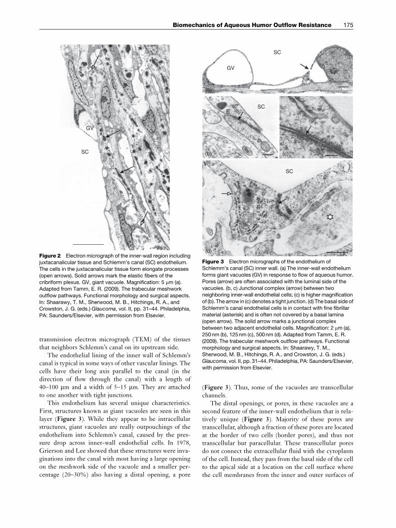
This endothelium has several unique characteristics.First, structures known as giant vacuoles are seen in thislayer (Figure 3). While they appear to be intracellularstructures, giant vacuoles are really outpouchings of theendothelium into Schlemm’s canal, caused by the pres-sure drop across inner-wall endothelial cells. In 1978,Grierson and Lee showed that these structures were inva-ginations into the canal with most having a large openingon the meshwork side of the vacuole and a smaller per-centage (20–30%) also having a distal opening, a pore
(Figure 3). Thus, some of the vacuoles are transcellularchannels.
The distal openings, or pores, in these vacuoles are asecond feature of the inner-wall endothelium that is rela-tively unique (Figure 3). Majority of these pores aretranscellular, although a fraction of these pores are locatedat the border of two cells (border pores), and thus nottranscellular but paracellular. These transcellular poresdo not connect the extracellular fluid with the cytoplasmof the cell. Instead, they pass from the basal side of the cellto the apical side at a location on the cell surface wherethe cell membranes from the inner and outer surfaces of
GV
SC
Figure 2 Electron micrograph of the inner-wall region includingjuxtacanalicular tissue and Schlemm’s canal (SC) endothelium.
The cells in the juxtacanalicular tissue form elongate processes
(open arrows). Solid arrows mark the elastic fibers of thecribriform plexus. GV, giant vacuole. Magnification: 5 mm (a).
Adapted from Tamm, E. R. (2009). The trabecular meshwork
outflow pathways. Functional morphology and surgical aspects.
In: Shaarawy, T. M., Sherwood, M. B., Hitchings, R. A., andCrowston, J. G. (eds.) Glaucoma, vol. II, pp. 31–44. Philadelphia,
PA: Saunders/Elsevier, with permission from Elsevier.
(a)
(b)
ESC
SC
GV
SC
(d)
(c)
Figure 3 Electron micrographs of the endothelium ofSchlemm’s canal (SC) inner wall. (a) The inner-wall endothelium
forms giant vacuoles (GV) in response to flow of aqueous humor.
Pores (arrow) are often associated with the luminal side of the
vacuoles. (b, c) Junctional complex (arrow) between twoneighboring inner-wall endothelial cells; (c) is higher magnification
of (b). The arrow in (c) denotes a tight junction. (d) The basal side of
Schlemm’s canal endothelial cells is in contact with fine fibrillarmaterial (asterisk) and is often not covered by a basal lamina
(open arrow). The solid arrow marks a junctional complex
between two adjacent endothelial cells. Magnification: 2 mm (a),
250 nm (b), 125nm (c), 500nm (d). Adapted from Tamm, E. R.(2009). The trabecular meshwork outflow pathways. Functional
morphology and surgical aspects. In: Shaarawy, T. M.,
Sherwood, M. B., Hitchings, R. A., and Crowston, J. G. (eds.)
Glaucoma, vol. II, pp. 31–44. Philadelphia, PA: Saunders/Elsevier,with permission from Elsevier.
Biomechanics of Aqueous Humor Outflow Resistance 175

the cell have come together and fused. The pore is thusmembrane-lined on its surface. These pores usually formon giant vacuoles, since it is this region in which the cell isgreatly attenuated and the cytoplasm becomes thin.
Fusion of the inner and outer cell membranes and theformation of transcellular pores are not entirely unique tothese cells. Pores associated with giant vacuoles are alsofound on the arachnoid villi in the drainage pathway forthe cerebrospinal fluid. When cell thickness is reducedbelow a critical measure, vascular endothelium can formtranscellular pores involved in transport processes.
The inner-wall endothelium of Schlemm’s canal issupported by a discontinuous basement membrane. Thismakes Schlemm’s canal a somewhat unique vessel havinga continuous endothelium with tight junctions betweenneighboring cells, supported by a discontinuous base-ment membrane. Blood vessel endothelia have a continu-ous endothelium with a continuous basement membrane,while lymphatics have a discontinuous endothelium witha discontinuous basement membrane.
The region immediately underlying the inner wall andbasement membrane and extending to the last trabecularbeam is the JCT. It has many large, apparently emptyspaces and is typically 2–15 mm thick.
The Generation of Flow Resistance in the Inner-Wall Region of Schlemm’s Canal
While there is agreement among most investigators thatthe bulk of aqueous humor outflow resistance is generatedin the immediate vicinity of the inner-wall endotheliumof Schlemm’s canal, the precise location and the mecha-nism by which this dissipation occurs is still a topic ofactive debate and research.
The JCT
With its tortuous submicron-sized flow pathways, theJCT is a natural location to investigate as to its role ingenerating outflow resistance. Surprisingly, morphomet-ric analyses combined with theoretical calculations haveindicated that, unless these apparently open spaces areactually filled with an extracellular matrix gel, they wouldgenerate an insignificant fraction of the total outflowresistance.
This conclusion follows from porous media theory anapproach that has been used to characterize flow resis-tance of other connective tissues. Several different para-meters can be used to characterize the fluid transportcapacity of a tissue. The flow resistance (R¼DP/Q ) ofa tissue is the ratio between the pressure drop across thattissue (DP) and the flow rate generated by that pressuredrop (Q ); the inverse of this quantity is known as the totalhydraulic conductance of this tissue. The conductance perunit surface area is known as the hydraulic conductivity(Lp), while the conductance of the tissue normalized for
surface area, tissue length in the flow wise direction (L),and fluid viscosity (m) is known as the specific hydraulicconductivity (K).
Darcy’s law relates the flow resistance (R) of a tissue tothe specific hydraulic conductivity (K) of that tissue,
R ¼ DPQ
¼ mLKA
½1�
and the specific hydraulic conductivity is related to thehydraulic conductivity as:
K ¼ Lp mL ½2�Typical values of K and Lp for a variety of tissues are foundin Tables 1 and 2.
The K value that characterizes the flow resistance of atissue can be measured experimentally by determining theother parameters in eqn [1], all of which are easy to deter-mine or estimate for aqueous humor outflow with theexception of the length (L) over which the pressure dropoccurs. However, since the bulk of the pressure drop occurssomewhere in or near the inner wall of Schlemm’s canal, itcan be concluded that this length is less than roughly 10mm,or so, an estimate supported by experimental studies. Using
Table 1 Specific hydraulic conductivity (k) of connective
tissues
Tissue K � 1014 (cm2)
Lens capsulea 0.1
Descemet’s membraneb 0.1–0.2
Bruch’s membranec 0.5–1.5Glomerulus basement membraned 2
Aortic walle 0.5–2.5
Corneal stromae 0.5–2.5
Sclerae 1.4Cartilagef 1–10
Synoviumg 1.5–7
Vitreous humorh 1500–1800
aFels, I. G. (1970). Permeability of the anterior bovine lens capsule.Experimental Eye Research 10: 8–14.bFatt, I. (1969). Permeability of Descemet’s membrane to water.
Experimental Eye Research 8: 34–354.cStarita, C., Hussain, A. A., et al. (1997). Localization of the majorsite of resistance to fluid transport in Bruch’s membrane. Investi-
gative Ophthalmology and Visual Science 38: 762–767.dRobertson, G. B. and Walton, H. A. (1989). Glomerular base-ment membrane as a compressible ultrafilter. Microvascular
Research 38: 36–48.eLevick, J. R. (1987). Flow through interstitium and other
fibrous matrices. Quarterly Journal of Experimental Physiology72: 409–437.fMow, V. C., Holmes, M. H., et al. (1984). Fluid transport and
mechanical properties of articular cartilage: a review. Journal of
Biomechanics 17: 377.gLevick, J. R., Price, F. M., et al. (1996). Synovial matrix–synovial
fluid system of joints. In: Comper, W. D. (ed.) Extracellular Matrix,
vol I, pp. 328–377. Amsterdam: Harwood Academic.hFatt, I. (1977). Hydraulic flow conductivity of the vitreous gel.Investigative Ophthalmology and Visual Science 16: 565–568.
176 Biomechanics of Aqueous Humor Outflow Resistance

a flow rate through the aqueous outflow pathway of 2 mlmin–1 passing through a cross-sectional area of between0.054 and 0.13 cm2 (canalwidth of 150–350 mm; canal lengtharound the eye of 3.6 cm), and a pressure drop of 5mmHg,it can then determined that K of the resistance-causingregion in the aqueous outflow pathway must be less than65 � 10–14 cm2. Unless the length over with the pressuredrop occurs (L) is much lesser than 10 mm, the specifichydraulic conductivity of the connective tissue elements inthe outflow pathway is greater than that of any other con-nective tissue with the exception of the vitreous humor(Table 1).
K can also be estimated from photomicrographsshowing the ultrastructure of a tissue. This can potentiallyallow an evaluation of which structures in the aqueousoutflow pathway are generating the measured outflowresistance. Carmen–Kozeny theory relates the structureof a porous medium to K as:
K ¼ " D2h
80½3�
where Dh is the hydraulic diameter of the open spacesavailable for flow and e is the porosity, or fraction of openspace of the medium (note that at porosities higher thanroughly 0.8, this equation becomes inaccurate). UsingCarmen–Kozeny theory combined with conventionalTEM, it was found (in immersion-fixed eyes) that theporosity of the JCTwas approximately 0.15–0.25, Dh wasapproximately 1–1.5 mm, and, most importantly, K of theJCT was calculated to be approximately 2000–10 00010–14 cm2 based on the photomicrographs. This is, atleast, 30 times greater than K based upon measured prop-erties of the outflow system.
In 1986, Ethier and coworkers concluded that theJCT, as visualized using conventional TEM, could notgenerate a significant fraction of outflow resistance. Otherinvestigators have confirmed this conclusion, includingstudies in which the eyes were fixed by perfusion. It thusfollowed that either this region was filled with an extra-cellular matrix gel that was poorly visualized using
Table 2 Hydraulic conductivity (Lp: cm2* s g�1) for a variety
of physiological membranes
Membrane Type Lp � 1011
Kidney epithelial cells (MDCK cells) a 0.075
Xenopus oocytes15 a 0.2
Xenopus oocytes + CHIP2815 a 1.6
Proximal tubule epithelial cells1,2 a 1.2Red blood cells12,14 a 1–1.6
Gall bladder epithelial cells13 a 4–9
Corneal epithelium2,9 b 0.04–0.7
Gall bladder epithelium12,13 b 1.3–3.6Proximal tubule epithelium1,12 b 7.5–5.5
Retinal pigment epithelium3 b 16
Brain capillary4 c 0.03Corneal endothelium2,5 c 0.14–5
Lung capillary4 c 3.4
Skeletal muscle capillary4,7 c 2.5–7
Cardiac muscle capillary4,7 c 8.6Aorta6 c 9
Mesentery, omentum4 c 50
Intestinal mucosa4,7 d 32–130
Synovium (knee)7 d 120Renal peritubulal capillaries7 d 225–700
Renal glomerulus4,7 d 400–3100
Descemet’s membrane9 e 15–37Lens capsule10 e 17–50
Bruch’s membrane11 e 2000–12,500
Kidney tubule basement membrane8 e 6300–13,700
a: cell membranes, b: unfenestrated epithelium, c: unfenestratedendothelium, d: fenestrated epithelia, and e: basement mem-
branes.1Timbs, M. M. and Spring, K. R. (1996). Hydraulic properties of
MDCK cell epithelium. Journal of Membrane Biology 153: 1–11.2Klyce, S. D. and Russell, S. R. (1979). Numerical solution of
coupled transport equations applied to corneal hydration dynam-
ics. Journal of Physiology 292: 107–134.3Tsuboi, S. (1987). Measurement of the volume flow and hydrau-lic conductivity across the isolated dog retinal pigment epitheli-
um. Investigative Ophthalmology and Visual Science 28:
1776–1782.4Renkin, E. M. (1977). Multiple pathways of capillary permeabili-
ty. Circulation Research 41: 735–743.5Hedbys, B. O. and Mishima, S. (1962). Flow of water in corneal
stroma. Experimental Eye Researcy 1: 262–275.6Vargas, C. B., Vargas, F. F., et al. (1979). Hydraulic conductivity
of the endothelial and outer layers of rabbit aorta. American
Journal of Physiology 236: H53–H60.7Levick, J. R. (1987). Flow through interstitium and other fibrousmatrices. Quarterly Journal of Experimental Physiology 72:
409–437.8Welling, L. andWelling, D. (1978). Physical properties of isolated
perfused basement membranes from rabbit loop of Henle. Amer-ican Journal of Physiology 234: F54–F58.9Fatt, I. (1969). Permeability of Descemet’s membrane to water.
Experimental Eye Research 8: 34–354.10Fisher, R. F. (1982). The water permeability of basement mem-
brane under increasing pressure: evidence for a new theory of
permeability. Proceedings of the Royal Society of London Series
B 216: 475–496.11Starita, C., Hussain, A., et al. (1996). Hydrodynamics of ageing
Bruch’s membrane: implications for macular disease. Experi-
mental Eye Research 62: 565–572; Bentzel, C. and Reczek, P.
(1978). Permeability changes in Necturus proximal tubule during
volume expansion. American Journal of Physiology 234:F225–F234.12Gonzalez, E., Carpi-Medina, P., et al. (1982). Cell osmotic
water permeability of isolated rabbit proximal straight tubules.
American Journal of Ophthalmology 242: F321–F330.13Persson, B. E. and Spring, K. R. (1982). Gallbladder epithelial
cell hydraulic water permeability and volume regulation. Journal
of General Physiology 79: 481–505.14Solomon, A. K., Chasan, B., et al. (1983). The aqueous pore inthe red cell membrane: band 3 as a channel for anions, cations,
nonelectrolytes, and water. Annals of the New York Academy of
Sciences 414: 97–124.15Preston, G. M., Carroll, T. P., et al. (1992). Appearance of waterchannels in Xenopus oocytes expressing red cell CHIP28 protein.
Science 256: 385–387.
Biomechanics of Aqueous Humor Outflow Resistance 177

conventional TEM techniques, or, that this region was notthe primary site of outflow resistance. The age-relatedaccumulation of plaque-like material in this region that isreported to be enhanced in glaucomatous eyes would haveno influence on this conclusion.
More recently, in 2002, Gong and coworkers usedquick-freeze/deep-etch (QFDE) methodology to exam-ine the apparent open spaces seen in the JCT region ingreater detail. QFDE is a morphological technique thatpreserves the cellular and extracellular ultrastructure inexquisite detail and allows visualization of structurespoorly preserved or not seen at all using conventionalTEM tissue preparation techniques. A more elaborateand extensive extracellular matrix was seen in the JCTusing QFDE as compared to conventional methods ofpreparation for TEM (Figure 4); however, micron-sizedopen spaces were still seen in this region, casting doubt onwhether a significant fraction of outflow resistance couldbe generated in this region. An important caveat pointedout by the Gong group regarding their studies was that itwas not clear whether or not QFDE can visualize theglycosaminoglycans (GAGs) in their uncollapsed state,
and this leaves uncertain the question of generation ofappreciable flow resistance in the JCT region.
The role that GAGs and other extracellular matrixelements found in the JCT might play in generatingoutflow resistance is unclear. While it has been shownthat enzymes that degrade macromolecules (GAGases)increase outflow facility in a number of species (e.g.,cow, guinea pig, dog, and rabbit), the evidence regardingprimates is conflicting, and there has been no confirmeddata yet showing that GAGases decrease outflow resis-tance in human eyes. Matrix metalloproteinases (MMPs)have been shown to reversibly increase outflow facility inperfused human anterior segment organ culture, and thisis a strong indicator that the extracellular matrix maygenerate significant aqueous outflow resistance.
Perhaps the strongest experimental evidence implicat-ing the JCT as a major site of outflow resistance is the1992 study by Maepea and Bill in which they used micro-pipettes and localized the pressure drop to occurringsomewhere between 7 and 14 mm from the inner wall ofSchlemm’s canal (i.e., in the JCT). While this result isfrequently cited in the literature, it is not widely appre-ciated that this measurement was made with micropip-ettes whose tip sizes were as large as the measurementzone. When this reservation is combined with the fact thatthe inner wall of Schlemm’s canal distends several tomany microns during the process of penetration by themicropressure probe (unpublished work by Dr. MilkoIliev in collaboration with Dr. Johnson’s laboratory), itleads to the conclusion that the bulk of the aqueoushumor pressure drop occurs near the inner wall ofSchlemm’s canal (within 5–10 mm), but that no furtherquantitative conclusions are possible.
The basement membrane
The basement membrane of the inner wall of Schlemm’scanal is another possible location for generation of out-flow resistance. Table 1 shows that the specific hydraulicconductivities of basement membranes (top four tissues intable) are among the lowest of connective tissues.
However, basement membranes tend to be quite thinand this limits the flow resistance they can generate (seeeqn [1]). Table 2 shows the hydraulic conductivity of avariety of physiological membranes, including cells mem-branes, epithelia, and basement membranes. The largevariation in Lp of basement membranes is due not onlyto variation in the specific hydraulic conductivity of thesetissues, but also to significant differences in their thick-nesses (ranging from 0.15 mm for Bruch’s membrane to7 mm for Descemet’s membrane).
It is interesting to compare the values seen in this tablewith the estimated value of the hydraulic conductivity ofthe outflow pathway. From the definition of hydraulicconductivity (or by combining eqns [1] and [2]), Lp=Q /A/DP. Using the values characterizing the aqueous
(a)
(b)
V VSC
SC
V
V
V
Figure 4 Enucleated human eye fixed by perfusion at 15mmHg:
(5A) vacuoles (V) in the inner wall of Schlemm’s canal (SC) in tissueprepared for TEM using conventional methods; notice the large
open space in the region of the JCT immediately under these
vacuoles; (4B) the same region as seen in tissue prepared using
QFDE; notice that while open spaces still exist under the vacuoles,a more complex and extensive extracellular matrix is seen.
Magnification�4860. Adapted from Gong, H., Ruberti, J., Overby,
D., Johnson,M., and Freddo, T. F. (2002). A new view of the human
trabecular meshwork using quick-freeze, deep-etch electronmicroscopy. Experimental Eye Research 75: 347–358, with
permission from Elsevier.
178 Biomechanics of Aqueous Humor Outflow Resistance

outflow pathway presented above, we can estimate that Lpfor the aqueous humor outflow pathway is between 4000� 10–11 and 9000 � 10–11 cm2 s g–1. Note that this is not atheoretical calculation but an estimate based on measuredquantities.
Table 2 shows that several basement membranes, inparticular those of the renal system, have hydraulic con-ductivities similar to that of the aqueous outflow pathway.This suggests that the basement membrane of the inner-wall endothelium of Schlemm’s canal might be an impor-tant contributor to aqueous humor outflow resistance.
However, as noted above, this basement membrane isdiscontinuous (Figure 3), as recently confirmed in 2002by Gong and coworkers. If there are breaks in the base-ment membrane, it is difficult to see how a significant flowresistance could be generated by this tissue.
The inner-wall endothelium of Schlemm’s canal
The inner-wall endothelium of Schlemm’s canal has beenan attractive candidate for the generation of outflow resis-tance since the time that light microscopes and laterelectron microscopes were focused on the inner-wallendothelium. Comparison of the hydraulic conductivityof this tissue (4000–9000 � 10–11 cm2 s g–1) with that ofother endothelia and epithelia as seen in Table 2, leadsone to conclude that this vessel lining must have one ofthe highest hydraulic conductivities in the body. Com-pared to other tissues in Table 2, it is clear that onlyfenestrated endothelia (and some basement membranes)have such high hydraulic conductivities.
While the inner-wall endothelium is not fenestrated,this endothelium is unique in that it contains micron-sized pores. It is interesting that even early investigators(e.g., Seidel) had concluded that such pores existed, longbefore they were first seen by electron microscopy. Thisconclusion was based on early filtration studies that exam-ined the sizes of microparticulates that would passthrough the outflow pathway. It was found in these studiesthat a filtration barrier existed for particles larger thanroughly 0.5 mm or so, and thus it was concluded that poresnearly 1 mm in size must exist. More recent studies haveconfirmed this conclusion using microparticles and latexmicrospheres.
While there is some debate concerning the existence ofthese pores (which is discussed below), no group hasoffered an alternate explanation for the extraordinarilyhigh hydraulic conductivity of the aqueous outflow path-way. Nor has any alternate explanation been offered forthe relatively easy passage of microparticles 200–500 nmin diameter through the outflow pathway, except throughthese large pores. Since there are tight junctions(Figure 3) between the inner-wall cells (presumably toprevent blood reflux into the eye that can occur duringperiods of transient increases in ocular venous pressure),there are no other structures apparent in this endothelium
that could explain the high hydraulic conductivity of thistissue or its filtration characteristics.
The flow resistance generated by these pores wasfirst considered by Bill and Svedbergh in 1972. In an ex-haustive study using scanning electron microscopy, theycharacterized the size distribution of these pores, and thenused hydrodynamic theory to calculate for hydraulic con-ductivity of these pores. Using Sampson’s law, that gives thehydraulic conductivity for a single pore of diameter d,
Lp ¼ d
6pm½4�
they found that the inner-wall endothelium could present,at most, 10% of the outflow resistance. That is, the hydrau-lic conductivity of the pores in the inner-wall endotheliumis, at least, 10-fold higher than the measured hydraulicconductivity of the outflow pathway. This conclusion hasbeen confirmed in a number of studies.
These results would appear to rule out the inner-wallendothelium as a major site of outflow resistance. How-ever, a number of experimental findings are at variancewith this conclusion. In particular, when chelating agents(ethylenediaminetetraacetic acid (EDTA) and ethyleneglycol tetraacetic acid (EGTA)) or a proteolytic enzyme(a-chymotrypsin) were perfused through the outflowpathway of live primates, it was found that ruptures ofthe inner-wall endothelium were produced by theseagents that decreased outflow resistance more thancould be explained by the calculated flow resistanceof the inner-wall pores. A hydrodynamic interaction (thefunneling effect) between the inner-wall pores and theJCT, which lies immediately below these pores, has beenproposed as an explanation of the findings (see Figure 5).
In this scenario, the pores and vacuoles themselvescontribute negligible flow resistance, but since theyforce aqueous humor to funnel through those regions ofthe JCT nearest the pores and vacuoles, the vacuole size
Trabecular meshwork
Inner wallSchlemm’s canal
JCT
Figure 5 Schematic of the funneling of aqueous humor
through the JCT, toward a vacuole and pore that allows this fluidto pass through the inner-wall endothelium. Adapted from
Overby, D., Gong, H., Qiu, G., Freddo, T. F., and Johnson,
M. (2002). The mechanism of increasing outflow facility during
washout in the bovine eye. Investigative Ophthalmology andVisual Science 43: 3455–3464. ãThe Association for Research in
Vision and Ophthalmology.
Biomechanics of Aqueous Humor Outflow Resistance 179

and pore density can have a significant effect on theeffective hydraulic conductivity (Lp) of the JCT:
Lp ¼ 2KnD
m½5�
Here, K is the specific hydraulic conductivity of the JCTregion, n is the number of pores per unit area in the innerwall, D is the diameter of the vacuoles in the inner wall,and m is the viscosity of the aqueous humor.
The funneling model suggests that while the bulk ofoutflow resistance is actually generated in the JCT orbasement membrane, its magnitude is modulated by thepores and vacuoles of the inner-wall endothelium ofSchlemm’s canal. This model explains many of the char-acteristics of the outflow pathways discussed above. How-ever, two recent studies failed to find a correlationbetween outflow facility and inner-wall pore density aswould be expected if eqn [5] describes the hydraulicconductivity of the outflow pathway. Furthermore, thesestudies found that at least some inner-wall pores may beartifacts of the fixation process.
Pores as possible fixation artifacts and the
possible importance of flow through inner-wall cell
junctions or through water channelsThe micron-sized pores that pass through the endothelialcells of the inner-wall endothelium of Schlemm’s canalare relatively unique to these cells and to the cells of thearachnoid villi, a tissue of the cerebrospinal fluid pathway.This unique character has led some investigators to doubtthat these are physiologic structures, but instead considerthem to be artifacts of the tissue-preparation process. Thepossibility that pores form during tissue preparation forelectron microscopy was supported in recent studies thatshowed that during tissue fixation under flow conditions,the inner-wall endothelium pore density increased as afunction of the volume of fixative perfused through theoutflow pathway (see Figure 6).
It has been postulated by several investigators thatinstead of passing through the pores in the inner-wallendothelium, a significant fraction of the aqueous humorpasses through gaps between the tight-junction strands ofthese cells. In 2001, Ethier and Chan found that cationizedferritin perfused into enucleated human eyes acted todecreased outflow facility and was seen accumulating injunctions between inner-wall endothelial cells. WhileEthier and Chan argue that the decreased outflow facilitycaused by cationized ferritin was due to pore blocking thatwas also seen to occur in this study, others have inter-preted their results as consistent with a significant flowthrough the junctional complexes.
There are a several reasons why this hypothesis isuntenable. Two strong arguments against this possibility
were suggested above, namely that the uniquely highhydraulic conductivity of the aqueous outflow pathwayis inconsistent with flow through junctions, and that therelatively free passage of microspheres 200–500 nm indiameter through the outflow pathway would be pre-cluded if transport was primarily through cell junctions.It is well known in the vascular system that macromole-cules larger than roughly 10–20 nm are largely excludedfrom passing through cell junctions, for either fenestratedor nonfenestrated vessels.
In 1981, Raviola and Raviola examined the tight junc-tions of the inner-wall endothelium and calculated theflow that would be expected through these junctions.The gaps they found available for transport around thetight-junctional strands were nanometers in size, and theyconcluded that any flow that might occur through thesespaceswould be negligible. In 1997, Ye and coworkers usedfreeze-fracture techniques to examine these junctions ineyes fixed under flow conditions and did not report signif-icant differences from the dimensions reported by Raviolaand Raviola. The notion that microparticles that areroughly 0.5 mm in size could relatively easily pass throughsuch openings seems improbable, at best.
The study of Ye and coworkers did find that the tightjunctions of the inner-wall cells simplified with increasingIOP. They speculated that the junctional simplificationthat occurred with increasing perfusion pressure mightlead to formation of pores at locations of focal separationin the tight junctions. While they did not find a statisticallysignificant relationship between junctional complexity andoutflow facility in this small series of eyes, they did find atrend in that direction, consistent with their hypothesis.
0
500
1000
1500
0
Por
e de
nsity
(m
m–2
)
Volume of fixed perfused (ml)
2000
2500
3000
3500
0.1 0.2 0.3 0.4 0.5 0.6
Figure 6 Pore density as a function of volume of fixative
perfused through the outflow pathway of normal eyes (filledsymbols) and eyes with POAG (open). Lines are best fit with
outliers excluded. Adapted from Johnson, M., Chan, D., Read,
A. T., et al. (2002). Glaucomatous eyes have a reduced pore
density in the inner wall endothelium of Schlemm’s canal.Investigative Ophthalmology and Visual Science 43: 2950–2955.
ãThe Association for Research in Vision and Ophthalmology.
180 Biomechanics of Aqueous Humor Outflow Resistance

Thus, while there are a number of strong argumentsagainst the possibility of transport of a significant quantityof aqueous humor across the inner wall of Schlemm’scanal passing directly through cell junctions, simpli-fication of these junctions might be involved in the pro-cess of pores formation. In particular, paracellular flowthrough border pores could be consistent with the 1991findings of Epstein and Rohen of dilations of this para-cellular space.
It has also been suggested that a fraction of outflowmight pass through water channels (aquaporins) in the cellmembrane of the inner-wall cells. Red blood cells andrenal proximal tubules are cell types expressing high levelsof aquaporin 1. The hydraulic conductivities of the waterchannels in these cell membranes have been measured, ashave those of cells in which aquaporins are overexpressed(seeTable 2). All of these values are more than 1000 timessmaller than the hydraulic conductivity of aqueous out-flow pathway. It goes without saying that water channelsalso cannot explain the relatively free passage of 0.5-mm-sized particles through the outflow pathway.
The only pathway that appears consistent with theavailable physiologic evidence is through the inner-wallpores whose existence was already postulated more than80 years ago by Seidel in 1921, and that now are easilyseen using scanning electron microscopy of this tissue.Importantly, the size of these pores is consistent with thepredictions of Seidel. Whether this transport is primarilythrough border pores or is also through the transcellularpores remains to be determined.
The question remains as to why the number of thesepores increases during the fixation process, as shown inFigure 6. A further finding of the studies was that thenumber density of pores in the inner-wall endotheliumdecreases as a function of postmortem time, somethingnot expected of an artifact. Instead, it may be that fixationunder flow conditions generates stresses in the inner walldue both to pressure-induced stretching of the inner wallof Schlemm’s canal and also to shrinkage of the tissuefollowing fixation. Pressure-induced stress is likely whatcauses the formation of these pores under physiologicconditions. Then, fixative-induced pore formation mightbe an artifact associated with a physiological process,namely stress on the inner-wall endothelium.
In this scenario, the y-intercepts of the lines seen onFigure 6would then represent the true physiological poredensity of normal and glaucomatous eyes, respectively.Importantly, Figure 6 shows this density to be greatlyreduced in glaucomatous eyes. Through their importanceto funneling (eqn [5]), a decrease of inner-wall poreswould be expected to decrease the hydraulic conductivityof the outflow pathway and thereby increase IOP.
This decrease pore density in the inner-wall endo-thelium of Schlemm’s canal may then represent the
cause of the elevated pressure characteristic of glaucoma.Future work may then indicate why this pore density isdecreased.
See also: Biological Properties of the Trabecular Mesh-
work Cells; The Biology of Schlemm’s Canal; The
Cytoskeletal Network of the Trabecular Meshwork; The
Fibrillar Extracellular Matrix of the Trabecular Meshwork;
Functional Morphology of the Trabecular Meshwork;
Primary Open-Angle Glaucoma; Role of Proteoglycans
in the Trabecular Meshwork; Structural Changes in the
Trabecular Meshwork with Primary Open Angle Glauco-
ma; Uveoscleral Outflow.
Further Reading
Bill, A. (1975). Blood circulation and fluid dynamics in the eye.Physiological Reviews 55: 383–416.
Bill, A. and Maepea, O. (1994). Mechanisms and routes of aqueoushumor drainage. In: Albert, D. M. and Jakobiac, F. A. (eds.)Principles and Practice of Ophthalmology. Vol I: Basic Sciences,ch. 12, pp. 2577–2595. Philadelphia, PA: Saunders.
Bill, A. and Svedbergh, B. (1972). Scanning electron microscopicstudies of the trabecular meshwork and the canal of Schlemm – anattempt to localize the main resistance to outflow of aqueous humorin man. Acta Ophthamologica 50: 295–320.
Epstein, D. L. and Rohen, J. W. (1991). Morphology of the trabecularmeshwork and inner-wall endothelium after cationized ferritinperfusion in the monkey eye. Investigative Ophthalmology and VisualScience 32(1): 160–171.
Ethier, C. R. (2002). The inner wall of Schlemm’s canal (review).Experimental Eye Research 74: 161–172.
Ethier, C. R., Coloma, F. M., Sit, A. J., and Johnson, M. (1998).Two pore types in the inner wall endothelium of Schlemm’scanal. Investigative Ophthalmology and Visual Science 39:2041–2048.
Ethier, C. R., Kamm, R. D., Palaszewski, B. A., Johnson, M. C., andRichardson, T. M. (1986). Calculations of flow resistance in thejuxtacanalicular meshwork. Investigative Ophthalmology and VisualScience 27(12): 1741–1750.
Gong, H., Ruberti, J., Overby, D., Johnson, M., and Freddo, T. F.(2002). A new view of the human trabecular meshwork using quick-freeze, deep-etch electron microscopy. Experimental Eye Research75: 347–358.
Gong, H., Tripathi, R. C., and Tripathi, B. J. (1996). Morphology of theaqueous outflow pathway. Microscopy Research and Technique33: 336–367.
Grant, W. M. (1963). Experimental aqueous perfusion in enucleatedhuman eyes. Archives of Ophthalmology 69: 783–801.
Johnson, M. (2006). What controls aqueous humour outflowresistance? Experimental Eye Research 82: 545–557.
Johnson, M. and Erickson, K. (2000). Mechanisms and routes ofaqueous humor drainage. In: Albert, D. M. and Jakobiec, F. A. (eds.)Principles and Practice of Ophthalmology. Vol. 4: Glaucoma,ch. 193B, pp. 2577–2595. Philadelphia, PA: WB Saunders.
Johnstone, M. A. and Grant, W. M. (1973). Pressure dependentchanges in the structures of the aqueous outflow system ofhuman and monkey eyes. American Journal of Ophthalmology75: 365.
Leber, T. (1873). Studien uber den Flussigkeitswechsel im Auge.(Albrecht von Graefes) Archiv fur Ophthalmologie 19: 87–106.
Maepea, O. and Bill, A. (1992). Pressures in the juxtacanalicular tissueand Schlemm’s canal in monkeys. Experimental Eye Research 54(6):879–883.
Biomechanics of Aqueous Humor Outflow Resistance 181

Raviola, G. and Raviola, E. (1981). Paracellular route of aqueous outflowin the trabecular meshwork and canal of Schlemm. InvestigativeOphthalmology and Visual Science 21: 52–72.
Seidel, E. (1921). Weitere experimentelle Untersuchungen uber dieQuelle und den Verlauf der intraokularen Saftstromung. IX Mitteilung.Uber den Abfluss des Kammerwassers aus der vorderenAugenKammer. Graefe’s Archive for Clinical and ExperimentalOphthalmology 104: 357–402.
Tamm, E. R. (2009). The trabecular meshwork outflow pathways.Functional morphology and surgical aspects. In: Shaarawy, T. M.,
Sherwood, M. B., Hitchings, R. A., and Crowston, J. G. (eds.)Glaucoma, vol. II, pp. 31–44. Philadelphia, PA: Saunders/Elsevier.
Tripathi, R. C. (1974). Comparative physiology and anatomy of theaqueous outflow pathway. In: Davson, H. and Graham, L. T. (eds.)The Eye. Comparative Physiology, vol. 5, ch. 3, pp. 163–356.London: Academic Press.
Ye, W., Gong, H., Sit, A., Johnson, M., and Freddo, T. F. (1997).Interendothelial junctions in normal human Schlemm’s canalrespond to changes in pressure. Investigative Ophthalmology andVisual Science 38(12): 2460–2468.
182 Biomechanics of Aqueous Humor Outflow Resistance