altered mechanobiology of schlemm s canal … mechanobiology of schlemm’s canal endothelial cells...
TRANSCRIPT

Altered mechanobiology of Schlemm’s canalendothelial cells in glaucomaDarryl R. Overbya,1, Enhua H. Zhoub,1,2, Rocio Vargas-Pintoc,1, Ryan M. Pedrigia, Rudolf Fuchshoferd,Sietse T. Braakmana, Ritika Guptaa, Kristin M. Perkumase, Joseph M. Sherwooda, Amir Vahabikashic, Quynh Dangb,Jae Hun Kimb, C. Ross Ethiera,f,g, W. Daniel Stamere,h, Jeffrey J. Fredbergb, and Mark Johnsonc,i,3
aDepartment of Bioengineering, Imperial College London, London SW7 2AZ, United Kingdom; bProgram in Molecular and Integrative Physiological Sciences,Department of Environmental Health, Harvard School of Public Health, Boston, MA 02115; cDepartment of Biomedical Engineering, Northwestern University,Evanston, IL 60208; dInstitute of Human Anatomy and Embryology, Universität Regensburg, 93053 Regensburg, Germany; eDepartment of Ophthalmology,Duke University, Durham, NC 27710; fWallace H. Coulter Department of Biomedical Engineering, Georgia Institute of Technology, Atlanta, GA 30332;gDepartments of Biomedical Engineering and Ophthalmology, Emory University, Atlanta, GA 30322; hDepartment of Biomedical Engineering, DukeUniversity, Durham, NC 27710; and iDepartments of Mechanical Engineering and Ophthalmology, Northwestern University, Evanston, IL 60208
Edited by David A. Weitz, Harvard University, Cambridge, MA, and approved August 18, 2014 (received for review June 9, 2014)
Increased flow resistance is responsible for the elevated intraoc-ular pressure characteristic of glaucoma, but the cause of thisresistance increase is not known. We tested the hypothesis thataltered biomechanical behavior of Schlemm’s canal (SC) cells con-tributes to this dysfunction. We used atomic force microscopy,optical magnetic twisting cytometry, and a unique cell perfusionapparatus to examine cultured endothelial cells isolated fromthe inner wall of SC of healthy and glaucomatous human eyes.Here we establish the existence of a reduced tendency for poreformation in the glaucomatous SC cell—likely accounting for in-creased outflow resistance—that positively correlates with ele-vated subcortical cell stiffness, along with an enhanced sensitivityto the mechanical microenvironment including altered expressionof several key genes, particularly connective tissue growth factor.Rather than being seen as a simple mechanical barrier to filtration,the endothelium of SC is seen instead as a dynamic material whoseresponse to mechanical strain leads to pore formation and therebymodulates the resistance to aqueous humor outflow. In the glau-comatous eye, this process becomes impaired. Together, theseobservations support the idea of SC cell stiffness—and its bio-mechanical effects on pore formation—as a therapeutic targetin glaucoma.
cell mechanics | primary open-angle glaucoma | modulus | cytoskeleton
Aqueous humor flows across the inner wall endothelium ofSchlemm’s canal (SC) and generates a transendothelial
pressure gradient from the cellular base to the cellular apex.From a biomechanical perspective, the direction of this gradientis remarkable considering that the endothelium of the systemicvasculature experiences a pressure gradient in precisely the op-posite direction. In the healthy eye, this basal-to-apical trans-cellular pressure gradient is of sufficient magnitude to partiallyseparate the SC cell from its supporting basement membrane,inflate dome-shaped structures known as giant vacuoles, andgenerate cellular mechanical strains exceeding 50% (Fig. 1) (1).The formation of giant vacuoles leads to substantial thinningof the SC endothelial cell and is thought to be associated withformation of pores that provide an outflow pathway across theSC endothelium (2). Although reported dysfunction of the poreformation process might be expected to affect outflow resistanceand elevate intraocular pressure (IOP) (3, 4), mechanisms forsuch dysfunction have never before been established, in largepart because SC cells from healthy eyes are so difficult to isolatetechnically, but also because isolated SC cells from the glau-comatous eye are a resource that has been exceedingly scarce.Here for the first time to our knowledge we show that the pro-cess of pore formation differs substantially between cells fromthe healthy versus the glaucomatous human eye and show, fur-ther, that this difference depends upon cytoskeletal stiffness thatis altered in the glaucomatous SC cell, likely owing to altered
substrate sensitivity and gene expression in these cells. Specifi-cally, stiffer glaucomatous cells impede pore formation and therebyelevate IOP.
ResultsPore Formation in SC Cells Is Altered in Glaucomatous Cell Strains. Toexamine pore formation in SC cells, we used an in vitro mono-layer perfusion system to mimic the biomechanical and filtrationenvironment of SC endothelium in vivo (5). As described pre-viously, SC cells were isolated from normal and glaucomatoushuman donors and extensively characterized (Materials andMethods) (6, 7). When perfused in a basal-to-apical directionpores formed in SC cell monolayers, with pores passing trans-cellularly through individual SC cells or paracellularly betweenneighboring SC cells, consistent with the two pore types observedalong the SC endothelium in situ (Fig. 2A) (8). The density ofpores (pores per cell area) increased significantly with perfusionpressure (P < 3 × 10⁻⁵; Fig. 2B), and porosity (pore area per cellarea) showed a similar dependence upon pressure (P < 0.003;
Significance
Glaucoma is a leading cause of blindness. The elevated in-traocular pressure characteristic of many cases of glaucoma isattributable to increased resistance to aqueous humor outflow.However, the cause of this increased flow resistance haseluded investigators for over 140 y. Here we demonstrate thatcells from the canal of Schlemm of glaucomatous eyes havealtered gene expression and increased cytoskeletal stiffnessthat leads to reduced pore formation in these cells, likelyaccounting for increased outflow resistance associated withglaucoma. These findings thus establish that dysfunctionalcytoskeletal mechanics may lie at the heart of this diseaseprocess and thereby motivate development of glaucomatherapeutics that target cell stiffness.
Author contributions: D.R.O., E.H.Z., R.V.-P., R.M.P., R.F., S.T.B., A.V., C.R.E., W.D.S., J.J.F.,and M.J. designed research; E.H.Z., R.V.-P., R.M.P., R.F., S.T.B., R.G., K.M.P., A.V., Q.D., andJ.H.K. performed research; D.R.O., E.H.Z., R.V.-P., K.M.P., J.M.S., J.H.K., W.D.S., J.J.F., andM.J. contributed new reagents/analytic tools; D.R.O., E.H.Z., R.V.-P., R.M.P., R.F., S.T.B.,R.G., K.M.P., J.M.S., A.V., J.H.K., C.R.E., W.D.S., J.J.F., and M.J. analyzed data; and D.R.O.,E.H.Z., R.V.-P., R.M.P., R.F., S.T.B., J.M.S., A.V., C.R.E., W.D.S., J.J.F., and M.J. wrotethe paper.
The authors declare no conflict of interest.
This article is a PNAS Direct Submission.
Freely available online through the PNAS open access option.1D.R.O., E.H.Z., and R.V.-P. contributed equally to this work.2Present address: Ophthalmology, Novartis Institutes of BioMedical Research, Cambridge,MA 02139.
3To whom correspondence should be addressed. Email: [email protected].
This article contains supporting information online at www.pnas.org/lookup/suppl/doi:10.1073/pnas.1410602111/-/DCSupplemental.
13876–13881 | PNAS | September 23, 2014 | vol. 111 | no. 38 www.pnas.org/cgi/doi/10.1073/pnas.1410602111

online supplement). The increase in pore density with perfu-sion pressure was observed for both transcellular and para-cellular pores (P < 0.005; SI Text). Apical-to-basal perfusionof these monolayers showed no such dependence of pore density(Fig. 2B) or porosity (SI Text) on perfusion pressure, consistentwith previous studies showing rectified flow across this endo-thelium and its role as part of the blood–aqueous barrier (9).Compared with the pore density measured in normal SC cell
strains perfused in a basal-to-apical direction at 6 mmHg, poredensity in glaucomatous SC cell strains was markedly reduced;pore density in glaucomatous cells was threefold smaller and thedifference was highly statistically significant (P < 2 × 10⁻⁴;Fig. 2C). Pore density seen in glaucomatous SC cell strainsperfused at 6 mmHg was comparable to unperfused normalcontrols (Fig. 2 B and C). Porosity was similarly reduced inglaucomatous SC cells compared with SC cells from normaleyes (P < 0.04; SI Text) and was attributable to a reduction intranscellular and paracellular pores, although neither aloneachieved statistical significance.
Glaucomatous SC Cells Demonstrate Elevated Subcortical Stiffness.We reasoned that it would be more difficult for a pore to form instiffer SC cells. To investigate this possibility, we measured thestiffness of SC cells isolated from normal and glaucomatoushuman donors (Materials and Methods) using atomic force mi-croscopy (AFM) using both sharp tips (20-nm tip radius) androunded tips (4.5 μm and 10 μm) (10). In other cellular systems,AFM measurements using a sharp tip characterize the cell cor-tex, whereas larger, spherical tips probe the subcortical cyto-skeleton (10). Here we use the term “cortex” to refer to theactin-dense region of the cell lying immediately beneath theplasma membrane, and the term “subcortical cytoskeleton” torefer to the internal cytoskeleton underlying the cortex (11). Forall tip geometries, elastic moduli were found to be similar be-tween nuclear and peripheral regions of the cell, and there wasno systematic variation between Young’s modulus and donor age(SI Text). Cell stiffness measured with sharp tips was 10-foldhigher than that measured with the larger, spherical tips (Fig.3B), consistent with the prominent actin-rich cell cortex foundin SC cells and other endothelia (Fig. 3A).Measured with a sharp AFM tip, we found no difference in
stiffness between normal versus glaucomatous SC cells (P > 0.85;Fig. 3B). Cortex thickness as measured by structured illumina-tion microscopy was similar between normal (400 ± 20 nm, n = 3cell strains) and glaucomatous SC cells (380 ± 60 nm, n = 2).However, when measured with the larger, spherical AFM tips,we found systematic differences in stiffness between glaucoma-tous SC versus normal SC cells (Fig. 3 B and C). With a 4.5-μmtip, the modulus of glaucomatous SC cells was 1.47 ± 0.29 kPa(n = 5 cell strains; m = 128 measurements), whereas that ofnormal SC cells was measured as 1.01 ± 0.12 kPa (n = 6; m =104) (P < 0.12). Using a 10-μm tip, the modulus of glaucomatousSC cells was 1.24 ± 0.11 kPa (n = 5; m = 120), whereas that ofnormal SC cells was 0.79 ± 0.10 kPa (n = 6; m = 153) (P < 0.02).Relative to the normal SC cells, glaucomatous SC cells revealedsubstantially elevated subcortical stiffness. Both cortical andsubcortical SC cell stiffness were greatly reduced by latrunculin-A,consistent with an important role for actin in determiningstiffness (Fig. 3A); however, there was no difference in the rel-ative decrease in cell stiffness following latrunculin between
normal and glaucomatous SC cells (SI Text), suggesting thatperhaps another constituent of the subcortical cytoskeleton [e.g.,intermediate filaments (11)] may be altered in glaucomatousSC cells.For two normal and three glaucomatous SC cell strains in
which both cell stiffness and pore density were measured weexamined the relationship between these parameters. Subcorticalstiffness (10-μm spherical tip) was related inversely to poredensity (P < 0.002; Fig. 3D) and porosity (P < 0.012; SI Text). Arelationship was apparent between subcortical stiffness and poredensity for both pore subtypes; however, subcortical stiffnessshowed a more significant correlation with transcellular poredensity (P < 0.02) compared with paracellular pore density (P <0.07) (SI Text). These data do not establish causality but dostrongly support the idea that increased subcortical cell stiffnessand decreased pore formation go hand in hand.
On Increasingly Stiffer Gels, Both Normal and Glaucomatous SC CellsStiffen. We asked next what might cause this stiffness difference.One possibility is mechanotransduction of the mechanicalproperties of the SC cell microenvironment (12, 13). We thusinvestigated how substrate stiffness might influence SC cell
Fig. 1. Aqueous humor flow pathway. (Left) Sche-matic of anterior segment of eye showing the di-rection of aqueous humor flow in red. (Center)Enlargement of the iris-cornea angle (boxed regionin left panel) to show the conventional outflowpathway. (Right) Transmission electron micrographof endothelial cells forming the inner wall of SC;aqueous humor crosses the endothelium throughpores to enter the lumen of SC. V, giant vacuoles.C is reproduced with permission from ref. 41.
Fig. 2. Pore density in perfused SC monolayers. (A) Representative image oftranscellular and paracellular pores in normal (SC52) and glaucomatous SC(SC62g) cells. (B) Pore density increases in monolayers formed from threenonglaucomatous SC cell strains with transcellular (basal-to-apical) pressuredrop; in one SC cell strain (SC67) perfused in the apical-to-basal direction(AB), pore densities are similar to unperfused controls at 0 mmHg. (C) Poredensity is reduced in glaucomatous compared with normal SC cells followingperfusion at 6 mmHg in the basal-to-apical direction. Bars are SEM.
Overby et al. PNAS | September 23, 2014 | vol. 111 | no. 38 | 13877
CELL
BIOLO
GY

stiffness and gene expression. Because of the need to examinea large number of cells on substrates of a variety of stiffnesses, weused optical magnetic twisting cytometry (OMTC) (Materials andMethods) to study SC cells isolated from normal and glaucom-atous human donors (Materials and Methods).Grown on rigid substrates, we found no difference in stiffness
between normal and glaucomatous SC cells strains (SI Text), and,as expected, these results were consistent with the AFM findingsusing a sharp tip described above (11, 14). We also examinedhow SC cells grown on rigid substrates responded to drugs withknown effects on outflow resistance. Similar to our finding pre-viously reported for normal SC cells (15), we found in glau-comatous SC cells that every agent that we examined thatdecreased outflow resistance also decreased cell stiffness, andevery agent that increased outflow resistance also increased cellstiffness (SI Text).We then examined the influence of substrate stiffness on
the cells. The physiological substrate of the SC cell is the tra-becular meshwork, and its compressive Young’s modulus hasbeen reported to be substantially increased in glaucoma (16).Normal and glaucomatous SC cells were cultured on collagen-coated polyacrylamide gels of tunable stiffness (Materials andMethods) with Young’s moduli ranging from 1.1 kPa to 34.4 kPa,the former mimicking normal trabecular meshwork and the lat-ter mimicking glaucomatous trabecular meshwork (albeit notmodeling the complex geometry of the basement membrane andjuxtacanalicular connective tissue that underlie the SC cells).With increasing gel stiffness SC cells exhibited more prom-
inent actin stress fibers and vinculin-containing focal adhesions(compare Fig. 4 A and B), suggestive of increased cytoskeletalcontractility and/or elevated cell stiffness. OMTC measurements
showed that normal SC cells stiffened in response to increasedsubstrate stiffness (P = 10−6; Fig. 4 C and D) and were 131%stiffer when cultured on the stiffest gel compared with the softestgel. Glaucomatous SC cells showed a much greater stiffeningresponse (P = 0.011 comparing normal versus glaucoma), in-creasing by 371% over the same range of substrate stiffness (Fig.4 E and F). Thus, similar to other endothelial cells, SC endo-thelial cells stiffen in response to increasing substrate stiffness.Compared with the healthy SC cell, the glaucomatous SC cellexhibits a strikingly enhanced stiffening response.
Expression of Glaucoma-Related Genes Is Dependent upon SubstrateStiffness and Exaggerated in Glaucomatous Cell Strains. In endo-thelial cells and fibroblasts, substrate stiffness is known to modulategene expression (17–19). Using real-time quantitative PCR as afunction of substrate stiffness in normal and glaucomatous SCcells (Materials and Methods), we examined the expression levelsof 13 genes (Table 1) previously linked to mechanosensing, glau-coma, ECM remodeling, or TGF-β2/connective tissue growthfactor (CTGF) signaling.The mRNA expression of Col1a1 was up-regulated by up to
20-fold with increasing substrate stiffness for both normal andglaucomatous cells (P < 10−9), with no significant differencebetween normal and glaucomatous cells (P > 0.4) (Fig. 5A) (seeMaterials and Methods for statistical treatment). Significant
Fig. 3. Young’s modulus for normal and glaucomatous SC cells as measuredby AFM. (A) Structured illumination microscopy images of normal andglaucomatous SC cells labeled with actin filament marker (42) rAV-LifeAct-TagGFP2 before and after application of latrunculin-A. Thick arrows, cortex;thin arrows, stress fibers. (B) Median and SEs of the modulus of six normal(blue) and five glaucomatous (red) nonconfluent SC cell strains as measuredwith three different AFM tips. Modulus is determined from force-deformationcurves using a modified Hertzian analysis (10); *P = 0.117, **P = 0.017. (C)Box and whisker plot (43) of individual AFM measurements of cell modulususing a 10-μm tip for each of the six normal and five glaucomatous SC cellstrains examined. (D) There is a significant correlation (dark line) betweenpore density and the modulus of the subcortical cytoskeleton, as measuredby AFM using a 10-μm spherical tip. Bars represent SEM on pore density andmodulus. Light curves in D represent 95% confident intervals on the slope ofthe GLM linear regression.
2.5 kPa 11.9 kPa
A B R
atio
of s
tiffe
ning
Substrate stiffness (kPa) Substrate stiffness (kPa)
Cel
l stif
fnes
s (P
a/nm
)
E F
C D
Fig. 4. Influence of substrate stiffness on the biomechanical properties ofSC cells. As the substrate stiffness increases, the stiffness of SC cells increasesby different amounts in a donor- and disease-dependent manner. (A and B)Fluorescent micrographs of normal SC cells labeled for f-actin (red), vinculin(green), and DNA (blue) at two levels of substrate stiffness; black dotsare 4.5-μm magnetic beads used for OMTC. (Scale bars: 50 μm.) (C and D)Cell stiffness index (g) of normal (blue) and glaucomatous (red) SC cells asmeasured by OMTC and expressed for individual cell strains (numbers abovefigure indicate cell strain) (C) or averages over all cell strains (D). (E and F)Stiffness index normalized by the value at the lowest substrate stiffness,expressed for individual cell strains (E) or averages over all cell strains (F).Median ± SEM with n > 600 beads for C and E; mean ± SEM with n = 5 cellstrains each for D and F. Note that because the embedding depth of thebeads in the cells is not known, an index of cell stiffness, g, is presentedrather than an absolute value (44).
13878 | www.pnas.org/cgi/doi/10.1073/pnas.1410602111 Overby et al.

increases with increasing substrate stiffness were also seen forSPARC (P < 10−6), TGM2 (P < 10−4), ACTA2 (P < 10−5),MMP2(P < 10−4), PAI1 (P < 0.005), BMP4 (P < 10−4), andGREM1 (P <10−5) (Fig. 5 B, C, H– J, L, and M). Marginally statisticallysignificant increases (0.01 < overall P < 0.05) in TPM1 andTGFβ2 were observed with increasing substrate stiffness (Fig. 5D and F). These results indicate that normal and glaucomatousSC cells share some common molecular responses to elevatedsubstrate stiffness.We also identified three genes that were differentially modu-
lated by substrate stiffness in glaucomatous compared with normalSC cells. PTGS2 had a marginally significant negative associationwith substrate stiffness in glaucomatous cells (overall P < 0.03)but not in normal cells (Fig. 5K). Importantly, CTGF and DCNwere more strongly up-regulated by elevated substrate stiffnessin glaucomatous SC cells (P < 0.05, P < 10−3, respectively) thanin normals (Fig. 5 E and G). Of note, the absolute increase inCTGF gene expression in glaucomatous cell strains, comparedwith normals (P < 0.05), was the highest of all of the genesinvestigated (Fig. 5N). Other genes with higher expression inglaucomatous SC cells included TGF-β2 (P < 0.05) and PAI1(P < 0.01). Genes with lower expression in glaucomatous SCcells included DCN (P < 0.05) and BMP4 (P < 0.01).Together, these data demonstrate that SC cells modulate their
gene expression in tandem with substrate stiffness and that glau-comatous SC cells have altered substrate sensitivity that affectskey genes, particularly CTGF and DCN. In a mouse model ofglaucoma, CTGF has been associated with increased stress fi-ber formation, IOP elevation, and glaucomatous optic neu-ropathy (20). Here we establish a link between the expressionof these same genes and changes of substrate stiffness.
DiscussionThe cause of the elevated pressure and increased outflow re-sistance characteristic of glaucoma is unknown despite beinga topic of investigation for over 140 y (21). Recent studies havefocused on the role of decreased extracellular matrix perme-ability (22) or increased extracellular matrix stiffness (16) in theglaucomatous process. Our studies here suggest that the cells of
the inner wall of SC may play a fundamental role in generatingincreased outflow resistance in the diseased eye. The densityof pores in glaucomatous eyes is lower than in normal eyes(3, 4). Pores in the inner wall endothelium of SC are thoughtto modulate aqueous outflow resistance through a hydrodynamicinteraction with the flow of aqueous humor passing through thetrabecular meshwork (23, 24). Thus, decreased pore density isexpected to increase the resistance to outflow of aqueous humorfrom the eye and thereby increase IOP, a characteristic of manycases of glaucoma. Moreover, in the glaucomatous eye the ul-trastructure and material properties of the trabecular meshworkthat supports the SC cell are altered (16, 25, 26). Because SCcells from glaucomatous human eyes comprise a scarce experi-mental resource, an innate limitation of this study is that thedifferences reported between normal versus glaucomatous SCcells may be inherent to the disease process itself or may ariseinstead from chronic exposure to drugs used to treat the disease.Although we cannot distinguish between these possibilities, wedo establish that these glaucomatous SC cells exhibit elevatedsubcortical cell stiffness, enhanced sensitivity to the mechanical
Table 1. Genes investigated and the proteins they code for
Gene Protein
ACTA2 Alpha smooth muscle actin (SMA)Col1A1 α-1 type I collagenCTGF Connective tissue growth factorDCN DecorinMMP2 Matrix metalloproteinase-2PAI1 Plasminogen activator inhibitor-1PTGS2 Prostaglandin-endoperoxide synthase 2 (COX-2)SPARC Secreted protein acidic and rich in cysteineTGF-β2 Transforming growth factor- β2TGM2 Tissue transglutaminaseTPM1 Tropomyosin α-1 chainBMP4 Bone morphogenetic protein 4GREM1 Gremlin 1
Fig. 5. Increases in substrate stiffness modulated SCcell gene expression. (A–M) The increases in substratestiffness expression levels in normal or glaucomatouscell strains relative to that on the softest gel of thatcell strain. Increased substrate stiffness led to in-creased expression in all genes except PTGS2 thatshowed constant or decreased expression. (N) Theexpression levels of 13 genes averaged across sub-strate stiffness and across donors were comparedbetween normal and glaucomatous cell strains, nor-malized to the averaged expression level in the normalcells on the softest gel. Statistically significant differ-ences between normal and glaucomatous cells in-dicated by *P < 0.05 and **P < 0.01. Mean ± SEMwith n = 5 for A–M; mean ± SEM with n = 25 for N.
Overby et al. PNAS | September 23, 2014 | vol. 111 | no. 38 | 13879
CELL
BIOLO
GY
microenvironment, and altered gene expression, notably CTGF,which has been shown to lead to ocular hypertension and glau-comatous optic neuropathy in mice (20). Furthermore, we havedemonstrated that these altered material properties of theglaucomatous SC cells render them less able to form pores andthus presumably lead to increased IOP.To lower IOP in glaucoma, two classes of new drugs are cur-
rently in clinical trials—Rho kinase inhibitors and actin depoly-merizers (27, 28)—both of which lower outflow resistance (29,30). The exact site of action in the conventional outflow tract ofthese drugs in lowering IOP in glaucoma is unknown, but it isinteresting to note that both classes cause cell stiffness to de-crease (15). We demonstrate here that both normal and glau-comatous SC cells alter their stiffness when treated with drugsthat alter outflow resistance. These findings emphasize the im-portance of cell stiffness and the contractile state to the modu-lation of aqueous humor outflow resistance and control of IOP.The mechanosensitivity of SC cells thus represents an interestingtherapeutic target for restoring the function of the conventionaloutflow pathway. Specifically, targeting SC cell stiffness is likelyto provide an efficacious therapeutic approach to lower IOP forglaucoma therapy, with minimal off-target effects.
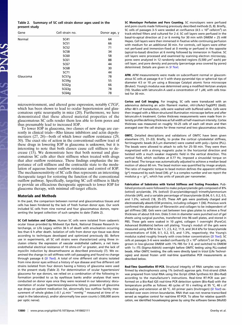
Materials and MethodsIn the past, the comparison between normal and glaucomatous tissues andcells has been hindered by the lack of fresh human donor eyes. Our workincluded SC cells from nine normal and four glaucomatous donors, repre-senting the largest collection of such samples to date (Table 2).
SC Cell Isolation and Culture. Human SC cells were isolated from cadavericocular tissue provided by Midwest Eye Bank, National Disease Research In-terchange, or Life Legacy within 36 h of death with enucleation occurringless than 6 h after death. Isolation of cells from donor eye tissue was doneaccording to techniques developed and optimized previously (6). Beforeuse in experiments, all SC cell strains were characterized using three in-clusion criteria: the expression of vascular endothelial cadherin, a net trans-endothelial electrical resistance of 10 ohms·cm2 or greater, and the lack ofmyocilin induction by dexamethasone as described previously (7). We ex-amined the change in cell stiffness with cell passaging and found no changethrough passage 6 (SI Text). A total of nine different cell strains isolatedfrom nine donor eyes without a history of eye disease and five different cellstrains isolated from four donors having a history of glaucoma were usedin the present study (Table 2). For determination of ocular hypertension/glaucoma for eye donors, we relied on a combination of the following in-formation provided to us by eye/tissue banks and/or analyses that wereconducted on donor eyes once they were received in our laboratory: docu-mentation of ocular hypertension/glaucoma history, presence of glaucomaeye drops on patient medication list, abnormally low outflow facility mea-surement of whole globes (<0.1 μL·min−1·mmHg−1 measured at time of re-ceipt in the laboratory), and/or abnormally low axon counts (<500,000 axonsper optic nerve).
SC Monolayer Perfusion and Pore Counting. SC monolayers were perfusedand pore counts made following previously described methods (5, 8). Briefly,SC cells of passages 3–5 were seeded at confluence (4.5 × 10⁴ cells/cm2) ontrack-etched filters and cultured for 2 d. SC cell layers were perfused in thebasal-to-apical direction at 2 or 6 mmHg for 30 min with DMEM + 25 mMHepes. Cell layers were then immersed in fixative while continuing perfusionwith medium for an additional 30 min. For controls, cell layers were eithernot perfused and immersion-fixed at 0 mmHg or perfused in the opposite(apical-to-basal) direction at 6 mmHg followed by immersion in fixative. SCcell layers were processed and examined by scanning electron microscopy,pores were analyzed in 12 randomly selected regions (5,500 μm2 each) percell layer, and pore density and porosity (percentage area covered by pores)determined. Details are given in SI Text.
AFM. AFM measurements were made on subconfluent normal or glaucom-atous SC cells at passage 4 or 5 with sharp pyramidal tips or spherical tips ofdiameter 4.5 or 10 μm using a Bioscope Catalyst atomic force microscope(Bruker). Young’s modulus was determined using a modified Hertzian analysis(10). Studies with latrunculin-A used a concentration of 1 μM, with cells trea-ted for 30 min.
Cortex and Cell Imaging. For imaging, SC cells were transduced with anadenovirus delivering an actin filament marker, rAV-LifeAct-TagGFP2 (ibidi).After 48 h of transduction, cells were washed with buffered saline. Cells werethen imaged with a Nikon-structured illumination microscope before and afterlatrunculin-A treatment. Cortex thickness measurements were made from in-tensity profilesdefining thickness as fullwidthathalf-maximum intensity. Cortexthickness was measured on roughly 10–25 cells of each cell strain and thenaveraged over the cell strains for three normal and two glaucomatous strains.
OMTC. Detailed descriptions and validations of OMTC have been givenelsewhere (15, 31–33). Briefly, to probe the cortical cytoskeletal stiffness,ferrimagnetic beads (4.5-μm diameter) were coated with poly-L-lysine (PLL).The beads were allowed to attach to cells for 25–50 min. They were thenmagnetized with a strong magnetic pulse in the horizontal direction andtwisted with a much weaker magnetic field in the vertical direction. Thisvertical field, which oscillates at 0.77 Hz, imposed a sinusoidal torque oneach bead. The torque was automatically adjusted to achieve a median beadmotion of about 60 nm. The bead motion was quantified by image analysis.The ratio of magnetic torque to bead motion defines the apparent stiffness(g*) measured by each bead (34). g* is a complex number and we report themodulus g = jg*j, which has units of pascals per nanometer.
Fabrication of Substrates with Varied Stiffness and Testing Procedure. Pub-lished protocols were followed tomake polyacrylamide gels composed of 8%(wt/vol) acrylamide, 3% (wt/vol) (3-acrylamidepropyl) trimethylammoniumchloride (API), and a variable percentage of bisacrylamide (0.04, 0.1, 0.2, 0.5,and 1.3%; wt/vol) (18, 35–37). These API gels were positively charged andelectrostatically absorb ECM proteins, including collagen 1 (36). Previous workconfirmed the absorption of fibronectin and collagen to be independent ofgel stiffness (38). Gels were cast between two glass plates to achieve a finalthickness of about 0.8 mm. Disks 5 mm in diameter were punched out of gelsheets using surgical punches, transferred into 96-well plates, and stored inPBS. These gels were soaked in 10 μg/mL collagen 1 overnight (PureCol;Advanced BioMatrix) before cell plating. Young’s moduli of the gels weremeasured using AFM to be 1.1, 2.5, 4.2, 11.9, and 34.4 kPa for bisacrylamideconcentrations of 0.04, 0.1, 0.2, 0.5, and 1.3%, respectively; the Young’smodulus scaled roughly linearly with cross-linker concentration (SI Text). SCcells at passages 3–6 were seeded confluently (3 × 10⁴ cells/cm2) on the gels,grown in low-glucose DMEM with 1% FBS for 3 d, and switched to DMEMwith 1× ITS (Sigma-Aldrich) overnight before OMTC testing using PLL-coatedbeads. After OMTC twisting, the cells were directly lysed in trizol (Life Technol-ogies) and stored frozen until real-time quantitative PCR measurements asdescribed below.
Real-Time Quantitative RT-PCR. Structural integrity of RNA samples was con-firmed by electrophoresis using 1% (wt/vol) agarose gels. First-strand cDNAwas prepared from total RNA using the iScript cDNA Synthesis Kit (Bio-Rad)according to the manufacturer’s instructions. Real-time RT-PCR was per-formed on a Bio-Rad iQ5 real-time PCR detection system (Bio-Rad) with thetemperature profile as follows: 40 cycles of 10 s melting at 95 °C, 40 s ofannealing and extension at 60 °C. All primer pairs (Invitrogen) (SI Text) ex-tended over exon–intron boundaries. RNA that was not reverse-transcribedserved as negative control for real-time RT-PCR. To allow for relative quantifi-cation, we identified housekeeping genes by using the software Genex (MultiD
Table 2. Summary of SC cell strain donor ages used in thepresent study
Gene Cell strain no. Donor age, y
Normal SC41 64SC51 66SC52 71SC58 34SC60 58SC61 88SC67 44SC68 30SC71 44
Glaucoma SC57g 78SC59g 55SC62g 66SC63g 78SC64g 78
13880 | www.pnas.org/cgi/doi/10.1073/pnas.1410602111 Overby et al.
Analysis) (39). In initial experiments, real-time RT-PCR for the potentialhousekeeping genes GNB2L1, GAPDH, and RPL32 was performed for each ofthe treatment protocols. Cycle threshold values were loaded to the softwarethat distinguishes genes that are regulated in a specific condition from thosethat are very likely not. No differences were obtained between GAPDH andGNB2L1, so GNB2L1was used for relative quantification of the real-time RT-PCRexperiments. Quantification was performed using Bio-Rad iQ5 standard edition(Version 2.0.148.60623) software (Bio-Rad).
Statistical Methods. In general, the statistical analysis was done using SPSS(version 12.0; IBM). Because the cell stiffness for each donor is approximatelylog-normally distributed (SI Text), we reported it as median ± SE, which wascalculated based on logarithmically transformed data (34).
Regression analysis for the studies of the effect of substrate stiffness oncell stiffness and gene expression used the following relationship:
VariableðESubstrateÞVariableð1:1 kPaÞ − 1= c1*
�ESubstrate1:1 kPa
− 1�+ c2*
�ESubstrate1:1 kPa
− 1�*Normal,
where VariableðESubstrateÞ is the value of the parameter being measured (cellstiffness or gene expression) at a given value of substrate stiffness (Esubstrate).Normal is 1 for normal cell strains and 0 for glaucomatous cell strains. Cor-relations were taken as statistically significant when the correlation had anoverall significance of P < 0.01 and either substrate stiffness and/or glau-coma affected the fit with P < 0.05 (unless otherwise noted). In all cases
where a statistically significant difference between glaucomatous cell strainsand normals was reported, the addition of donor age as an additionalcovariate to the equation did not affect this conclusion (SI Text).
Because pore density is a discrete random variable comprising finite countsof sparse events, Poisson statistics were applied and an E-test (40) was used tocompare Poisson-distributed pore densities between different perfusionpressures and between normal and glaucomatous SC cell strains. The gen-eralized linear model (GLM) was used for regression analysis of pore densityversus cell stiffness (as measured by AFM with a 10-μm spherical tip) applyinga logarithmic link function. Differences in porosity were analyzed using atwo-tailed two-sample Student t test. GLM analysis was also used for regressionanalysis of porosity versus cell stiffness with an identity link function.
ACKNOWLEDGMENTS. We gratefully acknowledge support from the NationalGlaucoma Research program of the Bright Focus Foundation; National Institutesof Health Grants R01 EY 01969, R21 EY018373, and T32 EY007128; theWhitakerInternational Scholars Program; the Deutsche Forschungsgemeinschaft (FOR1075, TP3); the Georgia Research Alliance; and the Royal Society WolfsonResearch Excellence Award. Northwestern University’s Atomic and NanoscaleCharacterization Experimental Center (used for atomic force microscopy stud-ies) is supported by the National Science Foundation-Nanoscale Science andEngineering Center, the National Science Foundation-Materials Research Sci-ence and Engineering Centers, the Keck Foundation, the State of Illinois, andNorthwestern University. Imaging studies were done at the Nikon ImagingCenter, Feinberg School of Medicine, Northwestern University.
1. Ethier CR (2002) The inner wall of Schlemm’s canal. Exp Eye Res 74(2):161–172.2. Johnson M (2006) ‘What controls aqueous humour outflow resistance?’. Exp Eye Res
82(4):545–557.3. Allingham RR, et al. (1992) The relationship between pore density and outflow facility
in human eyes. Invest Ophthalmol Vis Sci 33(5):1661–1669.4. Johnson M, et al. (2002) The pore density in the inner wall endothelium of Schlemm’s
canal of glaucomatous eyes. Invest Ophthalmol Vis Sci 43(9):2950–2955.5. Pedrigi RM, Simon D, Reed A, Stamer WD, Overby DR (2011) A model of giant vacuole
dynamics in human Schlemm’s canal endothelial cells. Exp Eye Res 92(1):57–66.6. Stamer WD, Roberts BC, Howell DN, Epstein DL (1998) Isolation, culture, and char-
acterization of endothelial cells from Schlemm’s canal. Invest Ophthalmol Vis Sci39(10):1804–1812.
7. Perkumas KM, Stamer WD (2012) Protein markers and differentiation in culture forSchlemm’s canal endothelial cells. Exp Eye Res 96(1):82–87.
8. Ethier CR, Coloma FM, Sit AJ, Johnson M (1998) Two pore types in the inner-wallendothelium of Schlemm’s canal. Invest Ophthalmol Vis Sci 39(11):2041–2048.
9. Pederson JE, MacLellan HM, Gaasterland DE (1978) The rate of reflux fluid movementinto the eye from Schlemm’s canal during hypotony in the rhesus monkey. InvestOphthalmol Vis Sci 17(4):377–381.
10. Vargas-Pinto R, Gong H, Vahabikashi A, Johnson M (2013) The effect of the endo-thelial cell cortex on atomic force microscopy measurements. Biophys J 105(2):300–309.
11. Guo M, et al. (2013) The role of vimentin intermediate filaments in cortical and cy-toplasmic mechanics. Biophys J 105(7):1562–1568.
12. Byfield FJ, et al. (2009) Absence of filamin A prevents cells from responding to stiff-ness gradients on gels coated with collagen but not fibronectin. Biophys J 96(12):5095–5102.
13. Solon J, Levental I, Sengupta K, Georges PC, Janmey PA (2007) Fibroblast adaptationand stiffness matching to soft elastic substrates. Biophys J 93(12):4453–4461.
14. Hoffman BD, Massiera G, Van Citters KM, Crocker JC (2006) The consensus mechanicsof cultured mammalian cells. Proc Natl Acad Sci USA 103(27):10259–10264.
15. Zhou EH, et al. (2012) Mechanical responsiveness of the endothelial cell of Schlemm’scanal: Scope, variability and its potential role in controlling aqueous humour outflow.J R Soc Interface 9(71):1144–1155.
16. Last JA, et al. (2011) Elastic modulus determination of normal and glaucomatoushuman trabecular meshwork. Invest Ophthalmol Vis Sci 52(5):2147–2152.
17. Liu F, et al. (2010) Feedback amplification of fibrosis through matrix stiffening andCOX-2 suppression. J Cell Biol 190(4):693–706.
18. Schlunck G, et al. (2008) Substrate rigidity modulates cell matrix interactions andprotein expression in human trabecular meshwork cells. Invest Ophthalmol Vis Sci49(1):262–269.
19. Dupont S, et al. (2011) Role of YAP/TAZ in mechanotransduction. Nature 474(7350):179–183.
20. Junglas B, et al. (2012) Connective tissue growth factor causes glaucoma by modifyingthe actin cytoskeleton of the trabecular meshwork. Am J Pathol 180(6):2386–2403.
21. Leber T (1873) Studien über den Flüssigkeitswechsel im Auge. Albrecht Von GraefesArch Ophthalmol 19:87–106.
22. Keller KE, Acott TS (2013) The juxtacanalicular region of ocular trabecular meshwork:A tissue with a unique extracellular matrix and specialized function. J Ocul Biol 1(1):3.
23. Johnson M, Shapiro A, Ethier CR, Kamm RD (1992) Modulation of outflow resistanceby the pores of the inner wall endothelium. Invest Ophthalmol Vis Sci 33(5):1670–1675.
24. Overby DR, Stamer WD, Johnson M (2009) The changing paradigm of outflow re-sistance generation: Towards synergistic models of the JCT and inner wall endothe-lium. Exp Eye Res 88(4):656–670.
25. Lütjen-Drecoll E, Futa R, Rohen JW (1981) Ultrahistochemical studies on tangentialsections of the trabecular meshwork in normal and glaucomatous eyes. Invest Oph-thalmol Vis Sci 21(4):563–573.
26. Camras LJ, Stamer WD, Epstein D, Gonzalez P, Yuan F (2014) Circumferential tensilestiffness of glaucomatous trabecular meshwork. Invest Ophthalmol Vis Sci 55(2):814–823.
27. Chen J, Runyan SA, Robinson MR (2011) Novel ocular antihypertensive compounds inclinical trials. Clin Ophthalmol 5:667–677.
28. Williams RD, Novack GD, van Haarlem T, Kopczynski C, AR-12286 Phase 2A StudyGroup (2011) Ocular hypotensive effect of the Rho kinase inhibitor AR-12286 in pa-tients with glaucoma and ocular hypertension. Am J Ophthalmol 152(5):834–841, e1.
29. Tian B, Geiger B, Epstein DL, Kaufman PL (2000) Cytoskeletal involvement in theregulation of aqueous humor outflow. Invest Ophthalmol Vis Sci 41(3):619–623.
30. Rao VP, Epstein DL (2007) Rho GTPase/Rho kinase inhibition as a novel target for thetreatment of glaucoma. BioDrugs 21(3):167–177.
31. Fabry B, et al. (2001) Selected contribution: Time course and heterogeneity of con-tractile responses in cultured human airway smooth muscle cells. J Appl Physiol (1985)91(2):986–994.
32. Fabry B, et al. (2003) Time scale and other invariants of integrative mechanical be-havior in living cells. Phys Rev E Stat Nonlin Soft Matter Phys 68(4 Pt 1):041914.
33. Zhou EH, et al. (2009) Universal behavior of the osmotically compressed cell and itsanalogy to the colloidal glass transition. Proc Natl Acad Sci USA 106(26):10632–10637.
34. Fabry B, et al. (2001) Scaling the microrheology of living cells. Phys Rev Lett 87(14):148102.
35. Engler AJ, Sen S, Sweeney HL, Discher DE (2006) Matrix elasticity directs stem celllineage specification. Cell 126(4):677–689.
36. de Rooij J, Kerstens A, Danuser G, Schwartz MA, Waterman-Storer CM (2005) Integrin-dependent actomyosin contraction regulates epithelial cell scattering. J Cell Biol171(1):153–164.
37. McKee CT, et al. (2011) The effect of biophysical attributes of the ocular trabecularmeshwork associated with glaucoma on the cell response to therapeutic agents.Biomaterials 32(9):2417–2423.
38. Wood JA, et al. (2011) Substratum compliance regulates human trabecular meshworkcell behaviors and response to latrunculin B. Invest Ophthalmol Vis Sci 52(13):9298–9303.
39. Vandesompele J, et al. (2002) Accurate normalization of real-time quantitative RT-PCRdata by geometric averaging of multiple internal control genes. Genome Biol 3(7):research0034.1–research0034.11.
40. Krishnamoorthy K, Thomson J (2004) A more powerful test for comparing two Pois-son means. J Stat Plan Inference 119(1):23–35.
41. Gong H, Ruberti J, Overby D, Johnson M, Freddo TF (2002) A new view of the humantrabecular meshwork using quick-freeze, deep-etch electron microscopy. Exp Eye Res75(3):347–358.
42. Riedl J, et al. (2008) Lifeact: A versatile marker to visualize F-actin. Nat Methods 5(7):605–607.
43. Pagano M, Gauvreau K (2000) Principles of Biostatistics (Cengage Learning, In-dependence, KY), Vol 2.
44. Mijailovich SM, Kojic M, Zivkovic M, Fabry B, Fredberg JJ (2002) A finite elementmodel of cell deformation during magnetic bead twisting. J Appl Physiol 93(4):1429–1436.
Overby et al. PNAS | September 23, 2014 | vol. 111 | no. 38 | 13881
CELL
BIOLO
GY