b lood
TRANSCRIPT

BLOOD
http://health.howstuffworks.com/medicine/medication/aspirin3.htm

OVERVIEW OF BLOOD CIRCULATION
Circulates continuously
Gas exchange occurs across capillary walls
O2 rich blood leaves lungs and is pumped systemically via the heart
CO2 rich blook enters heart and is pumped into lungs to be oxygenated

COMPOSITION OF BLOOD
Fluid tissue
Liquid plasma and formed elements
Hematocrit – the percentage of RBCs out of the total blood volume

PHYSICAL CHARACTERISTICS AND VOLUME Blood is a sticky, opaque, metallic taste
Color varies from scarlet to dark red
The pH of blood is 7.35–7.45
Temperature is 38C
Blood accounts for approximately 8% of body weight
Average volume: 5–6 L for males, and 4–5 L for females
FUNCTIONS AND DISTRIBUTION OF BLOOD
Protects against fluid loss
Provides immunity
Regulates pH
Maintains osmotic pressure

BLOOD PLASMA Blood plasma contains over 100 solutes, including:
Proteins: albumin, transferrin, globulins, complement and clotting proteins
Waste products: lactic acid, urea, creatinine
Nutrients – glucose, carbohydrates, amino acids
Electrolytes – sodium, potassium, calcium, chloride, bicarbonate
Respiratory gases – oxygen and carbon dioxide

FORMED ELEMENTS
Erythrocytes, leukocytes, and platelets Life span is variable
Stem cell renewal or division

ERYTHROCYTES (RBCS)
Biconcave discs
Anucleate
Essentially no organelles
Spectrin allows flexibility
Hemoglobin (97%)
Anerobic ATP generation
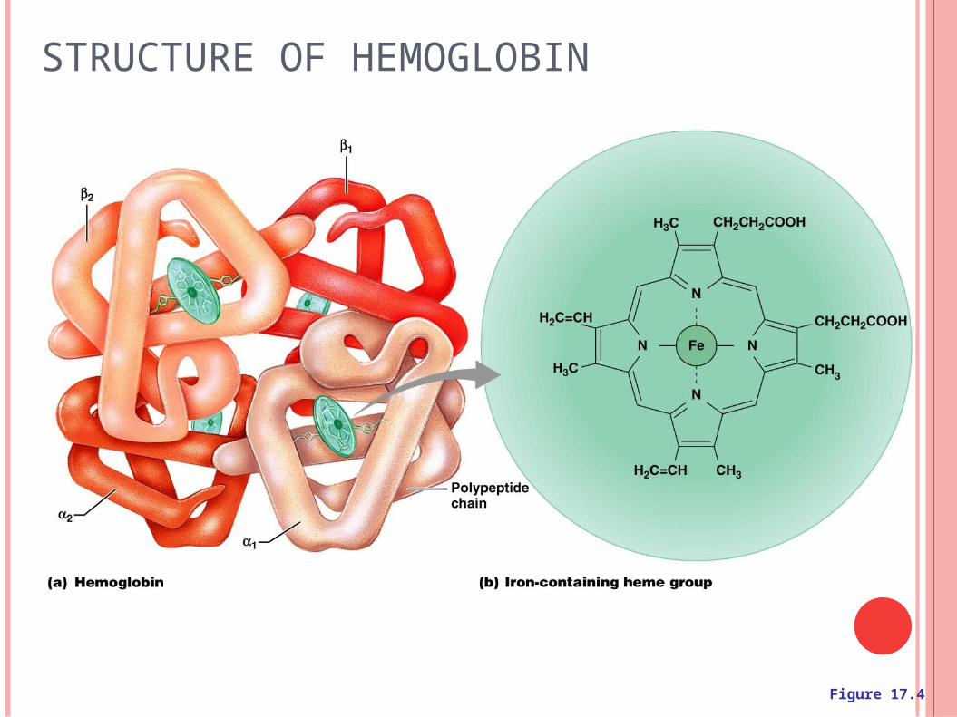
STRUCTURE OF HEMOGLOBIN
Figure 17.4
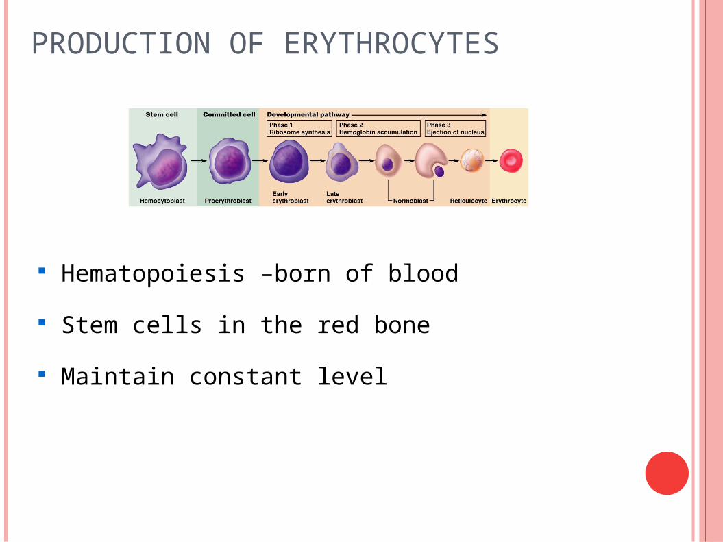
PRODUCTION OF ERYTHROCYTES
Hematopoiesis –born of blood
Stem cells in the red bone
Maintain constant level

ERYTHROPOIESIS REGULATION Indirectly through thyroxine, androgens, GH
Vitamin B12
Erythropoietin (EPO)
release by the kidneys
triggered by hypoxia
Increases division of erythroblasts and maturation rate
Oxygen carrying ability of the blood
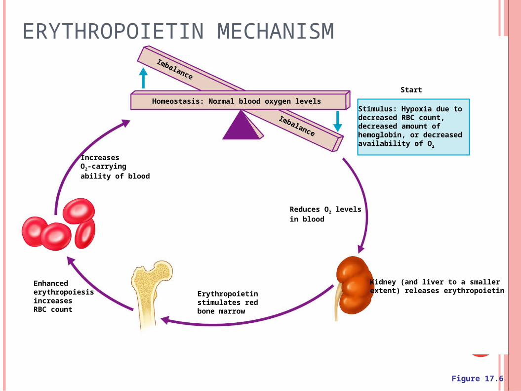
Homeostasis: Normal blood oxygen levels
IncreasesO2-carryingability of blood
Erythropoietinstimulates redbone marrow
Reduces O2 levelsin blood
Kidney (and liver to a smallerextent) releases erythropoietin
Enhancederythropoiesisincreases RBC count
Stimulus: Hypoxia due todecreased RBC count,decreased amount of hemoglobin, or decreased availability of O2
Start
Imbalance
Imbalance
ERYTHROPOIETIN MECHANISM
Figure 17.6

FATE AND DESTRUCTION OF ERYTHROCYTES The life span =100–120 days
Dying RBCs are engulfed by macrophages
Iron returned to blood bound by transferrin
Heme is degraded into bilirubin, bile, feces
Globin metabolized or excreted

Hemoglobin
Aminoacids
Globin
Raw materials aremade available inblood for erythrocytesynthesis.
Iron is bound to transferrin and released to blood from liver as needed for erythropoiesis
Food nutrients,including aminoacids, Fe, B12,and folic acidare absorbedfrom intestineand enter blood
Heme
Circulation
Iron storedas ferritin,hemosiderin
Bilirubin
Bilirubin is picked up fromblood by liver, secreted intointestine in bile, metabolizedto stercobilin by bacteriaand excreted in feces
Erythropoietin levelsrise in blood.
Erythropoietin and necessaryraw materials in blood promoteerythropoiesis in red bone marrow.
New erythrocytesenter bloodstream;function about120 days.
Low O2 levels in blood stimulatekidneys to produce erythropoietin.
Aged and damaged redblood cells are engulfed bymacrophages of liver, spleen,and bone marrow; the hemoglobinis broken down.
1
2
3
4
5
6
Figure 17.7
ERYTHROCYTE RECYCLING

ERYTHROCYTE DISORDERS Anemia – blood has abnormally low oxygen-
carrying capacity
It is a symptom rather than a disease itself
Blood oxygen levels cannot support normal metabolism
ERYTHROCYTE DISORDERS
Hemorrahgic anemia
Hemolytic anemia
Aplastic anemia
Iron-deficiency anemia
Pernicious anemia
Thalassemias
Sickel-cell anemia
Polycythemia

LEUKOCYTES (WBCS)
Leukocytes, the only blood components that are complete cells:
Are less numerous than RBCs
Make up 1% of the total blood volume
Capable of diapedesis and margination

PERCENTAGES OF LEUKOCYTES
Figure 17.9

SUMMARY OF FORMED ELEMENTS
Table 17.2.1

(a) (b) (c) (d) (e)
Hemocytoblast
Myeloid stem cell Lymphoid stem cell
Myeloblast MyeloblastMyeloblast Lymphoblast
Stem cells
Committedcells
Promyelocyte PromyelocytePromyelocyte Promonocyte Prolymphocyte
Eosinophilicmyelocyte
Neutrophilicmyelocyte
Basophilicmyelocyte
Eosinophilicband cells
Neutrophilicband cells
Basophilicband cells
Develop-mentalpathway
Eosinophils NeutrophilsBasophils
Granular leukocytes
Plasma cells
Some become
Monocytes Lymphocytes
Macrophages (tissues)
Agranular leukocytes
Some become
Figure 17.11

LEUKOCYTES DISORDERS
Leukocytosis
Leukemia refers to cancerous conditions involving WBCs
Myelocytic leukemia
Lymphocytic leukemia
Acute leukemia
Chronic leukemia
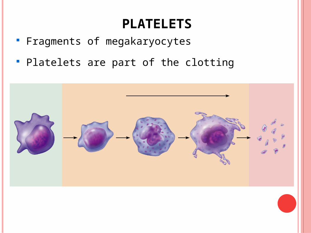
PLATELETS Fragments of megakaryocytes
Platelets are part of the clotting

HEMOSTASIS
A series of reactions for stoppage of bleeding
Three phases occur in rapid sequence
Vascular spasms
Platelet plug formation
Coagulation (blood clotting)

DETAILED EVENTS OF COAGULATION
Figure 17.13b
CLOT RETRACTION AND REPAIR Clot retraction – stabilization of the clot by squeezing
serum from the fibrin strands
Repair
Platelet-derived growth factor (PDGF) stimulates rebuilding of blood vessel wall
Fibroblasts form a connective tissue patch
Endothelial cells multiply , restoring the lining

FACTORS PREVENTING UNDESIRABLE CLOTTING
Endothelial lining of blood vessels
Platelet adhesion is prevented by:
Heparin and PGI2 secreted by endothelial cells
Vitamin E quinone, a potent anticoagulant
Aspirin
Wafarin (coumadin)

HEMOSTASIS DISORDERS
Thrombus
Embolus
Thrombocytopenia
Hemophilias
Hemophilia A, B, C

HUMAN BLOOD TYPING
Humans have 30 varieties of naturally occurring RBC antigens
The antigens of the ABO and Rh blood groups cause vigorous transfusion reactions when they are improperly transfused
Other blood groups (M, N, Dufy, Kell, and Lewis) are mainly used for legalities

ABO BLOOD GROUPS
Table 17.4