propionibacterium acnes and its phages lood, rolf · propionibacterium acnes and its phages rolf...
TRANSCRIPT

LUND UNIVERSITY
PO Box 117221 00 Lund+46 46-222 00 00
Propionibacterium acnes and its phages
Lood, Rolf
2011
Link to publication
Citation for published version (APA):Lood, R. (2011). Propionibacterium acnes and its phages. Department of Clinical Sciences, Lund University.
General rightsCopyright and moral rights for the publications made accessible in the public portal are retained by the authorsand/or other copyright owners and it is a condition of accessing publications that users recognise and abide by thelegal requirements associated with these rights.
• Users may download and print one copy of any publication from the public portal for the purpose of private studyor research. • You may not further distribute the material or use it for any profit-making activity or commercial gain • You may freely distribute the URL identifying the publication in the public portalTake down policyIf you believe that this document breaches copyright please contact us providing details, and we will removeaccess to the work immediately and investigate your claim.


Propionibacterium acnes and its phages
Rolf LoodDepartment of Clinical Sciences, Lund
Division of Infection MedicineFaculty of Medicine
Lund University, Sweden
Doctoral dissertationWith due permission from the Medical Faculty at Lund University this doctoral thesis is to
be publicly defended on the 7th of October 2011, at 9.00 in Segerfalksalen, BiomedicalCenter, Sölvegatan 17, Lund.
Faculty opponentDr. Holger Brüggemann
Department of Molecular BiologyMax Planck Institute for Infection Biology
Berlin, Germany
Propionibacterium acnes and its phages
Rolf LoodDepartment of Clinical Sciences, Lund
Division of Infection MedicineFaculty of Medicine
Lund University, Sweden
Lund 2011
Rolf LoodDepartment of Clinical Sciences, LundDivision of Infection MedicineFaculty of MedicineLund UniversityBiomedical Center, B14221 84 LundSwedenE-mail: [email protected] phone: +46 739 032 117Phone: +46 46 222 98 73Fax: +46 46 157 756
Cover image:The lysis of Propionibacterium acnes infected with bacteriophages, and the subsequentrelease of the bacteriophages, as visualized using scanning electron microscopy. MariaBaumgarten and Dr. Matthias Mörgelin are acknowledged for the negative staining andelectron microscopy, respectively.
Printed by E-huset tryckeri, Lund, Sweden© Rolf Lood, 2011© John Wiley and Sons, 2008
ISSN 1652-8220ISBN 978-91-86871-43-7
Lund University, Faculty of Medicine Doctoral Dissertation Series 2011:94
To Mom and Dad
The first to present his case seems right,till another comes forward and questions him
-Proverbs 18:17
Table of Contents
1 Introduction 101.1 List of papers . . . . . . . . . . . . . . . . . . . . . . . . . . . . . . . . . 101.2 Abbreviations . . . . . . . . . . . . . . . . . . . . . . . . . . . . . . . . . 121.3 Acknowledgements . . . . . . . . . . . . . . . . . . . . . . . . . . . . . . 141.4 Populärvetenskaplig sammanfattning . . . . . . . . . . . . . . . . . . . . . 17
2 Summary 21
The Bad Guys 22
3 Propionibacterium acnes 233.1 Nomenclature . . . . . . . . . . . . . . . . . . . . . . . . . . . . . . . . . 233.2 Genetics of the Propionibacteriaceae family . . . . . . . . . . . . . . . . . 243.3 Different types of P. acnes . . . . . . . . . . . . . . . . . . . . . . . . . . 243.4 Plasmids in Propionibacteria . . . . . . . . . . . . . . . . . . . . . . . . . 253.5 Transformation systems in Propionibacteria . . . . . . . . . . . . . . . . . 263.6 Morphological characteristics . . . . . . . . . . . . . . . . . . . . . . . . . 263.7 Growth characteristics . . . . . . . . . . . . . . . . . . . . . . . . . . . . 273.8 Antibiotic resistance . . . . . . . . . . . . . . . . . . . . . . . . . . . . . 283.9 Virulence of P. acnes . . . . . . . . . . . . . . . . . . . . . . . . . . . . . 293.10 Acne . . . . . . . . . . . . . . . . . . . . . . . . . . . . . . . . . . . . . . 293.11 Prostate cancer . . . . . . . . . . . . . . . . . . . . . . . . . . . . . . . . 303.12 Prosthesis removal - biofilm formation . . . . . . . . . . . . . . . . . . . . 303.13 Characterized proteins from P. acnes . . . . . . . . . . . . . . . . . . . . . 32
4 Virulence factors 344.1 Introduction . . . . . . . . . . . . . . . . . . . . . . . . . . . . . . . . . . 344.2 Virulence factors . . . . . . . . . . . . . . . . . . . . . . . . . . . . . . . 344.3 Triplets in virulence factors . . . . . . . . . . . . . . . . . . . . . . . . . . 35
4.3.1 Biofilms . . . . . . . . . . . . . . . . . . . . . . . . . . . . . . . . 354.3.2 Sialidases . . . . . . . . . . . . . . . . . . . . . . . . . . . . . . . 384.3.3 Protection from reactive oxygen species . . . . . . . . . . . . . . . 40
4.4 How to study virulence factors in P. acnes . . . . . . . . . . . . . . . . . . 42
8
The Good Guys 44
5 Bacteriophages 455.1 Introduction . . . . . . . . . . . . . . . . . . . . . . . . . . . . . . . . . . 455.2 Classification . . . . . . . . . . . . . . . . . . . . . . . . . . . . . . . . . 45
5.2.1 Introduction . . . . . . . . . . . . . . . . . . . . . . . . . . . . . . 455.2.2 Classification . . . . . . . . . . . . . . . . . . . . . . . . . . . . . 455.2.3 Identification of phages . . . . . . . . . . . . . . . . . . . . . . . . 46
5.3 Life cycle . . . . . . . . . . . . . . . . . . . . . . . . . . . . . . . . . . . 485.3.1 Introduction . . . . . . . . . . . . . . . . . . . . . . . . . . . . . . 485.3.2 Attachment . . . . . . . . . . . . . . . . . . . . . . . . . . . . . . 485.3.3 DNA injection . . . . . . . . . . . . . . . . . . . . . . . . . . . . 505.3.4 Life cycle decision . . . . . . . . . . . . . . . . . . . . . . . . . . 505.3.5 Integration . . . . . . . . . . . . . . . . . . . . . . . . . . . . . . 525.3.6 Phage release . . . . . . . . . . . . . . . . . . . . . . . . . . . . . 52
5.4 Phage mediated virulence . . . . . . . . . . . . . . . . . . . . . . . . . . . 535.5 Phages in the industry . . . . . . . . . . . . . . . . . . . . . . . . . . . . . 56
5.5.1 Industrial problems with phages . . . . . . . . . . . . . . . . . . . 565.5.2 Phage display . . . . . . . . . . . . . . . . . . . . . . . . . . . . . 56
5.6 Phage therapy . . . . . . . . . . . . . . . . . . . . . . . . . . . . . . . . . 565.6.1 Phage therapy using whole phages . . . . . . . . . . . . . . . . . . 565.6.2 Phage therapy using phage endolysins . . . . . . . . . . . . . . . . 59
6 References 61
9
1 Introduction
1.1 List of papers
Articles included in this thesis
1. Lood, R∗., Garbe, J∗., and Collin, M. Development of a genetic toolbox for studiesof Propionibacterium acnes. Manuscript in preparation.
2. Holmberg, A., Lood, R., Mörgelin, M., Söderquist, B., Holst, E., Collin, M., Chris-tensson, B., and Rasmussen, M. 2009. Biofilm formation by Propionibacterium ac-nes is a characteristic of invasive isolates. Clinical Microbiology & Infection 15(8):787-95.
3. Lood, R., Olin, AI, Collin, M., and Allhorn, M. Commensal skin bacteria increasethe viability of human cells by protecting them from harmful reactive oxygen species.Submitted.
4. Lood, R., Mörgelin, M., Holmberg, A., Rasmussen, M., and Collin, M. 2008. In-ducible Siphoviruses in superficial and deep tissue isolates of Propionibacterium ac-nes. BMC Microbiology 8:139.
5. Lood, R., and Collin, M. 2011. Characterization and genome sequencing of two Pro-pionibacterium acnes phages displaying pseudolysogeny. BMC Genomics 12:198.
Paper 2 has been reprinted with the permission from John Wiley & Sons.Paper 4 and 5 have been reprinted under the Creative Commons license.
10
Articles not included in this thesis
1. Karlsson, C., Mörgelin, M., Collin, M., Lood, R., Andersson, ML., Schmidtchen, A.,Björck, L., and Frick, IM. 2009. SufA - a bacterial enzyme that cleaves fibrinogenand blocks fibrin network formation. Microbiology 155:238-248.
11

1.2 Abbreviations
AHL - acyl homoserine lactoneB. acnes - Bacillus acnesC. acnes - Corynebacterium acnesC. parvum - Corynebacterium parvumCAMP - Christie, Atkins, and Munch-Petersencfu - colony forming unitsCXCL8 - Interleukin 8 (IL-8)DNA - deoxyribonucleic acidDNAse - deoxyribonucleasedsDNA - double stranded deoxyribonucleic aciddsRNA - double stranded ribonucleic acidE. coli - Escherichia coliGFP - green fluorescent proteinICTV - International committee on taxonomy of virusesIgA - immunoglobulin AIL-8 - interleukin 8kb - kilo baseskDa - kilo DaltonLPS - lipopolysaccharideMAC - membrane attack complexMb - mega basesNCBI - National Center for Biotechnology InformationORF - open reading frameP. acidifaciens - Propionibacterium acidifaciensP. acidipropionici - Propionibacterium acidipropioniciP. acnes - Propionibacterium acnesP. australiense - Propionibacterium australienseP. avidum - Propionibacterium avidumP. cyclohexanicum - Propionibacterium cyclohexanicumP. freudenreichii - Propionibacterium freudenreichiiP. granulosum - Propionibacterium granulosum
12
P. humerusii - Propionibacterium humerusiiP. jensenii - Propionibacterium jenseniiP. lymphophilum - Propionibacterium lymphophilumP. microaerophilum - Propionibacterium microaerophilumP. propionicus - Propionibacterium propionicusP. thoenii - Propionibacterium thoeniiPCR - polymerase chain reactionpfu - plaque forming unitsRDE - receptor destroying enzymeROS - reactive oxygen speciesrRNA - ribosomal ribonucleic acidRT-PCR - reverse transcriptase polymerase chain reactionSDS-PAGE - sodium dodecyl sulfate polyacrylamide gel electrophoresisSOD - superoxide dismutasessDNA - single stranded deoxyribonucleic acidssRNA - single stranded ribonucleic acidTNF-α - tumor necrosis factor alpha
13
1.3 Acknowledgements
My supervisorMattiasThanks for everything! Your supporting leadership, your enthusiasm and for many manyinteresting discussions where you always have let me come up with my own ideas. I cannot imagine any better supervisor for me than you. Also thanks for friendship, not only inthe lab but also outside the lab. I have really enjoyed working with you these years. Yourways of supervising and performing science will always serve as an example to me.
My co-supervisorsInga-MariaThanks for always being a support and for introducing me to the world of science. Alsothanks for believing in me when I still was an undergraduate student, even though there wasa small accident with the centrifuge, and the pH-meter, and...! I have much to thank youfor.
MatthiasThanks for all ”friendly” comments in the corridors, and for being more than helpful withelectron microscopy. I hope you enjoyed looking for phages as much as I did! The thesiswould not have had any good figures if it had not been for your help!
The Collin-groupMariaMy room-partner. Thanks for your never ending enthusiasm and happiness, it has helpedme a lot! And for lots of discussions – it can not always have been easy to share the officewith me!
UllaFor having a really good sense of humor and not being afraid of telling me how thingsshould be done... And also for teaching me the importance of starting early in the morning– I think I won most of the days?
Julia
14
Thanks for skillfully making some of my more crazy ideas working, and also for lettingme know what ideas I probably should not think any more on. I have really enjoyed ourcollaboration! Furthermore, a huge thanks for teaching me some basics in LaTex, I had lotsof fun! Who is going to press the electroporation button when I am leaving?
MartaThanks for taking lots of uncomfortable phone calls for me, reminding me of booking aplace to defend my thesis in, and being a great support with the thesis. And of course withall things concerning the party afterwards! I’m looking forward to read your book!
SaraThanks for letting me help you in the beginning, making me feel smart. Unfortunately, thatdid not last long... Also thanks for all encouraging comments and helpful advices!
JonathanFor showing me that recombinant proteins can be expressed at the first time, I do not envyyou at all... Also thanks for being a great friend in the lab, and for the moment at thesunrise...
B14A big thanks to all persons at B14 – group leaders, post docs, PhD students, lab assistantsand students, for making this floor so much fun working on. Especially thanks to Lasse forgenerating this open atmosphere, and Anita for helping me with keys and paper work in anexcellent way. Also an extra thanks to Maria B for all work with EM and to Christofer forinvesting lots of time in me during my first summer projects here. I am sorry I forced you tostart early in the mornings! Finally, an extra thanks to Pontus, whose thesis served both as agreat example and as a way to lift up the laptop a few inches from the table; and to Helenafor most helpful advices for my post doc and for letting me use ”her” office.
My coauthorsThanks to all my coauthors, for helping me during these four years. Especially thanksto Magnus Rasmussen and Anna Holmberg for introducing me to P. acnes and giving greatfeedback to many of my questions. Also thanks to Anna for letting me do some calculationsnow and then, making me believe that I actually can math.
15
My studentsTo all my gifted students, Erik & Sofia, Jennifer, Sofia and Peter, that helped me throughthe summers with experiments and made the labwork easier, as well as funnier. A hugethanks also to all the other students in Lab Collin - Frida, Eleni, Marcus, Johan, Abdigani& Yasmeen making me think about other things than the thesis!
My friendsTo all my friends in Charisma, Röke Blås, and Game Over, and to Anders (for lots of pokergames!), that have supported me and helped me to think about other things but microbiology,and from time to time got me realizing that there are other things in the world but P. acnesto think about.
My familyTo mom and dad, who, even though not reading all my papers, and maybe not understoodall details, still listened when I tried to explain my work for them, and nodded in the rightplaces! Thanks for all support and for motivating me to continue developing as a researcherin order to be spared from gathering stones at the farm... It really did help!
CFor all intense discussions about the work, and for a much helpful competition. I think wecan call it a tie, even though I finished ”a year” before you!
16

1.4 Populärvetenskaplig sammanfattning
Mikroorganismer finns överallt! De klarar avatt överleva i extrema miljöer, extrema pH, ex-trema temperaturer och kan motstå strålning ochgift i doser som vi inte skulle överleva. Fram-förallt klarar mikroorganismer av att överlevai en specifik extrem miljö - människan. Hun-dratals olika mikroorganismer bor på och i oss,i ett antal som långt överskrider vårt eget cellan-tal.
Mikroorganismer är ett samlingsnamn förflera olika typer av små (’mikro’) organismer,som bakterier och virus. Många av oss as-socierar nog dessa med infektioner och olikasjukdomar, vilket också ett fåtal orsakar. Mentvärtemot vad man kan tro är de flesta bakterierofarliga, och till och med gynnsamma, genomatt de hjälper oss att ta upp näring i tarmarnaoch skyddar oss mot farligare bakterier. Dessabakterier tillhör vår normalflora och har en stordel i vårt välbefinnande.
Vår hud har flera olika bakterier. Några avde vanligare bakterierna är Propionibakterier,Stafylokocker och Streptokocker. Många av osskan ha dessa på vår hud utan att de ger nå-gra symptom, då de tillhör normalfloran. Men,då tillfälle ges kan även dessa bakterier orsakaopportunistiska infektioner. Detta sker fram-förallt om vårt försvar mot dem på något sättär påverkat, genom exempelvis sår (skador ellerkirurgiska ingrepp) eller vid nedsatt immun-försvar.
En av de hudbakterier som oftast är inblandadi opportunistiska infektioner är Propionibac-terium acnes. Bakterien kallades ursprungligen
för Bacillus acnes eftersom den var stavformad(Bacillus-lik) och isolerades från acne. Fastänbakterien isolerades från acne för mer än 100 årsedan råder det fortfarande tveksamheter kringom P. acnes verkligen är orsaken bakom utveck-landet av sjukdomen. Anledningarna till detta ärmånga. Först och främst är vi alla koloniseradeav P. acnes utan att utveckla några sjukdomar.Därför hävdar många att en isolering av P. ac-nes från huden enbart är en kontaminering avprovet. Vidare är P. acnes svår att arbeta med avtvå anledningar. Dels växer P. acnes långsamtoch behöver leva i en miljö utan syre. Delsfinns väldigt få metoder utvecklade för att stud-era denna bakterie.
Intresset för denna bakterie har på de senasteåren ökat drastiskt. Fortfarande är intresset stortför den potentiella rollen av P. acnes i utveck-lingen av acne. Men senare forskning har ävenpekat på att P. acnes har en stor roll vid led-implantatsinflammationer, där ett byte av im-plantat ofta är nödvändigt för att bli av med in-fektionen då dessa bakterier ofta är väldigt re-sistenta mot antibiotika. Detta beror på att bak-terien kan bilda ett tjockt lager av socker kringsig (biofilm), som ett pansar som skyddar P. ac-nes mot antibiotikan. Där kan bakterierna ligga iflera månader till år utan att orsaka någon skada,för att sedan skapa en inflammation. Vidare ty-der forskning på att P. acnes kan vara delaktig iutvecklingen av prostatacancer.
Trots ett ökat intresse för att studera dennaopportunistiska bakterie, var möjligheten tilldetta begränsad, då det saknades flera nöd-
17
vändiga verktyg för detta. Därför utveckladevi flera verktyg för att på ett enkelt sätt kunnastudera hur delar av bakterien kunde medverkatill att utveckla sjukdom (Paper I). Dessa verk-tyg består av två plasmider, som är cirkulärtDNA. Dessa plasmider är konstruerade till attfungera som fabriker i P. acnes och produceraolika protein som önskas studeras. Då studier avproteiner i bakterier är en av de grundläggandesätten för att utvärdera hur en bakterie orsakarsjukdom, kommer dessa verktyg att underlättastudier av P. acnes i framtiden.
Då P. acnes var så vanligt förekommandevid inflammationer kring ledimplantat, isoler-ade vi flera av dessa stammar och undersöktevad som skiljde dem från de P. acnes som vihade på huden. Vi fann då att de P. acnes somhade förmåga att orsaka protesinflammationeralla kunde bilda mer biofilm än de bakterier somisolerades från huden (Paper II). I övrigt hittadesinga skillnader mellan de två grupperna av bak-terier, och vi kunde fastslå att den troligtvis vik-tigaste faktorn för att P. acnes orsakade protesin-flammationer berodde på dess förmåga att bildabiofilm.
Bortsett från att vara förmögen att orsakasjukdom bär de flesta av oss på P. acnes utanatt bli sjuka. Därför är en rimlig tanke att dessabakterier också kan vara nyttiga för oss, menpå vilket sätt de potentiellt skulle kunna skyddaoss har inte utretts. Vi valde att studera ett pro-tein som P. acnes producerar i stora mängder.Detta protein är ovanligt, för det liknar inget hit-tills undersökt protein i andra bakterier. Därförslogs vi av tanken att detta protein potentielltkunde vara av stor betydelse för bakteriens in-teraktion med oss (Paper III). Det visade sig att
detta protein, vidare kallat RoxP, kunde skyddavåra hudceller mot reaktiva syreradikaler sombildas bland annat av UV-ljus. Hudceller sombehandlades med RoxP mådde mycket bättre änceller som inte hade fått RoxP. Detta indikeraratt för en frisk person är kolonisering av P. acnesviktigt, då detta bidrar till att skydda vår hud.Därför är det viktigt att inte behandla ospecifikt(eg antibiotika) mot P. acnes om inte denna ärupphovet till infektionen.
Men P. acnes är inte ensam på spelplanen.Motståndarlaget har en minst lika bred truppav spelare, och är svurna fiender till P. acnes.Mötena dem sinsemellan brukar alltid resulterai vinst åt något av hållen, men kan även geett lika-resultat. Det handlar om bakteriofager.Virus som är ofarliga för oss, men som är dedik-erade till att eliminera bakterier. Dessa bakteri-ofager har två principiellt olika spelarstilar. Enav dem är anfallaren, som bara vill komma åtbakterien och förstöra för den. Lyckligtvis förbakterien känner den till ett och annat knep föratt skydda sig mot dessa tacklingar.
Den andra spelartypen är mer utav enförsvarsspelare. Han gillar att komma nära mot-ståndaren och interagera med dem, även tillfäl-ligt hjälpa dem, för att få motståndaren attkänna sig ohotad. Sedan, då tillfälle ges, slårförsvarsspelaren till med full kraft. Däremot kanbakterierna även klara sig ur dessa situationergenom olika försvar.
Då bakteriofager är kända för att kunna hjälpabakterierna tillfälligt, var vi intresserade av attundersöka ifall P. acnes som kunde orsaka led-protesinflammationer också i större utsträckn-ing hade hjälp av bakteriofager (Paper IV). Vifann att P. acnes i väldigt stor utsträckning hade
18
bakteriofager (70%), men att där inte var någonskillnad mellan sjukdomsframkallande P. acnesoch normalflora. Med andra ord, det verkadeinte som om bakteriofagerna samspelade medP. acnes i sjukdomsprocessen. Vad vi däremotfann var att bägge lagen hade olika kvaliteterpå sina spelare. En del bakteriofager kundespela anfallare mot nästan alla bakterier medframgång, medan vissa fager enbart kunde drib-bla bort ett fåtal motståndare. På samma sättvar kvaliteten i bakterielaget varierande. Medanvissa kunde försvara sig mot nästan alla fager-nas anfallare, förutom deras stjärnspelare, bordemånga av de andra snarare sitta på utbytar-bänken då de blev bortdribblade i varje anfall.
Även om det inte verkade som om bakteri-ofagerna kunde hjälpa P. acnes, bestämde vi ossför att granska detta mer, genom att undersökatvå av fagerna närmare. Detta bestod i att viplockade fram deras DNA, och läste av det, föratt se om de hade någon förmåga att hjälpa bak-
terierna (Paper V). Genom detta fann vi att dessabakteriofager inte verkar kunna samspela medbakterierna. Däremot kom vi fram till att dessabakteriofager hade en märklig ”spelstil”, då deinte var strikta anfallare, men inte heller striktaförsvarare. Inte enbart var deras spelstil annor-lunda, dessutom var deras DNA olikt DNA frånandra bakteriofager.
Sammanfattningsvis har jag i denna avhan-dling utvecklat verktyg för att bättre och lättarekunna studera P. acnes. Denna bakterie harbåde positiva och negativa egenskaper. Positivagenom att den hjälper vår hud att må bra, mennegativa då den genom att bilda biofilmer med-verkar till inflammation av proteser. Men P. ac-nes är inte ensam på spelplanen, utan har mot-ståndare i form av bakteriella virus, så kalladebakteriofager. Dessa bakteriofager är vanligahos P. acnes, men verkar inte bidra till att görabakterien farligare.
19

Propionibacterium acnes and its phages
Is there anything of which we can say:’Look! This is something new’?It was there already, long ago;
it was there before our time.-Ecclesiastes 1:10
20
2 Summary
Microorganisms are everywhere! They cangrow in acidic [715] and alkali [516] environ-ments, in high salt concentrations [542], at tem-peratures exceeding 100◦C [200] and below 0◦C[577], as well as being highly resistant to radi-ation [318], and poisons such as arsenic [311].More importantly, microorganisms can grow onus and in us. Several hundreds of different bac-teria colonize us, outnumbering our own cellsmore than ten times [55, 647].
Even though many of us associate the wordbacteria with infections and disease, not all bac-teria are bad for us. On the contrary, manybacteria are crucial for us [746], helping us totake up nutrients in the intestine [743], fight offpathogenic bacteria either by themselves [277],or by stimulating host cells to produce antimi-crobial agents [302]. Furthermore, commensals(eg. the normal flora) can regulate the immuneresponse to certain pathogenic bacteria [544].Thus, it is important for us to be colonized bybacteria.
The human skin harbors several different bac-terial species, mainly belonging to the Gram-positive bacteria Propionibacterium, Staphylo-coccus and Streptococcus [247]. The coloniza-tion of the skin with Gram-negative bacteriaas Pseudomonas and Klebsiella is much lower[129], compared to the Gram-positive bacteria,due to their differences in cell wall structure, andthereby their lower resistance to dry areas [129].
However, even though classified as commen-sals, several bacteria can act as opportunistic
pathogens, causing diseases only when the hostimmune system is compromised. One of themost prominent skin bacteria regarded as an op-portunistic pathogen is Propionibacterium ac-nes.
In this thesis, I have investigated some ofthe factors from P. acnes possibly associatedwith the development of disease. Furthermore,since P. acnes frequently is infected by bacterialviruses, eg. bacteriophages, I have also charac-terized the phages morphologically and geneti-cally.
In the first chapter, Propionibacterium acnes,I will discuss some of what is known about thisbacterium, before going on to a wider discussionabout factors necessary for causing disease (Vir-ulence factors). This will be followed by threepapers, describing in detail the development ofa genetic toolbox to more feasibly study P. ac-nes [417](Paper I), how biofilm formation con-tributes to the invasive characteristics of P. ac-nes [301] (Paper II), and the characterization ofa highly secreted heme oxygenase from P. acnesthat is beneficial for its host [416] (Paper III).
After having presented ”The bad guys”, thethesis will change focus and take a closer lookon ”The good guys” - the enemies of the bac-teria, the bacteriophages (Bacteriophages), andhow they might be used as a novel therapeutic.This will be followed by two papers describingthe isolated phages from P. acnes in more detail[415, 418] (Paper IV & V).
21
Part IThe Bad Guys
Research should be conducted at a secluded place, free from the alarm of the unlettered mob, whereyou can enjoy the philosophical serenity, to which scholars and astute people can get, while thecommon people, who do not understand such things and do not attach to them their true value, canbe kept away.-free translation of Tycho Brahe, Astronomiae Instauratae Mechanica 1598
22

3 Propionibacterium acnes
3.1 Nomenclature
Propionibacterium acnes has historically beenclassified as Bacillus acnes [230], Corynebac-terium acnes [57], and Corynebacteriumparvum [452]. The bacterium was first identi-fied 1896 in a sample from acne vulgaris [690],but was not cultivated until the year after [595].Gilchrist was the first to name the bacteriumas B. acnes [230] due to its rod-like shape andthe site of isolation. Later, in 1923, Bergeyet al. reclassified the bacterium as belongingto the Corynebacterium group due to its mor-phology [57]. However, this classification didnot last many years either, before it was ques-
tioned. In 1946, Douglas & Gunter proposedthat even though this bacterium shares mor-phological characteristics of Corynebacterium,several of those characteristics are present in thePropionibacterium family as well. Thereby theyproposed that the bacterium should be classifiedas P. acnes [168]. This classification was val-idated in 1963 by Moore & Cato [484] and in1967 when Moss et al. compared several Propi-onibacteria with C. acnes with respect to theirfatty acid composition and their fermentationpattern [491]. Even C. parvum was later con-cluded to be a mixture of different Propionibac-teria, mainly P. acnes and some P. granulosum[152].
Table 1: Species of Propionibacteriaceae
Species Habitat Identifier
P. acidifaciens cutaneous Downes & Wades 2009 [170]P. acidipropionici classical Orla-Jensen 1909 [528]P. acnes cutaneous Douglas & Gunter 1946 [168]P. australiense cutaneous Bernard et al. 2002 [59]P. avidum cutaneous Eggerth 1935 [184]P. cyclohexanicum classical Kusano et al. 1997 [370]P. freudenreichii subsp. freudenreichii classical van Niel 1928 [511]P. freudenreichii subsp. shermanii classical van Niel 1928 [511]P. granulosum cutaneous Prevot 1938 [553]P. humerusii cutaneous Butler-Wu et al. 2011 [106]P. jensenii classical van Niel 1928 [511]P. lymphophilum cutaneous Johnson & Cummins 1972 [328]P. microaerophilum classical Koussémon et al. 2001 [367]P. propionicum cutaneous Charfreitag et al. 1988 [125]P. thoenii classical van Niel 1928 [511]
23
3.2 Genetics of thePropionibacteriaceae family
Propionibacteria belong to the Actinobacteriaphylum in the Actinomycetales order and thefamily Propionibacteriaceae. The genus Propi-onibacterium consists of many different species(Table 1 and Figure 1), with the most well stud-ied species being P. acnes, P. granulosum andP. avidum that make up a part of the normalflora [546], and P. freudenreichii which is aspecies frequently used in the manufacturing ofdifferent cheeses [379]. ”Classical” (also called”dairy”) Propionibacteria are those identified inthe dairy industry, while ”cutaneous” Propioni-bacteria are those living on the skin as com-mensals or opportunistic pathogens. Some ofthe members of the Propionibacteriaceae fam-ily are also heat and acid resistant, exemplifiedby P. cyclohexanicum that can survive at 90◦Cfor 10 min and grow at pH 3.2 [370].
So far, 6 Propionibacteria have been fully se-quenced. The first to be sequenced was the P.acnes strain KPA171202 in 2004 by Brügge-mann et al. [93, 95]. The genome was ap-proximately 2.56 Mb, encoded 2333 putativeproteins, and had a GC content of 60%. Thesecond P. acnes genome from strain SK137was released six years later, in 2010, with ahighly similar genome to KPA171202. Thiswas followed by the release of the genome forstrain 266 in 2011 [102]. Furthermore, ac-cording to the genome projects listed at NCBI[506] at the end of 2010, 73 more genomesfor P. acnes are currently during either assem-bly or in progress for sequencing, indicatingthat the genomic data available for P. acnes
shortly will be overwhelming. Other than P. ac-nes, two more Propionibacterium species havebeen sequenced, P. humerusii and P. freuden-reichii subsp. shermanii CIRM-BIA1. Thegenome of the latter was concluded to be ap-proximately 2.62 Mb encoding 2439 putativeproteins, and had a GC content of 67% [190].Even though P. freudenreichii is related toP. acnes, and shares many characteristic fea-tures, this particular strain showed genetically amuch less pathogenic potential, as indicated bythe absence of endoglyceramidases, sialidases,hemolysins, CAMP-factors and toxins [190].Since P. humerusii just recently was sequenced,a proper annotation is still lacking [106].
3.3 Different types of P. acnes
P. acnes can be divided into different types (IA,IB, II, and III), and several methods have beendeveloped to distinguish them from each other,such as the usage of bacteriophages [720] andPCR-based identification [620]. In 1972 John-son & Cummins started to use antibodies to dif-ferentiate between type I and II with agglutina-tion tests [328], and 1975 Cummins found thattype II was unable to ferment sorbitol [151]. Itwas not until 2005 that McDowell et al. showedthat the differences between type I and II was re-flected by specific point mutations in recA [457].They also concluded that some of the P. acnesstrains used in the study reacted atypical withthe antibodies used. However the recA sequenc-ing revealed that they belonged to type I, andthey were later concluded to belong to a seper-ate type, IB [692]. Recently, McDowell et al.found a fourth P. acnes type, type III [456].
24
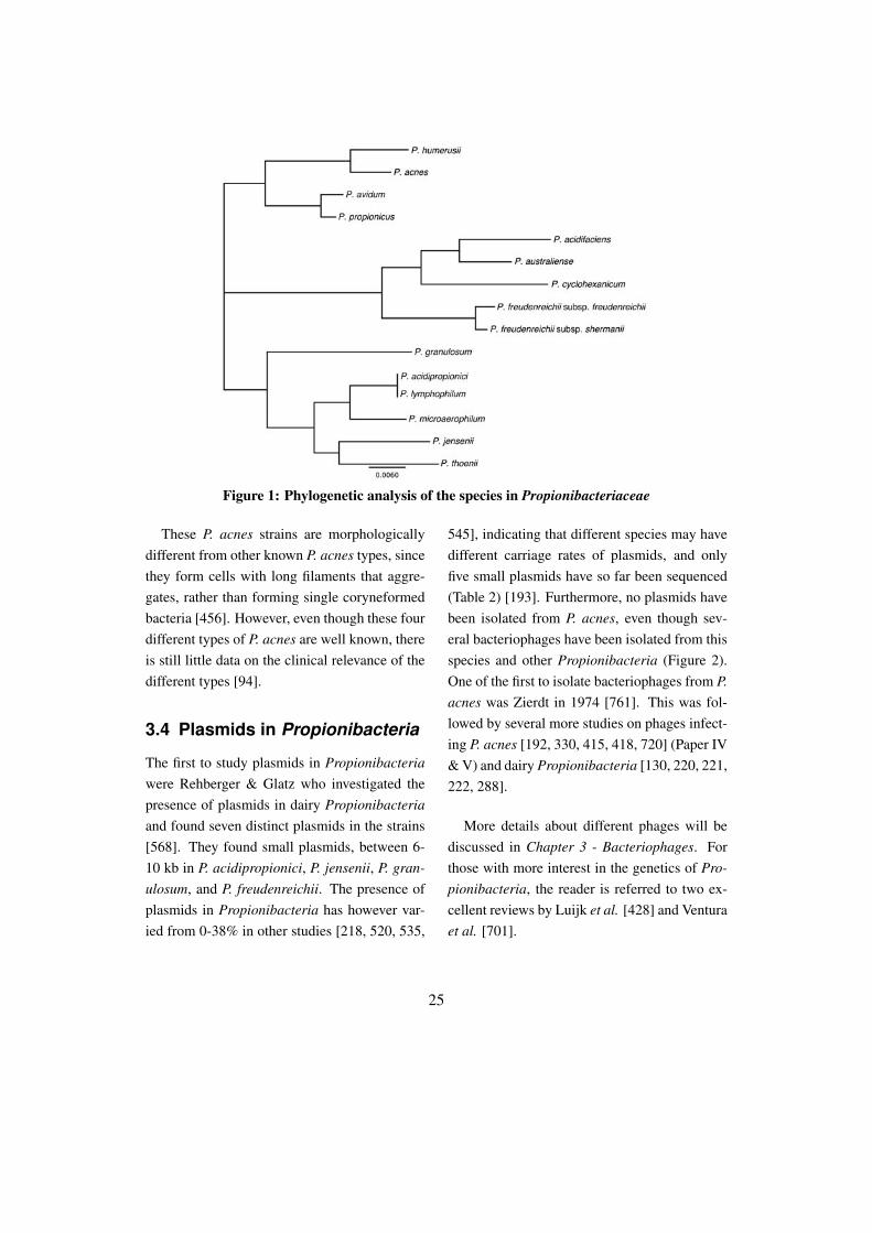
Figure 1: Phylogenetic analysis of the species in Propionibacteriaceae
These P. acnes strains are morphologicallydifferent from other known P. acnes types, sincethey form cells with long filaments that aggre-gates, rather than forming single coryneformedbacteria [456]. However, even though these fourdifferent types of P. acnes are well known, thereis still little data on the clinical relevance of thedifferent types [94].
3.4 Plasmids in Propionibacteria
The first to study plasmids in Propionibacteriawere Rehberger & Glatz who investigated thepresence of plasmids in dairy Propionibacteriaand found seven distinct plasmids in the strains[568]. They found small plasmids, between 6-10 kb in P. acidipropionici, P. jensenii, P. gran-ulosum, and P. freudenreichii. The presence ofplasmids in Propionibacteria has however var-ied from 0-38% in other studies [218, 520, 535,
545], indicating that different species may havedifferent carriage rates of plasmids, and onlyfive small plasmids have so far been sequenced(Table 2) [193]. Furthermore, no plasmids havebeen isolated from P. acnes, even though sev-eral bacteriophages have been isolated from thisspecies and other Propionibacteria (Figure 2).One of the first to isolate bacteriophages from P.acnes was Zierdt in 1974 [761]. This was fol-lowed by several more studies on phages infect-ing P. acnes [192, 330, 415, 418, 720] (Paper IV& V) and dairy Propionibacteria [130, 220, 221,222, 288].
More details about different phages will bediscussed in Chapter 3 - Bacteriophages. Forthose with more interest in the genetics of Pro-pionibacteria, the reader is referred to two ex-cellent reviews by Luijk et al. [428] and Venturaet al. [701].
25
3.5 Transformation systems inPropionibacteria
Several attempts have been done in order to de-velop transformation systems in Propionibacte-ria. The first successful protocol was devel-oped by Gautier et al. in 1995, using puri-fied phage B22 DNA to transform P. freuden-reichii [219], generating a maximum efficiencyof more than 105 cfu/µg DNA. However, thisDNA was isolated from a bacteriophage infect-ing Propionibacteria. Jore et al. found, whendeveloping an E. coli - P. freudenreichii shuttlevector, that DNA isolated from E. coli severelydecreased the transformation efficiency to 10-30cfu/µg DNA, while the same plasmid DNA iso-lated from a P. freudenreichii strain increasedthe efficiency to more than 108 cfu/µg DNA[331]. They concluded that this was due to arestriction-modification system in Propionibac-teria [331]. In order to increase the transforma-tion efficiency, Cheong et al. used dam− E. colistrains for the transformation of P. acnes andincreased the efficiency to approximately 104
cfu/µg DNA [127].The first knock-out in P. acnes was demon-
strated in 2010 by Sörensen et al. [642], wherethey used homologous recombination to gen-erate knock-out mutants of two co-hemolysin
genes. This group, even though using a dam−
E. coli strain, had to use several µg plasmidDNA in order to get a few colonies [642] in-dicating that much work still is needed in op-timizing a transformation protocol for Propi-onibacteria. Other groups have also devel-oped different genetic tools used to knock-outgenes in P. acidipropionici [664], to produce5-aminolevulinic acid using expression vectors[357] and developed shuttle vectors between P.freudenreichii and E. coli [356]. More recently,we developed a system for the homologous ex-pression of recombinant proteins in P. acnes[417] (Paper I), which will facilitate the expres-sion and characterization of proteins from P. ac-nes.
3.6 Morphologicalcharacteristics
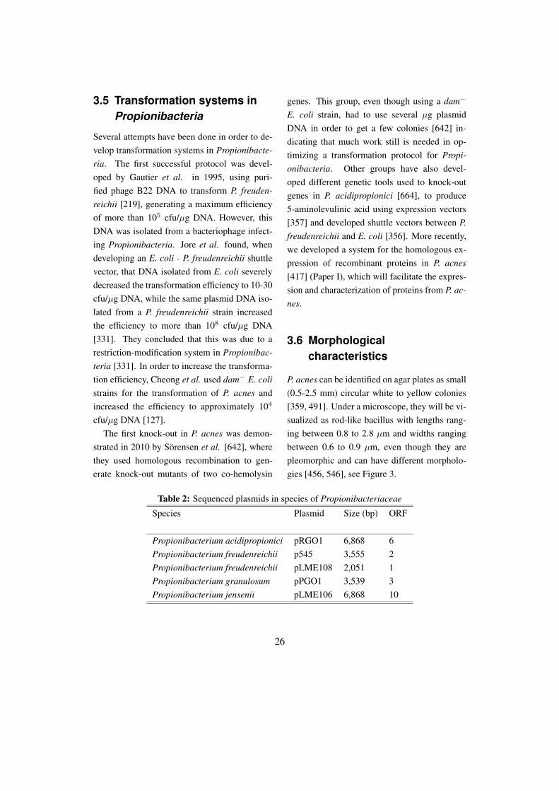
P. acnes can be identified on agar plates as small(0.5-2.5 mm) circular white to yellow colonies[359, 491]. Under a microscope, they will be vi-sualized as rod-like bacillus with lengths rang-ing between 0.8 to 2.8 µm and widths rangingbetween 0.6 to 0.9 µm, even though they arepleomorphic and can have different morpholo-gies [456, 546], see Figure 3.
Table 2: Sequenced plasmids in species of Propionibacteriaceae
Species Plasmid Size (bp) ORF
Propionibacterium acidipropionici pRGO1 6,868 6Propionibacterium freudenreichii p545 3,555 2Propionibacterium freudenreichii pLME108 2,051 1Propionibacterium granulosum pPGO1 3,539 3Propionibacterium jensenii pLME106 6,868 10
26
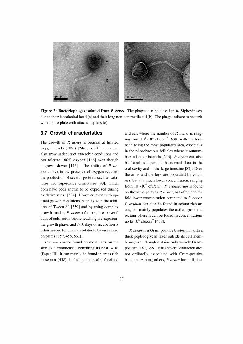
Figure 2: Bacteriophages isolated from P. acnes. The phages can be classified as Siphoviruses,due to their icosahedral head (a) and their long non-contractile tail (b). The phages adhere to bacteriawith a base plate with attached spikes (c).
3.7 Growth characteristics
The growth of P. acnes is optimal at limitedoxygen levels (10%) [246], but P. acnes canalso grow under strict anaerobic conditions andcan tolerate 100% oxygen [146] even thoughit grows slower [145]. The ability of P. ac-nes to live in the presence of oxygen requiresthe production of several proteins such as cata-lases and superoxide dismutases [93], whichboth have been shown to be expressed duringoxidative stress [584]. However, even with op-timal growth conditions, such as with the addi-tion of Tween 80 [359] and by using complexgrowth media, P. acnes often requires severaldays of cultivation before reaching the exponen-tial growth phase, and 7-10 days of incubation isoften needed for clinical isolates to be visualizedon plates [359, 458, 561].
P. acnes can be found on most parts on theskin as a commensal, benefiting its host [416](Paper III). It can mainly be found in areas richin sebum [458], including the scalp, forehead
and ear, where the number of P. acnes is rang-ing from 101-104 cfu/cm2 [639] with the fore-head being the most populated area, especiallyin the pilosebaceous follicles where it outnum-bers all other bacteria [216]. P. acnes can alsobe found as a part of the normal flora in theoral cavity and in the large intestine [87]. Eventhe arms and the legs are populated by P. ac-nes, but at a much lower concentration, rangingfrom 101-102 cfu/cm2. P. granulosum is foundon the same parts as P. acnes, but often at a tenfold lower concentration compared to P. acnes.P. avidum can also be found in sebum rich ar-eas, but mainly populates the axilla, groin andrectum where it can be found in concentrationsup to 103 cfu/cm2 [458].
P. acnes is a Gram-positive bacterium, with athick peptidoglycan layer outside its cell mem-brane, even though it stains only weakly Gram-positive [187, 358]. It has several characteristicsnot ordinarily associated with Gram-positivebacteria. Among others, P. acnes has a distinct
27
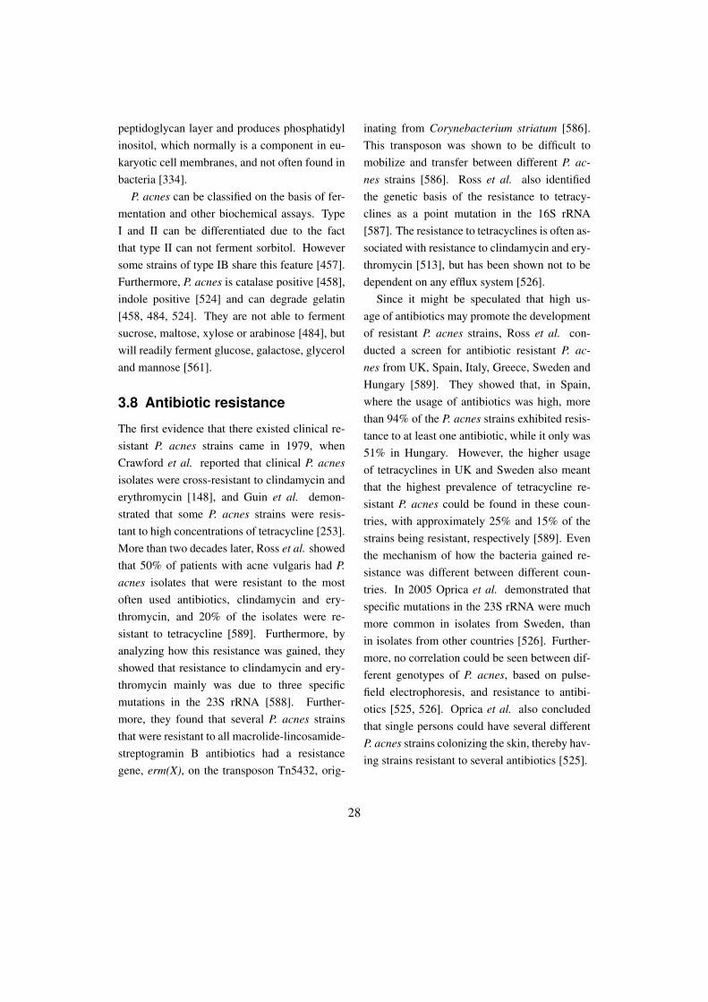
peptidoglycan layer and produces phosphatidylinositol, which normally is a component in eu-karyotic cell membranes, and not often found inbacteria [334].
P. acnes can be classified on the basis of fer-mentation and other biochemical assays. TypeI and II can be differentiated due to the factthat type II can not ferment sorbitol. Howeversome strains of type IB share this feature [457].Furthermore, P. acnes is catalase positive [458],indole positive [524] and can degrade gelatin[458, 484, 524]. They are not able to fermentsucrose, maltose, xylose or arabinose [484], butwill readily ferment glucose, galactose, glyceroland mannose [561].
3.8 Antibiotic resistance
The first evidence that there existed clinical re-sistant P. acnes strains came in 1979, whenCrawford et al. reported that clinical P. acnesisolates were cross-resistant to clindamycin anderythromycin [148], and Guin et al. demon-strated that some P. acnes strains were resis-tant to high concentrations of tetracycline [253].More than two decades later, Ross et al. showedthat 50% of patients with acne vulgaris had P.acnes isolates that were resistant to the mostoften used antibiotics, clindamycin and ery-thromycin, and 20% of the isolates were re-sistant to tetracycline [589]. Furthermore, byanalyzing how this resistance was gained, theyshowed that resistance to clindamycin and ery-thromycin mainly was due to three specificmutations in the 23S rRNA [588]. Further-more, they found that several P. acnes strainsthat were resistant to all macrolide-lincosamide-streptogramin B antibiotics had a resistancegene, erm(X), on the transposon Tn5432, orig-
inating from Corynebacterium striatum [586].This transposon was shown to be difficult tomobilize and transfer between different P. ac-nes strains [586]. Ross et al. also identifiedthe genetic basis of the resistance to tetracy-clines as a point mutation in the 16S rRNA[587]. The resistance to tetracyclines is often as-sociated with resistance to clindamycin and ery-thromycin [513], but has been shown not to bedependent on any efflux system [526].
Since it might be speculated that high us-age of antibiotics may promote the developmentof resistant P. acnes strains, Ross et al. con-ducted a screen for antibiotic resistant P. ac-nes from UK, Spain, Italy, Greece, Sweden andHungary [589]. They showed that, in Spain,where the usage of antibiotics was high, morethan 94% of the P. acnes strains exhibited resis-tance to at least one antibiotic, while it only was51% in Hungary. However, the higher usageof tetracyclines in UK and Sweden also meantthat the highest prevalence of tetracycline re-sistant P. acnes could be found in these coun-tries, with approximately 25% and 15% of thestrains being resistant, respectively [589]. Eventhe mechanism of how the bacteria gained re-sistance was different between different coun-tries. In 2005 Oprica et al. demonstrated thatspecific mutations in the 23S rRNA were muchmore common in isolates from Sweden, thanin isolates from other countries [526]. Further-more, no correlation could be seen between dif-ferent genotypes of P. acnes, based on pulse-field electrophoresis, and resistance to antibi-otics [525, 526]. Oprica et al. also concludedthat single persons could have several differentP. acnes strains colonizing the skin, thereby hav-ing strains resistant to several antibiotics [525].
28
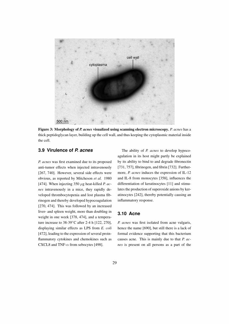
Figure 3: Morphology of P. acnes visualized using scanning electron microscopy. P. acnes has athick peptidoglycan layer, building up the cell wall, and thus keeping the cytoplasmic material insidethe cell.
3.9 Virulence of P. acnes
P. acnes was first examined due to its proposedanti-tumor effects when injected intravenously[267, 740]. However, several side effects wereobvious, as reported by Mitcheson et al. 1980[474]. When injecting 350 µg heat-killed P. ac-nes intravenously in a mice, they rapidly de-veloped thrombocytopenia and lost plasma fib-rinogen and thereby developed hypocoagulation[270, 474]. This was followed by an increasedliver- and spleen weight, more than doubling inweight in one week [378, 474], and a tempera-ture increase to 38-39◦C after 2-4 h [122, 270],displaying similar effects as LPS from E. coli[472], leading to the expression of several proin-flammatory cytokines and chemokines such asCXCL8 and TNF-α from sebocytes [498].
The ability of P. acnes to develop hypoco-agulation in its host might partly be explainedby its ability to bind to and degrade fibronectin[731, 757], fibrinogen, and fibrin [732]. Further-more, P. acnes induces the expression of IL-12and IL-8 from monocytes [358], influences thedifferentiation of keratinocytes [11] and stimu-lates the production of superoxide anions by ker-atinocytes [242], thereby potentially causing aninflammatory response.
3.10 Acne
P. acnes was first isolated from acne vulgaris,hence the name [690], but still there is a lack offormal evidence supporting that this bacteriumcauses acne. This is mainly due to that P. ac-nes is present on all persons as a part of the
29

normal flora, and due to the lack of good mod-els of acne. However some efforts to mimic thepathogenesis of acne have been done and differ-ent models have been evaluated [157, 300, 501,502]. The difficulty of addressing if P. acnes isinvolved in the progress of acne vulgaris mightalso be attributed to the complex nature of thedisease. For further reading in this topic, thereader is recommended three reviews by Toyoda& Morohashi [680], Bojar & Holland [75], andDessinioti & Katsambas [161].
Even though the contribution of P. acnes toacne vulgaris is questioned, the association be-tween acne vulgaris and P. acnes is not. Höffleret al. showed in 1977 that P. acnes was the mostfrequently isolated Propionibacteria from acnevulgaris, and also the most enzymatically ac-tive [294], but also that P. granulosum only wasisolated from patients with acne [223]. How-ever, Propionibacteria are not the only bacte-ria found in acne lesions, since Staphylococ-cus and Malassezia are also frequently isolated[388]. Furthermore, Bek-Thomsen et al. foundthat all follicles from normal skin were colo-nized only by P. acnes, while follicles from pa-tients with acne also included S. epidermidis anda few other bacteria [52]. Furthermore, Höf-fler et al. showed that the secretion of differ-ent enzymes differed between isolates from pa-tients with acne and from normal skin, with thefirst producing more sialidases [296], and moreDNAse and lecithinase, even though these iso-lates produced less proteolytic enzymes [295].Even though not yet formally proved, many pa-pers describe the contribution of P. acnes in thedevelopment of acne vulgaris [719] and puta-tive virulence factors [194] that are supposed to
mediate the inflammatory response. It has how-ever recently been shown that some clones ofP. acnes are associated with severe acne, whileother clones are associated with the normal flora[414].
3.11 Prostate cancer
P. acnes has been associated with prostate can-cer, with several groups constructing oligonu-cleotides (primers) for the detection of P. acnesin prostate tissue [618, 620]. In 2005 Cohenet al. found that 35% of the prostate samplesfrom patients with prostate cancer had infiltra-tion of P. acnes [136], which was significantlyassociated with inflammation. A similar studywas performed by Alexeyev et al. 2007 usingfluorescent in situ hybridization to detect P. ac-nes in prostate tissue [15], showing that P. ac-nes can persist for several years in the prostategland and possibly establish a persistent infec-tion [14, 15]. Recently, Fassi Fehri et al. showedthat P. acnes could be found in more than 80% ofcancer prostate tissues, while being absent fromhealthy prostate and from other cancer tissues[195]. Furthermore, P. acnes isolated from can-cer prostate tissue were able to alter cell prolifer-ation and cellular transformation, indicating thatP. acnes might contribute to the development ofprostate cancer [195].
3.12 Prosthesis removal - biofilmformation
P. acnes is suggested to have a role in the re-moval of prostheses due to infection, based onits ability to form biofilms [301] (Paper II). P.acnes readily forms biofilms on foreign material
30

(Figure 4) and when in a biofilm state, the resis-tance to several antibiotics increases more than10 folds [562] and also the production of extra-cellular lipases and quorum-sensing moleculesincreases [134]. It is estimated that between2-15% of all revision hip operations are dueto infections [40, 375]. However, this numbermight be vastly underestimated, since Tunney
et al. 1998 found that by improving the detec-tion method by using ultrasonication and trans-fer to an anaerobic milieu the detection of bac-teria from removed hip prostheses was almost22% [684]. Besides, 87% of tissue from pa-tients without any culturable bacteria had in-flammatory cells, suggesting that also these pa-tients might have had bacterial infection [684].
Figure 4: Biofilm formation by P. acnes. The bacteria are visible as coryneformed rods (a) insidea biofilm matrix (b).
31

In a following study in 1999, Tunney etal. showed, using immuno-fluorescence mi-croscopy that 63% of the prostheses had ei-ther P. acnes or Staphylococcus [683]. Fur-thermore, 72% of all prostheses were posi-tive for 16S rRNA amplification using univer-sal primers, and 73% of all patients had infil-tration of inflammatory cells in the associatedtissue [683]. Further studies confirmed that P.acnes could cause several different types of in-fections, differing between common late chronicinfections and much more seldom acute post-operative infections [759]. The median infec-tion occurred 7 months after surgery resulting injoint pain and local inflammation [335]. Malesand those that had a history of surgical proce-dures in the specific joint were concluded hav-ing a higher risk of infections [335]. In 2009,Sampedro et al. showed that P. acnes type Iwas more often found in biofilms from prosthe-ses than type II or type III [599]. However, therewas no significant difference between types andnormal skin or infection, suggesting that the dif-ferent types can form biofilm equally well [599].
P. acnes has also been suggested to have arole in the development of infective endocardi-tis [637] and inflammations in the cornea [150].
3.13 Characterized proteins fromP. acnes
Two of the more characterized proteins fromP. acnes are also two of the most abundant se-creted proteins in P. acnes [299], lipases andsialidases, two enzymes involved in degradinglipids and carbohydrates, respectively. Since P.acnes is thought to be involved in the patho-genesis of acne vulgaris it is not surprising that
one of the first proteins to be studied from P.acnes was the lipase encoded by gehA [473].This 33 kDa protein was shown to be the majorsecreted lipase from P. acnes, even though thecomplete genome of P. acnes has several pro-teins annotated as putative lipases [95]. Further-more, 2009 Iinuma et al. showed that P. acnesinduces the formation of lipids in sebocytes dueto increased synthesis of triacylglycerols [315],but as Zouboulis summarizes the finding: ”Therole of P. acnes in sebaceous gland function re-mains uncertain” [766].
Sialidases from P. acnes have also been stud-ied for more than 40 years. Müller describedin 1971 that P. acnes had enzymes with sial-idase activity that could cleave the sialic acidfrom several plasma proteins, including hap-toglobin, α2-macroglobulin, transferrin and IgA[492]. The abundance of sialidases in P. acneswas shown a couple of years later when Höffleret al. compared the activity of sialidases fromdifferent Propionibacteria and found that 83%of P. acnes had sialidase activity, to be comparedwith 20% of P. avidum and 6% of P. granulosum[297]. This conclusion was supported by a fur-ther analysis by von Nicolai et al. in 1980 thatalso concluded that 84% of P. acnes has siali-dase activity, and found this activity in both thecell wall and as a secreted enzyme [510], whichnowadays is supported by the genomic informa-tion from KPA171202, that suggests that thereare both cell wall bound and secreted sialidases[95]. Furthermore, von Nicolai et al. purifiedan enzyme with a molecular weight of 33 kDafrom the culture medium, and they concludedthat the enzyme had the highest activity againstoligosaccharides, rather than to glycoproteins,
32
indicating that this enzyme is not a virulencefactor [510]. However, in contrast to that, Höf-fler et al. found in 1981 that P. acnes isolatesfrom patients with acne had sialidase activity in90% of the cases, compared to strains from thenormal skin which only had activity in 73% ofthe cases [296]. Furthermore, the strains frompatients with acne had almost twice as muchsialidase activity as compared to the strains fromnormal skin, indicating that this enzyme mightcontribute to the development of acne [296].Nakatsuji et al. evaluated this hypothesis on amore molecular level in 2008 when they puri-fied a cell wall anchored sialidase from P. ac-nes [503]. This sialidase was shown to increasethe adhesion of P. acnes to sebocytes and also
increase the cytotoxicity. Mice pre-immunizedwith the sialidase did not develop any inflamma-tory response when they were subcutaneouslyinjected with P. acnes suggesting that a P. ac-nes vaccine based on sialidases might decreasethe inflammatory response caused by P. acnes[503].
Other factors from P. acnes have so far notgained that much attention. However, lately, se-creted CAMP-factors from P. acnes have beenshown to be expressed at high levels [299], withdifferent levels depending on type of P. acnes[692]. Two of the five CAMP-factors were re-cently knocked-out, but did not seem to affectthe virulence of P. acnes [642].
33

4 Virulence factors
4.1 Introduction
The prefix of different bacteria can sometimesbe very hard to interpret. Some are calledpathogenic bacteria and others normal flora,and even further more are called opportunisticpathogens or facultative pathogens. But whatdefines the pathogenicity of a microbe? It seemsclear that those microbes capable of inducing apathogenic state have the ability to produce cer-tain proteins or substances that mediates this ef-fect. But what, in this context, distinguish anopportunistic pathogen from a pathogen? Andwhat factors are involved in this pathogenicity?
4.2 Virulence factors
The ability of a microorganism to induce a dis-ease (to be pathogenic) is based on its virulence.The higher virulence, the higher capacity to in-duce disease. Though this concept is widely ac-cepted, the actual meaning of virulence has beendebated for many decades [118], and its mean-ing has changed from being solely focused onthe bacteria to rather focus on the bacteria-hostinteraction [119]. In the beginning pathogenicbacteria were believed to have different attackand defense mechanisms, which were the onlycauses to the disease [635]. Thus, one factor ofthe bacteria was used to protect itself from thehostile host environment, thereby increasing thepersistence, and one factor of the bacteria wasused to cause damage in the host [762].
The fact that many bacterial virulence factorscan be found in mobile genetic elements such
as bacteriophages, plasmids and pathogenicityislands [514, 533] and can be differently regu-lated during the growth by bacterial sigma fac-tors [345] might have contributed to the theorythat only the bacteria was important for the vir-ulence. Later, scientists began to understandthat bacterial virulence was also dependent onthe host [635], and soon it was widely recog-nized that even though the bacterial proteinsand structures were involved in the pathogene-sis, they alone were not responsible for the viru-lence (Figure 1) [716].
Modern scientist have further extended thisdiscussion and proposed the use of a damage-response context to describe virulence [116]and define virulence as a relative capacity ofa microbe to cause damage in the host [117].The focus is no longer only on the microbe,but also the host, and the damage caused tothe host might be mediated by the microbe,but also by the host [116]. Furthermore, viru-lence should not necessary be defined as a fac-tor that affects virulence but not viability [739],since this would exclude several important cellwall structures such as LPS, which also is re-garded as a virulence factor [64, 289, 702]. Sev-eral other well-known virulence factors includethe pneumolysin [406, 444, 590] from Strep-tococcus pneumonia, polysaccharide capsules[551, 655, 672], neuraminidases from Influenzaviruses [67, 658], cholera toxin from Vibriocholerae [695], and immunoglobulin modulat-ing enzymes from Streptococcus pyogenes [18,517, 543].
34
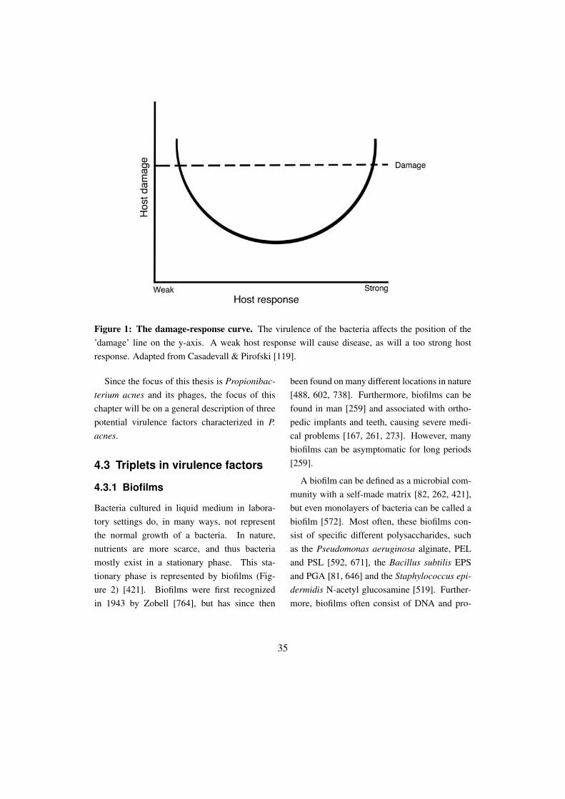
Figure 1: The damage-response curve. The virulence of the bacteria affects the position of the’damage’ line on the y-axis. A weak host response will cause disease, as will a too strong hostresponse. Adapted from Casadevall & Pirofski [119].
Since the focus of this thesis is Propionibac-terium acnes and its phages, the focus of thischapter will be on a general description of threepotential virulence factors characterized in P.acnes.
4.3 Triplets in virulence factors
4.3.1 Biofilms
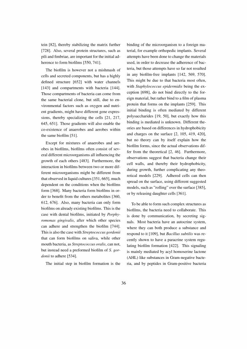
Bacteria cultured in liquid medium in labora-tory settings do, in many ways, not representthe normal growth of a bacteria. In nature,nutrients are more scarce, and thus bacteriamostly exist in a stationary phase. This sta-tionary phase is represented by biofilms (Fig-ure 2) [421]. Biofilms were first recognizedin 1943 by Zobell [764], but has since then
been found on many different locations in nature[488, 602, 738]. Furthermore, biofilms can befound in man [259] and associated with ortho-pedic implants and teeth, causing severe medi-cal problems [167, 261, 273]. However, manybiofilms can be asymptomatic for long periods[259].
A biofilm can be defined as a microbial com-munity with a self-made matrix [82, 262, 421],but even monolayers of bacteria can be called abiofilm [572]. Most often, these biofilms con-sist of specific different polysaccharides, suchas the Pseudomonas aeruginosa alginate, PELand PSL [592, 671], the Bacillus subtilis EPSand PGA [81, 646] and the Staphylococcus epi-dermidis N-acetyl glucosamine [519]. Further-more, biofilms often consist of DNA and pro-
35
tein [82], thereby stabilizing the matrix further[728]. Also, several protein structures, such aspili and fimbriae, are important for the initial ad-herence to form biofilms [550, 741].
The biofilm is however not a mishmash ofcells and secreted components, but has a highlydefined structure [652] with water channels[143] and compartments with bacteria [144].Those compartments of bacteria can come fromthe same bacterial clone, but still, due to en-vironmental factors such as oxygen and nutri-ent gradients, might have different gene expres-sions, thereby specializing the cells [21, 217,645, 651]. Those gradients will also enable theco-existence of anaerobes and aerobes withinthe same biofilm [51].
Except for mixtures of anaerobes and aer-obes in biofilms, biofilms often consist of sev-eral different microorganisms all influencing thegrowth of each others [483]. Furthermore, theinteraction in biofilms between two or more dif-ferent microorganisms might be different fromthat observed in liquid cultures [351, 665], muchdependent on the conditions when the biofilmsform [368]. Many bacteria form biofilms in or-der to benefit from the others metabolites [360,612, 676]. Also, many bacteria can only formbiofilms on already existing biofilms. This is thecase with dental biofilms, initiated by Porphy-romonas gingivalis, after which other speciescan adhere and strengthen the biofilm [744].This is also the case with Streptococcus gordoniithat can form biofilms on saliva, while othermouth bacteria, as Streptococcus oralis, can not,but instead need a preformed biofilm of S. gor-donii to adhere [534].
The initial step in biofilm formation is the
binding of the microorganism to a foreign ma-terial, for example orthopedic implants. Severalattempts have been done to change the materialsused, in order to decrease the adherence of bac-teria, but those attempts have so far not resultedin any biofilm-free implants [142, 569, 570].This might be due to that bacteria most often,with Staphylococcus epidermidis being the ex-ception [698], do not bind directly to the for-eign material, but rather bind to a film of plasmaprotein that forms on the implants [259]. Thisinitial binding is often mediated by differentpolysaccharides [19, 50], but exactly how thisbinding is mediated is unknown. Different the-ories are based on differences in hydrophobicityand charges on the surface [2, 105, 419, 420],but no theory can by itself explain how thebiofilm forms, since the actual observations dif-fer from the theoretical [2, 46]. Furthermore,observations suggest that bacteria change theircell walls, and thereby their hydrophobicity,during growth, further complicating any theo-retical models [229]. Adhered cells can thenspread on the surface, using different suggestedmodels, such as ”rolling” over the surface [385],or by releasing daughter cells [361].
To be able to form such complex structures asbiofilms, the bacteria need to collaborate. Thisis done by communication, by secreting sig-nals. Most bacteria have an autocrine system,where they can both produce a substance andrespond to it [109], but Bacillus subtilis was re-cently shown to have a paracrine system regu-lating biofilm formation [422]. This signalingis mainly mediated by acyl homoserine lactone(AHL) like substances in Gram-negative bacte-ria, and by peptides in Gram-positive bacteria
36
[421]. But, even low concentrations of othersmall molecules, as antibiotics, can trigger theformation of biofilms [298, 750]. Furthermore,signaling is not restricted to signaling betweenone species, but also exists in mixed biofilms[459, 597]. A similar phenomenon was ob-served with an E. coli strain that even thoughnot producing any AHL, still had a homologousreceptor for AHL [694].
One of the largest problems with biofilms isthat they are very difficult to eradicate. Eventhough phages and phage derived proteins havebeen suggested as a treatment [308, 309], thisis not standard procedure today. The prob-lem is mainly due to the higher resistance todifferent antibiotics and antimicrobial peptides[56, 143, 188, 259, 355, 435, 574]. The in-creased resistance to antibiotics could partly
be explained by the thick polysaccharide layer[650], but also by the existence of non-dividingpersister cells in the biofilm [396]. The resis-tance to different substances is further compli-cated in mixed biofilms, where one species cansecrete enzymes protecting the other [342], orby its mere existence physically hinder the sub-stance from reaching sensitive bacteria [401].Even though bacteria generally are resistant toantibiotics when the biofilms are formed, pre-treatment of implants with antibiotics seems todecrease the adherence of bacteria, thereby alsoreducing the formation of biofilms [571, 747].However, the resistance to antibiotics is not onlydependent on what species form the biofilm, butalso on what material the biofilm is formed on[27, 540].
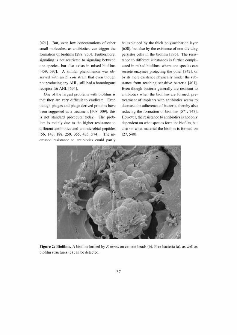
Figure 2: Biofilms. A biofilm formed by P. acnes on cement beads (b). Free bacteria (a), as well asbiofilm structures (c) can be detected.
37
4.3.2 Sialidases
Sialidases were first called Receptor-DestroyingEnzyme (RDE), since the first observation sug-gested that this enzyme from Vibrio cholera de-stroyed the receptor site for Influenza viruses[104], which later was concluded to be dueto sialidase activity [6, 7]. However, alreadyin 1941 Hirst demonstrated sialidase activityin Influenza virus [292]. A few years later,Gottshcalk named the enzyme ”neuraminidase”since it released N-acetylneuraminic acids[239]. Since then, both the word ”neu-raminidase” and ”sialidase” has been used fordescribing this activity [189].
Sialidases work by cleaving terminal sialicacids from glycoproteins and glycolipids [730].They usually display some substrate specificity[141], with the V. cholerae sialidase being ableto cleave 2,3; 2,4; 2,6; and 2,8 α-glycosidicbonds [171], while other sialidases only cancleave certain bonds [240]. Furthermore, sialicacids are not a single substrate, but consist of afamily of more than 30 different [581] nine car-bon sugars with 2-keto-3-deoxy-5-acetamido-D-glycero-D-galacto-nonulosonic acid structure[705], with the most common sialic acid beingN-acetylneuraminic acid (Figure 3) [730]. Sialicacids got their name from the first site of isola-tion, which was in the saliva (from Greek sialon)[71]. Due to this complexity of sialic acids,some researchers have claimed that: ”Sialicacids are not only the most interesting moleculesin the world, but also the most important” [704].
Not all organisms have the ability to pro-duce sialidases. Most plants do not, neither do
most metazoans, but some animals and microor-ganisms can [494, 581]. However, this abilitycan differ between species, and also betweenbacterial isolates [305, 476, 548, 580]. Theability of several different species to producesialidases has made several researchers to con-clude that sialidases have a common ancestorgene [581], since they usually share between 20-30% amino acid identity [305, 581], and have asimilar architecture between bacteria and mam-mals [305, 704, 706]. The sialidases are notbiochemically similar, but do share a similartertiary structure [581], and thereby phyloge-netic trees based on primary sequences do notgive the full picture [24]. Thus, this implicatesthat specific bacterial sialidases can be moresimilar to prokaryotic sialidases on a structurallevel than to other bacterial sialidases [305].Sialidases with a lower molecular weight havea rather similar primary and tertiary structure[305, 704], while larger sialidases (>60 kDa) of-ten have both an enzymatic domain and a do-main conferring the specificity of the enzyme[305, 673, 704], and are thus not as similar toeach other. However, all sialidases have someparts in common. They all have the same cat-alytic site with seven conserved amino acids,among others an arginine triad [704]. Further-more, almost all sialidases have Asp-box mo-tifs, even though the exact mechanism of theseboxes still is unknown [704]. Many sialidasesalso need calcium in order to have enzymaticactivity [305]. But even though the biochemicalpart of sialidases is rather well characterized, thebiological function of the enzyme is not [704].
38

Figure 3: Sialic acids. A representative figure of the most common sialic acid - N-acetylneuraminicacid.
Many microorganisms use sialidases in orderto get nutrition [141, 547], and sialic acids canin E. coli function as the sole carbon and nitro-gen source [445, 547]. The importance of suchsystem is understandable since sialic acids areabundant in the human body. In human serum,there exists 2 mM sialic acids, even though al-most all are in a bound format [625], and evena single red blood cell has more than 10 millionsialic acid molecules bound to its surface [697].Due to this abundance, many commensal bac-teria use sialic acids as a mean of fast energy,without disturbing the host [304].
However, not only commensal bacteria usesialic acids, but also pathogenic microorganisms[140, 141, 172, 566, 687], where they are sug-gested to have a role in the pathogenesis [566],by activating and stimulating cells [429, 566].Even though the sialidases can be both cell wallbound and secreted, it is the latter that is re-
garded to be the most potent to cause differentphysiological effects [141], and help to spreadthe bacteria [140, 493, 495]. So even thoughboth commensals and pathogenic bacteria cansecrete sialidases, only some use it as a viru-lence factor [141]. One of the differences is thatcommensals often produce sialidases constitu-tively, while pathogenic bacteria need a stimu-lus, as free sialic acids or oligosaccharides, toinduce their gene expression [141, 303].
Even though commonly regarded as a viru-lence factor, the main function of sialidases formost bacteria probably is nutritional [305, 496,673]. However, there is a connection betweenhigh levels of sialidase activity and pathogenic-ity for certain species [141]. Group B strepto-cocci start to produce sialidases late in their ex-ponential phase [471], together with many otherdifferent virulence factors, but they do not seemto be necessary to cause disease [497]. How-
39
ever, this sialidase activity can partly desialylateIgA1 in the saliva, and thereby increase the pro-teolytic effect [573]. Furthermore, it has latelybeen shown that S. pneumonia sialidase NanAis essential for the adherence to human brainendothelial cells [688], and decreases phago-cytosis by neutrophils [153]. However, themost characterized sialidase is from InfluenzaA viruses, where it is essential for pathogene-sis [327]. This Influenza sialidase is also ableto increase the adhesion of Neisseria meningi-tides to human cells [563]. In general, sialicacids are removed in order to reveal new re-ceptors [348, 585, 610], but also to increaseadhesion due to the decreased negative surfacecharge [140, 286, 765]. Furthermore, desialy-lation reveals sites for sialic acid-binding Ig su-perfamily lectin (Siglec) recognition sites [567],which, when activated, often modulates the im-mune system [149]. A similar effect can be seenwith desialylated IgG that changes its affinity tothe Fcγ receptors [337].
Not all microorganisms use sialic acids onlyfor nutrition or revealing adhesion sites, but alsofor building capsules protecting them from thehost immune system [323, 703]. The first bacte-rial sialic acids were actually identified from anE. coli capsule [44], which decreases the phago-cytosis [674]. This molecular mimicry bene-fits the bacteria due to an increased immunetolerance [703]. In S. pneumoniae, the sialicacid capsule also lead to an inhibition of the in-sertion of the MAC-complex in the membrane[708]. At least three different methods to gethold on sialic acids for bacteria exist. Eitherthey can synthetize the molecules themselves,as E. coli and N. meningitidis, use exogenous
sialic acids, as N. gonorrhoea [539], or by usingtrans-sialidases, as Trypanosoma cruzi [552].
4.3.3 Protection from reactiveoxygen species
Reactive oxygen species (ROS) is a collectivename for radicals and non-radicals derived fromO2 [264], and is a substance that all living or-ganisms will encounter. The production of ROSin different tissues in human was first suggestedby Gerschman and colleagues [227, 228], andhas since then been concluded to be both ben-eficial and dangerous for the host [131]. Afew years after the discovery of ROS, the firstsuperoxide dismutase (SOD) was characterizedand suggested to have a protective role againstoxidative stress [454]. Furthermore, it wasshown that ROS was able to protect human cellsfrom bacteria [31]. When phagocytes becomeactivated by microbes, they will assembly anNADPH oxidase and produce and secrete largeamounts of the superoxide anion radical O2
−
[31, 32, 362]. This product is not very reactivein itself, but will produce hydrogen peroxidewhich is a known mutagenesis factor [249]. Fur-thermore, hydrogen peroxide can react and pro-duce the highly reactive radical OH∗ [49], eventhough the exact mechanism of this in vivo is un-known [469, 724]. This radical is able to reactwith carbohydrates, lipids, proteins and DNA[249], but will only react where it is formed[137] due to its short diffusion life time of ap-proximately 0.5 µm [468].
All production of ROS from the phagocyteswill eventually cause pathogenic conditions inthe host if not taken care of correctly [137].This damage can be direct or indirect, by de-
40
grading substrates or by activating proteases[137]. Phagocytes will also secrete myeloper-oxidases that can convert hydrogen peroxide tohypochlorous acid which is a much more po-tent radical [362], which will inhibit the ef-fect of several anti-proteases [726] and also ac-tivate secreted proteases from the phagocytes[726]. Furthermore, it has been shown thatROS can signal via the immune regulator NF-κB [613], altering the inflammatory response[137], by increasing the expression of the pro-inflammatory cytokines IL-2, IL-6 and TNF-α[306, 398, 619]. However, the addition of SODand catalase has been shown experimentally toreduce tissue damage due to the activity of freeradicals [132, 243, 329, 628]. Furthermore, anE. coli deficient in SOD will have a much highermutagenesis rate on the DNA when exposed toROS [679]. Those enzymes, together with glu-tathione peroxidase are well established intra-cellular proteins that protect the cells from ROS(Figure 4) [66, 137, 213, 266, 624, 679]. How-ever, there are several other known mechanismsof how to protect the cells from free radicals, us-ing antioxidants.
Antioxidants are defined as a substrate thatcan protect or delay an oxidation of a substrate,even at low concentrations [266]. This effectcan be due to a lowered O2-concentration, abinding of O2 to stable proteins and bindingof otherwise reactive metal ions [263]. Eventhough mainly thought as being an intracellularprotein, several reports have described an extra-cellular SOD [340, 341, 442], even though thesemainly seem to be bound to cells. However,
there is a great abundance of antioxidants in thehuman serum [463]. Inside the cell, reducedglutathione is an important antioxidant [609],however the low extracellular concentration isnot enough to generate a good response againstfree radicals. Furthermore, transferrin is notloaded to more than 20-30% of its capacity withiron, which enables it to bind iron fast beforethe iron can react and form free radicals [257],since when bound the iron is unable to gener-ate OH∗ [28, 256, 257]. Other proteins in theplasma with antioxidant properties are bilirubin[653], ascorbic acid [212, 654], and alpha-1 mi-croglobulin [12, 16, 17]. Furthermore, vitaminC and E are important antioxidants inhibitingoxidation of several substrates [124, 265, 532].A function similar to transferrin is shared withhaptoglobin and hemopexin that will bind freehemoglobin and heme, respectively, before theycan react and release free radicals and stimulateoxidation [255, 258, 265, 596].
A free form of heme from damagedhemoglobin or from heme-proteins is a stimu-lator of oxidation [103, 231, 670], and can gen-erate oxidative stress [258, 669, 670] which hasbeen shown in vitro on several molecules [8, 9].Furthermore, the synthesis of heme can alsogenerate ROS [481]. This is due to the manyreactive properties of some of the building sub-stances [169, 291, 523]. Thus it is of importanceto degrade free heme before it reacts, whichis the function of heme oxygenases [594], thatprotect cells from oxidative stress by degradingheme to biliverdin (which will generate biliru-bin), CO and iron [489].
41

Figure 4: Protective systems against free radicals. Superoxide is dismutated to hydrogen perox-ide, and can then by converted to water by either catalases or peroxidases.
The enzyme was discovered in 1968 [675],is inducible by UVA and hydrogen peroxide[353], and will increase the tolerance of oxida-tive stress to the cells [667]. Heme oxygenasesprotect from oxidative stress both by decreas-ing the amount of free heme, but also by thebyproducts they form when heme is degraded[25, 352, 353, 354]. Low concentration ofCO is anti-inflammatory by downregulating pro-inflammatory cytokines and upregulating IL-10[490, 529] and protects against oxidative stress[530]. Bilirubin protects cells from hydrogen-peroxide [36] and is one of the most importantantioxidant factors in serum [235]. An excessof free iron from the reaction would harm thehost. However, a mouse with a heme oxyge-nase knock-out showed increased levels of iron,compared to the wildtype, indicating that theheme oxygenase has a beneficial role [160]. Ithas been argued that heme oxygenases can in-crease oxidation [372]. This is due to the releaseof small quantities of hydrogen peroxide duringthe reaction, and due to the abundance of free
iron which could benefit a production of OH∗
[178, 512, 626].ROS have lately been gaining interest as sig-
naling molecules [207]. The NADPH oxidaseNox2 is expressed in most cells, even thoughat much lower level than in phagocytes, and arelikely to have a signaling function in those cells[376, 707]. This signaling regulates apoptosisvia the JNK-pathway [159, 162, 407], and willfurthermore due to the ability to modify proteins[209] affect the faith of the cell [260, 448, 449,486]. The most probable signaling molecule isregarded to be hydrogen peroxide [656] that canbe transported out of the cell using aquaporines[68]. However, data also indicate that O2
− canfunction as a signal molecule, even though themechanism still is unknown [434].
4.4 How to study virulencefactors in P. acnes
Several putative virulence factors in P. acneshave already been studied to some extent, suchas the lipase GehA [473], sialidases [503],
42

and CAMP-factors [299, 642]. However, eventhough these studies proved successful, the ex-pression of recombinant proteins from P. ac-nes has proven difficult. Miskin et al. [473]optimized the expression by lowering the tem-perature to 27◦C and adding 0.45 M sucrose.Nakatsuji et al. [503], instead of optimizingthe expression, denatured and renatured inclu-sion bodies, so that the sialidase was soluble andactive. Sörensen et al. [642] purified recom-binant CAMP-factors from P. acnes. However,since these proteins only were used for develop-ing antibodies, and their activity was not tested,it is impossible to know if these proteins wereactive or not.
However, this author has also had problemswith recombinant expression of proteins from P.acnes and its phages in E. coli, where most pro-teins investigated have formed inclusion bod-ies. Even though some of these inclusion bodieswere able to be dissolved using 6 M guanidinehydrochloride, and refolded to a soluble state,this method is quite unsatisfactory since muchof the protein activity might be lost during thisharsh purification. Therefore we decided to de-velop tools to both be able to express recombi-nant proteins from P. acnes in P. acnes, and to beable to complement knock-outs (Figure 5) [417](Paper I).
Figure 5: Vector p1340 developed for the recombinant expression of homologous proteins inP. acnes. The vector has resistance cassettes for selection in E. coli (Amp) and in P. acnes (Ery andCm).
43
Part IIThe Good Guys
You still do not know what you are dealing with, do you?Perfect organism. Its structural perfection is matched only by its hostility...
I admire its purity; unclouded by conscience, remorse, or delusions of morality-Ash to Ripley in Alien 1979
44
5 Bacteriophages
5.1 Introduction
Not living, nor dead. Not friends, nor foes. Theycan be infectious for decades [382] and foundwherever their hosts are [722] - in deserts [554],in food [349], in our blood [536], in urine andsaliva [33, 749], and in ocean water where theycan reach numbers of 106 specimens/ml [58]. Infact they are so common in our bodies that morethan 1200 different genotypes can be identifiedfrom feces only [85]. They are the most com-mon entity found in the world, outnumberingbacteria 10:1 [281, 729], reaching an impressivetheoretical number of 1031 [281, 662]. Usingmathematical models, it has been estimated that10-20% of all their hosts in the water die everysingle day due to infections [733]. Furthermore,if placed after each other, they would form abridge to our second closest star Alpha Centauri,4.35 light years away - 46 million times [282]!Bacteriophages truly are impressive!
5.2 Classification
5.2.1 Introduction
Bacteriophages, or phages, are viruses that in-fect bacteria, and are estimated to have beenevolved when bacteria diverged from Eukary-oteae and Archeae [280]. They were first iden-tified by Fredrick Twort 1915 [685] and Felixd’Hérelle 1917 [163]. Even though they bothsoon realized that phages could be used to treatinfectious diseases [659], they did not know that
they were working with viruses. It was not until1942 that phages were recognized as viruses us-ing electron microscopy [430], and phages con-tinued for a long period of time to be the mainfocus of electron microscopy [23]. Further ma-jor scientific discoveries using the phages in-clude the finding in 1952 that DNA is the ge-netic material [287]. This knowledge was im-plemented when the first genome ever was se-quenced from phage φX174 1978 [600], fol-lowed a few years later by the sequence of phageλ in 1982 [601]. However, already from the startof phage biology the structural appearance ofthe phage has been one of the most importantfeatures to classify the phages together with thenature of the genomic material [191].
5.2.2 Classification
Phages can be classified into 12 different classesof phages (Table 1). The most common groupidentified is Caudovirales, constituting morethan 95% of all known phages [453]. Caudovi-rales are phages with tails, and could be di-vided into Myoviruses (long contractile tails),Siphoviruses (long non-contractile tails) (Figure1) and Podoviruses (short non-contractile tails)[438]. In the genomic era, several suggestionshave been proposed in order to facilitate theclassification of phages without the necessity ofelectron microscopy. However, so far no goldenstandard exists and thus the International Com-mittee on Taxonomy of Viruses (ICTV) systemis the most widely used [120, 191].
45

Table 1: Phage families (adapted from Veiga-Crespo & Villa [699] and ICTV [191])
Family Genetic material Morphology Host
Myoviridae dsDNA contractile long tail bacteriaSiphoviridae dsDNA non-contractile long tail bacteriaPodoviridae dsDNA non-contractile short tail bacteriaTectiviridae dsDNA double capsid bacteriaPlasmaviridae dsDNA pleomorphic bacteriaCorticoviridae dsDNA internal lipids bacteriaGuttaviridae dsDNA droplet shaped archeaRudiviridae dsDNA filamentous; non-enveloped archeaLipothrixviridae dsDNA filamentous; enveloped archeaFuselloviridae dsDNA lemon shaped archeaMicroviridae ssDNA capsid with spike bacteriaInoviridae ssDNA filamentous phages bacteriaCystoviridae ssRNA segmented genome bacteriaLeviviridae ssRNA spherical bacteria
Some of the proposed methods to classify thephages are similar to the ICTV standard [383],while other methods use a proteomic approach[583]. Even though there is no counterpart tothe bacterial recA in phages [583], attempts havebeen made to classify phages based on their cap-sule genes, since they to some extent are con-served in all known phages [557].
The difficulty by using a genetic approach toclassify phages is their modular nature. Phagesin the Caudovirales family generally consist ofseveral different genetic modules that can bechanged between different phages [283], eventhough phages isolated from the dairy industryare more conserved, potentially due to the selec-tion of the material [98]. However, it is unlikelythat modular recombination will take place inmodules encoding structural head genes, sincesuch modulations in most cases will lead to de-
fect phages due to the complexity and interac-tions between those proteins [332]. The trans-fer of modules is mainly regulated by horizon-tal gene transfer [114], but there has been the-oretical calculations estimating phage mediatedtransduction to happen in a frequency of 1013
times/year [325, 722]. Furthermore, phageshave the capacity to load their capsules withup to 4 kb more DNA, enabling the phages totake with them new integrated genes [121]. Themodule theory was founded more than 30 yearsago by Botstein and is still considered valid[78].
5.2.3 Identification of phages
Phages are usually identified using an overlay-plate approach (Figure 2), where the phages arepropagated against a selected host bacterium,
46
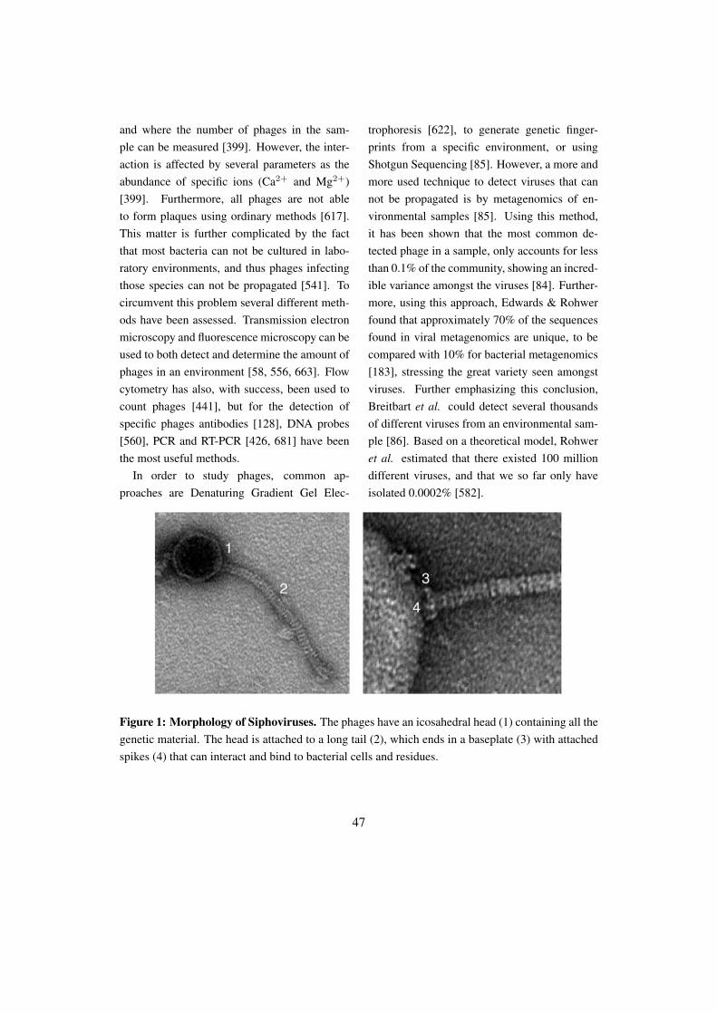
and where the number of phages in the sam-ple can be measured [399]. However, the inter-action is affected by several parameters as theabundance of specific ions (Ca2+ and Mg2+)[399]. Furthermore, all phages are not ableto form plaques using ordinary methods [617].This matter is further complicated by the factthat most bacteria can not be cultured in labo-ratory environments, and thus phages infectingthose species can not be propagated [541]. Tocircumvent this problem several different meth-ods have been assessed. Transmission electronmicroscopy and fluorescence microscopy can beused to both detect and determine the amount ofphages in an environment [58, 556, 663]. Flowcytometry has also, with success, been used tocount phages [441], but for the detection ofspecific phages antibodies [128], DNA probes[560], PCR and RT-PCR [426, 681] have beenthe most useful methods.
In order to study phages, common ap-proaches are Denaturing Gradient Gel Elec-
trophoresis [622], to generate genetic finger-prints from a specific environment, or usingShotgun Sequencing [85]. However, a more andmore used technique to detect viruses that cannot be propagated is by metagenomics of en-vironmental samples [85]. Using this method,it has been shown that the most common de-tected phage in a sample, only accounts for lessthan 0.1% of the community, showing an incred-ible variance amongst the viruses [84]. Further-more, using this approach, Edwards & Rohwerfound that approximately 70% of the sequencesfound in viral metagenomics are unique, to becompared with 10% for bacterial metagenomics[183], stressing the great variety seen amongstviruses. Further emphasizing this conclusion,Breitbart et al. could detect several thousandsof different viruses from an environmental sam-ple [86]. Based on a theoretical model, Rohweret al. estimated that there existed 100 milliondifferent viruses, and that we so far only haveisolated 0.0002% [582].
Figure 1: Morphology of Siphoviruses. The phages have an icosahedral head (1) containing all thegenetic material. The head is attached to a long tail (2), which ends in a baseplate (3) with attachedspikes (4) that can interact and bind to bacterial cells and residues.
47
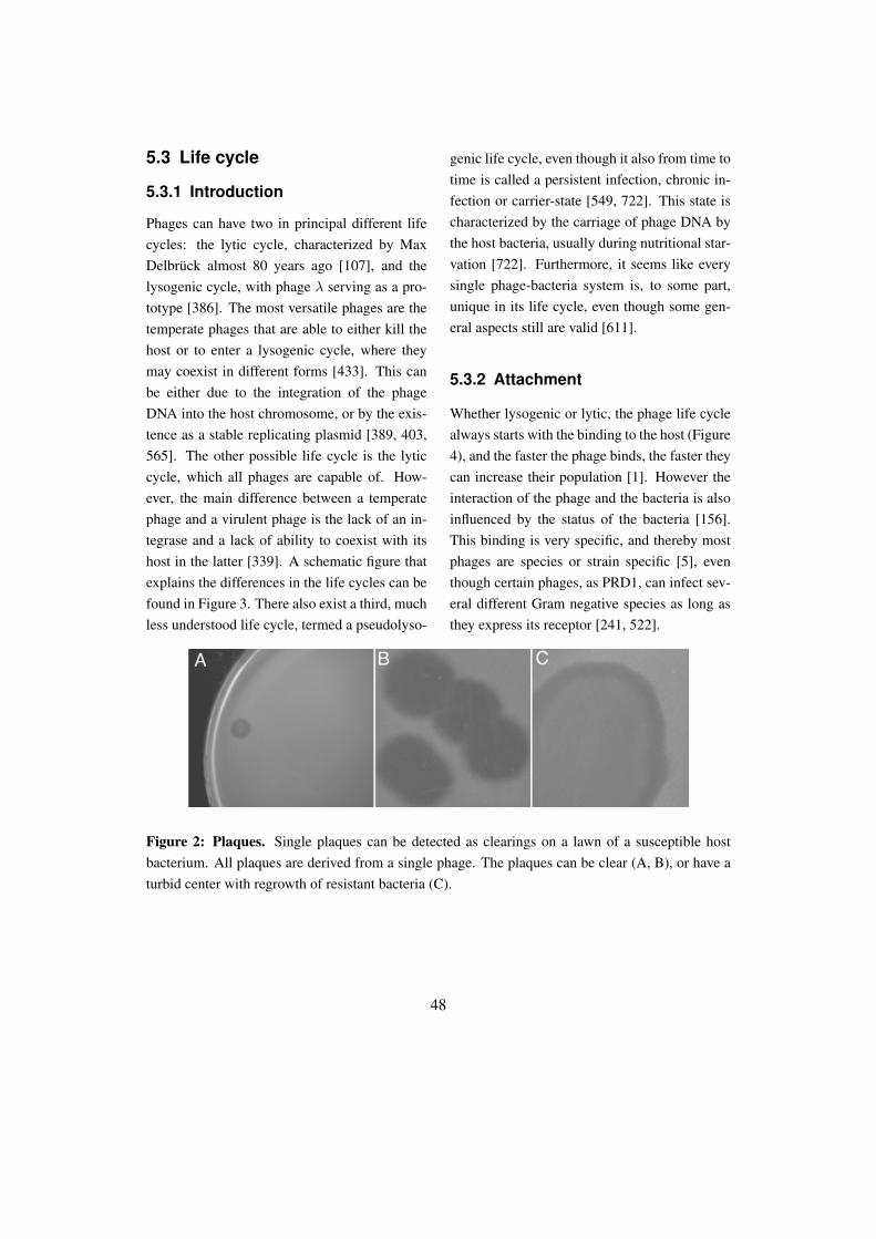
5.3 Life cycle
5.3.1 Introduction
Phages can have two in principal different lifecycles: the lytic cycle, characterized by MaxDelbrück almost 80 years ago [107], and thelysogenic cycle, with phage λ serving as a pro-totype [386]. The most versatile phages are thetemperate phages that are able to either kill thehost or to enter a lysogenic cycle, where theymay coexist in different forms [433]. This canbe either due to the integration of the phageDNA into the host chromosome, or by the exis-tence as a stable replicating plasmid [389, 403,565]. The other possible life cycle is the lyticcycle, which all phages are capable of. How-ever, the main difference between a temperatephage and a virulent phage is the lack of an in-tegrase and a lack of ability to coexist with itshost in the latter [339]. A schematic figure thatexplains the differences in the life cycles can befound in Figure 3. There also exist a third, muchless understood life cycle, termed a pseudolyso-
genic life cycle, even though it also from time totime is called a persistent infection, chronic in-fection or carrier-state [549, 722]. This state ischaracterized by the carriage of phage DNA bythe host bacteria, usually during nutritional star-vation [722]. Furthermore, it seems like everysingle phage-bacteria system is, to some part,unique in its life cycle, even though some gen-eral aspects still are valid [611].
5.3.2 Attachment
Whether lysogenic or lytic, the phage life cyclealways starts with the binding to the host (Figure4), and the faster the phage binds, the faster theycan increase their population [1]. However theinteraction of the phage and the bacteria is alsoinfluenced by the status of the bacteria [156].This binding is very specific, and thereby mostphages are species or strain specific [5], eventhough certain phages, as PRD1, can infect sev-eral different Gram negative species as long asthey express its receptor [241, 522].
Figure 2: Plaques. Single plaques can be detected as clearings on a lawn of a susceptible hostbacterium. All plaques are derived from a single phage. The plaques can be clear (A, B), or have aturbid center with regrowth of resistant bacteria (C).
48

Figure 3: Principal life cycles for bacteriophages. The phages adhere to the bacterial cell wall,and inject their genetic material. The phage can then decide if it should enter a lysogenic cycle ora lytic cycle. In the lysogenic cycle, the phage either integrates its genetic material into its hostchromosome, or exist as a stable plasmid. In the lytic cycle, the phage will replicate, degrade thepeptidoglycan layer of the host using endolysins, and escape from the host.
The knowledge that phages generally werespecific was rapidly used to develop several socalled phage typing sets, where the ability ofphages to infect certain bacterial strains wereused to classify the bacteria [293], with oneof the first type-system developed in 1938 forSalmonella [147]. Even though this system tosome extent still is used today, the importanceof phage typing has decreased [638]. Further-more, the host-range of phages has been shownto depend on which bacterial strain is used for
propagation, due to different DNA modifica-tion systems in the bacteria. This phenomenonis known as host-induced modifications of thephages [63, 432].
Phages initially bind reversible and then ir-reversible to a second structure [279, 390, 436,480, 564, 714]. Bacteriophages infecting Grampositive bacteria usually first bind to sugar moi-eties in the peptidoglycan layer, followed by anirreversible binding to proteins [26, 482], whilephages infecting Gram negative bacteria usually
49

interacts with LPS [278]. But several differ-ent receptors have been identified for differentphages, including both proteins [83, 564, 693,756] and carbohydrates [175, 225]. Since thisis the first contact between the phage and thebacterium, inhibition of attachment is one ofthe most commonly used strategies for bacte-ria to gain resistance to phages [20, 165], andmore than 50 different mechanisms to gain resis-tance to phages have been characterized [135].One of the resistance mechanisms is to formcapsules, which is known to inhibit the attach-ment of certain phages [62]. However, phageshave counter-measurements to degrade capsules[48], and this polysaccharide hydrolyzing ac-tivity has been found in lots of phages infect-ing a wide diversity of Gram negative bacteria[13, 38, 45, 254, 309, 373, 575, 696, 758]. Eventhough the phage adhesins bind to very differentstructures, most of the adhesins have similaritiesin secondary structure with a homo tetramer anda high percentage of beta structures, leading to ahighly stable heat and protease tolerant protein[475, 721].
5.3.3 DNA injection
Once bound to the receptor, the next step ofthe phage infection is to inject the DNA, butin contrast to many human viruses, the phagesleave their structural proteins outside their hosts[392]. This presents another problem, howto get the DNA over the membrane, and alsothrough the thick peptidoglycan layer of Grampositive bacteria, which can be between 20-50nm thick [65, 374]. This is often solved byphage structural peptidoglycan hydrolyzing en-zymes [336, 477, 593], as has been shown for
Lactococcus phage Tuc2009 that has a cell-walldegrading tail fiber [350].
Even though phage genomes can be verysmall and constitute less than 12 genes [108],the length of DNA can vary between 4-640 kb[99]. However, the concentration of DNA inthe capsule is always approximately the same indifferent phages, eg 450 mg/ml [72]. This highpressure introduced when packaging the DNA isthought to help during the injection [179, 576],since it is known for certain phages (T5) that itis only necessary with a binding to the recep-tor in order to release the phage DNA [515]. Ithas also been hypothesized that phage DNA isinjected into the bacterium due to proton gra-dients at the cytosolic membrane [248]. Eventhough this mechanism in general increases theinflux of DNA [79], it seems like there exist sev-eral methods for phages. For example phage T5starts by injecting a small portion of its DNA,then pauses for 4 min until it injects the rest ofthe DNA [380].
The injection of the DNA is facilitated byphage helper proteins that form a channel for theDNA to pass through the peptidoglycan layer[158, 252]. Those DNA transportation proteinscan even cross lipid membranes [74, 199]. It haseven been shown that the tape measure proteinof the phage, that usually determines the lengthof the tail, is used as a DNA transporter protein[579, 608]. The DNA is finally injected in theopposite direction as it was loaded [232, 479].
5.3.4 Life cycle decision
It is not entirely known how the decision to entera lysogenic or a lytic cycle is made, but factorsaffecting the choice have been identified. What
50

is known is that the decision is not immediatelymade after injection, since the fast propagatingphage lambda needs 10-15 min before it deter-mines what cycle it should enter [181]. Dur-ing this time, the phage can ”sense” the statusof the bacteria and adopt its cycle to that, us-ing a genetic switch [559]. For example if thebacteria are growing very slowly, due to nutri-tional limitations, the phage can adapt and pro-long the eclipse period (time when the phageproduces structural proteins), and enter a pseu-dolysogenic cycle, since this is the most benefi-
cial state during these circumstances [37, 427].Furthermore, the decision is influenced by fac-tors such as nutrients, ions, stress and the ratiobetween the bacteria and the phage [723], wherea low number of bacteria with many phages pro-mote a lysogenic decision [180, 649]. Therealso exist theories that the morphology of thephages influences the choice of life cycle, sinceMyoviruses mainly are lytic and Siphovirusesmainly are lysogenic [662], but this is so far notsupported by any experimental data.
Figure 4: The attachment of bacteriophages to the surface of P. acnes. The two phages to the lefthave still not injected their DNA, which can be detected as white lines in the head. The third phageto the left has already injected the DNA and thus the head is deformed due to the pressure appliedusing negative staining.
51
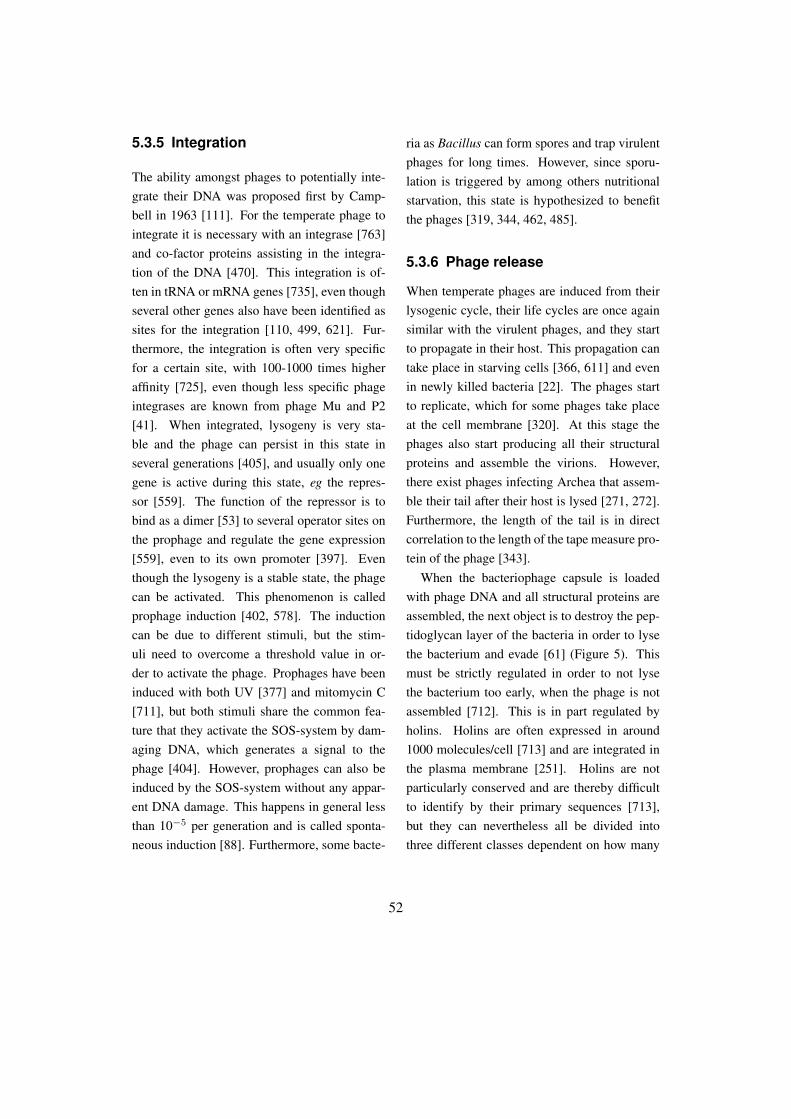
5.3.5 Integration
The ability amongst phages to potentially inte-grate their DNA was proposed first by Camp-bell in 1963 [111]. For the temperate phage tointegrate it is necessary with an integrase [763]and co-factor proteins assisting in the integra-tion of the DNA [470]. This integration is of-ten in tRNA or mRNA genes [735], even thoughseveral other genes also have been identified assites for the integration [110, 499, 621]. Fur-thermore, the integration is often very specificfor a certain site, with 100-1000 times higheraffinity [725], even though less specific phageintegrases are known from phage Mu and P2[41]. When integrated, lysogeny is very sta-ble and the phage can persist in this state inseveral generations [405], and usually only onegene is active during this state, eg the repres-sor [559]. The function of the repressor is tobind as a dimer [53] to several operator sites onthe prophage and regulate the gene expression[559], even to its own promoter [397]. Eventhough the lysogeny is a stable state, the phagecan be activated. This phenomenon is calledprophage induction [402, 578]. The inductioncan be due to different stimuli, but the stim-uli need to overcome a threshold value in or-der to activate the phage. Prophages have beeninduced with both UV [377] and mitomycin C[711], but both stimuli share the common fea-ture that they activate the SOS-system by dam-aging DNA, which generates a signal to thephage [404]. However, prophages can also beinduced by the SOS-system without any appar-ent DNA damage. This happens in general lessthan 10−5 per generation and is called sponta-neous induction [88]. Furthermore, some bacte-
ria as Bacillus can form spores and trap virulentphages for long times. However, since sporu-lation is triggered by among others nutritionalstarvation, this state is hypothesized to benefitthe phages [319, 344, 462, 485].
5.3.6 Phage release
When temperate phages are induced from theirlysogenic cycle, their life cycles are once againsimilar with the virulent phages, and they startto propagate in their host. This propagation cantake place in starving cells [366, 611] and evenin newly killed bacteria [22]. The phages startto replicate, which for some phages take placeat the cell membrane [320]. At this stage thephages also start producing all their structuralproteins and assemble the virions. However,there exist phages infecting Archea that assem-ble their tail after their host is lysed [271, 272].Furthermore, the length of the tail is in directcorrelation to the length of the tape measure pro-tein of the phage [343].
When the bacteriophage capsule is loadedwith phage DNA and all structural proteins areassembled, the next object is to destroy the pep-tidoglycan layer of the bacteria in order to lysethe bacterium and evade [61] (Figure 5). Thismust be strictly regulated in order to not lysethe bacterium too early, when the phage is notassembled [712]. This is in part regulated byholins. Holins are often expressed in around1000 molecules/cell [713] and are integrated inthe plasma membrane [251]. Holins are notparticularly conserved and are thereby difficultto identify by their primary sequences [713],but they can nevertheless all be divided intothree different classes dependent on how many
52

transmembrane domains they have [713]. Theholins are in their turn regulated by anti-holins[755], which are expressed before the holins[70]. Anti-holins and holins often only differin 2-3 amino acids in the N-terminal part of theprotein since the anti-holins starts at an alter-native start codon a few bases upstream of theholin [713]. However, this small difference en-ables the anti-holin to interact with the mem-brane in a different way than the holin, andthereby inhibit its effect, until enough holinshave been expressed [244, 250].
When the holin is assembled the preformedlysins can gain access to the peptidoglycan layerand hydrolyze their substrates [752]. Lysinscan have four different activities: N-acetyl-β-D-glucosaminidase, N-acetyl-β-D-muramidase,N-acetylmuramoyl-L-alanine amidase and en-dopeptidase (Figure 6) [604]. Lysins usuallyonly have one enzymatic domain even thoughlysins with several different active sites havebeen characterized [505]. Except for their ac-tive domain, which usually is N-terminal, mostlysins have a C-terminal that generates the spe-cific binding [203, 285, 412, 423]. Further-more, lysins from phages infecting Gram neg-ative bacteria are usually considerably smallerthan lysins from phages infecting Gram positivebacteria [754]. It has also recently been foundthat lysins might have signal peptides for secre-tion and are thereby secreted out of the bacteria[604]. This has experimentally been verified fora phage infecting Oenococcus oeni, where thelysin needs to be secreted in order to get acti-vated [605, 606], but several more phage lysinsshare the same signal peptides and are therebythought to be secreted [333, 603, 606].
Even though the above described lysis of bac-teria is the most common mechanism for themore complex dsDNA phages, other phages em-ploy other as refined methods to evade fromtheir hosts. Smaller phages often only use a sin-gle protein [753], interfering with the formationof the peptidoglycan layer [60, 61]. Those pepti-doglycan inhibitory proteins of non-lysin originare commonly called amurins [61]. Filamen-tous phages employ yet another strategy, sincethey are being released from the host bacteriumwithout causing lysis [439, 446], by expressingproteins that allows them to be secreted [636]through small channels [196, 591].
5.4 Phage mediated virulence
Since temperate phages have the ability to inte-grate their DNA into the host chromosome, it isnot surprising that bacterial genomes are full ofprophages. Studies have found that between 3-10% of the bacterial genome is constituted byphage-like modules [114, 120], but it can be alot more. For example S. pyogenes strain SF370has more than 16% of its genome covered byprophages [197]. Some of those modules aredefective prophages that can not replicate [112],others might be satellite phages that can repli-cate but do not encode all their structural pro-teins by themselves, but rather steal them fromother phages [120]. Further domains might con-stitute gene transfer agents that are phage-likeparticles that assemble bacterial DNA [310].
Even though the prophages are so common,the annotation of them is still mainly performedmanually [384]. There have been several at-tempts to develop software that could detectprophage regions in bacterial genomes, but so
53

far with limited success [174, 682]. A usualcharacteristic of prophages is the integrase, re-pressors and lysins [113, 120], but even the rep-etition of Gly-X-Y is a characteristic motif oftenfound in phage tail fibers [634], and can be usedfor the detection of prophages. Furthermore, thefunctionality of the phage proteins are most of-ten assigned based on similarities to other char-acterized proteins [531], and secondary struc-ture homologues using Pfam [640].
In order to be accepted by the bacterium,prophages often confer some advantages to theirhost [99, 182]. This advantage could either bean increased pathogenicity [80] or an increased
fitness in a specific environment [700] (Table2). However, some prophages have been re-ported to be beneficial for the outcome of patho-genesis, since E. coli without a specific phagemore often causes disturbances in the gastroin-testinal tract [275]. But, it has also been shownthat the pathogenic E. coli strain O157:H7 has18 prophages that together constitute more than50% of the genetic difference between O157:H7and a non-pathogenic E. coli strain [521]. Thesame phenomenon could be seen in S. pyogeneswhere more than 75% of the genetic variationsbetween pathogenic and non-pathogenic strainscan be due to prophages [34].
Figure 5: The lysis of bacteria and the subsequent release of phages. P. acnes infected withbacteriophages are lysed from within and release a huge amount of cytoplasmic material and phagesas it dies.
54
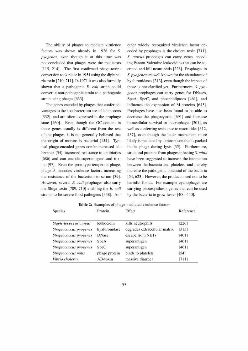
The ability of phages to mediate virulencefactors was shown already in 1926 for S.pyogenes, even though it at this time wasnot concluded that phages were the mediators[115, 214]. The first confirmed phage-toxin-conversion took place in 1951 using the diphthe-ria toxin [210, 211]. In 1971 it was also formallyshown that a pathogenic E. coli strain couldconvert a non-pathogenic strain to a pathogenicstrain using phages [633].
The genes encoded by phages that confer ad-vantages to the host bacterium are called morons[332], and are often expressed in the prophagestate [460]. Even though the GC-content inthose genes usually is different from the restof the phages, it is not generally believed thatthe origin of morons is bacterial [154]. Typ-ical phage-encoded genes confer increased ad-herence [54], increased resistance to antibiotics[686] and can encode superantigens and tox-ins [97]. Even the prototype temperate phage,phage λ, encodes virulence factors increasingthe resistance of the bacterium to serum [39].However, several E. coli prophages also carrythe Shiga toxin [709, 710] enabling the E. colistrains to be severe food pathogens [338]. An-
other widely recognized virulence factor en-coded by prophages is the cholera toxin [711].S. aureus prophages can carry genes encod-ing Panton-Valentine leukocidins that can be se-creted and kill neutrophils [226]. Prophages inS. pyogenes are well known for the abundance ofhyaluronidases [313], even though the impact ofthose is not clarified yet. Furthermore, S. pyo-genes prophages can carry genes for DNases,SpeA, SpeC, and phospholipases [461], andinfluence the expression of M-proteins [643].Prophages have also been found to be able todecrease the phagocytosis [691] and increaseintracellular survival in macrophages [201], aswell as conferring resistance to macrolides [312,437], even though the latter mechanism morelikely is mediated by a transposon that is packedin the phage during lysis [35]. Furthermore,structural proteins from phages infecting S. mitishave been suggested to increase the interactionbetween the bacteria and platelets, and therebyincrease the pathogenic potential of the bacteria[54, 623]. However, the products need not to beharmful for us. For example cyanophages arecarrying photosynthesis genes that can be usedby the bacteria to grow faster [400, 440].
Table 2: Examples of phage mediated virulence factors
Species Protein Effect Reference
Staphylococcus aureus leukocidin kills neutrophils [226]Streptococcus pyogenes hyaluronidase degrades extracellular matrix [313]Streptococcus pyogenes DNase escape from NETs [461]Streptococcus pyogenes SpeA superantigen [461]Streptococcus pyogenes SpeC superantigen [461]Streptococcus mitis phage protein binds to platelets [54]Vibrio cholerae AB-toxin massive diarrhea [711]
55
Even when phages are induced, they mightstill contribute to the virulence for the bacte-ria, since this induction also triggers the produc-tion of the potential virulence factors encodedby the phage [3, 276, 748]. Activators of thisprocess might be environmental, such as cell ex-tracts [90, 91] or hydrogen peroxide from neu-trophils [709]. The activating signal can evenbe antibiotics, thereby enhancing the pathogen-esis rather than decreasing it [365, 450]. Activa-tion of prophages can also lead to spread of vir-ulence, mediated by phages, to non-pathogenicstrains of the normal flora [89].
5.5 Phages in the industry
5.5.1 Industrial problems withphages
Fermentation, a process to change carbohy-drates into alcohols, is often used in several in-dustries to generate among others yogurt andvinegar. This fermentation is processed by bac-teria, and is hence sensitive for the activity ofphages [73]. It has been estimated that between0.1-10% of all fermentations are destroyed, de-pending on the industry [73, 478]. This prob-lem is mainly due to that phages are difficultto eradicate, and investigations have shown thepresence of phages in the air of fermentation in-dustries, reaching concentrations of 105 pfu/m3
[509]. Due to this problem, a huge effort hasbeen made to characterize phages infecting lac-tic acid bacteria, and more than 100 phages havebeen sequenced [4, 98], mainly being classifiedas Siphoviruses [69]. Several strategies havebeen assessed in order to solve this problem,among others by using different bacterial rota-
tion schemes [177, 627] and the usage of bacte-rial strains resistant to most phages [208].
5.5.2 Phage display
The technique for phage display was inventedalmost 25 years ago [630]. This is a techniquewhere peptides can be displayed on the surfaceon phages, usually using their capsule proteins[630]. It has so far only been used in phagesfrom E. coli [648, 734, 745], but has shown tobe a valuable tool in the development of mono-clonal antibodies [736] and for vaccination stud-ies [47, 465].
5.6 Phage therapy
5.6.1 Phage therapy using wholephages
When Twort in 1915 first discovered virusesthat could infect bacteria he soon realized thatphages could be used as antibacterial agents[659]. He was, nevertheless, not the first to iden-tify the antibacterial effect of phages. Alreadyin 1896, Hankin could demonstrate that waterfrom rivers could kill Vibrio cholerae [268], butdid not conclude that this was due to bacterialviruses. Furthermore, in 1918 d’Hérelle alsounderstood the therapeutic potential amongstphages and used them to treat dysentery [661],and cholera in India [164], where the mortalitydecreased from 25% to 0% [96]. Soon, manymore scientists followed their lead and usedphage therapy to treat different bacterial infec-tions [101, 155, 644]. Phage therapy was mainlyused to treat dysentery [30], but also against dif-ferent lung diseases [317] and wounds [760].
56
Most phage therapies have so far been con-ducted in former Soviet [464, 629, 657], wherethey used phages against S. aureus and E. coli,even during World War II [660], which also wastrue for Germany [274]. Phage therapy is stillbeing used in some former Soviet states withproposed beneficial effects [364, 598]. How-ever, since most trials were conducted in the for-mer East block, the Western countries had prob-lems to interpret the results [659]. Simultane-ously, penicillin was introduced, and phage ther-apy was in West only suggested to be better thanno treatment at all. Due to those coincidences,research in phage therapy was forgotten in West,but still continued in East.
Even though phage therapy was a forgottensubject in parts of the world, it recently startedto gain increased attention from the scientificcommunity. The reason for this was mainly thelack of development of new antibacterial drugsfrom the pharmaceutical industry [508, 666].Furthermore, some scientists have raised con-cerns that we might come back to the timebefore antibiotics were invented [381] due tothe increasing resistance seen amongst bacte-ria. This need for a novel class of antibacterialsubstances restarted the research in phage biol-ogy and the potential of phage therapy. The re-search was partly done in order to develop vet-erinary applications [29, 42, 307, 632]. Oneof the more ambitious phage therapy experi-ments was performed by Smith & Huggins in1982 by using an E. coli infection model [631].They could conclude that the presence of phagesseverely diminished the effect of a bacterial in-fection [631]. Further studies by other groupscould show that phage therapy was effective
even against antibiotic resistant bacteria [629],and even had better effect than several antibi-otics [464]. Several other groups could concludethat phage therapy could rescue animals fromsevere infections [42, 451, 641], and again con-clude its superiority versus antibiotics, since theanimals in the study did not lose as much weightwhen treated with phages, as did animals treatedwith antibiotics [742]. However, it is still notconcluded if the combination of phages and an-tibiotics is beneficial, since evidence points atboth directions [466, 660].
Still, even though some results were promis-ing, there were also issues that remained to besolved. One of the major reasons that phagetherapy was not used even more extensivelywith success during the first years was due tothe lack of basic knowledge of phage biology[371] and due to the fast development of phageresistant bacterial strains [186, 431]. However,this resistance is often expensive for bacteria inthe sense that they will have a loss of fitness[186, 631]. This was experimentally shown byHarcombe et al. in 2005 when phage resis-tant E. coli was outcompeted by a Salmonellastrain, not able to outcompete the wildtype E.coli [269]. Therefore, a potential developmentof resistance to phages might be beneficial fora therapeutic purpose [395], and might only bea laboratory problem since the same problemdoes not seem to exist in in vivo assays [431].Furthermore, this resistance is not gained whenthe therapy is applied, but already exist in asmall fraction of the bacterial community [269].The potential problem with the development ofphage resistant bacterial strains could partly besolved by using phage cocktails [173]. This has
57
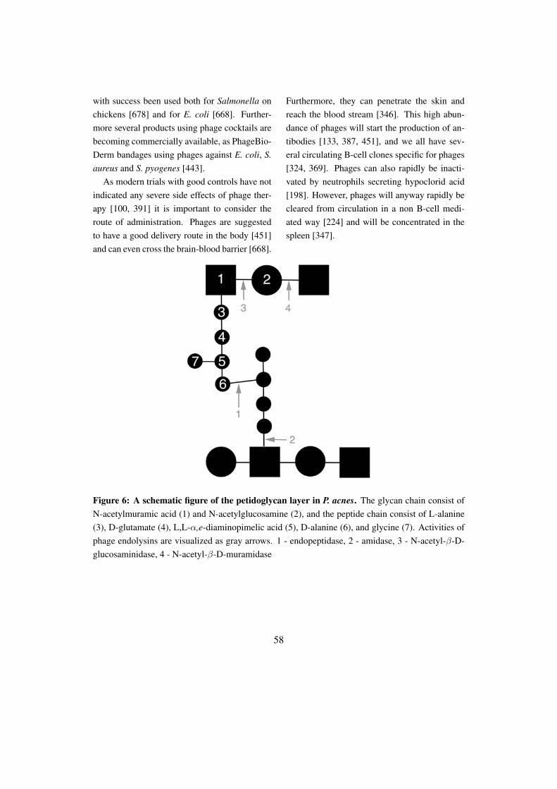
with success been used both for Salmonella onchickens [678] and for E. coli [668]. Further-more several products using phage cocktails arebecoming commercially available, as PhageBio-Derm bandages using phages against E. coli, S.aureus and S. pyogenes [443].
As modern trials with good controls have notindicated any severe side effects of phage ther-apy [100, 391] it is important to consider theroute of administration. Phages are suggestedto have a good delivery route in the body [451]and can even cross the brain-blood barrier [668].
Furthermore, they can penetrate the skin andreach the blood stream [346]. This high abun-dance of phages will start the production of an-tibodies [133, 387, 451], and we all have sev-eral circulating B-cell clones specific for phages[324, 369]. Phages can also rapidly be inacti-vated by neutrophils secreting hypoclorid acid[198]. However, phages will anyway rapidly becleared from circulation in a non B-cell medi-ated way [224] and will be concentrated in thespleen [347].
Figure 6: A schematic figure of the petidoglycan layer in P. acnes. The glycan chain consist ofN-acetylmuramic acid (1) and N-acetylglucosamine (2), and the peptide chain consist of L-alanine(3), D-glutamate (4), L,L-α,e-diaminopimelic acid (5), D-alanine (6), and glycine (7). Activities ofphage endolysins are visualized as gray arrows. 1 - endopeptidase, 2 - amidase, 3 - N-acetyl-β-D-glucosaminidase, 4 - N-acetyl-β-D-muramidase
58
In the human body, phages have several pro-posed functions, unrelated to their antibacterialeffect. Phages have been suggested to bind tointegrins on human cells [236], thereby inhibit-ing signaling through NF-κB and thus be im-munosuppressive [237, 238]. They also seem todecrease the production of ROS [558, 718] andcould protect against other viruses [238, 467]such as herpes simplex [43, 123]. The antiviralactivity is suggested to be due to an increase ofinterferon when stimulated with phages [363],which also is seen when free phage DNA is in-jected [316, 487]. Furthermore, phages, and inparticular holins, have been proposed to haveanti-tumor effects [10, 238].
The usage of phages as a therapy has severalbenefits. Not only are they specific, thereby notaffecting the commensals [245]. The commen-sal flora is not even affected if the therapy is di-rected to that particular species, potentially dueto different susceptibilities [100]. Furthermore,phages can replicate at the site where they areneeded the most [699] and are not hindered bybiofilms [139]. It has also been reported thatphages have effect on intracellular pathogens,by hijacking a bacterium outside the cell. Oncethe phages are inside the cell, they can kill allresting bacteria [92]. However, what makesphages advantageous is also their weakness as atherapeutic agent. Their high specificity gives aneed to identify the infectious agent before treat-ment [284]. Furthermore, there is a fear that thephages might be able to transport genetic ma-terial [326] and even integrate in human cells[614, 615]. The fast destruction of bacteria willalso generate a huge amount of free endotox-ins from the cell walls that might influence the
pathogenesis in an unwanted direction [504].Even though several issues still remain to be
solved considering phage therapy, they are usedin several countries [717, 718], and are proposedto be used mainly as veterinary applications toreduce the Salmonella and Campylobacter bur-den on chickens [138, 233, 290, 408], againstE. coli in cattle, in fish farms [500, 538] andto protect fresh fruit from bacteria [393, 394].However, several more applications are beingdeveloped based on the phages, for usage in bothmedicine and biotechnology.
Except being capable of killing bacterialpathogens, the phages can also be used as spe-cific delivery transportation molecules [133],and deliver DNA or antimicrobial substances[234, 591, 727]. Furthermore, many phageshave the ability to degrade biofilms [48] that cur-rently is a huge medical problem. On a morebiotechnological level, both luciferase genes[689] and GFP (green fluorescent protein) [215]have been coupled to phages both in order tostudy phage infection cycles, but also to be ableto identify contaminating bacteria [413].
5.6.2 Phage therapy using phageendolysins
Due to the potential drawbacks using wholephages, researchers have started to investigatethe potential of using purified phage lytic en-zymes. Those enzymes, commonly calledlysins, are more correctly called endolysins dueto their effect from the inside (Figure 6) [321],but have also been called virolysin [537] eventhough this word seldom is used. The first lysinever identified came from Streptococcus phageC1, almost 40 years ago, and was identified by
59
Fischetti [206]. Many years later, the potent ef-fect of this lysin was demonstrated when Loef-fler in 2003 lysed more than 107 group A Strep-tococci in less than 5 seconds using no morethan 10 ng lysin [409]. The effects seen usingthose enzymes lead scientists to think of phagelysins as ”enzybiotics” [507]. However, so farthe only available phage lysin, plyG, is activeagainst B. anthracis and is used by the US Cen-ter for Disease Control and Prevention in At-lanta [76].
Just as the phages are specific, so are thelysins, even though the specificity is to speciesrather than to single isolates [205], and eventhough exceptions exist that have activity toseveral species [751]. Lysins are quite potentagainst Gram-positive bacteria even from theoutside [203]. Gram-negative bacteria should intheory be resistant to this treatment due to theircell wall structure. However, some data suggestthat even Gram-negative bacteria might be af-fected by lysin treatment [176, 447, 527], andthere is ongoing research to increase the effi-ciency versus Gram-negative bacteria by pen-etrating the lipid layer [314]. Furthermore, agroup has successfully anchored lysins to thecell surface of E. coli using a fusion proteinwith OmpC in order to more easily distribute thelysin and have the ability to express more lysinat the site where it is needed [677]. Anotherexample of increasing the efficiency of phagelysins was demonstrated by Lopez by creatingchimeric lysins, with N- and C-terminal parts ofthe lysin from different phages [424, 425].
Another major concern regarding the usageof phage lysins is the potential development ofresistance towards the proteins. Even though re-sistance to lysins seemed unlikely due to theirconserved and essential binding sites [77, 205],
this was nevertheless investigated. So far nolysin resistant strains have been identified [203],not even after repeated exposure to low levels ofpurified enzymes [411, 616]. However it shouldbe mentioned that lysins have a decreased ef-fect against bacteria in stationary phase due toan altered peptidoglycan layer [411, 555]. Fur-thermore, not even antibodies directed towardsthe lysins will inhibit their effect [409]. So far,the only bacterial ”resistance” known to lysins issporulation. However, even the spores are sen-sitive to lysins as soon as they start to germinate[616].
Lysins have been used in several animal mod-els, without displaying any severe side effects,even though the treatment results in a rapid re-lease of endotoxins [185]. This will lead toa release of proinflammatory cytokines, whichis higher than compared to treatment with van-comycin [185]. However, some studies alsoindicate that the levels of proinflammatory cy-tokines are lower using lysins compared to regu-lar antibiotics [737]. The combination of lysinsand antibiotics has been a successful combina-tion in the experimental eradication of S. pneu-moniae [166]. Furthermore, lysins are ableto eradicate antibiotic resistant bacterial strains[409, 410, 411] in biofilms [607], and have beenshown to be able to cure several animal mod-els from bacterial infections [126, 322, 507, 518,616].
These data have made scientists to suggestthat lysins could be used as prophylaxis [204]in order to decrease the number of potentialpathogens before they can cause disease andthus prevent pathogenic conditions [202]. Thistherapy would furthermore decrease the risk ofsecondary viral infections [455].
60
References[1] Abedon, S., Hyman, P. & Thomas, C. (2003) Experimental examination
of bacteriophage latent-period evolution as a response to bacterial avail-ability. Appl Environ Microbiol, 69: 7499–7506.
[2] Absolom, D., Lamberti, F., Policova, Z., Zingg, W., van Oss, C. & Neu-mann, A. (1983) Surface thermodynamics of bacterial adhesion. ApplEnviron Microbiol, 46: 90–97.
[3] Acheson, D. & Donohue-Rolfe, A. (1989) Cancer-associated hemolyticuremic syndrome: a possible role of mitomycin in relation to Shiga-liketoxins. J Clin Oncol, 7: 1943.
[4] Ackermann, H. (2001) Frequency of morphological phage descriptionsin the year 2000. Brief review. Arch Virol, 146: 843–857.
[5] Ackermann, H. & DuBow, M. (1987) Viruses of prokaryotes, vol. 1. CRCPress, Boca Raton.
[6] Ada, G. & French, E. (1959) Purification of bacterial neuraminidase(receptor-destroying enzyme). Nature, 183: 1740–1741.
[7] Ada, G. & French, E. (1959) Stimulation of the production of neu-raminidase in Vibrio cholerae cultures by N-acetyl-mannosamine. J GenMicrobiol, 21: 561–568.
[8] Aft, R. & Mueller, G. (1983) Hemin-mediated DNA strand scission. JBiol Chem, 258: 12069–12072.
[9] Aft, R. & Mueller, G. (1984) Hemin-mediated oxidative degradation ofproteins. J Biol Chem, 259: 301–305.
[10] Agu, C., Klein, R., Schwab, S., Konig-Schuster, M., Kodajova, P.,Ausserlechner, M., Binishofer, B., Bläsi, U., Salmons, B., Gunzburg,W. & Hohenadl, C. (2006) The cytotoxic activity of the bacteriophageλ-holin protein reduces tumour growth rates in mammary cancer cellxenograft models. J Gene Med, 8: 229–241.
[11] Akaza, N., Akamatsu, H., Kishi, M., Mizutani, H., Ishii, I., Nakata, S.& Matsunaga, K. (2009) Effects of Propionibacterium acnes on variousmRNA expression levels in normal human epidermal keratinocytes invitro. J Dermatol, 36: 213–223.
[12] Åkerstrom, B., Maghzal, G., Winterbourn, C. & Kettle, A. (2007)The lipocalin α1-microglobulin has radical scavenging activity. J BiolChem, 282: 31493–31503.
[13] Albert, M., Bhuiyan, N., Rahman, A., Ghosh, A., Hultenby, K., Wein-traub, A., Nahar, S., Kibriya, A., Ansaruzzaman, M. & Shimada, T.(1996) Phage specific for Vibrio cholerae O139 Bengal. J Clin Micro-biol, 34: 1843–1845.
[14] Alexeyev, O., Olsson, J. & Elgh, F. (2009) Is there evidence for a role ofPropionibacterium acnes in prostatic disease? Urology, 73: 220–224.
[15] Alexeyev, O., Marklund, I., Shannon, B., Golovleva, I., Olsson, J., An-dersson, C., Eriksson, I., Cohen, R. & Elgh, F. (2007) Direct visualiza-tion of Propionibacterium acnes in prostate tissue by multicolor fluores-cent in situ hybridization assay. J Clin Microbiol, 45: 3721–3728.
[16] Allhorn, M., Berggård, T., Nordberg, J., Olsson, M. & Åkerstrom, B.(2002) Processing of the lipocalin α1-microglobulin by hemoglobin in-duces heme-binding and heme-degradation properties. Blood, 99: 1894–1901.
[17] Allhorn, M., Klapyta, A. & Åkerstrom, B. (2005) Redox properties of thelipocalin α1-microglobulin: reduction of cytochrome c, hemoglobin,and free iron. Free Radic Biol Med, 38: 557–567.
[18] Allhorn, M., Olin, A., Nimmerjahn, F. & Collin, M. (2008) HumanIgG/FcγR interactions are modulated by streptococcal IgG glycan hy-drolysis. PLoS One, 3: e1413.
[19] Allison, D. & Sutherland, I. (1987) The role of exopolysaccharides inadhesion of freshwater bacteria. Microbiology, 133: 1319–1327.
[20] Allison, G. & Klaenhammer, T. (1998) Phage resistance mechanisms inlactic acid bacteria. Int Dairy Jour, 8: 207–226.
[21] An, D. & Parsek, M. (2007) The promise and peril of transcriptionalprofiling in biofilm communities. Curr Opin Microbiol, 10: 292–296.
[22] Anderson, T. (1948) The growth of T2 virus on ultraviolet-killed hostcells. J Bacteriol, 56: 403–410.
[23] Anderson, T. (1966) Electron microscopy of phages. Phage and the ori-gins of molecular biology (edited by T. Cairns, G. Stent & J. Watson),pp. 63–78, Cold Spring Harbor Laboratory of Quantitative Biology, ColdSpring Harbor.
[24] Angata, T. & Varki, A. (2002) Chemical diversity in the sialic acids andrelated α-keto acids: an evolutionary perspective. Chem Rev, 102: 439–469.
[25] Applegate, L., Luscher, P. & Tyrrell, R. (1991) Induction of heme oxyge-nase: a general response to oxidant stress in cultured mammalian cells.Cancer Res, 51: 974–978.
[26] Archibald, A. (1980) Phage receptors in Gram-positive bacteria. VirusesReceptors Part I - receptors and recognition (edited by L. Randall &L. Philipson), pp. 7–26, Chapman & Hall, London.
[27] Ramirez de Arellano, E., Pascual, A., Martinez-Martinez, L. & Perea, E.(1994) Activity of eight antibacterial agents on Staphylococcus epider-midis attached to Teflon catheters. J Med Microbiol, 40: 43–47.
[28] Aruoma, O. & Halliwell, B. (1987) Superoxide-dependent andascorbate-dependent formation of hydroxyl radicals from hydrogen per-oxide in the presence of iron. Are lactoferrin and transferrin promotersof hydroxyl-radical generation? Biochem J, 241: 273–278.
[29] Atterbury, R., Dillon, E., Swift, C., Connerton, P., Frost, J., Dodd, C.,Rees, C. & Connerton, I. (2005) Correlation of Campylobacter bacte-riophage with reduced presence of hosts in broiler chicken ceca. ApplEnviron Microbiol, 71: 4885–4887.
[30] Babalova, E., Katsitadze, K., Sakvarelidze, L., Imnaishvili, N.,Sharashidze, T., Badashvili, V., Kiknadze, G., Meipariani, A.,Gendzekhadze, N., Machavariani, E., Gogoberidze, K., Gozalov, E. &Dekanosidze, N. (1968) [Preventive value of dried dysentery bacterio-phage]. Zh Mikrobiol Epidemiol Immunobiol, 45: 143–145.
[31] Babior, B., Kipnes, R. & Curnutte, J. (1973) Biological defense mecha-nisms. The production by leukocytes of superoxide, a potential bacteri-cidal agent. J Clin Invest, 52: 741–744.
[32] Babior, B., Lambeth, J. & Nauseef, W. (2002) The neutrophil NADPHoxidase. Arch Biochem Biophys, 397: 342–344.
61
[33] Bachrach, G., Leizerovici-Zigmond, M., Zlotkin, A., Naor, R. & Stein-berg, D. (2003) Bacteriophage isolation from human saliva. Lett ApplMicrobiol, 36: 50–53.
[34] Banks, D., Beres, S. & Musser, J. (2002) The fundamental contributionof phages to GAS evolution, genome diversification and strain emer-gence. Trends Microbiol, 10: 515–521.
[35] Banks, D., Porcella, S., Barbian, K., Martin, J. & Musser, J. (2003)Structure and distribution of an unusual chimeric genetic element en-coding macrolide resistance in phylogenetically diverse clones of groupA Streptococcus. J Infect Dis, 188: 1898–1908.
[36] Baranano, D., Rao, M., Ferris, C. & Snyder, S. (2002) Biliverdin reduc-tase: a major physiologic cytoprotectant. Proc Natl Acad Sci USA, 99:16093–16098.
[37] Barksdale, L. & Arden, S. (1974) Persisting bacteriophage infections,lysogeny, and phage conversions. Annu Rev Microbiol, 28: 265–299.
[38] Barnet, Y. & Humphrey, B. (1975) Exopolysaccharide depolymerases in-duced by Rhizobium bacteriophages. Can J Microbiol, 21: 1647–1650.
[39] Barondess, J. & Beckwith, J. (1990) A bacterial virulence determinantencoded by lysogenic coliphage λ. Nature, 346: 871–874.
[40] Barrack, R. & Harris, W. (1993) The value of aspiration of the hip jointbefore revision total hip arthroplasty. J Bone Joint Surg, 75: 66.
[41] Barreiro, V. & Haggård-Ljungquist, E. (1992) Attachment sites for bac-teriophage P2 on the Escherichia coli chromosome: DNA sequences,localization on the physical map, and detection of a P2-like remnant inE. coli K-12 derivatives. J Bacteriol, 174: 4086–4093.
[42] Barrow, P., Lovell, M. & Berchieri, A.J. (1998) Use of lytic bacte-riophage for control of experimental Escherichia coli septicemia andmeningitis in chickens and calves. Clin Diagn Lab Immunol, 5: 294–298.
[43] Barrow, P. & Soothill, J. (1997) Bacteriophage therapy and prophylaxis:rediscovery and renewed assessment of potential. Trends Microbiol, 5:268–271.
[44] Barry, G. (1957) Isolation of N-acetylneuraminic acid from colominicacid. Science, 126: 1230–1230.
[45] Bartell, P., Lam, G. & Orr, T. (1968) Purification and propertiesof polysaccharide depolymerase associated with phage-infected Pseu-domonas aeruginosa. J Biol Chem, 243: 2077–2080.
[46] Barton, A., Sagers, R. & WG, P. (1996) Bacterial adhesion to orthopedicimplant polymers. J Biomed Mater Res, 30: 403–410.
[47] Bastien, N., Trudel, M. & Simard, C. (1999) Complete protection ofmice from respiratory syncytial virus infection following mucosal deliv-ery of synthetic peptide vaccines. Vaccine, 17: 832–836.
[48] Bayer, M., Thurow, H. & Bayer, M. (1979) Penetration of the polysac-charide capsule of Escherichia coli (Bi161/42) by bacteriophage K29.Virology, 94: 95–118.
[49] Beckman, J., Beckman, T., Chen, J., Marshall, P. & Freeman, B. (1990)Apparent hydroxyl radical production by peroxynitrite: implications forendothelial injury from nitric oxide and superoxide. Proc Natl Acad SciUSA, 87: 1620–1624.
[50] Beech, I. & Gaylarde, C. (1991) Microbial polysaccharides and corro-sion. Int Biodet, 27: 95–107.
[51] de Beer, D., Stoodley, P., Roe, F. & Lewandowski, Z. (1994) Effects ofbiofilm structures on oxygen distribution and mass transport. BiotechnolBioeng, 43: 1131–1138.
[52] Bek-Thomsen, M., Lomholt, H. & Kilian, M. (2008) Acne is not associ-ated with yet-uncultured bacteria. J Clin Microbiol, 46: 3355–3360.
[53] Bell, C., Frescura, P., Hochschild, A. & Lewis, M. (2000) Crystal struc-ture of the λ repressor C-terminal domain provides a model for cooper-ative operator binding. Cell, 101: 801–811.
[54] Bensing, B., Siboo, I. & Sullam, P. (2001) Proteins PblA and PblB ofStreptococcus mitis, which promote binding to human platelets, are en-coded within a lysogenic bacteriophage. Infect Immun, 69: 6186–6192.
[55] Berg, R. (1996) The indigenous gastrointestinal microflora. Trends Mi-crobiol, 4: 430–435.
[56] Bergamini, T., Peyton, J. & Cheadle, W. (1992) Prophylactic antibioticsprevent bacterial biofilm graft infection. J Surg Res, 52: 101–105.
[57] Bergey, D., Breed, R., Hammer, R., Harrison, F. & Huntoon, F.(1923) Bergey’s manual of determinative bacteriology. The Willimasand Wilkins Co, Baltimore.
[58] Bergh, O., Borsheim, K., Bratbak, G. & Heldal, M. (1989) High abun-dance of viruses found in aquatic environments. Nature, 340: 467–468.
[59] Bernard, K., Shuttleworth, L., Munro, C., Forbes-Faulkner, J., Pitt, D.,Norton, J. & Thomas, A. (2002) Propionibacterium australiense sp novderived from granulomatous bovine lesions. Anaerobe, 8: 41–47.
[60] Bernhardt, T., Wang, I., Struck, D. & Young, R. (2001) A protein an-tibiotic in the phage Qβ virion: diversity in lysis targets. Science, 292:2326–2329.
[61] Bernhardt, T., Wang, I., Struck, D. & Young, R. (2002) Breaking free:"protein antibiotics" and phage lysis. Res Microbiol, 153: 493–501.
[62] Bernheimer, H. & Tiraby, J. (1976) Inhibition of phage infection bypneumococcus capsule. Virology, 73: 308–309.
[63] Bertani, G. & Weigle, J. (1953) Host controlled variation in bacterialviruses. J Bacteriol, 65: 113–121.
[64] Beutler, B. (2002) LPS in microbial pathogenesis: promise and fulfil-ment. J Endotoxin Res, 8: 329–335.
[65] Beveridge, T. & Graham, L. (1991) Surface layers of bacteria. MicrobiolMol Biol Rev, 55: 684.
[66] Beyer, W. & Fridovich, I. (1988) Catalases - with and without heme.Oxygen radicals in biology and medicine (edited by M. Simic, K. Tay-lor, J. Ward & C. von Sonntag), pp. 651–661, Plenum Press, New York.
[67] Bhatia, A. & Kast, R. (2007) How Influenza’s neuraminidase promotesvirulence and creates localized lung mucosa immunodeficiency. Cell MolBiol Lett, 12: 111–119.
62
[68] Bienert, G., Möller, A., Kristiansen, K., Schulz, A., Möller, I., Schjoer-ring, J. & Jahn, T. (2007) Specific aquaporins facilitate the diffusion ofhydrogen peroxide across membranes. J Biol Chem, 282: 1183–1192.
[69] Bissonnette, F., Labrie, S. & Deveau, H. (2000) Characterization ofmesophilic mixed starter cultures used for the manufacture of aged ched-dar cheese. J Dairy Sci, 83: 620–627.
[70] Bläsi, U., Chang, C., Zagotta, M., Nam, K. & Young, R. (1990) Thelethal λ S gene encodes its own inhibitor. EMBO J, 9: 981–989.
[71] Blix, G. (1936) Concerning the carbohydrate groups of submaxillarymucin. Hoppe-Seylers Zeitschrift Fur Physiologische Chemie, 240: 43–54.
[72] Bloomfield, V. (1996) DNA condensation. Curr Opin Struct Biol, 6:334–341.
[73] Bogosian, G. (2005) Control of bacteriophage in commercial microbiol-ogy and fermentation facilities. The bacteriophages (edited by R. Calen-dar), Oxford University Press, New York.
[74] Böhm, J., Lambert, O., Frangakis, A., Letellier, L., Baumeister, W. &Rigaud, J. (2001) FhuA-mediated phage genome transfer into liposomes:a cryo-electron tomography study. Curr Biol, 11: 1168–1175.
[75] Bojar, R. & Holland, K. (2004) Acne and Propionibacterium acnes. ClinDermatol, 22: 375–379.
[76] Borysowski, J. & Gorski, A. (2010) Enzybiotics and their potential appli-cations in medicine. Enzybiotics: antibiotic enzymes as drugs and ther-apeutics (edited by T. Villa & P. Veiga-Crespo), pp. 1–26, John Wiley &Sons, Hoboken, New Jersey.
[77] Borysowski, J., Weber-Dabrowska, B. & Górski, A. (2006) Bacterio-phage endolysins as a novel class of antibacterial agents. Exp Bio Med,231: 366–377.
[78] Botstein, D. (1980) A theory of modular evolution for bacteriophages.Ann NY Acad Sci, 354: 484–490.
[79] Boulanger, P. & Letellier, L. (1988) Characterization of ion channels in-volved in the penetration of phage T4 DNA into Escherichia coli cells. JBiol Chem, 263: 9767–9775.
[80] Boyd, E. (2005) Bacteriophages and bacterial virulence. Bacterio-phages: Biology and Applications (edited by E. Kutter & A. Su-lakvelidze), pp. 223–266, CRC Press, Boca Raton.
[81] Branda, S., Chu, F., Kearns, D., Losick, R. & Kolter, R. (2006) A majorprotein component of the Bacillus subtilis biofilm matrix. Mol Microbiol,59: 1229–1238.
[82] Branda, S., Vik, Å., Friedman, L. & Kolter, R. (2005) Biofilms: the ma-trix revisited. Trends Microbiol, 13: 20–26.
[83] Braun, V., Schaller, K. & Wolff, H. (1973) A common receptor proteinfor phage T5 and colicin M in the outer membrane of Escherichia coliB. Biochim Biophys Acta, 323: 87–97.
[84] Breitbart, M., Felts, B., Kelley, S., Mahaffy, J., Nulton, J., Salamon, P.& Rohwer, F. (2004) Diversity and population structure of a near-shoremarine-sediment viral community. Proc Biol Sci, 271: 565–574.
[85] Breitbart, M., Hewson, I., Felts, B., Mahaffy, J., Nulton, J., Salamon,P. & Rohwer, F. (2003) Metagenomic analyses of an uncultured viralcommunity from human feces. J Bacteriol, 185: 6220–6223.
[86] Breitbart, M., Salamon, P., Andresen, B., Mahaffy, J., Segall, A., Mead,D., Azam, F. & Rohwer, F. (2002) Genomic analysis of uncultured ma-rine viral communities. Proc Natl Acad Sci USA, 99: 14250–14255.
[87] Brook, I. & Frazier, E. (1991) Infections caused by Propionibacteriumspecies. Rev Infect Dis, 13: 819–822.
[88] Brooks, K. & Clark, A. (1967) Behavior of λ bacteriophage in a recom-bination deficient strain of Escherichia coli. J Virol, 1: 283–293.
[89] Broudy, T. & Fischetti, V. (2003) In vivo lysogenic conversion of Tox(-) Streptococcus pyogenes to Tox(+) with lysogenic Streptococci or freephage. Infect Immun, 71: 3782–3786.
[90] Broudy, T., Pancholi, V. & Fischetti, V. (2001) Induction of lysogenicbacteriophage and phage-associated toxin from group a streptococci dur-ing coculture with human pharyngeal cells. Infect Immun, 69: 1440–1443.
[91] Broudy, T., Pancholi, V. & Fischetti, V. (2002) The in vitro interaction ofStreptococcus pyogenes with human pharyngeal cells induces a phage-encoded extracellular DNase. Infect Immun, 70: 2805–2811.
[92] Broxmeyer, L., Sosnowska, D., Miltner, E., Chacón, O., Wagner, D.,McGarvey, J., Barletta, R. & Bermudez, L. (2002) Killing of Mycobac-terium avium and Mycobacterium tuberculosis by a mycobacteriophagedelivered by a nonvirulent mycobacterium: a model for phage therapy ofintracellular bacterial pathogens. J Infect Dis, 186: 1155–1160.
[93] Brüggemann, H. (2005) Insights in the pathogenic potential of Propioni-bacterium acnes from its complete genome. Semin Cutan Med Surg, 24:67–72.
[94] Brüggemann, H. (2010) Skin: Acne and Propionibacterium acnes.Handbook of hydrocarbon and lipid microbiology (edited by K. Tim-mis), pp. 3215–3225, Springer, Berlin.
[95] Brüggemann, H., Henne, A., Hoster, F., Liesegang, H., Wiezer, A.,Strittmatter, A., Hujer, S., Dürre, P. & Gottschalk, G. (2004) The com-plete genome sequence of Propionibacterium acnes, a commensal of hu-man skin. Science, 305: 671–673.
[96] Brüssow, H. (2007) Phage therapy: the Western perspective. Bacterio-phage Genetics and Molecular Biology (edited by S. McGrath & D. vanSinderen), pp. 159–192, Caister Academic Press, Norfolk.
[97] Brüssow, H., Canchaya, C. & Hardt, W. (2004) Phages and the evolu-tion of bacterial pathogens: from genomic rearrangements to lysogenicconversion. Microbiol Mol Biol Rev, 68: 560–602.
[98] Brüssow, H. & Desiere, F. (2001) Comparative phage genomics and theevolution of Siphoviridae: insights from dairy phages. Mol Microbiol,39: 213–222.
[99] Brüssow, H. & Hendrix, R. (2002) Phage genomics: small is beautiful.Cell, 108: 13–16.
[100] Bruttin, A. & Brüssow, H. (2005) Human volunteers receiving Es-cherichia coli phage T4 orally: a safety test of phage therapy. AntimicrobAgents Chemother, 49: 2874–2878.
63

[101] Bruynoghe, R. & Maisin, J. (1921) Essais de thérapeutique au moyen dubacteriophage. Comptes Rendus Des Seances De La Societe De BiologieEt De Ses Filiales, 85: 1120–1121.
[102] Brzuskiewicz, E., Weiner, J., Wollherr, A., Thürmer, A., Hüpeden, J.,Lomholt, H., Kilian, M., Gottschalk, G., Daniel, R., Mollenkopf, H.,Meyer, T. & Brüggemann, H. (2011) Comparative genomics and tran-scriptomics of Propionibacterium acnes. PLoS One, 6: e21581.
[103] Bunn, H. & Jandl, J. (1968) Exchange of heme among hemoglobins andbetween hemoglobin and albumin. J Biol Chem, 243: 465–475.
[104] Burnet, F., McCrea, J. & Stone, J. (1946) Modification of human redcells by virus action; the receptor gradient for virus action in human redcells. Br J Exp Pathol, 27: 228–236.
[105] Busscher, H., Weerkamp, A., van der Mei, H., van Pelt, A., de Jong, H.& Arends, J. (1984) Measurement of the surface free energy of bacterialcell surfaces and its relevance for adhesion. Appl Environ Microbiol, 48:980–983.
[106] Butler-Wu, S., Sengupta, D., Kittichotirat, W., Matsen, F. & Bumgar-ner, R. (2011) Genome sequence of a novel species, Propionibacteriumhumerusii. J Bact, 193: 3678.
[107] Cairns, J., Stent, G. & Watson, J. (1966) Phage and the origins of molec-ular biology. Cold Spring Harbor Laboratory, Cold Spring Harbor, 1 ed.
[108] Calendar, R. & Inman, R. (2005) Phage biology. Phages: their rolein bacterial pathogenesis and biotechnology (edited by M. Waldor,D. Friedman & S. Adhya), pp. 18–36, ASM Press, Washington.
[109] Camilli, A. & Bassler, B. (2006) Bacterial small-molecule signalingpathways. Science, 311: 1113–1116.
[110] Campbell, A., Schneider, S. & Song, B. (1992) Lambdoid phages aselements of bacterial genomes (integrase/phage21/Escherichia coli K-12/icd gene). Genetica, 86: 259–267.
[111] Campbell, A. (1963) Episomes. Advances in genetics, 11: 101–145.
[112] Campbell, A. (1996) Cryptic prophages. E. coli and Salmonella: cellularand molecular biology (edited by F. Neidhardt, R. Curtis III, J. Ingraham,E. Lin, K. Low, B. Magasarik, W. Reznikoff, M. Riley, M. Schaechter &H. Umbarher), pp. 2041–2046, ASM Press, Washington.
[113] Canchaya, C., Fournous, G. & Brüssow, H. (2004) The impact ofprophages on bacterial chromosomes. Mol Microbiol, 53: 9–18.
[114] Canchaya, C., Fournous, G., Chibani-Chennoufi, S., Dillmann, M. &Brüssow, H. (2003) Phage as agents of lateral gene transfer. Curr OpinMicrobiol, 6: 417–424.
[115] Cantacuzene, J. & Boncieu, O. (1926) Modifications subies par desstreptococques d’origine non-scarlatineuse qu contact des produits scar-latineux filtres. C. R. Acad. Sci., 182: 1185.
[116] Casadevall, A. (2009) Virulence factors and their mechanisms of action:the view from a damage-response framework. J Wat Health, 7: S2–S18.
[117] Casadevall, A. & Pirofski, L. (1999) Host-pathogen interactions: re-defining the basic concepts of virulence and pathogenicity. Infect Immun,67: 3703–3713.
[118] Casadevall, A. & Pirofski, L. (2002) What is a pathogen? Ann Med, 34:2–4.
[119] Casadevall, A. & Pirofski, L. (2003) The damage-response frameworkof microbial pathogenesis. Nat Rev Microbiol, 1: 17–24.
[120] Casjens, S. (2005) Comparative genomics and evolution of the tailed-bacteriophages. Curr Opin Microbiol, 8: 451–458.
[121] Catalano, C., Cue, D. & Feiss, M. (1995) Virus DNA packaging: thestrategy used by phage lambda. Mol Microbiol, 16: 1075–1086.
[122] Cederholm-Williams, S., King, A., Allington, M., Gill, P., Sharp, A. &Britton, B. (1978) Coagulation and fibrinolysis during the infusion ofCorynebacterium parvum in man. Br J Cancer, 37: 1074–1077.
[123] Centifanto, Y. (1965) A therapeutic antiviral from an extract of lambdainfected E. coli (phagicin). Proc Soc Exp Biol Med, 120: 607–611.
[124] Chan, A. (1993) Partners in defense, vitamin E and vitamin C. Can JPhysiol Pharmacol, 71: 725–731.
[125] Charfreitag, O., Collins, M. & Stackebrandt, E. (1988) Reclassificationof Arachnia propionica as Propionibacterium propionicus comb. nov. IntJ Syst Bacteriol, 38: 354.
[126] Cheng, Q., Nelson, D., Zhu, S. & Fischetti, V. (2005) Removal of groupB streptococci colonizing the vagina and oropharynx of mice with a bac-teriophage lytic enzyme. Antimicrob Agents Chemother, 49: 111–117.
[127] Cheong, D., Lee, H. & So, J. (2008) Optimization of electrotransforma-tion conditions for Propionibacterium acnes. J Microbiol Methods, 72:38–41.
[128] Chibani Azaiez, S., Fliss, I., Simard, R. & Moineau, S. (1998) Mono-clonal antibodies raised against native major capsid proteins of Lacto-coccal c2-like bacteriophages. Appl Environ Microbiol, 64: 4255.
[129] Chiller, K. & Selkin, B. (2001) Skin microflora and bacterial infectionsof the skin. J Infect Dis, 6: 170–174.
[130] Chopin, M., Rouault, A., Ehrlich, S. & Gautier, M. (2002) Filamentousphage active on the gram-positive bacterium Propionibacterium freuden-reichii. J Bacteriol, 184: 2030–2033.
[131] Circu, M. & Aw, T. (2010) Reactive oxygen species, cellular redox sys-tems, and apoptosis. Free Radic Biol Med, 48: 749–762.
[132] Clark, D., Fornabaio, D., McNeill, H., Mullane, K., Caravella, S. &Miller, M. (1988) Contribution of oxygen-derived free radicals to ex-perimental necrotizing enterocolitis. Am J Pathol, 130: 537–542.
[133] Clark, J. & March, J. (2004) Bacterial viruses as human vaccines? Ex-pert Rev Vaccines, 3: 463–476.
[134] Coenye, T., Peeters, E. & Nelis, H. (2007) Biofilm formation by Propi-onibacterium acnes is associated with increased resistance to antimicro-bial agents and increased production of putative virulence factors. ResMicrobiol, 158: 386–392.
[135] Coffey, A. & Ross, R. (2002) Bacteriophage-resistance systems in dairystarter strains: molecular analysis to application. Antonie Van Leeuwen-hoek, 82: 303–321.
64
[136] Cohen, R., Shannon, B., McNeal, J., Shannon, T. & Garrett, K. (2005)Propionibacterium acnes associated with inflammation in radical prosta-tectomy specimens: a possible link to cancer evolution? J Urol, 173:1969–1974.
[137] Conner, E. & Grisham, M. (1996) Inflammation, free radicals, and an-tioxidants. Nutrition, 12: 274–277.
[138] Connerton, P., Loc Carrillo, C., Swift, C., Dillon, E., Scott, A., Rees,C., Dodd, C., Frost, J. & Connerton, I. (2004) Longitudinal study ofCampylobacter jejuni bacteriophages and their hosts from broiler chick-ens. Appl Environ Microbiol, 70: 3877–3883.
[139] Corbin, B., McLean, R. & Aron, G. (2001) Bacteriophage T4 multiplica-tion in a glucose-limited Escherichia coli biofilm. Can J Microbiol, 47:680–684.
[140] Corfield, A., Mikchalki, J. & Schauer, R. (1981) The substrate specificityof sialidases from microorganisms and mammals. Sialidases and siali-dosis: perspectives in inherited metabolic diseases (edited by G. Tetta-manti, P. Durand & S. Di Donato), pp. 3–70, Ermes, Milan.
[141] Corfield, T. (1992) Bacterial sialidases–roles in pathogenicity and nutri-tion. Glycobiology, 2: 509–521.
[142] Cormio, L., Vuopio-Varkila, J., Siitonen, A., Talja, M. & Ruutu, M.(1996) Bacterial adhesion and biofilm formation on various double-Jstents in vivo and in vitro. Scand J Urol Nephrol, 30: 19–24.
[143] Costerton, J., Lewandowski, Z., Caldwell, D., Korber, D. & Lappin-Scott, H. (1995) Microbial biofilms. Annu Rev Microbiol, 49: 711–745.
[144] Costerton, J., Lewandowski, Z., DeBeer, D., Caldwell, D., Korber, D. &James, G. (1994) Biofilms, the customized microniche. J Bacteriol, 176:2137–2142.
[145] Cove, J., Holland, K. & Cunliffe, W. (1983) Effects of oxygen concentra-tion on biomass production, maximum specific growth rate and extracel-lular enzyme production by three species of cutaneous propionibacteriagrown in continuous culture. J Gen Microbiol, 129: 3327–3334.
[146] Cove, J., Holland, K. & Cunliffe, W. (1987) The effects of oxygen on cu-taneous propionibacteria grown in continuous culture. FEMS MicrobiolLett, 43: 61–65.
[147] Craigie, J. & Yen, C. (1938) The demonstration of types of B typhosusby means of preparations of type II Vi phage. II the stability and epi-demiological significance of V form types of B typhosus. Can. PublicHealth J, 29: 484–496.
[148] Crawford, W., Crawford, I., Stoughton, R. & Cornell, R. (1979) Labo-ratory induction and clinical occurrence of combined clindamycin anderythromycin resistance in Corynebacterium acnes. J Invest Dermatol,72: 187–190.
[149] Crocker, P. & Varki, A. (2001) Siglecs in the immune system. Immunol-ogy, 103: 137–145.
[150] Csukás, Z., Banizs, B. & Rozgonyi, F. (2004) Studies on the cytotoxiceffects of Propionibacterium acnes strains isolated from cornea. MicrobPathog, 36: 171–174.
[151] Cummins, C. (1976) Identification of Propionibacterium acnes and re-lated organisms by precipitin tests with trichloroacetic acid extracts. JClin Microbiol, 2: 104–110.
[152] Cummins, C. & Johnson, J. (1974) Corynebacterium parvum: a syn-onym for Propionibacterium acnes? J Gen Microbiol, 80: 433–442.
[153] Dalia, A., Standish, A. & Weiser, J. (2010) Three surface exoglycosi-dases from Streptococcus pneumoniae, NanA, BgaA, and StrH, promoteresistance to opsonophagocytic killing by human neutrophils. Infect Im-mun, 78: 2108–2116.
[154] Daubin, V., Lerat, E. & Perriere, G. (2003) The source of laterally trans-ferred genes in bacterial genomes. Genome Biol, 4: R57.
[155] Davidson, W. (1922) The bacteriolysant therapy of bacillary dysenteryin children: therapeutic application of bacteriolysants; D’Herelle’s phe-nomenon. Am J Dis Child, 23: 531–534.
[156] Day, M. (2004) Bacterial sensitivity to bacteriophage in the aquatic en-vironment. Sci Prog, 87: 179–191.
[157] De Young, L., Young, J., Ballaron, S., Spires, D. & Puhvel, S. (1984)Intradermal injection of Propionibacterium acnes: a model of inflam-mation relevant to acne. J Invest Dermatol, 83: 394–398.
[158] Demchick, P. & Koch, A. (1996) The permeability of the wall fabric ofEscherichia coli and Bacillus subtilis. J Bacteriol, 178: 768–773.
[159] Deng, Y., Ren, X., Yang, L., Lin, Y. & Wu, X. (2003) A JNK-dependentpathway is required for TNFα-induced apoptosis. Cell, 115: 61–70.
[160] Dennery, P., Spitz, D., Yang, G., Tatarov, A., Lee, C., Shegog, M. &Poss, K. (1998) Oxygen toxicity and iron accumulation in the lungs ofmice lacking heme oxygenase-2. J Clin Invest, 101: 1001–1011.
[161] Dessinioti, C. & Katsambas, A. (2010) The role of Propionibacteriumacnes in acne pathogenesis: facts and controversies. Clin Dermatol, 28:2–7.
[162] Dhanasekaran, D. & Reddy, E. (2008) JNK signaling in apoptosis. Onco-gene, 27: 6245–6251.
[163] d’Herelle, F. (1917) An invisible antagonist microbe of dysentery bacil-lus. Comptes Rendus Hebdomadaires Des Seances De L Academie DesSciences, 165: 373–375.
[164] d’Herelle, F., Malone, F. & Lahiri, M. (1930) Studies on Asiatic cholera.Indian Med Res Memoirs, 14: 1–161.
[165] Dinsmore, P. & Klaenhammer, T. (1995) Bacteriophage resistance inLactococcus. Mol Biotech, 4: 297–314.
[166] Djurkovic, S., Loeffler, J. & Fischetti, V. (2005) Synergistic killing ofStreptococcus pneumoniae with the bacteriophage lytic enzyme Cpl-1and penicillin or gentamicin depends on the level of penicillin resistance.Antimicrob Agents Chemother, 49: 1225–1228.
[167] Donlan, R. (2008) Biofilm on central venous catheters: is eradicationpossible? Curr Top Microbiol Immun, 322: 133–161.
[168] Douglas, H. & Gunter, S. (1946) The taxonomic position of Corynebac-terium acnes. J Bacteriol, 52: 15–23.
[169] Douki, T., Onuki, J., Medeiros, M., Bechara, E., Cadet, J. & Di Mascio,P. (1998) Hydroxyl radicals are involved in the oxidation of isolated andcellular DNA bases by 5-aminolevulinic acid. FEBS Lett, 428: 93–96.
65
[170] Downes, J. & Wade, W. (2009) Propionibacterium acidifaciens sp. nov.,isolated from the human mouth. Int J Syst Evol Microbiol, 59: 2778–2781.
[171] Drzeniek, R. (1967) Differences in splitting capacity of virus and V.cholerae neuraminidases on sialic acid type substrates. Biochem BiophysRes Commun, 26: 631–638.
[172] Drzeniek, R. (1973) Substrate specificity of neuraminidases. HistochemJ, 5: 271–290.
[173] Dubos, R., Straus, J. & Pierce, C. (1943) The multiplication of bacterio-phage in vivo and its protective effect against an experimental infectionwith Shigella dysenteriae. J Exp Med, 78: 161–168.
[174] Dufraigne, C., Fertil, B., Lespinats, S., Giron, A. & Deschavanne, P.(2005) Detection and characterization of horizontal transfers in prokary-otes using genomic signature. Nucleic Acids Res, 33: e6.
[175] Dupont, K., Janzen, T., Vogensen, F., Josephsen, J. & Stuer-Lauridsen,B. (2004) Identification of Lactococcus lactis genes required for bacte-riophage adsorption. Appl Environ Microbiol, 70: 5825–5832.
[176] Düring, K., Porsch, P., Mahn, A., Brinkmann, O. & Gieffers, W. (1999)The non-enzymatic microbicidal activity of lysozymes. FEBS Lett, 449:93–100.
[177] Durmaz, E. & Klaenhammer, T. (1995) A starter culture rotation strategyincorporating paired restriction/modification and abortive infection bac-teriophage defenses in a single Lactococcus lactis strain. Appl EnvironMicrobiol, 61: 1266–1273.
[178] Dwyer, B., Lu, S., Laitinen, J. & Nishimura, R. (1998) Protectiveproperties of tin- and manganese-centered porphyrins against hydrogenperoxide-mediated injury in rat astroglial cells. J Neurochem, 71: 2497–2504.
[179] Earnshaw, W. & Casjens, S. (1980) DNA packaging by the double-stranded DNA bacteriophages. Cell, 21: 319–331.
[180] Echols, H. (1972) Developmental pathways for the temperate phage: ly-sis vs lysogeny. Annu Rev Genet, 6: 157–190.
[181] Echols, H. (1986) Bacteriophageλ development: temporal switches andthe choice of lysis or lysogeny. Trends in Genetics, 2: 26–30.
[182] Edlin, G., Lin, L. & Kudrna, R. (1975) λ lysogens of E. coli reproducemore rapidly than non-lysogens. Nature, 255: 735–737.
[183] Edwards, R. & Rohwer, F. (2005) Viral metagenomics. Nat Rev Micro-biol, 3: 504–510.
[184] Eggerth, A. (1935) The Gram-positive non-spore-bearing anaerobicbacilli of human feces. J Bacteriol, 30: 277–299.
[185] Entenza, J., Loeffler, J., Grandgirard, D., Fischetti, V. & Moreillon, P.(2005) Therapeutic effects of bacteriophage Cpl-1 lysin against Strepto-coccus pneumoniae endocarditis in rats. Antimicrob Agents Chemother,49: 4789–4792.
[186] Esber, H., Ganfield, D. & Rosenkrantz, H. (1985) Staphage lysate:an immunomodulator of the primary immune response in mice. Im-munopharm, 10: 77–82.
[187] Esteban, J., García-Calvo, G., Jiménez-Castillo, P. & Soriano, F. (1996)Failure of Gram stain to detect Propionibacterium acnes in specimensfrom clinically significant infections. J Clin Microbiol, 34: 2051.
[188] Evans, D., Allison, D., Brown, M. & Gilbert, P. (1991) Susceptibil-ity of Pseudomonas aeruginosa and Escherichia coli biofilms towardsciprofloxacin: effect of specific growth rate. J Antimicrob Chemother,27: 177–184.
[189] Faillard, H. (1989) The early history of sialic acids. Trends Biochem Sci,14: 237–241.
[190] Falentin, H., Deutsch, S., Jan, G., Loux, V., Thierry, A., Parayre, S.,Maillard, M., Dherbécourt, J., Cousin, F., Jardin, J., Siguier, P., Couloux,A., Barbe, V., Vacherie, B., Wincker, P., Gibrat, J., Gaillardin, C. & Lor-tal, S. (2010) The complete genome of Propionibacterium freudenreichiiCIRM-BIA1, a hardy actinobacterium with food and probiotic applica-tions. PLoS One, 5: e11748.
[191] Faquet, C., Mayo, M., Maniloff, J., Desselberger, U. & Ball, L. (2005)Virus taxonomy - Classification and nomenclature of viruses. Eight re-port of the International Committee on the Taxonomy of Viruses. ElsevierAcademic Press, London.
[192] Farrar, M., Howson, K., Bojar, R., West, D., Towler, J., Parry, J., Pelton,K. & Holland, K. (2007) Genome sequence and analysis of a Propioni-bacterium acnes bacteriophage. J Bacteriol, 189: 4161–4167.
[193] Farrar, M., Howson, K., Emmott, J., Bojar, R. & Holland, K. (2007)Characterisation of cryptic plasmid pPG01 from Propionibacteriumgranulosum, the first plasmid to be isolated from a member of the cu-taneous Propionibacteria. Plasmid, 58: 68–75.
[194] Farrar, M., Ingham, E. & Holland, K. (2000) Heat shock proteins andinflammatory acne vulgaris: molecular cloning, over expression and pu-rification of a Propionibacterium acnes GroEL and DnaK homologue.FEMS Microbiol Lett, 191: 183–186.
[195] Fassi Fehri, L., Mak, T., Laube, B., Brinkmann, V., Ogilvie, L., Mol-lenkopf, H., Lein, M., Schmidt, T., Meyer, T. & Brüggemann, H. (2010)Prevalence of Propionibacterium acnes in diseased prostates and its in-flammatory and transforming activity on prostate epithelial cells. Int JMed Microbiol, 301: 69–78.
[196] Feng, J., Russel, M. & Model, P. (1997) A permeabilized cell systemthat assembles filamentous bacteriophage. Proc Natl Acad Sci USA, 94:4068–4073.
[197] Ferretti, J., McShan, W., Ajdic, D., Savic, D., Savic, G., Lyon, K.,Primeaux, C., Sezate, S., Suvorov, A., Kenton, S., Lai, H., Lin, S., Qian,Y., Jia, H., Najar, F., Ren, Q., Zhu, H., Song, L., White, J., Yuan, X.,Clifton, S., Roe, B. & McLaughlin, R. (2001) Complete genome se-quence of an M1 strain of Streptococcus pyogenes. Proc Natl Acad SciUSA, 98: 4658–4663.
[198] Ferrini, U., Mileo, A., Nista, A., Mattei, E. & Orofino, A. (1989) Poly-morphonuclear leukocyte stimulation measured by phage inactivation.Int Arch Allergy Appl Immunol, 90: 207–212.
[199] Feucht, A., Schmid, A., Benz, R., Schwarz, H. & Heller, K. (1990) Poreformation associated with the tail-tip protein pb2 of bacteriophage T5. JBiol Chem, 265: 18561–18567.
66
[200] Fiala, G. & Stetter, K. (1986) Pyrococcus furiosus sp. nov. represents anovel genus of marine heterotrophic archaebacteria growing optimally at100 ◦C. Arch Microbiol, 145: 56–61.
[201] Figueroa-Bossi, N. & Bossi, L. (1999) Inducible prophages contribute toSalmonella virulence in mice. Mol Microbiol, 33: 167–176.
[202] Fischetti, V. (2003) Novel method to control pathogenic bacteria on hu-man mucous membranes. Ann NY Acad Sci, 987: 207–214.
[203] Fischetti, V. (2005) Bacteriophage lytic enzymes: novel anti-infectives.Trends Microbiol, 13: 491–496.
[204] Fischetti, V. (2005) The use of phage lytic enzymes to control bacterialinfections. Bacteriophages: Biology and Applications (edited by E. Kut-ter & A. Sulakvelidze), pp. 321–334, CRC Press, Boca Raton.
[205] Fischetti, V. (2008) Bacteriophage lysins as effective antibacterials. CurrOpin Microbiol, 11: 393–400.
[206] Fischetti, V., Gotschlich, E. & Bernheimer, A. (1971) Purification andphysical properties of group C streptococcal phage-associated lysin. JExp Med, 133: 1105–1117.
[207] Fisher, A. (2009) Redox signaling across cell membranes. Antioxid Re-dox Signal, 11: 1349–1356.
[208] Forde, A. & Fitzgerald, G. (1999) Bacteriophage defence systems in lac-tic acid bacteria. Antonie Van Leeuwenhoek, 76: 89–113.
[209] Forman, H., Fukuto, J. & Torres, M. (2004) Redox signaling: thiol chem-istry defines which reactive oxygen and nitrogen species can act as sec-ond messengers. Am J Physiol Cell Physiol, 287: C246–56.
[210] Freeman, V. (1951) Studies on the virulence of bacteriophage-infectedstrains of Corynebacterium diphtheriae. J Bacteriol, 61: 675–688.
[211] Freeman, V. & Morse, I. (1952) Further observations on the changeto virulence of bacteriophage-infected a virulent strains of Corynebac-terium diphtheria. J Bacteriol, 63: 407–414.
[212] Frei, B., England, L. & Ames, B. (1989) Ascorbate is an outstandingantioxidant in human blood plasma. Proc Natl Acad Sci USA, 86: 6377–6381.
[213] Fridovich, I. (1989) Superoxide dismutases. An adaptation to a param-agnetic gas. J Biol Chem, 264: 7761–7764.
[214] Frobisher, M. & Brown, J. (1927) Transmission toxigenicity of strepto-cocci. Bull. Johns Hopkins Hosp., 41: 167–173.
[215] Funatsu, T., Taniyama, T., Tajima, T., Tadakuma, H. & Namiki, H.(2002) Rapid and sensitive detection method of a bacterium by usinga GFP reporter phage. Microbiol Immunol, 46: 365–369.
[216] Funke, G., von Graevenitz, A., Clarridge, J.r. & Bernard, K. (1997) Clin-ical microbiology of coryneform bacteria. Clin Microbiol Rev, 10: 125–159.
[217] Fux, C., Costerton, J., Stewart, P. & Stoodley, P. (2005) Survival strate-gies of infectious biofilms. Trends Microbiol, 13: 34–40.
[218] Gautier, M. (1990) Screening of plasmids in strains of Propionibac-terium. XXIII International Dairy Congress, Montreal.
[219] Gautier, M., Rouault, A. & Lemée, R. (1995) Electrotransfection of Pro-pionibacterium freudenreichii TL 110. Lett Appl Microbiol, 20: 125–129.
[220] Gautier, M., Rouault, A., Lortal, S., Leloir, Y. & Patureau, D. (1992)Characterization of a phage infecting Propionibacterium freudenreichii.Le Lait, 72: 431–435.
[221] Gautier, M., Rouault, A., Sommer, P. & Briandet, R. (1995) Occurrenceof Propionibacterium freudenreichii bacteriophages in swiss cheese.Appl Environ Microbiol, 61: 2572–2576.
[222] Gautier, M., Rouault, A., Sommer, P., Briandet, R. & Cassin, D. (1995)Bacteriophages infecting dairy propionibacteria. Le Lait, 75: 427–434.
[223] Gehse, M., Höffler, U., Gloor, M. & Pulverer, G. (1983) Propionibacte-ria in patients with acne vulgaris and in healthy persons. Arch Derm Res,275: 100–104.
[224] Geier, M., Trigg, M. & Merril, C. (1973) Fate of bacteriophage lambdain non-immune germ-free mice. Nature, 246: 221–223.
[225] Geller, B., Ngo, H., Mooney, D., Su, P. & Dunn, N. (2005) Lactococcal936-species phage attachment to surface of Lactococcus lactis. J DairySci, 88: 900–907.
[226] Genestier, A., Michallet, M., Prévost, G., Bellot, G., Chalabreysse, L.,Peyrol, S., Thivolet, F., Etienne, J., Lina, G., Vallette, F., Vandenesch, F.& Genestier, L. (2005) Staphylococcus aureus Panton-Valentine leuko-cidin directly targets mitochondria and induces Bax-independent apop-tosis of human neutrophils. J Clin Invest, 115: 3117–3127.
[227] Gerschman, R., Gilbert, D., Nye, S., Dwyer, P. & Fenn, W. (1954) Oxy-gen poisoning and x-irradiation: a mechanism in common. Science, 119:623–626.
[228] Gilbert, D., Gerschman, R., Ruhm, K. & Price, W. (1958) The produc-tion of hydrogen peroxide by high oxygen pressures. J Gen Physiol, 41:989–1003.
[229] Gilbert, P., Evans, D., Evans, E., Duguld, I. & Brown, M. (1991) Sur-face characteristics and adhesion of Escherichia coli and Staphylococcusepidermidis. J Appl Bacteriol, 71: 72–77.
[230] Gilchrist, T. (1900) A bacteriological and microscopical study of overthree hundred vesicular and pustular lesions of the skin, with a researchupon the etiology of acne vulgaris. John Hopkins Hosp Rep, 9: 409–430.
[231] Goldman, D., Breyer, R.r., Yeh, D., Brockner-Ryan, B. & Alayash, A.(1998) Acellular hemoglobin-mediated oxidative stress toward endothe-lium: a role for ferryl iron. Am J Physiol, 275: H1046–53.
[232] González-Huici, V., Salas, M. & Hermoso, J. (2004) The push-pullmechanism of bacteriophage Φ29 DNA injection. Mol Microbiol, 52:529–540.
[233] Goode, D., Allen, V. & Barrow, P. (2003) Reduction of experimentalSalmonella and Campylobacter contamination of chicken skin by appli-cation of lytic bacteriophages. Appl Environ Microbiol, 69: 5032–5036.
[234] Goodridge, L., Chen, J. & Griffiths, M. (1999) The use of a fluorescentbacteriophage assay for detection of Escherichia coli O157:H7 in inoc-ulated ground beef and raw milk. Int J Food Microbiol, 47: 43–50.
67
[235] Gopinathan, V., Miller, N., Milner, A. & Rice-Evans, C. (1994) Biliru-bin and ascorbate antioxidant activity in neonatal plasma. FEBS Lett,349: 197–200.
[236] Gorski, A., Dabrowska, K., Switala-Jelen, K., Nowaczyk, M., Weber-Dabrowska, B., Boratynski, J., Wietrzyk, J. & Opolski, A. (2003) Newinsights into the possible role of bacteriophages in host defense and dis-ease. Med Immunol, 2: 2.
[237] Górski, A., Kniotek, M., Perkowska-Ptasinska, A., Mroz, A., Przerwa,A., Gorczyca, W., Dabrowska, K., Weber-Dabrowska, B. & Nowaczyk,M. (2006) Bacteriophages and transplantation tolerance. TransplantProc, 38: 331–333.
[238] Górski, A. & Weber-Dabrowska, B. (2005) The potential role of endoge-nous bacteriophages in controlling invading pathogens. Cell Mol LifeSci, 62: 511–519.
[239] Gottschalk, A. (1957) Neuraminidase: the specific enzyme of influenzavirus and Vibrio cholerae. Biochim Biophys Acta, 23: 645–646.
[240] Gottschalk, A. & Bhargava, A. (1971) Neuraminidases. The Enzymes(edited by P.D. Boyer), pp. 321–342, Academic Press, New York.
[241] Grahn, A., Haase, J., Lanka, E. & Bamford, D. (1997) Assembly of afunctional phage PRD1 receptor depends on 11 genes of the IncP plas-mid mating pair formation complex. J Bacteriol, 179: 4733–4740.
[242] Grange, P., Chéreau, C., Raingeaud, J., Nicco, C., Weill, B., Dupin, N.& Batteux, F. (2009) Production of superoxide anions by keratinocytesinitiates P. acnes-induced inflammation of the skin. PLoS Pathog, 5:e1000527.
[243] Granger, D., Rutili, G. & McCord, J. (1981) Superoxide radicals in felineintestinal ischemia. Gastroenterology, 81: 22–29.
[244] Graschopf, A. & Bläsi, U. (1999) Functional assembly of the λ S holinrequires periplasmic localization of its N-terminus. Arch Microbiol, 172:31–39.
[245] Greer, G. (2005) Bacteriophage control of foodborne bacteria. J FoodProt, 68: 1102–1111.
[246] Gribbon, E., Shoesmith, J., Cunliffe, W. & Holland, K. (1994) The mi-croaerophily and photosensitivity of Propionibacterium acnes. J ApplBacteriol, 77: 583–590.
[247] Grice, E., Kong, H., Conlan, S., Deming, C., Davis, J., Young, A., Bouf-fard, G., Blakesley, R., Murray, P., Green, E., Turner, M. & Segre, J.(2009) Topographical and temporal diversity of the human skin micro-biome. Science, 324: 1190–1192.
[248] Grinius, L. (1980) Nucleic acid transport driven by ion gradient acrosscell membrane. FEBS Lett, 113: 1–10.
[249] Grisham, M. (1992) Reactive oxygen metabolism. Reactive metabolitesof oxygen and nitrogen in biology and medicine (edited by M. Grisham),RG Lander Company, Austin.
[250] Gründling, A., Bläsi, U. & Young, R. (2000) Biochemical and geneticevidence for three transmembrane domains in the class I holin, λ S. JBiol Chem, 275: 769–776.
[251] Gründling, A., Manson, M. & Young, R. (2001) Holins kill withoutwarning. Proc Natl Acad Sci USA, 98: 9348–9352.
[252] Guihard, G., Boulanger, P. & Letellier, L. (1992) Involvement of phageT5 tail proteins and contact sites between the outer and inner membraneof Escherichia coli in phage T5 DNA injection. J Biol Chem, 267: 3173–3178.
[253] Guin, J., Huber, D. & Gielerak, P. (1979) Antibiotic sensitivity of come-donal Propionibacterium acnes. Acta Derm Venereol, 59: 552–554.
[254] Gupta, D., Jann, B. & Jann, K. (1983) Enzymatic degradation of the cap-sular K5-antigen of E. coli by coliphage K5. FEMS Microbiol Lett, 16:13–17.
[255] Gutteridge, J. (1987) The antioxidant activity of haptoglobin towardshaemoglobin-stimulated lipid peroxidation. Biochim Biophys Acta, 917:219–223.
[256] Gutteridge, J., Paterson, S., Segal, A. & Halliwell, B. (1981) Inhibitionof lipid peroxidation by the iron-binding protein lactoferrin. Biochem J,199: 259–261.
[257] Gutteridge, J., Rowley, D. & Halliwell, B. (1982) Superoxide-dependentformation of hydroxyl radicals and lipid peroxidation in the presence ofiron salts. Detection of ’catalytic’ iron and anti-oxidant activity in extra-cellular fluids. Biochem J, 206: 605–609.
[258] Gutteridge, J. & Smith, A. (1988) Antioxidant protection by haemopexinof haem-stimulated lipid peroxidation. Biochem J, 256: 861–865.
[259] Habash, M. & Reid, G. (1999) Microbial biofilms: their developmentand significance for medical device-related infections. J Clin Pharma-col, 39: 887–898.
[260] Haddad, J. (2004) Hypoxia and the regulation of mitogen-activated pro-tein kinases: gene transcription and the assessment of potential pharma-cologic therapeutic interventions. Int Immunopharmacol, 4: 1249–1285.
[261] Hall-Stoodley, L., Costerton, J. & Stoodley, P. (2004) Bacterial biofilms:from the natural environment to infectious diseases. Nat Rev Microbiol,2: 95–108.
[262] Hall-Stoodley, L. & Stoodley, P. (2009) Evolving concepts in biofilminfections. Cell Microbiol, 11: 1034–1043.
[263] Halliwell, B. (1990) How to characterize a biological antioxidant. FreeRadic Res Commun, 9: 1–32.
[264] Halliwell, B. & Cross, C. (1994) Oxygen-derived species: their relationto human disease and environmental stress. Environ Health Perspect, 102Suppl 10: 5–12.
[265] Halliwell, B. & Gutteridge, J. (1990) The antioxidants of human extra-cellular fluids. Arch Biochem Biophys, 280: 1–8.
[266] Halliwell, B. & Gutteridge, J. (1989) Free radicals in biology andmedicine. Clarendon Press, Oxford, 2 ed.
[267] Halpern, B., Biozzi, G., Stiffel, C. & Mouton, D. (1966) Inhibition ofumour growth by administration of killed Corynebacterium parvum. Na-ture, 212: 853–854.
[268] Hankin, E. (1896) L’action bactericide des eaux de la Jumna et du Gangesur le microbe du cholera. Ann Inst Pasteur, 10: 511–523.
68

[269] Harcombe, W. & Bull, J. (2005) Impact of phages on two-species bacte-rial communities. Appl Environ Microbiol, 71: 5254–5259.
[270] Harenberg, J., Baumgärtner, A., Fritze, D. & Zimmermann, R. (1982)Hypercoagulability after immunotherapy with Corynebacterium parvumin man. Ann Hematology, 44: 241–247.
[271] Häring, M., Rachel, R., Peng, X., Garrett, R. & Prangishvili, D. (2005)Viral diversity in hot springs of Pozzuoli, Italy, and characterization of aunique archaeal virus, Acidianus bottle-shaped virus, from a new family,the Ampullaviridae. J Virol, 79: 9904–9911.
[272] Haring, M., Vestergaard, G., Rachel, R., Chen, L., Garrett, R. &Prangishvili, D. (2005) Virology: independent virus development out-side a host. Nature, 436: 1101–1102.
[273] Hatt, J. & Rather, P. (2008) Role of bacterial biofilms in urinary tractinfections. Curr Top Microbiol Immun, 322: 163–192.
[274] Häusler, T. (2003) Gesund durch Viren. Piper Verlag, München.
[275] Havelaar, A., Furuse, K. & Hogeboom, W. (1986) Bacteriophages andindicator bacteria in human and animal faeces. J Appl Bacteriol, 60:255–262.
[276] Head, S., Petric, M., Richardson, S., Roscoe, M. & Karmali, M. (1988)Purification and characterization of verocytotoxin-2. FEMS MicrobiolLett, 51: 211–215.
[277] Heikkilä, M. & Saris, P. (2003) Inhibition of Staphylococcus aureus bythe commensal bacteria of human milk. J Appl Microbiol, 95: 471–478.
[278] Heller, K. & Braun, V. (1979) Accelerated adsorption of bacterio-phage T5 to Escherichia coli F, resulting from reversible tail fiber-lipopolysaccharide binding. J Bacteriol, 139: 32–38.
[279] Heller, K. (1992) Molecular interaction between bacteriophage and thegram-negative cell envelope. Arch Microbiol, 158: 235–248.
[280] Hendrix, R. (1999) Evolution: the long evolutionary reach of viruses.Curr Biol, 9: R914–7.
[281] Hendrix, R. (2003) Bacteriophage genomics. Curr Opin Microbiol, 6:506–511.
[282] Hendrix, R. (2005) Bacteriophage evolution and the role of phages inhost evolution. Phages: their role in bacterial pathogenesis and biotech-nology (edited by M. Waldor, D. Friedman & S. Adhya), pp. 55–65,ASM Press, Washington.
[283] Hendrix, R., Smith, M., Burns, R., Ford, M. & Hatfull, G. (1999) Evo-lutionary relationships among diverse bacteriophages and prophages: allthe world’s a phage. Proc Natl Acad Sci USA, 96: 2192–2197.
[284] Hermoso, J., García, J. & García, P. (2007) Taking aim on bacterialpathogens: from phage therapy to enzybiotics. Curr Opin Microbiol, 10:461–472.
[285] Hermoso, J., Monterroso, B., Albert, A., Galán, B., Ahrazem, O., García,P., Martínez-Ripoll, M., García, J. & Menéndez, M. (2003) Structuralbasis for selective recognition of pneumococcal cell wall by modular en-dolysin from phage Cp-1. Structure, 11: 1239–1249.
[286] Herold, B., Siston, A., Bremer, J., Kirkpatrick, R., Wilbanks, G., Fugedi,P., Peto, C. & Cooper, M. (1997) Sulfated carbohydrate compounds pre-vent microbial adherence by sexually transmitted disease pathogens. An-timicrob Agents Chemother, 41: 2776–2780.
[287] Hershey, A. & Chase, M. (1952) Independent functions of viral proteinand nucleic acid in growth of bacteriophage. J Gen Physiol, 36: 39–56.
[288] Herve, C., Coste, A., Rouault, A., Fraslin, J. & Gautier, M. (2001) Firstevidence of lysogeny in Propionibacterium freudenreichii subsp. sher-manii. Appl Environ Microbiol, 67: 231–238.
[289] Heumann, D. & Roger, T. (2002) Initial responses to endotoxins andGram-negative bacteria. Clin Chim Acta, 323: 59–72.
[290] Higgins, J., Higgins, S., Guenther, K., Huff, W., Donoghue, A.,Donoghue, D. & Hargis, B. (2005) Use of a specific bacteriophage treat-ment to reduce Salmonella in poultry products. Poult Sci, 84: 1141–1145.
[291] Hiraku, Y. & Kawanishi, S. (1996) Mechanism of oxidative DNA dam-age induced by δ-aminolevulinic acid in the presence of copper ion. Can-cer Res, 56: 1786–1793.
[292] Hirst, G. (1941) The agglutination of red cells by allantoic fluid of chickembryos infected with influenza virus. Science, 94: 22–23.
[293] Hirszfeld, L., Galis-Mikolajczyk, A., Sembrat Niewiadomska, Z. &Zwierz, C. (1948) Bacteriophages and their role in the diagnosis of ty-phoid fever. Pol. Tyg. Lek, 14: 417–422.
[294] Höffler, U. (1977) Enzymatic and hemolytic properties of Propionibac-terium acnes and related bacteria. J Clin Microbiol, 6: 555–558.
[295] Höffler, U., Gehse, M., Gloor, M. & Pulverer, G. (1985) Enzyme pro-duction of Propionibacteria from patients with acne vulgaris and healthypersons. Acta Derm Venereol, 65: 428–432.
[296] Höffler, U., Gloor, M. & von Nicolai, H. (1981) Neuraminidase pro-duction by Propionibacterium acnes-strains isolated from patients withacne vulgaris, seborrheic eczema and healthy subjects. Zentralbl Bakte-riol Mikrobiol Hyg A, 250: 122–126.
[297] Höffler, U., Köhler, H., Adam, R. & Mauff, G. (1978) Neuraminidaseproduction of Propionibacterium acnes and related bacteria. FEMS Mi-crobiol Lett, 4: 177–179.
[298] Hoffman, L., D’Argenio, D., MacCoss, M., Zhang, Z., Jones, R. &Miller, S. (2005) Aminoglycoside antibiotics induce bacterial biofilmformation. Nature, 436: 1171–1175.
[299] Holland, C., Mak, T., Zimny-Arndt, U., Schmid, M., Meyer, T., Jung-blut, P. & Brüggemann, H. (2010) Proteomic identification of secretedproteins of Propionibacterium acnes. BMC Microbiology, 10: 230.
[300] Holland, D., Bojar, R., Jeremy, A., Ingham, E. & Holland, K. (2008) Mi-crobial colonization of an in vitro model of a tissue engineered humanskin equivalent–a novel approach. FEMS Microbiol Lett, 279: 110–115.
[301] Holmberg, A., Lood, R., Mörgelin, M., Söderquist, B., Holst, E., Collin,M., Christensson, B. & Rasmussen, M. (2009) Biofilm formation by Pro-pionibacterium acnes is a characteristic of invasive isolates. Clin Micro-biol Infect, 15: 787–795.
69
[302] Hooper, L., Stappenbeck, T., Hong, C. & Gordon, J. (2003) Angio-genins: a new class of microbicidal proteins involved in innate immunity.Nat Immunol, 4: 269–273.
[303] Hoskins, L., Agustines, M., McKee, W., Boulding, E., Kriaris, M. &Niedermeyer, G. (1985) Mucin degradation in human colon ecosystems.Isolation and properties of fecal strains that degrade ABH blood groupantigens and oligosaccharides from mucin glycoproteins. J Clin Invest,75: 944–953.
[304] Hoskins, L., Boulding, E., Gerken, T., Harouny, V. & Kriaris, M. (1992)Mucin glycoprotein degradation by mucin oligosaccharide-degradingstrains of human faecal bacteria. Characterization of saccharide cleav-age products and their potential role in nutritional support of larger faecalbacterial populations. Microb Ecol Health Dis, 5: 193–207.
[305] Hoyer, L., Hamilton, A., Steenbergen, S. & Vimr, E. (1992) Cloning, se-quencing and distribution of the Salmonella typhimurlum LT2 siaiidasegene, nanH, provides evidence for interspecies gene transfer. Mol Mi-crobiol, 6: 873–884.
[306] Hoyos, B., Ballard, D., Bohnlein, E., Siekevitz, M. & Greene, W. (1989)κB-specific DNA binding proteins: role in the regulation of humaninterleukin-2 gene expression. Science, 244: 457–460.
[307] Huff, W., Huff, G., Rath, N., Balog, J. & Donoghue, A. (2002) Pre-vention of Escherichia coli infection in broiler chickens with a bacterio-phage aerosol spray. Poult Sci, 81: 1486–1491.
[308] Hughes, K., Sutherland, I. & Clark, J. (1998) Bacteriophage and asso-ciated polysaccharide depolymerases – novel tools for study of bacterialbiofilms. J Appl Microbiol, 85: 583–590.
[309] Hughes, K., Sutherland, I. & Jones, M. (1998) Biofilm susceptibility tobacteriophage attack: the role of phage-borne polysaccharide depoly-merase. Microbiology, 144 ( Pt 11): 3039–3047.
[310] Humphrey, S., Stanton, T., Jensen, N. & Zuerner, R. (1997) Purificationand characterization of VSH-1, a generalized transducing bacteriophageof Serpulina hyodysenteriae. J Bacteriol, 179: 323–329.
[311] Huysmans, K. & Frankenberger, W. (1990) Arsenic resistant microor-ganisms isolated from agricultural drainage water and evaporation pondsediments. Water, 53: 159–168.
[312] Hyder, S. & Streitfeld, M. (1978) Transfer of erythromycin resistancefrom clinically isolated lysogenic strains of Streptococcus pyogenes viatheir endogenous phage. J Infect Dis, 138: 281–286.
[313] Hynes, W. & Ferretti, J. (1989) Sequence analysis and expression in Es-cherichia coli of the hyaluronidase gene of Streptococcus pyogenes bac-teriophage H4489A. Infect Immun, 57: 533–539.
[314] Ibrahim, H., Aoki, T. & Pellegrini, A. (2002) Strategies for new an-timicrobial proteins and peptides: lysozyme and aprotinin as modelmolecules. Current pharmaceutical design, 8: 671–693.
[315] Iinuma, K., Sato, T., Akimoto, N., Noguchi, N., Sasatsu, M., Nishijima,S., Kurokawa, I. & Ito, A. (2009) Involvement of Propionibacterium ac-nes in the augmentation of lipogenesis in hamster sebaceous glands invivo and in vitro. J Invest Dermatol, 129: 2113–2119.
[316] Iizuka, A., Watanabe, T., Kubo, T., Yamamoto, M., Ogawa, K., Ohkuma,T. & Kaji, A. (1994) M13 bacteriophage DNA inhibits duck hepatitis Bvirus during acute infection. Hepatology, 19: 1079–1087.
[317] Ioseliani, G., Meladze, G., Chkhetiia, N., Mebuke, M. & Kiknadze, N.(1980) Use of bacteriophage and antibiotics for prevention of acute post-operative empyema in chronic suppurative lung diseases. Grudn Khir,pp. 63–67.
[318] Ito, H., Watanabe, H., Takehisa, M. & Iizuka, H. (1983) Isolationand identification of radiation-resistant cocci belonging to the genusDeinococcus from sewage sludges and animal feeds. Agri Biol Chem,47: 1239–1247.
[319] Ito, J., Meinke, W., Hathaway, G. & Spizizen, J. (1973) Studies on Bacil-lus subtilis bacteriophage φ15. Virology, 56: 110–122.
[320] Ivarie, R. & Pene, J. (1973) DNA replication in bacteriophage φ29: Therequirement of a viral-specific product for association of φ29 DNA withthe cell membrane of Bacillus amyloliquefaciens. Virology, 52: 351–362.
[321] Jacob, F. & Fuerst, C. (1958) The mechanism of lysis by phage studiedwith defective lysogenic bacteria. J Gen Microbiol, 18: 518–526.
[322] Jado, I., López, R., García, E., Fenoll, A., Casal, J. & García, P.(2003) Phage lytic enzymes as therapy for antibiotic-resistant Strepto-coccus pneumoniae infection in a murine sepsis model. J AntimicrobChemother, 52: 967–973.
[323] Jann, B. & Jann, K. (1990) Structure and biosynthesis of the capsularantigens of Escherichia coli. Curr Top Microbiol Immunol, 150: 19–42.
[324] Jerne, N. (1956) The presence in normal serum of specific antibodyagainst bacteriophage T4 and its increase during the earliest stages ofimmunization. J Immunol, 76: 209–216.
[325] Jiang, S. & Paul, J. (1998) Gene transfer by transduction in the marineenvironment. Appl Environ Microbiol, 64: 2780–2787.
[326] Joerger, R. (2003) Alternatives to antibiotics: bacteriocins, antimicrobialpeptides and bacteriophages. Poult Sci, 82: 640–647.
[327] Johansson, B., Grajower, B. & Kilbourne, D. (1993) Infection-permissive immunization with influenza virus neuraminidase preventsweight loss in infected mice. Vaccine, 11: 1037–1039.
[328] Johnson, J. & Cummins, C. (1972) Cell wall composition and deoxyri-bonucleic acid similarities among the anaerobic coryneforms, classicalpropionibacteria, and strains of Arachnia propionica. J Bacteriol, 109:1047–1066.
[329] Johnson, K. & Ward, P. (1981) Role of oxygen metabolites in immunecomplex injury of lung. J Immunol, 126: 2365–2369.
[330] Jong, E., Ko, H. & Pulverer, G. (1975) Studies on bacteriophages ofPropionibacterium acnes. Med Microbiol Immunol, 161: 263–271.
[331] Jore, J., van Luijk, N., Luiten, R., van der Werf, M. & Pouwels, P.(2001) Efficient transformation system for Propionibacterium freuden-reichii based on a novel vector. Appl Environ Microbiol, 67: 499–503.
[332] Juhala, R., Ford, M., Duda, R., Youlton, A., Hatfull, G. & Hendrix, R.(2000) Genomic sequences of bacteriophages HK97 and HK022: perva-sive genetic mosaicism in the lambdoid bacteriophages. J Mol Biol, 299:27–51.
70
[333] Kakikawa, M., Yokoi, K., Kimoto, H., Nakano, M., Kawasaki, K.,Taketo, A. & Kodaira, K. (2002) Molecular analysis of the lysis pro-tein Lys encoded by Lactobacillus plantarum phage φg1e. Gene, 299:227–234.
[334] Kamisango, K., Saiki, I., Tanio, Y., Okumura, H., Araki, Y., Sekikawa,I., Azuma, I. & Yamamura, Y. (1982) Structures and biological activitiesof peptidoglycans of Listeria monocytogenes and Propionibacterium ac-nes. J Biochem, 92: 23–33.
[335] Kanafani, Z., Sexton, D., Pien, B., Varkey, J., Basmania, C. & Kaye, K.(2009) Postoperative joint infections due to Propionibacterium species:a case-control study. Clin Infect Dis, 49: 1083–1085.
[336] Kanamaru, S., Ishiwata, Y., Suzuki, T., Rossmann, M. & Arisaka, F.(2005) Control of bacteriophage T4 tail lysozyme activity during the in-fection process. J Mol Biol, 346: 1013–1020.
[337] Kaneko, Y., Nimmerjahn, F. & Ravetch, J. (2006) Anti-inflammatory ac-tivity of immunoglobulin G resulting from Fc sialylation. Science, 313:670–673.
[338] Kaper, J. & O’Brien, A. (1998) Escherichia coli O157:H7 and otherShiga toxin-producing E. coli strains. ASM Press, Washington.
[339] Karam, J. (1994) Molecular biology of bacteriophage T4. ASM Press,Washington DC.
[340] Karlsson, K. & Marklund, S. (1988) Extracellular superoxide dismutasein the vascular system of mammals. Biochem J, 255: 223–228.
[341] Karlsson, K. & Marklund, S. (1988) Plasma clearance of humanextracellular-superoxide dismutase C in rabbits. J Clin Invest, 82: 762–766.
[342] Karthikeyan, S., Wolfaardt, G., Korber, D. & Caldwell, D. (1999) Iden-tification of synergistic interactions among microorganisms in biofilmsby digital image analysis. Int Microbiol, 2: 241–250.
[343] Katsura, I. & Hendrix, R. (1984) Length determination in bacteriophagelambda tails. Cell, 39: 691–698.
[344] Kawamura, F. & Ito, J. (1974) Bacteriophage gene expression in sporu-lating cells of Bacillus subtilis 168. Virology, 62: 414–425.
[345] Kazmierczak, M., Wiedmann, M. & Boor, K. (2005) Alternative sigmafactors and their roles in bacterial virulence. Microbiol Mol Biol Rev, 69:527–543.
[346] Keller, R. (1958) Passage of bacteriophage particles through intact skinof mice. Science, 128: 718–719.
[347] Keller, R. & Engley, F.J. (1958) Fate of bacteriophage particles intro-duced into mice by various routes. Proc Soc Exp Biol Med, 98: 577–580.
[348] Kelly, R. & Greiff, D. (1970) Toxicity of pneumococcal neuraminidase.Infect Immun, 2: 115–117.
[349] Kennedy, J.J. & Bitton, G. (1987) Bacteriophages in food. Phage Ecol-ogy (edited by S. Goyal, G. Gerba & G. Bitton), pp. 289–316, John Wiley& Sons, New York.
[350] Kenny, J., McGrath, S., Fitzgerald, G. & van Sinderen, D. (2004) Bacte-riophage Tuc2009 encodes a tail-associated cell wall-degrading activity.J Bacteriol, 186: 3480–3491.
[351] Kerr, B., Riley, M., Feldman, M. & Bohannan, B. (2002) Local dispersalpromotes biodiversity in a real-life game of rock-paper-scissors. Nature,418: 171–174.
[352] Keyse, S., Applegate, L., Tromvoukis, Y. & Tyrrell, R. (1990) Oxidantstress leads to transcriptional activation of the human heme oxygenasegene in cultured skin fibroblasts. Mol Cell Biol, 10: 4967–4969.
[353] Keyse, S. & Tyrrell, R. (1989) Heme oxygenase is the major 32-kDastress protein induced in human skin fibroblasts by UVA radiation, hy-drogen peroxide, and sodium arsenite. Proc Natl Acad Sci USA, 86: 99–103.
[354] Keyse, S. & Tyrrell, R. (1990) Induction of the heme oxygenase gene inhuman skin fibroblasts by hydrogen peroxide and UVA (365 nm) radia-tion: evidence for the involvement of the hydroxyl radical. Carcinogen-esis, 11: 787–791.
[355] Khardori, N., Wong, E., Nguyen, H., Jeffery-Wiseman, C., Wallin, E.,Tewari, R. & Bodey, G. (1991) Effect of subinhibitory concentrationsof clindamycin and trospectomycin on the adherence of Staphylococcusin an in vitro model of vascular catheter colonization. J Infect Dis, 164:108–113.
[356] Kiatpapan, P., Hashimoto, Y., Nakamura, H., Piao, Y., Ono, H., Ya-mashita, M. & Murooka, Y. (2000) Characterization of pRGO1, a plas-mid from Propionibacterium acidipropionici, and its use for develop-ment of a host-vector system in Propionibacteria. Appl Environ Micro-biol, 66: 4688–4695.
[357] Kiatpapan, P. & Murooka, Y. (2001) Construction of an expression vec-tor for propionibacteria and its use in production of 5-aminolevulinicacid by Propionibacterium freudenreichii. Appl Microbiol Biotech, 56:144–149.
[358] Kim, J. (2005) Review of the innate immune response in acne vulgaris:activation of Toll-like receptor 2 in acne triggers inflammatory cytokineresponses. Dermatology, 211: 193–198.
[359] Kishishita, M., Ushijima, T., Ozaki, Y. & Ito, Y. (1980) New medium forisolating Propionibacteria and its application to assay of normal flora ofhuman facial skin. Appl Environ Microbiol, 40: 1100–1105.
[360] Kives, J., Guadarrama, D., Orgaz, B., Rivera-Sen, A., Vazquez, J. &SanJose, C. (2005) Interactions in biofilms of Lactococcus lactis ssp.cremoris and Pseudomonas fluorescens cultured in cold UHT milk. JDairy Sci, 88: 4165–4171.
[361] Kjelleberg, S., Humphrey, B. & Marshall, K. (1982) Effect of interfaceson small, starved marine bacteria. Appl Environ Microbiol, 43: 1166.
[362] Klebanoff, S. (1992) Phagocytic cells: Products of oxygen metabolism.Inflammation: Basic principles and clinical correlates (edited byJ. Gallen, I. Goldstein & R. Synderman), Raven Press, New York.
[363] Kleinschmidt, W., Douthart, R. & Murphy, E. (1970) Interferon produc-tion by T4 coliphage. Nature, 228: 27–30.
[364] Kochetkova, V., Marmontov, A., Moskovtseva, R., Erastova, E., Trofi-mov, E., Popov, M. & Dzhubalieva, S. (1989) Phagotherapy of post-operative suppurative-inflammatory complications in patients with neo-plasms. Sov. Med., pp. 23–26.
71
[365] Köhler, B., Karch, H. & Schmidt, H. (2000) Antibacterials that are usedas growth promoters in animal husbandry can affect the release of Shiga-toxin-2-converting bacteriophages and Shiga toxin 2 from Escherichiacoli strains. Microbiology, 146 ( Pt 5): 1085–1090.
[366] Kokjohn, T. & Sayler, G. (1991) Attachment and replication of Pseu-domonas aeruginosa bacteriophages under conditions simulating aquaticenvironments. Microbiology, 137: 661–666.
[367] Koussémon, M., Combet-Blanc, Y., Patel, B., Cayol, J., Thomas, P.,Garcia, J. & Ollivier, B. (2001) Propionibacterium microaerophilum sp.nov., a microaerophilic bacterium isolated from olive mill wastewater.Int J Syst Evol Microbiol, 51: 1373–1382.
[368] Kreth, J., Merritt, J., Shi, W. & Qi, F. (2005) Competition and coexis-tence between Streptococcus mutans and Streptococcus sanguinis in thedental biofilm. J Bacteriol, 187: 7193–7203.
[369] Kucharewicz-Krukowska, A. & Slopek, S. (1987) Immunogenic effectof bacteriophage in patients subjected to phage therapy. Arch ImmunolTher Exp (Warsz), 35: 553–561.
[370] Kusano, K., Yamada, H., Niwa, M. & Yamasato, K. (1997) Propionibac-terium cyclohexanicum sp. nov., a new acid-tolerant ω-cyclohexyl fattyacid-containing Propionibacterium isolated from spoiled orange juice.Int J Syst Bacteriol, 47: 825–831.
[371] Kutter, E. (2001) Phage therapy: Bacteriophage as natural self-limitingantibiotics. AstraZeneca Research Foundation India, Bangalore, 1 ed.
[372] Kvam, E., Hejmadi, V., Ryter, S., Pourzand, C. & Tyrrell, R. (2000)Heme oxygenase activity causes transient hypersensitivity to oxidativeultraviolet A radiation that depends on release of iron from heme. FreeRadic Biol Med, 28: 1191–1196.
[373] Kwiatkowski, B., Beilharz, H. & Stirm, S. (1975) Disruption of Vi bac-teriophage III and localization of its deacetylase activity. J Gen Virol, 29:267–280.
[374] Labischinski, H., Goodell, E., Goodell, A. & Hochberg, M. (1991) Di-rect proof of a ”more-than-single-layered” peptidoglycan architecture ofEscherichia coli W7: a neutron small-angle scattering study. J Bacteriol,173: 751–756.
[375] Lachiewicz, P., Rogers, G. & Thomason, H. (1996) Aspiration of the hipjoint before revision total hip arthroplasty. Clinical and laboratory fac-tors influencing attainment of a positive culture. J Bone Joint Surg Am,78: 749–754.
[376] Lambeth, J., Kawahara, T. & Diebold, B. (2007) Regulation of Nox andDuox enzymatic activity and expression. Free Radic Biol Med, 43: 319–331.
[377] Lamont, I., Brumby, A. & Egan, J. (1989) UV induction of coliphage186: prophage induction as an SOS function. Proc Natl Acad Sci USA,86: 5492–5496.
[378] Lampert, I., Jones, P., Sadler, T. & Castro, J. (1977) Intravascular coag-ulation resulting from intravenous injection of C. parvum in mice. Br JCancer, 36: 15–22.
[379] Langsrud, T., Sörhaug, T. & Vegarud, G. (1995) Protein degradation andamino acid metabolism by propionibacteria. Le Lait, 75: 325–330.
[380] Lanni, Y. (1968) First-step-transfer deoxyribonucleic acid of bacterio-phage T5. Bacteriol Rev, 32: 227–242.
[381] Larson, E. (2007) Community factors in the development of antibioticresistance. Ann Rev Publ Health, 28: 435–447.
[382] Lawrence, J., Chan, A. & Suttle, C. (2002) Viruses causing lysis ofthe toxic bloom-forming alga Heterosigma akashiwo (Raphidopyceae)are widespread in coastal sediments of British Columbia, Canada. LimOcean, 47: 545–550.
[383] Lawrence, J., Hatfull, G. & Hendrix, R. (2002) Imbroglios of viral taxon-omy: genetic exchange and failings of phenetic approaches. J Bacteriol,184: 4891–4905.
[384] Lawrence, J. & Ochman, H. (1998) Molecular archaeology of the Es-cherichia coli genome. Proc Natl Acad Sci USA, 95: 9413–9417.
[385] Lawrence, J. & Caldwell, D. (1987) Behavior of bacterial stream popu-lations within the hydrodynamic boundary layers of surface microenvi-ronments. Microbial Ecol, 14: 15–27.
[386] Lederberg, E. & Lederberg, J. (1953) Genetic studies of lysogenicity inEscherichia coli. Genetics, 38: 51–64.
[387] Lee, B., Murakami, M., Mizukoshi, M., Shinomiya, N. & Yada, J. (1985)[Effects of staphylococcal phage lysate (SPL) on immunoglobulin pro-duction in human peripheral lymphocyte cultures]. Yakugaku Zasshi,105: 574–579.
[388] Leeming, J., Holland, K. & Cuncliffe, W. (1988) The microbial coloniza-tion of inflamed acne vulgaris lesions . Bri Jour Derm, 118: 203–208.
[389] Lehnherr, H. (2006) Bacteriophage P1. The bacteriophages (edited byR. Calendar), Oxford University Press, New York.
[390] Leiman, P., Kostyuchenko, V., Shneider, M., Kurochkina, L.,Mesyanzhinov, V. & Rossmann, M. (2000) Structure of bacteriophageT4 gene product 11, the interface between the baseplate and short tailfibers. J Mol Biol, 301: 975–985.
[391] Leszczynski, P., Weber-Dabrowska, B., Kohutnicka, M., Luczak, M.,Gorecki, A. & Gorski, A. (2006) Successful eradication of methicillin-resistant Staphylococcus aureus (MRSA) intestinal carrier status in ahealthcare worker - case report. Folia Microbiologica, 51: 236–238.
[392] Letellier, L., Plancon, L. & Boulanger, P. (2007) Transfer of DNA fromphage to host. Bacteriophage Genetics and Molecular Biology (edited byS. McGrath & D. van Sinderen), pp. 209–228, Caister Academic Press,Norfolk.
[393] Leverentz, B., Conway, W., Alavidze, Z., Janisiewicz, W., Fuchs, Y.,Camp, M., Chighladze, E. & Sulakvelidze, A. (2001) Examination ofbacteriophage as a biocontrol method for Salmonella on fresh-cut fruit:a model study. J Food Prot, 64: 1116–1121.
[394] Leverentz, B., Conway, W., Camp, M., Janisiewicz, W., Abuladze, T.,Yang, M., Saftner, R. & Sulakvelidze, A. (2003) Biocontrol of Listeriamonocytogenes on fresh-cut produce by treatment with lytic bacterio-phages and a bacteriocin. Appl Environ Microbiol, 69: 4519–4526.
[395] Levin, B. & Bull, J. (2004) Population and evolutionary dynamics ofphage therapy. Nat Rev Microbiol, 2: 166–173.
72
[396] Lewis, K. (2005) Persister cells and the riddle of biofilm survival. Bio-chemistry (Mosc), 70: 267–274.
[397] Li, M., Moyle, H. & Susskind, M. (1994) Target of the transcriptionalactivation function of phage lambda cI protein. Science, 263: 75–77.
[398] Libermann, T. & Baltimore, D. (1990) Activation of interleukin-6 geneexpression through the NF-κB transcription factor. Mol Cell Biol, 10:2327–2334.
[399] Lillehaug, D. (1997) An improved plaque assay for poor plaque-producing temperate lactococcal bacteriophages. J Appl Microbiol, 83:85–90.
[400] Lindell, D., Sullivan, M., Johnson, Z., Tolonen, A., Rohwer, F. &Chisholm, S. (2004) Transfer of photosynthesis genes to and fromProchlorococcus viruses. Proc Natl Acad Sci USA, 101: 11013–11018.
[401] Lindsay, D., Brözel, V., Mostert, J. & von Holy, A. (2002) Differentialefficacy of a chlorine dioxide-containing sanitizer against single speciesand binary biofilms of a dairy-associated Bacillus cereus and a Pseu-domonas fluorescens isolate. J Appl Microbiol, 92: 352–361.
[402] Little, J. (1993) LexA cleavage and other self-processing reactions. JBacteriol, 175: 4943–4950.
[403] Little, J. (2005) Lysogeny, prophage induction and lysogenic conversion.Phages: their role in bacterial pathogenesis and biotechnology (editedby M. Waldor, D. Friedman & S. Adhya), pp. 37–54, ASM Press, Wash-ington.
[404] Little, J. (2007) Prophage induction of phage lambda. Bacteriophage Ge-netics and Molecular Biology (edited by S. McGrath & D. van Sinderen),pp. 251–272, Caister Academic Press, Norfolk.
[405] Little, J., Shepley, D. & Wert, D. (1999) Robustness of a gene regulatorycircuit. EMBO J, 18: 4299–4307.
[406] Littmann, M., Albiger, B., Frentzen, A., Normark, S., Henriques-Normark, B. & Plant, L. (2009) Streptococcus pneumoniae evades hu-man dendritic cell surveillance by pneumolysin expression. EMBO MolMed, 1: 211–222.
[407] Liu, H., Lo, C. & Czaja, M. (2002) NF-κB inhibition sensitizes hepa-tocytes to TNF-induced apoptosis through a sustained activation of JNKand c-Jun. Hepatology, 35: 772–778.
[408] Loc Carrillo, C., Atterbury, R., el Shibiny, A., Connerton, P., Dillon,E., Scott, A. & Connerton, I. (2005) Bacteriophage therapy to reduceCampylobacter jejuni colonization of broiler chickens. Appl Environ Mi-crobiol, 71: 6554–6563.
[409] Loeffler, J., Djurkovic, S. & Fischetti, V. (2003) Phage lytic enzyme Cpl-1 as a novel antimicrobial for pneumococcal bacteremia. Infect Immun,71: 6199–6204.
[410] Loeffler, J. & Fischetti, V. (2003) Synergistic lethal effect of a com-bination of phage lytic enzymes with different activities on penicillin-sensitive and -resistant Streptococcus pneumoniae strains. AntimicrobAgents Chemother, 47: 375–377.
[411] Loeffler, J., Nelson, D. & Fischetti, V. (2001) Rapid killing of Strepto-coccus pneumoniae with a bacteriophage cell wall hydrolase. Science,294: 2170–2172.
[412] Loessner, M. (2005) Bacteriophage endolysins–current state of researchand applications. Curr Opin Microbiol, 8: 480–487.
[413] Loessner, M., Rudolf, M. & Scherer, S. (1997) Evaluation of luciferasereporter bacteriophage A511::luxAB for detection of Listeria monocyto-genes in contaminated foods. Appl Environ Microbiol, 63: 2961–2965.
[414] Lomholt, H. & Kilian, M. (2010) Population genetic analysis of Propi-onibacterium acnes identifies a subpopulation and epidemic clones as-sociated with acne. PLoS One, 5: e12277.
[415] Lood, R. & Collin, M. (2011) Characterization and genome sequenc-ing of two Propionibacterium acnes phages displaying pseudolysogeny.BMC Genomics, 12: 198.
[416] Lood, R., Collin, M. & Allhorn, M. (2011) Commensal skin bacteriaincrease the viability of human cells by protecting them from harmfulreactive oxygen species. Manuscript in preparation.
[417] Lood, R., Garbe, J. & Collin, M. (2011) Development of a genetic tool-box for studies of Propionibacterium acnes. Manuscript in preparation.
[418] Lood, R., Mörgelin, M., Holmberg, A., Rasmussen, M. & Collin, M.(2008) Inducible Siphoviruses in superficial and deep tissue isolates ofPropionibacterium acnes. BMC Microbiology, 8: 139.
[419] van Loosdrecht, M., Lyklema, J., Norde, W., Schraa, G. & Zehnder, A.(1987) Electrophoretic mobility and hydrophobicity as a measure to pre-dict the initial steps of bacterial adhesion. Appl Environ Microbiol, 53:1898–1901.
[420] van Loosdrecht, M., Lyklema, J., Norde, W., Schraa, G. & Zehnder, A.(1987) The role of bacterial cell wall hydrophobicity in adhesion. ApplEnviron Microbiol, 53: 1893–1897.
[421] López, D., Vlamakis, H. & Kolter, R. (2010) Biofilms. Cold Spring HarbPerspect Biol, 2: a000398.
[422] López, D., Vlamakis, H., Losick, R. & Kolter, R. (2009) Paracrine sig-naling in a bacterium. Genes Dev, 23: 1631–1638.
[423] López, R. & García, E. (2004) Recent trends on the molecular biologyof pneumococcal capsules, lytic enzymes, and bacteriophage. FEMS Mi-crobiol Rev, 28: 553–580.
[424] Lopez, R., Garcia, E., Garcia, P. & Garcia, J. (1995) Architecture anddomain interchange of the pneumococcal cell wall lytic enzymes. DevBiol Stand, 85: 273–281.
[425] López, R., García, E., García, P. & García, J. (1997) The pneumococcalcell wall degrading enzymes: A modular design to create new lysins?Microb Drug Resist, 3: 199–211.
[426] Lopez-Pila, J. (1993) Detection of viruses in water and soil by molecularhybridization techniques and by PCR: pitfalls and limitations. Trends inMicrobial Ecology (edited by R. Guerrero & C. Pedros-Allo), pp. 595–600, Spanish Society for Microbiology, Barcelona.
[427] Los, M., Wegrzyn, G. & Neubauer, P. (2003) A role for bacteriophageT4 rI gene function in the control of phage development during pseu-dolysogeny and in slowly growing host cells. Res Microbiol, 154: 547–552.
73

[428] van Luijk, N., Stierli, M., Miescher Schwenninger, S., Hervé, C., Dasen,G., Jore, J., Pouwels, P., van Der Werf, M., Teuber, M. & Meile, L.(2002) Genetics and molecular biology of propionibacteria. Le Lait, 82:45–57.
[429] Lundgren, G. & Simmons, R. (1971) Effect of neuraminidase on thestimulatory capacity of cells in human mixed lymphocyte cultures. ClinExp Immunol, 9: 915–926.
[430] Luria, S. & Anderson, T. (1942) The identification and characterizationof bacteriophages with the electron microscope. Proc Natl Acad Sci USA,28: 127–130 1.
[431] Luria, S. & Delbrück, M. (1943) Mutations of bacteria from virus sensi-tivity to virus resistance. Genetics, 28: 491–511.
[432] Luria, S. & Human, M. (1952) A nonhereditary, host-induced variationof bacterial viruses. J Bacteriol, 64: 557–569.
[433] Lwoff, A. (1953) Lysogeny. Bacteriol Rev, 17: 269–337.
[434] Lynch, R. & Fridovich, I. (1978) Effects of superoxide on the erythrocytemembrane. J Biol Chem, 253: 1838–1845.
[435] Mah, T. & O’Toole, G. (2001) Mechanisms of biofilm resistance to an-timicrobial agents. Trends Microbiol, 9: 34–39.
[436] Makhov, A., Trus, B., Conway, J., Simon, M., Zurabishvili, T.,Mesyanzhinov, V. & Steven, A. (1993) The short tail-fiber of bacterio-phage T4: molecular structure and a mechanism for its conformationaltransition. Virology, 194: 117–127.
[437] Malke, H. (1974) Genetics of resistance to macrolide antibiotics and lin-comycin in natural isolates of Streptococcus pyogenes. Mol Gen Genet,135: 349–367.
[438] Maniloff, J. & Ackermann, H. (1998) Taxonomy of bacterial viruses:establishment of tailed virus genera and the order Caudovirales. ArchVirol, 143: 2051–2063.
[439] Maniloff, J., Cadden, S. & Putzrath, R. (1981) Maturation of an en-veloped budding phage: mycoplasmavirus L2. Prog Clin Biol Res, 64:503–513.
[440] Mann, N., Cook, A., Millard, A., Bailey, S. & Clokie, M. (2003) Marineecosystems: bacterial photosynthesis genes in a virus. Nature, 424: 741.
[441] Marie, D., Brussaard, C., Thyrhaug, R., Bratbak, G. & Vaulot, D. (1999)Enumeration of marine viruses in culture and natural samples by flowcytometry. Appl Environ Microbiol, 65: 45.
[442] Marklund, S. (1984) Extracellular superoxide dismutase in human tis-sues and human cell lines. J Clin Invest, 74: 1398–1403.
[443] Markoishvili, K., Tsitlanadze, G., Katsarava, R., Morris, J.J. &Sulakvelidze, A. (2002) A novel sustained-release matrix based onbiodegradable poly(ester amide)s and impregnated with bacteriophagesand an antibiotic shows promise in management of infected venous stasisulcers and other poorly healing wounds. Int J Dermatol, 41: 453–458.
[444] Marriott, H., Mitchell, T. & Dockrell, D. (2008) Pneumolysin: a double-edged sword during the host-pathogen interaction. Curr Mol Med, 8:497–509.
[445] Martinez, J., Steenbergen, S. & Vimr, E. (1995) Derived structure ofthe putative sialic acid transporter from Escherichia coli predicts a novelsugar permease domain. J Bacteriol, 177: 6005–6010.
[446] Marvin, D. (1998) Filamentous phage structure, infection and assembly.Curr Opin Struct Biol, 8: 150–158.
[447] Masschalck, B. & Michiels, C. (2003) Antimicrobial properties oflysozyme in relation to foodborne vegetative bacteria. Crit Rev Micro-biol, 29: 191–214.
[448] Matés, J., Segura, J., Alonso, F. & Márquez, J. (2008) Intracellular redoxstatus and oxidative stress: implications for cell proliferation, apoptosis,and carcinogenesis. Arch Toxicol, 82: 273–299.
[449] Matkar, S., Wrischnik, L. & Hellmann-Blumberg, U. (2008) Productionof hydrogen peroxide and redox cycling can explain how sanguinarineand chelerythrine induce rapid apoptosis. Arch Biochem Biophys, 477:43–52.
[450] Matsushiro, A., Sato, K., Miyamoto, H., Yamamura, T. & Honda, T.(1999) Induction of prophages of enterohemorrhagic Escherichia coliO157:H7 with norfloxacin. J Bacteriol, 181: 2257–2260.
[451] Matsuzaki, S., Yasuda, M., Nishikawa, H., Kuroda, M., Ujihara,T., Shuin, T., Shen, Y., Jin, Z., Fujimoto, S., Nasimuzzaman, M.,Wakiguchi, H., Sugihara, S., Sugiura, T., Koda, S., Muraoka, A. & Imai,S. (2003) Experimental protection of mice against lethal Staphylococ-cus aureus infection by novel bacteriophage φMR11. J Infect Dis, 187:613–624.
[452] Mayer, G. (1926) Corynebacterium parvum infectiosum. Zentralbl Bak-teriol A, 98: 370–371.
[453] McAuliffe, O., Ross, R. & Fitzgerald, G. (2007) The new phage biology:from genomics to application. Bacteriophage Genetics and MolecularBiology (edited by S. McGrath & D. van Sinderen), pp. 1–42, CaisterAcademic Press, Norfolk.
[454] McCord, J. & Fridovich, I. (1969) Superoxide dismutase. An enzymicfunction for erythrocuprein (hemocuprein). J Biol Chem, 244: 6049–6055.
[455] McCullers, J. (2006) Insights into the interaction between Influenza virusand Pneumococcus. Clin Microbiol Rev, 19: 571–582.
[456] McDowell, A., Perry, A., Lambert, P. & Patrick, S. (2008) A new phy-logenetic group of Propionibacterium acnes. J Med Microbiol, 57: 218–224.
[457] McDowell, A., Valanne, S., Ramage, G., Tunney, M., Glenn, J., McLori-nan, G., Bhatia, A., Maisonneuve, J., Lodes, M., Persing, D. & Patrick,S. (2005) Propionibacterium acnes types I and II represent phylogeneti-cally distinct groups. J Clin Microbiol, 43: 326–334.
[458] McGinley, K., Webster, G. & Leyden, J. (1978) Regional variations ofcutaneous Propionibacteria. Appl Environ Microbiol, 35: 62–66.
[459] McNab, R., Ford, S., El-Sabaeny, A., Barbieri, B., Cook, G. & Lamont,R. (2003) LuxS-based signaling in Streptococcus gordonii: autoinducer2 controls carbohydrate metabolism and biofilm formation with Porphy-romonas gingivalis. J Bacteriol, 185: 274–284.
74
[460] McShan, W. (2005) The bacteriophages of group A streptococci. Gram-Positive pathogens (edited by V. Fischetti, R. Novick, J. Ferretti, D. Port-noy & J. Roods), ASM Press, Washington.
[461] McShan, W. & Ferretti, J. (2007) Prophages and their contributon to hostcell phenotype. Bacteriophage Genetics and Molecular Biology (editedby S. McGrath & D. van Sinderen), pp. 229–250, Caister AcademicPress, Norfolk.
[462] Meijer, W., Castilla-Llorente, V., Villar, L., Murray, H., Errington, J. &Salas, M. (2005) Molecular basis for the exploitation of spore formationas survival mechanism by virulent phageφ29. EMBO J, 24: 3647–3657.
[463] Meister, A. (1988) Glutathione metabolism and its selective modifica-tion. J Biol Chem, 263: 17205–17208.
[464] Meladze, G., Mebuke, M., Chkhetiia, N., Kiknadze, N. & Koguashvili,G. (1982) [Efficacy of staphylococcal bacteriophage in the treatment ofpurulent lung and pleural diseases]. Grudn Khir, pp. 53–56.
[465] Meola, A., Delmastro, P., Monaci, P., Luzzago, A., Nicosia, A., Felici, F.,Cortese, R. & Galfre, G. (1995) Derivation of vaccines from mimotopes.Immunologic properties of human hepatitis B virus surface antigen mi-motopes displayed on filamentous phage. J Immunol, 154: 3162–3172.
[466] Merril, C., Scholl, D. & Adhya, S. (2003) The prospect for bacteriophagetherapy in Western medicine. Nat Rev Drug Discov, 2: 489–497.
[467] Miedzybrodzki, R., Fortuna, W., Weber-Dabrowska, B. & Górski, A.(2005) Bacterial viruses against viruses pathogenic for man? Virus Res,110: 1–8.
[468] Mikkelsen, R. & Wardman, P. (2003) Biological chemistry of reactiveoxygen and nitrogen and radiation-induced signal transduction mecha-nisms. Oncogene, 22: 5734–5754.
[469] Miles, A., Owens, M., Milligan, S., Johnson, G., Fields, J., Ing, T.,Kottapalli, V. & Keshavarzian, A.and Grisham, M. (1995) Nitric oxidesynthase in circulating vs. extravasated polymorphonuclear leukocytes.J Leukoc Biol, 58: 616–622.
[470] Miller, H. & Friedman, D. (1980) An E. coli gene product required forλ site-specific recombination. Cell, 20: 711–719.
[471] Milligan, T., Straus, D. & Mattingly, S. (1977) Extracellular neu-raminidase production by group B streptococci. Infect Immun, 18: 189–195.
[472] Miragliotta, G., Marcuccio, C., Ricci, G., Barone, G. & Fumarola, D.(1985) In vitro generation of procoagulant activity by Corynebacteriumparvum-stimulated mononuclear leukocytes. J Leukoc Biol, 37: 101–108.
[473] Miskin, J., Farrell, A., Cunliffe, W. & Holland, K. (1997) Propionibac-terium acnes, a resident of lipid-rich human skin, produces a 33 kDaextracellular lipase encoded by gehA. Microbiology, 143 ( Pt 5): 1745–1755.
[474] Mitcheson, H., Hilgard, P., McCraw, A. & Castro, J. (1980) Mechanismsof C. parvum-induced coagulopathy in mice. Br J Cancer, 41: 117–122.
[475] Mitraki, A., Miller, S. & van Raaij, M. (2002) Review: conformationand folding of novel beta-structural elements in viral fiber proteins: thetriple beta-spiral and triple beta-helix. J Struct Biol, 137: 236–247.
[476] Mizan, S., Henk, A., Stallings, A., Maier, M. & Lee, M. (2000) Cloningand characterization of sialidases with 2-6’ and 2-3’ sialyl lactose speci-ficity from Pasteurella multocida. J Bacteriol, 182: 6874–6883.
[477] Moak, M. & Molineux, I. (2000) Role of the Gp16 lytic transglycosy-lase motif in bacteriophage T7 virions at the initiation of infection. MolMicrobiol, 37: 345–355.
[478] Moineau, S. (2005) Control of bacteriophages in industrial fermenta-tions. Bacteriophages: biology and application (edited by E. Kutter &A. Sulakvelidze), pp. 285–296, CRC Press, Boca Raton.
[479] Molineux, I. (2001) No syringes please, ejection of phage T7 DNA fromthe virion is enzyme driven. Mol Microbiol, 40: 1–8.
[480] Montag, D., Hashemolhosseini, S. & Henning, U. (1990) Receptor-recognizing proteins of T-even type bacteriophages. The receptor-recognizing area of proteins 37 of phages T4 TuIa and TuIb. J Mol Biol,216: 327–334.
[481] Monteiro, H., Bechara, E. & Abdalla, D. (1991) Free radicals involve-ment in neurological porphyrias and lead poisoning. Mol Cell Biochem,103: 73–83.
[482] Monteville, M., Ardestani, B. & Geller, B. (1994) Lactococcal bacte-riophages require a host cell wall carbohydrate and a plasma membraneprotein for adsorption and ejection of DNA. Appl Environ Microbiol, 60:3204–3211.
[483] Moons, P., Michiels, C. & Aertsen, A. (2009) Bacterial interactions inbiofilms. Crit Rev Microbiol, 35: 157–168.
[484] Moore, W. & Cato, E. (1963) Validity of Propionibacterium acnes(Gilchrist) Douglas and Gunter Comb. Nov. J Bacteriol, 85: 870–874.
[485] Moreno, F. (1977) Development of bacteriophage φ29 in sporulatingand non-sporulating cells of Bacillus subtilis 168. Ann Microbiol (Paris),128B: 3–18.
[486] Morgan, M., Kim, Y. & Liu, Z. (2008) TNFα and reactive oxygenspecies in necrotic cell death. Cell Res, 18: 343–349.
[487] Mori, K., Kubo, T., Kibayashi, Y., Ohkuma, T. & Kaji, A. (1996) Anti-vaccinia virus effect of M13 bacteriophage DNA. Antiviral Research, 31:79–86.
[488] Morris, C., Monier, J. & Jacques, M. (1998) A technique To quantifythe population size and composition of the biofilm component in com-munities of bacteria in the phyllosphere. Appl Environ Microbiol, 64:4789–4795.
[489] Morse, D. & Choi, A. (2005) Heme oxygenase-1: from bench to bedside.Am J Respir Crit Care Med, 172: 660–670.
[490] Morse, D., Pischke, S., Zhou, Z., Davis, R., Flavell, R., Loop, T., Otter-bein, S., Otterbein, L. & Choi, A. (2003) Suppression of inflammatorycytokine production by carbon monoxide involves the JNK pathway andAP-1. J Biol Chem, 278: 36993–36998.
[491] Moss, C., Dowell, V.J., Lewis, V. & Schekter, M. (1967) Cultural char-acteristics and fatty acid composition of Corynebacterium acnes. J Bac-teriol, 94: 1300–1305.
75

[492] Müller, H. (1971) Über das vorkommen von Neuraminidase beiCorynebacterium acnes. Z Med Mikrobiol Immunol, 156: 240–249.
[493] Müller, H. (1974) Neuraminidase activity in Streptococcus sanguis andin the viridans group, and occurrence of acylneuraminate lyase in viri-dans organisms isolated from patients with septicemia. Infect Immun, 9:323–328.
[494] Müller, H. (1974) Neuraminidase of bacteria and protozoa and theirpathogenic role. Behring Inst Res Mitt, 55: 34–56.
[495] Müller, H. (1976) Neuraminidase als Pathogenitätsfaktor bei mikro-biellen Infektionen. Zentralbl Bakteriol Hyg A, pp. 106–110.
[496] Müller, H. (1992) Beziehungen zwischen Ökologie und Pathogenität vonMikroorganismen. Bioforum, 15: 16–22.
[497] Musser, J., Mattingly, S., Quentin, R., Goudeau, A. & Selander, R.(1989) Identification of a high-virulence clone of type III Streptococcusagalactiae (group B Streptococcus) causing invasive neonatal disease.Proc Natl Acad Sci USA, 86: 4731–4735.
[498] Nagy, I., Pivarcsi, A., Kis, K., Koreck, A., Bodai, L., McDowell, A.,Seltmann, H., Patrick, S., Zouboulis, C. & Kemény, L. (2006) Propioni-bacterium acnes and lipopolysaccharide induce the expression of antimi-crobial peptides and proinflammatory cytokines/chemokines in humansebocytes. Microbes Infect, 8: 2195–2205.
[499] Nakagawa, I., Kurokawa, K., Yamashita, A., Nakata, M., Tomiyasu,Y., Okahashi, N., Kawabata, S., Yamazaki, K., Shiba, T., Yasunaga, T.,Hayashi, H., Hattori, M. & Hamada, S. (2003) Genome sequence of anM3 strain of Streptococcus pyogenes reveals a large-scale genomic re-arrangement in invasive strains and new insights into phage evolution.Genome Res, 13: 1042–1055.
[500] Nakai, T. & Park, S. (2002) Bacteriophage therapy of infectious diseasesin aquaculture. Res Microbiol, 153: 13–18.
[501] Nakatsuji, T., Kao, M., Fang, J., Zouboulis, C., Zhang, L., Gallo, R.& Huang, C. (2009) Antimicrobial property of lauric acid against Pro-pionibacterium acnes: its therapeutic potential for inflammatory acnevulgaris. J Invest Dermatol, 129: 2480–2488.
[502] Nakatsuji, T., Liu, Y., Huang, C., Zoubouis, C., Gallo, R. & Huang,C. (2008) Antibodies elicited by inactivated Propionibacterium acnes-based vaccines exert protective immunity and attenuate the IL-8 produc-tion in human sebocytes: relevance to therapy for acne vulgaris. J InvestDermatol, 3: e1551.
[503] Nakatsuji, T., Liu, Y., Huang, C., Zoubouis, C., Gallo, R. & Huang, C.(2008) Vaccination targeting a surface sialidase of P. acnes: implicationsfor new treatment of acne vulgaris. Plos One, 128: 2451–2457.
[504] Nau, R. & Eiffert, H. (2002) Modulation of release of proinflammatorybacterial compounds by antibacterials: potential impact on course of in-flammation and outcome in sepsis and meningitis. Clin Microbiol Rev,15: 95–110.
[505] Navarre, W., Ton-That, H., Faull, K. & Schneewind, O. (1999) Multipleenzymatic activities of the murein hydrolase from staphylococcal phageφ11. Identification of a D-alanyl-glycine endopeptidase activity. J BiolChem, 274: 15847–15856.
[506] NCBI (2011) Entrez Genome Projects. http://www.ncbi.nlm.nih.gov.
[507] Nelson, D. & Loomis, L. (2001) Prevention and elimination of upperrespiratory colonization of mice by group A streptococci by using a bac-teriophage lytic enzyme. Proc Natl Acad Sci USA, 98: 4107–4112.
[508] Nelson, R. (2003) Antibiotic development pipeline runs dry. New drugsto fight resistant organisms are not being developed, experts say. Lancet,362: 1726–1727.
[509] Neve, H., Kemper, U., Geis, A. & Heller, K. (1994) Monitoring andcharacterization of lactococcal bacteriophages in a dairy plant. KielerMilchwirtschaftliche Forschungsberichte, 46: 167–178.
[510] von Nicolai, H., Höffler, U. & Zilliken, F. (1980) Isolation, purifica-tion, and properties of neuraminidase from Propionibacterium acnes.Zentralbl Bakteriol A, 247: 84–94.
[511] van Niel, C. (1928) The propionic acid bacteria. Ph.D. thesis, J. W. Bois-sevain and Co, Haarlem.
[512] Noguchi, M., Yoshida, T. & Kikuchi, G. (1983) A stoichiometric studyof heme degradation catalyzed by the reconstituted heme oxygenase sys-tem with special consideration of the production of hydrogen peroxideduring the reaction. J Biochem, 93: 1027–1036.
[513] Nord, C. & Oprica, C. (2006) Antibiotic resistance in Propionibacteriumacnes. Microbiological and clinical aspects. Anaerobe, 12: 207–210.
[514] Novick, R., Christie, G. & Penades, J. (2010) The phage-related chromo-somal islands of Gram-positive bacteria. Nat Rev Microbiol, 8: 541–551.
[515] Novick, S. & Baldeschwieler, J. (1988) Fluorescence measurement ofthe kinetics of DNA injection by bacteriophage λ into liposomes. Bio-chemistry, 27: 7919–7924.
[516] Nowlan, B., Dodia, M., Singh, S. & Patel, B. (2006) Bacillus okhen-sis sp. nov., a halotolerant and alkalitolerant bacterium from an Indiansaltpan. Int J Syst Evol Microbiol, 56: 1073–1077.
[517] Nyberg, P., Rasmussen, M. & Björck, L. (2004) α2-Macroglobulin-proteinase complexes protect Streptococcus pyogenes from killing by theantimicrobial peptide LL-37. J Biol Chem, 279: 52820–52823.
[518] O’Flaherty, S., Coffey, A., Meaney, W., Fitzgerald, G. & Ross, R. (2005)The recombinant phage lysin LysK has a broad spectrum of lytic activityagainst clinically relevant staphylococci, including methicillin-resistantStaphylococcus aureus. J Bacteriol, 187: 7161–7164.
[519] O’Gara, J. (2007) ica and beyond: biofilm mechanisms and regulation inStaphylococcus epidermidis and Staphylococcus aureus. FEMS Micro-biol Lett, 270: 179–188.
[520] Ogata, K., Aminov, R., Nagamine, T., Benno, Y., Sekizaki, T., Mit-sumori, M., Minato, H. & Itabashi, H. (1996) Structural organizationof pRAM4, a cryptic plasmid from Prevotella ruminicola. Plasmid, 35:91–97.
[521] Ohnishi, M., Kurokawa, K. & Hayashi, T. (2001) Diversification ofEscherichia coli genomes: are bacteriophages the major contributors?Trends Microbiol, 9: 481–485.
[522] Olsen, R., Siak, J. & Gray, R. (1974) Characteristics of PRD1, a plasmid-dependent broad host range DNA bacteriophage. J Virol, 14: 689–699.
76

[523] Onuki, J., Medeiros, M., Bechara, E. & Di Mascio, P. (1994) 5-Aminolevulinic acid induces single-strand breaks in plasmid pBR322DNA in the presence of Fe2+ ions. Biochimica et Biophysica Acta,1225: 259–263.
[524] Oomi, H., Azuma, R., Suzuki, S., Watanabe, T. & Murakami, S. (2002)Propionibacterium from liver and lung of pigs and guinea pigs. 1: phys-iological identification. Anaerobe, 8: 223–231.
[525] Oprica, C., Emtestam, L., Lapins, J., Borglund, E., Nyberg, F., Stenlund,K., Lundeberg, L., Sillerström, E. & Nord, C. (2004) Antibiotic-resistantPropionibacterium acnes on the skin of patients with moderate to severeacne in Stockholm. Anaerobe, 10: 155–164.
[526] Oprica, C., Löfmark, S., Lund, B., Edlund, C., Emtestam, L. & Nord,C. (2005) Genetic basis of resistance in Propionibacterium acnes strainsisolated from diverse types of infection in different European countries.Anaerobe, 11: 137–143.
[527] Orito, Y., Morita, M., Hori, K., Unno, H. & Tanji, Y. (2004) Bacillusamyloliquefaciens phage endolysin can enhance permeability of Pseu-domonas aeruginosa outer membrane and induce cell lysis. Appl Micro-biol Biotechnol, 65: 105–109.
[528] Orla-Jensen, S. (1909) Die Hauptlinien des naturlichen Bakteriensys-tems. Zentr. Bakt. Parasitenk. II, 22: 305–346.
[529] Otterbein, L., Bach, F., Alam, J., Soares, M., Tao Lu, H., Wysk,M., Davis, R., Flavell, R. & Choi, A. (2000) Carbon monoxide hasanti-inflammatory effects involving the mitogen-activated protein kinasepathway. Nat Med, 6: 422–428.
[530] Otterbein, L., Mantell, L. & Choi, A. (1999) Carbon monoxide providesprotection against hyperoxic lung injury. Am J Physiol, 276: L688–94.
[531] Ouzounis, C. (2002) Bioinformatics and the theoretical foundations ofmolecular biology. Bioinformatics, 18: 377–378.
[532] Padh, H. (2009) Vitamin C: newer insights into its biochemical func-tions. Nutrition Rev, 49: 65–70.
[533] Pallen, M. & Wren, B. (2007) Bacterial pathogenomics. Nature, 449:835–842.
[534] Palmer, R.J., Kazmerzak, K., Hansen, M. & Kolenbrander, P. (2001) Mu-tualism versus independence: strategies of mixed-species oral biofilmsin vitro using saliva as the sole nutrient source. Infect Immun, 69: 5794–5804.
[535] Panon, G. (1988) Presence of plasmids in propionic acid bacteria. LeLait, 68: 103–107.
[536] Parent, K. & Wilson, I. (1971) Mycobacteriophage in Crohn’s disease.Gut, 12: 1019–1020.
[537] Parisien, A., Allain, B., Zhang, J., Mandeville, R. & Lan, C. (2008)Novel alternatives to antibiotics: bacteriophages, bacterial cell wall hy-drolases, and antimicrobial peptides. J Appl Microbiol, 104: 1–13.
[538] Park, S. (2003) Bacteriophage control of Pseudomonas plecoglossicidainfection in ayu Plecoglossus altivelis. Dis Aqu Org, 53: 33–39.
[539] Parsons, N., Patel, P., Tan, E., Andrade, J., Nairn, C., Goldner, M., Cole,J. & Smith, H. (1988) Cytidine 5’-monophospho-N-acetyl neuraminicacid and a low molecular weight factor from human blood cells inducelipopolysaccharide alteration in gonococci when conferring resistance tokilling by human serum. Microb Pathog, 5: 303–309.
[540] Pascual, A., Garcia, I., Ramirez de Arellano, E. & Perea, E. (1995) Ac-tivity of sparfloxacin on Staphylococcus epidermidis attached to plasticcatheters. J Antimicrob Chemother, 36: 425–430.
[541] Paul, J., Sullivan, M., Segall, A. & Rohwer, F. (2002) Marine phagegenomics. Comp Biochem Phys B: Biochem Mol Biol, 133: 463–476.
[542] Paul, S., Bag, S., Das, S., Harvill, E. & Dutta, C. (2008) Molecular sig-nature of hypersaline adaptation: insights from genome and proteomecomposition of halophilic prokaryotes. Genome Biol, 9: R70.
[543] von Pawel-Rammingen, U. & Björck, L. (2003) IdeS and SpeB:immunoglobulin-degrading cysteine proteinases of Streptococcus pyo-genes. Curr Opin Microbiol, 6: 50–55.
[544] Pena, J. & Versalovic, J. (2003) Lactobacillus rhamnosus GG decreasesTNF-α production in lipopolysaccharide-activated murine macrophagesby a contact-independent mechanism. Cell Microbiol, 5: 277–285.
[545] Perez Chaia, A., Sesma, F., Pesce de Ruiz Holgado, A. & Oliver,G. (1988) Screening of plasmids in strains of Propionibacterium andmesophilic lactobacilli isolated from Swiss type cheeses. Microbiol Ali-ments Nutr, 6: 171–174.
[546] Perry, A. & Lambert, P. (2006) Propionibacterium acnes. Lett Appl Mi-crobiol, 42: 185–188.
[547] Plumbridge, J. & Vimr, E. (1999) Convergent pathways for utilizationof the amino sugars N-acetylglucosamine, N-acetylmannosamine, andN-acetylneuraminic acid by Escherichia coli. J Bacteriol, 181: 47–54.
[548] Popoff, M. & Dodin, A. (1985) Survey of neuraminidase productionby Clostridium butyricum, Clostridium beijerinckii, Clostridium difficilestrains from clinical and nonclinical sources. J Clin Microbiol, 22: 873–876.
[549] Porter, K., Russ, B. & Dyall-Smith, M. (2007) Virus-host interactions insalt lakes. Curr Opin Microbiol, 10: 418–424.
[550] Pratt, L. & Kolter, R. (1998) Genetic analysis of Escherichia coli biofilmformation: roles of flagella, motility, chemotaxis and type I pili. Mol Mi-crobiol, 30: 285–293.
[551] Preston, J. & Dockrell, D. (2008) Virulence factors in pneumococcal res-piratory pathogenesis. Future Microbiol, 3: 205–221.
[552] Previato, J., Andrade, A., Pessolani, M. & Mendonca-Previato, L. (1985)Incorporation of sialic acid into Trypanosoma cruzi macromolecules. Aproposal for a new metabolic route. Mol Biochem Parasit, 16: 85–96.
[553] Prevot, A. (1938) Études de systématics bactérienne. Ann Inst Pasteur,60: 285–307.
[554] Prigent, M., Leroy, M., Confalonieri, F., Dutertre, M. & DuBow, M.(2005) A diversity of bacteriophage forms and genomes can be isolatedfrom the surface sands of the Sahara Desert. Extremophiles, 9: 289–296.
[555] Pritchard, D., Dong, S., Baker, J. & Engler, J. (2004) The bifunctionalpeptidoglycan lysin of Streptococcus agalactiae bacteriophage B30. Mi-crobiology, 150: 2079–2087.
77

[556] Proctor, L. & Fuhrman, J. (1990) Viral mortality of marine bacteria andcyanobacteria. Nature, 343: 60–62.
[557] Proux, C., van Sinderen, D., Suarez, J., Garcia, P., Ladero, V., Fitzgerald,G., Desiere, F. & Brüssow, H. (2002) The dilemma of phage taxonomyillustrated by comparative genomics of Sfi21-like Siphoviridae in lacticacid bacteria. J Bacteriol, 184: 6026–6036.
[558] Przerwa, A., Zimecki, M., Switala-Jelen, K., Dabrowska, K., Krawczyk,E., Luczak, M., Weber-Dabrowska, B., Syper, D., Miedzybrodzki, R. &Górski, A. (2006) Effects of bacteriophages on free radical productionand phagocytic functions. Med Microbiol Immunol, 195: 143–150.
[559] Ptashne, M. (2004) A genetic switch: phage lambda revisited. ColdSpring Harbor Press, Cold Spring Harbor, 3 ed.
[560] Puig, M., Jofre, J. & Girones, R. (2000) Detection of phages infectingfragilis HSP40 using a specific DNA probe. J Vir Met, 8: 163–173.
[561] Pulverer, G. & Ko, H. (1973) Fermentative and serological studies onPropionibacterium acnes. Appl Microbiol, 25: 222–229.
[562] Ramage, G., Tunney, M., Patrick, S., Gorman, S. & Nixon, J. (2003) For-mation of Propionibacterium acnes biofilms on orthopaedic biomaterialsand their susceptibility to antimicrobials. Biomaterials, 24: 3221–3227.
[563] Rameix-Welti, M., Zarantonelli, M., Giorgini, D., Ruckly, C., Maras-escu, M., van der Werf, S., Alonso, J., Naffakh, N. & Taha, M. (2009)Influenza A virus neuraminidase enhances meningococcal adhesion toepithelial cells through interaction with sialic acid-containing meningo-coccal capsules. Infect Immun, 77: 3588–3595.
[564] Randall-Hazelbauer, L. & Schwartz, M. (1973) Isolation of the bacterio-phage lambda receptor from Escherichia coli. J Bacteriol, 116: 1436–1446.
[565] Ravin, N. (2006) N15: the linear plasmid prophage. The bacteriophages(edited by R. Calendar), Oxford University Press, New York.
[566] Ray, P. (1977) Bacterial neuraminidase and altered immunological be-havior of treated mammalian cells. Adv Appl Microbiol, 21: 227–267.
[567] Razi, N. (1999) Cryptic sialic acid binding lectins on human bloodleukocytes can be unmasked by sialidase treatment or cellular activation.Glycobiology, 9: 1225–1234.
[568] Rehberger, T. & Glatz, B. (1990) Characterization of Propionibacteriumplasmids. Appl Environ Microbiol, 56: 864–871.
[569] Reid, G., Denstedt, J., Kang, Y., Lam, D. & Nause, C. (1992) Microbialadhesion and biofilm formation on ureteral stents in vitro and in vivo. JUrol, 148: 1592–1594.
[570] Reid, G., Lam, D., Policova, Z. & Neumann, A. (1993) Adhesion of twouropathogens to silicone and lubricious catheters: influence of pH, ureaand creatinine. J Mat Sci: Mat Med, 4: 17–22.
[571] Reid, G., Sharma, S., Advikolanu, K., Tieszer, C., Martin, R. & Bruce,A. (1994) Effects of ciprofloxacin, norfloxacin, and ofloxacin on invitro adhesion and survival of Pseudomonas aeruginosa AK1 on urinarycatheters. Antimicrob Agents Chemother, 38: 1490.
[572] Reid, G., Tieszer, C. & Bailey, R. (1995) Bacterial biofilms on devicesused in nephrology. Nephrology, 1: 269–275.
[573] Reinholdt, J., Tomana, M., Mortensen, S. & Kilian, M. (1990) Molecularaspects of immunoglobulin A1 degradation by oral streptococci. InfectImmun, 58: 1186–1194.
[574] Richards, G., Gagnon, R. & Prentis, J. (1991) Comparative rates ofantibiotic action against Staphylococcus epidermidis biofilms. ASAIOTrans, 37: M160–2.
[575] Rieger-Hug, D. & Stirm, S. (1981) Comparative study of host capsuledepolymerases associated with Klebsiella bacteriophages. Virology, 113:363–378.
[576] Riemer, S. & Bloomfield, V. (1978) Packaging of DNA in bacteriophageheads: some considerations on energetics. Biopolymers, 17: 785–794.
[577] Riley, M., Staley, J., Danchin, A., Wang, T., Brettin, T., Hauser, L., Land,M. & Thompson, L. (2008) Genomics of an extreme psychrophile, Psy-chromonas ingrahamii. BMC Genomics, 9: 210.
[578] Roberts, J. & Devoret, R. (1983) Lysogenic induction. Lambda II (editedby R. Hendrix, J. Roberts, F. Stahl & R. Weisberg), pp. 123–144, ColdSpring Harbor Press, Cold Sping Harbor.
[579] Roessner, C. & Ihler, G. (1984) Proteinase sensitivity of bacteriophagelambda tail proteins gpJ and pH in complexes with the lambda receptor.J Bacteriol, 157: 165–170.
[580] Roggentin, P., Gutschker-Gdaniec, G., Schauer, R. & Hobrecht, R.(1985) Correlative properties for a differentiation of two Clostridiumsordellii phenotypes and their distinction from Clostridium bifermen-tans. Zentralbl Bakteriol Mikrobiol Hyg A, 260: 319–328.
[581] Roggentin, P., Schauer, R. & Hoyer, L. (1993) The sialidase superfamilyand its spread by horizontal gene transfer. Mol Microbiol, 9: 915–921.
[582] Rohwer, F. (2003) Global Phage Diversity. Cell, 113: 141.
[583] Rohwer, F. & Edwards, R. (2002) The Phage Proteomic Tree: a genome-based taxonomy for phage. J Bacteriol, 184: 4529–4535.
[584] Rolfe, R., Hentges, D., Campbell, B. & Barrett, J. (1978) Factors relatedto the oxygen tolerance of anaerobic bacteria. Appl Environ Microbiol,36: 306–313.
[585] Rosen, S., Chi, S., True, D., Singer, M. & Yednock, T. (1989) Intra-venously injected sialidase inactivates attachment sites for lymphocyteson high endothelial venules. J Immunol, 142: 1895–1902.
[586] Ross, J., Eady, E., Carnegie, E. & Cove, J. (2002) Detection of transpo-son Tn5432-mediated macrolide-lincosamide-streptogramin B (MLSB)resistance in cutaneous Propionibacteria from six European cities. J An-timicrob Chemother, 49: 165–168.
[587] Ross, J., Eady, E., Cove, J. & Cunliffe, W. (1998) 16S rRNA mutationassociated with tetracycline resistance in a gram-positive bacterium. An-timicrob Agents Chemother, 42: 1702–1705.
[588] Ross, J., Eady, E., Cove, J., Jones, C., Ratyal, A., Miller, Y., Vyakr-nam, S. & Cunliffe, W. (1997) Clinical resistance to erythromycin andclindamycin in cutaneous propionibacteria isolated from acne patients isassociated with mutations in 23S rRNA. Antimicrob Agents Chemother,41: 1162–1165.
78
[589] Ross, J., Snelling, A., Carnegie, E., Coates, P., Cunliffe, W., Bettoli,V., Tosti, G., Katsambas, A., Galvan Peréz Del Pulgar, J., Rollman, O.,Török, L., Eady, E. & Cove, J. (2003) Antibiotic-resistant acne: lessonsfrom Europe. Br J Dermatol, 148: 467–478.
[590] Rubins, J. & Janoff, E. (1998) Pneumolysin: a multifunctional pneumo-coccal virulence factor. J Lab Clin Med, 131: 21–27.
[591] Russel, M. (1995) Moving through the membrane with filamentousphages. Trends Microbiol, 3: 223–228.
[592] Ryder, C., Byrd, M. & Wozniak, D. (2007) Role of polysaccharides inPseudomonas aeruginosa biofilm development. Curr Opin Microbiol,10: 644–648.
[593] Rydman, P. & Bamford, D. (2002) The lytic enzyme of bacteriophagePRD1 is associated with the viral membrane. J Bacteriol, 184: 104–110.
[594] Ryter, S. & Tyrrell, R. (2000) The heme synthesis and degradation path-ways: role in oxidant sensitivity. Heme oxygenase has both pro- andantioxidant properties. Free Radic Biol Med, 28: 289–309.
[595] Sabouraud, R. (1897) La séborrhée grasse et la pelade. Ann Inst Pasteur,11: 134–159.
[596] Sadrzadeh, S., Graf, E., Panter, S., Hallaway, P. & Eaton, J. (1984)Hemoglobin. A biologic fenton reagent. J Biol Chem, 259: 14354–14356.
[597] Saito, Y., Fujii, R., Nakagawa, K., Kuramitsu, H., Okuda, K. & Ishihara,K. (2008) Stimulation of Fusobacterium nucleatum biofilm formation byPorphyromonas gingivalis. Oral Microbiol Immunol, 23: 1–6.
[598] Sakandelidze, V. (1991) The combined use of specific phages and antibi-otics in different infectious allergoses. Vrach Delo, 3: 60–63.
[599] Sampedro, M., Piper, K., McDowell, A., Patrick, S., Mandrekar, J.,Rouse, M., Steckelberg, J. & Patel, R. (2009) Species of Propionibac-terium and Propionibacterium acnes phylotypes associated with ortho-pedic implants. Diagn Microbiol Infect Dis, 64: 138–145.
[600] Sanger, F., Coulson, A., Friedmann, T., Air, G., Barrell, B., Brown, N.,Fiddes, J., Hutchison III, C., Slocombe, P. & Smith, M. (1978) The nu-cleotide sequence of bacteriophage φX174. J Mol Biol, 125: 225–246.
[601] Sanger, F., Coulson, A., Hong, G., Hill, D. & Petersen, G. (1982) Nu-cleotide sequence of bacteriophage λ DNA. J Mol Biol, 162: 729–773.
[602] Santo Domingo, J., Berry, C., Summer, M. & Fliermans, C. (1998)Microbiology of spent nuclear fuel storage basins. Curr Microbiol, 37:387–394.
[603] Sao-Jose, C. (2003) Triggering of host-cell lysis by double-strandedDNA bacteriophages: fundamental concepts, recent developments andemerging applications. Recent research developments in bacteriology(edited by S. Pandalai), pp. 103–130, Research Signpost, TransworldResearh Network, Trivandrum.
[604] Sao-Jose, C., Nascimento, J., Parreira, R. & Santos, M. (2007) Re-lease of progeny phages from infected cells. Bacteriophage Genetics andMolecular Biology (edited by S. McGrath & D. van Sinderen), pp. 307–334, Caister Academic Press, Norfolk.
[605] Sao-José, C., Parreira, R., Vieira, G. & Santos, M. (2000) The N-terminalregion of the Oenococcus oeni bacteriophage fOg44 lysin behaves as abona fide signal peptide in Escherichia coli and as a cis-inhibitory el-ement, preventing lytic activity on oenococcal cells. J Bacteriol, 182:5823–5831.
[606] Sao-José, C., Santos, S., Nascimento, J., Brito-Madurro, A., Parreira,R. & Santos, M. (2004) Diversity in the lysis-integration region ofoenophage genomes and evidence for multiple tRNA loci, as targets forprophage integration in Oenococcus oeni. Virology, 325: 82–95.
[607] Sass, P. & Bierbaum, G. (2007) Lytic activity of recombinant bacterio-phage φ11 and φ12 endolysins on whole cells and biofilms of Staphylo-coccus aureus. Appl Environ Microbiol, 73: 347–352.
[608] Scandella, D. & Arber, W. (1976) Phage λ DNA injection into Es-cherichia coli pel− mutants is restored by mutations in phage genesV or H. Virology, 69: 206–215.
[609] Schafer, F. & Buettner, G. (2001) Redox environment of the cell asviewed through the redox state of the glutathione disulfide/glutathionecouple. Free Radic Biol Med, 30: 1191–1212.
[610] Schauer, R. (1991) Biosynthesis and function of N- and O-substitutedsialic acids. Glycobiology, 1: 449–452.
[611] Schrader, H., Schrader, J., Walker, J., Bruggeman, N., Vanderloop, J.,Schaffer, J. & Kokjohn, T. (1997) Effects of host starvation on bacte-riophage dynamics. Bacteria in Oligotrophic environment. Starvation-Survival lifestyle. (edited by R.Y. Morita), pp. 368–385, Chapman &Hall, New York.
[612] Schramm, A., Larsen, L., Revsbech, N., Ramsing, N., Amann, R. &Schleifer, K. (1996) Structure and function of a nitrifying biofilm as de-termined by in situ hybridization and the use of microelectrodes. ApplEnviron Microbiol, 62: 4641–4647.
[613] Schreck, R., Meier, B., Männel, D., Dröge, W. & Baeuerle, P. (1992)Dithiocarbamates as potent inhibitors of nuclear factor κB activation inintact cells. J Exp Med, 175: 1181–1194.
[614] Schubbert, R., Hohlweg, U., Renz, D. & Doerfler, W. (1998) On the fateof orally ingested foreign DNA in mice: chromosomal association andplacental transmission to the fetus. Mol Gen Genet, 259: 569–576.
[615] Schubbert, R., Renz, D., Schmitz, B. & Doerfler, W. (1997) Foreign(M13) DNA ingested by mice reaches peripheral leukocytes, spleen, andliver via the intestinal wall mucosa and can be covalently linked to mouseDNA. Proc Natl Acad Sci USA, 94: 961–966.
[616] Schuch, R., Nelson, D. & Fischetti, V. (2002) A bacteriolytic agent thatdetects and kills Bacillus anthracis. Nature, 418: 884–889.
[617] Sellmer, S., Sievers, M. & Teuber, M. (1992) Morphology, virulence andepidemiology of bacteriophage particles isolated from industrial vinegerfermentations. Syst Appl Microbiol, 15: 610–616.
[618] Sfanos, K. & Isaacs, W. (2008) An evaluation of PCR primer sets usedfor detection of Propionibacterium acnes in prostate tissue samples.Prostate, 68: 1492–1495.
[619] Shakhov, A., Collart, M., Vassalli, P., Nedospasov, S. & Jongeneel, C.(1990) κB-type enhancers are involved in lipopolysaccharide-mediatedtranscriptional activation of the tumor necrosis α gene in primarymacrophages. J Exp Med, 171: 35–47.
79

[620] Shannon, B., Cohen, R. & Garrett, K. (2006) Polymerase chain reaction-based identification of Propionibacterium acnes types isolated from themale urinary tract: evaluation of adolescents, normal adults and menwith prostatic pathology. BJU Int, 98: 388–392.
[621] Shimizu-Kadota, M., Kiwaki, M., Sawaki, S., Shirasawa, Y., Shibahara-Sone, H. & Sako, T. (2000) Insertion of bacteriophage φFSW into thechromosome of Lactobacillus casei strain Shirota (S-1): characterizationof the attachment sites and the integrase gene. Gene, 249: 127–134.
[622] Short, S. & Suttle, C. (1999) Use of the polymerase chain reaction anddenaturing gradient gel electrophoresis to study diversity in natural viruscommunities. Hydrobiologia, 401: 19–32.
[623] Siboo, I., Bensing, B. & Sullam, P. (2003) Genomic organization andmolecular characterization of SM1, a temperate bacteriophage of Strep-tococcus mitis. J Bacteriol, 185: 6968–6975.
[624] Sies, H. (1985) Oxidative Stress. Academic Press, London.
[625] Sillanaukee, P., Pönniö, M. & Jääskeläinen, I. (1999) Occurrence ofsialic acids in healthy humans and different disorders. Eur J Clin Invest,29: 413–425.
[626] da Silva, J., Morishita, T., Escalante, B., Staudinger, R., Drummond, G.,Goligorsky, M., Lutton, J. & Abraham, N. (1996) Dual role of hemeoxygenase in epithelial cell injury: contrasting effects of short-term andlong-term exposure to oxidant stress. J Lab Clin Med, 128: 290–296.
[627] Sing, W. & Klaenhammer, T. (1993) A strategy for rotation of differ-ent bacteriophage defenses in a Lactococcal single-strain starter culturesystem. Appl Environ Microbiol, 59: 365–372.
[628] Skaleric, U., Allen, J., Smith, P., Mergenhagen, S. & Wahl, S. (1991)Inhibitors of reactive oxygen intermediates suppress bacterial cell wall-induced arthritis. J Immunol, 147: 2559–2564.
[629] Slopek, S., Weber-Dabrowska, B., Dabrowski, M. & Kucharewicz-Krukowska, A. (1987) Results of bacteriophage treatment of suppura-tive bacterial infections in the years 1981-1986. Arch Immunol Ther Exp(Warsz), 35: 569–583.
[630] Smith, G. (1985) Filamentous fusion phage: novel expression vectorsthat display cloned antigens on the virion surface. Science, 228: 1315–1317.
[631] Smith, H. & Huggins, M. (1982) Successful treatment of experimentalEscherichia coli infections in mice using phage: its general superiorityover antibiotics. J Gen Microbiol, 128: 307–318.
[632] Smith, H. & Huggins, M. (1983) Effectiveness of phages in treating ex-perimental Escherichia coli diarrhoea in calves, piglets and lambs. J GenMicrobiol, 129: 2659–2675.
[633] Smith, H. & Linggood, M. (1971) The transmissible nature of entero-toxin production in a human enteropathogenic strain of Escherichia coli.J Med Microbiol, 4: 301–305.
[634] Smith, M., Burns, N., Sayers, J., Sorrell, J., Casjens, S. & Hendrix, R.(1998) Bacteriophage collagen. Science, 279: 1834.
[635] Smith, T. (1913) An attempt to interpret present-day uses of vaccines. JAm Med Ass, 60: 1591–1599.
[636] Snyder, S. (2003) Molecular genetics of bacteria. ASM Press, Washing-ton.
[637] Sohail, M., Gray, A., Baddour, L., Tleyjeh, I. & Virk, A. (2009) In-fective endocarditis due to Propionibacterium species. Clin MicrobiolInfect, 15: 387–394.
[638] Sola, C., Cortes, P., Saka, H., Vindel, A. & Bocco, J. (2006) Evolutionand molecular characterization of methicillin-resistant Staphylococcusaureus epidemic and sporadic clones in Cordoba, Argentina. J Clin Mi-crobiol, 44: 192–200.
[639] Somerville, D. & Murphy, C. (1973) Quantitation of Corynebacteriumacnes on healthy human skin. J Infect Dis, 60: 213–233.
[640] Sonnhammer, E., Eddy, S., Birney, E., Bateman, A. & Durbin, R. (1998)Pfam: multiple sequence alignments and HMM-profiles of protein do-mains. Nucleic Acids Res, 26: 320–322.
[641] Soothill, J. (1992) Treatment of experimental infections of mice withbacteriophages. J Med Microbiol, 37: 258–261.
[642] Sörensen, M., Mak, T., Hurwitz, R., Ogilvie, L., Mollenkopf, H., Meyer,T. & Brüggemann, H. (2010) Mutagenesis of Propionibacterium acnesand analysis of two CAMP factor knock-out mutants. J Microbiol Meth-ods, 83: 211–216.
[643] Spanier, J. & Cleary, P. (1980) Bacteriophage control of antiphagocyticdeterminants in group A streptococci. J Exp Med, 152: 1393–1406.
[644] Spence, R. & Mckinley, E. (1924) The therapeutic value of the bacterio-phage in treatment of bacillary dysentery. South Med Jour, 17: 563.
[645] Spormann, A. (2008) Physiology of microbes in biofilms. Curr Top Mi-crobiol Immun, 322: 17–36.
[646] Stanley, N. & Lazazzera, B. (2005) Defining the genetic differences be-tween wild and domestic strains of Bacillus subtilis that affect poly-γ-DL-glutamic acid production and biofilm formation. Mol Microbiol, 57:1143–1158.
[647] Steinhoff, U. (2005) Who controls the crowd? New findings and oldquestions about the intestinal microflora. Immunol Lett, 99: 12–16.
[648] Sternberg, N. & Hoess, R. (1995) Display of peptides and proteins on thesurface of bacteriophage λ. Proc Natl Acad Sci USA, 92: 1609–1613.
[649] Stewart, F. & Levin, B. (1984) The population biology of bacterialviruses: why be temperate. Theor Popul Biol, 26: 93–117.
[650] Stewart, P. (1996) Theoretical aspects of antibiotic diffusion into micro-bial biofilms. Antimicrob Agents Chemother, 40: 2517.
[651] Stewart, P. & Franklin, M. (2008) Physiological heterogeneity inbiofilms. Nat Rev Microbiol, 6: 199–210.
[652] Stewart, P., Peyton, B., Drury, W. & Murga, R. (1993) Quantitative ob-servations of heterogeneities in Pseudomonas aeruginosa biofilms. ApplEnviron Microbiol, 59: 327.
[653] Stocker, R., Glazer, A. & Ames, B. (1987) Antioxidant activity ofalbumin-bound bilirubin. Proc Natl Acad Sci USA, 84: 5918–5922.
80
[654] Stocks, J., Gutteridge, J., Sharp, R. & Dormandy, T. (1974) Assay usingbrain homogenate for measuring the antioxidant activity of biologicalfluids. Clin Sci Mol Med, 47: 215–222.
[655] Stollerman, G. & Dale, J. (2008) The importance of the group A Strep-tococcus capsule in the pathogenesis of human infections: a historicalperspective. Clin Infect Dis, 46: 1038–1045.
[656] Stone, J. & Yang, S. (2006) Hydrogen peroxide: a signaling messenger.Antioxid Redox Signal, 8: 243–270.
[657] Stone, R. (2002) Bacteriophage therapy. Stalin’s forgotten cure. Science,298: 728–731.
[658] Su, B., Wurtzer, S., Rameix-Welti, M., Dwyer, D., van der Werf, S., Naf-fakh, N., Clavel, F. & Labrosse, B. (2009) Enhancement of the influenzaA hemagglutinin (HA)-mediated cell-cell fusion and virus entry by theviral neuraminidase (NA). PLoS One, 4: e8495.
[659] Sulakvelidze, A., Alavidze, Z. & Morris, J.J. (2001) Bacteriophage ther-apy. Antimicrob Agents Chemother, 45: 649–659.
[660] Sulakvelidze, A. & Kutter, E. (2005) Bacteriophage therapy in humans.Bacteriophages: biology and applications (edited by E. Kutter & A. Su-lakvelidze), pp. 381–436, CRC Press, Boca Raton.
[661] Summers, W. (1999) Félix d’Hérelle and the origins of molecular biol-ogy. Yale University Press, New Haven.
[662] Suttle, C. (2005) The viriosphere: the greatest biological diversity onEarth and driver of global processes. Environ Microbiol, 7: 481–482.
[663] Suttle, C. & Chan, A. (1990) Infection of phytoplankton by viruses andreduction of primary productivity. Nature, 347: 467–469.
[664] Suwannakham, S., Huang, Y. & Yang, S. (2006) Construction and char-acterization of ack knock-out mutants of Propionibacterium acidipropi-onici for enhanced propionic acid fermentation. Biotechnol Bioeng, 94:383–395.
[665] Tait, K. & Sutherland, I. (2002) Antagonistic interactions amongstbacteriocin-producing enteric bacteria in dual species biofilms. J ApplMicrobiol, 93: 345–352.
[666] Talbot, G., Bradley, J., Edwards, J.J., Gilbert, D., Scheld, M. & Bartlett,J. (2006) Bad bugs need drugs: an update on the development pipelinefrom the Antimicrobial Availability Task Force of the Infectious Dis-eases Society of America. Clin Infect Dis, 42: 657–668.
[667] Tang, G., Berg, J., White, J., Lumb, P., Lee, C. & Tsan, M. (1994) Pro-tection against oxygen toxicity by tracheal insufflation of endotoxin: roleof Mn SOD and alveolar macrophages. Am J Physiol, 266: L38–45.
[668] Tanji, Y., Shimada, T., Fukudomi, H., Miyanaga, K., Nakai, Y. & Unno,H. (2005) Therapeutic use of phage cocktail for controlling Escherichiacoli O157:H7 in gastrointestinal tract of mice. J Biosci Bioeng, 100:280–287.
[669] Tappel, A. (1953) The mechanism of the oxidation of unsaturated fattyacids catalyzed by hematin compounds. Arch Biochem Biophys, 44:378–395.
[670] Tappel, A. (1955) Unsaturated lipide oxidation catalyzed by hematincompounds. J Biol Chem, 217: 721–733.
[671] Tart, A. & Wozniak, D. (2008) Shifting paradigms in Pseudomonasaeruginosa biofilm research. Curr Top Microbiol Immun, 322: 193–206.
[672] Taylor, C. & Roberts, I. (2005) Capsular polysaccharides and their rolein virulence. Contrib Microbiol, 12: 55–66.
[673] Taylor, G., Vimr, E., Garman, E. & Laver, G. (1992) Purification, crys-tallization and preliminary crystallographic study of neuraminidase fromVibrio cholerae and Salmonella typhimurium LT2. J Mol Biol, 226:1287–1290.
[674] Telang, S., Vimr, E., Mahoney, J., Law, I., Lundqvist-Gustafsson, H.,Qian, M. & Eaton, J. (2001) Strain-specific iron-dependent virulence inEscherichia coli. J Infect Dis, 184: 159–165.
[675] Tenhunen, R., Marver, H. & Schmid, R. (1968) The enzymatic conver-sion of heme to bilirubin by microsomal heme oxygenase. Proc NatlAcad Sci USA, 61: 748–755.
[676] Thiele, J., Chartrain, M. & Zeikus, J. (1988) Control of interspecies elec-tron flow during anaerobic digestion: role of floc formation in syntrophicmethanogenesis. Appl Environ Microbiol, 54: 10–19.
[677] Tolmasky, M. & Alonso, J. (2007) Method of making active biologicalcontainment factors for use in selectively killing target bacteria. Patent.
[678] Toro, H., Price, S., McKee, S., Hoerr, F., Krehling, J., Perdue, M. &Bauermeister, L. (2005) use of bacteriophages in combination with com-petitive exclusion to reduce Salmonella from infected chickens. Aviandiseases, 49.
[679] Touati, D. (1989) The molecular genetics of superoxide dismutase in E.coli. An approach to understanding the biological role and regulation ofSODs in relation to other elements of the defence system against oxygentoxicity. Free Radic Res Commun, 8: 1–9.
[680] Toyoda, M. & Morohashi, M. (2001) Pathogenesis of acne. Med ElectronMicrosc, 34: 29–40.
[681] Tsai, Y., Sobsey, M., Sangermano, L. & Palmer, C. (1993) Simplemethod of concentrating enteroviruses and hepatitis A virus from sewageand ocean water for rapid detection by reverse transcriptase-polymerasechain reaction. Appl Environ Microbiol, 59: 3488–3491.
[682] Tsirigos, A. & Rigoutsos, I. (2005) A new computational method forthe detection of horizontal gene transfer events. Nucleic Acids Res, 33:922–933.
[683] Tunney, M., Patrick, S., Curran, M., Ramage, G., Hanna, D., Nixon,J., Gorman, S., Davis, R. & Anderson, N. (1999) Detection of pros-thetic hip infection at revision arthroplasty by immunofluorescence mi-croscopy and PCR amplification of the bacterial 16S rRNA gene. J ClinMicrobiol, 37: 3281–3290.
[684] Tunney, M., Patrick, S., Gorman, S., Nixon, J., Anderson, N., Davis, R.,Hanna, D. & Ramage, G. (1998) Improved detection of infection in hipreplacements. A currently underestimated problem. J Bone Joint SurgBr, 80: 568–572.
[685] Twort, F. (1915) An investigation on the nature of ultra-microscopicviruses. Lancet, 186: 1241–1243.
81
[686] Ubukata, K., Konno, M. & Fujii, R. (1975) Transduction of drug resis-tance to tetracycline, chloramphenicol, macrolides, lincomycin and clin-damycin with phages induced from Streptococcus pyogenes. J Antibiot(Tokyo), 28: 681–688.
[687] Uchida, Y., Tsukada, Y. & Sugimori, T. (1974) Production of micro-bial neuraminidases induced by colominic acid. Biochimica et Biophys-ica Acta, 350: 425–431.
[688] Uchiyama, S., Carlin, A., Khosravi, A., Weiman, S., Banerjee, A.,Quach, D., Hightower, G., Mitchell, T., Doran, K. & Nizet, V. (2009)The surface-anchored NanA protein promotes pneumococcal brain en-dothelial cell invasion. J Exp Med, 206: 1845–1852.
[689] Ulitzur, S. & Kuhn, J. (2000) Construction of lux bacteriophages andthe determination of specific bacteria and their antibiotic sensitivities.Biolum Chemilum, Pt C, 305: 543–557.
[690] Unna, P. (1979) Dermatopathology in historical perspective. Thehistopathology of the diseases of the skin. Am J Dermatopathol, 1: 153–156.
[691] Vaca-Pacheco, S., Paniagua-Contreras, G., García-González, O. & de laGarza, M. (1999) The clinically isolated FIZ15 bacteriophage causeslysogenic conversion in Pseudomonas aeruginosa PAO1. Curr Micro-biol, 38: 239–243.
[692] Valanne, S., McDowell, A., Ramage, G., Tunney, M., Einarsson, G.,O’Hagan, S., Wisdom, G., Fairley, D., Bhatia, A., Maisonneuve, J.,Lodes, M., Persing, D. & Patrick, S. (2005) CAMP factor homologues inPropionibacterium acnes: a new protein family differentially expressedby types I and II. Microbiology, 151: 1369–1379.
[693] Valyasevi, R., Sandine, W. & Geller, B. (1991) A membrane protein isrequired for bacteriophage c2 infection of Lactococcus lactis subsp. lac-tis C2. J Bacteriol, 173: 6095–6100.
[694] Van Houdt, R., Aertsen, A., Moons, P., Vanoirbeek, K. & Michiels, C.(2006) N-acyl-L-homoserine lactone signal interception by Escherichiacoli. FEMS Microbiol Lett, 256: 83–89.
[695] Vanden Broeck, D., Horvath, C. & De Wolf, M. (2007) Vibrio cholerae:cholera toxin. Int J Biochem Cell Biol, 39: 1771–1775.
[696] Vandenbergh, P. & Cole, R. (1986) Cloning and expression in Es-cherichia coli of the polysaccharide depolymerase associated withbacteriophage-Infected Erwinia amylovora. Appl Environ Microbiol, 51:862–864.
[697] Varki, A. & Schauer, R. (2008) Sialic Acids. Essentials of Glycobiol-ogy (edited by A. Varki, R. Cummings, J. Esko, H. Freeze, P. Stanley,C. Bertozzi, G. Hart & M. Etzler), Cold Spring Harbor Press.
[698] Veenstra, G., Cremers, F., van Dijk, H. & Fleer, A. (1996) Ultrastructuralorganization and regulation of a biomaterial adhesin of Staphylococcusepidermidis. J Bacteriol, 178: 537–541.
[699] Veiga-Crespo, P. & Villa, T. (2010) Advantages and disadvantages in theuse of antibiotics or phages as therapeutic agents. Enzybiotics: antibi-otic enzymes as drugs and therapeutics (edited by P. Veiga-Crespo &T. Villa), pp. 27–58, john Wiley & Sons, Inc, New Jersey.
[700] Ventura, M., Canchaya, C., Kleerebezem, M., de Vos, W., Siezen, R. &Brüssow, H. (2003) The prophage sequences of Lactobacillus plantarumstrain WCFS1. Virology, 316: 245–255.
[701] Ventura, M., Canchaya, C., Tauch, A., Chandra, G., Fitzgerald, G.,Chater, K. & van Sinderen, D. (2007) Genomics of Actinobacteria: trac-ing the evolutionary history of an ancient phylum. Microbiol Mol BiolRev, 71: 495–548.
[702] Verheul, A., Snippe, H. & Poolman, J. (1993) Meningococcallipopolysaccharides: virulence factor and potential vaccine component.Microbiological Reviews, 57: 34–49.
[703] Vimr, E. & Lichtensteiger, C. (2002) To sialylate, or not to sialylate: thatis the question. Trends Microbiol, 10: 254–257.
[704] Vimr, E. (1994) Microbial sialidases: does bigger always mean better?Trends Microbiol, 2: 271–277.
[705] Vimr, E., Kalivoda, K., Deszo, E. & Steenbergen, S. (2004) Diversity ofmicrobial sialic acid metabolism. Microbiol Mol Biol Rev, 68: 132–153.
[706] Vimr, E., Lawrisuk, L., Galen, J. & Kaper, J. (1988) Cloning and ex-pression of the Vibrio cholerae neuraminidase gene nanH in Escherichiacoli. J Bacteriol, 170: 1495–1504.
[707] van der Vliet, A. (2008) NADPH oxidases in lung biology and pathol-ogy: host defense enzymes, and more. Free Radic Biol Med, 44: 938–955.
[708] Vogel, U. & Frosch, M. (1999) Mechanisms of neisserial serum resis-tance. Mol Microbiol, 32: 1133–1139.
[709] Wagner, P., Acheson, D. & Waldor, M. (2001) Human neutrophils andtheir products induce Shiga toxin production by enterohemorrhagic Es-cherichia coli. Infect Immun, 69: 1934–1937.
[710] Wagner, P., Livny, J., Neely, M., Acheson, D., Friedman, D. & Waldor,M. (2002) Bacteriophage control of Shiga toxin 1 production and releaseEscherichia coli. Mol Microbiol, 44: 957–970.
[711] Waldor, M. & Mekalanos, J. (1996) Lysogenic conversion by a filamen-tous phage encoding cholera toxin. Science, 272: 1910–1914.
[712] Wang, I. (2006) Lysis timing and bacteriophage fitness. Genetics, 172:17–26.
[713] Wang, I., Smith, D. & Young, R. (2000) Holins: the protein clocks ofbacteriophage infections. Annu Rev Microbiol, 54: 799–825.
[714] Wang, J., Hofnung, M. & Charbit, A. (2000) The C-terminal portion ofthe tail fiber protein of bacteriophage lambda is responsible for bindingto LamB, its receptor at the surface of Escherichia coli K-12. J Bacteriol,182: 508–512.
[715] Ward, N., Challacombe, J., Janssen, P., Henrissat, B., Coutinho, P., Wu,M., Xie, G., Haft, D., Sait, M., Badger, J., Barabote, R., Bradley, B.,Brettin, T., Brinkac, L., Bruce, D., Creasy, T., Daugherty, S., David-sen, T., DeBoy, R., Detter, J., Dodson, R., Durkin, A., Ganapathy, A.,Gwinn-Giglio, M., Han, C., Khouri, H., Kiss, H., Kothari, S., Madupu,R., Nelson, K., Nelson, W., Paulsen, I., Penn, K., Ren, Q., Rosovitz,M., Selengut, J., Shrivastava, S., Sullivan, S., Tapia, R., Thompson, L.,Watkins, K., Yang, Q., Yu, C., Zafar, N., Zhou, L. & Kuske, C. (2009)Three genomes from the phylum Acidobacteria provide insight into thelifestyles of these microorganisms in soils. Appl Environ Microbiol, 75:2046–2056.
82
[716] Watson, D. & Brandly, C. (1949) Virulence and pathogenicity. AnnualReviews Inc, Stanford.
[717] Weber-Dabrowska, B., Mulczyk, M. & Gorski, A. (2000) Bacteriophagetherapy of bacterial infections: an update of our institute’s experience.Arch Immunol Ther Exp (Warsz), 48: 547–551.
[718] Weber-Dabrowska, B., Mulczyk, M. & Górski, A. (2003) Bacterio-phages as an efficient therapy for antibiotic-resistant septicemia in man.Transplant Proc, 35: 1385–1386.
[719] Webster, G. (2002) Acne vulgaris. Br Med J, 325: 475–479.
[720] Webster, G. & Cummins, C. (1978) Use of bacteriophage typing to dis-tinguish Propionibacterium acne types I and II. J Clin Microbiol, 7: 84–90.
[721] Weigele, P., Haase-Pettingell, C., Campbell, P., Gossard, D. & King, J.(2005) Stalled folding mutants in the triple β-helix domain of the phageP22 tailspike adhesin. J Mol Biol, 354: 1103–1117.
[722] Weinbauer, M. (2004) Ecology of prokaryotic viruses . FEMS MicrobiolRev, 28: 127–181.
[723] Weinbauer, M., Agis, M., Bonilla-Findji, O., Maltis, A. & Winter, C.(2007) Bacteriophage in the environment. Bacteriophage Genetics andMolecular Biology (edited by S. McGrath & D. van Sinderen), pp. 61–92, Caister Academic Press, Norfolk.
[724] Weinberg, J., Misukonis, M., Shami, P., Mason, S., Sauls, D., Dittman,W., Wood, E., Smith, G., McDonald, B., Bachus, K., Haney, A. & DL,G. (1995) Human mononuclear phagocyte inducible nitric oxide syn-thase (iNOS): analysis of iNOS mRNA, iNOS protein, biopterin, and ni-tric oxide production by blood monocytes and peritoneal macrophages.Blood, 86: 1184–1195.
[725] Weisberg, R. & Landy, A. (1983) Site-specific recombination in phagelambda. Lambda II (edited by J. Roberts, R. Hendrix, F. Stahl & R. Weis-berg), pp. 211–250, Cold Spring Harbor Press, Cold Spring Harbor.
[726] Weiss, S. (1989) Tissue destruction by neutrophils. N Engl J Med, 320:365–376.
[727] Westwater, C., Kasman, L., Schofield, D., Werner, P., Dolan, J., Schmidt,M. & Norris, J. (2003) Use of genetically engineered phage to deliverantimicrobial agents to bacteria: an alternative therapy for treatment ofbacterial infections. Antimicrob Agents Chemother, 47: 1301–1307.
[728] Whitchurch, C., Tolker-Nielsen, T., Ragas, P. & Mattick, J. (2002) Ex-tracellular DNA required for bacterial biofilm formation. Science, 295:1487.
[729] Whitman, W., Coleman, D. & Wiebe, W. (1998) Prokaryotes: the unseenmajority. Proc Natl Acad Sci USA, 95: 6578–6583.
[730] Wiggins, R., Hicks, S., Soothill, P., Millar, M. & Corfield, A. (2001)Mucinases and sialidases: their role in the pathogenesis of sexually trans-mitted infections in the female genital tract. Sex Transm Infect, 77: 402–408.
[731] Wikström, M. & Linde, A. (1986) Ability of oral bacteria to degradefibronectin. Infect Immun, 51: 707–711.
[732] Wikström, M., Dahlén, G. & Linde, A. (1983) Fibrinogenolytic and fib-rinolytic activity in oral microorganisms. J Clin Microbiol, 17: 759–767.
[733] Wilhelm, S. & Suttle, C. (1999) Viruses and nutrient cycles in the sea -Viruses play critical roles in the structure and function of aquatic foodwebs. Bioscience, 49: 781–788.
[734] Willats, W. (2002) Phage display: practicalities and prospects. Plant MolBiol, 50: 837–854.
[735] Williams, K. (2002) Integration sites for genetic elements in prokaryotictRNA and tmRNA genes: sublocation preference of integrase subfami-lies. Nucleic Acids Res, 30: 866–875.
[736] Winter, G., Griffiths, A., Hawkins, R. & Hoogenboom, H. (1994) Mak-ing antibodies by phage display technology. Annu Rev Immunol, 12:433–455.
[737] Witzenrath, M., Schmeck, B., Doehn, J., Tschernig, T., Zahlten, J., Lo-effler, J., Zemlin, M., Müller, H., Gutbier, B., Schütte, H., Hippenstiel,S., Fischetti, V., Suttorp, N. & Rosseau, S. (2009) Systemic use of theendolysin Cpl-1 rescues mice with fatal pneumococcal pneumonia. CritCare Med, 37: 642–649.
[738] Wollin, T., Tieszer, C., Riddell, J., Denstedt, J. & Reid, G. (1998) Bacte-rial biofilm formation, encrustation, and antibiotic adsorption to ureteralstents indwelling in humans. J Endourol, 12: 101–111.
[739] Wood, W. & Davis, B. (1980) Host-parasite relations in bacterial infec-tions. Harper & Row, Cambridge.
[740] Woodruff, M. & Boak, J. (1966) Inhibitory effect of injection ofCorynebacterium parvum on the growth of tumour transplants in iso-genic hosts. Br J Cancer, 20: 345–355.
[741] Wright, K., Seed, P. & Hultgren, S. (2007) Development of intracellu-lar bacterial communities of uropathogenic Escherichia coli depends ontype 1 pili. Cell Microbiol, 9: 2230–2241.
[742] Xie, H., Zhuang, X., Kong, J., Ma, G. & Zhang, H. (2005) BacteriophageEsc-A is an efficient therapy for Escherichia coli 3-1 caused diarrhea inchickens. J Gen Appl Microbiol, 51: 159–163.
[743] Xu, J., Bjursell, M., Himrod, J., Deng, S., Carmichael, L., Chiang,H., Hooper, L. & Gordon, J. (2003) A genomic view of the human-Bacteroides thetaiotaomicron symbiosis. Science, 299: 2074–2076.
[744] Yamada, M., Ikegami, A. & Kuramitsu, H. (2005) Synergistic biofilmformation by Treponema denticola and Porphyromonas gingivalis.FEMS Microbiol Lett, 250: 271–277.
[745] Yamamoto, M., Kominato, Y. & Yamamoto, F. (1999) Phage displaycDNA cloning of protein with carbohydrate affinity. Biochem BiophysRes Commun, 255: 194–199.
[746] Yan, F. & Polk, D. (2004) Commensal bacteria in the gut: learning whoour friends are. Curr Opin Gastroenterol, 20: 565–571.
[747] Yassien, M., Khardori, N., Ahmedy, A. & Toama, M. (1995) Modula-tion of biofilms of Pseudomonas aeruginosa by quinolones. AntimicrobAgents Chemother, 39: 2262–2268.
[748] Yee, A., De Grandis, S. & Gyles, C. (1993) Mitomycin-induced synthe-sis of a Shiga-like toxin from enteropathogenic Escherichia coli H.I.8.Infect Immun, 61: 4510–4513.
83

[749] Yeung, M. & Kozelsky, C. (1997) Transfection of Actinomyces spp. bygenomic DNA of bacteriophages from human dental plaque. Plasmid,37: 141–153.
[750] Yim, G., Wang, H. & Davies, J. (2007) Antibiotics as signallingmolecules. Philos Trans R Soc Lond B Biol Sci, 362: 1195–1200.
[751] Yoong, P., Schuch, R., Nelson, D. & Fischetti, V. (2004) Identifica-tion of a broadly active phage lytic enzyme with lethal activity againstantibiotic-resistant Enterococcus faecalis and Enterococcus faecium. JBacteriol, 186: 4808–4812.
[752] Young, I., Wang, I. & Roof, W. (2000) Phages will out: strategies of hostcell lysis. Trends Microbiol, 8: 120–128.
[753] Young, R. (1992) Bacteriophage lysis: mechanism and regulation. MolBiol Rev, 56: 430–481.
[754] Young, R. (2005) Phage lysis. Phages: their role in bacterial pathogen-esis and biotechnology (edited by M. Waldor, D. Friedman & S. Adhya),ASM Press, Washington.
[755] Young, R. & Bläsi, U. (1995) Holins - form and function in bacterio-phage lysis. FEMS Microbiol Rev, 17: 191–205.
[756] Yu, F. & Mizushima, S. (1982) Roles of lipopolysaccharide and outermembrane protein OmpC of Escherichia coli K-12 in the receptor func-tion for bacteriophage T4. J Bacteriol, 151: 718–722.
[757] Yu, J., Månsson, R., Flock, J. & Ljungh, Å. (1997) Fibronectin bindingby Propionibacterium acnes. FEMS Immunol Med Microbiol, 19: 247–253.
[758] Yurewicz, E., Ghalambor, M. & Heath, E. (1971) The structure of Aer-obacter aerogenes capsular polysaccharide. J Biol Chem, 246: 5596–5606.
[759] Zeller, V., Ghorbani, A., Strady, C., Leonard, P., Mamoudy, P. & De-splaces, N. (2007) Propionibacterium acnes: an agent of prosthetic jointinfection and colonization. J Infect, 55: 119–124.
[760] Zhukov-Verezhnikov, N., Peremitina, L., Berillo, E., Komissarov, V. &Bardymov, V. (1978) Therapeutic effect of bacteriophage preparationsin the complex treatment of suppurative surgical diseases. Sov. Med., pp.64–66.
[761] Zierdt, C. (1974) Properties of Corynebacterium acnes bacteriophageand description of an interference phenomenon. J Virol, 14: 1268–1267.
[762] Zinsser, H. (1914) Infection and resistance - an exposition of the biolog-ical phenomena underlying the occurrence of infection and the recoveryof the animal body from infectious disease. The Macmillan Company,New York.
[763] Zissler, J. (1967) Integration-negative (int) mutants of phageλ. Virology,31: 189.
[764] Zobell, C. (1943) The effect of solid surfaces upon bacterial activity. JBacteriol, 46: 39–56.
[765] Zopf, D. & Roth, S. (1996) Oligosaccharide anti-infective agents.Lancet, 347: 1017–1021.
[766] Zouboulis, C. (2009) Propionibacterium acnes and sebaceous lipogene-sis: a love-hate relationship? J Invest Dermatol, 129: 2093–2096.
84