amphibian evolution (the life of early land vertebrates) || the amphibian soft body
TRANSCRIPT

Amphibian Evolution: The Life of Early Land Vertebrates, First Edition. Rainer R. Schoch. © 2014 Rainer R. Schoch. Published 2014 by John Wiley & Sons, Ltd.
4 The Amphibian Soft Body
The staple diet of paleontologists is the study of skeletons, the only body parts to be preserved in the great majority of fossils. Yet there are exceptions, in which soft tissues have been partially preserved. These are often unexpected in occurrence and depend on the type of fossil deposit. In the case of extinct amphibians, a rich body of evidence has accumulated from numerous geological formations preserving traces of gills, skin, eye pigments, gut contents, and even the outline of intestines. These data, although highly fragmentary and selective, shed light on otherwise unknown aspects of amphibian paleobiology. They play an important role in providing anatomical information for evolutionary hypotheses.
Soft-body preservation forms only one line of evidence. In order to understand the evolution of a group, primary fossil evidence must be supplemented by anatomical data from extant taxa. The present chapter outlines how this is done in the least hypothetical way. To this end, several body regions are discussed which permit the reconstruction of organ systems that played a significant role in the evolution of land vertebrates. For the early tetrapods, the existence of three surviving amphibian clades – caecilians, salamanders, and frogs – can be regarded as fortunate. They provide much anatomical information inaccessible to paleontology.

H O W T O I N F E R S O F T T I S S U E S I N E X T I N C T T A X A 107
4.1 How to infer soft tissues in extinct taxa
The crucial aspect of the reconstruction procedure is that it relies on two different lines of evidence which are independent from each other: (1) direct fossil evidence (skeleton, soft-tissue preservation) and (2) phylogenetic reasoning. On closer inspec-tion, fossil evidence is not always as “hard” as one might like. Bones are often not adequately preserved, and hardly any two deposits preserve fossils in the same way. The most common situa-tion is the presence of disarticulated material, which needs to be identified bit by bit, referred to the same taxon (which requires knowledge of more complete finds from other deposits), and
finally reconstructed in three dimensions. Most early tetrapod taxa are known from incomplete finds, but many are represented by more than one specimen – again a fortunate case for paleontology. This permits incomplete finds to complement one another, but also grants insight into individual variation, development, and geographical varia-tion or evolutionary patterns on a small scale. Provided that all these criteria are fulfilled, a fossil taxon may provide rich data on the anatomy of an extinct species.
The second line of evidence (phylogenetic reasoning) is derived from phylogenetic systemat-ics and employs anatomical (or other) data gathered from extant taxa (Figure 4.1). It forms an indirect means of assessing the soft-anatomical features that a given fossil taxon may have had – but it will
Actin
opeter
rygian
s
Lissam
phibian
s
Coe
laca
nths
Extant Phylogenetic Bracket (EPB)
Lung
fishe
s
Acantho-
stegaEryops Anthraco-
saurusAm
niot
es
Tetrapoda
Soft-anatomical
feature
Skeletal correlate
present
Skeletal correlate
absent
Presence of feature
inferred from bracket
Presence of feature
uncertainOsteichthyes
(bony fishes)
Figure 4.1 The extant phylogenetic bracket (EPB) exemplified by early tetrapods. To trace the presence of soft- anatomical features, their distribution among extant bracket groups is first assessed (bracket in bold). In a second step, skeletal correlates of these features are examined and their distribution mapped on the cladogram. Together, these lines of evidence indicate the plausibility that a given structure was present in a given taxon.

T H E A M P H I B I A N S O F T B O D Y108
Figure 4.2 Features of the soft body are sometimes preserved in early tetrapods. This ranges from skin and tail fins (A, G, temnospondyl Sclerocephalus) over whole-body contours (B, C, branchiosaurid Apateon) and external larval gills (B, C) to fillings of ear capsules (D, E, larval newt Chelotriton), intestines (F, branchiosaurid Apateon), and eye pigments (D).

F O S S I L E V I D E N C E : S O F T T I S S U E P R E S E R V A T I O N 109
never reach the degree of certainty that a well-pre-served fossil skeleton does. That said, phyloge-netic reasoning can be a powerful tool that supplements direct fossil evidence (Bryant and Russell 1990; Bryant and Seymour 1992). The approach has been coined “extant phylogenetic bracketing” or EPB (Witmer 1995). It makes use of two particular aspects of cladistics: outgroup comparison and parsimony.
The EPB requires substantial knowledge of relationships in a studied group. That is, a sound phylogenetic hypothesis must exist before the approach can be attempted at all. Then, two extant clades that contain the fossil taxon are to be found. What does “contain” mean here? Suppose we want to reconstruct an unfossilized structure in the stem-tetrapod Acanthostega (it is irrelevant here whether we call it a tetrapod or a tetrapodo-morph). The most important question is whether we can find two living taxa that form a phyloge-netic frame into which Acanthostega can be placed – this frame will be the extant phylogenetic bracket. In the present case, lungfishes and crown tetrapods are considered the two closest extant relatives. In other words, Acanthostega nests higher than dipnoans but below tetrapods, thus forming the sister group of Tetrapoda in the present three-taxon statement.
Although the dipnoan–tetrapod monophyly is only one among several hypotheses, we may choose this as our preferred framework (Mickoleit 2004). The impact of an alternative, e.g., the actinistian–tetrapod hypothesis of Schultze (1991), can always be compared at a later stage. Unlike extant dipnoans (three genera), tetrapods are highly speciose and diverse. Therefore it is advisable to select a representative tetrapod clade that is likely to retain the plesiomorphic tetrapod condition in the studied aspect. In most of the present cases, salamanders are almost always the best choice.
The EPB procedure takes three steps: (1) one needs to find a skeletal (osteological) correlate for a soft-anatomical feature. For instance, this may be the attachment site for a muscle or cartilage on a bone surface that is preserved in fossils; (2) the similarities of the soft-anatomical feature between the extant taxa are hypothesized to be inherited from a common ancestor; (3) this hypothesis is
tested by searching for a skeletal correlate of the soft- anatomical character in the fossil taxon (Figure 4.1). When the hypothesis passes the test, the soft- anatomical feature can be inferred in the extinct taxon with a relatively high degree of confidence.
In many cases, the EPB has to be expanded because the available outgroup taxa lack the studied feature altogether. Unfortunately, this is often the case in early tetrapod anatomy. In fact, both lungfishes and the living coelacanth (Latimeria) are too modified to serve as a reasona-ble guide for soft-anatomical inference in the head and pectoral girdle. In Latimeria, the skull and cranial musculature have been greatly modified, and many skull elements shared by crown tetrapods and fossil tetrapod taxa are absent. Lungfishes are even less suited, because the living forms have highly reduced skeletons, and the Devonian ancestors had various bones that cannot be homologized with other groups. In both cases, the long separate evolution has remodeled the head extensively. Thus, the EPB would be incom-plete on the fish side, unless another, more distant outgroup taxon is chosen. Fortunately, this is possible. Basal ray-finned fishes (Polypterus, Amia, Lepisosteus) preserve the closed skull of early bony fishes that characterized most Paleozoic tetrapods (Allis 1897, 1922; Lauder 1980). They retain the full set of bones primitive for stem- tetrapods. The cranial and visceral muscles associated with basal actinopterygian skulls are therefore likely to represent the primitive condition.
4.2 Fossil evidence: soft tissue preservation
Skin. The most common soft-part preservation in early tetrapods consists of faint traces of the skin (Figure 4.2A). In most cases, they form dark shadows with no clearly defined shape. In Rotliegend deposits of Europe, black silhouettes contouring the body outline are common in some fine-grained mudstones (Boy 1972). Similar finds are known from much younger deposits in the Paleogene, such as from Messel (Franzen and Schaal 2000). The bony scales, which were very thin in larval temnospondyls and microsaurs, are often embedded in such dark matrices. Willems and

T H E A M P H I B I A N S O F T B O D Y110
Wuttke (1987) have shown that at least some of this skin preservation in the Rotliegend does not preserve the skin itself but rather a sheet of bacteria that were feeding on the skin and themselves became petrified. In the branchiosaurid Apateon from Odernheim, such fossilized microbes have produced very well-preserved dark halos.
Gills. In some rare cases of skin preservation, remains of gills are also present (Bulman and Whittard 1926; Boy 1974; Milner 1982). These are usually only contours that do not preserve fine structures (Figure 4.2B,C). Imprints of internal gills of Eusthenopteron were described by Jarvik (1980), but otherwise only external larval gills are known from direct preservation (Witzmann 2004). They cover a wide range of taxa, known from several seymouriamorphs (Klembara 1995) and temnospondyls (Boy 1974; Werneburg 1991; Witzmann 2006b).
Braincase. Calcareous fillings of the ear cap-sules are common in branchiosaurids and known in modern anuran analogs (Boy 1972). They indicate the position and size of the otic capsules, which were cartilaginous and not themselves preserved (Figure 4.2D,E). In exceptional cases, even other parts of the braincase were filled in and thus preserved, such as the endolymphatic sacs inside the ear capsules (Boy 1974).
Pigments. Werneburg (2007) described colora-tion patterns in the skin of branchiosaurids from the Permian of Thuringia, Germany. These are likely to contain regionally variable patterns of pigments. Round black patches, resembling skin preservation, are reported from inside the orbit in small temnospondyl larvae in Rotliegend sediments, and these have been interpreted as eye pigments (Boy 1974). They are clearly distinct from scleral rings, and both co-occur occasionally (Schoch 1992).
Intestine fillings. In carbonate nodules from Mazon Creek, Milner (1982) reported the excep-tional case of larval amphibamid temnospondyls with intestine outlines. These are caused by the filling of these organs by a matrix different from that of the surrounding sediment.
Cartilage. Usually, only skeletal elements that contained some bony tissue or enamel are preserved in fossils. However, histology has revealed that cartilage may be enclosed in bone and preserved
with it (de Ricqlès 1975; Sanchez et al. 2010). Imprints of cartilaginous ceratobranchials occur in Archegosaurus (Witzmann 2006a) and Glanochthon latirostre (Schoch and Witzmann 2009)
Early bone formation. In various Carboniferous–Permian Lagerstätten, small larvae are preserved in which the skeleton was only partially ossified. Preservation of tiny bone primordia, early stages of bone formation, permitted the study of ossifica-tion sequences and direction of bone growth (Boy 1974; Schoch 1992, 2002, 2004).
4.3 Head and visceral skeleton
In contrast to the lightly built lissamphibians, the closed and heavy skulls of Paleozoic tetrapods are a substantial constraint on the reconstruction of muscle arrangements in the head. Such skulls are also typical of extant basal actinopterygians (Polypterus, Lepisosteus, Amia) and all known fossil tetrapodomorphs, as exemplified by Eusthenopteron and Acanthostega. The primitive condition for crown tetrapods is therefore a closed skull with head musculature attaching along the internal side of the cheek, the skull table, and the lateral wall of the braincase (Figure 4.3). Indeed, many Paleozoic tetrapods preserve muscle attach-ment sites that pass the EPB test when compared with such skeletal correlates in extant tetrapods and bony fishes.
Epaxial musculature. Elevation of the head is usually the first movement in the feeding process, and throughout bony fishes and tetrapods it is mediated by epaxial muscles (EA).
Adductor mandibulae. The jaw-closing muscles of extant bony fishes and tetrapods are relatively consistent in number and arrangement, although the latter have modified skulls with large open-ings. These muscles always insert inside the fossa and along the medial side of the mandible. Many taxa share three branches of the adductor mandib-ulae (AM), but in Polypterus, Amia, and teleosts, different terminologies are in use from those of tetrapods (Allis 1897, 1922; Luther 1914; Jarvik 1980; Diogo et al. 2008). In bony fishes, the AM attaches to the braincase, parasphenoid, cheek (quadrate and preoperculum), and hyomandibula; in tetrapods, it originates from the squamosal,

H E A D A N D V I S C E R A L S K E L E T O N 111
(A) (B) (E) (F)
(G)(C)
(D) (H)
AMe
AMi
GH
AMe
AMe
AMi
AMi
AMp
AMe
DM
GH
DM
DM
BH
EA
EA
AMi
DM
EA
Operculum
LB
RB
GH
OP
Figure 4.3 Reconstruction of musculature relies on the EPB, here shown by (A–D) the actinopterygian fish Polypterus (adapted from Allis 1922) and (E–H) the salamander Dicamptodon (Schoch, unpublished data). Abbreviations are explained in text.
otic capsule, parietal, and frontal (Luther 1914; Carroll and Holmes 1980; Iordansky 1990). In tetrapods, the adductor mandibulae has three main portions: external (AMe), internal (AMi), and posterior (AMp).
Following the EPB approach, a configuration similar to Polypterus is likely for stem- tetrapods and Paleozoic crown tetrapods (temnospondyls,
embolomeres, seymouriamorphs, many lepo-spondyls). A notable difference between actinop-terygians and sarcopterygians is the possession of additional skull elements (jugal, squamosal), which make the cheek substantially longer in tetrapodomorphs (Janvier 1996). This probably corresponds to the different proportions of the adductor muscles.

T H E A M P H I B I A N S O F T B O D Y112
Levator palatoquadrati. Where present, this muscle raises the cheek and upper jaw relative to the braincase. It is present in its plesiomorphic form in all bony fishes, best represented by Polypterus and Amia, which retain the moveable palatoquadrate. Although there are no direct skeletal correlates of this muscle preserved, the levator palatoquadrati (LPQ) was probably lost or had changed its role in taxa with basicranial articulation, such as in early stem-amphibians (temnospondyls). In anthracosaurs and chronio-suchians, the muscle may still have been present in its plesiomorphic state.
Arcus palatini musculature. Like the LPQ, the levator arcus palatini (LAP) raises the palatoquad-rate against the braincase, with its antagonist being the adductor arcus palatini (AAP). The LAP has four portions in actinopterygians, of which only one is retained in lungfishes and tetrapods (Lubosch 1938). In tetrapods, the LAP still attaches along the pterygoid and braincase, but as these skulls are largely consolidated between braincase and palatoquadrate, they have adopted different functions. In adult salamanders and frogs, the LAP is large and raises the eye (Iordansky 1990), form-ing a further example of an exaptation (see below).
Subcranial muscle. The subdivided, kinetic braincase is an autapomorphy of sarcopterygians, and known also from numerous well-preserved tetrapodomorphs (Jarvik 1980; Janvier 1996). However, among extant taxa only Latimeria retains such a joint. In this taxon, the subcranial muscle mediates movement of the anterior braincase block (Thomson 1967). The intracranial joint disappeared in tetrapodomorphs, with Acanthostega already having a solid single-unit braincase (Clack 1998). The subcranial muscle, which is comparably large in Latimeria, has been homologized with the retractor bulbi (RB) of tetrapods (Janvier 1996).
Opercular muscles. In bony fishes, the opercu-lum articulates with the underlying hyomandibula, and the opercular and branchiostegal elements are interconnected by a series of muscles on the medial side, the hyohyoideus superioris (HHS) (Allis 1897). Opening or closing of the opercular series – which permits water to flow out of the gill chamber – is mediated by rotation of the hyomandibula. This is made possible by muscles attaching along different sides of the hammer-shaped hyomandibula, the
dilatator operculi (DOP). Another muscle, the adductor operculi (AOP), attaches directly along the operculum. The opercular muscles (OP) are not present in their original form in any tetrapod, and the opercular elements are completely absent. In stem- tetrapods, the opercular series was already absent in Tiktaalik, with Panderichthys being the last tetrapodomorph in which the gill cover worked in the plesiomorphic way. The muscles attaching to the hyomandibula underwent modification along with this element (see section 4.5, below).
Visceral muscles. In bony fishes, numerous muscles connect the hyoid and gill arches with the mandible and pectoral girdle. Some of these muscles are retained in larval lissamphibians (Lauder and Shaffer 1985), but substantially modified in their metamorphosing adults (Drüner 1901; Wake and Deban 2000). An important role in feeding is played by the sternohyoideus (SH), which connects the hypohyals with the pectoral girdle (in salamanders it is often called the rectus cervicis). In both osteichthyans and larval salamanders, this muscle ranks among the pri-mary mouth-opening muscles. The branchioman-dibularis (BM) runs from the tip of the mandible to the hypobranchials, and the coracomandibularis (CM) connects the mandible with the pectoral girdle. The geniohyoideus (GH) connects the mandible with the branchial arches. Finally, the branchiohyoideus (BH) unfolds the branchial basket in order to enlarge the buccal cavity (Deban and Wake 2000). Together with a range of others, these muscles form a complex network with interconnected skeletal elements, in concert mediating the depression of the lower jaw, hyoid arch, and branchial arches in bony fishes and lar-val salamanders (Lauder 1980; Lauder and Shaffer 1985; Deban and Wake 2000).
Depressor mandibulae. The jaw-opening depres-sor mandibulae (DM) is confined to dipnoans and tetrapods. Embryology reveals that it derives from a hyoid muscle (constrictor hyoideus, CH) in both groups, but only one of the two portions present in tetrapods is actually homologous in the two groups (Diogo et al. 2008). This is the anterior depressor mandibulae (DMa), which attaches to the squamosal and braincase in tetrapods and inserts on the mandible behind the jaw articulation. It is not difficult to imagine a

R E S P I R A T O R Y O R G A N S 113
slight shift from the hyoid arch to the mandible. The posterior portion (DMp), attaching along the epaxial musculature in salamanders, is not homol-ogous to the dipnoan depressor (Diogo et al. 2008).
In Paleozoic tetrapods, the presence of a depressor mandibulae is indicated by a retroar-ticular process, a bony projection behind the jaw articulation. In temnospondyls, such a process is generally present, albeit of a different length. It often preserves muscle scars pointing dorsally and posteriorly, which is consistent with the alignment of the DM in salamanders and frogs (Lubosch 1938).
Eye musculature. The eye-raising muscle of tet-rapods, the levator bulbi (LB), is the homolog of the palatoquadrate muscle (LAP) of bony fishes. Its antagonist is the retractor bulbi (RB), which is a tetrapod character judged by its function and attachment, but derived from the subcranial muscle (SM) of sarcopterygians. In batrachians, this muscle is large and originates along the margin of the parasphenoid where the anterior process merges into the quadrangular plate. Similar mus-cle attachments are found in temnospondyls. The slit-like palatal windows of stem-tetrapods and stem-amniotes permitted such a muscle to attach in a similar way to the parasphenoid. This is consistent with the presence of the RB throughout tetrapods (Mickoleit 2004).
The retention of several visceral muscles in larval salamanders that are otherwise unknown from tetrapods highlights the importance of stud-ying all phases of development. Here, salamanders can indeed be viewed as a fortunate case in which crucial functional components of bony fishes have been retained in tetrapods (see Chapter 5).
4.4 Respiratory organs
When tetrapods left the water they had to tackle numerous problems, but the physical properties of air also provided some huge advantages: it is much easier to take up oxygen from air than from water (Schmidt-Nielsen 1997). There are three main reasons for this: (1) one liter of air contains 209 milliliters of O2, whereas the same amount of water has only 0.7 milliliters of dissolved oxygen;
(2) pumping air through a respiratory organ requires much less energy in air than in the more viscous water; and (3) the diffusion rate in air is 10 000 times higher than in water. This suggests that once the appropriate organs were available, respiration on land could be made an effective process – and indeed early tetrapods made use of two different organs.
Small animals rely entirely on diffusion of oxygen and carbon dioxide, but in vertebrates specialized organs and a blood circulatory system evolved to transport respiratory gases through the voluminous body. There are three different respiratory organs: gills, lungs, and epithelial surfaces. Gills and lungs are structurally opposite solutions to the problem of surface increase: gills are inversions, lungs protuberances. It is true that gills evolved under water and are not used in air in modern vertebrates, but there is no reason in principle why they could not work on land. Lungs, in turn, evolved under water as well, and were ready to work on land. However, in contrast to the water-processing lungs of some invertebrates, vertebrate lungs were air-breathing from the start. The best-suited tissues for respiration purpose are epithelia, such as the outer layer of the skin ( epidermis) or the internal layer of the mouth, pharyngeal, and intestinal cavities. Respiratory organs have consequently evolved in both body regions, and they did so repeatedly. The plesiomorphic condition of bony fishes is respira-tion with gills, which form in pouches between the head and pectoral girdle. They require a water current running from the mouth cavity over the gills to the gill slits, the openings of the gill pouches within the body wall. Any respiratory epithelia inside the pouches are called internal gills, while those outside the wall are external gills. At this stage, these terms are only descriptive, without reference to homology.
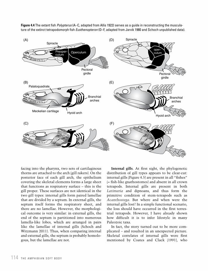
Both types of gills are associated with the gill arch skeleton, which is homologous throughout gnathostomes (Janvier 1996). These arches are composed of curved bows, primitively five arranged in a series, each consisting of several rod-like elements (ceratobranchials, epibranchials, pharyngobranchials) (Figure 4.4). They articulate with unpaired elements in the midline of the pharyngeal floor (basibranchials). Internally,
T H E A M P H I B I A N S O F T B O D Y114
facing into the pharynx, two sets of cartilaginous thorns are attached to the arch (gill rakers). On the posterior face of each gill arch, the epithelium covering the skeletal elements forms a large sheet that functions as respiratory surface – this is the gill proper. These surfaces are not identical in the two gill types: internal gills form paired lamellae that are divided by a septum. In external gills, the septum itself forms the respiratory sheet, and there are no lamellae. However, the morphologi-cal outcome is very similar: in external gills, the end of the septum is partitioned into numerous lamella-like lobes, which are arranged in pairs like the lamellae of internal gills (Schoch and Witzmann 2011). Thus, when comparing internal and external gills, the septum is probably homolo-gous, but the lamellae are not.
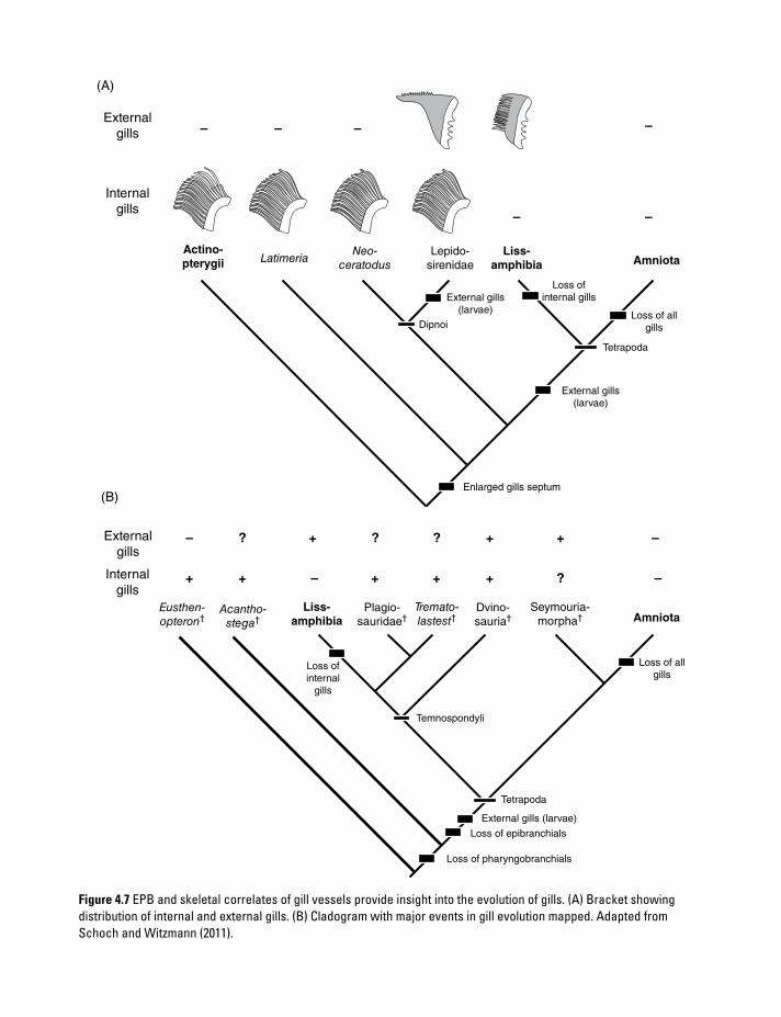
Internal gills. At first sight, the phylogenetic distribution of gill types appears to be clear-cut: internal gills (Figure 4.5) are present in all “fishes” (= fish-like gnathostomes) and absent in all crown tetrapods. Internal gills are present in both Latimeria and dipnoans, and thus form the primitive condition of stem-tetrapods such as Acanthostega. But where and when were the internal gills lost? In a simple functional scenario, the loss should have occurred in the first terres-trial tetrapods. However, I have already shown how difficult it is to infer lifestyle in many Paleozoic taxa.
In fact, the story turned out to be more com-plicated – and resulted in an unexpected picture. Skeletal correlates of internal gills were first mentioned by Coates and Clack (1991), who
Spiracle(A)
(B)
(C) (F)
(E)
(D)
Pectoral
girdle Pectoral
girdle
Spiracle
Palatoquadrate
Meckelian cartilageHyoid arch
Hyoid arch
Branchial
archesBranchial
arches
OperculumPreoper-culum
Figure 4.4 The extant fish Polypterus (A–C, adapted from Allis 1922) serves as a guide in reconstructing the muscula-ture of the extinct tetrapodomorph fish Eusthenopteron (D–F, adapted from Jarvik 1980 and Schoch unpublished data).

R E S P I R A T O R Y O R G A N S 115
discovered grooves on the posterior side of the gill arch elements in Acanthostega. Such grooves, they argued, are only found in bony fishes with internal gills, but not in salamander larvae, which have external gills. Schoch and Witzmann (2011) found the reason for this: the gill arteries lie close to the gill arch in all internal gills, running in
grooves along the skeleton (Figure 4.6A,C). In salamanders, the septum bifurcates (into septal “lamellae”) at a considerable distance from the skeletal element, and there lie the arteries (Figure 4.6B,D). They are far away from the gill arch and consequently do not leave traces on the bone like the grooves in bony fishes. The
(A)
(B)
(C)
Spiracle
Preoper-
culumOperculum
Pectoral
girdleForefin
Gill
lamellae
Gill
lamellae
Gill
raker
Branchial arches
Branchial arch
Figure 4.5 Anatomy of the gill region in (A, B) a bony fish (Polypterus, adapted from Allis 1922) and (C) a salamander (Dicamptodon, unpublished data). (A) With dermal bones covering the branchial region (opercular series in black); (B) without.

T H E A M P H I B I A N S O F T B O D Y116
discovery of such grooves, along with other corre-lates of internal gills, may not be surprising in an aquatic stem-tetrapod like Acanthostega. In the meantime, they were also found in Ichthyostega, which has also increasingly been viewed as water-dwelling (Clack 2012). However, evidence of internal gills also comes from a very different group: Schoch and Witzmann (2011) recently highlighted that such grooves exist in temnospon-
dyls. These were recognized by Bystrow (1938), but at the time were interpreted as support for external gills.
External gills. External gills are only present in larvae, and indeed the “larval stage” is often defined by the presence of external gills in lissam-phibians. External gills in larvae of bony fishes are exceptional, and are certainly not homologous to those of lissamphibians (Figure 4.7) (Witzmann
(A)(B)
(C) (D)
Lamellla
Septum
Cerato-
branchial
Gill raker
Gill raker
Gill
rakerGill
raker
Lamella
Septum
Septum
Septum
Arteries
Arteries
Cerato-
branchial
Cerato-
branchial
Cerato-
branchial
Filaments
formed by
septum
Figure 4.6 The two different types of gills in bony fishes. (A, C) The internal adult gills, common to all bony fishes, are formed by two sheets of lamellae separated by a septum. (B, D) The external larval gills of lissamphibians are instead formed by the septum, and there are no homologs of fish lamellae. (A, B) Lateral view; (C, D) cross-section. Adapted from Schoch and Witzmann (2011).
(A)
(B)
External
gills – – –
– –
Amniota
AmniotaLiss-
amphibia
Loss of all
gills
Loss of all
gills
Loss of
internal gills
Loss of
internal
gills
External gills
(larvae)
Dipnoi
Liss-amphibia
Lepido-
sirenidae
Seymouria-
morpha†Dvino-
sauria†Plagio-
sauridae†Tremato-
lastest†Acantho-
stega†
Eusthen-
opteron†
Neo-
ceratodusLatimeria
Actino-pterygii
Tetrapoda
Tetrapoda
Temnospondyli
External gills
(larvae)
External gills (larvae)
Loss of pharyngobranchials
Loss of epibranchials
Enlarged gills septum
–
–
–
? ? ?
?
+
+ + +
+
–
–+
++
Internal
gills
External
gills
Internal
gills
Figure 4.7 EPB and skeletal correlates of gill vessels provide insight into the evolution of gills. (A) Bracket showing distribution of internal and external gills. (B) Cladogram with major events in gill evolution mapped. Adapted from Schoch and Witzmann (2011).

T H E A M P H I B I A N S O F T B O D Y118
2004). As mentioned above, the external gills are formed by the septum, which together with the skeletal element is the only homologous part between external and internal gills. In caecilians and salamanders, the larval gills develop on branchial arches II, IV, and V; in anurans the poste-rior one is usually absent (Duellman and Trueb 1994). Caecilians have three external larval gills, forming expanded sheets in typhlonectid embryos (Wake 1977) plus fimbriate ones in embryos of Ichthyophis (Dünker et al. 2000). Salamanders have three external gills of various shapes and sizes, correlating with properties of the water body (stream type, pond type). In plethodontid salamanders, the encapsulated larvae undergo direct development but still retain larval gills, and in some species they are leaf-like; in viviparous Salamandra, gill fimbriae are elongated, presuma-bly to take up oxygen within the oviducts (Duellman and Trueb 1994). Finally, in anuran tad-poles external larval gills are overgrown by a flap of skin. Hence, in this clade, external larval gills become secondarily internal (Schmalhausen 1968).
4.5 Lateral lines, electroreception, and ears
The capacity to hear is an ancient trait of vertebrates, and the hearing organs are diverse. Both hearing and balancing senses rely on receptor cells that develop locally from the ectoderm. Based on their possession of hair-like structures, they are called hair cells. They are arranged in clusters and the hairs are sensitive to deflection, generating an electrical response in the cell. Depending on the organ, these receptors are called neuromasts ( lateral sense), maculae and cristae (vestibular or balancing sense), or papillae (auditory sense). Strictly speaking, only the auditory sense is referred to as hearing, but functionally the lateral-line system of fishes is a hearing organ as well.
Lateral line. The lateralis organs (lateral-line sys-tem) form an ancient trait of vertebrates (Mickoleit 2004). They consist of numerous separate mechan-oreceptors located in the skin. Each sensory organ (neuromast) consists of a group of receptor cells bearing sensitive hairs (cilia) that are enclosed in a
gelatinous capsule (cupula). Neuromasts may be located as single units or arranged in lateral lines. In bony fishes, they are located within the dermal bones and connected to the outer surface by means of pores; in tetrapods they lie in open grooves or simply within the dermis. The lateral-line neuro-masts are sensitive to changes in velocity and per-mit orientation under water independent of sight. Based on their anatomical and functional consist-ency, the homology between lateral-line organs of bony fishes and lissamphibians is generally accepted (Mickoleit 2004). Occurring throughout ontogeny in fishes, they are confined to larval stages in amphibians, with the exception of neotenic species, where they persist in aquatic adults (Figure 4.8), and a few aquatic anurans retain-ing them in the adult stage (Pipa, Xenopus). Lateral-line organs only function in organisms that return to the water regularly. They were evidently present in stem-tetrapods, where they were located in bony canals (Clack 2012). That is, anatomical correlates in dermal skull bones indicate the presence of the lateral-line system, confirming the presence of lateral lines in the bracket taxa (bony fishes and lissamphibians). In Paleozoic tetrapods, both stem-amphibians and stem- amniotes, lateral lines were located in grooves aligned in exactly the same pattern as the closed canals of bony fishes. This indicates that lateral lines were not re-invented in lissamphibians, and that they were finally lost in the stem-group of amniotes, where they persisted in seymouriamorphs and lepospondyls. Clack (2012) pointed out that the open lateral-line sulci in tetrapods are a pedomorphic trait with respect to the enclosed canals of their fish-like ancestors. In bony fishes, the canal neuromasts form superfi-cially in the epidermis, and sink into a furrow formed by dermis and epidermis.
Electroreception. A second group of sensory organs of use under water are the electroreceptors of sharks and bony fishes, which are similar in receptor anatomy to the lateralis organs. Electrosensory organs help in the detection and identification of conspecifics and prey items. In addition to orientation, electrosensory organs may also be used to generate electric fields, a feat accomplished by specialized electroreceptors. Certain rays, eels, and catfishes have indepen-dently evolved this capacity in order to threaten

L A T E R A L L I N E S , E L E C T R O R E C E P T I O N , A N D E A R S 119
enemies or paralyze or even kill prey. Electrosensory organs are present in larval salamanders and cae-cilians (Fritzsch and Wahnschaffe 1983), but absent in other tetrapods. Klembara (1994) suggested that depressed, densely pitted regions in the skull roof of Permian seymouriamorphs (Discosauriscus) may have housed electroreceptors.
Balance and sound organs. The organs for sound perception and balance are both located in the inner ear. Together, they are referred to as the stato-acoustic sense. The static or vestibular organ is an autapomorphy of vertebrates, which use it for maintaining balance in the water. Whereas hagfishes and lampreys have only two semicircular canals, gnathostomes have three, corresponding with the three dimensions of space. In contrast to all other sense organs, the vestibular apparatus does not provide information on the environment, but on the orientation and movement of the body
itself. The vestibular system has not essentially changed with the fish–tetrapod transition. The receptors for the vestibular sense are called maculae and cristae, and they are sensitive to displacement occurring when the body changes its orientation.
The second, acoustic, system involves recep-tors (papillae) sensitive to pressure changes. As in the lateral-line organs, papillae are capable of detecting vibrations in the water. In terrestrial tetrapods, airborne vibrations are perceived, but because of their much smaller amplitude an impedance-matching system evolved: the middle ear. In tetrapods, the acoustic organ system thus falls in two separate components: (1) the sensory receptor (papilla) in the water-filled inner ear cavity and (2) the middle ear, an air-filled canal housing the ear ossicle, which acts as sound transmitter (Figure 4.9).
Figure 4.8 Many Paleozoic tetrapods were more or less aquatic. Lateral lines, homologous to those of fishes, are found as closed canals or open grooves in many stem-tetrapods, anthracosaurs, and temnospondyls. (A) Skull roof of neotenic temnospondyl Micromelerpeton. (B) Hyobranchial skeleton (black, ossified; white, unossified; inferred from relatives in which these structures are preserved), branchial dentition, and external gills. Adapted from Schoch (2009a).

T H E A M P H I B I A N S O F T B O D Y120
In bony fishes, hearing in the inner ear is performed by the maculae, which are covered by a gelatinous cupula that contains mineralized bodies. In actinop-terygians, Latimeria, and dipnoans, the mineralized parts, called otoliths (ear stones), are large and formed of aragonite (Nolf 1985). In tetrapods, the same organs contain small calcite crystals. In addition to these receptors, tetrapods also have an acoustic sense, formed by the already mentioned papillae. Common to most tetrapods is the papilla basilaris. As this is absent in dipnoans and other bony fishes, it had long been considered a tetrapod autapomorphy. However, Fritzsch (1987) reported a papilla basilaris in Latimeria, and it is therefore likely that this papilla was lost in dipnoans (Mickoleit 2004).
This reasoning indicates that the papilla basilaris was the first receptor of the auditory sense and can be inferred to have existed in tetrapodomorphs. A second receptor (papilla amphibiorum) is present only in lissamphibians (Parsons and Williams 1963; Duellman and Trueb 1994). Amniotes are thus considered to retain the plesiomorphic condition, with a single papilla covering the entire range of frequencies. Only in modern amphibians has auditory processing been divided into low- and high-frequency streams, with the ear drum of frogs associated with the papilla basilaris, mediating the high-frequency end of the spectrum. Hearing mediated by the papilla basilaris thus evolved under water, first
(A)
(C) (D)
(B)Stapes
Tympanum
Ventral
process
Operculum
Opercular muscle
Operculum
Inner ear
Ear capsule
Stapes
Operculum
Round
window
Opercular
muscle
Stapedial
muscle
Tympanum
(ear drum)Pap. bas.
<1.000 Hz
>1.000 Hz
Pap. am.
Middle ear
Scapula
Figure 4.9 Anatomy of the amphibian ear. (A) Stapes and batrachian operculum in the extant goliath frog (Conraua, unpublished data). (B) Operculum and opercularis muscle in a salamander (adapted from Duellman and Trueb 1994). (C, D) Inner and middle ear of a frog in cross-section (adapted from Wever 1985). Pap am, papilla amphibiorum; pap bas, papilla basilaris.

L A T E R A L L I N E S , E L E C T R O R E C E P T I O N , A N D E A R S 121
confined to low-frequency sound, transmitted by vibrations of the whole skull (Christensen-Dalsgaard and Carr 2008).
Spiracle and middle ear. The spiracle is a canal connecting the pharyngeal cavity with the dorsal side of the skull in jawed vertebrates. It is associated with the hyoid arch, specifically with one component (hyomandibula) that forms part of its lateral wall. Like the gill pouches, the spiracle is primitively water-filled, and has often been considered a modified gill slit. However, its orientation is different from the branchial arches and water flows through it in reversed fashion, from dorsal to ventral. A spiracle is present in sharks and rays, in Polypterus and other basal actinopterygians, as well as in Latimeria, but it is only vestigial, without openings, in lungfishes (Rauther 1930; Bartsch 1994). In many sharks and all rays, the spiracle controls the influx of respira-tory water (von Wahlert 1966). In bony fishes, its role is less clear. Budgett (1903) and Magid (1966) observed the intake of air through the spiracle in Polypterus, when the fish is at the water surface. This happened on many occasions, but especially during phases of excitement or raised activity, or in water that was short of oxygen. This confirms that the spiracle is used as a respiratory canal for the lung in some bony fishes, in contrast to its use in sharks. A bony canal consistent with the features of the spiracle has been identified in many tetrapodomorph fishes, where it is largely similar to that of Polypterus (Jarvik 1980). It is therefore generally accepted that the spiracular canal was present in stem-tetrapods, and a similar anatomy is known from temnospondyls and anthracosaurs (Clack 1993).
The spiracle is considered a homolog of the middle ear cavity in tetrapods (Clack 1993). In tet-rapods, the hyomandibula (stapes) is not attached to the spiracular wall, but enclosed in the spiracular canal, which is always air-filled. Like the spiracle, this middle ear cavity opens ventrally into the pharynx, by means of a narrow channel known as the eustachian tube. Dorsally, the middle ear is closed by a membrane, referred to as the ear drum (tympanum). The ear drum holds the same position as the dorsal spiracular opening in bony fishes, a region known as the temporal notch (squamosal embayment). A middle ear cavity of
this type is present in frogs and most amniotes and may be considered a synapomorphy of tetrapods, although other evidence contradicts this (see below).
The amphibian ear. The evolutionary transfor-mation of the fish hyomandibula into the tetrapod stapes ranks among the most interesting topics in vertebrate evolution. The hyomandibula is a massive bone that tightly integrates numerous anatomical structures (muscles, ligaments, the gill-covering operculum). Movement of the hyomandibula is mediated by several muscles, contributing to the opening of the operculum, changing the shape of the spiracle, and constrain-ing movements of the mandible, palatoquadrate, and braincase. In tetrapods, however, the stapes is not involved in any such role – cranial mobility has been largely reduced, the spiracle has become the middle ear cavity that contains the stapes, and the opercular bones are lost. The massive hyoman-dibula is thus a feature found in groups that primarily feed and breathe under water: actinop-terygians, Latimeria, and stem-tetrapods (Jarvik 1980; Janvier 1996). By contrast, extant lungfishes have a small rudimentary hyomandibula and the opercular region is largely soft with a reduced operculum (Bartsch 1994), but Devonian stem taxa are more consistent with other bony fishes in this set of characters.
In tetrapods, the stapes is shorter than the hyomandibula and largely freed from connections to other skeletal elements, except for its articula-tion with the otic capsule. The reduced impor-tance and connectivity of the hyomandibula/stapes in lungfishes and tetrapods is considered a convergence: fossils show that both stem- dipnoans and stem-tetrapods retained the primitive condi-tion of bony fishes, encompassing a complete hyoid arch. The hyomandibula of Eusthenopteron was still a large and solid element with numerous muscle attachments (Jarvik 1980; Brazeau and Ahlberg 2006).
The tetrapod stapes attaches to the margin of an opening in the ear capsule, the oval window. This round opening evolved from a slit-like fontanelle in bony fishes, but the morphology of the oval window and the mode of attachment are exclusive to and found throughout tetrapods. Distally, the stapes is thin and lightly built,

T H E A M P H I B I A N S O F T B O D Y122
attaching to the tympanum in frogs and amniotes. In salamanders and caecilians, the stapes is more robust and rudimentary, attaching either to the quadrate or the squamosal. In both groups the tympanum, middle ear cavity, and eustachian tube are consistently absent.
Hence, there are two divergent types of middle ears in lissamphibians – the anuran and salaman-der–caecilian types. The similarities between the anuran and amniote ears are usually interpreted as convergences (Lombard and Bolt 1979). This is concluded from inconsistent anatomical struc-tures in anurans and amniotes, especially the course and position of nerves and blood vessels relative to the middle ear and tympanum, which indicate a convergent origin of tympanum and middle ear cavity. However, the absence of these structures in salamanders and caecilians is probably a derived state rather than inherited from stem-tetrapods. This conclusion is based on an entirely phylogenetic argument: the most likely stem-group of all three lissamphibian clades are the dissorophoid temnospondyls, which all possessed a large tympanum, a delicate anuran-like stapes, and a middle ear cavity similar to that of extant frogs (Bolt and Lombard 1985; Maddin et al. 2012). If the temnospondyl origin of lissam-phibians is accepted, the primitive condition of the amphibian ear should therefore be the posses-sion of a tympanum, middle ear cavity, and eustachian tube, with the stapes completely enclosed within the air-filled middle ear cavity. Here, this set of structures is referred to as the tympanic ear. As stem-amniotes lack evidence of a tympanum and middle ear cavity, they are generally not considered to have possessed a tympanic ear – this indicates the independent evolution of such ears in lissamphibians and amniotes. This hypothesis is supported by the presence of massive stapes in stem-amphibians and stem-amniotes, which often articulated with the quadrate or squamosal.
Among lissamphibians, anurans and salaman-ders have a second ear ossicle that is formed by an isolated piece of the ear capsule (Figure 4.9). It is often bony, but may also be cartilaginous. Unfortunately, this element is referred to as the operculum, although it is neither homologous nor functionally comparable to the gill-covering
elements of bony fishes. It is an endoskeletal element, in contrast with the dermal origin of the fish operculum. To avoid confusion, I refer to this element as the batrachian operculum. This element is located posterior to the oval window and forms the origin of a muscle that attaches to the scapula. Thus, the batrachian operculum and the so-called opercularis muscle connect the inner ear with the shoulder girdle and forelimb, forming an independent hearing apparatus from that of the stapes. This apparatus transmits low-frequency vibrations from the ground to the inner ear, which are perceived by the papilla amphibiorum (Wever 1985). The fact that the papilla amphibiorum and the opercular apparatus are functionally coupled suggests that the ancestors of caecilians probably possessed an operculum, although the extant taxa lack it; the massive footplate of the caecilian stapes might well include an operculum.
References
Allis, P. (1897) The cranial muscles and cranial and first spinal nerves in Amia calva. Journal of Morphology 12, 487–762.
Allis, P. (1922) The cranial anatomy of Polypterus, with special reference to Polypterus bichir. Journal of Anatomy 56, 189–291.
Bartsch, P. (1994) Development of the cranium of Neoceratodus forsteri, with a discussion of the suspensorium and the opercular apparatus in Dipnoi. Zoomorphology 114, 1–31.
Bolt, J.R. & Lombard, R.E. (1985) Evolution of the amphibian tympanic ear and the origin of frogs. Biological Journal of the Linnean Society 24, 83–99.
Boy, J.A. (1972) Die Branchiosaurier (Amphibia) des saarpfälzischen Rotliegenden (Perm, SW-Deutschland). Abhandlungen des Hessischen Landesamts für Bodenforschung 65, 1–137.
Boy, J.A. (1974) Die Larven der rhachitomen Amphibien (Amphibia: Temnospondyli, Karbon-Trias). Paläontologische Zeitschrift 48, 236–268.
Brazeau, M. D. & Ahlberg, P. E. (2006) Tetrapod-like middle ear architecture in a Devonian fish. Nature 439, 318–321.
Bryant, H.N. & Russell, A.P. (1992) The role of phylogenetic analysis in the inference of

R E F E R E N C E S 123
unpreserved attributes of extinct taxa. Philo-sophical Transactions of the Royal Society of London B 337, 405–418.
Bryant, H.N. & Seymour, K.L. (1990) Observations and comments on the reliability of muscle recon-struction in fossil vertebrates. Journal of Mor-phology 206, 109–117.
Budgett, J.S. (1903) Notes on the spiracles of Polypterus. Proceedings of the Zoological Society of London 1903, 10.
Bulman, O.M.B. & Whittard, W.F. (1926) On Branchiosaurus and allied genera (Amphibia). Proceedings of the Zoological Society of London 1926, 533–579.
Bystrow, A.P. (1938) Dvinosaurus als neotenische Form der Stegocephalen. Acta Zoologica 19, 209–295.
Carroll, R.L. & Holmes, R. (1980) The skull and jaw musculature as guides to the ancestry of salamanders. Zoological Journal of the Linnean Society 68, 1–40.
Christensen-Dalsgaard, J. & Carr, C.E. (2008) Evolution of a sensory novelty: tympanic ears and the associated neural processing. Brain Research Bulletin 75, 365–370.
Clack, J.A. (1993) Homologies in the fossil record: the middle ear as a test case. Acta Biotheoretica 41, 391–410.
Clack, J.A. (1998) The neurocranium of Acanthostega gunnari Jarvik and the evolution of the otic region in tetrapods. Zoological Journal of the Linnean Society 122, 61–97.
Clack, J.A. (2012) Gaining Ground: the Origin and Evolution of Tetrapods, 2nd edition. Bloomington: Indiana University Press.
Coates, M.I. & Clack, J.A. (1991) Fish-like gills and breathing in the earliest known tetrapod. Nature 352, 234–236.
Deban, S.M. & Wake, D.B. (2000) Aquatic feeding in salamanders. In: K. Schwenk (ed.), Feeding: Form, Function, and Evolution in Tetrapod Vertebrates. Boston: Academic Press, pp. 65–94.
de Ricqlès, A. (1975) Quelques remarques paléo-his-tologiques sur le problème de la néotenie chez les stégocéphales. CNRS Colloquium International 218, 351–363.
Diogo, R., Hinits, Y., & Hughes, S. (2008) Development of mandibular, hyoid and hypo-branchial muscles in the zebrafish: homologies
and evolution of these muscles in bony fishes and tetrapods. BMC Evolutionary Biology 8, 24–46.
Drüner, L. (1901) Studien zur Anatomie der Zungenbein-, Kiemenbogen- und Kehlkopf-muskulatur der Urodelen. I. Theil. Zoologisches Jahrbuch für Anatomie und Ontogenie 15, 435–622.
Duellman, W.E. & Trueb, L. (1994) Biology of Amphibians. Baltimore: Johns Hopkins Univer-sity Press.
Dünker, N., Wake, M.H., & Olson, W.M. (2000) Embryonic and larval development in the caecil-ian Ichthyophis kohtaoensis (Amphibia, Gymnophiona): a staging table. Journal of Morphology 243, 3–34.
Franzen, J.L. & Schaal, S. (2000) Der eozäne See von Messel. In: G. Pinna (ed.), Europäische Fossillagerstätten. Berlin: Springer, pp. 177–183.
Fritzsch, B. (1987) Inner ear of the coelacanth fish Latimeria has tetrapod affinities. Nature 327, 153–154.
Fritzsch, B. & Wahnschaffe, U. (1983) The elec-troreceptive ampullary organs of urodeles. Cell and Tissue Research 229, 483–503.
Iordansky, N.N. (1990) Evolution of Complex Adaptations. The Jaw Apparatus of Amphibians and Reptiles. Nauka, Moscow. (In Russian.)
Janvier, P. (1996) Early Vertebrates. Oxford Mono-graphs on Geology and Geophysics 33. Oxford: Oxford Univesity Press.
Jarvik, E. (1980) Basic Structure and Evolution of Vertebrates. Vols. 1–2. London and New York: Academic Press.
Klembara J. (1994) Electroreceptors in the Lower Permian Discosauriscus austriacus. Palaeon-tology 37, 609–626.
Klembara, J. (1995) The external gills and orna-mentation of skull-roof bones of the Lower Permian Discosauriscus (Kuhn 1933) with remarks to its ontogeny. Paläontologische Zeitschrift 69, 265–281.
Lauder, G.V. (1980) Evolution of the feeding mech-anism in primitive actinopterygian fishes: a functional anatomical analysis of Polypterus, Lepisosteus, and Amia. Journal of Morphology 163, 283–317.
Lauder, G.V. & Shaffer, H.B. (1985) Functional morphology of the feeding mechanism in

T H E A M P H I B I A N S O F T B O D Y124
aquatic ambystomatid salamanders. Journal of Morphology 185, 297–326.
Lombard, R.E. & Bolt, J.R. (1979) Evolution of the tetrapod ear: an analysis and reinterpretation. Biological Journal of the Linnean Society 11, 19–76.
Lubosch, W. (1938) Muskeln des Kopfes. Viscerale Muskulatur. In: L. Bolk, E. Göppert, E. Kallius, & W. Lubosch (eds.), Handbuch der vergleichenden Anatomie der Wirbeltiere. Berlin: Urban & Schwarzenberg, pp. 1011–1106.
Luther, A. (1914) Über die vom N. trigeminus versorgte Muskulatur der Amphibien. Acta Societatis Scientarum Fennicae 44, 1–151.
Maddin, H., Jenkins, F.A., & Anderson, J.S. (2012) The braincase of Eocaecilia micropodia (Lissamphibia, Gymnophiona) and the origin of caecilians. PloS ONE 7, e50743.
Magid, A.M.A. (1966) Breathing and function of the spiracles in Polypterus senegalus. Animal Behavior 14, 530–533.
Mickoleit, G. (2004) Phylogenetische Systematik der Wirbeltiere. Munich: Pfeil.
Milner, A.C., Milner, A.R., & Walsh, S.A. (2009) A new specimen of Baphetes from Nýrany, Czech Republic and the intrinsic relationships of the Baphetidae. Acta Zoologica 90, 318–334.
Milner, A.R. (1982) Small temnospondyl amphib-ians from the Middle Late Carboniferous of Illinois. Palaeontology 25, 635–664.
Milner, A.R. (2007) Mordex laticeps and the base of the Trematopidae. Journal of Vertebrate Paleontology 27, 118A.
Milner, A.R. & Sequeira, S.E.K. (1994) The temno-spondyl amphibians from the Viséan of East Kirkton, West Lothian, Scotland. Transactions of the Royal Society of Edinburgh: Earth Sciences 84, 331–361.
Nolf, D. (1985) Otolithi piscum. In: H.-P. Schultze (ed.), Handbook of Paleoichthyology. Stuttgart: Gustav Fischer, Vol. 10, pp. 1–145.
Parsons, T.S. & Williams, E.E. (1963) The relation-ships of the modern Amphibia: a re-examination. Quarterly Review of Biology 38, 26–53.
Rauther, W. (1930) Kiemen der Anamnier: Kiemenderivate der Cyclostomen und Fische. In: L. Bolk, E. Göppert, E. Kallius, & W. Lubosch (eds.), Handbuch der vergleichenden Anatomie der Wirbeltiere, vol. 3, pp. 211–276.
Sanchez, S., de Ricqlès, A., Schoch, R.R., & Steyer, J.S. (2010) Developmental plasticity of limb bone microstructural organization in Apateon: histological evidence of paedomorphic conditions in branchiosurs. Evolution and Development 12, 315–328.
Schmalhausen, I.I. (1968) The Origin of Terrestrial Vertebrates. London and New York: Academic Press.
Schmidt-Nielsen, K. (1997) Animal Physiology: Adaptation and Environment. Cambridge: Cam-bridge University Press.
Schoch, R.R. (1992) Comparative ontogeny of Early Permian branchiosaurid amphibians from southwestern Germany. Developmental stages. Palaeontographica A 222, 43–83.
Schoch, R.R. (2002) The early formation of the skull in extant and Paleozoic amphibians. Paleobiology 28, 378–396.
Schoch, R.R. (2004) Skeleton formation in the Branchiosauridae as a case study in comparing ontogenetic trajectories. Journal of Vertebrate Paleontology 24, 309–319.
Schoch, R.R. (2009) The evolution of life cycles in early amphibians. Annual Review of Earth and Planetary Sciences 37, 135–162.
Schoch, R.R. & Witzmann, F. (2009) Osteology and relationships of the temnospondyl Sclero-cephalus. Zoological Journal of the Linnean Society London 157, 135–168.
Schoch, R.R. & Witzmann, F. (2011) Bystrow’s paradox: gills, forssils, and the fish-to-tetrapod transition. Acta Zoologica 92, 251–265.
Schultze, H.-P. (1991) A comparison of controversial hypotheses on the origin of tetrapods. In: H.-P. Schultze & L. Trueb (eds.) Origins of the Higher Groups of Tetrapods: Controversy and Consensus. Ithaca: Cornell University Press, pp. 29–67.
Thomson, K.S. (1967). Mechanisms of intracr-anial kinesis in fossil rhipidistian fishes (Crossopterygii) and their relatives. Zoological Journal of the Linnean Society 46, 223–253.
von Wahlert, G. (1966) Atemwege und Schädelbau der Fische. Stuttgarter Beiträge zur Naturkunde A 159, 1–10.
Wake, D.B. & Deban, S.M. (2000) Terrestrial feeding in salamanders. In: K. Schwenk (ed.), Feeding: Form, Function, and Evolution in Tetrapod Vertebrates. Boston: Academic Press, pp. 95–116.

R E F E R E N C E S 125
Wake, M.H. (1977) The reproductive biology of caecilians: an evolutionary perspective. In: D.H. Taylor & S.I. Guttman (eds.), The Reproductive Biology of Amphibians. New York: Plenum, pp. 73–101.
Werneburg, R. (1991) Die Branchiosaurier aus dem Unterrotliegend des Döhlener Beckens bei Dresden. Veröffentlichungen des Naturhistorischen Museums Schleusingen 6, 75–99.
Werneburg, R. (2007) Timeless design: colored pattern of skin in Early Permian branchiosaurid (Temnospondyli: Dissorophoidea). Journal of Vertebrate Paleontology 27, 1047–1050.
Wever, E.G. (1985) The Amphibian Ear. Princeton: Princeton University Press.
Willems, H. & Wuttke, M. (1987) Lithogenese lakustriner Dolomite und mikrobiell induzierte “Weichteilerhaltung” bei Tetrapoden des Unter-Rotliegenden (Perm, Saar-Nahe-Becken, SW-Deutschland). Neues Jahrbuch für Geologie und Paläontologie Abhandlungen 174, 213–238.
Witmer, L. M. (1995). The extant phylogenetic bracket and the importance of reconstructing soft tissues in fossils. In: J. J. Thomason (ed.), Functional Morphology in Vertebrate Paleon-tology. Cambridge: Cambridge University Press, pp. 19–33.
Witzmann F. (2004) The external gills of Palaeozoic amphibians. Neues Jahrbuch für Geologie und Paläontologie Abhandlungen 232, 375–401.
Witzmann F. (2006a). Morphology and palaeobiol-ogy of the Permo-Carboniferous temnospondyl amphibian Archegosaurus decheni Goldfuss, 1847 from the Saar-Nahe Basin, Germany. Transactions of the Royal Society of Edinburgh: Earth Sciences 96, 131–162.
Witzmann F. (2006b). Developmental patterns and ossification sequence in the Permo-Carboniferous temnospondyl Archegosaurus decheni (Saar-Nahe Basin, Germany). Journal of Vertebrate Paleontology 26, 7–17.