amphibian biology - wakelab.berkeley.edu
TRANSCRIPT

Amphibian Biology
Edited by
Harold Heatwole
Volume 5
Osteology
Co-editor for this volume
Margaret Davies
Published by
Surrey Beatty & Sons

CHAPTER 5
Morphological Variation, Development, and Evolution of the Limb Skeleton of Salamanders
Neil H. Shubin and David B. Wake
I. Introduction
II. The Salamander Limb
III. Phylogenetic Diversity A. Progenitors of Salamander Limbs B. The Limbs of the First Salamanders C. Variation in Living Taxa
1. Hynobiidae and Cryptobranchidae 2. Sirenidae, Proteidae and
Amphiumidae 3. Rhyacotritonidae 4. Dicamptodontidae and
Ambystomatidae 5. Salamandridae 6. Plethodontidae
D. Phylogenetic Significance of Interfamilial Variation
IV. Ontogeny A. Common Features of Developing
Salamander Limbs 1. Stylopodium and Zeugopodium 2. Autopodium
B. Apomorphies of Salamander Limb Development
C. Developmental Variation 1. Regional Patterns of Developmental
Variation A. Preaxial Column B. Central Series C. Digital Arch
2. Species with Reduced Limbs 3. Ecological Variation
A. Pond Larvae of Salamandrids B. Pond Larvae of Hynobiids C. Stream Larvae Diverse Species D. Direct Development
V. Variation within Populations VI. Developmental Basis of Character Evolution
VII. Phylogenetic Uses of Developmental Data A. The Hypothesis of the Polyphyly of
Tetrapods B. The Hypothesis of the Neomorphic
Origin of the Salamander Autopodium C. The Hypothesis of a Conserved
Developmental Trajectory VIII. Prospects for Future Research IX. Acknowledgements X. References
I. INTRODUCTION
SALAMANDER limbs serve as a natural laboratory for addressing problems of 1 morphological evolution. This utility results from the fact that important aspects of
limb biology have become the focus of comparative analysis. Recent studies have produced large sets of data on natural variation, both of adult form and of limb development. Comparative investigations of ontogeny have provided an understanding of the ways that organogenesis varies in diverse clades living in different ecological settings (e.g., Blanco and Alberch 1992; Vorobyeva and Hinchliffe 1996). In addition, new studies of intraspecific

SHUBIN and WAKE: LIMB SKELETON OF SALAMANDERS 1783
variation of adult limb patterns span much of the phylogenetic and morphological diversity of salamanders (e.g., Hanken 1983; Rienesl and Wagner 1992; Zaffaroni et al. 1992; Shubin et al. 1995). With the convergence of these lines of evidence, and die emergence of stable molecular phylogenies of the Order Caudata (Larson and Dimmick 1993), salamander limb diversity now can be used to address the mechanisms behind the origin of morphological novelty.
II. THE SALAMANDER LIMB
The morphology of the limb was thoroughly described for the salamandrid species Salamandra salamandra by Francis (1934), who presented excellent figures of the skeleton, musculature and innervation (see also Duellman and Trueb [1986] for a general summary). This chapter focuses on the skeleton, which has been the main subject of limb investigations.
For the purposes of nomenclature, the limb skeleton of a generalized species (that is, one not having undergone evident reductions in numbers of digits or phalanges) is described. The taxon selected is the genus Dicamptodon, a large terrestrial salamander with well-formed limbs. Forelimbs bear a manus having no more than four digits, and hind limbs bear a pes having no more than five digits (Fig. 1; Wake and Shubin [1999]). The forelimb contains a stylopodium consisting of a humerus, a zeugopodium consisting of a radius and an ulna, and an autopodium (manus) consisting of a mesopodium, a metapodium, and the digits. The mesopodium contains a variable number of elements (eight in Dicamptodon), including a row of basal elements (radiale, intermedium and ulnare, from preaxial to postaxial), a centrale, and distal elements (element y, basale commune, and distal carpals three and four). The metapodium contains metacarpals I-IV The digits display limited variation in numbers of phalanges, the generalized Dicamptodon having a phalangeal formula (from preaxial to postaxial) of 2-2-3-2. In the hind limb, the stylopodium contains the femur, the zeugopodium contains the tibia and fibula, and the autopodium (pes) contains the mesopodium and metapodium with digits l-V. The mesopodium of the hind limb consists of a variable number of elements (nine in Dicamptodon): a row of basals (tibiale, intermedium, fibulare), a centrale, and a series of distal elements (element y, basale commune, and distal tarsals three, four and five). The metapodium contains the five metatarsals. The digits of the hind limb show a limited range of variation in terms of numbers of phalanges; Dicamptodon retains a generalized phalangeal formula of 2-2-3-3-2. All the elements of the limb may be ossified in the adults of some taxa (e.g., most species of the family Salamandridae). In the majority of clades, however, the mesopodium remains cartilaginous in whole or in part.
Among living tetrapods, salamanders display several features of die limb that constitute important morphological apomorphies of Caudata and its outgroups. The phylogenetic hypothesis of Larson and Dimmick (1993) serves as the foundation for analysis (Fig. 2).
1. Four digits on the manus. With anurans, salamanders share a manus with no more than four digits. Many temnospondyls also possess a four-digited manus. Assuming the monophyly of the Lissamphibia, this character is likely to be a synapomorphy inclusive of temnospondyls and lissamphibians, or of Lissamphibia plus some subset of temnospondyls. An alternative interpretation has the Lissamphibia deeply embedded within the lepospondyls (Laurin 1998), but the analysis on which this is based includes relatively few taxa and does not include limb characters discussed herein. While lepospondyl limbs are not well known, and many taxa have reduced limbs or no limbs at all, those forms that do have well-preserved forelimbs seem to show reductions to four or fewer digits on the manus (Carroll and Gaskill 1978; Carroll 1988). The striking conservation of manual phalangeal formulae in various temnospondyls and lissamphibians suggests that the reduction to four digits happened prior to the evolution of lissamphibians. Overall phalangeal formulae in temnospondyls, basal salamanders and basal frogs varies from 2-2-3-3 (colosteids, Apateon, Viaerella, Notobatrachus,

Fig. I. Limbs of Dicamptodon ensatus. A. Forelimb. B. Hind limb. Stippling indicates cartilage. For abbreviations see Appendix at the back of the book. Modified from Wake and Shubin (1999).
Eryops
Trematops
Amphibamus
Doleserpeton
Sirenidae
Cryptobranchidae
Hynobiidae
Amphiumidae
Plethodontidae
Rhyacotritonidae
Salamandridae
Ambystomatidae
Dicamptodontidae
Proteidae
Sirenoidea
Cryptobranchoidea
Fig. 2. The phylogenetic hypothesis used in this chapter, based on molecular and morphological characters. From Larson and Dimmick (1993).
Salamandroidea

SHUBIN and WAKE: LIMB SKELETON OF SALAMANDERS 1785
Eodiscoglossus) to 2-2-3-2 (Eryops, Karaurus, hynobiids). The fact that four digits are present in each of these taxa, and that the phalangeal counts are also highly conserved, suggests that the digits are homologous.
2. A basale commune in the carpus. This is a unique synapomorphy of the Order Caudata. It represents a combination of distal carpals 1 and 2. These elements are separate in the temnospondyls (Fig. 3) and lepospondyls where this portion of the mesopodium is preserved.
3 A basale commune in the tarsus. The parallelism between the carpus and tarsus of tetrapods has long been evident. Stem temnospondyls and lepospondyls both have separate distal tarsals 1 and 2. The fusion of these bones is unique to Caudata.
4. Preaxial dominance in development. Digits two and one are the first to form in salamanders, whereas in frogs and amniotes the postaxial portion of the limb typically develops first (usually digit four). The digital arch typically develops in a preaxial to postaxial direction, the reverse of that in frogs and amniotes (Shubin and Alberch 1986). This character pertains to both the manus and pes.
5. Precocial development of the basale commune and first two metapodial elements. These elements typically chondrify before elements in the centre of the mesopodium (Shubin and Alberch 1986). This character pertains to both the manus and pes.
6. Loss of the element identified as the mediale (m) in the tarsus. This element, first identified by Schmalhausen (1917) as a rare variant in salamanders, is found in some temnospondyls, and is a variant condition in populations of some extant caudates (Shubin et al. 1995).
7. The loss of the prepollex. Features of the limb that are characteristic of caudates, but not useful for phylogenetic analysis, include the five digits of the hind limb, the relatively large size of the mesopodium, the large number of mesopodial elements and their tendency to remain cartilaginous, and the maximal phalangeal formulae of 2-2-3-2 and
Fig. 3. Limbs of salamander outgroups. A. Forelimb of Eryops. B. Hind limb of Trematops (modified from Schaeffer 1941). For abbreviations see Appendix at the back of the book.
1786 AMPHIBIAN BIOLOGY
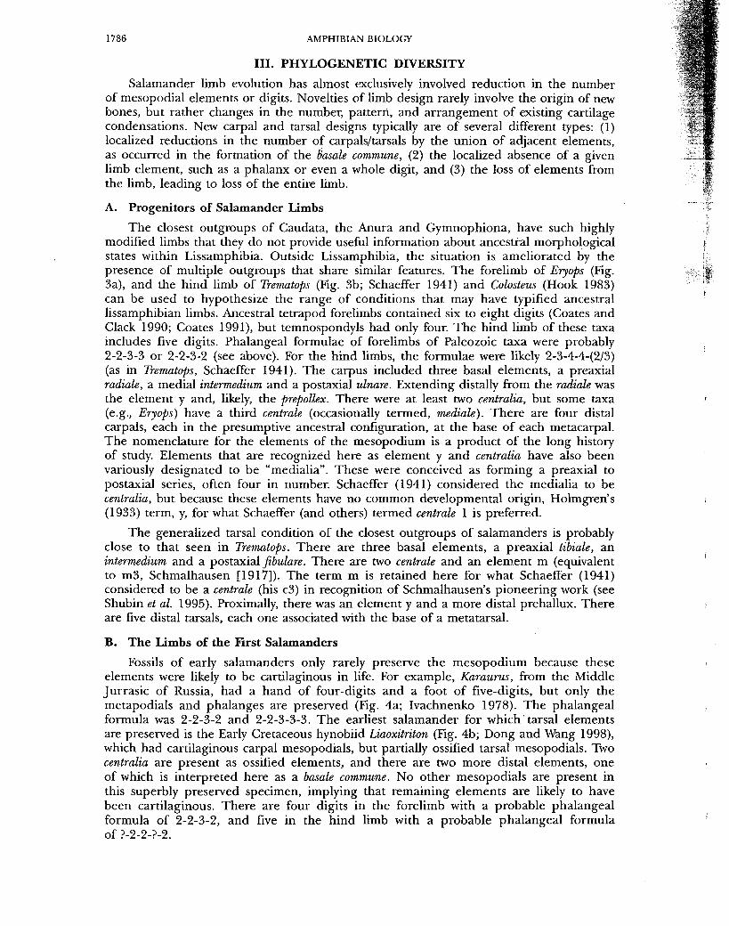
III. PHYLOGENETIC DIVERSITY
Salamander limb evolution has almost exclusively involved reduction in the number of mesopodial elements or digits. Novelties of limb design rarely involve the origin of new bones, but rather changes in the number, pattern, and arrangement of existing cartilage condensations. New carpal and tarsal designs typically are of several different types: (1) localized reductions in the number of carpals/tarsals by the union of adjacent elements, as occurred in the formation of the basale commune, (2) the localized absence of a given limb element, such as a phalanx or even a whole digit, and (3) the loss of elements from the limb, leading to loss of the entire limb.
A. Progenitors of Salamander Limbs
The closest outgroups of Caudata, the Anura and Gymnophiona, have such highly modified limbs that they do not provide useful information about ancestral morphological states within Lissamphibia. Outside Lissamphibia, the situation is ameliorated by the presence of multiple outgroups that share similar features. The forelimb of Eryops (Fig. 3a), and the hind limb of Trematops (Fig. 3b; Schaeffer 1941) and Colosteus (Hook 1983) can be used to hypothesize the range of conditions that may have typified ancestral lissamphibian limbs. Ancestral tetrapod forelimbs contained six to eight digits (Coates and Clack 1990; Coates 1991), but temnospondyls had only four. The hind limb of these taxa includes five digits. Phalangeal formulae of forelimbs of Paleozoic taxa were probably 2-2-3-3 or 2-2-3-2 (see above). For the hind limbs, the formulae were likely 2-3-4-4-(2/3) (as in Trematops, Schaeffer 1941). The carpus included three basal elements, a preaxial radiale, a medial intermedium and a postaxial ulnare. Extending distally from the radiate was the element y and, likely, the prepollex. There were at least two centralia, but some taxa (e.g., Eryops) have a third centrale (occasionally termed, mediale). There are four distal carpals, each in the presumptive ancestral configuration, at the base of each metacarpal. The nomenclature for the elements of the mesopodium is a product of the long history of study. Elements that are recognized here as element y and centralia have also been variously designated to be "medialia". These were conceived as forming a preaxial to postaxial series, often four in number. Schaeffer (1941) considered the medialia to be centralia, but because these elements have no common developmental origin, Holmgren's (1933) term, y, for what Schaeffer (and others) termed centrale 1 is preferred.
The generalized tarsal condition of the closest outgroups of salamanders is probably close to that seen in Trematops. There are three basal elements, a preaxial tibiale, an intermedium and a postaxial fibulare. There are two centrale and an element m (equivalent to m3, Schmalhausen [1917]). The term m is retained here for what Schaeffer (1941) considered to be a centrale (his c3) in recognition of Schmalhausen's pioneering work (see Shubin et al. 1995). Proximally, there was an element y and a more distal prehallux. There are five distal tarsals, each one associated with the base of a metatarsal.
B. The Limbs of the First Salamanders
Fossils of early salamanders only rarely preserve the mesopodium because these elements were likely to be cartilaginous in life. For example, Karaurus, from the Middle Jurrasic of Russia, had a hand of four-digits and a foot of five-digits, but only the metapodials and phalanges are preserved (Fig. 4a; Ivachnenko 1978). The phalangeal formula was 2-2-3-2 and 2-2-3-3-3. The earliest salamander for which tarsal elements are preserved is the Early Cretaceous hynobiid Liaoxitriton (Fig. 4b; Dong and Wang 1998), which had cartilaginous carpal mesopodials, but partially ossified tarsal mesopodials. Two centralia are present as ossified elements, and there are two more distal elements, one of which is interpreted here as a basale commune. No other mesopodials are present in this superbly preserved specimen, implying that remaining elements are likely to have been cartilaginous. There are four digits in the forelimb with a probable phalangeal formula of 2-2-3-2, and five in the hind limb with a probable phalangeal formula of ?-2-2-?-2.

SHUBIN and WAKE: LIMB SKELETON OF SALAMANDERS 1787
Fig. 4. Hind limbs of basal salamanders. A. Kaururus (Ivachnenko 1978). B. Liaoxitriton (Dong and Wang 1998). C. Onychodactylus. D. Liua. Stippling indicates cartilage. For abbreviations see Appendix at the back of the book.

be dt dt dt be 3 4 ©
5 R 4 ©
y cx
c
cx
c t
i f
Liu a
be dt 3
dt 4
O
dt 5
y cx
c
i
dt 4
O
t
cx
c
i f
£§ Hi
Ranodon
|p? dt 1
d 2
dt 3
dt 4
dt 5
y cx %
c - f
'' - f
Tr em at ops
be dt 3
dt 4
dt 5 P3
be dt 3
dt 4 + 5 IP?
be dt 3
dt 4
O
dt 5
y c © m
y c +© y c
dt 4
O
dt 5
t 1 t c
f t f t 1 t i
f t f
Andhas Schaeffer 1941
Cryptobranchus Cryptobranchus alleganiensis schaeffer 1941
dope 1889
BASAL TAXA Fig. 5. Block diagrams (following conventions of Shubin and Wake [1996]) of
basal salamanders on reduced cladogram. For abbreviations see Appendix at the back of the book.

SHUBIN and WAKE: LIMB SKELETON OF SALAMANDERS 1789
C. Variation in Living Taxa
1. Hynobiidae and Cryptobranchidae
The limbs of cryptobranchids and hynobiids contain more elements and tend to be more variable than those of other taxa (Figs 4b, 5, 6c,d). No single carpal and tarsal pattern characterizes these families because interspecific variability is very high (Fig. 5). The limbs of most species of cryptobranchids and hynobiids retain plesiomorphic elements that have been lost in the standard arrangements of other salamanders. Some taxa retain two centralia (Liua, Ranodon, Salamandra, Paradactylodon, Batrachuperus and Cryptobranchus alleganiensis) whereas others possess a single central element. Element m appears as a variant in populations of some taxa in both families (Shubin and Wake 1996). Some taxa retain the postminimus seen in many temnospondyl and lepospondyl limbs (Liua, Ranodon, Salamandrella, Paradactylodon, Batrachuperus and Andrias) and two taxa possess a prehallux (Cryptobranchus, Salamandrella). Baur (1888) cited the case of a remnant complete prepollex in the preaxial column consisting of the basal tibiale, e lement y and two more distal elements; this appears to be the most complete prepollex known from salamanders, but it is a rare variant condition. Some hynobiids and cryptobranchids display a carpal a r rangement not found elsewhere among living salamanders but present in some temnospondyls (e.g., Eryops). In some species of Batrachuperus, for example, the large basal centrale may articulate directly with the radius, which is larger (at least wider distally) than in other taxa. In such instances the centrale separates the radiate from the intermedium.
Hind limb digits are reduced in number from five to four in Salamandrella, Paradactylodon, and Batrachuperus, and in intraspecific variants of some species oiHynobius.
2. Sirenidae, Proteidae and Amphiumidae
These taxa all have reduced limbs (Figs 6c, 7). Sirenids have only forelimbs, which bear four digits in Siren and three in Pseudobranchus. The carpals are reduced in number. Baur (1888) reported seven elements in Siren, which he interpreted as radiale, centrale + intermedium, and ulnare. The phalangeal formula is 1-2-3-2. Element y is often very reduced in size compared to other salamanders. There is a basale commune and two distal carpals. In Pseudobranchus the phalangeal formula is 2-2-2 and there are only four carpals, which is interpreted here as radiale+y, centrale + intermedium+ulnare, basale commune, and one distal carpal.
Amphiumids have forelimbs and hind limbs that are greatly reduced in size and complexity, with reduced numbers of bones in the manus and pes (Fig. 7). Digits vary in number from one to three. There is some variation in the number of mesopodials, but the standard number is three, two basal elements (radiale or tibiale, intermedium + ulnare or fibulare) and a single distal element representing minimally the basale commune. The preaxial basal element apparently is combined with element y.
Proteiids have reduced numbers of toes (Fig. 7), with Necturus having four digits on each limb and Proteus having three digits on the forelimb but only two on the hind limb. Proteus is the only salamander that has more digits on the forelimb than the hind limb. T h e mesopodials of Necturus have the standard configuration. Mesopodial elements of Proteus are greatly reduced in number, with the forelimb having a radiale, intermedium + ulnare, and basale commune; the hind limb has a tibiale, intermedium + fibulare, and distal basale commune.
3. Rhyacotritonidae
The "standard" carpal pattern contains six mesopodials, three basal elements with the intermedium incorportating the centrale, and the radiale incorporating element y (Figs 6e, 8c), but there is much variability (Good and Wake 1992). The most common tarsal pattern contains the standard nine mesopodial elements seen in other taxa (Fig. 8). The commonest phalangeal formulae are 2-2-3-2 and 2-2-3-3-2.

*£pi
1790 AMPHIBIAN BIOLOGY
-41
6. Forelimbs of diverse salamanders. A. Taricha (from Shubin et al. 1995). B. Onychodactylus. C. Siren. D. Paramesotriton. E. Rhyacotriton (from Good and Wake 1992). Stippling indicates cartilage. For abbreviations see Appendix at the back of the book.

SHUBIN and WAKE: LIMB SKELETON OF SALAMANDERS 1791
fpf be dt
3 dt
4 + 5 P3 y cx O
t c f
t i
f
Salamandrella Necturus Amphiuma Proteus Cope 1889 Cope 1889
Kg. 7. Block diagrams of the hind limb of species with reductions in skeletal elements (after Shubin and Wake 1996). Note that Salamandrella and Necturus have four digits, Amphiuma tridactyla has three, and Proteus anguinius has two. For abbreviations see Appendix at the back of the book.
4. Dicamptodontidae and Ambystomatidae
These taxa share the generalized condition described earlier (Fig. 2).
5. Salamandridae
Members of this family share the standard pattern, but with much variation within certain clades. Intraspecific variation has been well-studied in this family. In the manus, the most common departure from the standard condition is the fusion of the ulnare and intermedium (e.g., species of Taricha and Triturus; [RienesI and Wagner 1992; Zaffaroni et al. 1992; Shubin et al. 1995]). The phalangeal formula is 1-2-3-2 and 1-2-3-3-2. In the pes the most significant variation in other taxa includes the universal loss of the fifth digit in the hind limb of Salamandrina terdigitata, and the fusion of distal tarsals 4 and 5 in Cynops, Euproctus, and species of Triturus).
6. Plethodontidae
Some members of this family possess the standard pattern (Wake 1963, 1966; Wake and Elias 1983), but there is variation in the number, shape and articulations of manual and pedal elements. The most significant variation is the loss of the fifth digit in all members of the genera Hemidactylium and Batrachoseps, and the Eurycea quadridigitata species complex. T h e standard phalangeal formula is 1-2-3-2 and 1-2-3-3-2. There is substantial variation in size and shape of the limbs, digits and phalanges, the extent of interdigital webbing, and in the number and arrangements of the carpals and tarsals, especially in miniaturized species (Wake 1966; Hanken 1985). The most common pattern of interspecific variation is the fusion of distal tarsals 4 and 5 (e.g., Bolitoglossa, Bradytriton, Dendrotriton, Lineatriton, Nototriton, Nyctanolis, Pseudoeurycea). In some genera (e.g., Oedipina, Thorius), the centrale may also fuse with distal tarsal/carpal 4 or a combined distal tarsal 4 and 5.
D. Phylogenetic Significance of Interfamilial Variation
If one accepts the phylogenetic hypothesis in Figure 2, limb characters add support to the clade of salamandroid taxa; this clade also is supported by a biologically significant synapomorphy, internal fertilization (Larson and Dimmick 1993). These synapomorphies would constitute one or two derived conditions of the limbs for those clades that have the otherwise standard organizational pattern of limbs (as in Fig. 1). All these characters would involve the loss of elements seen in basal taxa: the postminimus, prepollex, and some centralia. Whereas these elements are typically present in temnospondyls, they are variably present in species of extant basal families (hynobiids and cryptobranchids). This distribution of characters implies that the loss of these primitive elements may have been homoplastic, with reductions evolving in basal clades and within the common ancestor of salamandroids.
be dt 3
dt 4
y c O
t i + f

B > s; •0
2 o r O S3
Fig. 8. Hind limbs of derived salamanders. A. Salamandrina terdigitata. B. Taricha granulosa (from Shubin et al. 1995). C. Rhyacotriton olympicus (from Good and Wake 1992). D. Chiropterotriton multidentatus (from Wake and Elias 1983). E. Batrachoseps gabrieli. F. Pseudoeurycea leprosa (from Wake and Elias 1983). Stippling indicates cartilage. For abbreviations see Appendix at the back of the book.
SHUBIN and WAKE: LIMB SKELETON OF SALAMANDERS 1793
Proteids, amphiumids and sirenids have limbs that are far more reduced than those of other taxa. However, there is no evidence that these taxa are close relatives, implying that these reduced limbs, distinctively unique in each taxon, have been independently derived.
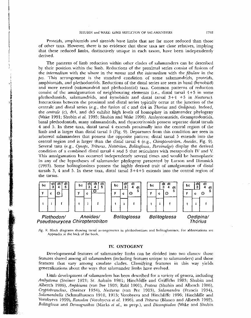
The patterns of limb reduction within other clades of salamanders can be described by their position, within the limb. Reductions of the proximal series consist of fusions of the intermedium with the ulnare in the manus and the intermedium with the fibulare in the pes. This ar rangement is the standard condition of some salamandrids, proteids, amphiumids, and plethodontids. Reductions of the distal series are seen in basal (hynobiid) and more nested (salamandrid and plethodontid) taxa. Common patterns of reduction consist of the amalgamation of neighbouring elements (i.e., distal tarsal 4 + 5 in some plethodontids, salamandrids, and hynobiids and distal tarsal 3 + 4 + 5 in Necturus). Interactions between the proximal and distal series typically occur at the junction of the centrale and distal series (e.g., the fusion of c and dt4 in Thorius and Oedipina). Indeed, the centrale (c), dt4, and dt5 exhibit high levels of homoplasy in salamander phylogeny (Wake 1991; Shubin et al. 1995; Shubin and Wake 1996). Ambystomatids, dicamptodontids, basal plethodontids, many salamandrids, and rhyacotritonids possess separate distal tarsals 4 and 5. In these taxa, distal tarsal 4 extends proximally into the central region of the limb and is larger than distal tarsal 5 (Fig. 9). Departures from this condition are seen in arboreal salamanders that possess the opposite pattern; distal tarsal 5 extends into the central region and is larger than the distal tarsal 4 (e.g., Chiropterotriton, Aneides, Fig. 9). Several taxa (e.g., Cynops, Triturus, Nototriton, Bolitoglossa, Parvimolge) display the derived condition of a combined distal tarsal 4 and 5 that articulates with metapodials IV and V. This amalgamation has occurred independently several times and would be homoplastic in any of the hypotheses of salamander phylogeny presented by Larson and Dimmick (1993). Some bolitoglossines possess the highly derived trait of amalgamation of distal tarsals 3, 4 and 5. In these taxa, distal tarsal 3 + 4 + 5 extends into the central region of the tarsus.
be dt 3
dt 4
O
dt 5
y c
dt 4
O
dt 5
t
c
t i f
be dt 3
dt 4 dt
o 5 y c
dt 4 dt
o 5
t
c
f t i f
be dt 3
dt 4 dt
y c o 5
t f t i f
be dt dt 3 4 dt
y c 0 "
t
c
f t i f
be dt 3
dt 4
dt 5
y
_.
o O
y
_.
o
t _.
o
f
Plethodon/ Aneides/ Bolitoglossa Bolitoglossa Oedipina/ Pseudoeurycea Chiropterotriton Thorius
Fig. 9. Block diagrams showing tarsal arrangements in plethodontines and bolitoglossines. For abbreviations see Appendix at the back of the book.
IV. ONTOGENY
Developmental features of salamander limbs can be divided into two classes: those features shared among all salamanders (including features unique to salamanders) and those features that vary among caudate clades. Classifying features in this way yields generalizations about the ways that salamander limbs have evolved.
Limb development of salamanders has been described for a variety of genera, including Ambystoma (Steiner 1921; St. Aubain 1981; Hinchliffe and Griffiths 1983; Shubin and Alberch 1986), Amphiuma (van Pee 1903; Rabl 1901), Proteus (Shubin and Alberch 1986), Cryptobranchus, (Steiner 1934), Necturus (van Pee 1903), Salamandra (Francis 1934), Salamandrella (Schmalhausen 1910, 1915; Vorobyeva and Hinchliffe 1996; Hinchliffe and Vorobyeva 1999), Ranodon (Vorobyeva et al. 1996), and Triturus (Blanco and Alberch 1992), Bolitoglossa and Desmognathus (Marks et al., in prep.), and Dicamptodon (Wake and Shubin

1794 AMPHIBIAN BIOLOGY
1999). This diversity of taxa affords an opportunity to discuss the phylogeny and variability of limb ontogeny in salamanders.
A. Common Features of Developing Salamander Limbs
Some developmental features appear to be highly stable and are seen in all salamanders that have limbs (Fig. 10). Salamander limbs contain fewer and larger cells than the limbs of other tetrapods. During early development, limb buds are composed of a morphologically uniform field of mesenchymal cells. T h e first visible step of chondrogenesis is the condensation of mesenchymal cells to form zones of high cell density (Hinchliffe and Johnson 1980). In later stages, these condensations differentiate into two zones of cells: an inner zone of pre-chondrogenic cells that are crescentic in shape, and an outer zone of flattened, fibroblastic cells that surround the cartilage blastema. These patterns (known as "Aniken patterns" [Aniken 1929; Gould et al. 1972, 1974; Hinchliffe and Johnson 1980; Ede 1983]) are pronounced in amphibians, particularly caudates. Finally, salamanders lack an apical epidermal ridge during limb development (Hanken 1986).
1. Stylopodium and Zeugopodium
The humerus/femur is the first element to form in the limb. Later, the anlagen of the radius/tibia and the ulna/fibula develop within a "y"-shaped condensation that extends from the humerus/femur (Fig. 9a). The radius/tibia and ulna/fibula begin to differentiate within the preaxial and postaxial arms of this cellular extension. The condensations of the radius/tibia and ulna/fibula are elongate and ovate in shape in the earliest stages. In general, the radius/tibia differentiates at an earlier stage than does the ulna/fibula.
?^°o ~°
VJ
Fig. 10. The diverse precartilage connections in developing salamander limbs. A. Photograph of hind limb bud of Dicamptodon tenebrosus (after Wake and Shubin 1999) labelled with antibodies to Type II collagen and revealing a ring of precartilagenous cells. B. A diagrammatic depiction of the developmental patterns seen in A. This pattern includes three columns of chondrogenesis: a preaxial column (that extends from the radius or tibia), a central series (that extends distally from the ulna or fibula) and a digital arch (that typically extends from the basale commune). This latter column is highly variable. C. The developmental connections of Triturus marmomtus, as described by Blanco and Alberch (1992) with several variant features, including a distal-to-proximal pattern of development of the central series, and the independence of distal tarsal five from the digital arch. D. The pattern of development of Ambystoma (after St. Aubain [1981] and Shubin and Alberch [1986]), fundamentally similar to that described for Dicamptodon. E. Salamandrella limb development with several variant features including (1) an extension of the preaxial column that develops into a prehallux, (2) independent condensation of the centralia, and (3) independent condensation of the postaxial distal tarsalia (Vorobyeva and Hinchliffe 1996). Arrows indicate the sequence of chondrogenesis.

SHUBIN and WAKE: LIMB SKELETON OF SALAMANDERS 1795
2. Autopodium
The autopodium is derived from three zones of condensation: the preaxial column, the central series, and the digital arch (Fig. 10a). The preaxial column is a zone of condensation that initially extends from the radius and tibia. The radiale/tibiale and element "y" differentiate within this condensation and later separate from each other. The central series is a zone.of condensation that extends as a "y"-shaped condensation attached to the ulna/fibula. The preaxial branch of this condensation continues distally from the ulna/ fibula to connect to the basale commune. The intermedium and centrale will differentiate within this zone. A postaxial branch contains the pr imordium of the ulnare/fibulare. This condensation is independent or contiguous with the digital arch in different species (Fig. 10). The development of the digital arch is variable in salamanders, but minimally it contains the condensations of digits I—III, the basale commune, and distal carpal/tarsal III. The first visible stage of the development of the digital arch consists of the condensations of digits I and II and the basale commune. There is, in general, a preaxial to postaxial sequence in the development of the digital arch: digits III, IV, and V develop after the preaxial digits.
B. Apomorphies of Salamander Limb Development
Several features of limb development differ from those of other tetrapods and appear to be apomorphic. Most of these differences relate to the sequence in the formation of digits and their associated mesopodials. Chondrogenesis in most te t rapod limbs and sarcopterygian fins follows a general proximal to distal sequence. In salamanders, a distal element (the basale commune) often appears before more proximal ones (the intermedium and centrale). This is a pattern not seen either in anurans or irr amniotes. In addition, digits of salamanders differentiate in a different sequence than in other tetrapods; in larval salamanders the digits differentiate in a preaxial to postaxial sequence, in contrast to the postaxial to preaxial sequence seen in other tetrapods. Salamanders are also unique in that preaxial zeugopodial and mesopodial elements generally precede postaxial ones in development. The radius/tibia and radiale/tibiale develop earlier than corresponding postaxial elements (ulna/fibula and ulnare/fibulare).
C. Developmental Variation
Most developmental variation is the result of alteration of the patterns of the formation and fusion of condensations. Main patterns of variation are reviewed, with special attention given to evolutionary ecology.
1. Regional Patterns of Developmental Variation
Much of the developmental variation of salamander limbs is highly localized. When this variation is grouped according to its location within the limb, as below, several general features arise.
A. PREAXIAL COLUMN
The variation in the preaxial column lies in the size and in the number of elements that it contains. The preaxial column can contain one, two or three elements (Fig. 10). These elements normally segment from a continuous condensation that extends from the radius/tibia. In taxa with only a single preaxial mesopodial element, such as Proteus, this condensation is reduced in length.
B. CENTRAL SERIES
The central series is labile in its sequence of development (Fig. 10). It can develop in a proximal to distal sequence (Ambystoma) or a distal to proximal sequence (Triturus). In addition, a centrale can arise within a condensation that extends from the basale commune (e.g., Triturus; [Blanco and Alberch 1992]) or, alternatively, from a condensation that arises from the intermedium (e.g., Ambystoma; [Shubin and Alberch 1986]).

1796 AMPHIBIAN BIOLOGY
The intermedium and the ulnare form a combined element in several derived species. In Triturus, the only species where the development of this feature has been observed, the intermedium and ulnare first develop as separate condensations. These two elements fuse during later stages of chondrogenesis.
C. DIGITAL ARCH
Most of the variation in the development of the digital arch is localized in the postaxial region. Indeed, the development of the preaxial digits is relatively stable. In the postaxial portion of the limb, distal carpal/tarsal 4 and 5 can either be associated with the digital arch in early development or arise as an extension of the ulnare Ifibulare.
2. Species with Reduced Limbs
The development of the highly reduced limbs of Proteus anguinus (Shubin and Alberch 1986) and Amphiuma (Rabl 1901) results from a truncation of primitive developmental processes. In these taxa, the digital arch can contain fewer than three digits and two distal carpalialtarsalia. The preaxial column is present but only contains one condensation (the radiale/tibiale) rather than two. The central series is also present, but is reduced to only two elements.
3. Ecological Variation
The major differences in limb development relate to the timing of the development of the limbs and digits, which in turn appears to correlate with the evolutionary ecology of reproduction. Salamanders with larvae that develop predominantly in ponds (such as Ambystoma) typically form their forelimbs well in advance of the hind limbs. Within the forelimb, there is a lag in the development of postaxial structures; digits IV and V emerge well after digits I, II and III have developed. These gradients are not so marked in salamanders whose larvae develop in streams (Wake and Shubin 1999). While the lag between the emergence of the forelimbs and hind limbs typically is less than that in pond-dwelling forms (the stream larva of Dicamptodon for example), the hind limb bears only two of the ultimate five digits upon hatching (Wake and Shubin 1999). Species that undergo direct development lay eggs in terrestrial settings, and hatchlings emerge fully metamorphosed, with a full complement of mesopodials and metapodials both in the forelimbs and the hind limbs. The emergence of forelimb and hind limb buds occurs nearly simultaneously compared with other salamanders; hind limb buds are delayed by as little as one week in the direct-developing plethodontid, Bolitoglossa pesrubra (Marks et ai, in prep.).
There is considerable variation in the relative appearance of forelimb and hind limb buds in salamanders (data summarized by Collazo and Marks 1994). In general, the forelimbs develop before hind limbs, with limb buds being separated by as many as 20 days in the pond-breeding salamandrid Pleurodeles waltl (Gallien and Durocher 1957), but only one to four days for direct-developing plethodontids. Stream-breeding plethodontids show a separation of between 5 and 17 days, and the direct-developing plethodontid Bolitoglossa pesrubra, which develops extraordinarily slowly, has a separation of one to two weeks.
Several detailed studies of limb development reveal a modest range in diversity. These are briefly summarized in the following paragraphs.
A. POND LARVAE OF SALAMANDRIDS
Blanco and Alberch (1992) described unique aspects of limb development in Triturus marmoratus (Fig. 10c). As in Ambystoma (Fig. 10b), the forelimbs develop well before the hind limbs. Key differences from Ambystoma are found in the development of the centrale, intermedium and distal carpal 4. Triturus has a unique distal to proximal sequence of development within the central axis. The basale commune is initially connected to a

SHUBIN and WAKE: LIMB SKELETON OF SALAMANDERS 1797
condensation that develops in a distal to proximal direction as it approaches the ulnarel fibulare ("central axis" of Blanco and Alberch [1992]). Both the centrale and intermedium develop within this axis. Another unique feature involves the development of the most postaxial distal carpal/tarsal. In the forelimb, distal carpal 4 originally forms as an extension of the ulnare, and in the hind limb, distal tarsal 5 originally extends from the fibulare. Blanco and Alberch (1992) argued that the unique features of limb development in Triturus are related to adaptations of larvae.
B. POND LARVAE OF HYNOBIIDS
Studies of hynobiids offer the opportunity to understand limb development in forms that possess relatively primitive limb designs. In hynobiids with pond larvae, forelimbs develop well before hind limbs. Hatchlings have only tiny hind limb buds, and the forelimbs of early larvae are conical. In some instances only a small protrusion marks the site where the forelimb bud will appear and there is no sign of the hind limb bud (stage 40 of Hynobius nigrescens, [Iwasawa and Yamashita 1991]). As the forelimbs develop, the first two digits differentiate and a web-like filament forms between them and grows into an elongate, filamentous structure (Sitina et al. 1987; Vorobyeva and Hinchliffe 1996). This structure is resorbed later in larval life and does not appear to be involved in skeletogenesis. Vorobyeva et al. (1997) described several common features of the limb development of Ranodon and Salamandrella. Several unique features relate to the timing and pat tern of chondrogenesis (Fig. lOe). The preaxial column differentiates relatively earlier and more rapidly than in other salamanders. In addition, the digital arch develops an early connection to the postaxial basal mesopodial (the ulnarel fibulare), a condition seen in direct-developers and in amniotes. Ranodon is similar to Triturus in that the condensations of distal tarsals 4 and 5 are separate.
C. STREAM LARVAE OF DIVERSE SPECIES
Development in dicamptodontids {Dicamptodon [Wake and Shubin 1999]) closely resembles that o£ Ambystoma, especially with respect to patterns of primary and secondary connectivity of rudiments and condensations in the mesopodium (fig. 10a,b) (Wake and Shubin 1999). The major difference is that a ring-like series of condensations occurs in the posterior mesopodium, involving the basale commune, distal tarsals 3 and 4, the fibulare, intermedium and centrale. A similar formation has been observed in the hynobiid Salamandrella keyserlingii (Schmalhausen 1910), which has pond larvae. The digital arch is more truncated than in Ambystoma, and resembles that seen in Triturus. In the plethodontid Desmognathus quadramaculatus limb development is similar to that seen in Dicamptodon and Ambystoma, although details are lacking. The plethodontid Gyrinophilus porphyriticus has well-developed limbs at hatching, with four digits on the forelimb and three on the hind limb (71 days after egg deposition, Collazo and Marks 1994). This sequence of digital development contrasts with other stream-breeding plethodontids in which the full adult complement of digits is present at the time of hatching.
In the stream-dwelling hynobiid, Onychodactylus japonicus, hatching occurs about 142 days after egg deposition, at a time when the forelimb bears three digits but the hind limb is a small, undifferentiated bud (Iwasawa and Kera 1980). A fourth finger appears at hatching or within a few days. The first finger bears a claw at hatching and a claw appears on the second within days (with stream-dwelling larvae the hatchlings have relatively well-developed limbs and have at least some differentiated digits). Cryptobranchoids also have stream larvae; at about the time of hatching there are three or four differentiated digits on the forelimb but the hind limb bud bears no differentiated digits (Andrias japonicus [Iwama 1968]).
D. DIRECT DEVELOPMENT
Direct-developing species that have been studied to date, Bolitoglossa pesrubra and Desmognathus aeneus, display a mosaic of tetrapod features (Marks et al., in prep.). These

1798 AMPHIBIAN BIOLOGY
taxa resemble amniotes and anurans in some respects, but the structures are small and development proceeds in the entire autopodium more synchronously than in other taxa. Accordingly, it is difficult to determine the precise sequence of events. However, the limb buds become more paddle-like than conical, and the digital arch appears to be part of a continuous column of cells that extends from the ulnare/fibulare towards the anterior side of the limb. In D. aeneus condensation of the digital arch occurs very early, just after observation of the "y"-shaped condensation that includes the humerus/femur, radius/tibia and ulna/fibula. The digital arch arises from the ulnar/fibular side of the limb and extends in a preaxial direction. While there is a preaxial to postaxial gradient of differentiation once the digital arch is in place, it is weak, so that there is a relative synchrony of digital differentiation.
V. VARIATION WITHIN POPULATIONS
The use of studies of intraspecific variation in the analysis of the origin of morphological novelties has a long history. Often, these approaches used an analysis of intraspecific variation to formulate hypotheses of homology. Schmalhausen (1917), for example, studied variation in Ranodon sibiricus, a hynobiid salamander, and described three "states" of the hynobiid tarsus consisting of different configurations of the centralia, distal tarsal four, and distal tarsal five. Change in any one of these elements usually involves reciprocal changes in other elements within this region of the foot because dt4, c, and dt5 interact with one another during ontogenesis. A major player in this interaction is a small mediate (his m3, and our m). In Ranodon, and presumably in other salamanders, it can merge with dt4, with dt5, or it can form an amalgamation with dt4 and dt5 (Wake 1991). Many fossil amphibians possess an independent m, a small rudimentary bone between the centralia and the distal tarsals (Fig. 3). This bone is not part of the standard morphology of any extant salamander and it is presumed to have been lost during phylogenesis of salamanders. Schmalhausen (1917) noted that an independent m is a common variant condition in the foot of Ranodon, Cryptobranchns, and Onychodactylus. When an independent m is present in these limbs, dt4, dt5 are both reduced in size. These observations suggested a "latent" homology, whereby m is lost from the standard conditions of extant taxa but the developmental interactions that form it are retained. Furthermore, these studies imply that dt4, dt5, c, and m are the product of interaction during ontogeny that serve to integrate and regulate their phenotypic expression. Under this hypothesis, the latent homology of m would be an incidental byproduct of the conservatism of this generative process.
Recent analyses of salamander limb variation extend and amplify Schmalhausen's (1917) observations. The parallel between patterns of intrapopulational variability and homoplastic characters is so common as to be the expected situation in salamander limb diversity. Studies on the intraspecific and interspecific variability of salamander mesopodia have revealed biases in the production of novel phenotypes (e.g., Alberch 1983; Hanken 1983; Vogl and Rienesl 1991; Rienesl and Wagner 1992; Shubin et al. 1995). Observed patterns of variability are often predictable from a combination of developmental and phylogenetic considerations (Shubin et al. 1995, see below). Many variant mesopodial arrangements are homoplastic and are commonly encountered in different species. Distal tarsal 4+5 is a particularly widespread variant found in (1) taxa that normally retain separate distal tarsal 4 and distal tarsal 5 (e.g., Plethodon cinereus, Taricha granulosa) and (2) species that are standard for a combined distal tarsal 3+4+5 (Bolitoglossa dofleini) (Alberch 1983). Variation in the former parallels homoplastic conditions of highly nested plethodontids whereas variation in the latter reflects atavistic restoration of an ancestral condition. Other types of atavism also occur; in Triturus cristatus (a species that normally possesses a joined distal tarsal 4+5) the ancestral pattern of independent elements is a natural variant (Rienesl and Wagner 1992).
Analysis of variation reveals atavisms that restore conditions at different hierarchical levels of salamander phylogeny (Figs 11, 12, 13). Paleozoic temnospondyls had a
SHUBIN and WAKE: LIMB SKELETON OF SALAMANDERS 1799
postminimus, prehallux, supernumerary centralia, and an independent m. No single salamander species possesses all these primitive elements, but they all appear either as features of different taxa, or as common atavistic variants. While more basal clades (cryptobranchoids in particular) display these features most commonly, they also appear as rare variants in more deeply nested clades. The most commonly observed variants (those characteristic of the patterns of intraspecific variation of many different clades) include the prepollex, prehallux, m, and supernumerary centralia). In addition, variants of highly nested clades (e.g., bolitoglossines) may correspond to less general, but understandable (see below) characters (e.g., dt3+dt4+dt5 in B. adspersa; d4+d5 in B. dolfleni; independent dt5 larger than dt4 in B. pesrubra).
Salamanders share patterns of variation because they share a common generative system, and homoplasy is both expected and (within limits) predictable (Fig. 13). There are homoplastic patterns of fixation of particular patterns (such as amalgamated dt4+dt5) derived from standard structural designs. Significantly, no new carpal or tarsal elements have evolved during salamander phylogenesis; limb diversification instead is characterized by the fixation of variant patterns found in other taxa (Shubin et al. 1995).
VI. DEVELOPMENTAL BASIS OF CHARACTER EVOLUTION
All variation of the salamander limb takes place within a framework of stability that arises from a combination of phylogenetic history and ontogeny. Strong evidence of the monophyly of living salamanders encouraged the development of a model of salamander limb development that was used to establish the identity of individual components (Shubin and Wake 1996). To understand deviations from what here is termed the ground plan, knowledge of phylogeny and ontogeny is used to make interpretations of specific instances.
Salamander limb evolution is highly ordered; some portions of the limb pattern are invariant while others are labile. The development of the salamander limb enables us to
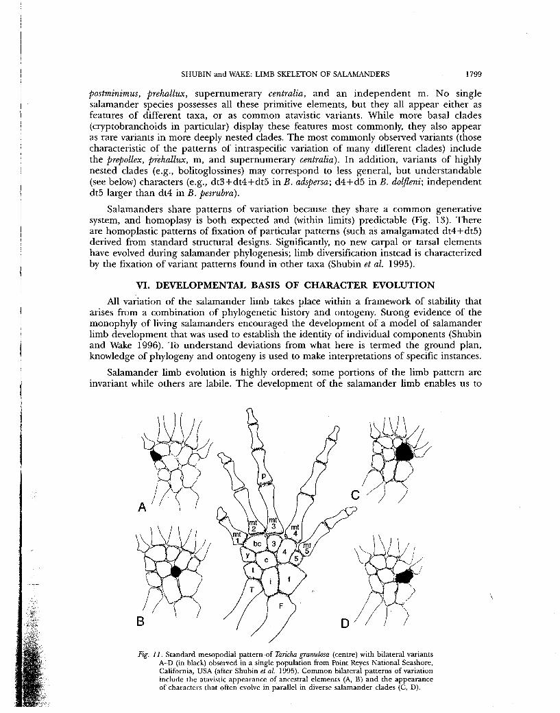
Fig. 11. Standard mesopodial pattern of Taricha granulosa (centre) with bilateral variants A-D (in black) observed in a single population from Point Reyes National Seashore, California, USA (after Shubin et al. 1995). Common bilateral patterns of variation include the atavistic appearance of ancestral elements (A, B) and the appearance of characters that often evolve in parallel in diverse salamander clades (C, D).

1800 AMPHIBIAN BIOLOGY
fig. 12. Standard (centre) and variant tarsal designs (A-E, in black) in Plethodon cinereus (after Hanken 1983). Hanken's preaxial c as shown in the diagram is the element y of the present chapter. Hanken's distal tarsal 1/2 as shown in the figure is the basale commune of the present chapter. The most common variants consist of the combining of neighbouring elements.
classify this variability (Riensel and Vogl 1992; Wagner and Riensel 1995; Shubin and Wake 1996; Vorobyeva and Hinchliffe 1996). Anlagen of skeletal elements mainly arise as extensions of existing cartilagnous elements that later separate from one another. Three major patterns of developmental connectivity are most frequently observed in salamander limbs: bifurcation, segmentation, and de novo condensation. Bifurcation occurs when two Anlagen arise within a "Y"-shaped extension from an existing element, whereas segmentation results from a single element dividing from an existing extension. An element that arises in isolation is a de novo condensation. In salamanders with generalized patterns

SHUBIN and WAKE: LIMB SKELETON OF SALAMANDERS 1801
Fig. IB. Ancestral and highly derived tarsal patterns transiently appear during development. A. Element m (in black), an ancestral component of the limb of salamanders (Trematops). B. Element m transiently appearing during the normal development of a more derived species, Ambystoma mexicanum. At this stage, distal tarsals four and five are still connected to one another. This feature is similar to the condition that arises in highly derived hynobiids, plethodontids, and salamandrids. C. Element m (in black) in a variant of Plethodon cinereus. During the normal development of Ambystoma, the primordial element m amalgamates with the centrale to form a single element. Distal tarsals four and five separate to form two distinct cartilagenous foci. This diagram reveals that simple shifts in the timing of development can lead to the origin of both ancestral and highly derived features. This situation obtains because many of the major adult mesopodial patterns can be seen transiently in the development of diverse species.
of development, two major axes of development are observed: a proximodistal axis initiates at the humerus/femur, radius/tibia-ulna/fibula and extends to the proximal mesopodials, while an antero-posterior (digital) axis arises on the anterior (preaxial) border and development of both digits and distal mesopodials progresses posteriorly (postaxially). The two axes of development terminate in the region of the centrale and ulnare/fibulare, where they converge and often merge. As a consequence of these developmental patterns, reductions in numbers of mesopodial elements result from one of three processes: truncation, fusion or amalgamation.
A relatively easy and direct way of evolving reduction is truncation of a developmental trajectory: an element may simply fail to form during development because ontogeny is stopped. An example of this is the loss of distal carpal 4 of die paedomorphic cave-dwelling salamander Proteus. With two digits in the foot and three in the hand, the limb of Proteus is highly reduced and has most likely evolved via truncation of a primitive developmental trajectory. The abridgment of development led to the loss of postaxial structures of the digital arch and central series, which are the last to develop in related taxa. Parallel evolution of this transformation is seen in sirenids and amphiumids. Less dramatic reductions have independently evolved in other taxa. Some plethodontid, salamandrid and hynobiid species have independently lost the fifth toe. In species where development has been studied, no rudiment of this toe is ever present during development. Indeed, some of the most paedomorphic species, such as Proteus, also have the most exaggerated patterns of truncated limb development. This holds despite die fact that the adult pattern of Proteus does not match any single stage in the development of any other salamander (Shubin and Alberch 1986).
The vast majority of mesopodial reductions in salamander phylogeny appear to be the result of the retention of embryonic connections into adult stages (Fig. 13). Examples of this mode of evolution are seen during the evolution of very general apomorphies (i.e., synapomorphies of Caudata) as well as characters of highly derived groups. Element m is likely to be a component of the ancestral salamander limb. While this element has been lost in salamander evolution, it is still transiently observed during normal development. In Ambystoma, for example, m arises as a condensation that is connected to the postaxial

1802 AMPHIBIAN BIOLOGY
side of the centrale (Fig. 13). This condensation is initially a distinct focus of condensed cells. Eventually m is no longer seen as an independent element as it never cleaves from the centrale. Other types of amalgamation are seen in the preaxial column (the combination of r + y in some rhyacotritonids), the central series (the combination of the intermedium and centrale in some rhyacotritonids), and the digital arch (the combination of neighbouring distal carpals/tarsals). Amalgamation is a form of highly localized paedomorphosis and, indeed, is often seen in neotenic forms.
Amalgamations of mesopodials also appear to be the rule in salamanders with miniaturized limbs. The plethodontid genera Thorius and Oedipina possess miniature limbs and have large genomes and cells. While Thorius is a true miniature, Oedipina is an attenuated salamander with a relatively elongate body axis and small appendages. Species in both genera have a four-digited hand and a five-toed foot; there have been no apparent truncations of limb structure. The variablity of the mesopodium is very high in these species (Fig. 14). Indeed, much of this variation appears to result from the failure of neighbouring elements to separate. In other cases different carpal patterns result from different patterns of amalgamation. Many of the resulting elements are unique to a taxon, or occur only in a few taxa. One such amalgamation joins the centrale and distal carpal 4. This exceptional case is the only known case of the fusion of an element of the digital arch with the central series, and while it is encountered in populations of a few plethodontids and some salamandrids, it reaches high frequency only in Thorius and Oedipina.
Mesopodial reductions also are produced by the fusion of independent condensations. Fusion is a mode of mesopodial reduction that does not appear to involve paedomorphosis. This mode of limb reduction typically results in an enlarged "combined" element in place of two separate ones. The most frequent fusion in salamander limb phylogeny occurs between the intermedium and ulnare. In Triturus marmoratus, the only species where this fusion has actually been observed during development, the condensations of the intermedium and ulnare are initially connected to the ulna (Blanco and Alberch 1992). At this stage, both condensations are apparently separate and independent Anlage. Immediately after they cleave from the ulna, the condensations merge to form one cartilaginous unit.
Salamander limb evolution has never involved the addition of mesopodials or digits to the limb. Despite this, variants containing supernumerary elements are seen within populations of diverse species. There is a fundamental regularity in the types of supernumerary elements that appear: almost exclusively these elements are atavistic restorations of ancient characters (e.g., m, postminimus, prepollex, and supernumerary centralia). If the evolution of salamander mesopodia consist mostly of reduction, then atavisms will exclusively involve additions to the standard patterns of different species.
Developmental patterns enable one to predict the location of supernumerary elements in variant limbs. It is most parsimonious to assume that new elements arise from extended sequences of segmentation or bifurcation, or from novel patterns of de novo condensation. De novo condensations can appear anywhere in the limb and do not offer a means of predicting the location of variant supernumerary condensations. A few assumptions are invoked to predict the location of supernumerary elements. First, superumeraries are assumed to arise within existing tracts of connectivity (e.g., the preaxial column, the digital arch, and the central series). Second, it is assumed that the evolution of supernumeraries is a peramorphic event, largely adding elements to existing developmental sequences. The addition of segmentation events to the preaxial column would result in the presence of more than two condensations in the preaxial column. In fact, this is the ancestral condition for salamanders. Extra segmentations, or bifurcations, in the central series would result in more than one centrale. This, too, is a primitive condition with basal amphibians possessing from one to four centralia. Finally, extra bifurcation events could be added to the digital arch. If these additions are peramorphic, then supernumerary elements would arise postaxially. Indeed, most supernumeraries associated with the digital arch are postaxial. The postminumus, for example, lies on the postaxial border of distal tarsal 5.

re w
s a.
I w en
W r w
z o 1 )
> s > z o w CO
fig-. / 4 . Extreme hind limb variation in the plethodontid miniature salamander, Thorius. The standard pattern for Thorius (A) contrasts with the numerous diverse conditions (other drawings) seen in populations of this species (modified from Hanken 1985). Stippling indicates cartilage. Scale bar = 1 mm.

1804 AMPHIBIAN BIOLOGY
The sequence of development lends a predictability to the evolution of salamander digits. The last digits to develop are typically the first to be lost during limb reduction. When an entire digit is lost, that digit is from the postaxial side of the limb. Four-toed salamanders lose the fifth toe. In highly reduced limbs of some amphiumids and proteids, the only digits to be unaffected are digits I and II, the most preaxial digits in the limb. With the basale commune, these two digits are the first elements of the digital arch to form. Indeed, these are among the most stable features of the evolution of salamander limbs; the basale commune is a highly informative synapomophy of Caudata, and digits I and II are never lost, even in the most reduced appendages.
Patterns of mesopodial evolution are also predictable from a knowledge of ontogeny (Fig. 13). Most patterns of mesopodial variation involve the combination of elements that were originally connected in earlier stages of embryogenesis. The phylogenetic outcome of this is that patterns of consolidation of elements involve neighbouring elements that lie within one of the three series of the mesopodium: the preaxial column, the central series and the digital arch (Figs 11, 12). Condensations in these tracks largely remain distinct from one another and rarely fuse. This observation holds for both intraspecific and interspecific variation; most mesopodial reduction involves the retention of embryonic precartilagenous connections into adult stages. The fusions of elements that are derived from different series is less common. The most significant exceptions are seen in some minaturized limbs (Fig. 14). The amalgamation of dc4 and the centrale in Thorius and Oedipina involves merging condensations from the digital arch and centrale series. This particular case is only one of many bizarre conditions seen in mesopodia of these groups (Hanken 1985). The unique patterns of variation in these groups suggest that different factors may be at work in structuring their morphological variation. The observation that these species contain miniaturized limbs, large genomes, and large cells may be relevant in explaining the exceptional variation seen in these taxa. If the number of condensations that appears in the limb is partially controlled by the number of cells per unit volume, then the limbs of Oedipina and Thorius may have a constraint on the number of discrete mesopodial elements present in the limb.
Digital loss appears to be an all or none situation in salamanders. In species of the Plethodontidae with large genomes and cells, some extraordinary variants have been reported which show that digits are not reduced and lost by gradually reducing the size and numbers of phalanges in a digit, but rather by loss of an entire digit. In one large but paedomorphic species of Bolitoglossa with fully-webbed hands and feet an individual was reported that had only four digits in one pes as a result of the extreme reduction and attenuation of the skeleton of the fifth digit, which was conjoined with the fourth (Alberch and Gale 1985). In a miniaturized individual of Parvimolge townsendi only four digits were evident from external examination, but a radiograph disclosed that in one pes there were two full digital skeletons in a single integumentary digit, whereas in the other pes there was a much thickened last digital skeleton (in effect, a combined fourth and fifth digital skeleton) (Wake 1991, 1999). In another exceptional instance, by far the largest individual of the four-toed species Batrachoseps wrighti had five toes on one pes. A radiograph disclosed that this specimen had only three metapodials, but the last two were both much enlarged and had bifurcated, each supporting two digital skeletons (Wake 1991).
VII. PHYLOGENETIC USES OF DEVELOPMENTAL DATA
Development of the salamander limb has led to several distinctive phylogenetic hypotheses about the early evolution of limb patterns.
A. The Hypothesis of the Polyphyly of Tetrapods
The possibility that salamanders evolved entire limbs independent ly from other tetrapods was raised by Holmgren (1933, 1939, 1942), who presented his hypothesis in great detail in a series of papers based on his embryological studies. The critical evidence

SHUBIN and WAKE: LIMB SKELETON OF SALAMANDERS 1805
had to do with the position of the metapterygial axis relative to the intermedium and the fact that digits one and two were the first to form in salamanders, whereas digit four formed first in anurans and amniotes (reviewed by Shubin and Alberch [1986]). Jarvik (1965) presented other evidence which he interpreted as supporting the polyphyletic origin of tetrapods, and embraced Holmgren's arguments about limbs as additional support for his views. In neither case was any phylogenetic analysis conducted. Rather, each worker emphasized the' ways in which salamanders differed from anurans and amniotes and Jarvik in particular sought correlates between characters of salamanders and porolepiform fishes, which he thought gave rise to them. The limbs of Paleozoic amphibians played little role in the thinking of either worker. Shubin and Alberch (1986) showed how the data for limb development in salamanders are compatible with the hypothesis of the monophyly of tetrapods; the most comprehensive phylogenetic analyses of large data sets also support such an explanation (e.g., Ahlberg and Milner [1994]; but for an alternative view see Laurin [1998]).
B. The Hypothesis of the Neomorphic Origin of the Salamander Autopodium
Perhaps die postaxial portion of the salamander autopodium is a secondary neomorph, having been re-evolved from an ancestral extremely reduced state (Wagner 1999; Wagner et al. 1999). Wagner and colleagues studied expression patterns of the Hoxa-11 gene in the salamandrid Notophthalmus and the anuran Xenopus. This gene is expressed in the zeugopodium of mice and chickens, where there is a sharp limit of expression at the distal limit of the zeugopodium, and it has been argued that the pattern is causally important for the differentiation of the autopodium from the zeugopodium. In Xenopus the entire expression pattern is basically similar to that in amniotes. In Notophthalmus, the distal limit of expression is less well defined. After formation of the first two digits, expression ceases. Later reexpression is seen in the autopodium coincident with the appearance of digit III, and there is variable expression in the distal tips of all the growing fingers. These observations led the authors to postulate that living salamanders are derived from an ancestral lineage that had undergone digital reduction and to suggest that such reduction would have followed Morse's Law (Morse 1872), which they believed would have left digits III and IV in a two-digited ancestor. From this state they argued that three new postaxial digits formed, which they interpreted as neomorphs. Thus, they argued that digits I and II of salamanders are the homologues of digits III and IV of anurans and amniotes, and that there are no homologues of salamander digits III, IV and V. Wagner et al. (1999) observed that three families of salamanders have reduced limbs and that there is a strong tendency to reduce limbs in other taxa (see above), concluding that a bias in this evolutionary direction is associated with aquatic life. In addition, one of these families, the Sirenidae, sits in a basal position among caudates. Wagner et al. (1999) suggested that a similar tendency was present in aquatic ancestors of Caudata.
This argument, while plausible, is not parsimonious from a phylogenetic point of view. Ultimately, this hypothesis would involve the loss and re-appearance of a four-digited limb with identical phalangeal formulae in different taxa. Assuming that the ancestor of lissamphibians is to be found among temnospondyls, one might expect to see limb reductions, but the majority of temnospondyls have very salamander-like limbs, with four digits on the forelimb, five on the hind limb, and with phalangeal formulae remarkably similar to those of living salamanders (2-2-3-2 for the forelimb, 2-3-4-4-3 or some slight reduction in the hind limb). In addition, the earliest fossil evidence of salamanders does not lend support to the notion of reduction. The sister taxon of the Caudata represented by a complete fossil, Karaurus, has four digits on the forelimb and five on the hind limb. There are fragmentary Jurassic fossils that have been assigned to the Proteidae, but the first complete fossils of salamanders are from the earliest Cretaceous of China (Dong and Wang 1998). These fossils are not distinguishable from living members of the Hynobiidae, and they have typical hynobiid limbs with four digits on the forelimbs and five on the hind limbs.

1806 AMPHIBIAN BIOLOGY
The neomorphic scenario leads to certain predictions, one being that the be should be an anterior extension from the postaxial basal mesopodial region. Wagner et al. (1999) cited a few observations that they believed gave evidence of such an origin. While it is true that in some species there is a stage when the be is connected to a "ring" of condensations in the postaxial mesopodium (as shown by Wake and Shubin [1999, their Fig. 5] for Dicamptodon), this occurs after the precocial and independent condensation of be (and the rudiments of digits I«and II). This hypothesis also predicts that early salamander fossils with highly-reduced limbs may yet be discovered from early Mesozoic sediments.
C. The Hypothesis of a Conserved Developmental Trajectory
Working within a phylogenetic framework and taking cognizance of the full array of data from fossil and living forms, developmental data can be placed within a mechanistic model that attempts to explain the persistent morphogenetic processes responsible for the diversity of limbs in living salamanders and their extinct relatives. The resultant hypothesis posits a conserved ontogenetic trajectory that has persisted for hundreds of millions of years during tetrapod evolution. This hypothesis holds that homologous adult structures can develop by different ontogenetic sequences. Salamander digits, then, would be homologous to those of other tetrapods as suggested by the congruence of molecular and morphological characters in tetrapod phylogeny. This homology would account for the similar phalangeal formulae between basal salamanders, basal anurans and temnospondyls. The main differences between the limbs of these salamanders and other tetrapods are developmental, in particular with regard to the timing of digit formation. These developmental differences are hypothesized to be due to larval adaptation of salamanders (Blanco and Alberch 1992; Shubin et al. 1995; Hinchliffe and Vorobyeva 1999; Wake and Shubin 1999). This hypothesis yields predictions for the evolution of salamander limbs that are concordant with existing data. Major departures from standard developmental patterns should correlate with ecological and phylogenetic differences in larval biology. This hypothesis posits an homologous 4-digited hand among temnospondyls and lissamphibians. Larval adaptation in the clade leading to caudates involved a change in early stages of autopodial morphogenesis. This larval adaptation included the precocial formation of digits 1 and 2 and the development of the distinctive preaxial column. Next came the establishment of the basale commune and the directional change (from preaxial to postaxial) in digital arch formation. Fingers 1-4 are homologous in all four-fingered or five-fingered tetrapods, but details of their development are not. Unexplained in this hypothesis is the hind limb, and a second hypothesis of concerted evolution, necessary to explain the parallelisms found in forelimbs and hind limbs of other tetrapods is required.
The strength of this hypothesis is that it is readily tested and is parsimonious. Furthermore, it is to some degree tree-independent, in that it is compatible with several alternative phylogenetic hypotheses, including monophyly of the lissamphibians, polyphyly of the lissamphibians, monophyly of tetrapods and even polyphyly of tetrapods, although at present the weight of evidence seems to favour monophyly in both cases. Thus, while this hypothesis generates some characters of phylogenetic significance, in itself it does not challenge existing orthodoxy or alternative phylogenetic hypotheses.
VIII. PROSPECTS FOR FUTURE RESEARCH .
Although substantial effort has been focused on comparative studies of salamander limb development and morphology, significant challenges remain. While some data exist with respect to limb development of species lacking a larval stage (Marks et al., in prep.), there is no detailed description of limb development in any direct-developing species, a group constituting more than one-half of the living species of salamanders. Another challenge is to extend studies of patterns of gene-expression to additional taxa, especially those having unusual limb structure. Unfortunately, these species, in the families Sirenidae, Amphiumidae, and Necturidae, are exceptionally difficult to study developmentally. The

SHUBIN and WAKE: LIMB SKELETON OF SALAMANDERS 1807
strange absence of an apical epidermal ridge in salamanders needs to be resolved. This structure is essential for normal limb development in tetrapods, and it is highly desirable to know whether in salamanders this is primary absence or secondary loss. Finally, the ancestry of salamanders badly needs resolution. For nearly 40 years the monophyly of the Lissamphibia has been accepted, but this taxon has few if any unique synapomorphies and there are reasons to question monophyly. Until this question is resolved several unusual features of salamander limb morphology and development will remain problematic.
IX. ACKNOWLEDGEMENTS
Our collaborative research was initiated with the support of the National Science Foundation. We thank Andrew Crawford for his assistance in making skeletal preparations and Karen Klitz and Jason Downs for assistance in preparation of some of the illustrations.
X. REFERENCES Ahlberg, P. and Milner, A. R., 1994. The origin
and early diversification of tetrapods. Nature Lond. 395: 507-514.
Alberch, P., 1983. Morphological variation in the neotropical salamander genus Bolitoglossa. Evolution 37: 906-919.
Alberch, P. and Gale, E. A., 1985. A developmental analysis of an evolutionary trend: digital reduction in amphibians. Evolution 39: 8-23.
Aniken, A. W., 1929. Das morphogene Feld der Knorpelbildung. Archiv fur Entwicklungsmechanik der Organismen 114: 549-578.
Baur, G., 1888. Beitrage zur Morphogenie des Carpus und Tarsus der Vertebraten, Vol. 1. Batrachia. Jena.
Blanco, M. J. and Alberch, P., 1992. Caenogenesis, developmental variability, and evolution in the carpus and tarsus of the marbled newt, Triturus marmoratus. Evolution 46: 677-687.
Carroll, R. L., 1988. "Vertebrate Paleontology and Evolution". W. H. Freeman and Co., New York.
Carroll, R. L. and Gaskill, P., 1978. The Order Microsauria. Mem. Amer. Philo. Soc. 126: 1-211.
Coates, M. I., 1991. New paleontological contributions to limb ontogeny and phylogeny. Pp. 314-338 in "Developmental Patterning of the Vertebrate Limb", ed by J. R. Hinchliffe, J . M. Hurle and D. Summerbell. Plenum Press, New York.
Coates, M. I. and Clack, J . A., 1990. Polydactyly in the earliest known tetrapod limbs. Nature Lond. 347: 66-69.
Collazo, A. and Marks, S. B., 1994. Development of Gyrinophilus porphyriticus: identification of the ancestral developmental pattern in the salamander family Plethodontidae./ . Exp. Zool. 268: 239-258.
Dong, Z. and Wang, X., 1998. [Title in Chinese]. Vertebrate Palasiatica 36: 159-172.
Duellman, W and Trueb, L., 1986. "Biology of Amphibians". McGraw-Hill, New York.
Ede, D. A., 1983. Cell condensations and chondro-genesis. Pp. 143-185 in "Cartilage", ed by B. Hall. Academic Press, New York.
Francis, E. T. B., 1934. "The Anatomy of the Salamander". Clarendon Press, Oxford.
Gallien, L. and Durocher, M., 1957. Table chronologique du developpement chez Pleurodeles waltii Michah. Bull. Biol. FT. Belg. 91: 97-114.
Good, D. A. and Wake, D. B., 1992. Geographic variation and speciation in the torrent salamanders of the genus Rhyacotriton. Univ. California Publ. Zool. 126: 1-91.
Gould, R., Day, A. and Wolpert, L., 1972. Mesenchymal condensation and cell contact in early morphogenesis of the chick limb bud. Expert. Cell Res. 72: 325-336.
Gould, R., Selwood, L. and Day, A., 1974. The mechanisms of cellular orientation during cartilage formation in the chick limb and the regenerating amphibian limb. Experi. Cell Res. 83: 287-296.
Hanken, J., 1983. High incidence of limb skeletal variation in a peripheral population of the red-backed salamander, Plethodon cinereus (Amphibia, Plethodontidae) from Nova Scotia. Canadian J. Zool. 61: 1925-1931.
Hanken, J., 1985. Morphological novelty in the limb skeleton accompanies miniaturization in salamanders. Science 229: 871-874.
Hanken, J., 1986. Developmental evidence for amphibian origins. Evolution. Biol. 20: 389-417.
Hinchliffe, J . R. and Griffiths, P., 1983. The pre-chondrogenic patterns in tetrapod limb development and their phylogenetic significance. Pp. 99-121 in "Development and Evolution", ed by B. Goodwin, N. Holder and C. Wylie. Cambridge University Press, Cambridge.
Hinchliffe, J . R. and Johnson, D. R., 1980. "The Development of the Vertebrate Limb". Oxford University Press, Oxford.
Hinchliffe, J . R. and Vorobyeva, E. I., 1999. Developmental basis of limb homology in urodeles: heterochronic evidence from the primitive hynobiid family. Pp. 93-109 in "Homology", ed by G. R. Bock and G. Cardew. John Wiley and Sons, Chichester.
Holmgren, N., 1933. On the origin of the tetrapod limb. Acta Zoologica 14: 185-295.
Holmgren, N., 1939. Contribution to the question of the origin of the tetrapod limb. Acta Zoologica 20: 89-124.
Holmgren, N., 1942. On the tetrapod limb — again. Acta Zoologica 30: 485-508.

1808 AMPHIBIAN BIOLOGY
Hook, R. W, 1983. Colosteus scutellatus (Newberry), a primitive temnospondyl amphibian from the Middle Pennsylvanian of Linton, Ohio. Amer. Mus. Novit. 2770: 1-41.
Ivachnenko, K. R, 1978. Urodelans from the Triassic and Jurassic of Soviet Central Asia. Paleontol. J. 12: 362-368.
Iwama, H., 1968. Normal table of Megalobafrachus japonicus. Nagoya, Japan.
Iwasawa, H. and Kera, Y., 1980. Normal stages of development of the Japanese lungless salamander, Onychodactylus japonicus (Houttayn). Japanese J. Herp. 8: 73-89.
Iwasawa, H. and Yamashita, K., 1991. Normal stages of development of a hynobiid salamander, Hynobius nigrescens Stejneger. Japanese J. Herp. 14: 39-62.
Jarvik, E., 1965. On the origin of girdles and paired fins. Israel J. Zool. 14: 141-172.
Larson, A. and Dimmick, W., 1993. Phylogenetic relationships of the salamander families: An analysis of congruence among morphological and molecular characters. Herpetol. Monog. 7: 77-93.
Laurin, M., 1988. The importance of global parsimony and historical bias in understanding tetrapod evolution. Part I — systematics, middle ear evolution, and jaw suspension. Ann. Sci. Nat., Zool., 13 Ser 19: 99-114.
Morse, E., 1872. On the carpus and tarsus of birds. Ann. Lyceum Nat. Hist., New York 10: 3-22.
Rabl, C , 1901. Gedanken und Studien iiber den Ursprung der Extremitaten. Zeitschrift. F. wiss. Zool. 70: 474-558.
Rienesl, J . and Wagner, G. P., 1992. Constancy and change of basipodial variation patterns: A comparative study of crested and marbled newts — Triturus cristatus, Triturus marmoratus — and their natural hybrids./. Evol. Biol. 5: 307-324.
St. Aubain, M. L., 1981. Amphibian limb ontogeny and its bearing on the phytogeny of the group. Zeitschrift fur Zoologische Systematik und Evolutionforschung 19: 174-193.
Schaeffer, B., 1941. The morphology and evolution of the tarsus in amphibians and reptiles. Bull. Am. Mus. Nat. Hist. 78: 395-472.
Schmalhausen, I. I., 1910. Die Entwickelung des Extremitatenskelettes von Salamandrella kayserlingii. Anatomischer Anzeiger 37: 431-446.
Schmalhausen, I. I., 1915. "Development of the Extremities of the Amphibia and their significance to the Question of the Origins of the Vertebrates". University Press, Moscow.
Schmalhausen, I. I., 1917. On the extremeties of Ranidens sibiricus Kessl. Rev. Zool. Russe 2: 129-135.
Shubin, N. and Alberch, P., 1986. A morphogenetic approach to the origin and basic organization of the tetrapod limb. Evol. Biol. 20: 319-387.
Shubin, N. and Wake, D. B., 1996. Phylogeny, variation, and morphological integration. Amer. Zool. 36: 51-60.
Shubin, N., Wake, D. B. and Crawford, A. J., 1995. Morphological variation in the limbs of Taricha granulosa (Caudata: Salamandridae): evolutionary and phylogenetic implications. Evolution 49: 874-884.
Sitina, L."A., Medvedeva, I. M. and Godina, L. B., 1987. "Development of Hynobius keyserlingii. Akademia Nauka, Moscow, [in Russian].
Steiner, H., 1921. Hand und Fuss der Amphibien, ein Beitrag zur Extremitatenfrage. Anatomischer Anzeiger 53: 513-542.
Steiner, H., 1934. Uber die embryonale Hand und Fuss-Skelett-Anlage bei den Crocodiliern, sowie iiber ihre Beziehungen zur Vogel-Flugelanlage und zur ursprunglichen Tetrapoden-Extremitat. Revue Suisse de Zoologie 41: 383-396.
van Pee, J., 1904. Les membres chez Amphiuma. Anatomischer Anzeiger 24: 476-482.
Vogl, C. and Rienesl, J., 1991. Testing for developmental constraints: Carpal fusions in urodeles. Evolution 45: 1516-1519.
Vorobyeva, E. I. and Hinchliffe, J . R., 1996. Developmental pattern and morphology of Salamandrella keyserlingii limbs (Amphibia: Hynobiidae) including some evolutionary aspects. Russian J. Herp. 3 : 68-81 .
Vorobyeva, E. I., Ol'shevskaya, O. P. and Hinchliffe, J . R., 1997. Specific features of development of the paired limbs in Ranodon sibiricus Kessler (Hynobiidae, Caudata). Russ. J. Dev. Biol. 28: 150-158.
Wagner, G. P., 1999. A research programme for testing the biological homology concept. Pp. 125-140 in "Homology", ed by G. R. Bock and G. Cardew. John Wiley & Sons, Chichester.
Wagner, G. P., Khan, P A., Blanco, M. J., Misof, B. and Liversage, R. A., 1999. Evolution of Hoxa-11 expression in amphibians: is the urodele autopodium an innovation? Amer. Zool. 39: 686-694.
Wake, D. B., 1963. Comparative osteology of the plethodontid salamander genus Aneides.J. Morphol. 113: 77-118.
Wake, D. B., 1966. Comparative osteology and evolution of the lungless salamanders, family Plethodontidae. Mem. So. Calif. Acad. Sci. 4: 1-111.
Wake, D. B., 1991. Homoplasy: The result of natural selection or evidence of design limitations? Am. Nat. 138: 543-567.
Wake, D. B. 1999. Homoplasy, homology and the problem of "sameness" in biology. Pp. 24-46 in "Homology", ed by G. R. Bock and G. Cardew. John Wiley & Sons, Chichester.
Wake, D. B. and Elias, P., 1983. New genera and a new species of Central American salamanders, with a review of the tropical genera (Amphibia, Caudata, Plethodontidae). Contributions in Science, Nat. Hist. Mus. LA. County 354: 1-19.
Wake, D. B. and Shubin, N., 1999 (1998). Limb development in the Pacific giant salamanders, Dicamptodon (Amphibia, Caudata, Dicamptodontidae). Can. J. Zool. 76: 2058-2066.
Zaffaroni, N. P., Arias, E. and Zavanella, T , 1992. Natural variation in the limb skeletal pattern of the crested newt, Triturus carniflex (Amphibia, Salamandridae)./. Morph. 213: 265-273.