active control of corneal thickness
TRANSCRIPT

Life Sciences
Yol. 5, pp. 2309-2314, 1966 . Pergagon Press Ltd.Printed in Great Britain.
ACTIVE CONTROL OF CORNEAL THICKNESS
Keith Green
Ophthalmological Research Unit, W.K . Kellogg FoundationLaboratories, The Wilmer Institute, Johns Hopkans University
School of Medicine, Baltimore, Maryland
(Received 5 August 1966 ; in final forte 5 October 1966)
There has been much speculation as to the mechanisms by which
the rabbit cornea maintains a constant thickness and hydration . A
large amount of evidence now indicates that the control is an
active process : conditions that inhibit cellular metabolism, such
as cooling below 10°C and various drugs, produce a reversible
corneal swelling . Many findings also support the concept that
corneal thickness Is controlled by an active ion transport system
(1) . Assuming the correctness of this view, the process would seem
to require an active movement of solute (and/or solvent) out of the
stroma into the bathing fluids . Recently, however, it has been
shown that there is an active inward transport of sodium and chlor-
ide across the epithelium from the tear film into the stroma (2,3) .
In view of these recent developments in corneal physiology, it has
been important to determine whether or not this active transport
system is the controlling factor in the maintenance of corneal
thickness .
Adult albino rabbits (2-3 kg) were killed with an intravenous
overdose of sodium pentobarbital (Nembutal) . The corneas were re-
moved and each was mounted on an open ended Lucite chamber ; a Lucite
plate, with a hole exactly the size of the chamber opening, was
placed over the open end of the chamber to hold the cornea firmly
in place, and was held tightly by four nylon screws . Both sides of
2309
2310
CORNEAL THICSNE88 CONTROL
Yol. 5, No . 24
the cornea were bathed initially in Krebs-bicarbonate Ringer's
solution (pH 7 .4) at 25 ° C . A 15cm head of Ringer was placed on the
aqueous humor side of the cornea to maintain a constant shape for
measuring thickness, which was performed using the instrument of
Maurice and Giardini (4) . Normal corneas were mounted as described
and one of two procedures followed . The first was that the thick-
ness was measured in normal corneas under three conditions : after
mounting, after 30 min in Ringer, and after 1 h of applied potential ;
all measurements were made at 25 ° C . An external voltage was applied
to the cornea in the same direction as the normal potential differ-
ence (P,D,) (endothelium positive with respect to the epithelium) .
The potential was supplied from a pHM4c Radiometer and connected
to the bathing solutions via calomel electrodes and agar/KC1 bridges .
The other procedure consisted of measuring corneal thickness under
these four conditions : after mounting, after 30 min in Ringer at
room temperature, after 2 h at 6°C and after 1 h of applied poten-
tial .
In this case the external voltage was applied in the opposite
direction to the normal corneal P,D, All the experiments were
performed to paired corneas, one of the pair in each case serving
as the control and the other receiving the applied voltage .
Zerahn (5) and Andersen (6) have shown that active ion trans-
port in biological membranes can readily be controlled by applying
an external voltage in one direction or the other across the
membrane . The effect is on the transport system alone and does not
produce large changes in rates of diffusion across the tissue .
Every pair of corneas were found to be of the same thickness
(± 0 .02 rtm) for the first two measurements . Application of a volt-
age such that the endothelial surface was made more positive caused
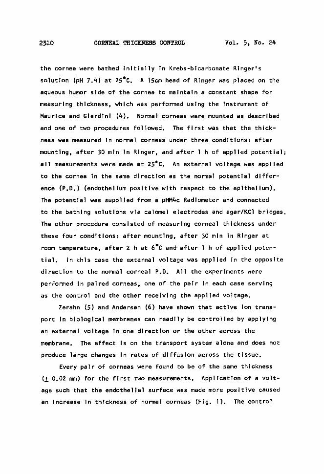
an increase in thickness of normal corneas (Fig . 1) . The control

Vol . 5, Ro . 24
COR'AEAL THICl~FSB CO1fTE0L
2311
O.SSI
É0.50'E
O.~.S'F-JWZ
Ou0.40
EMF
0 . 350 30 0 90
TIME ~1~ IIIÎ~
FIG . 1 .
Response of corneal thickness to an external voltage
applied opposite to the normal corneal P,D, External
voltage applied to experimental corneas atT EMF . C,
control corneas ; E, experimental corneas with applied
voltages in parenthesis . Values are the mean ± S,E .
of at least 8 corneas .
corneas usually increased by 0 .02 mm in the 90 min experimental
period (from 0 .38 ± 0.005 mm to 0.39 ± 0.005 mm : mean ± Standard

2312
coRAEAL Tszc>~>Jss cox~oL
vol . 5, xo. 24
FIG . 2,
Response of corneas swollen in the cold to an external
voltage applied in the same direction as the corneal P,D,
External voltage applied at ~ EMF,
C,
control corneas ;
E,
experimental corneas . Values are the mean ± S .E, of 8 corneas .
F,h-ror of 100 control corneas) . Application of a voltage in the
same direction as the corneal P,D� however, caused a greater
increase in corneal thickness . The increase in thickness was found
to be dependent upon the applied voltage, within the limits of
50 mV and 450 mV ; below 50 mV no change in thickness occurred as
compared to the control, and above 450 mV no further increases in
thickness were found (Fig . l) . The values seen after one hour of
applied voltage are steady state values, for maintenance of the
particular thickness continues for the duration of the period of

Qol . 5, No . 24
CORNEAL THICSNESS CONTROL
2313
voltage application .
Application of a voltage such that the positive electrode is
placed on the epithelial surface caused a decrease in thickness of
the swollen cornea (Fig . 2), although the application of such a
voltage caused no change in thickness of normal corneas . Cooling
of the cornea for 2 h caused an increase in thickness from 0,39 ±
0.005 mm to 0 .46 ± 0.01 mm (mean ± S,E, of 8 corneas) ; upon removal
from the refrigerator the Ringer was not t~eplaced with warm Ringer
but was allowed to regain heat from the room ; the temperature was
found to rise from 6°C to 18°C in 1 h . The control cornea continued
to swell after removal from the cold : 0 .46 + 0 .01 mm to 0.50 +
0.01 mm in .l h . However, following 1 h of an applied voltage of
500 mV (the minimum required to cause thinning was found to be
350 mV) the experimental cornea had thinned : 0 .46 + 0.01 mm to
0.43 ± 0 .01 mm (mean ± S.E, of 8 corneas.) .
On the basis that the applied poten~tal either accelerates or
nullifies the active transport system in the cornea, depending
upon the direction of the applied potential, the experiments show
that active ion transport may well be the factor by which corneal
thickness is maintained constant . This interpretation, however,
does not preclude the effects of the voltage on passive ion move-
ments under the influence of the applied current .
In the case of
the normal cornea, the applied voltage reduces the transport of ions
into the stroma . In the swollen cornea, where the transport
system is inhibited by cold, the applied voltage stimulates the
normal transport system or creates a greater driving force for the
ion transport, thereby increasing the ton concentration in the
stroma . The effects that are seen following application of the
applied voltage to the corneas in various states indicate that by

2~1~
COldiEAL TSIC)QPE88 CO1fTROL
Yol . 5, No. 24
controlling the active transport system one can control corneal
thickness . It is highly probable, therefore, that the active ion
transport system in the epithelium serves to maintain a constant
corneal hydration and thickness - a concept previously proposed
by Donn, Maurice and Mills (7) . The way In which corneal thickness
is controlled is at present unknown, but it may well be connected
to the binding of cations onto the acid mucopolysaccharide in the
stroma ; the transport pump may serve to control this process by
making ions available in greater or .lesser quantities and thereby
controlling the reversible imbibition of water .
Acknowledgements
This work was aided by U,S . Public Health Research Grant
N,B, 04854 from the National Institute of Neurological Diseases
and Blindness . The author wishes to thank Dr . T . Otorl for
performing the corneal thickness determinations .
References
1 .
Maurice,
D,M�
in The Eve , vol .
1,
(H .
Davson,
ed.) Academic
Press, London . (1962) .
2 . Green, K ., Am . J . Physiol . 20~, 1311 (1965) .
3 . Green, K., Exatl . Eve Res . ~, 110 (1966) .
4 . Maurice, D,M, and Glardini, A,A,, Brit . J . Ophthalmol . ~,,
169 (1958) .
5 . Zerahn, K ., Oxygen consumption and active sodium transport .
Universitetsforlaget . Aarhus . (1958) .
6 .
Andersen, B .,
reported by Ussing, H,H� J . Gen .
Physiol . 4~,,
135 (1960) .
7 .
Donn, A., Maurice,
D,M, and Mills, N,L� Arch . Ophthalmol . 6~2 ,
748 (1959) .