intraoperative corneal thickness changes during pulsed
TRANSCRIPT
Clinical StudyIntraoperative Corneal Thickness Changes duringPulsed Accelerated Corneal Cross-Linking Using IsotonicRiboflavin with HPMC
Ahmed M. Sherif,1 Nihal A. El-Gheriany,1 Yehia M. Salah El-Din,1 Lamiaa S. Aly,1
Amr A. Osman,1 Michael A. Grentzelos,2 and George D. Kymionis2,3
1Department of Ophthalmology, Faculty of Medicine, Cairo University, Cairo 11519, Egypt2Vardinoyiannion Eye Institute of Crete (VEIC), Faculty of Medicine, University of Crete, Heraklion, 71003 Crete, Greece3Bascom Palmer Eye Institute, University of Miami, Miller School of Medicine, Miami, FL 33136, USA
Correspondence should be addressed to Ahmed M. Sherif; [email protected]
Received 3 July 2015; Accepted 16 September 2015
Academic Editor: Neil Lagali
Copyright © 2016 Ahmed M. Sherif et al. This is an open access article distributed under the Creative Commons AttributionLicense, which permits unrestricted use, distribution, and reproduction in any medium, provided the original work is properlycited.
Purpose. To evaluate corneal thickness changes during pulsed accelerated corneal cross-linking (CXL) for keratoconus using a newisotonic riboflavin formula.Methods. In this prospective, interventional, clinical study patientswith grades 1-2 keratoconus (Amsler-Krumeich classification) underwent pulsed accelerated (30mW/cm2) CXL after application of an isotonic riboflavin solution (0.1%)with HPMC for 10 minutes. Central corneal thickness (CCT) measurements were taken using ultrasound pachymetry before andafter epithelial removal, after riboflavin soaking, and immediately after completion of UVA treatment. Results. Twenty eyes of11 patients (4 males, 7 females) were enrolled. Mean patient age was 26 ± 3 (range from 18 to 30 years). No intraoperative orpostoperative complications were observed in any of the patients. Mean CCT was 507 ± 35 𝜇m (range: 559–459 𝜇m) before and475 ± 40 𝜇m (range: 535–420 𝜇m) after epithelial removal (𝑃 < 0.001). After 10 minutes of riboflavin instillation, there was astatistically significant decrease of CCTby 6.2% from 475±40 𝜇m(range: 535–420 𝜇m) to 446±31 𝜇m(range: 508–400) (𝑃 < 0.005).There was no other statistically significant change of CCT during UVA irradiation. Conclusions. A significant decrease of cornealthickness was demonstrated during the isotonic riboflavin with HPMC application while there was no significant change duringthe pulsed accelerated UVA irradiation.
1. Introduction
Corneal cross-linking (CXL) is a minimally invasive proce-dure that combines the use of riboflavin and ultraviolet-A(UVA) irradiation resulting in an increase of the biomechani-cal stability of the corneal tissue [1, 2]. A preoperative cornealthickness of 400 𝜇m as a minimum safety limit to avoidcorneal endothelial damage during CXL has been proposed[3]. However, endothelial failure has been reported veryoccasionally after CXL resulting in corneal edema postop-eratively [4, 5]. The etiology of such problems has not beenfully elucidated but may be due to severe stromal thinning
intraoperatively which has been reported by several authors[6, 7]. Hence, it has become important to monitor cornealthickness during the procedure.
Accelerated CXL is based on the Bunsen-Roscoe lawof reciprocity according to which reducing irradiation timeand correspondingly increasing irradiation intensity couldachieve the same photochemical effect.
The aim of this study was to evaluate the intraoperativepachymetric changes during CXL using isotonic riboflavin(0.1%) and HPMC (hydroxyl propyl methylcellulose, HPMC;VibeX Rapid, Avedro Inc., Waltham, MS, USA) and pulsedaccelerated UVA.
Hindawi Publishing CorporationJournal of OphthalmologyVolume 2016, Article ID 1471807, 4 pageshttp://dx.doi.org/10.1155/2016/1471807

2 Journal of Ophthalmology
2. Materials and Methods
In this prospective, interventional, clinical study patientswithgrades 1-2 keratoconus (Amsler-Krumeich classification)were enrolled. All patients underwent pulsed high intensityCXL using theKXL system (Avedro Inc.,Waltham,MS,USA)preceded by the application of an isotonic riboflavin (0.1%)and HPMC (hydroxyl propyl methylcellulose, HPMC; VibeXRapid, Avedro Inc.,Waltham,MS, USA) for 10minutes at EyeCare Center, Maadi, Cairo, Egypt, between August 2014 andFebruary 2015. The study was conducted within the tenets ofthe Declaration of Helsinki after obtaining the institutionalreview board approval. A written informed consent wasobtained from all patients.
Inclusion criteria were progressive keratoconus (pro-gression was confirmed if there was an increase in the𝐾max on Pentacam maps of 1.00 diopter [D], increase ofmanifest refraction cylinder of 1.00D, or increase of manifestrefraction spherical equivalent of 0.50D over the period ofone year) and corneal thickness more than 400𝜇m at thethinnest location. Exclusion criteria were corneal scars oropacities, pregnancy or lactation, active anterior segmentpathologic features, previous corneal or anterior segmentsurgery, systemic connective tissue disease, atopic syndrome,and dry eye syndrome. Preoperative data obtained from thecase records included patient age and gender, Pentacam cen-tral corneal thickness (CCT) and thinnest corneal thickness(TCT) values, and CCT values obtained by ultrasonic cornealpachymetry (Sonomed 300P PacScan Pachymeter; EscalonMedical Corp.), which takes the mean of 256 measurementsin each scan.
2.1. Surgical Technique. Corneal cross-linking (CXL) wasconducted under sterile conditions. One drop of pilocarpine1% eye drops was instilled 15 minutes before the procedure.After topical application of benoxinate hydrochloride 0.4%eye drops (Benox; Eipico Inc., Cairo, Egypt), an eye speculumwas placed and CCT was measured just before epithelialremoval. The probe tip of the ultrasonic pachymetry wasdisinfected using alcohol swab and was held perpendicular tothe cornea. Three consecutive measurements were obtainedat the center of the cornea of each eye; the thinnest measure-ment is used in the statistical analysis.Then, the central 8mmof the corneal epithelium was removed mechanically usinga blunt spatula. After corneal epithelial removal, CCT wasmeasured. Next, dextran-free riboflavin 0.1% with hydroxylpropyl methylcellulose (HPMC; VibeX Rapid, Avedro Inc.,Waltham, MS, USA) was instilled every 2 minutes for 10minutes after whichCCTwas remeasured. Pulsed acceleratedUVA irradiation was next performed using KXL system(Avedro Inc., Waltham, MS, USA) with 1 sec. on/1 sec. offof UVA irradiation of 30mW/cm2 for a total duration of8 minutes and 40 seconds. A final CCT measurement wasobtained immediately after completion of UVA irradiation. Atherapeutic contact lens was applied and removed at the 3rdpostoperative day after complete reepithelialization.
2.2. Statistical Analysis. Statistical analysis was done usingpaired 𝑡-test. Statistical Package for the Social Sciences (SPSS)v.16 was used.
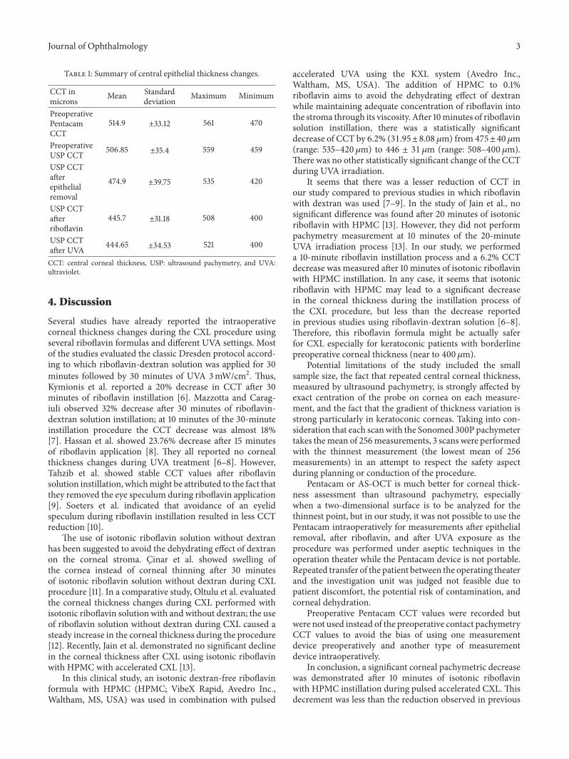
Afterepithelial
removal, 475After
riboflavinsoaking, 446
Time
6.2% decrease inthickness
350370390410430450470490510530550
CCT
(𝜇)
Figure 1: CCT changes (in microns) after riboflavin soaking.
Time
Afterriboflavin
soaking, 446After UVA
treatment, 445CCT
(𝜇)
350370390410430450470490510530550
Figure 2: CCT changes (in microns) during UVA treatment.
Because both eyes of some patients were used in thestudy, a nested analysis of variance was used to correct forany correlation between the right and left eyes of the samesubject. 𝑃 value less than 0.05 was considered significant.
3. Results
Twenty eyes of 11 patients were included. Four were malesand 7 were females. Mean patient age was 26 ± 3 (rangefrom 18 to 30 years). No intraoperative or postoperativecomplications were observed in any of the patients.
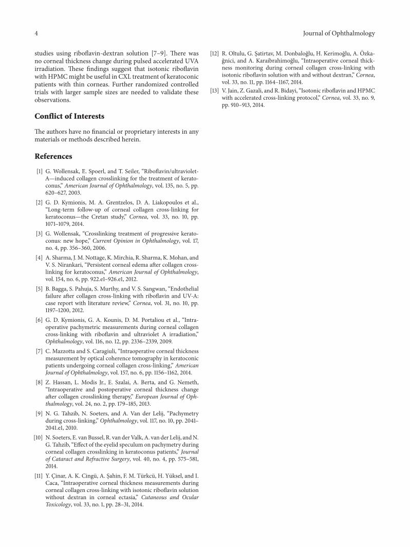
Mean USP CCT was 506.85 ± 35.4 𝜇m (range: 559–459 𝜇m) before and 474.9 ± 39.75 𝜇m (range: 535–420𝜇m)after epithelial removal (𝑃 < 0.001). After 10 minutes ofriboflavin installation, there was a statistically significantdecrease of CCT by 6.2% (31.95 ± 8.08 𝜇m) from 474.9 ±39.75 𝜇m (range: 535–420𝜇m) to 445.7 ± 31.18 𝜇m (range:508–400𝜇m) (𝑃 < 0.005) (Figure 1). There was no statisti-cally significant change of the CCT during UVA irradiation;CCT was 444.65 ± 34.53 𝜇m (range: 521–400𝜇m) at the endof UVA irradiation (𝑃 = 0.61) (Figure 2). However, therewas a statistically significant decrease by 62.2 ± 34.64 𝜇min CCT from 506.85 ± 35.4 𝜇m (range: 559–459 𝜇m) beforeepithelial removal to 444.65 ± 34.53 𝜇m (range: 521–400𝜇m)at the end of CXL (𝑃 < 0.001). Results are summarized inTable 1.
Journal of Ophthalmology 3
Table 1: Summary of central epithelial thickness changes.
CCT inmicrons Mean Standard
deviation Maximum Minimum
PreoperativePentacamCCT
514.9 ±33.12 561 470
PreoperativeUSP CCT 506.85 ±35.4 559 459
USP CCTafterepithelialremoval
474.9 ±39.75 535 420
USP CCTafterriboflavin
445.7 ±31.18 508 400
USP CCTafter UVA 444.65 ±34.53 521 400
CCT: central corneal thickness, USP: ultrasound pachymetry, and UVA:ultraviolet.
4. Discussion
Several studies have already reported the intraoperativecorneal thickness changes during the CXL procedure usingseveral riboflavin formulas and different UVA settings. Mostof the studies evaluated the classic Dresden protocol accord-ing to which riboflavin-dextran solution was applied for 30minutes followed by 30 minutes of UVA 3mW/cm2. Thus,Kymionis et al. reported a 20% decrease in CCT after 30minutes of riboflavin instillation [6]. Mazzotta and Carag-iuli observed 32% decrease after 30 minutes of riboflavin-dextran solution instillation; at 10 minutes of the 30-minuteinstillation procedure the CCT decrease was almost 18%[7]. Hassan et al. showed 23.76% decrease after 15 minutesof riboflavin application [8]. They all reported no cornealthickness changes during UVA treatment [6–8]. However,Tahzib et al. showed stable CCT values after riboflavinsolution instillation, whichmight be attributed to the fact thatthey removed the eye speculum during riboflavin application[9]. Soeters et al. indicated that avoidance of an eyelidspeculum during riboflavin instillation resulted in less CCTreduction [10].
The use of isotonic riboflavin solution without dextranhas been suggested to avoid the dehydrating effect of dextranon the corneal stroma. Cinar et al. showed swelling ofthe cornea instead of corneal thinning after 30 minutesof isotonic riboflavin solution without dextran during CXLprocedure [11]. In a comparative study, Oltulu et al. evaluatedthe corneal thickness changes during CXL performed withisotonic riboflavin solution with and without dextran; the useof riboflavin solution without dextran during CXL caused asteady increase in the corneal thickness during the procedure[12]. Recently, Jain et al. demonstrated no significant declinein the corneal thickness after CXL using isotonic riboflavinwith HPMC with accelerated CXL [13].
In this clinical study, an isotonic dextran-free riboflavinformula with HPMC (HPMC; VibeX Rapid, Avedro Inc.,Waltham, MS, USA) was used in combination with pulsed
accelerated UVA using the KXL system (Avedro Inc.,Waltham, MS, USA). The addition of HPMC to 0.1%riboflavin aims to avoid the dehydrating effect of dextranwhile maintaining adequate concentration of riboflavin intothe stroma through its viscosity. After 10minutes of riboflavinsolution instillation, there was a statistically significantdecrease of CCT by 6.2% (31.95±8.08 𝜇m) from 475±40 𝜇m(range: 535–420𝜇m) to 446 ± 31 𝜇m (range: 508–400𝜇m).There was no other statistically significant change of the CCTduring UVA irradiation.
It seems that there was a lesser reduction of CCT inour study compared to previous studies in which riboflavinwith dextran was used [7–9]. In the study of Jain et al., nosignificant difference was found after 20 minutes of isotonicriboflavin with HPMC [13]. However, they did not performpachymetry measurement at 10 minutes of the 20-minuteUVA irradiation process [13]. In our study, we performeda 10-minute riboflavin instillation process and a 6.2% CCTdecrease was measured after 10 minutes of isotonic riboflavinwith HPMC instillation. In any case, it seems that isotonicriboflavin with HPMC may lead to a significant decreasein the corneal thickness during the instillation process ofthe CXL procedure, but less than the decrease reportedin previous studies using riboflavin-dextran solution [6–8].Therefore, this riboflavin formula might be actually saferfor CXL especially for keratoconic patients with borderlinepreoperative corneal thickness (near to 400𝜇m).
Potential limitations of the study included the smallsample size, the fact that repeated central corneal thickness,measured by ultrasound pachymetry, is strongly affected byexact centration of the probe on cornea on each measure-ment, and the fact that the gradient of thickness variation isstrong particularly in keratoconic corneas. Taking into con-sideration that each scanwith the Sonomed 300P pachymetertakes themean of 256measurements, 3 scans were performedwith the thinnest measurement (the lowest mean of 256measurements) in an attempt to respect the safety aspectduring planning or conduction of the procedure.
Pentacam or AS-OCT is much better for corneal thick-ness assessment than ultrasound pachymetry, especiallywhen a two-dimensional surface is to be analyzed for thethinnest point, but in our study, it was not possible to use thePentacam intraoperatively for measurements after epithelialremoval, after riboflavin, and after UVA exposure as theprocedure was performed under aseptic techniques in theoperation theater while the Pentacam device is not portable.Repeated transfer of the patient between the operating theaterand the investigation unit was judged not feasible due topatient discomfort, the potential risk of contamination, andcorneal dehydration.
Preoperative Pentacam CCT values were recorded butwere not used instead of the preoperative contact pachymetryCCT values to avoid the bias of using one measurementdevice preoperatively and another type of measurementdevice intraoperatively.
In conclusion, a significant corneal pachymetric decreasewas demonstrated after 10 minutes of isotonic riboflavinwith HPMC instillation during pulsed accelerated CXL. Thisdecrement was less than the reduction observed in previous
4 Journal of Ophthalmology
studies using riboflavin-dextran solution [7–9]. There wasno corneal thickness change during pulsed accelerated UVAirradiation. These findings suggest that isotonic riboflavinwith HPMCmight be useful in CXL treatment of keratoconicpatients with thin corneas. Further randomized controlledtrials with larger sample sizes are needed to validate theseobservations.
Conflict of Interests
The authors have no financial or proprietary interests in anymaterials or methods described herein.
References
[1] G. Wollensak, E. Spoerl, and T. Seiler, “Riboflavin/ultraviolet-A—induced collagen crosslinking for the treatment of kerato-conus,” American Journal of Ophthalmology, vol. 135, no. 5, pp.620–627, 2003.
[2] G. D. Kymionis, M. A. Grentzelos, D. A. Liakopoulos et al.,“Long-term follow-up of corneal collagen cross-linking forkeratoconus—the Cretan study,” Cornea, vol. 33, no. 10, pp.1071–1079, 2014.
[3] G. Wollensak, “Crosslinking treatment of progressive kerato-conus: new hope,” Current Opinion in Ophthalmology, vol. 17,no. 4, pp. 356–360, 2006.
[4] A. Sharma, J.M.Nottage, K.Mirchia, R. Sharma, K.Mohan, andV. S. Nirankari, “Persistent corneal edema after collagen cross-linking for keratoconus,” American Journal of Ophthalmology,vol. 154, no. 6, pp. 922.e1–926.e1, 2012.
[5] B. Bagga, S. Pahuja, S. Murthy, and V. S. Sangwan, “Endothelialfailure after collagen cross-linking with riboflavin and UV-A:case report with literature review,” Cornea, vol. 31, no. 10, pp.1197–1200, 2012.
[6] G. D. Kymionis, G. A. Kounis, D. M. Portaliou et al., “Intra-operative pachymetric measurements during corneal collagencross-linking with riboflavin and ultraviolet A irradiation,”Ophthalmology, vol. 116, no. 12, pp. 2336–2339, 2009.
[7] C. Mazzotta and S. Caragiuli, “Intraoperative corneal thicknessmeasurement by optical coherence tomography in keratoconicpatients undergoing corneal collagen cross-linking,” AmericanJournal of Ophthalmology, vol. 157, no. 6, pp. 1156–1162, 2014.
[8] Z. Hassan, L. Modis Jr., E. Szalai, A. Berta, and G. Nemeth,“Intraoperative and postoperative corneal thickness changeafter collagen crosslinking therapy,” European Journal of Oph-thalmology, vol. 24, no. 2, pp. 179–185, 2013.
[9] N. G. Tahzib, N. Soeters, and A. Van der Lelij, “Pachymetryduring cross-linking,” Ophthalmology, vol. 117, no. 10, pp. 2041–2041.e1, 2010.
[10] N. Soeters, E. vanBussel, R. van derValk, A. van der Lelij, andN.G. Tahzib, “Effect of the eyelid speculum on pachymetry duringcorneal collagen crosslinking in keratoconus patients,” Journalof Cataract and Refractive Surgery, vol. 40, no. 4, pp. 575–581,2014.
[11] Y. Cinar, A. K. Cingu, A. Sahin, F. M. Turkcu, H. Yuksel, and I.Caca, “Intraoperative corneal thickness measurements duringcorneal collagen cross-linking with isotonic riboflavin solutionwithout dextran in corneal ectasia,” Cutaneous and OcularToxicology, vol. 33, no. 1, pp. 28–31, 2014.
[12] R. Oltulu, G. Satirtav, M. Donbaloglu, H. Kerimoglu, A. Ozka-gnici, and A. Karaibrahimoglu, “Intraoperative corneal thick-ness monitoring during corneal collagen cross-linking withisotonic riboflavin solution with and without dextran,” Cornea,vol. 33, no. 11, pp. 1164–1167, 2014.
[13] V. Jain, Z. Gazali, and R. Bidayi, “Isotonic riboflavin andHPMCwith accelerated cross-linking protocol,” Cornea, vol. 33, no. 9,pp. 910–913, 2014.

Submit your manuscripts athttp://www.hindawi.com
Stem CellsInternational
Hindawi Publishing Corporationhttp://www.hindawi.com Volume 2014
Hindawi Publishing Corporationhttp://www.hindawi.com Volume 2014
MEDIATORSINFLAMMATION
of
Hindawi Publishing Corporationhttp://www.hindawi.com Volume 2014
Behavioural Neurology
EndocrinologyInternational Journal of
Hindawi Publishing Corporationhttp://www.hindawi.com Volume 2014
Hindawi Publishing Corporationhttp://www.hindawi.com Volume 2014
Disease Markers
Hindawi Publishing Corporationhttp://www.hindawi.com Volume 2014
BioMed Research International
OncologyJournal of
Hindawi Publishing Corporationhttp://www.hindawi.com Volume 2014
Hindawi Publishing Corporationhttp://www.hindawi.com Volume 2014
Oxidative Medicine and Cellular Longevity
Hindawi Publishing Corporationhttp://www.hindawi.com Volume 2014
PPAR Research
The Scientific World JournalHindawi Publishing Corporation http://www.hindawi.com Volume 2014
Immunology ResearchHindawi Publishing Corporationhttp://www.hindawi.com Volume 2014
Journal of
ObesityJournal of
Hindawi Publishing Corporationhttp://www.hindawi.com Volume 2014
Hindawi Publishing Corporationhttp://www.hindawi.com Volume 2014
Computational and Mathematical Methods in Medicine
OphthalmologyJournal of
Hindawi Publishing Corporationhttp://www.hindawi.com Volume 2014
Diabetes ResearchJournal of
Hindawi Publishing Corporationhttp://www.hindawi.com Volume 2014
Hindawi Publishing Corporationhttp://www.hindawi.com Volume 2014
Research and TreatmentAIDS
Hindawi Publishing Corporationhttp://www.hindawi.com Volume 2014
Gastroenterology Research and Practice
Hindawi Publishing Corporationhttp://www.hindawi.com Volume 2014
Parkinson’s Disease
Evidence-Based Complementary and Alternative Medicine
Volume 2014Hindawi Publishing Corporationhttp://www.hindawi.com