a surgical pathology system for gross specimen examination
TRANSCRIPT
A SURGICAL PATHOLOGY SYSTEM FOR GROSS SPECIMENEXAMINATION
Domingos Cruz, MS 1,3, Mario Seixas, PhD 12
1. Institute of Molecular Pathology and Immunology of the University of Porto,Portugal (IPATIMUP)
2. Medical Faculty of the University of Porto, Portugal3. Higher Education Institute of Maia, Portugal
Rua Roberto Frias s/n Porto, PortugalTel: +351 2 5570700dcruz~ipatimup.pt
ABSTRACT
The concepts used in the storage of still digitalimages obtained during gross specimen examinationof tissues and organs in surgical pathology using adigital camera are described. We address thetechnical aspects related with the implementation ofa prototype tool to assist the pathologist during thesampling process as well the logic archive supportto store the acquired images. We describe, also, thehypermedia concepts that allow the navigation andthe efficient examination of the informationcontained in the stored images. The advantages, thetechnological and human limitations, and the effectsof using images in the documentation of a case arealso discussed.
KEYWORDS: Information, digital images, image-based documentation, anatomic pathology, grossspecimen examination.
INTRODUCTION
Diagnoses in Anatomic Pathology are obtainedusually by microscopic inspection (optical orelectronic) of slides of tissue fragments of tissuepieces, surgical specimens or biopsies submitted toanalysis.
In surgical pathology routine (gross examination),several samples are taken from the tissue piece ororgan (sampling domain) and a macroscopicalreport is elaborated; each sample is identified,morphologically described and precisely locatedwithin the specimen. This report contains essentialinformation for ulterior microscopic analysis andfor the final diagnosis.
Sampling is done, trying to represent thoroughlythe whole surgical specimen. The sampling processfaces frequently difficulties that may lead to majorinformation losses. Often, it is difficult or evenimpossible to describe the spatial location of thefragments. This occurs mainly because the tissuepiece or the organ under examination looses its
1091-8280/99/$5.00 © 1999 AMIA, Inc.
configuration and, eventually, thereferences, along with the sampling process.
spatial
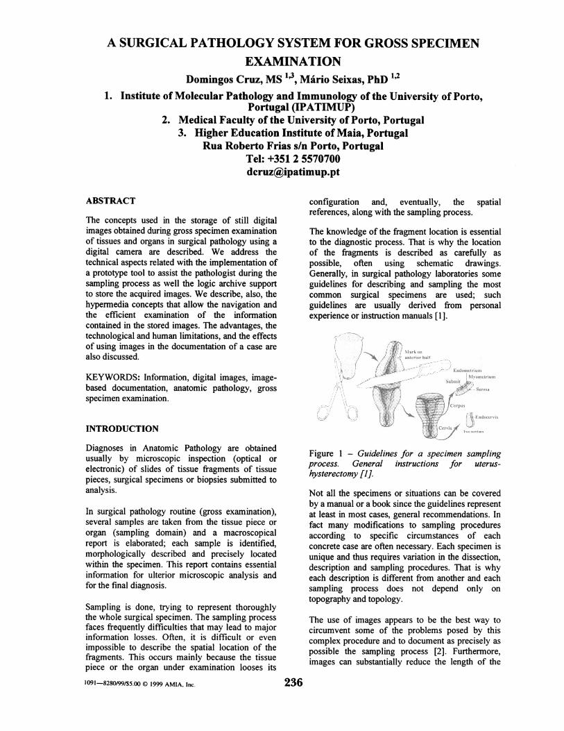
The knowledge of the fragment location is essentialto the diagnostic process. That is why the locationof the fragments is described as carefully aspossible, often using schematic drawings.Generally, in surgical pathology laboratories someguidelines for describing and sampling the mostcommon surgical specimens are used; suchguidelines are usually derived from personalexperience or instruction manuals [1].
i ; Ssrbw hoaw
Sub mist
Figure 1 - Guidelines for a specimen samplingprocess. General instructions for uterus-hysterectomy [1].
Not all the specimens or situations can be coveredby a manual or a book since the guidelines representat least in most cases, general recommendations. Infact many modifications to sampling proceduresaccording to specific circumstances of eachconcrete case are often necessary. Each specimen isunique and thus requires variation in the dissection,description and sampling procedures. That is whyeach description is different from another and eachsampling process does not depend only ontopography and topology.
The use of images appears to be the best way tocircumvent some of the problems posed by thiscomplex procedure and to document as precisely aspossible the sampling process [2]. Furthermore,images can substantially reduce the length of the
236
................. .............. .................................................... ..... ........ ..... ........... ....... ........................... ........ ...... ......................... ......................................
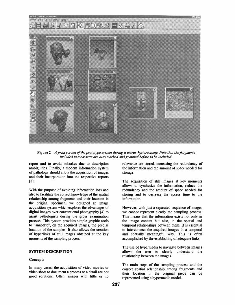
Figure 2 - A print screen ofthe prototype system during a uterus-hysterectomy. Note that thefragmentsincluded in a cassette are also marked andgrouped before to be included
report and to avoid mistakes due to descriptionambiguities. Finally, a modem information systemof pathology should allow the acquisition of imagesand their incorporation into the respective reports[3].
With the purpose of avoiding information loss andalso to facilitate the correct knowledge of the spatialrelationship among fragments and their location inthe original specimen, we designed an imageacquisition system which explores the advantages ofdigital images over conventional photography [4] toassist pathologists during the gross examinationprocess. This system provides simple graphic toolsto "annotate", on the acquired images, the preciselocation of the samples. It also allows the creationof hyperlinks of still images obtained at the keymoments of the sampling process.
SYSTEM DESCRIPTION
Concepts
In many cases, the acquisition of video movies orvideo shots to document a process or a detail are notgood solutions. Often, images with little or no
relevance are stored, increasing the redundancy ofthe information and the amount of space needed forstorage.
The acquisition of still images at key momentsallows to synthesize the information, reduce theredundancy and the amount of space needed forstoring and to decrease the access time to theinformation.
However, with just a separated sequence of imageswe cannot represent clearly the sampling process.This means that the information exists not only inthe image content but also, in the spatial andtemporal relationships between them. It is essentialto interconnect the acquired images in a temporaland spatially meaningful way. This is oftenaccomplished by the establishing of adequate links.
The use of hypermedia to navigate between imagesallows the user to clearly understand therelationship between the images.
The main steps of the sampling process and thecorrect spatial relationship among fragments andtheir location in the original piece can berepresented using a hypermedia model.
237
Links in hypermedia models are defined as part ofthe Dexter model for explicitly representingrelationships among object [5]. They specify alogical connection between two (or more) endpoints via the use of anchors. Most of thehypermedia systems allow the user to follow a linkas the basic form of interaction with the documentstructure. The use of links in hypermedia allows theusers to make a choice of which presentations toview and doing so the document structure becomesapparent. This lead to the question of where, in ourparticular case, links completely represent temporalor spatial relationship between images.
There are some theoretical approaches to solveproblems related with the use of multimedia objectsthat require spatial and temporal relations. TheAmsterdam Hypermedia Model [5] defineschannels and synchronization arcs to solve theproblems of spatial and temporal relationships.However in this case that kind of approach does notseem to be the most appropriate, due to thespecificity of the application.
Other option was to use Hypertext MarkupLanguage (HTML) images maps where thepathologist after the image acquisition just needs todecide where to put the hotspots (the areas thattrigger links), creating the document by linkingareas to images. That kind of solution has theadvantage of working with graphically orientedWeb browsers. However image maps need to havea separated image map file for presentation. Thus,the pathologist will have to define the mappingcoordinates for each acquired image with a mapeditor including these in a separated file.
Although, using a separated file to specify theimage maps is not the best solution for a systemthat was designed in order to concentrate, as muchas possible, all the information. To improveintegrity assurance, all images, annotations,mapping coordinates and navigation scheme shouldbe stored inside of a single file.
Implementation
To implement that concept, we defined a logicalstructure representing images with regions linked toother images in a single file.
We decided to use TIFF (Tag Image File Format) tostore images due to its ability to handle multiplepages and store extra information in a single file.TIFF allows, for each image, to store additionalinformation (text and objects) without damaging theimage contents. In fact, when we use an overlappinglayer it is possible to create objects (rectangles,ellipses, polygons, text boxes, etc) that areautomatically geo-referenced to the image (seeFigure 2).
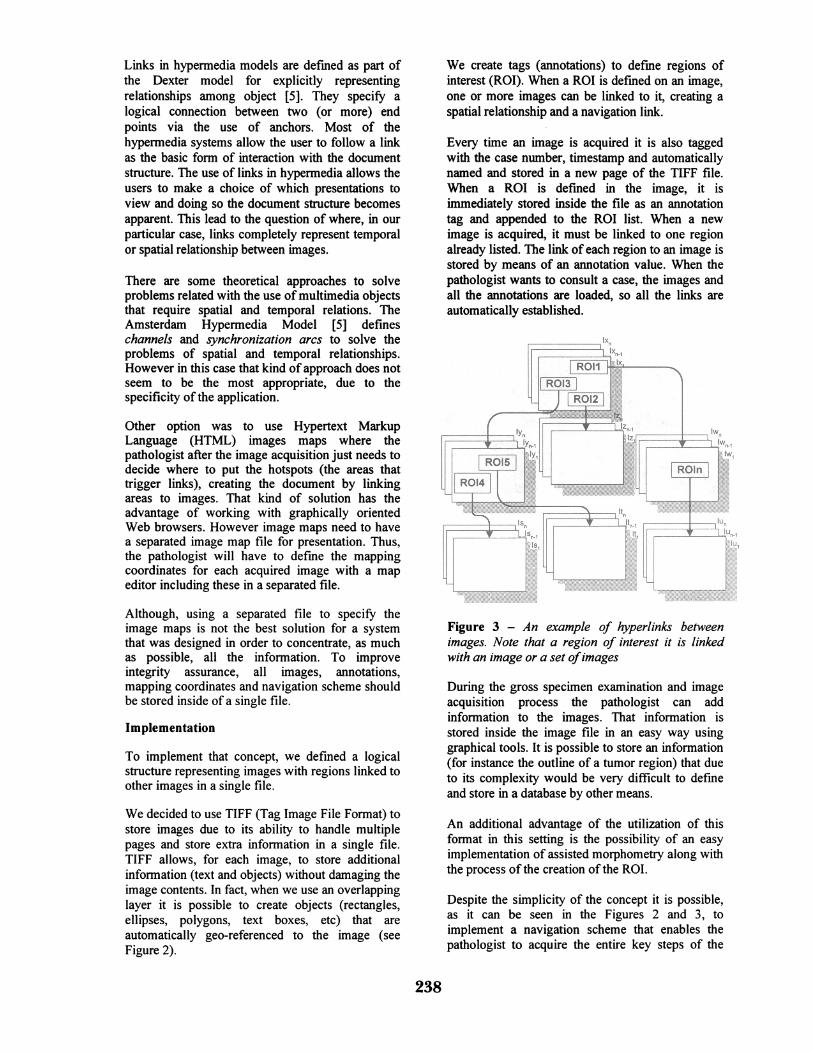
We create tags (annotations) to define regions ofinterest (ROI). When a ROI is defined on an image,one or more images can be linked to it, creating aspatial relationship and a navigation link.
Every time an image is acquired it is also taggedwith the case number, timestamp and automaticallynamed and stored in a new page of the TIFF file.When a ROI is defined in the image, it isimmediately stored inside the file as an annotationtag and appended to the ROI list. When a newimage is acquired, it must be linked to one regionalready listed. The link of each region to an image isstored by means of an annotation value. When thepathologist wants to consult a case, the images andall the annotations are loaded, so all the links areautomatically established.
Figure 3 - An example of hyperlinks betweenimages. Note that a region of interest it is linkedwith an image or a set ofimages
During the gross specimen examination and imageacquisition process the pathologist can addinformation to the images. That information isstored inside the image file in an easy way usinggraphical tools. It is possible to store an information(for instance the outline of a tumor region) that dueto its complexity would be very difficult to defineand store in a database by other means.
An additional advantage of the utilization of thisformat in this setting is the possibility of an easyimplementation of assisted morphometry along withthe process of the creation of the ROI.
Despite the simplicity of the concept it is possible,as it can be seen in the Figures 2 and 3, toimplement a navigation scheme that enables thepathologist to acquire the entire key steps of the
238
sampling process. Using this approach it is possibleto review all the steps from the piece to a singleimage or backtracking; it is indeed possible toreview different perspectives or views of a sampleand to pinpoint its precise origin.
The browsing strategies depend on use and on thetype of user. The application can be used for grossexamination, to assist the final report, to teach andto learn.
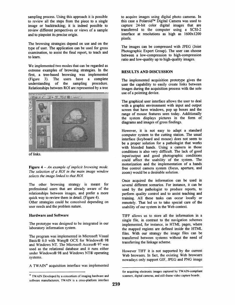
We implemented two modes that can be regarded asextreme examples of browsing strategies. In thefirst, a tree-based browsing was implemented(Figure 3). The users have a completeunderstanding of the sampling procedure.Relationships between ROI are represented by a tree
of links.
Figure 4 - An example of implicit browsing mode.The selection of a ROI in the main image windowselects the image linked to that ROI.
The other browsing strategy is meant forprofessional users that are already aware of therelationships between images, and prefer a morequick way to review them in detail. (Figure 4).Other strategies could be conceived depending onuser needs and the problem nature.
Hardware and Software
The prototype was designed to be integrated in ourlaboratory information system.
The program was implemented in Microsoft VisualBasicg 6.0 with Wang(® OCX for Windows® 98and Windows NT. The Microsoft Access® 97 wasused as the relational database and it runs eitherunder Windows® 98 and Windows NT® operatingsystems.
to acquire images using digital photo cameras. Inthis case a PolaroidTm Digital Camera was used tocapture 24-bit color digital images that aretransferred to the computer using a SCSI-2interface at resolutions as high as 1600x1200pixels.
The images can be compressed with JPEG (JointPhotographic Expert Group). The user can choosebetween a low-compression to high-compressionratio and low-quality up to high-quality images.
RESULTS AND DISCUSSION
The implemented acquisition prototype gives theuser the capability to easily create links betweenimages during the acquisition process with the soleuse of a pointing device.
The graphical user interface allows the user to dealwith a graphic environment with input and outputscreen that have windows, pop up boxes and therange of mouse features seen today. Additionallythe system displays pictures in the form ofdiagrams and images of gross findings.
However, it is not easy to adapt a standardcomputer system to the cutting station. The usualinterface (keyboard and mouse) does not seem tobe a proper solution for a pathologist that workswith blooded hands. Using a camera in thoseconditions is also very difficult. The lack of goodinput/output and good photographic conditionscould affect the usability of the system. Themotorization and the implementation of a handsfree control camera system (focus, aperture, andzoom) would be a desirable solution.
Once acquired the information can be used inseveral different scenarios. For instance, it can beused by the pathologist to produce reports, toperform quality control and to assist teaching andtraining. All these tasks can occur locally orremotely. That led us to take special care of theusability of our system in the Web context.
TIFF allows us to store all the information in asingle file, in contrast to the navigation schemesimplemented, for instance, in HTML pages, wherethe mapped regions are defined inside the HTMLfiles. With our strategy the image files can betransferred between systems without the need oftransferring the linkage scheme.
However TIFF it is not supported by the currentWeb browsers. In fact, the existing Web browsersnowadays only support GIF, JPEG and PNG image
A TWAIN* acquisition interface was implemented
* TWAIN Developed by a consortium of imaging hardware andsoftware manufacturers, TWAIN is a cross-platform interface
for acquiring electronic images captured by TWAIN-compliantscanners, digital cameras. and still-frame video capture boards.
239
formats.
We see three possible pathways to solve thisproblem:a) An application could generate a HMTL code,
mapping coordinates and image files toperform special browsing needed in standardWeb browsers.
b) The standard Web browser could beconfigured to lunch an external application tobrowse the images.
c) Finally, it is possible to extend a standard Webbrowser and make it capable of use new datatype, by means of a new protocol. For this, it isnecessary to make a content handler and aprotocol handler, and turn them available onthe Web server. Content and protocol handlerscould be downloaded over the Net, from thesame site that supplies the data.
Another drawback of TIFF is the file size.However, when acceptable, it is possible tocompress all the pages with JPEG turning a largerTIFF file into a small one.
The prototype has been used in our institution forthree months in parallel with the traditional process.So far the following conclusions were drawn:
Firstly, the graphic tools and the related tags need tobe specifically defined for the different types ofsurgical specimens and anatomic pathologicalprocedures.
Secondly, it is necessary to improve the human-machine interaction in the registry room in order toturn the system fully operational.
Finally, it is clear that some organs, due to theirpoorly defined anatomical limits (for instance breastsurgical specimens), do not benefit with the imagedocumentation. It seems that another approachshould be considered. Perhaps, in these cases,instead of images, standardized schematic drawingsshould be used.
REFERENCES
1. Rosai J, Guidlines for handling of mostcommom and important surgical specimens. In:Ackerman's Surgical Pathology. 8th ed. NewYork: Mosby-Year Book, Inc.; 1996. p. 2629-2725.
2. Furness PN, The use of digital images inpathology. Journal of Pathology 1997 Nov;183:3 253-2
3. Silverberg SG, DeLellis RA, Frable WJ,Computers in Surgical and Cytopathology. In:Principles and Practice Surgical Pathology and
Cytopathology. 3rd ed. New York: ChurchillLivingstone Inc.; 1997. P. 113-25
4. O'Brien MJ, Sotnikov AV, Digital imaging inanatomic pathology. American Journal ofClinical Pathology 1996 Oct; 106(4 Suppl 1):S25-S32..
5. Hardman L., Bulterman D. and Guido vanRossum, The Amsterdam Hypermedia Model:Extending Hypertext to Support RealMultimedia, Hypermedia Journal 5(1), 1993July, 47 - 69.
6. Rosai J, Information Systems in surgicalpathology. In: Ackerman's Surgical Pathology.8th ed. New York: Mosby-Year Book Inc.;1996. p. 2727-32.
240