md pathology specimen collection manualmdpathology.com/pdfs/mdp_usersguide.pdf · md pathology...
TRANSCRIPT

MD PATHOLOGY
SPECIMEN COLLECTION MANUAL AND USER’S GUIDE
Prepared by: Dr. Michael Deck Date Originally Prepared: 1993 Date Revised: January 11, 2018 Supersedes: Version of December 18, 2015 Date Effective: January 11, 2018
By: _Signature on file__ _1/11/2018_
Michael A. Deck, M.D. Date
Chief Pathologist
Reviewed:
Date: By:
_______________________ ________________________
_______________________ ________________________
_______________________ ________________________
_______________________ ________________________
_______________________ ________________________
_______________________ ________________________
_______________________ ________________________
NOTE: This procedure will be reviewed annually by the Medical Director or Assistant Medical Director of MD Pathology.

MD PATHOLOGY
Specimen Collection Manual
and User's Guide
October 9, 2017
Copyright © MD Pathology 2009, 2010, 2011, 2012, 2013, 2016, 2017 All Rights Reserved
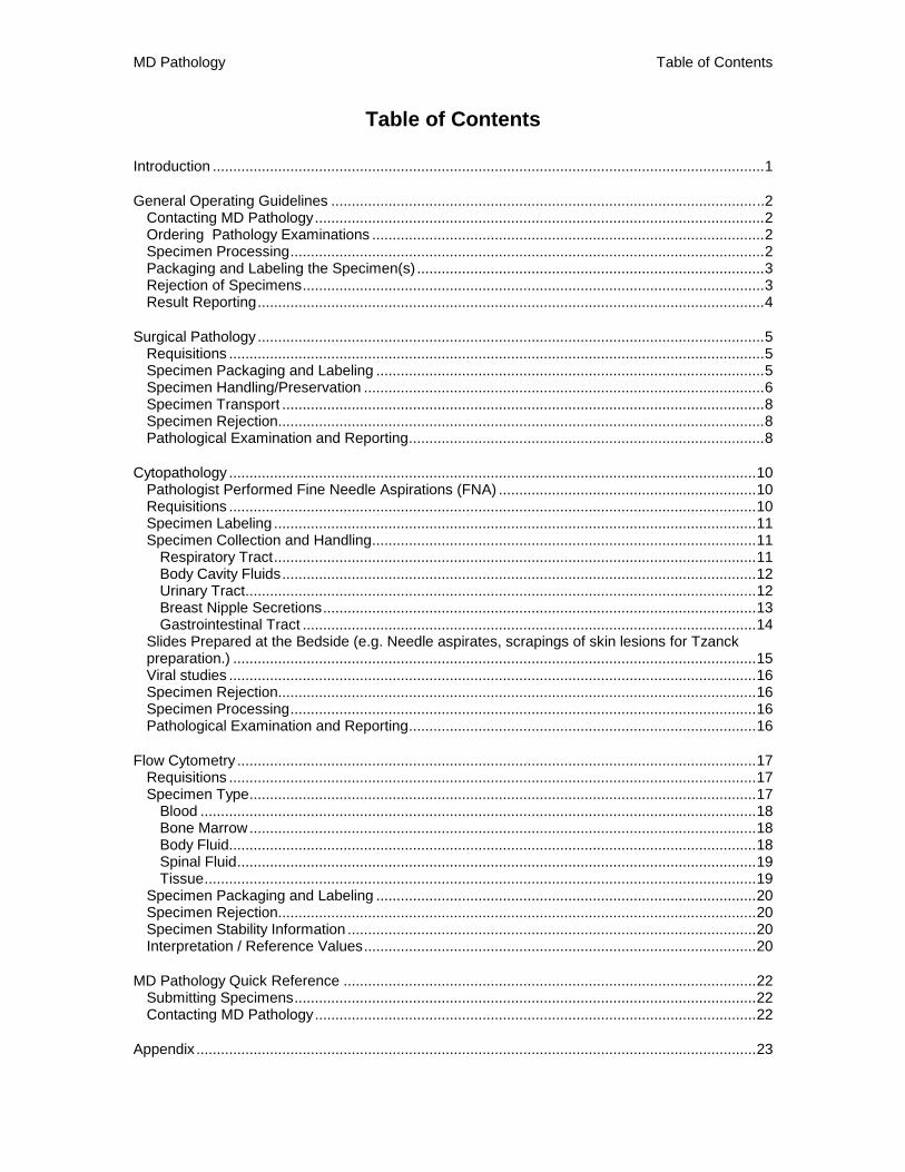
MD Pathology Table of Contents
Table of Contents
Introduction ....................................................................................................................................... 1
General Operating Guidelines ........................................................................................................ ..2 Contacting MD Pathology .............................................................................................................. 2 Ordering Pathology Examinations ................................................................................................ 2 Specimen Processing .................................................................................................................... 2 Packaging and Labeling the Specimen(s) ..................................................................................... 3 Rejection of Specimens ................................................................................................................. 3 Result Reporting ............................................................................................................................ 4
Surgical Pathology ............................................................................................................................ 5 Requisitions ................................................................................................................................... 5 Specimen Packaging and Labeling ............................................................................................... 5 Specimen Handling/Preservation .................................................................................................. 6 Specimen Transport ...................................................................................................................... 8 Specimen Rejection....................................................................................................................... 8 Pathological Examination and Reporting ....................................................................................... 8
Cytopathology ................................................................................................................................. 10 Pathologist Performed Fine Needle Aspirations (FNA) ............................................................... 10 Requisitions ................................................................................................................................. 10 Specimen Labeling ...................................................................................................................... 11 Specimen Collection and Handling .............................................................................................. 11
Respiratory Tract ...................................................................................................................... 11 Body Cavity Fluids .................................................................................................................... 12 Urinary Tract ............................................................................................................................. 12 Breast Nipple Secretions .......................................................................................................... 13 Gastrointestinal Tract ............................................................................................................... 14
Slides Prepared at the Bedside (e.g. Needle aspirates, scrapings of skin lesions for Tzanck preparation.) ................................................................................................................................ 15 Viral studies ................................................................................................................................. 16 Specimen Rejection..................................................................................................................... 16 Specimen Processing .................................................................................................................. 16 Pathological Examination and Reporting ..................................................................................... 16
Flow Cytometry ............................................................................................................................... 17 Requisitions ................................................................................................................................. 17 Specimen Type ............................................................................................................................ 17
Blood ........................................................................................................................................ 18 Bone Marrow ............................................................................................................................ 18 Body Fluid................................................................................................................................. 18 Spinal Fluid ............................................................................................................................... 19 Tissue ....................................................................................................................................... 19
Specimen Packaging and Labeling ............................................................................................. 20 Specimen Rejection..................................................................................................................... 20 Specimen Stability Information .................................................................................................... 20 Interpretation / Reference Values ................................................................................................ 20
MD Pathology Quick Reference ..................................................................................................... 22 Submitting Specimens ................................................................................................................. 22 Contacting MD Pathology ............................................................................................................ 22
Appendix ......................................................................................................................................... 23
MD Pathology Page 1 of 23
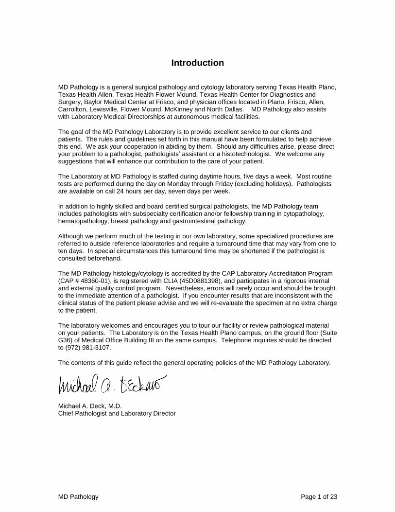
Introduction
MD Pathology is a general surgical pathology and cytology laboratory serving Texas Health Plano, Texas Health Allen, Texas Health Flower Mound, Texas Health Center for Diagnostics and Surgery, Baylor Medical Center at Frisco, and physician offices located in Plano, Frisco, Allen, Carrollton, Lewisville, Flower Mound, McKinney and North Dallas. MD Pathology also assists with Laboratory Medical Directorships at autonomous medical facilities.
The goal of the MD Pathology Laboratory is to provide excellent service to our clients and patients. The rules and guidelines set forth in this manual have been formulated to help achieve this end. We ask your cooperation in abiding by them. Should any difficulties arise, please direct your problem to a pathologist, pathologists’ assistant or a histotechnologist. We welcome any suggestions that will enhance our contribution to the care of your patient.
The Laboratory at MD Pathology is staffed during daytime hours, five days a week. Most routine tests are performed during the day on Monday through Friday (excluding holidays). Pathologists are available on call 24 hours per day, seven days per week.
In addition to highly skilled and board certified surgical pathologists, the MD Pathology team includes pathologists with subspecialty certification and/or fellowship training in cytopathology, hematopathology, breast pathology and gastrointestinal pathology.
Although we perform much of the testing in our own laboratory, some specialized procedures are referred to outside reference laboratories and require a turnaround time that may vary from one to ten days. In special circumstances this turnaround time may be shortened if the pathologist is consulted beforehand.
The MD Pathology histology/cytology is accredited by the CAP Laboratory Accreditation Program (CAP # 48360-01), is registered with CLIA (45D0881398), and participates in a rigorous internal and external quality control program. Nevertheless, errors will rarely occur and should be brought to the immediate attention of a pathologist. If you encounter results that are inconsistent with the clinical status of the patient please advise and we will re-evaluate the specimen at no extra charge to the patient.
The laboratory welcomes and encourages you to tour our facility or review pathological material on your patients. The Laboratory is on the Texas Health Plano campus, on the ground floor (Suite G36) of Medical Office Building III on the same campus. Telephone inquiries should be directed to (972) 981-3107.
The contents of this guide reflect the general operating policies of the MD Pathology Laboratory.
Michael A. Deck, M.D. Chief Pathologist and Laboratory Director

MD Pathology Page 2 of 23
General Operating Guidelines
Contacting MD Pathology
The following telephone numbers may be used for reaching various staff at MD Pathology:
Reaching Pathologists; General Inquiries; Specimen Pick-up
(972) 981-3107 (If you get the voice mail system, please indicate your location and that you have a pick-up for MD Pathology).
Histology Laboratory
(972) 981-3108 (Staffed 2:00 a.m. to 5:00 p.m.)
Gross Room (Specimen Processing)
(972) 981-8065 (Staffed 9:00 a.m. to 6:00 p.m.)
Ordering Pathology Examinations
Pathology consultations are ordered using paper requisitions provided by MD Pathology. Requisitions may be ordered from MD Pathology by calling (972) 981-3107. Orders will only be accepted from physicians licensed to practice medicine.
All specimens sent to the laboratory must be accompanied by a written requisition. Requisitions must be legible and complete. Incomplete requisitions may be cause for specimen rejection. Care must be taken so that all sections of the requisition are completed including the patient identification, the billing instructions, the specimen(s) submitted (clearly identifying the site of origin and laterality if appropriate) with type of examination requested and with pertinent clinical information (e.g. the clinical impression, the duration of the problem, the differential diagnosis, “rule out” conditions, pertinent past history such as prior tumors or inflammatory disease, other biopsy or cytology findings, etc.). The importance of clinical information cannot be overemphasized and will have direct bearing on the quality and specificity of the pathological opinion rendered!
It is the responsibility of the individual collecting the specimen to label the container(s) at the bedside (or immediately upon removal from surgical field) with the patient's name and second patient identifier such as date of birth or medical record number, date and time of collection, the anatomic site of origin, and initials of person collecting the specimen, or the person receiving specimen immediately from the surgical field. Double check and confirm that the name on the container and requisition are indeed the patient's. When pre-printed labels are used, it is easy to assume that the label has the correct patient name when it, in fact, does not. Identifiers may be in machine readable format, such as a barcode.
Please see that the specimen container is carefully sealed. Only non-leaking containers should be used.
For fine needle aspiration of palpable masses, we recommend that a pathologist skilled in this technique perform the aspiration. Please refer to the Pathologist Performed Fine Needle Aspirations (FNA) section below.

MD Pathology Page 3 of 23
Specimen Processing
Specimens received in the MD Pathology laboratory by 16:30 (4:30 p.m.) will be processed that day, and the pathologist will read the slides on the following working day. Exceptions will be made when the patient would be better served by allowing large specimens to fix in formalin overnight. See the section below entitled “Report Turnaround Times” for information regarding the time to issuance of the final report.
Results that are needed within a short time frame should be brought to the attention of the pathologist, preferably by a telephone call at the time the specimen is obtained. Specimen processing and interpretation can be expedited if clinically required.
Specimens submitted for frozen section are processed immediately. Results are usually available within twenty minutes from the time the specimen is received in the laboratory. Cases requiring multiple frozen section blocks (e.g. evaluation of margins from a large skin excision) will, at times, require more time with most cases completed within 30 to 45 minutes. Paraffin ("permanent") sections are available as described below.
Packaging and Labeling the Specimen(s)
All specimens submitted to the Laboratory must be accompanied by a requisition. The specimen container(s) must be labeled with the patient’s name, another unique identifier such as the medical record number or birthdate, date and time of collection, initials of person collecting the specimen, anatomic site of origin, and should be in an appropriate fixative (see below). The patient’s identity should be verified at the time of collection. All specimens must be placed into a leakproof container, labeled appropriately while at the patient’s bedside, and placed inside a plastic specimen bag for transport to the laboratory. For specimens with removable lids, the label should be affixed to the side of the container rather than the lid. It is not acceptable to label the transport bag and not label the specimens inside. The requisition and accompanying paperwork should be placed in the outside pocket of the specimen bag to avoid contamination. Specimens received without appropriate labeling will be rejected.
Surgical and cytology specimens may be delivered directly to the MD Pathology Laboratory in Medical Office Building III, on the Presbyterian Hospital of Plano Campus, Suite G36. Courier pick-up may be arranged by calling (972) 981-3107.
Rejection of Specimens
In the ultimate interest of patient well being and quality results, the laboratory may reject specimens for the following reasons:
1) No laboratory requisition.
2) Requisition label and specimen label do not match.
3) Inappropriate container or preservative.
4) Insufficient, inappropriate or contaminated sample.
5) Excessive delay in transport of specimen to laboratory, compromising specimen integrity.
6) Additional reasons specific to the requested procedures listed in the sections below.
7) Unlabeled or incompletely labeled specimen. Specimens rejected due to discrepancies in the specimen labeling or ambiguities in specimen identification shall be addressed by first contacting the individual who filled out the requisition or the submitting physician, either in person or over the telephone. If the discrepancy is resolved to the satisfaction of the person performing the gross examination of the case, the discrepant information is noted on the

MD Pathology Page 4 of 23
requisition with a notation on the requisition as to the person who provided the corrected information and the pathology staff member who obtained the corrected information. The specimen shall then be accessioned as usual. If the individual who prepared the requisition or the submitting physician is not available, or if the discrepancy is not satisfactorily resolved, the specimen(s) are returned to the physician’s office/facility for correction and completion of a Specimen Identity Confirmation form. Once discrepancies are resolved, the specimen(s) are returned to MD Pathology and processed accordingly.
The most common and potentially most serious problem is mislabeling of patient samples. Great care must be taken to avoid this error. The specimen container must be labeled as promptly as feasible while still at the patient's side.
Result Reporting
For most specimens, reports should be available before the close of business on the next working day following the submission of the specimen. Cases requiring special stains, decalcification, additional sections, or consultation with additional expert pathologists will require additional time (as much as a week or rarely longer). In such instance the pathologist working on the case will keep the ordering physician informed of preliminary impressions and updates as the workup progresses.
Written reports will be delivered either by Courier or by U.S. Mail. Faxed reports are issued upon sign out unless the submitting physician has specifically requested otherwise. Electronic delivery of reports (PDF) via an online report viewer is available upon request. Please contact MD Pathology for further details.
On occasion, amended reports will be issued. The amended version, which will contain the original report with the changes, will clearly be marked as such across the top of the first page. The pathologist of record will telephone amendments carrying significant clinical implications to the ordering physician.
Addendum reports will be issued when indicated, and shall contain all of the information in the original report as well as the additional findings (usually ancillary tests, results of consultations, additional study, etc.).

MD Pathology Page 5 of 23
Surgical Pathology
Surgical pathology entails histologic examination (i.e. microscopic examination of very thin tissue slices) of organs and tissues, also known as histology or “gross and microscopic examinations”. Order these examinations for tissue biopsies, organs removed at surgery, or portions thereof. For liquid specimens, body fluids, smears, scrapings, fine needle aspirations, etc., please refer to the “Cytopathology” section below.
Requisitions
All specimens submitted for surgical pathology examination must have an accompanying anatomic pathology requisition slip.
The source of the specimen (i.e. anatomic location including laterality if appropriate), the ordering physician, the day and time collected, the preoperative and postoperative diagnoses, and any special procedures desired (i.e. stain for AFB, Fungi, Pneumocystis) should be stated. In addition, the submitting physician should be consulted as to what relevant clinical information is to be included on the requisition.
If multiple specimens are obtained on the same patient, the source (i.e. anatomic site) of each specimen should be explicitly indicated on the requisition. Each requisition can accommodate up to six separate specimens.
Please specify the source of the specimen in the “specimen” section of the requisition and on the specimen container. Do not confuse diagnoses with specimen sources. Sources are anatomic sites. Diagnoses or “rule outs” are important information to provide to the laboratory but belong in the pertinent clinical information section.
Appropriate Specimen Labeling
Incorrect labeling…
Skin, right hand Mole
Left inguinal tissue Hernia sac
Mass, back Lipoma
Small bowel Rule out Whipple, Sprue
Specimen Packaging and Labeling
1) Each specimen must be placed in a sealed leak-proof specimen container and labeled at the bedside or promptly upon passage from the surgical field.
2) The label shall include the patient's name, another unique identifier such as the medical record number or birthdate, the date the specimen was collected, and the specific anatomic site of origin of the specimen.
3) The label should be placed on the container itself rather than the lid to insure unequivocal identification even while the lid is removed or should it become dislodged in transit. Specimens from separate sites should be placed in separate containers.
MD Pathology Page 6 of 23
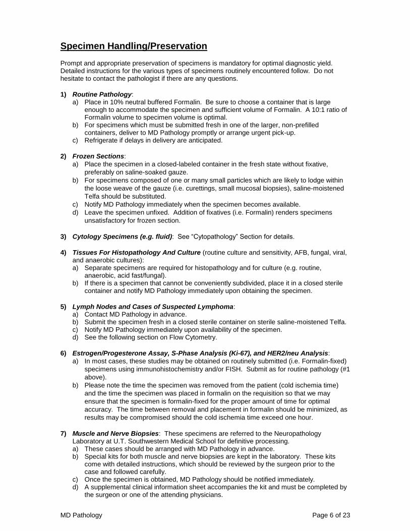
Specimen Handling/Preservation
Prompt and appropriate preservation of specimens is mandatory for optimal diagnostic yield. Detailed instructions for the various types of specimens routinely encountered follow. Do not hesitate to contact the pathologist if there are any questions.
1) Routine Pathology: a) Place in 10% neutral buffered Formalin. Be sure to choose a container that is large
enough to accommodate the specimen and sufficient volume of Formalin. A 10:1 ratio of Formalin volume to specimen volume is optimal.
b) For specimens which must be submitted fresh in one of the larger, non-prefilled containers, deliver to MD Pathology promptly or arrange urgent pick-up.
c) Refrigerate if delays in delivery are anticipated.
2) Frozen Sections:
a) Place the specimen in a closed-labeled container in the fresh state without fixative,
preferably on saline-soaked gauze.
b) For specimens composed of one or many small particles which are likely to lodge within
the loose weave of the gauze (i.e. curettings, small mucosal biopsies), saline-moistened
Telfa should be substituted.
c) Notify MD Pathology immediately when the specimen becomes available.
d) Leave the specimen unfixed. Addition of fixatives (i.e. Formalin) renders specimens
unsatisfactory for frozen section.
3) Cytology Specimens (e.g. fluid): See “Cytopathology” Section for details.
4) Tissues For Histopathology And Culture (routine culture and sensitivity, AFB, fungal, viral, and anaerobic cultures): a) Separate specimens are required for histopathology and for culture (e.g. routine,
anaerobic, acid fast/fungal). b) If there is a specimen that cannot be conveniently subdivided, place it in a closed sterile
container and notify MD Pathology immediately upon obtaining the specimen.
5) Lymph Nodes and Cases of Suspected Lymphoma: a) Contact MD Pathology in advance. b) Submit the specimen fresh in a closed sterile container on sterile saline-moistened Telfa. c) Notify MD Pathology immediately upon availability of the specimen. d) See the following section on Flow Cytometry.
6) Estrogen/Progesterone Assay, S-Phase Analysis (Ki-67), and HER2/neu Analysis:
a) In most cases, these studies may be obtained on routinely submitted (i.e. Formalin-fixed)
specimens using immunohistochemistry and/or FISH. Submit as for routine pathology (#1
above).
b) Please note the time the specimen was removed from the patient (cold ischemia time)
and the time the specimen was placed in formalin on the requisition so that we may
ensure that the specimen is formalin-fixed for the proper amount of time for optimal
accuracy. The time between removal and placement in formalin should be minimized, as
results may be compromised should the cold ischemia time exceed one hour.
7) Muscle and Nerve Biopsies: These specimens are referred to the Neuropathology Laboratory at U.T. Southwestern Medical School for definitive processing. a) These cases should be arranged with MD Pathology in advance. b) Special kits for both muscle and nerve biopsies are kept in the laboratory. These kits
come with detailed instructions, which should be reviewed by the surgeon prior to the case and followed carefully.
c) Once the specimen is obtained, MD Pathology should be notified immediately. d) A supplemental clinical information sheet accompanies the kit and must be completed by
the surgeon or one of the attending physicians.

MD Pathology Page 7 of 23
8) Bone Marrow Biopsies and Aspirates (call for special bone marrow collection kits): a) A specialized bone marrow requisition form should be completed to include patient
information, who performed the marrow, which physician needs to receive the report, what specimens are being submitted, the diagnosis under consideration, and any special studies requested (e.g. flow cytometry, cytogenetics, FISH testing, etc.).
b) Both aspiration and biopsy specimens are essential for optimal bone marrow evaluation. From these, aspirate smears, an aspirate clot in Formalin, a trephine (core needle) biopsy in formalin, and imprint smears should be submitted on each patient. Often additional aliquots of aspirate material should be submitted in anticoagulated tubes if flow cytometery, cytogenetics or FISH analyses are requested.
c) All specimens should be clearly labeled with the patient’s name and date of birth, as well as date and time of collection.
d) Bone marrow aspiration: At least two pulls should be made. The first pull is for aspirate smears and clot sections. Use a non-anticoagulated syringe and withdraw up to 4 cc—attempting to withdraw additional volume will cause the specimen to be diluted by peripheral blood emanating from the intra-marrow sinuses. Immediately extract the marrow particles using a pipette, and transfer the particles to clean, labeled slides and gently smear with a second slide. Four smears should be prepared, and allowed to air dry or, alternatively, promptly fixed in methanol. The remaining aspirate material should be allowed to clot in the syringe. After clotting, remove the plunger and pour the clot from the large end of the syringe barrel into a small container of 10% buffered formalin.
Flow Cytometery, Cytogenetics and FISH: Specimens for flow cytometry, cytogenetics, and FISH studies require additional 5 cc each of aspirate material after re-directing the needle (otherwise the specimens may be too dilute with peripheral blood to yield optimal results). A 5 cc sample in an EDTA (purple top) tube is required for flow cytometry and/or FISH studies, and additional smears prepared at MD Pathology for morphologic evaluation. A 5 cc sample in a sodium heparin (green top) tube (not lithium heparin) is required for cytogenetic analysis. Please see additional information under the Flow Cytometry section.
e) The core biopsy is generally performed on the posterior iliac crest and is strongly recommended in addition to aspirate clots and smears. The core provides additional critical information that may be gained by assessing the overall marrow architecture apparent only in the core biopsies. The actual biopsy, excluding any adherent cylindrical blood clot, should be at least 1 cm in length. It should be from a second site or after deliberately redirecting the needle along an axis distinct from that used during any prior aspirations from the same site. Otherwise, the biopsy will consist of bony trabecula with an empty marrow space due to aspiration artifact. A minimum of two imprint smears should be prepared by gently and repetitively touching the core biopsy to clean, labeled slides and allowed to air dry. Imprint smears are particularly important if no aspirate could be obtained. After making the imprints the core biopsy is then placed in a container with 10% buffered formalin.
f) A peripheral blood smear and a CBC report from the day of the bone marrow procedure should be submitted with the bone marrow specimen. This blood smear is very useful in evaluating cases for myelodysplasia, anemia, and in assessing circulating red blood cell, leukocyte and platelet morphology.
g) If microbiology cultures are requested, use sterile tubes or containers with either no preservative or sterile saline. In the event of a “dry tap”, submit a second core biopsy in a sterile container.
9) Immunofluorescence (Immune complex studies, Goodpasture's syndrome, certain infections such as Legionella, etc.): a) Notify MD Pathology in advance of obtaining specimens for immunofluorescence. b) Place the specimen in a closed container on a saline-moistened Telfa pad, or placed in
Michele’s or “Immunofluorescence Holding” solution. Notify MD Pathology immediately upon availability of the specimen.
10) Electron Microscopy: MD Pathology is not equipped with an electron microscope. Cases requiring EM will be referred to an institution with appropriate expertise on a case-by-case basis.

MD Pathology Page 8 of 23
a) These cases must be arranged with MD Pathology in advance, as a specific fixative (glutaraldehyde-based) is recommended and can be provided by MD Pathology if the laboratory is given proper notice.
b) If this special fixative cannot be obtained in a timely manner, the specimen should be fixed immediately in generous quantities of 10% neutral buffered Formalin. Loss of resolution under EM may occur with Formalin fixation.
11) Calculi:
a) Submit in dry container without addition of preservatives with a completed anatomic
pathology requisition. A gross examination will be performed and the specimen
forwarded for crystallographic analysis.
b) If crystallographic analysis is the only service desired, please submit calculi directly to the
clinical laboratory.
12) Cases of suspected gout:
a) Place specimen in reagent grade alcohol (95%).
b) If alcohol is not available, place the specimen on saline-moistened Telfa and arrange to
have promptly delivered to the MD Pathology laboratory.
13) Renal biopsies:
a) Needle biopsies of the kidney should be scheduled with MD Pathology in advance to
ensure proper handing and prompt transportation.
b) The tissue cores should be placed into a properly labeled specimen container on saline-
moistened Telfa.
c) Notify MD Pathology of the specimen’s availability or bring the specimen and the
appropriate paperwork to MD Pathology.
d) The specimen will be sent by a stat courier (within two hours) to a local reference
laboratory for evaluation and diagnosis.
14) Testicular biopsies:
a) Testicular biopsies for infertility should be scheduled with MD Pathology in advance to
ensure proper handling.
b) The biopsy should be placed in a properly labeled container and 10% neutral buffered
formalin should be added in a 10:1 ratio.
c) If touch preparations are made, they should be properly labeled and air-dried.
d) Please notify MD Pathology when the specimen is available.
Specimen Transport
Arrangements for transportation of specimens to MD Pathology will be made on an individual basis with each physician/facility.
Specimen Rejection
Specimens not submitted according to this protocol may be rejected as inappropriate. Specifically, specimens submitted without proper labeling, without a complete requisition, and without appropriate preservation will be subject to rejection. Rejected specimens will not be discarded without first contacting the operating physician. An opportunity to remedy the reason for rejection will be provided if the pathological interpretation and patient well-being is not compromised. In the event of insufficient or no fixative, fixatives will be added upon discovery by MD Pathology personnel.
Pathological Examination and Reporting
Gross examinations on specimens received Monday through Friday (excluding holidays) prior to 4:30 p.m. are performed the day the specimen is submitted. The pathologist may elect to allow overnight fixation for certain large and complex specimens prior to performing the gross

MD Pathology Page 9 of 23
examination. Specimens received after 4:30 p.m., on weekends, or on holidays are examined the next working day. If there is a more urgent need for gross examination and tissue processing, call MD Pathology.
The vast majority of specimens are given both gross and microscopic examination. A specimen may be given a “gross examination only”, but at the discretion of the pathologist. The pathologist reserves the right to perform histologic examination on all specimens submitted as “gross only” or “identification only”.
Processing for histologic examination is performed the night of the gross examination (unless more prolonged fixation is required), and the histologic sections are available for the pathologist's study the following morning. For uncomplicated cases, final written reports are completed and sent the working day following gross examination. Cases requiring special stains, decalcification, additional sections, or consultation will be reported as soon as is feasible. In diagnostically challenging cases in which a delay is anticipated, the pathologist will maintain communication with the submitting physician and keep him or her abreast of the progress on the case prior to rendering the final report.
Final reports are automatically faxed and/or electronically transferred to each physician/facility on a daily basis (Monday through Friday), and formal copies are mailed to each physician via U. S. mail.
On weekends and holidays, the pathologist on call gladly performs histologic examinations upon special request. Clerical support for production of the written report, however, may not be available until the next working day. Physicians requiring interpretations over weekends or holidays should make advance arrangements with MD Pathology.
MD Pathology Page 10 of 23
Cytopathology
Cytopathology involves deduction of the underlying disease process through the microscopic study of intact individual cells and multicellular fragments. Specimens amenable to cytologic examination include body fluids, fine needle aspirations, and touch and scrape preparations. For solid pieces of tissue or organs, please refer to the “Surgical Pathology” section above.
Pathologist Performed Fine Needle Aspirations (FNA)
The pathologists at MD Pathology are trained in performing fine needle aspirations of palpable masses. The procedure can be performed on an outpatient basis in the MD Pathology Suite or at the bedside for hospitalized patients. We are located on the ground floor of Medical Office Building III, Suite G36, on the Texas Health Presbyterian Hospital of Plano campus. FNA appointments are available Monday through Friday, and can be scheduled by calling (972) 981-3107.
There are advantages for both the patient and requesting physician when the pathologist performs the FNA procedure. These include:
Application of special training skills in fine needle aspiration and preparation of cytology smears, both very crucial in obtaining the most optimal specimen for interpretation.
Immediate assessment of specimen adequacy, thereby preventing delays in diagnosis.
Optimal results achieved through personal examination of the lesion in combination with a microscopic evaluation of the sample.
Specific preparations at the time of the procedure can be tailored to the differential diagnostic possibilities.
Therapeutic drainage of masses such as cysts and abscesses.
Confirmation of malignancies without compromising the integrity of the mass.
Immediate dialogue and feedback to clinicians. In most instances, a preliminary verbal diagnosis is provided within an hour of the procedure, followed by a written report the following work day.
Please contact MD Pathology at (972) 981-3107 to coordinate a Pathologist directed fine needle aspiration. We will assist in scheduling an appointment. We will provide a requisition to be completed by the requesting physician.
Clinicians performing fine needle aspiration biopsies are advised to follow the procedure described under “Prepared Slides” below.
We are also happy to assist in obtaining and preparing radiographically directed fine needle aspirations of deep-seated masses. Initiate such requests with the Radiology Department at Texas Health Presbyterian Hospital of Plano, Texas Health Presbyterian Hospital of Allen, Texas Health Presbyterian Hospital of Flower Mound, or Baylor Medical Center at Frisco. The radiologist will then coordinate the procedure with MD Pathology.
Requisitions
Specimens for cytologic examination should have an accompanying anatomic pathology requisition. The source of the specimen, the ordering physician, date and time collected, relevant

MD Pathology Page 11 of 23
clinical findings (especially history of prior therapy, biopsies or malignancy), and desired special procedures (if any) must be stated.
Specimen Labeling
If prepared slides are submitted, the patient's name and a second patient identifier (including patient ID number or date of birth), and the date collected must be written on the frosted end of the slide(s) in pencil (ink will be dissolved by the solvents used in processing, rendering the labeling illegible).
For liquid and semisolid specimens, each should be placed in a sealed container labeled with the patient’s name and a second patient identifier (including patient ID number or date of birth), date and time collected, and site of origin of the specimen.
Specimen Collection and Handling
Specimen collection techniques play a major role in rendering an accurate cytologic diagnosis. A basic concept and goal in specimen preparation is to reduce artificial cell changes while producing an optimal diagnostic cell sample. In order to achieve this goal, certain collection techniques must be followed to ensure that an optimal cell sample is achieved. Failure to follow the collection techniques for each specimen type can result in delay in processing time, loss of cellular material, loss of cellular detail, and having to recollect a specimen, which is an unnecessary inconvenience for the patient. The collection techniques are outlined below for each specimen type.
RESPIRATORY TRACT
1) Sputum and tracheal aspirations: a) Collect specimen in a clean specimen container. b) Have the patient produce an early morning deep cough directly into the sputum container.
The patient should be advised that oral and nasal secretions do not yield satisfactory material. If the patient cannot produce sputum spontaneously, he can usually be induced to do so by inhaling an aerosol containing an irritating or mucus-dissolving agent under the instruction and/or supervision of respiratory therapy. Submit the specimen fresh and refrigerate. Do not add fixative.
c) Label the container properly and submit with the appropriate requisition. d) Three consecutive early morning specimens are recommended to maximize diagnostic
yield when screening for malignancy.
2) Pneumocystis carinii: a) Patient must be HIV positive or immunosuppressed (i.e. transplant patient,
chemotherapy, etc.). b) Collect a fresh sample in a sterile specimen container. Highest yield is obtained from
transbronchial biopsies, but lung tap or bronchoalveolar lavage specimens are also useful. Do not add fixative.
c) Label the specimen properly and submit to the Laboratory with the requisition form. Refrigerate specimen if there is a delay in transporting it to the Laboratory.
d) NOTE: Clearly mark on the requisition that “special stains for Pneumocystis desired”. Tracheal and bronchial brush specimens, tracheal aspirates, bronchial washings and sputum are low-yield specimens and are discouraged.
e) Deliver immediately to the Laboratory and place in the refrigerator.
3) Bronchial washings: a) Collect washings in an electrolyte solution (i.e. saline or Plasma-lyte). b) Submit washings as fresh, unfixed specimens. Do not add formalin. c) Label the specimen properly and submit to the Laboratory with the requisition form.
Refrigerate specimen if there is a delay in transporting it to the Laboratory. d) Deliver to the Laboratory immediately and place in the refrigerator.

MD Pathology Page 12 of 23
4) Bronchial brushings: a) Preserve the specimen by cutting off the disposable brush and placing it in a sealable
tube containing 5-10 ml of saline, and cap tightly. b) Submit as fresh, unfixed specimens. Do not add fixative. c) Label the specimen properly and submit to the Laboratory with the requisition form.
Refrigerate specimen if there is a delay in transporting it to the Laboratory. d) Deliver to the Laboratory immediately and place in the refrigerator.
5) Sinus washings: a) Irrigate the sinuses with any balanced salt solution (Plasma-lyte). b) Collect the specimen in sterile specimen container. Do not add fixative. c) Label the specimen properly and submit to the Laboratory with the requisition form.
Refrigerate specimen if there is a delay in transporting it to the Laboratory. d) Deliver to the Laboratory immediately and place in the refrigerator.
6) Ciliary Motility: a) At start of case, call Pathology to notify the pathologist of impending ciliary motility
assessment.
b) NOTE: Cellular death begins as soon as the sample is removed from the body with progressive loss of ciliary motility. Maintaining the specimen at body temperature helps delay this inevitable ciliary motility loss.
c) Biopsies or brushings must be taken from portions of the aerodigestive tract lined by ciliated columnar epithelium. This includes the nasopharynx, trachea and bronchi.
d) Specimens should be placed promptly into normal saline which has been prewarmed to approximate body temperature.
e) Attempt to maintain the specimen near body temperature during transport by warming the specimen against your skin during transport.
f) Specimens should not be dropped off unattended, but rather handed directly to the pathologist as soon as possible after collection.
BODY CAVITY FLUIDS
The following collection procedures apply to urinary tract fluids, pleural fluids, peritoneal fluids, pericardial fluids, synovial fluids, cerebrospinal fluids, breast cyst fluids, cul-de-sac fluids, and other cyst fluids.
1) Submit as much fluid as is feasible (up to 100 ml of specimen) fresh and unfixed in a sterile specimen container. 5 ml is the minimum required. Do not add fixative. Volumes in excess of 100 ml may be discarded.
2) Label the specimen properly and submit to the Laboratory with the requisition form. Refrigerate specimen if there is a delay in transporting it to the Laboratory.
3) Deliver to the Laboratory immediately and place in the refrigerator.
NOTE: Aspirate a suspected breast cyst by using a 23 or 25 gauge
needle and a 20 ml syringe. Breast cyst fluid with any
appearance other than clear and yellow (which has no
significant cytologic value), should be examined
cytologically, particularly when bloody. Collect fresh and
unfixed in a sterile specimen container in the same fashion
as body cavity fluids. Add an equal volume of saline if
necessary.

MD Pathology Page 13 of 23
URINARY TRACT
1) Voided urine: a) Hydrate the patient by having him/her drink as much water as possible for 1½-2 hours
(i.e. one glass every 15 minutes). b) Have the patient void the urine and discard the urine at will during the period of hydration. c) Have the patient void the urine and discard it at the end of the hydration period. d) Collect the next voided urine (about ½ hour later) in a sterile specimen container. Collect
the specimen fresh and unfixed. If there is a delay in transport of specimen to MD Pathology, overnight/weekend, please add equal amount of reagent grade alcohol to specimen.
e) Collect 50-100 ml of urine. Volumes in excess of 100 ml may be discarded. f) Label the specimen properly and submit to the Laboratory with the requisition form.
Refrigerate specimen if there is a delay in transporting it to the Laboratory. g) Deliver to the Laboratory immediately and place in the refrigerator.
h) Repeat for three successive days if clinically indicated. NOTE: If patient cannot be hydrated, send random voided urine. Never send 24-hour urine or first morning urine.
2) Catheterized urine: a) Hydrate the patient with several glasses of water and exercise the patient if possible. b) Collect the specimen from the catheter directly into a sterile specimen container after a
good urine flow is established. NOTE: Do not submit urine obtained at the time of initial catheterization since it has been in the bladder for some time and contains only degenerated cells.
c) Collect 50-100 ml of fresh, unfixed specimen. Volumes in excess of 100 ml may be discarded. If there is a delay in transport of specimen to MD Pathology, overnight/weekend, please add equal amount of reagent grade alcohol to specimen.
d) Label the specimen properly and submit to the Laboratory with the requisition form. Refrigerate specimen if there is a delay in transporting it to the Laboratory.
e) Deliver to the Laboratory immediately and place in the refrigerator.
3) Bladder washings, cytoscopic urine, renal pelvis washings, and ureteral washings: a) Have the patient void the urine. b) Inject electrolyte solution into the bladder and retrieve. c) Collect 50-100 ml of fresh, unfixed specimen in a sterile specimen container. Do not add
fixative. Volumes in excess of 100 ml may be discarded. If there is a delay in transport of specimen to MD Pathology, overnight/weekend, please add equal amount of reagent grade alcohol to specimen.
d) Label the specimen properly and submit to the Laboratory with the requisition form. Refrigerate specimen if there is a delay in transporting it to the Laboratory.
e) Deliver to the Laboratory immediately and place in the refrigerator.
4) Renal pelvis brushings and ureteral brushings: a) Preserve the specimen by cutting off the disposable brush and placing it in 5-10 ml of
normal saline or Plasma-lyte in a sterile sealable tube and cap securely. b) Submit specimen fresh and unfixed. Do not add fixative. c) Label the specimen properly and submit to the Laboratory with the requisition form.
Refrigerate specimen if there is a delay in transporting it to the Laboratory. d) Deliver to the Laboratory immediately and place in the refrigerator.
MD Pathology Page 14 of 23
BREAST NIPPLE SECRETIONS
1) Write patient’s name on a frosted-end glass microscope slide.
2) Gently squeeze subareolar area and nipple using thumb and forefinger. Allow only a drop the size of a pea to accumulate on the apex of the nipple if secretion occurs. a) Support areola and nipple with one hand. b) With the other hand, place slide upon the nipple. Touch the emerging drop of secretion
onto the slide and allow it to spread slightly. Then draw the slide quickly across the nipple.
c) Allow the slides to air dry. d) Repeat the procedure until all the secretion obtainable from the nipple is sampled. e) Label the slides properly and place in a protective cardboard or plastic slide holder and
submit to the Laboratory with the requisition form. f) Deliver to the Laboratory immediately.
GASTROINTESTINAL TRACT
1) Esophageal washings: a) Collect washings in an electrolyte solution (i.e. saline or Plasma-lyte). b) Collect washings as fresh and unfixed specimens. Do not add fixative. c) Label the specimen properly and submit to the Laboratory with the requisition form.
Refrigerate specimen if there is a delay in transporting it to the Laboratory. d) Deliver to the Laboratory immediately and place in the refrigerator.
2) Esophageal, gastric and bile duct brushings: a) Preserve the brushings by cutting off the end of the disposable brush and dropping it in a
culture or other clean, sterile tube containing 5-10 ml of saline. b) Collect as fresh and unfixed specimen. Do not add fixative. c) Label the specimen properly and submit to the Laboratory with the requisition form.
Refrigerate specimen if there is a delay in transporting it to the Laboratory. d) Deliver to the Laboratory immediately and place in the refrigerator.
3) Colon and rectal brushings: a) Give the patient enemas (unless contraindicated) until return from the colon and rectum is
clear. b) Preserve the brushings by cutting off the end of the disposable brush and dropping it in a
culture or other clean, sterile tube containing 5-10 ml of saline. c) Submit as fresh and unfixed specimen. Do not add fixative. d) Label the specimen properly and submit to the Laboratory with the requisition form.
Refrigerate specimen if there is a delay in transporting it to the Laboratory. e) Deliver to the Laboratory immediately and place in the refrigerator.
4) Oral cavity smears: a) Write the patient’s name on a frosted-end glass microscope slide. b) Scrape the lesion directly and firmly with the edge of a tongue blade. c) Smear the collected material on the glass slide evenly. d) Immediately fix the slides with copious amounts of spray fixative by spraying the slides
from a distance of about six inches, or place the slides in a jar of 95% ethyl alcohol. e) Label the specimen properly and submit to the Laboratory with the requisition form. f) Deliver to the Laboratory immediately.
MD Pathology Page 15 of 23
SLIDES PREPARED AT BEDSIDE (E.G. FINE NEEDLE ASPIRATES, SCRAPINGS OF SKIN
LESIONS FOR TZANCK PREPARATION.)
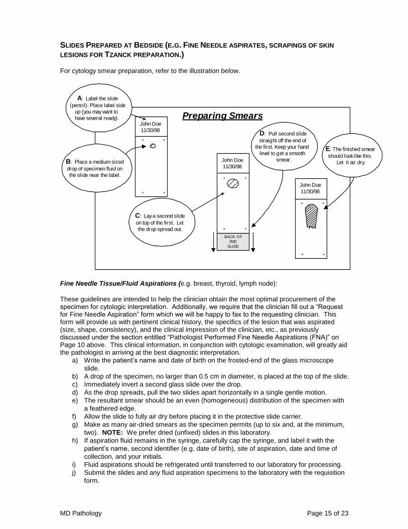
For cytology smear preparation, refer to the illustration below.
Fine Needle Tissue/Fluid Aspirations (e.g. breast, thyroid, lymph node):
These guidelines are intended to help the clinician obtain the most optimal procurement of the specimen for cytologic interpretation. Additionally, we require that the clinician fill out a “Request for Fine Needle Aspiration” form which we will be happy to fax to the requesting clinician. This form will provide us with pertinent clinical history, the specifics of the lesion that was aspirated (size, shape, consistency), and the clinical impression of the clinician, etc., as previously discussed under the section entitled “Pathologist Performed Fine Needle Aspirations (FNA)” on Page 10 above. This clinical information, in conjunction with cytologic examination, will greatly aid the pathologist in arriving at the best diagnostic interpretation.
a) Write the patient’s name and date of birth on the frosted-end of the glass microscope
slide.
b) A drop of the specimen, no larger than 0.5 cm in diameter, is placed at the top of the slide.
c) Immediately invert a second glass slide over the drop.
d) As the drop spreads, pull the two slides apart horizontally in a single gentle motion.
e) The resultant smear should be an even (homogeneous) distribution of the specimen with
a feathered edge.
f) Allow the slide to fully air dry before placing it in the protective slide carrier.
g) Make as many air-dried smears as the specimen permits (up to six and, at the minimum,
two). NOTE: We prefer dried (unfixed) slides in this laboratory.
h) If aspiration fluid remains in the syringe, carefully cap the syringe, and label it with the
patient’s name, second identifier (e.g. date of birth), site of aspiration, date and time of
collection, and your initials.
i) Fluid aspirations should be refrigerated until transferred to our laboratory for processing.
j) Submit the slides and any fluid aspiration specimens to the laboratory with the requisition
form.
John Doe
11/30/98
+ +
Preparing Smears
+ +
A: Label the slide
(pencil). Place label side
up (you may want to
have several ready).
C: Lay a second slide
on top of the first. Let
the drop spread out.
John Doe
11/30/98
+ +
+ +
John Doe
11/30/98
+ +
+ +
BACK OF
2ND
SLIDE
D: Pull second slide
straight off the end of
the first. Keep your hand
level to get a smooth
smear.B: Place a medium sized
drop of specimen fluid on
the slide near the label.
E: The finished smear
should look like this.
Let it air dry.

MD Pathology Page 16 of 23
5) Tzanck preparations to assess for herpesviruses: a) Write patient’s name and date of birth on frosted-end glass microscope slide.
b) A fresh, clear vesicle should be selected. Old, crusted lesions are unsatisfactory. c) The vesicle should be unroofed, and the base scraped with a small scalpel. d) The scrapings from the blade should be immediately placed at the top of a clean, labeled
slide. e) Immediately invert a second glass slide over the drop. f) As the drop spreads, pull the two slides apart horizontally in a single gentle motion. g) The resultant smear should be an even (homogeneous) distribution of the specimen with
a feathered edge. h) Allow the slide to fully air dry before placing it in the protective slide carrier. i) Make as many air-dried smears as the specimen permits (up to six and, at the minimum,
two). NOTE: We prefer dried (unfixed) slides in this laboratory. j) Submit specimen to the laboratory with the requisition form. No refrigeration is
necessary.
If there are any questions about appropriate handling, do not hesitate to contact MD Pathology.
VIRAL STUDIES
Viral culture or antigen assays are the recommended studies for identifying viruses. While less sensitive and specific, cytologic examination is a useful and rapid adjunct in certain clinical situations. If cytologic examination is desired, specimens should be submitted as described in the preceding sections. A separate specimen must be submitted for viral culture or antigen testing and should be submitted directly to an alternate laboratory.
Specimen Rejection
1) Specimens not submitted according to this protocol may be rejected as inappropriate. Specifically, specimens submitted without proper labeling, without a complete requisition (including appropriate history), and without appropriate preservation will be subject to rejection. The laboratory will retain rejected specimens and the submitting physician will be contacted promptly. If the problem may be rectified, processing will commence upon resolving the cause for rejection.
2) Specimens with sparse cellular material, excess blood contamination, excessive clotting, or excessively thick smears may be unsatisfactory (or suboptimal) for examination or interpretation. In such instances, a written report stating that the specimen is unsatisfactory will be issued and sent to the submitting physician.
Specimen Processing
Liquid and semisolid specimens will be processed at the discretion of the pathologist to yield optimal results, and may include such techniques as cytocentrifuge preparations, cell blocking, direct smearing or monolayer techniques.
Pathological Examination and Reporting
1) Routine cytologic processing occurs Monday through Friday. Specimens received in the laboratory prior to 4:30 p.m. will be processed that evening and will be available for interpretation the following working day.
2) A pathologist will examine all specimens.
3) The same guidelines for reporting as stated for surgical pathology apply to cytology specimens.

MD Pathology Page 17 of 23
Flow Cytometry
The MD Pathology Flow Cytometry Laboratory performs Leukemia/Lymphoma
Immunophenotyping assays for clinical diagnostic testing. Clinical disorders or scenarios for
which Flow Cytometry may be helpful include:
Evaluating lymphocytosis of undetermined etiology.
Identifying B- and T-cell lymphoproliferative disorders involving blood and bone marrow.
Distinguishing acute lymphoblastic leukemia (ALL) from acute myeloid leukemia (AML).
Immunologic subtyping of ALL.
Distinguishing reactive lymphocytes and lymphoid hyperplasia from malignant lymphoma.
Phenotypic subclassification of B- and T-cell lymphoproliferative disorders.
Recognizing monoclonal plasma cells.
Diagnostic hematopathology has become an increasingly complex subspecialty, particularly with
neoplastic disorders of blood and bone marrow. While morphologic assessment of blood smears,
bone marrow smears, and tissue sections remains the cornerstone of lymphoma and leukemia
diagnosis and classification, immunophenotyping is a very valuable and important complementary
tool.
Cellular immunophenotyping, characterizing cells by using antibodies directed against cell surface
markers, is generally regarded as a fundamental element in establishing a diagnosis of tissue
involvement by hematolymphoid malignancies, when used in conjunction with morphologic
assessment. It is also an essential component in subclassification of hematolymphoid
malignancies, when present.
This test is not appropriate for and cannot support diagnoses of sarcoidosis, hypersensitivity
pneumonitis, interstitial lung diseases, or differentiating between pulmonary tuberculosis and
sarcoidosis (requests for CD4/CD8 ratios). This test is not appropriate for quantitative lymphocyte
subset enumerations or quantitative CD34 enumerations. Specimens sent for these purposes will
be rejected.
Specimens submitted for evaluation for leukemia, lymphoma, plasma cell neoplasms, and
myelodysplasia are appropriate to send for Flow Cytometry.
Requisitions
All specimens submitted for Flow Cytometry must have an accompanying requisition form,
including either an MD Pathology Hematology requisition form for blood and bone marrow
specimens, or an anatomic pathology requisition form for tissue and cytology specimens. If the
specimen is a tissue or cytology specimen, special note should be made if leukemia or lymphoma
is suspected, or if Flow Cytometry is specifically requested. As with other specimens, the
following information should be included: patient name and date of birth, pertinent clinical history,
clinical or morphologic suspicion, specimen source, i.e. Peripheral Blood, Bone Marrow Aspirate,
etc, and date and time of collection.
Specimen Requirements
Flow cytometric analysis can be performed on anti-coagulated peripheral blood and bone marrow
aspirates, as well as tissues or body fluids.

MD Pathology Page 18 of 23
Submit one of the following specimens:
1) Blood
Container/Tube: Green top (Sodium Heparin) or Purple top (EDTA)
NOTE: Lithium Heparin Green Top tubes are NOT acceptable. The anti-coagulant must
be Sodium Heparin. Lithium Heparin samples will be rejected.
Specimen Volume: 2-5 mL; minimum volume 2 mL
Collection Instructions:
Do not transfer blood to other containers.
Include 1 to 2 unstained EDTA blood smears, if possible. If unable to provide
EDTA blood smears, then send EDTA tube to laboratory for smears to be made.
Specimen should be labeled with patient name, date of birth, “Peripheral Blood”
and date and time of collection.
Specimen Storage: Room temperature, do not refrigerate, do not allow specimen to
freeze.
2) Bone Marrow
Container/Tube: Green top (Sodium Heparin) or Purple top (EDTA)
Specimen Volume: 1-5 mL; minimum volume 1 mL
Collection Instructions:
Submission of bilateral specimens is not required.
Include 5 to 10 unstained bone marrow aspirate smears, if possible. If unable to
provide smears, then send EDTA (purple top) bone marrow aspirate to laboratory
for smears to be made.
Specimen should be labeled with patient name, date of birth, “Bone Marrow
Aspirate” and date and time of collection.
Specimen Storage: Room temperature, do not refrigerate, do not allow specimen to
freeze.
Additional Information:
Label specimen as bone marrow, with the date and time of collection.
If cytogenetic, FISH or molecular tests are also desired, an additional specimen
should be submitted. It is important that the specimen be obtained, processed,
and transported according to instructions for the other required tests.
3) Body Fluid (e.g. Serous Effusions)
Container/Tube: Body fluid container or body fluid transferred into Sodium Heparin
(green-top) or EDTA (purple) blood collection containers.

MD Pathology Page 19 of 23
Specimen Volume: 20 mL; minimum volume 5 mL
Collection Instructions:
The volume of fluid necessary to phenotype the lymphocytes or blasts in serous
effusions depends upon the cell count in the specimen. Usually 20 mL of pleural
or peritoneal fluid is sufficient. Smaller volumes can be used if there is a high cell
count. When a sample is very paucicellular, analysis may not be successful.
Label specimen with patient name, date of birth, specimen source, date and time
of collection
Specimen Storage: Refrigerate. Do not allow specimen to freeze.
4) Spinal Fluid
Container/Tube: Sterile vial
Specimen Volume: 1-5 mL; minimum volume 1 mL
Collection Instructions:
Spinal fluid specimen should arrive within 48 hours of collection.
The volume of fluid necessary to phenotype the lymphocytes or blasts in spinal
fluid depends upon the cell count in the specimen. Usually 3-8 mL of spinal fluid
is sufficient. Smaller volumes can be used if there is a high cell count. If cell count
is <10 cells/uL, a larger volume of spinal fluid may be required. When cell counts
drop below 5 cells/uL, the immunophenotypic analysis may not be successful.
Label specimen with patient name, date of birth, “CSF” and date and time of
collection.
Specimen Storage: Refrigerate. Do not allow specimen to freeze.
5) Tissue
Container/Tube: Sterile container with 15 mL of tissue culture medium (RPMI is
preferred to maintain cell survival. Sterile Saline or Hank’s Balanced Salt Solution can be
used if sample can be sent immediately)
Specimen Volume: 5 mm3 or larger biopsy; minimum volume 1 mm3
Collection Instructions:
Specimen is best when placed in RPMI solution, and received within 24 hours of
surgery. If unable to be sent within that time frame, the sample should be minced
and refrigerated until it can be delivered. Do not fix in Formalin.
Samples should be no older than 48 hours old when received.
If sample is unable to be placed in RPMI, then it should be delivered within 12
hours of surgery to maintain cell survival.

MD Pathology Page 20 of 23
Additional Information:
A pathology/diagnostic report and/or a brief history should be provided.
If, in the opinion of the MD Pathology Hematopathologist, the tissue is not
adequate for flow cytometric immunophenotyping, the specimen may be rejected.
Specimen Packaging and Labeling
Special MD Pathology collection kits are available for blood and bone marrow specimens. Please
call 972-981-3107 for questions regarding the availability of such kits. Tissue and fluid specimens
should be submitted WITHOUT FIXATIVE as specified under the Surgical Pathology and
Cytopathology sections.
Specimen Rejection
Specimens not submitted according to this protocol may be rejected as inappropriate, as
mentioned in prior sections. Additionally, specimens will be rejected for Flow Cytometry if the
specimen is submitted in Formalin or B5 fixative, is frozen, shows gross hemolysis, or is a
peripheral blood greater than 72 hours old. Mild hemolysis is acceptable. Lipemia or icterus is
acceptable.
Specimen Stability Information
Blood and Bone Marrow samples should be stored at ambient temperatures. Tissues and Body
Fluids should be refrigerated with RPMI or other approved solutions.
Avoid extreme temperatures (hot/cold). Specimens should be shipped with cold packs whenever
possible to prevent dramatic changes of temperature for all samples, even if the recommended
storage is at room temperature. Samples that are transported to MD Pathology from a location on
the Texas Health Plano campus do not require cold packs. Specimens should be analyzed within
24 hours of collection, if possible.
Interpretation / Reference Values This test will be processed as a laboratory consultation. An interpretation of the
immunophenotypic findings will be provided by a hematopathologist for every case. The report
will include an immunophenotypic description of cells present in the sample analyzed and an
interpretive conclusion based upon the correlation available data including clinical history,
morphologic features, and immunophenotypic results.
Cautions
Specimens will be initially triaged to determine which, if any, of the immunophenotyping panels
should be performed.
Viability will be assessed on all tissue and fluid specimens. In addition, viability will be assessed
on all blood and bone marrow aspirate specimens more than 24 hours old. Cases with viability of
less than 80% are considered suboptimal. Cases in which the viability is <50% are prone to false-
negative results and, therefore, must be interpreted with caution.
Cerebrospinal fluid (CSF), fine needle aspiration, and small biopsy specimens have a higher
frequency of insufficient specimens due to low cell counts and/or poor viability.

MD Pathology Page 21 of 23
For tissue specimens, it is well recognized that a negative flow cytometry result does not exclude
tissue involvement by hematolymphoid malignancy. This may be attributable to sampling bias,
although some malignancies, such as Hodgkin lymphoma, typically are not detected by this
technique. The tissue used for flow cytometry cannot be subsequently submitted for
histopathologic evaluation. For this reason, this technique should be avoided in small biopsy
specimens.
Method Description
A request for leukemia immunophenotyping initiates a process in the laboratory to determine the
most appropriate and cost-efficient immunophenotyping antibody panel. Panel selection is based
upon clinical history and the 2006 Besthesda Guidelines to determine which lineages should be
evaluated. Immunophenotyping is performed by multi-color flow cytometry using monoclonal
antibodies directly conjugated with different fluorochromes. Based upon the findings in this initial
panel, additional testing may be required and will be added to fully characterize disease states
with a charge per unique antibody tested.
If no abnormalities are detected by the initial panel, no further flow cytometric assessment will be
performed unless otherwise indicated by specific features of the clinical presentation or prior
laboratory results. In some instances, additional cytogenetic or molecular genetic studies may be
indicated by the immunophenotyping results. In these cases, the ordering physician will be notified
verbally by the pathologist interpreting the case.
Specimens are processed and reported Monday through Friday, with a typical analytic time of 1
working day. Results are typically available 1 business day after the sample is received in the
laboratory. Results from STAT samples may be available the same day that the sample is
received in the testing laboratory.
Any remaining specimen is retained in the laboratory for 7 days.

MD Pathology Page 22 of 23
MD Pathology Quick Reference
Michael A. Deck, M.D. Rocio P. Calle-Rodrigue, M.D.
David L. Ring, M.D. Benjamin J. Williams, M.D.
Imran Shahab, M.D. Rachel Rucker-Schmidt, M.D. Christopher M. Schuler, M.D.
MD Pathology is an Anatomic Pathology Laboratory providing surgical pathology and cytology services for physician offices and surgery centers in Plano, Frisco, Allen, North Dallas, Carrollton and surrounding communities. Pathologist performed fine needle aspirations of palpable masses are available by appointment. MD Pathology is fully accredited by the College of American Pathologists (CAP # 48360-01) and is registered with CLIA (45D0881398). Our goal is to provide the highest quality services in a friendly, cost effective and timely fashion.
Submitting Specimens
1) Following these instructions will help insure the most timely and accurate results for your patients. Routine tissue specimens should be fixed in Formalin solution as soon as collected. Cytology specimens (usually fluids) and specimens for Flow Cytometry should be submitted fresh with no fixation added. Allow cytology slides to air dry. Label each specimen container (on the side, not the lid) or slide (on the frosted end in pencil) with the patient’s name, date of birth, date and time of collection, collector’s initials, and anatomic site of the specimen.
2) Complete a requisition to include patient identifying information, age, sex, pertinent clinical information and list each specimen submitted.
3) Please be certain to include billing information or complete the billing section on the requisition. Please note that the only billing option available is directly to the patients or their insurance plan.
4) Place the specimen(s) into a biohazard bag, with the requisition in the outer compartment. Place the biohazard bag at the pick-up point.
5) If your site uses “on-call” pick-up services, notify the courier (see next section) that you have a specimen to be picked up.
Please refer to the “Specimen Collection Manual and User’s Guide” for more detailed information.
Contacting MD Pathology
Reason for Call Number Notes Routine Specimen Pick-Up 8:00 a.m. to 5:00 p.m.
(972) 981-3107 If you get voice mail, please indicate that a specimen is to be picked up for MD Pathology, the place of pick-up, and the latest time that someone will be available.
Fine Needle Aspiration Biopsy
(972) 981-3107 If you get voice mail, please leave a detailed message.
Frozen Sections, Fresh Specimens, Reports, Questions, or to contact a Pathologist
(972) 981-3107 If no answer, follow the instructions on the voice mail message for contacting the pathologist on-call.
Supplies
(972) 981-3107 If you get voice mail, indicate the supplies needed and the name of the account.
Histology/Cytology Lab (972) 981-3108 (972) 981-3236 Fax
Histology/Cytology is staffed from 2:00 a.m. to 5:00 pm. After hours leave voice mail or call (972) 981-3107.
Other information (972) 981-3107 If you have questions not covered in this manual, please contact the pathologist on call.
MD Pathology Page 23 of 23
Appendix: Handling Formalin
INTRODUCTION
Formalin is a 10% solution of formaldehyde in a buffered aqueous solution. It is a hazardous substance and potent irritant. It is also a known carcinogen. Formalin must be used in a well-ventilated area. The rule of thumb is if you can smell it, the concentration in the air is too high and there is a problem. Notify a supervisor if you smell any fumes. It should be kept over a sink or with a drip pan under the spout to catch a spill. It is important to keep your face a safe distance away from the formalin when using it. Wear gloves and protective eye gear when handling or dispensing formalin.
FIRST AID IN CASE OF CONTACT
If you should come in contact with formalin, wash with soap and water immediately. If it gets in your eyes, flush with water immediately or use an eye wash.
DISPENSING AND OPENING CUBITAINERS
When opening the container, place the box on the counter with the lid towards the ceiling. Gently pull the cap off and remove the screw cap and screw on the spout, making sure the valve is in the “off” position. The “off” position on the valve is with the handle to the right side; rotating it to the left will open the flow. Place the box with the spout pointing toward the floor over the drip pan or sink where it will be stored.
If the container will be kept on the counter with the spout hanging over the counter, place a drip pan filled with either kitty litter or diatomaceous earth, or an absorbent pad directly below. Keep an approved spill kit on hand should a spill occur.
FORMALIN SPILLS
If a moderate amount of formalin should spill on the floor locate the formalin spill kit. Spread enough of the granules over the entire area and let it soak for 10 minutes. The spill kit material reacts chemically with the formaldehyde to neutralize it. If possible close the door. Make sure to let all necessary personnel know about the spill and notify a supervisor. Using a dustpan and
broom, collect and place the absorbed spill kit into a biohazard bag and discard appropriately. If
the formaldehyde fumes are very strong or for large spills, close the door to contain the
fumes and notify your supervisor. DO NOT ENTER THE ROOM – call Haz-mat
IMMEDIATELY!
DISPENSING FORMALIN ONTO SPECIMENS
Place the specimen in a container large enough to accommodate the specimen and add enough formalin to cover it. A 10:1 ratio of formalin volume to specimen volume is optimal. Make sure the lid is threaded on the container properly and is leak proof. Label the container in the usual way. Place the specimen container in the sealable compartment of the specimen bag and place the requisition and billing sheets in the outside pocket of the specimen bag.