ก กpni.go.th/pnigoth/wp-content/uploads//2010/07/pr1-1.pdf · .. 2549 ก ˇก ˆ ˝˛˚˜ ˛...
TRANSCRIPT


������ก�ก����� ��������������
Clinical Practice Guidelines for Hydrocephalus
+,,-��

������ก�ก����� ���������������.� /01�/�2����2��-�/�3��45���6��ก�,3ก�
78���46����.�/9�� ��ก,��:�ก�; /�2���<6����<�:�7:9��=;>�ก��8��/�3��; �ก8<6
0?@9��46���6����<�:�:-���.0 �3�A3����; �48��-� 68��� ���B-��C >�������ก�ก���.�
<�->D-68�,��,6��ก�0E3,B3 =F8>D8����G0E3,B3�BกB-��<0H�ก68��� ���<78 >�ก5.�.�
�G��ก�5I�BกB-����ก<092��./9B4=;�.�����

������
���� �.�. 2549 �������� ������� ก��ก������� ������� ������!ก����ก"�#���$��! �%!&�'!�( ��)����*+����,����'����*��$��������������*����-'.%�ก)�����,���ก��� ������!/ ��!ก),�����0���(� +1��2���.��-�345�%6� ��&5�7% �&��������6!�ก��8*��'�*.����!���ก�� *�7'%��9�������!ก����ก"��-(�5� ��1�:��)���� ��� *�;����$�0���ก,�������ก�������<���)��1�)��ก"��-'�-�)��-��� ��=�#����ก8>(� ��!��(� �>!*5?� �&���5��-ก����9��������6!�)����� ������!/ *�7'%�5������+1�*ก-'��8�%! ������ ����0�5�7%�@���.��5�*ก�����$�0��������!�� ���ก��:��3��6���
#���$��! �%!&�'!�( � (Hydrocephalus) *ก��8>(����ก��+1��P���6กก)6,�%��6 Q>'!�- �*5.6����ก&���+���ก.�8%! �%!�.,ก �*��� *�7(%!%ก5�7%�6!�( ��� �%! ก��%�ก* �8%!*�7'%56�� �%! *)7%�%%ก��0�(��.�*�7'%56�� �%! 5�7%#��� �%!&�'!�( �8:�&������ �%!�ก.�
�������� ���������R��� �����0�(�� �������0�ก��$�&������� ���)�*�;�+1�� �������ก������ ������!ก����ก"�����$�&������� ��$��������ก���%�����������0�.� �>!*5?�&���5��-ก����9��������6!�)����� ������!ก����ก"�#���$��! �%!&�'!�( �8>(� *�7'%�5������+1�*ก-'��8�%! ������ ����0�5�7%� ����@���.��5�*ก�����$�0��������!�� ���ก��:��3��6���

�5 ���������ก�H7���������ก�ก����� ��������������
1. ��.���=�0 ��* � �-'��>ก"� 2. �4.ก�)��:� =-�����1)�� �-'��>ก"� 3. ��. ��� .�(!%�6:��)�U �-'��>ก"� 4. ��. ��! �3���� -5� ���=��$&�!ก�� 5. ��.0,%*�-�� *.$0V�� &:�� �!�� 6. ��.��.*%ก 5�! 1. &:�� �!�� 7. ��.�4.����� *5),�=�������� &:�� �!�� 8. ��.*ก�-�!��ก��Z �Q,*.-� &:�� �!�� 9. ��.0�&������ : ��!0��! &:�� �!�� 10. ��. ���. ��-%6��8�� &:�� �!�� 11. ��.#�:6 ����6! &:�� �!�� 12. ��.��*0"R *�=���ก"�0-� &:�� �!�� 13. �4.%�#���- )6 �� ��Z &:�� �!�� 14. �.�.=-�� .�(!���������1)�� &:�� �!�� 15. �.�.%�6��ก��Z *)-�!%6�� &:�� �!�� 16. �.�.���* ��R *%-'����-0�ก6) &:�� �!�� 17. �.�.�69��!"� R��$\�� &:�� �!�� 18. �.�.�6=�0�� � � ���.� &:�� �!�� 19. �.4. 6��!&� *�-������� &:�� �!�� 20. ����*%ก%���� ��'���) &:�� �!�� 21. ��.��9�0�� $0.������.�ก6) &:�� �!�� 22. ��.�4.�����: * ��:�!&� &:�� �!�� 23. ��.�!"���9�� �)�!"� &:�� �!�� 24. ��.ก6)��9�� �-� �� *)8��6ก�� 25. �. .%� �- .�-ก�) +1�0,��*)8��6ก�� 1

��,@ 5���
& ����� & �� � ������&:�� �!�� .���,% �����& ����� �%�!&6:#�� (Strength of recommendation) .��5)�กR����!��0�ก�� ���-' 1 �����!ก�������<���)�� ������ก"�+1��P���-' ! ���-#���$��! �%!&�'!�( � 1 (Diagnosis and Management of patients suspected of hydrocephalus ) ���-' 2 �����!ก�������<��8%!#���$��! �%!&�'!�( ���*�?ก 9 ���-' 3 �����!ก�������<�� Normal pressure hydrocephalus 12 ���-' 4 �����!ก����ก"�+1��P���-'�-#���$��! �%!&�'!�( � 14 ���-' 5 �����!ก�� ,!��>ก"� 16 ���-' 6 �����!ก��� ������ก"�5)�!+,�.�� CSF diversion 17 *%ก ��%��!%�! 21 #�&+��ก #�&+��ก�-' 1 %�ก��� �!8%!#���&��������ก�$5)ก�-�"� 1!��*�?ก 23 #�&+��ก�-' 2 Growth record for premature infants in relation to gestational age and fetal 24 and infant norms (combined sexes) #�&+��ก�-' 3 �+�#1��� �!&,�* ���%��!�-�"� (occipito-frontal circunference; OFC) 25 ��*�?ก%��6.�(!�.,��ก*ก���>! 3 �� #�&+��ก�-' 4 ����� %���9��ก�� Denver II <���#�"���� 26 #�&+��ก�-' 5 Type of shunt 28 #�&+��ก�-' 6 #���,����! -��+1��P���-'�-#���$��! �%!&�'!�( � 29
��,@B���
.���!�-' 1 %�ก���)�%�ก��� �!8%!&������ก�$5)ก�-�"� 1! 5 �)� �*5.6�-'���,%�8%!#���$��! �%!&�'!�( � ��,!.��ก)6,�%��6 .���!�-' 2 Etiology of macrocephaly 7

��,@�=��F�3 5���
�+�#1���-' 1 �����!ก�������<��+1��P���-'�-#���$��! �%!&�'!�( � 3 �+�#1���-' 2 �����!ก�������<��+1��P���-'�-#���$��! �%!&�'!�( ���*�?ก 9 �+�#1���-' 3 �����!ก�������<�� Normal pressure hydrocephalus 12 �+�#1���-' 4 �����!ก����ก"�+1��P���-'�-#���$��! �%!&�'!�( � 14 �+�#1���-' 5 �����!ก��� ������ก"�5)�!+,�.�� CSF diversion 17 �+�#1���-' 6 �����!ก��� ������ก"�+1��P���-' ! ���,��-#���$��! �%!&�'!�( ��,��ก�� 35 &��������ก�$5)ก�-�"� 1! �+�#1���-' 6.1 �����!ก��� ������ก"�+1��P���-'�-#���$��! �%!&�'!�( ���กก��%6�.��8%! 37 ����$��! �%! �+�#1���-' 6.2 �����!ก��� ������ก"�+1��P���-'�-#���$��! �%!&�'!�( ���กก��%6�.���( � 38 �8 ��5)�!�-'. ��5�,! foramen of Monro �+�#1���-' 6.3 �����!ก��� ������ก"�+1��P���-'�-#���$��! �%!&�'!�( ���กก��%6�.�� 40 �-'. ��5�,!.' �ก�,� third ventricle
��,@F0���
�1��-' 1 macrocephaly , setting sun sign, bulging anterior fontanel, dilated scalp vein (�1�����5���) 23 �1��-' 2 macrocephaly , setting sun sign, bulging anterior fontanel, dilated scalp vein (�1�����8��!) 23 �1��-' 3 �+�#1��� �!&,��ก.�8%!ก��*���4*.��$.8%!���ก�-'&)%�ก,%�ก �5�����>!%��6 1 �� 24 �1��-' 4 �+�#1��� �!&,�* ���%��!�-�"� (occipito-frontal circunference; OFC) 25 ��*�?ก%��6.�(!�.,��ก*ก���>! 3 �� �1��-' 5 Type of shunt 28 �1��-' 7 Burr hole shape 30 �1��-' 8 #�� MRI of HCP � �! dilatation of the lateral ventricle with 30 periventricular effusion �1��-' 9 #�� CT of HCP � �! symmetrical dilatation of all ventricles 31 �1��-' 10 MRI sagittal T2 � �! Flow void sign (signal void) in the aqueduct 32 of Sylvius in NPH �1� 11 #�� CT of NPH � �! dilatation of the ventricle disproportion to the sulci 33 �1� 12 #�� MRI of NPH � �! rounding of bilateral frontal horns with 34 periventricular effusion �1� 13 #�� CT of obstructive HCP from bleeding at left thalamus with secondary 34 intraventricular hemorrhage (IVH) �1� 14 #�� MRI of obstructive hydrocephalus from obstruction at posterior 29 part of 3rd ventricle with marked dilatation of the lateral ventricles
B�:-�
CNS = Central nervous system CSF = Cerebrospinal fluid CT = Computerized tomography DDST = Denver developmental screening test EVD = External ventricular drainage HCP = Hydrocephalus ICP = Increased intracranial pressure IVH = Intraventricular hemorrhage LP = Lumbar puncture MRI = Magnetic resonance imaging MRS = Magnetic resonance spectroscopy MRV = Magnetic resonance venography NPH = Normal pressure hydrocephalus PHH = Post hemorrhagic hydrocephalus US = Ultrasound
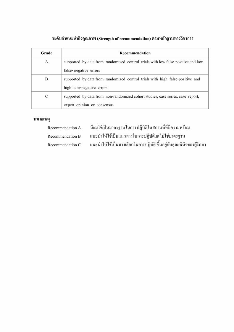
7,����� ����3��45��� (Strength of recommendation) B��9;กO������3D�ก�
Grade Recommendation
A supported by data from randomized control trials with low false-positive and low false- negative errors
B supported by data from randomized control trials with high false-positive and high false-negative errors
C supported by data from non-randomized cohort studies, case series, case report, expert opinion or consensus
9��:/9B4
Recommendation A �����0�*�;���.�R����ก���@���.��� ����-'�-'�-&������%� Recommendation B ���� ��5��0�*�;������!��ก���@���.��.,��,�0,��.�R�� Recommendation C ���� ��5��0�*�;���!*)7%ก��ก���@���.� 8>(�%�1,ก���6)������8%!+1���ก"�

1
����� 1
����ก ��������������� �ก������������������� ! ���"�#����$� (Diagnosis and management of patients suspected of hydrocephalus )
��������� !�"#$�%&'� (hydrocephalus) ()*%����+,$�-./01%230)4�5+6กก869 !�56 �/5 ,!6-#:;ก��<=�3�1%(/>ก ����%,&?'�@%ก:� �5�A;��,��;+5�ก8.กก��(ก;/ /#�%,&
1. ,ก����0��%&'�.D�#%E8#� (CSF) �ก2;/)ก:; (increase CSF secretion) 2. ,ก��!6/:#%+��(/;%%&'�E89!� !�@8�.D�#%E8#� (CSF pathway obstruction) 3. ,"�� 2;/)ก:;D!�ก��/3/MN ก8#-D!�%&'�.D�#%E8#�+��E8!/(8O!//'� (sagittal sinus)
(decreased CSF absorption)
#�����ก%����ก&"'(�)ก���� ! ���"�#����$�%�!�*�� � ��� ./0@ก9 1. ก����0�� CSF �ก(ก;% (P9% (%O&!�!กD!� Choroid plexus (choroid plexus papilloma) 2. ก��!6/:#%+��(/;%%&'�E89!� !�@8�.D�#%E8#� @-9�()*% 2 @--
2.1 Obstructive hydrocephalus E�O! Non communicating hydrocephalus ,ก��!6/:#%��E�9������� !�ก#-P9!�1:0(5O$!E60 � !�@8�.D�#%E8#� (subarachnoid space) ��(E:6 ,./0E8�5!59�� (P9% (%O&!�!ก� !� (8O!/!!ก1%����� !�@8�(%O&!� !� "�� �;ก��@:9ก'�(%;/ (aqueductal stenosis) ก��:;/(PO&! (P9% �5�A;:O/E 31%� !� (neurocysticcercosis) ()*%:0% ,"�� @:ก:9����E�9��"�� /#%1%����� !�ก#-P9!�1:0(5O$!E60 � !�@8�.D�#%E8#� (subarachnoid space) E�ก ,ก��(?��E8#�(�O$!��-�5%&'�E89!� !�@8�.D�#%E8#�?�+'�1E0(ก;/"�� @:ก:9����E�9��"�� /#%1%ก��E8กV,�W�@8�P9!�.D�#%E8#�+'�1E0(ก;/ก��("8O$!%D!�� !��9�% Cerebellum 29�% Foramen of magnum ก/ก0�%� !��9�281E0230)4�5(�,5P,�;: ?N�()*%D0!E0� 1%ก��(?��E8#� (lumbar puncture)
2.2 Communicating hydrocephalus ,ก��:;/:9!��E�9������� !�@8�P9!�1:0(5O$!E60 � !� (subarachnoid space) ก��!6/:#%(ก;/ #ก(ก;/DN&%%!ก����� !�+,$P9!�1:0(5O$!E60 � !� (subarachnoid space :Cistern) D!�� !� .D�#%E8#� @8� arachnoid villi ��(E:6+,$�--9!5+,$�6/"O!(8O!/!!ก1:0P9!�(5O$!E60 � !� (subarachnoid hemorrhage) @8�ก��:;/(PO&!D!�(5O$!E60 � !� ��(E:6!O$%Y+,$. 91P9ก��!6/:#%1%����� !� (P9% ก����0��E�O!ก��/3/MN %&'�E89!� !�@8�.D�#%E8#�2;/)ก:; 3. ก��/3/MN 2;/)ก:; ��(E:6?�ก ก��!6/:#%E8!/(8O!//'� (venous sinus thrombosis) E�O!ก��!#ก(�- arachnoiditis ?�กก��:;/(PO&!E�O!(8O!/!!ก ก9!1E0(ก;/ communicating hydrocephalus

2
�)(%+ (etiology) !�?@-9�ก869 ��(E:6/#�%,& 1. "�� 2;/)ก:;@:9ก'�(%;/ (congenital anomaly) ��(E:6+,$�--9!5+,$�6/ "O! aqueductal stenosis
��(E:6!O$%Y (P9% congenital hydrocephalus, myelomeningocoele, Dandy walker syndrome, Chiari malformation, intrauterine infection, perinatal hemorrhage
2. ��":9��Y (acquired disease) -�/(?>-+,$V,�W� (traumatic brain injury / traumatic subarachnoid hemorrhage ) ��"E8!/(8O!/� !� (CVA) ก��:;/(PO&! (CNS infection) (%O&!�!ก� !� (brain tumor)
3
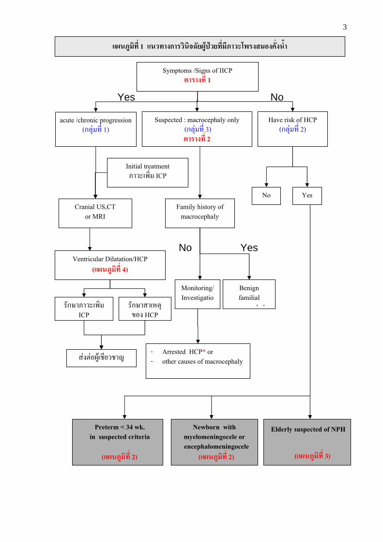
Symptoms /Signs of IICP % ���� 1
acute /chronic progression (ก869 +,$ 1)
Suspected : macrocephaly only (ก869 +,$ 3) % ���� 2
Have risk of HCP (ก869 +,$ 2)
Family history of macrocephaly
Monitoring/ Investigatio
n
Benign familial
macrocephaly
- Arrested HCP* or - other causes of macrocephaly
No Yes
Newborn with myelomeningocele or encephalomeningocele
(���������� 2)
Elderly suspected of NPH
(���������� 3)
Preterm < 34 wk. in suspected criteria
(���������� 2)
Initial treatment ����(�;$ ICP
Cranial US,CT or MRI
Ventricular Dilatation/HCP (���������� 4)
�#กW�����(�;$ ICP
�#กW���(E:6D!� HCP
�9�:9!230(P,$5�P�k
No Yes
No Yes
���������� 1 ����ก �������������������� ! ���"�#����$�
4
#�"*������������� 1
����ก �������L"��� ! ���"�#����$� @5ก:� 8#กW<�+��"8;%;ก+,$�'�"#k"O! ,!�ก��E�O!!�ก��@�/�D!�����"�� /#%1%ก��E8กV,�W��3�E�O!. 9
ก869 +,$ 1 ,!�ก��E�O!!�ก��@�/�D!�����"�� /#%1%ก��E8กV,�W��3�MN$�?� ,"�� @:ก:9��ก#%:� ก869 !�56 �/5(l���1%(/>ก (/#�:����+,$ 1) @8�:0!�ก��ก���;%;?l#5@8�ก���#กW�!59����/(�>� /#�@2%�3 ;+,$ 4
ก869 +,$ 2 . 9 ,!�ก��E�O!!�ก��@�/�D!�����"�� /#%1%ก��E8กV,�W��3�@:9()*%ก869 +,$?'�()*%:0!�(no����#���������� !�"#$�%&'� �/5@-9�()*%ก869 !�56 +,$ ,"�� (�,$5�(l��� @8�:0!�ก�� ก��:;/:� !�ก��@8� ก���;%;?l#5:� @%�+��1%@2%�3 ; 2 ./0@ก9
1. (/>ก"8!/ก9!%ก'�E%/+,$ ,����(8O!/!!ก1%� !� (premature baby with IVH) 2. (/>ก@�ก(ก;/+,$ , myelomeningocele E�O! encephalomeningocele 3. 230�3�!�56+,$ ,!�ก��@�/�D!����� NPH (normal pressure hydrocephalus)
ก869 +,$ 3 ,!�ก��@�/�+,$���#5�9�!�? ,����"�� /#%1%ก��E8กV,�W��3� "O! 1%(/>ก+,$ ,D%�/D!�V,�W��:ก�9�"9�)ก:; @:9. 9 ,!�ก��E�O!!�ก��@�/�!O$%YD!�����"�� /#%1%ก��E8กV,�W��3� �;$�+,$"��@5ก"O! 1%ก�<,+,$ ,)���#:;"�!-"�#�V,�W��:E8�5"%@:9. 9 ,!�ก��!O$%+,$2;/)ก:; +'�1E0";/pN����� benign familial macrocephaly MN$�1%ก869 %,&. 9?'�()*%:0!�ก��ก���;%;?l#5(�;$ (:; (�,5�@:91E0"'�@%�%'� %#/:;/:� !�ก��@8��#q%�ก��:� )ก:;(+9�%#&% 1%ก�<,+,$. 9 ,)���#:;"�!-"�#�/#�ก89��!�??�:0!�";/pN�����!O$%Y +,$+'�1E0(ก;/����V,�W��:ก�9�)ก:; (P9% arrested hydrocephalus*, megalencephaly, etc. (/#�:����+,$ 2) MN$�:0!�!�V#5ก��:��?�;%;?l#5(�;$ (:; (�O$!E���(E:6:9!.)
*�� arrested hydrocephalus / non-progressive hydrocephalus "O! 230)4�5+,$ ,��������� !�"#$�%&'�(ก;/DN&%@:9ก��/3/ MN D!�%&'�1%����� !�� /68ก#-����"#$�D!�%&'�1%����� !� 230)4�51%ก869 %,&!�? �/0�5D%�/V,�W��:ก�9�)ก:; @:9. 9 ,!�ก��./0 pN�!59��.�ก>:� ����%,&5#� ,�!ก��+,$?�(ก;/����"�� /#%1%ก��E8กV,�W��3�./0p0���������� !�"#$�%&'�(�;$ �กDN&% ?N�?'�()*%:0!�:;/:� 230)4�5!59��1ก80P;/
5
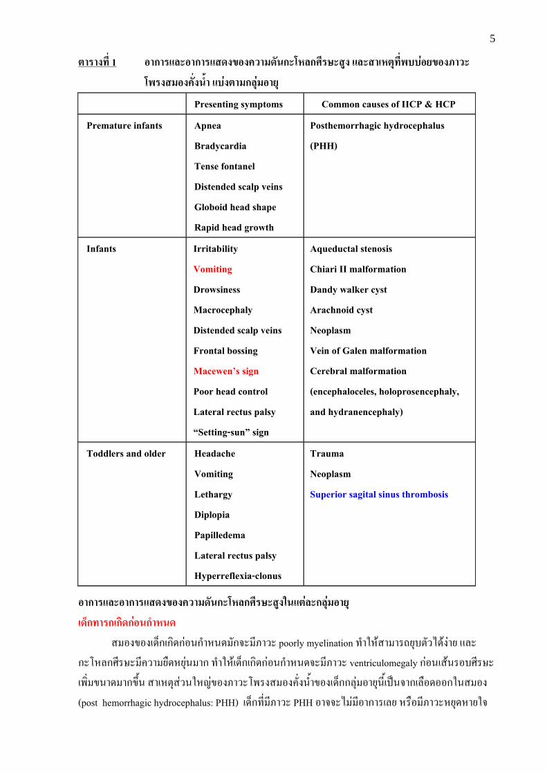
% ���� 1 "ก ���"ก ����L"�#����ก� (�กQ� ����� ����)(%+���!��&"�L"���
! ���"�#����$� ��&�%�ก�+&�"�+
Presenting symptoms Common causes of IICP & HCP
Premature infants Apnea
Bradycardia
Tense fontanel
Distended scalp veins
Globoid head shape
Rapid head growth
Posthemorrhagic hydrocephalus
(PHH)
Infants Irritability
Vomiting
Drowsiness
Macrocephaly
Distended scalp veins
Frontal bossing
Macewen^s sign
Poor head control
Lateral rectus palsy
`Setting-sunb sign
Aqueductal stenosis
Chiari II malformation
Dandy walker cyst
Arachnoid cyst
Neoplasm
Vein of Galen malformation
Cerebral malformation
(encephaloceles, holoprosencephaly,
and hydranencephaly)
Toddlers and older Headache
Vomiting
Lethargy
Diplopia
Papilledema
Lateral rectus palsy
Hyperreflexia-clonus
Trauma
Neoplasm Superior sagital sinus thrombosis
"ก ���"ก ����L"�#����ก� (�กQ� �����'��%&��ก�+&�"�+
)�fก� ก)ก��ก&"�ก�(�� � !�D!�(/>ก(ก;/ก9!%ก'�E%/ #ก?� ,���� poorly myelination +'�1E0�� ��p56-:#�./0�9�5 @8�ก��E8กV,�W� ,"�� 5O/E569% �ก +'�1E0(/>ก(ก;/ก9!%ก'�E%/?� ,���� ventriculomegaly ก9!%(�0%�!-V,�W�(�;$ D%�/ �กDN&% ��(E:6�9�%1Ek9D!���������� !�"#$�%&'�D!�(/>กก869 !�56%,&()*%?�ก(8O!/!!ก1%� !� (post hemorrhagic hydrocephalus: PHH) (/>ก+,$ ,���� PHH !�??�. 9 ,!�ก��(85 E�O! ,����E56/E�51?
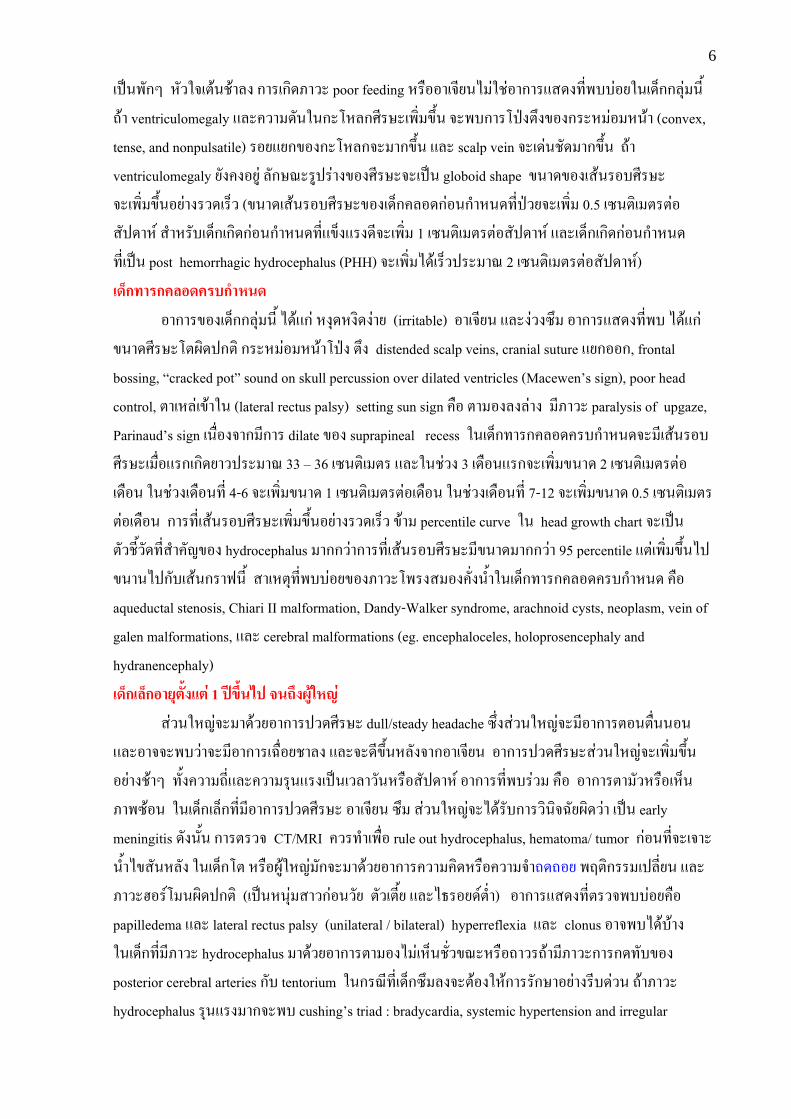
6
()*%�#กY E#�1?(:0%P0�8� ก��(ก;/���� poor feeding E�O!!�(?,5%. 91P9!�ก��@�/�+,$�--9!51%(/>กก869 %,& p0� ventriculomegaly @8�"�� /#%1%ก��E8กV,�W�(�;$ DN&% ?��-ก���)4�:N�D!�ก��E 9! E%0� (convex, tense, and nonpulsatile) �!5@5กD!�ก��E8ก?� �กDN&% @8� scalp vein ?�(/9%P#/ �กDN&% p0� ventriculomegaly 5#�"�!539 8#กW<��3)�9��D!�V,�W�?�()*% globoid shape D%�/D!�(�0%�!-V,�W� ?�(�;$ DN&%!59����/(�>� (D%�/(�0%�!-V,�W�D!�(/>ก"8!/ก9!%ก'�E%/+,$)4�5?�(�;$ 0.5 (M%:;( :�:9!�#)/�E= �'�E�#-(/>ก(ก;/ก9!%ก'�E%/+,$@D>�@��/,?�(�;$ 1 (M%:;( :�:9!�#)/�E= @8�(/>ก(ก;/ก9!%ก'�E%/ +,$()*% post hemorrhagic hydrocephalus (PHH) ?�(�;$ ./0(�>�)�� �< 2 (M%:;( :�:9!�#)/�E=) )�fก� ก#�"�# �ก�(�� !�ก��D!�(/>กก869 %,& ./0@ก9 E�6/E�;/�9�5 (irritable) !�(?,5% @8��9��MN !�ก��@�/�+,$�- ./0@ก9 D%�/V,�W��:2;/)ก:; ก��E 9! E%0��)4� :N� distended scalp veins, cranial suture @5ก!!ก, frontal bossing, tcracked potu sound on skull percussion over dilated ventricles (Macewenvs sign), poor head control, :�(E89(D0�1% (lateral rectus palsy) setting sun sign "O! :� !�8�89�� ,���� paralysis of upgaze, Parinaudvs sign (%O$!�?�ก ,ก�� dilate D!� suprapineal recess 1%(/>ก+��ก"8!/"�-ก'�E%/?� ,(�0%�!-V,�W�( O$!@�ก(ก;/5��)�� �< 33 x 36 (M%:;( :� @8�1%P9�� 3 (/O!%@�ก?�(�;$ D%�/ 2 (M%:;( :�:9!(/O!% 1%P9��(/O!%+,$ 4-6 ?�(�;$ D%�/ 1 (M%:;( :�:9!(/O!% 1%P9��(/O!%+,$ 7-12 ?�(�;$ D%�/ 0.5 (M%:;( :�:9!(/O!% ก��+,$(�0%�!-V,�W�(�;$ DN&%!59����/(�>� D0� percentile curve 1% head growth chart ?�()*%:#�P,&�#/+,$�'�"#kD!� hydrocephalus �กก�9�ก��+,$(�0%�!-V,�W� ,D%�/ �กก�9� 95 percentile @:9(�;$ DN&%.)D%�%.)ก#-(�0%ก��|%,& ��(E:6+,$�--9!5D!���������� !�"#$�%&'�1%(/>ก+��ก"8!/"�-ก'�E%/ "O! aqueductal stenosis, Chiari II malformation, Dandy-Walker syndrome, arachnoid cysts, neoplasm, vein of galen malformations, @8� cerebral malformations (eg. encephaloceles, holoprosencephaly and hydranencephaly) )�fก)�fก"�+%�$��%& 1 �gLh$�i� ��jh����'(k& �9�%1Ek9?� �/0�5!�ก��)�/V,�W� dull/steady headache MN$��9�%1Ek9?� ,!�ก��:!%:O$%%!% @8�!�??��-�9�?� ,!�ก��(lO$!5P�8� @8�?�/,DN&%E8#�?�ก!�(?,5% !�ก��)�/V,�W��9�%1Ek9?�(�;$ DN&%!59��P0�Y +#&�"�� p,$@8�"�� �6%@��()*%(�8��#%E�O!�#)/�E= !�ก��+,$�-�9� "O! !�ก��:� #�E�O!(E>%���M0!% 1%(/>ก(8>ก+,$ ,!�ก��)�/V,�W� !�(?,5% MN �9�%1Ek9?�./0�#-ก���;%;?l#52;/�9� ()*% early meningitis /#�%#&% ก��:��? CT/MRI "��+'�(�O$! rule out hydrocephalus, hematoma/ tumor ก9!%+,$?�(?��%&'�.D�#%E8#� 1%(/>ก�: E�O!2301Ek9 #ก?� �/0�5!�ก��"�� ";/E�O!"�� ?'�p/p!5 �~:;ก�� ()8,$5% @8� �����!�=� %2;/)ก:; (()*%E%69 ���ก9!%�#5 :#�(:,&5 @8�.A�!5/=:$'�) !�ก��@�/�+,$:��?�--9!5"O! papilledema @8� lateral rectus palsy (unilateral / bilateral) hyperreflexia @8� clonus !�?�-./0-0�� 1%(/>ก+,$ ,���� hydrocephalus �/0�5!�ก��:� !�. 9(E>%P#$�D<�E�O!p���p0� ,����ก��ก/+#-D!� posterior cerebral arteries ก#- tentorium 1%ก�<,+,$(/>กMN 8�?�:0!�1E0ก���#กW�!59���,-/9�% p0����� hydrocephalus �6%@�� �ก?��- cushingvs triad : bradycardia, systemic hypertension and irregular
7
breathing pattern and autonomic dysfunction p0��- cushing triad @�/��9� ,"�� /#%ก��E8กV,�W��3� �ก?'�()*%:0!�1E0ก���#กW�!59���,-/9�% ��������� !�"#$�%&'�+,$(ก;/DN&%1%ก869 !�56%,& ,��(E:6 �9�%1Ek9?�ก trauma, neoplasm
% ���� 2 Etiology of macrocephaly
• Achondroplasia
• Benign familial macrocephaly
• Cranioskeletal dysplasia
• Craniosynostosis
• Megalencephaly
• Leptomeningeal cyst
• Mucopolysaccharidoses
• Alexander disease
• Osteopetrosis
• Arrested hydrocephalus
• Etc.
ก % �������� ���� hydrocephalus 1. ก % �����!�� ����������� (diagnostic imaging) CT scan E�O! MRI brain
MN$�()*%�;A,ก��:��?�;%;?l#5E8#ก1%ก���;%;?l#5��������� !�"#$�%&'�1%)�??6-#% "�� 2;/)ก:;+,$?�:��?�- ./0@ก9 (/#����+,$ 7)
- ����� !��:ก�9�)ก:; ("���30�9������ !�)ก:;?�ก���+���#��,()*%!59��.� ) - ����� !��9�% temporal �:ก�9�)ก:; (dilate temporal horn > 2mm.) - ����� !��9�% frontal �:ก�9�)ก:; (dilate Frontal horn of lateral ventricle) - Frontal horn (FH)/ internal diameter of skull > 0.5 - Evan ratio : FH/Maximal biparietal diameter > 0.3 - Mickey mouse sign (ballooning frontal horn) - ก��-� %&'��!-����� !� (periventricular edema : periventricular low density on CT,
periventricular hyperintensity T2WI on MRI) - ก��()8,$5%:'�@E%9�D!�!�#5���!-����� !� (P9% (�0%(8O!/ �9�% corpus callosum
�ก9�:#�DN&%1% sagittal MRI (upward bowing of corpus callosum) E�O! angiogram 1%���+���#��,�;%;?l#5!�?�-��(E:6+,$+'�1E0 ,���� hydrocephalus (P9% (%O&!�!ก p6�%&'� ()*%:0%
2. ก % ����ก �����)L� ! ���"� (ventriculography) ()*%�;A,ก��+,$1P0�;%;?l#5��������� !�"#$�%&'�1%!/,: �;A,%,&?��-�9������ !��:E�O! ,ก��!6/:#%1%����� !�?�กก0!%(%O&!�!ก ก��l,/�,
8
(D0�(�0%(8O!/� !� (cerebral angiography) �-�9� ,ก��()8,$5%@)8�:'�@E%9�D!�E8!/(8O!/�!-����� !� (P9% thalamostriat vein 5ก:#�p9��DN&%-%E�O! stretching and bowing ACA
3. ก % ����#�l��)����"��% m�n (ultrasound) :��?E�"�� 2;/)ก:;:#&�@:91%"���=@8�1%(/>ก+��ก (MN$�ก��E8ก5#�-��@8� fontanel 5#�()�/) )�??6-#%�� ��p:��?�;%;?l#5/0�5"8O$%(�,5� !#8:��M��%= 3 ;:; (3D real time ultrasound) E�"�� 2;/)ก:;1%��--)����++,$�-�9� ก#-��������� !�"#$�%&'�:#&�@:9!�56"���= (gestational age) 8 �#)/�E=
4. ก % ����ก �&"�io�� (transillumination test) 1P0.|l�5�9!�E#�1%(/>ก(8>ก (ก��E8ก-��) +,$ ,��������� !�"#$�%&'�@��?��9!�29�%ก��E8กV,�W�@8�(E>%@����9����51%ก��E8ก (%O$!�?�ก ,%&'� �ก
5. ก )��(��� (lumbar puncture) (�O$!ก���;%;?l#5��":;/(PO&!@8�(8O!/!!ก1:0(5O$!-6� !�(subarachnoid hemorrhage) �/5%'�%&'�E89!� !�@8�.D�#%E8#�.):��? (�O$!ก���#กW� communicating hydrocephalus ( O$!5#�. 9�� ��p1�9��5��-�5%&'�1%����� !�./0(����ก��:;/(PO&! ,�)�:,%�3�(P9%ก��:;/(PO&!�#<��" ,( >/(8O!/@/� �ก ,�!ก��(ก;/ก��!6/:#%��51%��5��-�5%&'�1%����� !� @:9 ,D0!E0� 1%non-communicating (obstructive) hydrocephalus (%O$!�?�ก(ก;/ brain herniation +'�1E0(�,5P,�;:./0

9
����� 2
����ก �������������������� ! ���"�#����$�'�)�fก
Preterm baby with IVH
High risk infection, early repair, R/O Hydrocephalus
Cranial US for HCP
Clinically worse
No
Emergency CT/MRI, U/S if Chairi crisis
-Change in clinical -Bulging of anterior fontanel -Head growth > 2 cm./wk
U/S,CT Brain ~ 1wk after repair
Present HCP
Progressive Ventricular
dilation �9�:9!230(P,$5�P�k
Newborn. with Myelomeningocele or encephalomeningocele
Preterm < 34 wk. in suspected Criteria
Yes
F/U
No
Yes
F/U U/S
Stable
F/U
Stable
�9�:9!230(P,$5�P�k
���������� 2 ����ก �������������������� ! ���"�#����$�'�)�fก
1E0ก���#กW�(-O&!�:0%
10
#�"*������������� 2 ����ก �������������������� ! ���"�#����$�'�)�fก
ก�+&�� ก)ก��ก&"�ก�(���������)�l"�""ก'� ! ���"� (preterm with PHH)
?�กก��VNกW�D!� Ment @8�"<�1 1%(/>ก(ก;/ก9!%ก'�E%/+,$ ,!�56"���=%0!5ก�9� 34 �#)/�E= E�O! %&'�E%#ก%0!5ก�9� 1,500 ก�# �-�9� ,!6-#:;ก��<=D!�(8O!/!!ก1%����� !� (intraventricular hemorrhage: IVH) �0!58� 15 - 35 @8��0!58� 20 - 50 D!� IVH ?�(ก;/��������� !�"#$�%&'�1-4 (PHH) /#�%#&%1%(/>ก(ก;/ก9!%ก'�E%/+,$ , IVH "��:��?E��9� , PHH E�O!. 9
(/>ก(ก;/ก9!%ก'�E%/+,$ ,���� IVH @8����#5�9�?� , HCP @�+5="���;?��<�:��?� !�/0�5 ultrasound E�O! CT scan +6ก��5 (recommendation C) p0�. 9�-���� HCP 1E0:;/:� /3@8!59��1ก80P;/:9!.)
p0� ,���� HCP @8� ,!�ก��!59��1/!59��E%N$�/#�:9!.)%,& 1. Change in clinical condition (P9% alteration of consciousness, irritable, lethargy, vomiting ()*%:0% 2. Bulging of anterior fontanel 3. Head growth > 2 cm./wk (/3ก��|(�0%�!-V,�W�1%(/>ก(ก;/ก9!%ก'�E%/1%��"2%�ก+,$ 3)
1E0+'�ก���#กW�(-O&!�:0% (temporizing measures) (P9% LP5, EVD6, ventricular access device7-8, early shunting, medical treatment (P9% acetazolamide, furosemide ()*%:0% (recommendation C) @ 0�9�ก���#กW���������� !�"#$�%&'�/0�55� acetazolamide, furosemide 1%230)4�5-����5?�./028/,1 @:9 ,-����5��%�-�9�. 9./028@8�"���� #/���#�1%ก��1P05�D%�/�3�ก#-(/>ก(ก;/ก9!%ก'�E%/+,$ ,(8O!/!!ก1%����� !�9-10
(recommendation B) p0�1E0ก���#กW�(-O&!�:0%@80�230)4�5 ,!�ก��"�+,$1E0:;/:� /3!�ก��:9!.) E�ก230)4�5 ,!�ก�� �กDN&% (worsening of clinical conditions) E�O!. 9/,(+9�+,$"�� E�O! , progressive
ventricular dilation 1E0)�NกW�)����+V#85@�+5= (�O$!�;?��<�+'� permanent CSF diversion MN$�@%�%'�1E0+'�( O$!(/>ก ,%&'�E%#ก �กก�9� 1,500 ก�# @8� CSF protein %0!5ก�9� 500 mg%11 (recommendation C) E�ก5#�. 9./0(ก<�=/#�ก89��@%�%'�1E0"�ก���#กW�(-O&!�:0%:9!.) (%O$!�?�ก(/>ก ,�!ก��+,$?�(ก;/ shunt obstruction E�O! skin break down
p0�230)4�5 ,��������� !��:@:9. 9 ,!�ก��2;/)ก:;/#�ก89��D0��:0% 1E0:;/:� !�ก��@8�:��? cranial ultrasound E�O! CT scan ()*%"�#&�"���:� "�� (E ��� ก�+&�)�fก� ก#�"������ myelomeningocele ( l" encephalomeningocele
ก��VNกW�D!� Lorber 12 �-�9��0!58� 70 D!�(/>ก@�ก"8!/+,$ , myelomeningocele ?� ,��������� !�"#$�%&'� ?N�"��:;/:� 230)4�51%ก869 %,& (�O$!(no����#���������� !�"#$�%&'�!59��1ก80P;/ :� @2%�3 ;+,$ 2 230)4�5 myelomeningocele E�O! encephalomeningocele @�+5="��1E0"�� �'�"#k1%ก��29�:#/ repair myelomeningocele �/5(�>�(�O$!)o!�ก#%ก��:;/(PO&!+����--)����+ p0��p�%�5�-�81/ ,"�� ��0!
11
@%�%'�1E0+'� cranial ultrasound / CT scan brain (recommendation C) ก9!%29�:#/ (�O$!)��( ;%��������� !�"#$�%&'� @8�. 9+'�1E0ก��29�:#/89�P0� p0��- severe hydrocephalus !�?+'� CSF diversion /0�5 1%�p�%�5�-�8+,$. 9 ,)����+V#85@�+5= "��1E0230)4�5%!%"�$'�:8!/(�8�@8�+'� wet dressing .�0-% myelomeningocele ?�ก%#&%�9�230)4�5.)�-)����+V#85@�+5=(�O$!29�:#/�/5(�>� E8#�29�:#/ repair myelomeningocele :0!�:;/:� /3!�ก��230)4�5�9� ,!�ก��(8�8�E�O!. 9 (P9% !�ก��+,$@�/�����"�� /#%ก��E8กV,�W��3�E�O!!�ก��+,$-9��9�?� , Chairi crisis ./0@ก9 laryngeal stridor, drooling, increased tone in arms and legs, retropulsion of head (opisthotonic posturing), apneic spells ()*%:0% �9�% encephalomeningocele . 9�-�9� ก#- Chairi malformation ก�<, , Chairi crisis :0!��,-�9�:��? CT E�O! MRI brain (�O$!)��( ;%"�� 2;/)ก:;D!�� !��9�%E8#� (posterior fossa) @8���������� !�"#$�%&'��/5(�>� ��0! 1E0ก���#กW�l6ก(l;%+,$(E ��� (P9% posterior fossa decompression13 E�O! immediate ventriculostomy ()*%:0% ก�<,E8#�29�:#/ repair @80�!�ก��(8�8�@:9. 9 , Chairi crisis 1E0�9� ultrasound E�O! CT brain �/5(�>� (�O$!)��( ;%"�� �6%@��D!���������� !�"#$�%&'�?�กD%�/D!� ventricle @8� periventricular edema @8�1E0ก��-'�-#/�#กW�/#�%,&
1. p0� ,��������� !�"#$�%&'�)�%ก8��pN� �ก1E0+'� CSF diversion �/5(�>� 2. p0�. 9 ,��������� !�"#$�%&'�E�O! ,(8>ก%0!5 1E0:;/:� !�ก��!59��1ก80P;/ �/5ก���#/(�0%�!-V,�W�
@8� / E�O! :��? ultrasound E�O! CT brain ()*%"�#&�"��� ( O$!(/>ก ,!�56:#&�@:9 6 �#)/�E= DN&%.) @%�%'�1E0)��( ;%�#q%�ก��D!�(/>ก (psychomotor developmental assessment) !�+;(P9% Denver Developmental Screening Test14 (DDST) ()*%:0% (recommendation C)
E�ก:;/:� 230)4�5!59��1ก80P;/@80� 2.1 !�ก���6%@�� �กDN&% (P9% progressive HCP E�O! lower cranial nerve dysfunction E�O!
delayed psychomotor development 1E0�;?��<�+'� CSF diversion 2.2 !�ก��"�(/; , psychomotor development )ก:; . 9 ,!�ก��@�/�D!� Chairi crisis @8�( O$!
+'� CT E�O! U/S �-�9� , cerebral cortex E%� ( , cortical mantle > 3.5 cm ( O$!!�56 5 (/O!%)15-16 (recommendation C) 230)4�5�9�%1Ek9?� ,�5�ก�<=��"+,$/,
(%O$!�?�ก��������� !�"#$�%&'��9�% �ก?�(ก;/E8#�29�:#/ repair myelomeningocele / encephalomeningocele )�� �<E%N$��#)/�E= /#�%#&%p0�(/>ก ,!�ก��"�+,$1E0�;?��<�:��? ultrasound E�O! CT brain E8#�29�:#/)�� �<E%N$��#)/�E= (�O$!)��( ;%��������� !�"#$�%&'�

12
����� 3
����ก ������� Normal pressure hydrocephalus
Suspected of NPH !�56 > 60 )�
Triad of NPH 2 1% 3 - Gait disturbance - Dementia - Urinary incontinence
CT or MRI brain
Present ventricular dilatation / periventricular edema, Narrow CSF space at high convexity / midline, no mass, no asymmetry
Lumbar puncture
Pressure 7-24 CmH2O
Release 30 ml
�9�:9!230(P,$5�P�k �;?��<�+'� CSF diversion
�9�:9!230(P,$5�P�k (�O$! F/U Option: - Spinal drainage - ICP monitor
Improve Not improve
If indicated
���������� 3 ����ก ������� Normal pressure hydrocephalus

13
#�"*������������� 3 @%�+��ก���;%;?l#5 normal pressure hydrocephalus 230)4�5!�56 �กก�9� 60 )� NPH ()*%����+,$ ,D%�/D!� ventricle 1Ek9DN&%@:9ก��:��?�#/ opening pressure /0�5ก��+'�
lumbar puncture ,"9�!5391%(ก<�=)ก:; 230)4�5+,$ ,����%,& #ก ,!�ก�� triad "O! dementia, gait disturbance @8� urinary incontinence 230)4�5�9�%1Ek9 #ก , gait disturbance �!�1%�� , dementia @8�(�,5�"�N$�E%N$� , urinary incontinence p0�230)4�5 ,!�ก��(�,5�!59��1/!59��E%N$�?�:0!�@5ก!!ก?�ก��"!O$%Y (P9% Alzheimervs disease, dementia with Lewy body, vascular dementia, ����:� E8#� subarachnoidal hemorrhage E�O! meningitis ก �������'p�(��ก�&�q �����$
1) Clinical features #ก()*%1%230�3�!�56@8� ,!�ก�� "O! dementia, gait disturbance @8� urinary incontinence
2) Radiographic imaging D!� brain 2.1 CT scan , rounding of frontal horn of lateral ventricles with periventricular white
matter edema E�O! , symmetrical dilatation of lateral ventricles out of proportion to sulcal enlargement
2.2 MRI ก> #ก ,ก��:��?�-+,$"80�5ก#- CT scan E�O!-����5!�? , aqueductal flow void sign 3) ก��:��?(�O$!5O%5#%ก���;%;?l#5+,$%;5 ก#% �ก+,$�6/"O!ก��+'� Lumbar tap test +,$(�,5ก�9� fisher
test "O! ก����-�5%&'�.D�#%E8#� 30 - 50 ml @80�()�,5-(+,5- gait (speed, stride length) @8� cognitive function (reaction time, verbal memory @8� visual attention) ก9!%ก��+'�ก#-E8#�ก��+'�+,$ 30 @8� 60 %�+, ก��:��?%,& , positive predictive value 90 x 100% @:9 , negative predictive value 30 x 50% /#�%#&%"%+,$. 9 response :9!ก��:��?%,& ก>5#�!�?:!-�%!�/,:9!ก��+'� CSF shunting
p0�230)4�5/,DN&% �9�:9!230(P,$5�P�k(�O$!�;?��<�+'� CSF diversion p0�230)4�5!�ก��. 9/,DN&% 1E0�9�:9!230(P,$5�P�k(�O$!:;/:� !�ก��!59��1ก80P;/ 1%-����5!�?�;?��<�+'� spinal drainage E�O! ICP monitor :9!.)

14
����� 4
����ก �ก�������������� ! ���"�#����$�
F/U
�#กW�:� ��(E:6
�#กW�:� !�ก�� 1E0"'�@%�%'�@ก9230)4�5@8�k�:;
Communicating hydrocephalus
:��?E���(E:6?�ก CSF
,
!�ก��/,DN&% !�ก��. 9/,DN&%/ (8�8�
Non-communicating (obstructive) hydrocephalus
Clinical suspected NPH
Patient with hydrocephalus
Clinical ↑ICP progression
�#กW�:� ��(E:6
1P9 . 91P9
LP CSF analysis E� secondary cause (P9%
chronic CNS infection�
�9�)�NกW�)����+@�+5=E�O!)����+ก6 ��@�+5= �9�)�NกW�)����+V#85@�+5=(�O$!+'� CSF diversion
. 9/,
, . 9 ,
supportive treatment • Head elevation Hyperventilation
to a PCO2 of 26 to 30 mmHg • Intravenous mannitol
1 to1.5 g/kg :� /0�5 0.25 to 0.5 g/kg +6ก 6-8 P#$�� �
/,
Impending Herniation
)��( ;% surgical responsiveness
���������� 4 ����ก �ก�������������� ! ���"�#����$�
. 9 ,
15
#�"*������������� 4 ����ก �ก�������������� ! ���"�#����$�
������������� ! ���"�#����$� p��� communicating hydrocephalus
( O$!�-230)4�5+,$ ,��������� !�"#$�%&'� P%;/ communicating hydrocephalus "��:��?E���(E:6 �/5�;A,(?��:��?%&'�.D�#%E8#� (�O$!:��?E� infection, infestation and CSF cytology for spinal metastatic tumors @8�1E0�#กW�:� ��(E:6 p0�230)4�5 ,!�ก�� ,����"�� /#%1%ก��E8กV,�W��3�(ก;/DN&%!59����/(�>� MN$�!�?(ก;/���� brain herniation 1E0�#กW�����"�� /#%1%ก��E8กV,�W��3�(-O&!�:0%ก9!%�9�:9! @8��,-�9�:9!)�NกW�)����+V#85@�+5=+#%+, 1%230)4�5+,$ ,!�ก��. 9E%#ก @8�. 9 ,���� brain herniation 1E0�#กW�:� ��(E:6 p0�. 9/,DN&%�;?��<��9�)�NกW�)����+@�+5= E�O!)����+ก6 ��@�+5= ������������� ! ���"�#����$� p��� non-communicating (obstructive) hydrocephalus ( O$!�-230)4�5+,$ ,��������� !�"#$�%&'� P%;/ non-communicating (obstructive) hydrocephalus 1E0 �9�:9!)�NกW�)����+V#85@�+5= @:91%��5+,$ ,���� brain herniation @80�1E0�#กW�����"�� /#%1%ก��E8กV,�W��3�(-O&!�:0%ก9!%�9�:9! @8��,-�9�:9!)�NกW�)����+V#85@�+5=+#%+, ������ NPH 1%230)4�5+,$���#5 NPH 1E0�9�:��?%&'�.D�#%E8#� (�O$!E���(E:6 (P9% ก��:;/(PO&!:9��Y p0��-��(E:61E0�#กW�:� ��(E:6%#&% p0�. 9�-��(E:61E0)��( ;%D0!-9�P,&�9�ก��29�:#/./028/, (surgical responsiveness) ./0@ก9
1. Gait disturbance �ก9!%@8�()*%!�ก��+,$()*%)�kE�E8#ก 2. !�ก��%0!5ก�9� 6 (/O!% 3. . 9 , secondary cause D!� NPH 4. Response :9! CSF removal /,
D0!-9�P,&�9�ก��29�:#/./028. 9/, 1. ,!�ก�� dementia %'� �ก9!%@8� ,!�ก�� �ก 2. ,!�ก�� dementia � �กก�9� 2 )� 3. . 9 , gait disturbance 4. ,)���#:;ก��1P0(E80� �ก 5. MRI findings , marked white matter lesion, diffuse sulcal enlargement, medial temporal
atrophy
1%230)4�5 NPH ก���#กW�+,$/,+,$�6/"O!ก��+'� CSF shunting �9�%ก���#กW�:� !�ก�� (P9% ก��1E05� acetazolamide E�O! osmotic diuretics 5#�. 9 ,ก��VNกW�+,$��5��%�9�./028/,
16
����� 5
����ก �&�� hก�
����ก �&�� hก� (�'�E�#- GP)
1. �;?��<�:� !�ก��@8�!�ก��@�/�D!�����"�� /#%ก��E8กV,�W��3� (:����+,$ 1) 1.1 230)4�5+,$ ,!�ก��E%#ก "��1E0ก���#กW�����"�� /#%ก��E8กV,�W��3�(-O&!�:0%ก9!%�9�:9!
@8��,-�9�)�NกW�)����+V#85@�+5=����� (�O$!+'� CSF diversion @8��#กW���(E:6:9!.) 1.2 230)4�5+,$ ,!�ก��. 9E%#ก �� ��p�;%;?l#5���� HCP @8�E���(E:6D!���"./0 1E0 �#กW�@8�
�9�:9!:� @%�+��+,$@%�%'�.�01%-++,$ 2 @8� 3 2. �;?��<�:� P%;/@8��;A,ก���#กW�D!� HCP
2.1 Communicating HCP +,$�� ��p�#กW��/5 medical treatment 1E0)�NกW�)����+@�+5= E�O! )����+ก6 ��@�+5=
2.2 Obstructive HCP E�O! communicating HCP +,$:0!��#กW��/5 surgical treatment 1E0)�NกW�)����+V#85@�+5=
2.3 230)4�5(/>ก(ก;/ก9!%ก'�E%/ E�O! myelomeningocele E�O! NPH 1E0)�;-#:;%��������+,$ 2
2.4 230)4�5+,$1�9+9!��-�5%&'�1%����� !�+,$ ,����@+�กM0!% "���9�)�NกW�V#85@�+5=+#%+,:� @%�+��1%@2%�3 ;+,$ 5

17
����� 6
����ก ����� �ก�(����&%�� CSF diversion
1. ���������i�� ��ก �&%��'�& shunt
,!�ก��)�/+0!� +0!��:DN&% E�O!"8'�./0ก0!%1%+0!�
Ultrasound E�O! CT abdomen
+ CT brain
�-"�� 2;/)ก:;
230)4�5+,$./0�#-ก��29�:#/1�9 shunt
- !�ก��+����--)����+ �� +#&�"�� ?'� !�� <= ก��(/;% ก��)������ - :��?�9��ก�5 +����--)����+ :��?/3@28 @8�@%���55�� - :;/:� follow up
F/U +6ก 6 x 12 (/O!%
,!�ก��@8�!�ก��@�/�D!�"�� /#%1%ก��E8กV,�W��3�
���#5 shunt obstruction
+'�ก��+/�!- shunt function, film shunt series @8� CT brain
2;/)ก:;
)ก:;
R/O Slit ventricle syndrome
Refer �-)����+V#85@�+5=(�O$!+'� shunt revision
,!�ก��D!� overdrainage
,!�ก��@8�!�ก��@�/�+,$���#5�9�(ก;/ shunt infection
(?�� CBC hemoculture, ESR, C-reative protein (CRP), shunt tapping
�-"�� 2;/)ก:;+,$-9��9� ,ก��:;/(PO&!
Refer �-)����+V#85@�+5=(�O$!+'�ก���O-"0%(�;$ (:;
1%ก�<,+,$���#5�9�(ก;/"�� 2;/)ก:;��5E8#�ก��1�9 shunt
)ก:;
1E05�)�;P,�%�"�!-"86 (PO&!+,$�--9!5
���������� 5 ����ก ����� �ก�(����&%�� CSF diversion
18
#�"*������������� 5
1. 230)4�5+,$ ,!�ก��@8�!�ก��@�/�D!�"�� /#%1%ก��E8กV,�W��3����#5 shunt obstruction ./0@ก9
- 230)4�5+,$ ,!�ก��@8�!�ก��@�/�D!�"�� /#%1%ก��E8กV,�W��3� (symptoms and signs of increased intracranial pressure) /#�"'�!A;-�5:����+,$ 1
- 230+,$:��?�-ก��"#$�D!�%&'�1%-�;(�<+,$1�9 shunt reservoir .�0 �� pN�:� @%� distal catheter +#&�E /
- Shunt obstruction ก��!6/:#%D!���5 shunt !�?(ก;/��/(�>�+#%+,+#%1/E�O!"9!5Y (ก;/./0 MN$�+'�1E0!�ก��
D!�230)4�5:9��ก#%./0 �ก !�ก��D!�230)4�5+,$!�?�-./0 ./0@ก9 - MN 8� - )�/V,�W� - !�(?,5% - ก;%./0%0!5 ((P9%1%(/>ก+,$5#��3/�O$!���. 9./0) - P#ก - �#q%�ก��@598�
1%230)4�5+,$MN �ก (COMA ; glasgow coma score < 8) ?'�()*%:0!�1�9+9!P9�5E�51? @8��,-�9�:9!�/5(�9�/9�%
2. 230)4�5+,$ ,!�ก�� shunt overdrainage MN$�(ก;/?�กก����-�5%&'�!!ก �ก(ก;%.) !#%(%O$!�?�ก:#&� setting D!� pressure E�O!(8O!ก shunt pressure . 9(E ��� E�O!!�?(ก;/?�ก siphoning effect (ก8.ก@--ก�8#ก%&'�) +'�1E0��-�5%&'� CSF !!ก �ก1%+9�5O% ,28:9!230)4�5 ./0@ก9
- Postural headache @8� slit ventricle syndrome #ก ,!�ก��)�/V,�W�1%+9� upright !�ก��/,DN&% ( O$!%!%8�
- Subdural hygroma, subdural hematoma (ก;/?�ก���� over drainage +'�1E0� !�56-8� +'�1E0 ,%&'�"#$�E�O!(8O!/"#$�./0�9�5
- Deformed skull �-1%(/>ก+,$ suture 5#�. 9)����%ก#% ���� over drainage ?�+'�1E0ก��E8ก56-:$'�8�M0!%ก#% 2;/�3)
3. 230)4�5+,$���#5�9�(ก;/���� shunt infection ./0@ก9 230)4�5+,$ ,!�ก��@8�/E�O!!�ก��@�/� /#�%,& - 230)4�5+,$ ,.D0 "!@D>� - 230)4�5+,$ ,!�ก��)�/ -� @/� �0!%1%-�;(�<+,$1�9 shunt reservoir .�0 �� pN�:� @%�
distal catheter +#&�E / - 230)4�5+,$ , exposure D!� shunt system - 1%-��"�#&�230)4�5!�? ,@:9!�ก��D!� shunt malfunction �/5. 9 ,!�ก��D!� infection ก>./0

19
- 1%ก�<,+,$���#5 :0!��,-1E0ก���;%;?l#5@8��#กW��/5 high dose antibiotic (+,$29�%(D0� CNS ./0/,) (����E�ก89�P0�?�+'�1E0230)4�5(�,5P,�;:./0 ((PO&!+,$�--9!5 "O! staphylococcus epidermidis @8� staphylococcus aureus)
@%�+��ก���#กW�230)4�5+,$(ก;/���� shunt infection 5#�()*%+,$pก(p,5�1%)�??6-#% �/5 , 3 �;A,E8#ก1 ./0@ก9
- 1E05�)�;P,�%�(�,5�!59��(/,5� ?�(8O!ก1P0�;A,%,&1%ก�<,+,$230)4�5 ,"�� (�,$5�?�กก��29�:#/�3�(+9�%#&% (����./028. 9"9!5/,%#ก
- 1E05�)�;P,�%��9� ก#-(!���5 distal catheter !!ก �%!ก:#�230)4�5(�O$!��-�5%&'�.D�#%E8#� ( O$!28 CSF culture . 9�-(PO&!@80�?N�%'�230)4�5 �29�:#/()8,$5%��5 shunt +#&���--:9!.)
- 1E05�)�;P,�%��9� ก#-(!� shunt !!ก+#&���-- @8�1�9��5 ventriculostomy (�O$!��-�5%&'� .D�#%E8#�P#$�"��� ( O$!28 CSF culture . 9�-(PO&!@80�?N�%'�230)4�5 �29�:#/1�9 shunt 1E 9:9!.)
4. ก��+/�!- shunt function +'�./0�/5ก��ก/-�;(�<ก��()�� reservoir +,$V,�W�230)4�5 �/5@-9�!!ก()*% 2 D#&%:!% "O!
- ก/ shunt reservoir (�O$!+/�!-�9� ,ก��:#%D!� distal shunt catheter E�O!. 9 p0� ,ก��:#%?��-�9�ก/@80�@D>�ก�9�)ก:;E�O!ก/. 98�
- )89!5 reservoir E8#�?�กก/@80�(�O$!+/�!-�9� ,ก��:#%D!� proximal shunt catheter E�O! reservoir E�O!. 9 p0� ,ก��:#%?��-�9�)89!5@80� reservoir . 9)4!�DN&%!59����/(�>�E�O!. 9)4!�(85
5. Film shunt series "O! plain film x-ray (�O$!/3 shunt +#&���-- ./0@ก9 film skull AP, lateral chest x-ray 1%ก�<,+,$()*% VP shunt 1E0 film abdomen AP supine �9� /0�5 @8� "�� 2;/)ก:;+,$!�?�-./0 ./0@ก9 shunt fracture, shunt disconnection @8� shunt kinking
6. CT brain (+,5-ก#- CT brain (/; "�� 2;/)ก:;+,$-9�P,&�9�(ก;/���� shunt malfunction ./0@ก9 ventricle ,D%�/1Ek9DN&% @8� / E�O! :'�@E%9�D!� ventricular catheter . 9./0!5391% ventricle
7. Shunt tapping "O! ก��1P0(D> (-!�= 22 E�O!(8>กก�9� (?��.)+,$ shunt reservoir �/51P0 sterile technique @80�(ก>-:#�!59��%&'�.D�#%E8#�(�O$!�9�:��? cell count, differential cell count, protein, sugar, gram stain @8� culture
8. Ultrasound E�O! CT abdomen (�O$!/3�9� , pseudocyst E�O! ascites E�O!. 9 9. Shunt revision "O! ก��29�:#/(�O$!@ก0.D1E0 shunt ก8#- �+'���%./0:� )ก:; �/5�;A,ก��29�:#/
DN&%ก#-��(E:6@8�:'�@E%9�+,$(ก;/"�� 2;/)ก:; 1%-��ก�<,!�??'�()*%:0!�()8,$5%:'�@E%9�D!� distal catheter .).�0+,$�9�%!O$%D!� peritoneal cavity ()8,$5%()*% ventriculo-atrial shunt ()8,$5%()*% ventriculo-pleural shunt E�O!()8,$5% shunt 1E 9+#&���--

20
2. ���������i�� ��ก �&%��'�& programmable shunt
Programmable shunt ()*% shunt P%;/�;(VW+,$./0�#-ก��!!ก@--1E0�� ��p)�#- pressure setting D!� shunt ./029�%+��2;�E%#��/5. 9:0!�29�:#/ �/5+#$�.)1P0��--ก��:#&� pressure �/5@ 9(E8>ก 1%230)4�5+,$./0�#-ก��29�:#/1�9 programmable shunt :0!�E8,ก(8,$5�ก���# 2#�ก#-�%� @ 9(E8>ก (P9% MRI MN$��� ��p+'�1E0(ก;/ก��()8,$5%@)8�D!� pressure setting ./0 1%ก�<,+,$230)4�5�# 2#�ก#-�%� @ 9(E8>ก 230)4�5:0!�./0�#-ก�� x-ray shunt +,$V,�W�(�O$!/3�9� ,ก��()8,$5%@)8�D!� pressure setting E�O!. 9 p0� ,ก��()8,$5%@)8� 230)4�5:0!�./0�#-ก��)�#- pressure setting 1E0!5391%��/#-(/; ,��5��%�9��%� @ 9(E8>กD!��+�+#V%=�� ��p+'�1E0(ก;/ก��()8,$5%@)8�D!� pressure setting D!� programmable shunt ./01%(/>ก+,$P!-(89%�/51P0V,�W�@%-ก#-�+�+#V%= 1%230)4�5+,$1�9 programmable shunt @80� ,!�ก��2;/)ก:;@%�%'�1E0�9�:9!230)4�5�-)����+V#85@�+5=(�O$!+'�ก���O-"0%(�;$ (:; :9!.)
ก %��%������� (follow up)
�/5)ก:;230)4�5!539����5�-�8(�O$!29�:#/ (P9% (�8� 3-7 �#% E8#�29�:#/ 230)4�5"��./0�#-ก��:��?:;/:� !�ก�� "�#&�@�ก1%(�8���51% 1 (/O!% ?�ก�#%29�:#/ E8#�?�ก%#&%?�:0!�:;/:� !�ก��+6ก 6-12 (/O!%.):8!/P,�;:
ก��:��?:;/:� (%0%pN�����ก��+'���%D!�+9!��-�5%&'� @8�:#���"(/; +,$230)4�5()*%!539@8�:0!�p� pN�!�ก��D0��(",5�+,$!�?(ก;/DN&%/0�5
ก��M#ก)���#:; "��p� pN�!�ก��(/; +,$()*% (P9% "�� ?'� !�ก��D!�ก��(/;% ก��)������ MN$��9�%1Ek9!�ก��?�E%#กDN&% E�ก hydrocephalus ก8#-()*%DN&%1E 9
ก��:��?�9��ก�5+����--)����+ :0!�/31%�9�%@2829�:#/:� @%���55���9� ,ก��-� @/�E�O!�#$�MN E�O!. 9 1%-��"�#&� shunt system !�?�289+�86 �?�ก@2829�:#/ P9!�"8!/ E�O!+���E%#ก./0 p0��-8#กW<�2;/)ก:;/#�ก89�� ?'�()*%:0!��9�:9!(�O$!)�NกW�)����+V#85@�+5=�/5(�>�
21
)"ก� "��"��
1. Ment LR, Keller MS,Duncan CC. Intraventricular hemorrhage of the preterm neonate. In : Swaiman KF, Ashwal S. Pediatric neurology principles and practice (3rd edition). New York. 1999:205.
2. Szymonowicz W, Yu VY, Lewis EA. Post-haemorrhagic hydrocephalus in the preterm infant. Aust Paediatr J. 1985 Aug;21(3):175-9.
3. Kazan S, Gura A, Ucar T, Korkmaz E, Ongun H, Akyuz M. Hydrocephalus after intraventricular
hemorrhage in preterm and low-birth weight infants: analysis of associated risk factors for
ventriculoperitoneal shunting. Surg Neurol. 2005;64 Suppl 2:S77-81; discussion S81. 4. Rahman N, Murshid WR, Jamjoom ZA, Jamjoom A. Neurosurgical management of
intraventricular haemorrhage in preterm infants. J Pak Med Assoc. 1993 Oct;43(10):195-200. 5. Muller W, Urlesberger B, Maurer U, Kuttnig-Haim M, Reiterer F, Moradi G, Pichler G. Serial
lumbar tapping to prevent posthaemorrhagic hydrocephalus after intracranial haemorrhage in
preterm infants. Wien Klin Wochenschr. 1998 Oct 2;110(18):631-4. 6. Berger A, Weninger M, Reinprecht A, Haschke N, Kohlhauser C, Pollak A. Long-term experience
with subcutaneously tunneled external ventricular drainage in preterm infants. Childs Nerv Syst. 2000 Feb;16(2):103-9; discussion 110.
7. Hudgins RJ, Boydston WR, Gilreath CL. Treatment of posthemorrhagic hydrocephalus in the
preterm infant with a ventricular access device. Pediatr Neurosurg. 1998 Dec;29(6):309- 8. Richard E, Cinalli G, Assis D, Pierre-Kahn A, Lacaze-Masmonteil T. Treatment of post-
haemorrhage ventricular dilatation with an Ommaya's reservoir: management and outcome of
64 preterm infants. Childs Nerv Syst. 2001 May;17(6):334-40. 9. Kennedy CR, Ayers S, Campbell MJ, Elbourne D, Hope P, Johnson A. Randomized, controlled trial
of acetazolamide and furosemide in posthemorrhagic ventricular dilation in infancy: follow-up
at 1 year. Pediatrics. 2001 Sep;108(3):597-607. 10. [No authors listed] International randomised controlled trial of acetazolamide and furosemide in
posthaemorrhagic ventricular dilatation in infancy. International PHVD Drug Trial Group. Lancet. 1998 8;352(9126):433-40.
11. McCallum JE, Turbeville D. Cost and outcome in premature infants with intraventricular
hemorrhage. Pediatr Neurosurg. 1994;20:63-67 12. Lorber J. Results of treatment of myclomeningocele : an analysis of 524 unseluted cases with speacial
reference to posible selection for treatment. Dev Med Child Neurol 1971 : 13 : 279.

22
13. Vandertop WP, Asai A, Hoffman HJ, Drake JM, Humphreys RP, Rutka JT, Becker LE. Surgical
decompression for symptomatic Chiari II malformation in neonates with myelomeningocele. J Neurosurg. 1992 Oct;77(4):541-4.
14. Rekate HL. To shunt or not to shunt: hydrocephalus and dysraphism. Clin Neurosurg. 1985;32:593-607.
15. Young HF, Nulsen FE, Weiss MH, Thomas P. The relationship of intelligence and cerebral mantle in treated infantile hydrocephalus. (IQ potential in hydrocephalic children). Pediatrics. 1973 Jul;52(1):38-44.
16. Nulsen FE, Rekate HL. Results of treatment of hydrocephalus as a guide to pronosis and management. In: McLaurin RL, ed. Pediatric Neurosurgery of the developing Nervous System. New York: Grune and Stratton, 1982: 229-241
17. Elizabeth B. Claus, Charles Duncan. Shunt infection. In : H. Richard Winn, editor. Youmansv Neurological Surgery 5th ed. Pennsylvania : Saunders;2004:3419-25.
18. Browd SR, Ragel BT, Gottfried ON, Kestle JRW. Failure of cerebrospinal fluid shunts : Part I : Obstruction and mechanical failure. Pediatr Neurol. 2006;34:83-92.
19. Ann-Christine Duhaime. Evaluation and management of shunt infections in children with hydrocephalus. Clin Pediatr. 2006;45:705-13.
20. Christopher D. Goeser, Michael S. Mcleary, Lionel W. Young. Diagnostic imaging of ventriculoperitoneal shunt malfunctions and complications. Radiographics 1998;18:635-51.
21. Michiko Yukinaka, Masahiro Nomura, Tomomi Mitani, Yuki Kondo, Tomotsugu Tabata, Yutaka Nakaya, Susumu Ito. Cerebrospinal ascites developed 3 years after ventriculoperitoneal shunting in a hydrocephalic patient. Internal Medicine 1998;37:638-41.
22. Metin Kaplan, S. Karem Ozel, Bekir Akgun, Ahmet Kazez, Serpil Kaplan. Hepatic pseudocyst as a result of ventriculoperitoneal shunts: Case report and review of the literature. Pediatr Neurosurg 2007;43:501-3.
23. Chris M. Anderson, Donald L. Sorrells, Jeffrey D. Kerby. Intraabdominal pseudocysts as a complication of ventriculoperitoneal shunts. J Am Coll Surg 2003;196:297-300.
24. Satoshi Utsuki, Satoru Shimizu, Hidehiro Oka, Sachio Suzuki, Kiyotaka Fujii. Alteration of the pressure setting of a Codman-Hakim programmable valve by a television. Neurol Med Chir (Tokyo) 2006; 46:405-7.

23
�#��ก��� 1
"ก ����L"���#����'�ก� (�กQ� �����'�)�fก
�3)+,$ 1 : macrocephaly, setting sun sign, bulging anterior fontanel, dilated scalp vein (�3)/0�%E%0�)
�3)+,$ 2 : macrocephaly, setting sun sign, bulging anterior fontanel, dilated scalp vein (�3)/0�%D0��)
24
�#��ก��� 2
GROWTH RECORD FOR INFANTS
In relation to
GESTATIONAL AGE AND FETAL AND INFANT NORMS
(combined sexes)
Growth record for premature infants in relation to
gestational age and fetal and infant norms (combined sexes)
����� 3 �����������#&�ก%�L"�ก )� �k)%�� %L"�� ก���#�"�ก&"�ก�(��
��jh�"�+ 1 �g
�ก�%�$������$�(��ก �&���� )��� "��L"�Q� ��
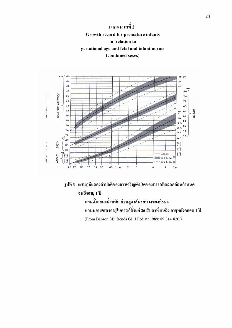
�ก��"�����"�+'�# �n%�$��%& 26 ����(n ��jh� "�+(���#�"� 1 �g (From Babson SB, Benda GI. J Pediatr 1989; 89:814-820.)
25
Month Year
����� 4 �����������#&)��� "��Q� �� (occipito-frontal circunference; OFC)
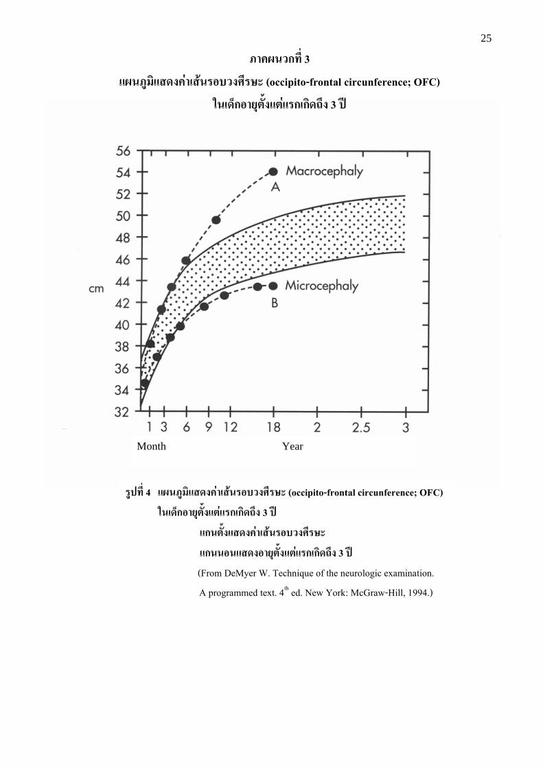
'�)�fก"�+%�$��%&� ก)ก��jh� 3 �g
�ก�%�$�����#&)��� "��Q� ��
�ก��"�����"�+%�$��%&� ก)ก��jh� 3 �g (From DeMyer W. Technique of the neurologic examination. A programmed text. 4th ed. New York: McGraw-Hill, 1994.)
�#��ก��� 3
�����������#&)��� "��Q� �� (occipito-frontal circunference; OFC)
'�)�fก"�+%�$��%&� ก)ก��jh� 3 �g

26 �#��ก��� 4

27
28
�#��ก��� 5
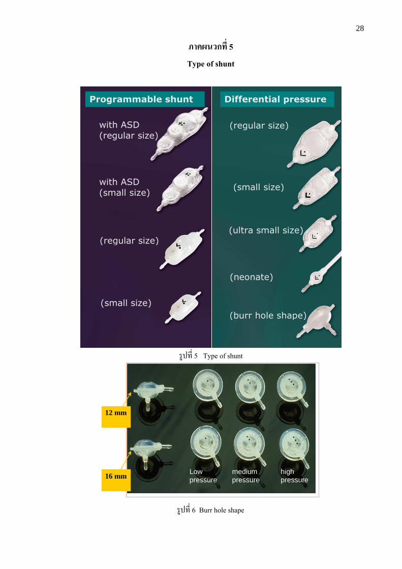
Type of shunt
�3)+,$ 5 Type of shunt
12 mm
16 mm
�3)+,$ 6 Burr hole shape
Low pressure
medium pressure
high pressure
with ASD
(regular size)
with ASD
(small size)
(regular size)
(small size)
(regular size)
(small size)
(ultra small size)
(neonate)
(burr hole shape)
Programmable shunt Differential pressure
valve
29
�#��ก��� 6
�!j&� ����'�������������� ! ���"�#����$�
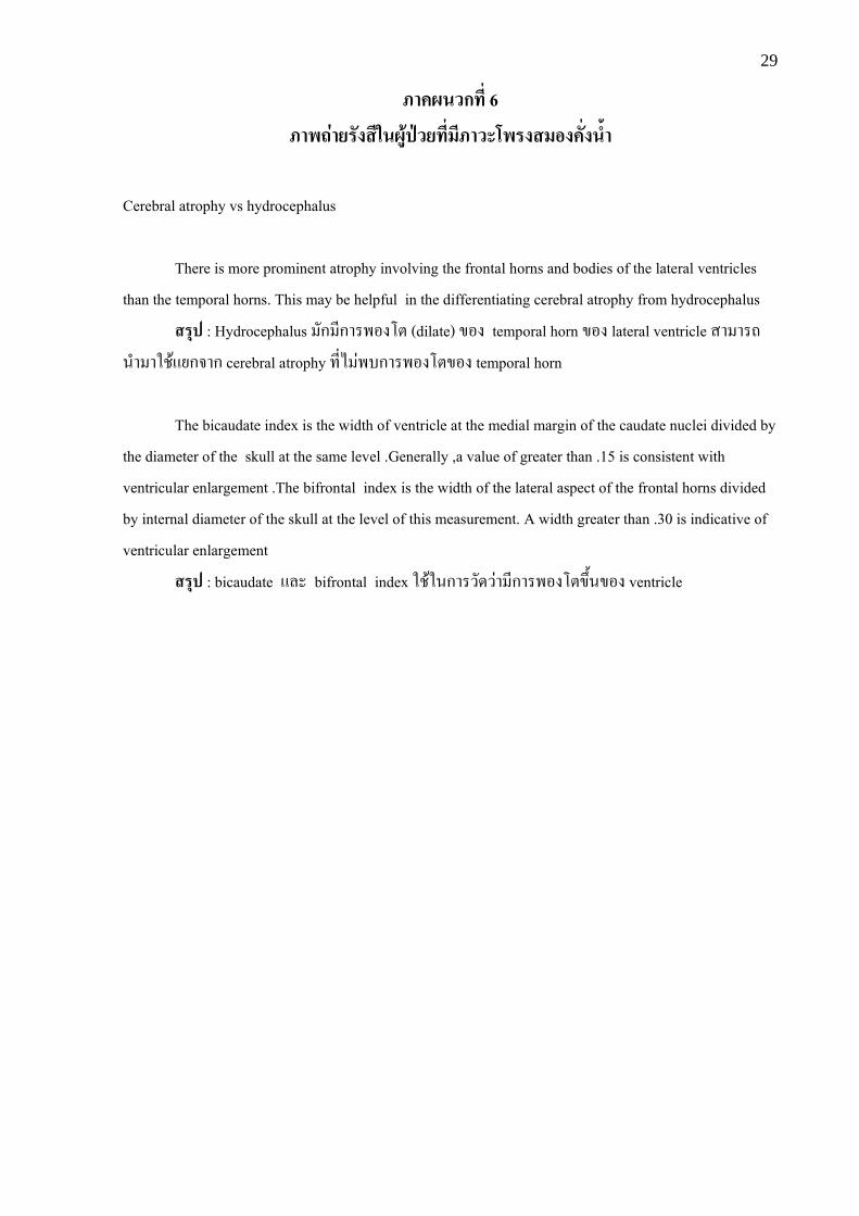
Cerebral atrophy vs hydrocephalus
There is more prominent atrophy involving the frontal horns and bodies of the lateral ventricles than the temporal horns. This may be helpful in the differentiating cerebral atrophy from hydrocephalus
� +� : Hydrocephalus #ก ,ก���!��: (dilate) D!� temporal horn D!� lateral ventricle �� ��p%'� �1P0@5ก?�ก cerebral atrophy +,$. 9�-ก���!��:D!� temporal horn
The bicaudate index is the width of ventricle at the medial margin of the caudate nuclei divided by the diameter of the skull at the same level .Generally ,a value of greater than .15 is consistent with ventricular enlargement .The bifrontal index is the width of the lateral aspect of the frontal horns divided by internal diameter of the skull at the level of this measurement. A width greater than .30 is indicative of ventricular enlargement
� +� : bicaudate @8� bifrontal index 1P01%ก���#/�9� ,ก���!��:DN&%D!� ventricle
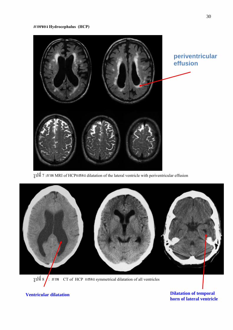
30
�!L"� Hydrocephalus (HCP)
�3)+,$ 7 :��� MRI of HCP@�/� dilatation of the lateral ventricle with periventricular effusion
�3)+,$ 8 : ��� CT of HCP @�/� symmetrical dilatation of all ventricles
periventricular effusion
Ventricular dilatation Dilatation of temporal horn of lateral ventricle

31
�!L"� Normal pressure hydrocephalus (NPH)
�3)+,$ 9 : MRI sagittal T2 @�/� Flow void sign (signal void) in the aqueduct of Sylvius in NPH
Signal void
32
�3)+,$ 10 : ���CT of NPH @�/� dilatation of the ventricle disproportion to the sulci
sulci
Ventricle

33
�3)+,$ 11 : ��� MRI of NPH @�/� rounding of bilateral frontal horns with periventricular effusion
Rounding of frontal horn
Perivetricular effusion
34
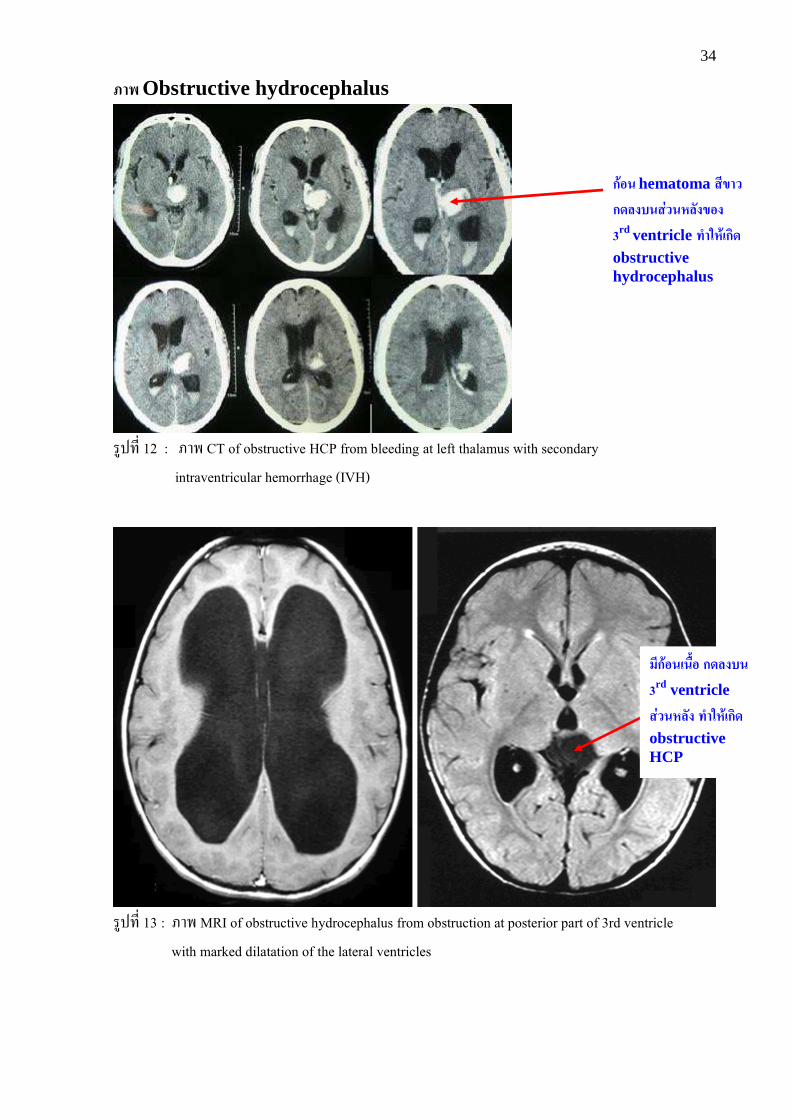
�! Obstructive hydrocephalus
�3)+,$ 12 : ��� CT of obstructive HCP from bleeding at left thalamus with secondary intraventricular hemorrhage (IVH)
�3)+,$ 13 : ��� MRI of obstructive hydrocephalus from obstruction at posterior part of 3rd ventricle with marked dilatation of the lateral ventricles
ก�"� hematoma ��L
ก������&�(���L"�
3rd ventricle ��'(�)ก�� obstructive hydrocephalus
��ก�"�)�l$" ก�����
3rd ventricle
�&�(��� ��'(�)ก�� obstructive HCP

35
Normal CSF***
Treatment of infection or parasitic infestation ± CSF diversion
CSF diversion Treatment of the causes accordingly + CSF diversion
�#��ก��� 7
Patients suspected of HCP with symptoms & signs of increased ICP
Cranial US, CT or MRI*
Ventricular dilatation
No Yes
Extraventricular obstruction
Ventricular obstruction
Treatment of the causes **
/3@2%�3 ;+,$ 2.1, 2.2, 2.3
LP for CSF examination for infection, infestation and CSF cytology for spinal metastatic tumors
CNS infection or parasitic infestation
Positive cytology for spinal metastatic tumors
Cause identifieda
No Yes
Treatment of the causesa accordingly
±CSF diversion
* 1%�p�%+,$+,$�� ��p:��?./0 ** 1%ก�<,+,$ ventricleD%�/)ก:; @�/��9� ���� increased ICP !�?(ก;/?�ก��(E:6!O$% (P9% space occupying lesion, pseudotumor cerebri, cryptococcal meningitis ()*%:0% *** 1%ก�<,+,$���#5 venous sinus obstruction !�?�;?��<�:��?(�;$ (:; (P9% MRV ()*%:0% a /31%"'�!A;-�5@2%�3 ;
1 2
3 4
5 6
7
���������� 6 @%�+��ก��-'�-#/�#กW�230)4�5+,$���#5�9� ,��������� !�"#$�%&'��9� ก#-"�� /#%1%ก��E8กV,�W��3�
( Management of patients suspected of HCP with increased intracranial pressure)
36
����ก ����� �ก���������������&���� ! ���"�#����$� &�ก��#����'�ก� (�กQ� �����
( Management of patients suspected of HCP with increased intracranial pressure)
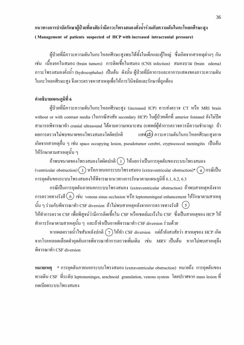
230)4�5+,$ ,����"�� /#%1%ก��E8กV,�W��3��-./0+#&�1%(/>ก@8�2301Ek9 MN$�(ก;/?�ก��(E:6:9��Y ก#% (P9% (%O&!�!ก1%� !� (brain tumors) ก��:;/(PO&!1%� !� (CNS infection) � !�-� (brain edema) ��������� !�"#$�%&'� (hydrocephalus) ()*%:0% /#�%#&% 230)4�5+,$ ,!�ก��@8�!�ก��@�/�D!�����"�� /#%1%ก��E8กV,�W��3� ?N�"��:��?E���(E:6(�O$!1E0ก���;%;?l#5@8��#กW�+,$p3ก:0!� #�"*������������� 6
230)4�5+,$ ,����"�� /#%1%ก��E8กV,�W��3� (increased ICP) "���9�:��? CT E�O! MRI brain without or with contrast media (1%ก�<,���#5 secondary HCP) 1%230)4�5(/>ก+,$ anterior fontanel 5#�. 9)�/ �� ��p�;?��<�+'� cranial ultrasound ./0:� "�� (E ��� (@�+5=230+'�ก��:��?"�� ,"�� P'�%�k) p0�28ก��:��?. 9�-D%�/D!������ !��:2;/)ก:; @�/��9� ����"�� /#%1%ก��E8กV,�W��3�!�?(ก;/?�ก��(E:6!O$% Y (P9% space occupying lesion, pseudotumor cerebri, cryptococcal meningitis ()*%:0% 1E0�#กW�:� ��(E:6%#&% Y
p0��-D%�/D!������ !��:2;/)ก:; 1E0@5ก�9�()*%ก��!6/:#%D!���--����� !� (ventricular obstruction) E�O!��5%!ก��--����� !� (extraventricular obstruction)* ก�<,()*%ก��!6/:#%D!���--����� !�1E0�;?��<�@%�+��ก���#กW�:� @2%�3 ;+,$ 6.1, 6.2, 6.3
ก�<,()*%ก��!6/:#%��5%!ก��--����� !� (extraventricular obstruction) p0��-��(E:6E8#�?�กก��:��?+���#��, (P9% venous sinus occlusion E�O! leptomeningeal enhancement 1E0�#กW�:� ��(E:6%#&% Y �9� ก#-�;?��<�+'� CSF diversion p0�. 9�-��(E:6E8#�?�กก��:��?+���#��, 1E0+'�ก��:��? CSF (�O$!�;�3?%=�9� ,ก��:;/(PO&!1% CSF E�O!(M88= �(�>�1% CSF MN$�()*%��(E:6D!� HCP 1E0+'�ก���#กW�:� ��(E:6%#&% Y @8�p0�?'�()*%!�?�;?��<�+'� CSF diversion �9� /0�5
E�ก28:��?%&'�.D�#%E8#�)ก:; 1E0+'� CSF diversion @:9p0�5#����#5�9� ��(E:6D!� HCP (ก;/?�ก��"E8!/(8O!//'�!6/:#%!�?�;?��<�+'�ก��:��?(�;$ (:; (P9% MRV ()*%:0% E�ก. 9�-��(E:6?N��;?��<�+'� CSF diversion (��)(%+ * ก��!6/:#%��5%!ก��--����� !� (extraventricular obstruction) E �5pN� ก��!6/:#%D!�+��(/;% CSF +,$��/#- leptomeninges, arachnoid granulation, venous system �/5)��V?�ก mass lesion +,$ก/(-,5/��--����� !�
1
5
2
3 4
6
7
37
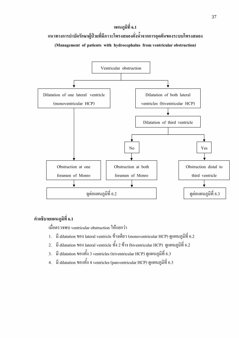
���������� 6.1
����ก ����� �ก�������������� ! ���"�#����$��กก "+�%��L"� ��� ! ���"� (Management of patients with hydrocephalus from ventricular obstruction)
#�"*������������� 6.1 ( O$!:��?�- ventricular obstruction 1E0@5ก�9�
1. , dilatation D!� lateral ventricle D0��(/,5� (monoventricular HCP) /3@2%�3 ;+,$ 6.2 2. , dilatation D!� lateral ventricle +#&� 2 D0�� (biventricular HCP) /3@2%�3 ;+,$ 6.2 3. , dilatation D!�+#&� 3 ventricles (triventricular HCP) /3@2%�3 ;+,$ 6.3 4. , dilatation D!�+#&� 4 ventricles (panventricular HCP) /3@2%�3 ;+,$ 6.3
Ventricular obstruction
Dilatation of one lateral ventricle (monoventricular HCP)
Dilatation of both lateral ventricles (biventricular HCP)
Dilatation of third ventricle
No Yes
Obstruction at both foramen of Monro
Obstruction at one foramen of Monro
Obstruction distal to third ventricle
/3:9!@2%�3 ;+,$ 6.3 /3:9!@2%�3 ;+,$ 6.2

38
���������� 6.2
����ก ����� �ก�������������� ! ���"�#����$� �กก "+�%���$�iL���(���
���%��(�&� foramen of Monro
(Management of patients with hydrocephalus from obstruction at foramen of Monro) * 1%ก�<,+,$���#5�9�5#� , active purulent meningitis / ventriculitis :0!�:��? CSF @8��#กW� infection 1E0E�5ก9!%?N��;?��<�+'� permanent CSF diversion
Obstruction at foramen of Monro
Obstruction at one foramen of Monro (monoventricular HCP)
Obstruction at both foramen of Monro (biventricular HCP)
No mass lesion Mass in third ventricle
Atresia of both foramen of Monro
Mass in lateral ventricle
Treatment of the causes accordingly + CSF diversion CSF diversion* CSF diversion*
39
#�"*������������� 6.2
( O$!:��?�- dilatation of lateral ventricle @:9 third @8� fourth ventricle ,D%�/)ก:;@�/��9� , obstruction at foramen of Monro 1E0�;?��<��9� ,ก��!6/:#%D!� foramen of Monro D0��(/,5�E�O! 2 D0�� 1. p0� ,ก��!6/:#%D0��(/,5�+'�1E0(ก;/ monoventricular HCP 1E0�;?��<��9� , mass lesion +,$ lateral ventricle E�O!. 9 1.1 p0� , mass lesion (P9% subependymal giant cell astrocytoma +,$�-1%��" tuberous sclerosis6 ()*%:0% 1E0�#กW�:� ��(E:6 �9� ก#-�;?��<�+'� CSF diversion :� "�� (E ��� 1.2 p0�. 9 , mass lesion 1E0�;?��<�+'� CSF diversion 5ก(�0%ก�<,���#5�9� , active purulent meningitis / ventriculitis :0!�:��? CSF @8��#กW� infection 1E0E�5ก9!%?N��;?��<�+'� permanent CSF diversion 2. p0� ,ก��!6/:#%+#&� 2 D0�� +'�1E0(ก;/ biventricular HCP 1E0�;?��<��9� , mass lesion +,$��/#- third ventricle E�O!. 9 2.1 p0� , mass lesion (P9% craniopharyngioma, ependymoma ()*%:0% 1E0�#กW�:� ��(E:6 �9� ก#-�;?��<�+'� CSF diversion :� "�� (E ��� 2.2 1%ก�<,+,$. 9 , mass �9�%1Ek9(ก;/?�ก atresia of both foramen of Monro 1E0�;?��<�+'� CSF diversion 5ก(�0%ก�<,���#5�9� , active purulent meningitis / ventriculitis :0!�:��? CSF @8��#กW� infection 1E0E�5ก9!%?N��;?��<�+'� permanent CSF diversion
40
���������� 6.3
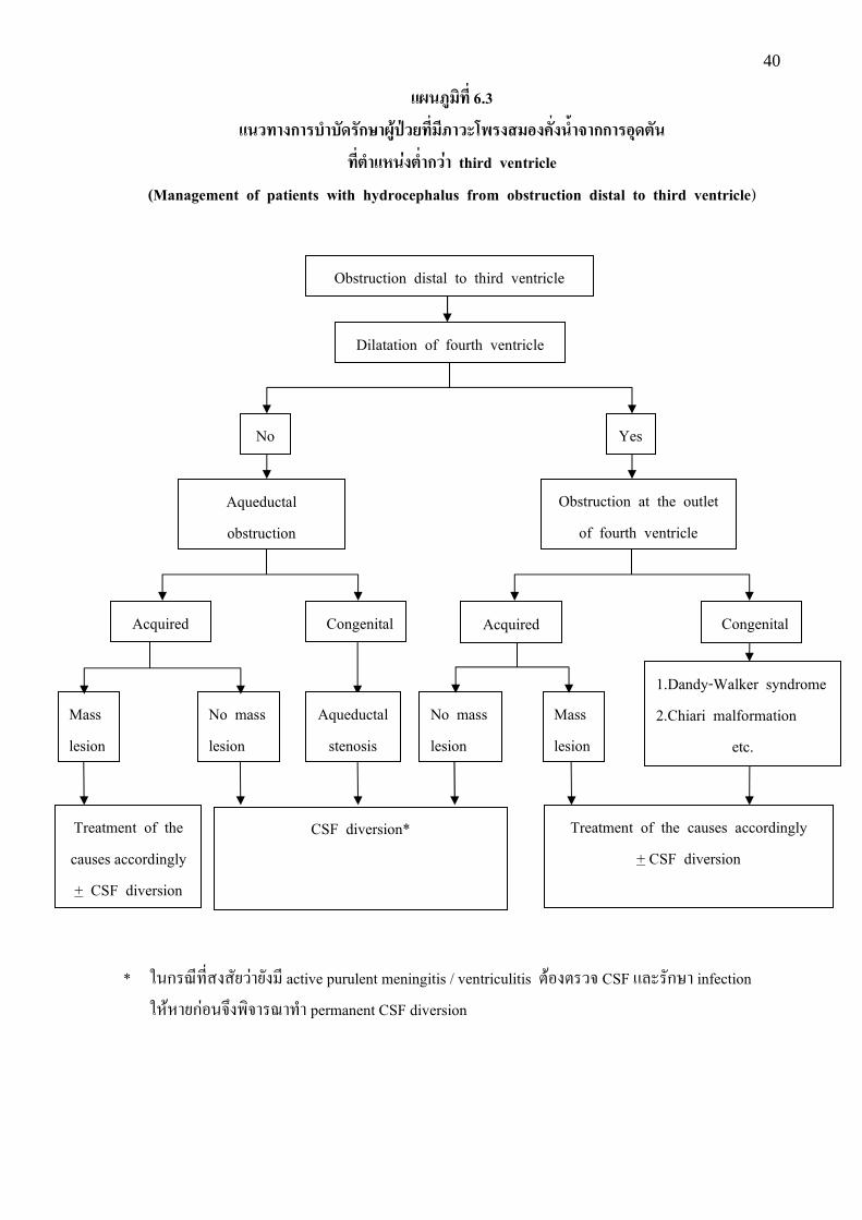
����ก ����� �ก�������������� ! ���"�#����$��กก "+�%��
���%��(�&�%��ก& third ventricle
(Management of patients with hydrocephalus from obstruction distal to third ventricle) * 1%ก�<,+,$���#5�9�5#� , active purulent meningitis / ventriculitis :0!�:��? CSF @8��#กW� infection 1E0E�5ก9!%?N��;?��<�+'� permanent CSF diversion
Obstruction distal to third ventricle
Dilatation of fourth ventricle
No Yes
Obstruction at the outlet of fourth ventricle
Aqueductal obstruction
Acquired Congenital Acquired Congenital
No mass lesion
Mass lesion
Mass lesion
Aqueductal stenosis
No mass lesion
Treatment of the causes accordingly + CSF diversion
CSF diversion* Treatment of the causes accordingly + CSF diversion
1.Dandy-Walker syndrome 2.Chiari malformation
etc.
41
#�"*������������� 6.3
( O$!:��?�- dilatation of lateral ventricles @8� third ventricle 1E0�;?��<�/3�9� , dilatation D!� fourth ventricle E�O!. 9 1. p0� fourth ventricle . 9�: �;%;?l#5�9�()*% aqueductal obstruction 1.1 p0� , mass lesion +,$()*%��(E:6D!� HCP (P9% tectal astrocytoma, pineal region tumor, posterior fossa tumor, cysticercosis ()*%:0% 1E0�#กW�:� ��(E:6%#&% Y �9� ก#-�;?��<�+'� CSF diversion
1.2 p0�. 9 , mass �;%;?l#5�9�()*% aqueductal stenosis MN$�!�?()*%"�� 2;/)ก:; �@:9ก'�(%;/ (congenital) E�O!(ก;/DN&%��5E8#� (acquired) 1E0ก���#กW��/5ก��+'� CSF diversion 1%ก�<,+,$���#5�9� , active purulent meningitis / ventriculitis :0!�:��? CSF @8��#กW� infection 1E0E�5ก9!%?N��;?��<�+'�permanent CSF diversion 2. p0� fourth ventricle �: 1E0@5ก�9�()*%"�� 2;/)ก:; �@:9ก'�(%;/ (congenital) (P9% Dandy Walker syndrome, Chiari malformation ()*%:0% 1E0�#กW�:� ��(E:6%#&%Y E�O!p0�()*%��(E:6+,$(ก;/DN&%��5E8#� (acquired) 1E0�;?��<��9� , mass lesion E�O!. 9 2.1 p0� , mass lesion +,$()*%��(E:6D!� HCP (P9% meningioma -�;(�< foramen magnum ()*%:0% 1E0�#กW�:� ��(E:6%#&%Y �9� ก#-�;?��<�+'� CSF diversion :� "�� (E ��� 2.2 p0�. 9 , mass lesion 1E0�;?��<�+'� CSF diversion 1%ก�<,+,$���#5�9�5#� , active purulent meningitis/ ventriculitis :0!�:��? CSF @8��#กW� infection 1E0E�5ก9!%?N��;?��<�+'� permanent CSF diversion

������������ ��ก����ก���������������������
Clinical Practice Guidelines for Hydrocephalus
(2����3��)
���56��7� ���������� ����� � ���� � ��� ����� ������� ������������������
�3� 6� 1 9:��;� ���<�9��;:=>:9:��;�
1. ! �1) ��� �2) �$�� 2. ��% �1) �� �ก��� 30 �) �2) 31-40 �) �3) 41-50 �) �4) ��กก��� 50 �) 3. ���-���� �1) ���- ��. �2) ��/�������0 �3) ���1 �����2/%............................................. 4. ��� ��� ��/��/45���0�������........................................................................................................................... ���/�........................................... ��6 .........................................�7���7�....................................................... 5. ��26���� ��/��/45���0������� �1) �. . �%��� �2) �. . �7��8� �3) �. . !9��. �4) �. . ���������7� �5) �. . ก�� �6) �. . 47�ก7�ก�2����ก����� �7) �. . < �2��� �8) �. . 47�ก7� ก��. �9) ����ก4����7� �10) ���1 �����2/%....................................................................................................................................... 6. �������0������� ��/�� ���� �0��
�3� 6� 2 ������@�>A�ก6�B�ก���� ��ก����ก��������������������� 7. ������� ?-�����ก���7ก@�6��2� ��4� �7���A��B </7/�0A��ก���9-��7ก@�C9��D�� 0���� �1) ����%ก��� �2) ���/����� ��2��E�� ��2.............. ��� ���ก �2.1) ������C9��D����26��0A�0�� � �2.2) �7ก@��9-���� �ก��/�A ����-�24���� ��� ��/���0��0!7ก�6� 49�ก��� �2.3) C9��D��/����� /��45��ก��E.8��4����5������-�����8���7A���� �3) 8��8����� ��� ���ก
�3.1) 8���0C9��D�� �3.2) 8���0��� ��� �3.3) 8���0- ��.< �2�������..................................................................................................... �3.4) �0-�����ก���7ก@�C9��D�� �9�-��� �2/%��� ����........................................................................ �3.5) ��������ก

8. ��2�����A ��� �-�����ก���7ก@�6��2� ��4� �7���A�� �����9:�=7�C��>� ก����<�=5:
>��9:� B�ก �3�B <�3�9:�=7 <@:=5: <�3<@:=5:
1. /��0� 1 -�����ก�������<7�-�2/��/7��7ก@�C9��D���0�4�47��06��2� ��4� �7���A��(Diagnosis and Management of patients suspected of hydrocephalus)
2. /��0� 2 -�����ก�������<7�� �6��2� ��4� �7���A�����[ก
3. /��0� 3 -�����ก�������<7� Normal pressure hydrocephalus
4. /��0� 4 -�����ก���7ก@�C9��D���0��06��2� ��4� �7���A��
5. /��0� 5 -�����ก��4����]ก@�
6. /��0� 6 -�����ก��/��/7��7ก@���7�C���7� CSF diversion
7. 6�C��ก�0� 1 �ก��-4��� �6��2����7���ก2���ก!0�@249����[ก
8. 6�C��ก�0� 2 Growth record for premature infants in relation to gestational age and fetal and infant norms (combined sexes)
9. 6�C��ก�0� 3 -C�69��-4����4��� /��!0�@2 (occipito-frontal circunference; OFC) ���[ก ��%�7A�-��-�กก��5]� 3 �)
10. 6�C��ก�0� 4 -//��4 / 7h��ก�� Denver II </7/6�@�8��
11. 6�C��ก�0� 5 Type of shunt
12. 6�C��ก�0� 6 6� 5����7�40��C9��D���0��06��2� ��4� �7���A��
13. ������0� 1 �ก��-�2 �ก��-4��� �����7�ก2���ก!0�@249� -�24���%�0� //� �� �6��2� ��4� �7���A�� -/�� ���ก�%�� ��%
14. ������0� 2 Etiology of macrocephaly
15. -C�69���0� 1 -�����ก�������<7�C9��D���0��06��2� ��4� �7���A��
16. -C�69���0� 2 -�����ก�������<7�C9��D���0��06��2� ��4� �7���A�� ���[ก
17. -C�69���0� 3 -�����ก�������<7� Normal pressure hydrocephalus
18. -C�69���0� 4 -�����ก���7ก@�C9��D���0��06��2� ��4� �7���A��
19. -C�69���0� 5 -�����ก��/��/7��7ก@���7�C���7� CSF diversion
20. -C�69���0� 6 -�����ก��/��/7��7ก@�C9��D���0�4�47�����06��2� ��4� �7���A������ก7/����7���ก2���ก!0�@249�
�����9:�=7�C��>� ก����<�=5: >��9:�
B�ก �3�B <�3�9:�=7 <@:=5: <�3<@:=5:
21. -C�69���0� 6.1 -�����ก��/��/7��7ก@�C9��D���0��06��2� ��4� �7���A����กก�� %��7�� ��2//� ��4� �
22. -C�69���0� 6.2 -�����ก��/��/7��7ก@�C9��D���0��06��2� ��4� �7���A����กก�� %��7��A��8�47���7��0����-���� foramen of Monro
23. -C�69���0� 6.3 -�����ก��/��/7��7ก@�C9��D���0��06��2� ��4� �7�� �A����กก�� %��7��0����-��������ก��� third ventricle
24. �9��0� 1 macrocephaly, setting sun sign, bulging anterior fontanel, dilated scalp vein (�9���������)
25. �9��0� 2 macrocephaly, setting sun sign, bulging anterior fontanel, dilated scalp vein (�9���������)
26. �9��0� 3 -C�69��-4�����ก��� �ก�����$��/��� ����ก�0�� �ก� �ก�������5]� ��% 1 �)
27. �9��0� 4 -C�69��-4����4��� /��!0�@2 (occipito-frontal circunference; OFC) ���[ก ��%�7A�-��-�กก��5]� 3 �)
28. �9��0� 5 Type of shunt
29. �9��0� 7 Burr hole shape
30. �9��0� 8 6� MRI of HCP -4�� dilatation of the lateral ventricle with periventricular effusion
31. �9��0� 9 6� CT of HCP -4�� symmetrical dilatation of all ventricles
32. �9��0� 10 MRI sagittal T2 -4�� Flow void sign (signal void) in the aqueduct of Sylvius in NPH
33. �9� 11 6� CT of NPH -4�� dilatation of the ventricle disproportion to the sulci
34. �9� 12 6� MRI of NPH -4�� rounding of bilateral frontal horns with periventricular effusion
35. �9� 13 6� CT of obstructive HCP from bleeding at left thalamus with secondary intraventricular hemorrhage (IVH)
36. �9� 14 6� MRI of obstructive hydrocephalus from obstruction at posterior part of 3rd ventricle with marked dilatation of the lateral ventricles
9. �����[���� ?-�����ก���7ก@�6��2� ��4� �7���A��B </7/�0A���ก7/�t$��� �C9��D�� 0���� �1) ��ก �2) �� � �3) ���ก��� �4) 8�����
10. �����[������A ��� � ?-�����ก���7ก@�6��2� ��4� �7���A��B </7/�0A���ก7/!7ก�6� � ���� ��/������ 0���� �1) ��ก �2) �� � �3) ���ก��� �4) 8�����
11. �����[���� ?-�����ก���7ก@�6��2� ��4� �7���A��B </7/�0A42��ก��ก���u�/7��������� 8�� �1) 42��ก �2) 8��42��ก �3) 8��-����
12. �����[���� ?-�����ก���7ก@�6��2� ��4� �7���A��B </7/�0A�0��2����.��� 8�� �1) �0 �2) 8���0 �3) 8��-����
13. ����8���7/����9� ��������ก�� ��� ?-�����ก���7ก@�6��2� ��4� �7���A��B </7/�0A��ก�� � 0���� �1) ��ก �2) �� � �3) ���ก��� �4) 8��8���7/
14. �����[����ก��C�- �� ?-�����ก���7ก@�6��2� ��4� �7���A��B </7/�0A�0����7��5]� 0���� �1) ��ก �2) �� � �3) 8���7��5]� ��2...............................................................................
15. �����[����?-�����ก���7ก@�6��2� ��4� �7���A��B </7/�0A�0C�ก�2�/�� - ��.����ก��59กvw ��� � ����8� �1) ����]A� �2) ���� �3) 8��ก0����� �
16. �����[��������7/�%�� ?-�����ก���7ก@�6��2� ��4� �7���A��B </7/�0A ����8� �1) 8���������� ���7/��%� �2) ��A �� �2.1) ��กก��8�
�2.2) �� �ก��8� �2.3) �7��� �0�������-��8�����กu����7�4� ����0A......................................................................
17. �� 4� -�2 ���1................................................................................................................................................... ...................................................................................................................................................................................... ...................................................................................................................................................................................... ......................................................................................................................................................................................

ก�������
������ �� ก��
����������������� (���������������ก��)
312 �.������ !"����!��
ก���!�#$
1 0 4 0 0
'����(��!�"� ) ��.(�)/2881 ��,.���!��'�
�-�.�ก���'����!�/0���-��1�2ก���0��3� ��ก�
���ก��4��ก�,������
9�9���DE 3� 6�ก�DE�=>:9:��;� 6���F����B5G ������@�3����������ก���
6� �DE����6 H�6ก�� ������D��5�ก��
��B=�� 6� 6 HD���� 2553
�L�������� �� B� L��5��L6 �9H��5� �6 ก�D�� �M 10400
� ���� 0 2354 7076 LQ� 83 H3� 3317, 0 2354 7085, 0 2354 7072