2014 3 03 neuroanatomy of the cerebellum
DESCRIPTION
Slideshow from Department of Anatomy. University of Szeged 2013/2014TRANSCRIPT
NEUROANATOMY OF THENEUROANATOMY OF THECEREBELLUMCEREBELLUM
19 September 20141
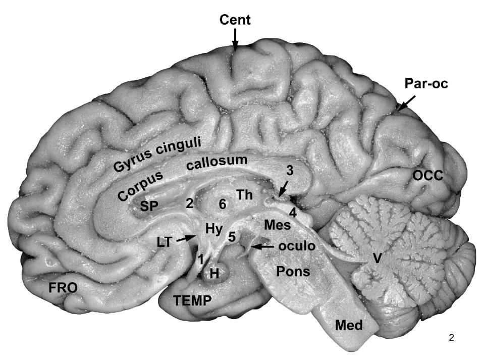
2

3
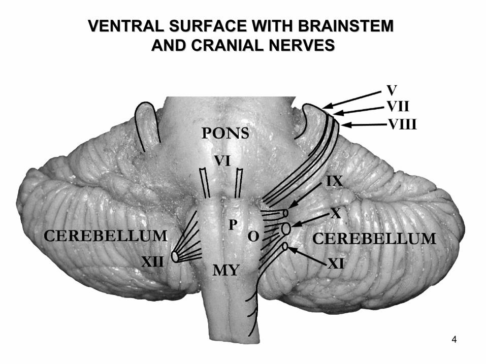
VENTRAL SURFACE WITH BRAINSTEM VENTRAL SURFACE WITH BRAINSTEM AND CRANIAL NERVESAND CRANIAL NERVES
4

DORSAL SURFACE WITH TECTUM AND PULVINARDORSAL SURFACE WITH TECTUM AND PULVINAR
1: primary fissure2: horizontal fissure 5

T: TONSIL; F: FLOCCULUS; PCM: MIDDLE CERBELLAR PEDUNCLE; T: TONSIL; F: FLOCCULUS; PCM: MIDDLE CERBELLAR PEDUNCLE; PCI: INFERIOR CEREBELLAR PEDUNCLE; CRUS: CEREBRAL PEDUNCLE; PCI: INFERIOR CEREBELLAR PEDUNCLE; CRUS: CEREBRAL PEDUNCLE;
FIP: INTERPEDUNCULAR FOSSAFIP: INTERPEDUNCULAR FOSSA
6
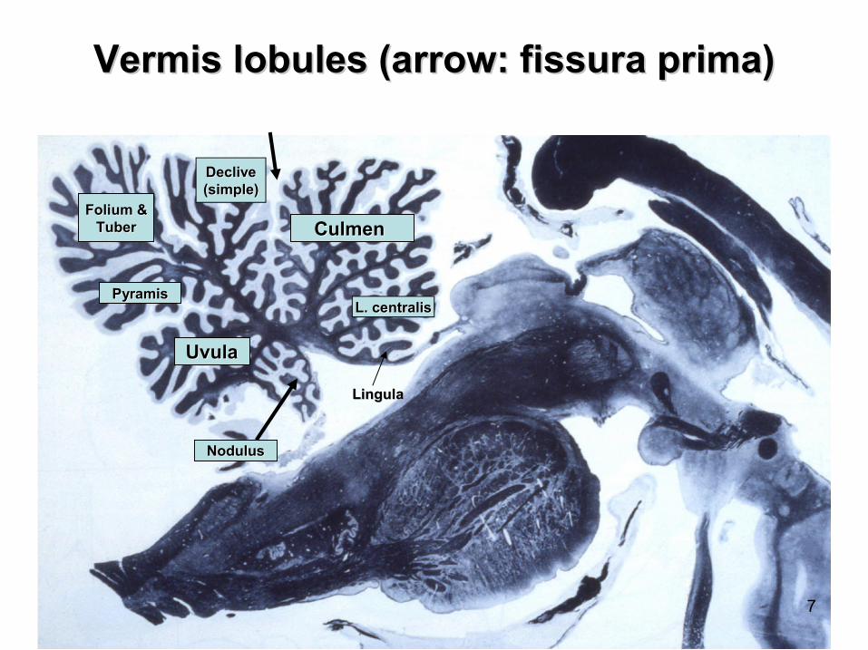
Vermis lobules (arrow: fissura prima)Vermis lobules (arrow: fissura prima)
Culmen Culmen
L. centralisL. centralis
Lingula Lingula
DecliveDeclive(simple)(simple)
Folium &Folium &TuberTuber
PyramisPyramis
UvulaUvula
NodulusNodulus
7
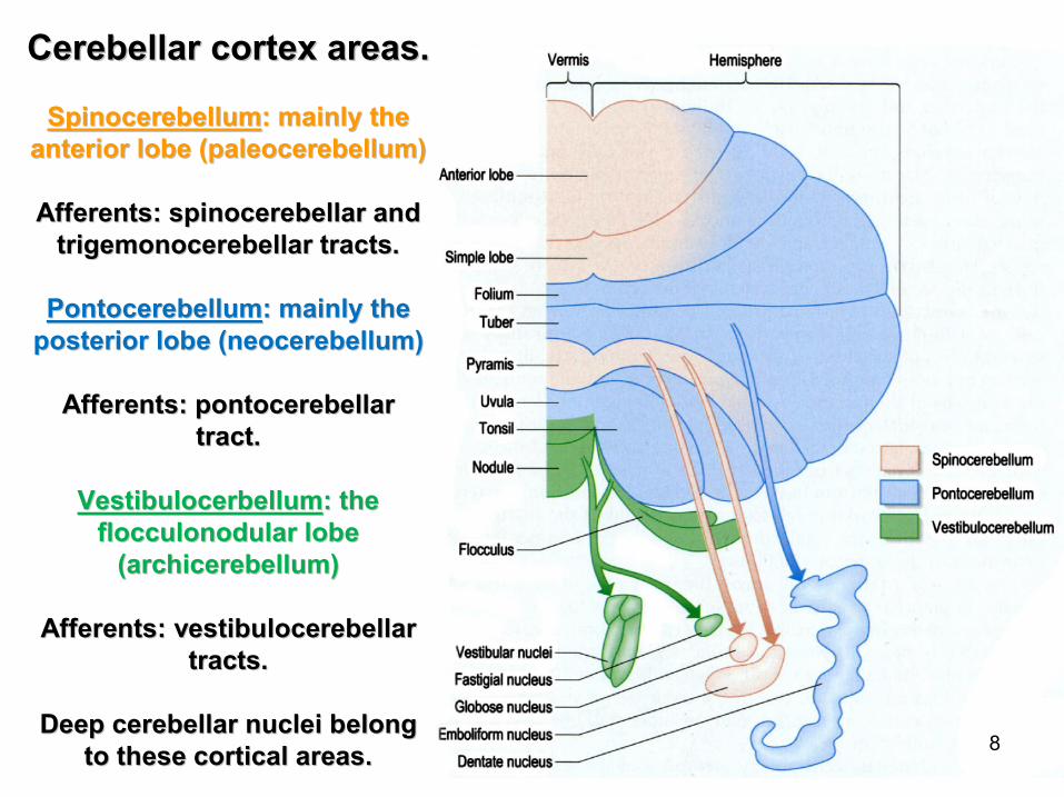
Cerebellar cortex areas.Cerebellar cortex areas.
SpinocerebellumSpinocerebellum: mainly the: mainly theanterior lobe (paleocerebellum)anterior lobe (paleocerebellum)
Afferents: spinocerebellar andAfferents: spinocerebellar andtrigemonocerebellar tracts.trigemonocerebellar tracts.
PontocerebellumPontocerebellum: mainly the: mainly theposterior lobe (neocerebellum)posterior lobe (neocerebellum)
Afferents: pontocerebellarAfferents: pontocerebellartract.tract.
VestibulocerbellumVestibulocerbellum: the: theflocculonodular lobe flocculonodular lobe
(archicerebellum)(archicerebellum)
Afferents: vestibulocerebellar Afferents: vestibulocerebellar tracts.tracts.
Deep cerebellar nuclei belongDeep cerebellar nuclei belongto these cortical areas.to these cortical areas.
8

The spinocerebellar and The spinocerebellar and trigeminocerebellar afferentstrigeminocerebellar afferents
project in somatotopic order: in project in somatotopic order: in spinocerebellum and vermal spinocerebellum and vermal pyramis and lobulus biventerpyramis and lobulus biventer
in the posterior lobe.in the posterior lobe.9

The folium ofThe folium ofthe cerebellarthe cerebellarcortex: cell typescortex: cell types
A: purkinje cellA: purkinje cellB: basket cellB: basket cellE: superficial stellate cellE: superficial stellate cellG: granule cellG: granule cellF: golgi cellF: golgi cellJ: bergmann glia cellJ: bergmann glia cellM: astrocyteM: astrocyteN: climbing fiberN: climbing fiberH: mossy fiberH: mossy fiberi: paralel fibersi: paralel fibers
Drawing from RamDrawing from Ramóón y Cajaln y Cajal
10

PURKINJEPURKINJE--CELL (GABAergic)CELL (GABAergic) GOLGIGOLGI--CELL (GABAergic)CELL (GABAergic)
11

Cortex cerebelli in numbers Cortex cerebelli in numbers (1 mm(1 mm33 of tissue)of tissue)
•• 500 Purkinje500 Purkinje--cellscells
•• 600 basket cells600 basket cells
•• 50 Golgi50 Golgi--cellscells
•• 3 000 000 granule cells3 000 000 granule cells
•• 600 000 cerebellar glomeruli600 000 cerebellar glomeruli
•• Area of the cerebellar cortex cca. 2000 Area of the cerebellar cortex cca. 2000 cmcm22
12
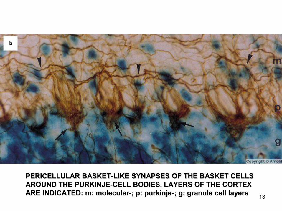
PERICELLULAR BASKETPERICELLULAR BASKET--LIKE SYNAPSES OF THE BASKET CELLSLIKE SYNAPSES OF THE BASKET CELLSAROUND THE PURKINJEAROUND THE PURKINJE--CELL BODIES. LAYERS OF THE CORTEXCELL BODIES. LAYERS OF THE CORTEXARE INDICATED: m: molecularARE INDICATED: m: molecular--; p: purkinje; p: purkinje--; g: granule cell layers; g: granule cell layers
13

MOSSYMOSSY
GOLGIGOLGIGRANULEGRANULE
GOLGIGOLGI
GRANULEGRANULE
CEREBELLAR GLOMERULUSCEREBELLAR GLOMERULUS 14
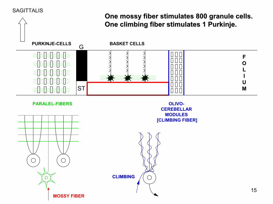
FFOOLLIIUUMM
SAGITTALIS
PARALEL-FIBERS
PURKINJE-CELLS BASKET CELLS
ST
MOSSY FIBER
OLIVO-CEREBELLAR
MODULES[CLIMBING FIBER]
G
CLIMBINGCLIMBING
One mossy fiber stimulates 800 granule cells.One mossy fiber stimulates 800 granule cells.One climbing fiber stimulates 1 Purkinje.One climbing fiber stimulates 1 Purkinje.
15

FFOOLLIIUUMM
SAGITTALIS
PARALEL-FIBERS
PURKINJE-CELL BASKET CELL
ST
MOSSY FIBERMODULE
OLIVO-CEREBELLAR
MODULES[CLIMBING FIBER]
G
MossyMossy--fiberfiber--module: group of Purkinjemodule: group of Purkinje--cells which are stimulated by a mossy fibercells which are stimulated by a mossy fiber(this module is sculptured by the basket cell which is also acti(this module is sculptured by the basket cell which is also activated by the mossyvated by the mossystimuli stimuli –– through the parallel fibers).through the parallel fibers).
Olivocerebellar module: one olivary neuron stimulates 10Olivocerebellar module: one olivary neuron stimulates 10PurkinjePurkinje--cells.cells.In this module there is no role for basketIn this module there is no role for basket--inhibition.inhibition.
16
Cerebellar functions I.Cerebellar functions I.
•• Regulation of muscle movements, muscle tone Regulation of muscle movements, muscle tone and posture.and posture.
•• Conversion of vestibular stimuli into movements Conversion of vestibular stimuli into movements and muscle tone adaptation.and muscle tone adaptation.
•• Comparison of the peripheral somatosensory Comparison of the peripheral somatosensory stimuli to the neocortically processed sensory stimuli to the neocortically processed sensory stimuli (somatosensory, visual and auditory), and stimuli (somatosensory, visual and auditory), and adjusting the movement and movement planning adjusting the movement and movement planning to the actual peripheral stimulus conditions.to the actual peripheral stimulus conditions.
17

Cerebellar functions II.Cerebellar functions II.
•• Neocerebellum: planning, initiating and Neocerebellum: planning, initiating and timing of movement.timing of movement.
•• Vestibulocerebellum: control of equilibrium Vestibulocerebellum: control of equilibrium of the body and vestibuloof the body and vestibulo--ocular reflexes ocular reflexes (eye movements).(eye movements).
•• Spinocerebellum: control of movements Spinocerebellum: control of movements and muscle tone of the trunk and limb and muscle tone of the trunk and limb musculature.musculature.
18

FUNCTIONAL MRI: RIGHT HAND FINGERS ARE MOVING.FUNCTIONAL MRI: RIGHT HAND FINGERS ARE MOVING.STRONG COSTRONG CO--ACTIVATION OF THE PRECENTRAL GYRUSACTIVATION OF THE PRECENTRAL GYRUS
AND THE ANTERIOR LOBE OF THE CEREBELLUM. AND THE ANTERIOR LOBE OF THE CEREBELLUM. (from: Gray(from: Gray’’s Anatomy, 2008)s Anatomy, 2008) 19

Cerebellar symptomsCerebellar symptoms•• Ataxia: lack of coordination of movementAtaxia: lack of coordination of movement
•• Intention tremorIntention tremor
•• Weakness of muscles (hypotonia)Weakness of muscles (hypotonia)
•• Gait disturbances (walk and stand Gait disturbances (walk and stand unsteadily)unsteadily)
•• Equilibrium disturbances (vertigoEquilibrium disturbances (vertigo--dizziness) dizziness)
•• Pathological eye movementsPathological eye movements
•• Speech disturbances (stuttering)Speech disturbances (stuttering)20
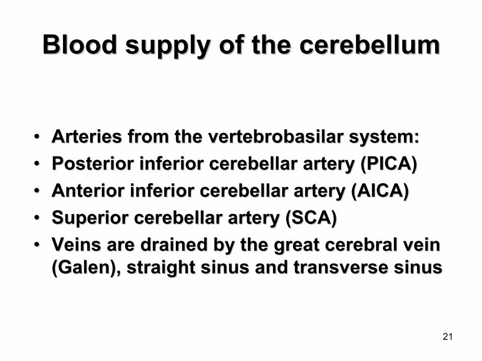
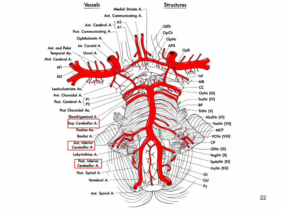
Blood supply of the cerebellumBlood supply of the cerebellum
•• Arteries from the vertebrobasilar system:Arteries from the vertebrobasilar system:
•• Posterior inferior cerebellar artery (PICA)Posterior inferior cerebellar artery (PICA)
•• Anterior inferior cerebellar artery (AICA)Anterior inferior cerebellar artery (AICA)
•• Superior cerebellar artery (SCA)Superior cerebellar artery (SCA)
•• Veins are drained by the great cerebral vein Veins are drained by the great cerebral vein (Galen), straight sinus and transverse sinus(Galen), straight sinus and transverse sinus
21
22
23
KKéépek forrpek forráásasa
•• Standring S (Ed): GrayStandring S (Ed): Gray’’s Anatomy. s Anatomy. ChurchillChurchill--Livingstone, 2008.Livingstone, 2008.
•• RRééthelyi M, Szentthelyi M, Szentáágothai J: Funkciongothai J: Funkcionáális lis anatanatóómia. Medicina, 2013.mia. Medicina, 2013.
•• Haines: Neuroanatomy. Williams and Haines: Neuroanatomy. Williams and Wilkins, 1995.Wilkins, 1995.
•• Szegedi AnatSzegedi Anatóómiai Intmiai Intéézet Mzet Múúzeuma zeuma éés s tudomtudomáányos nyos áábra gybra gyűűjtemjteméénye.nye.