1 copper import into the mitochondrial matrix in saccharomyces
TRANSCRIPT

Pic2 transports mitochondrial copper
1
Copper Import into the Mitochondrial Matrix in Saccharomyces cerevisiae is Mediated by Pic2, a Mitochondrial Carrier Family Protein *
Katherine E. Vest1, Scot C. Leary2, Dennis R. Winge3 and Paul A. Cobine1*
1. Department of Biological Sciences, Auburn University, Auburn, AL, USA, 36849
2. Department of Biochemistry, University of Saskatchewan, Saskatoon, SK, Canada, S7N 5E5 3. Departments of Biochemistry and Medicine, University of Utah, Salt Lake City, UT, USA, 84132
Running title: Pic2 transports mitochondrial copper
To whom correspondence should be addressed: Paul A. Cobine, Department of Biological Sciences, 101 Rouse Life Sciences, Auburn, AL 36849. Telephone: 344 844 1661 Email: [email protected] Keywords: copper, silver, cytochrome c oxidase, mitochondrial carrier family Background: Copper must enter the mitochondrial matrix prior to assembly into cytochrome c oxidase. Results: Pic2 transports mitochondrial copper in vivo and in vitro. Conclusions: Pic2 mediates copper import into the mitochondrial matrix. Significance: We have identified the first mitochondrial copper importer. SUMMARY Saccharomyces cerevisiae must import copper into the mitochondrial matrix for eventual assembly of cytochrome c oxidase. This copper is bound to an anionic, fluorescent molecule known as the copper ligand (CuL). Here, we identify for the first time a mitochondrial carrier family protein capable of importing copper into the matrix. In vitro transport of CuL into the mitochondrial matrix is saturable and temperature-dependent. Strains with a deletion of PIC2 grow poorly on copper-deficient, non-fermentable medium supplemented with silver and under respiratory conditions when challenged with a matrix targeted copper competitor. Mitochondria from pic2∆ cells have lower total mitochondrial copper and exhibit a decreased capacity for copper uptake. Heterologous expression of Pic2 in Lactococcus lactis significantly enhanced CuL transport into these cells. Therefore, we propose a novel role for Pic2 in copper import into mitochondria.
INTRODUCTION Metals are essential nutrients that pose a
management quandary for cells. They must be directed to the correct proteins and organelles through a maze of cellular components and opportunistic metal binding sites (1). Failure to control their delivery results in cellular stress, presumably due to inappropriate interactions and oxidative damage. Cells have adopted a protein-mediated delivery mechanism for copper within the cytosol. In the budding yeast Saccharomyces cerevisiae, copper enters the cell via specific (Ctr1) and non-specific transporters (e.g. Fet4), and is then trafficked to points of utilization by copper chaperone proteins (2). Copper is used as a cofactor in three major enzymes: the multi-copper oxidase Fet3 required for high affinity Fe uptake (3); Sod1, a Cu, Zn superoxide dismutase required for protection against oxidative stress and regulation of glucose signaling in yeast (4); and cytochrome c oxidase (CcO), the terminal enzyme complex of the electron transport chain (5). Atx1 is the copper chaperone responsible for delivering copper to the trans-Golgi vesicles via Ccc2, a P-type ATPase (6), while Ccs1 serves as the copper donor for Sod1 and acts as a post-transfer modifying enzyme by facilitating the formation of an essential disulfide bond within the enzyme itself (7). Though cytosolic copper trafficking has been well characterized, the pathway that delivers copper to mitochondria in yeast and in other eukaryotes is completely unknown.
CcO is a multimeric protein complex that contains two copper centers, a binuclear CuA site and a heme a3-CuB site. A number of assembly
http://www.jbc.org/cgi/doi/10.1074/jbc.M113.470674The latest version is at JBC Papers in Press. Published on July 11, 2013 as Manuscript M113.470674
Copyright 2013 by The American Society for Biochemistry and Molecular Biology, Inc.
by guest on April 16, 2018
http://ww
w.jbc.org/
Dow
nloaded from

Pic2 transports mitochondrial copper
2
factors act in concert to build both of these sites. The soluble intermembrane space (IMS) protein Cox17 delivers copper to both Sco1 and Cox11, which are integral inner membrane (IM) proteins that donate copper to the assembling holoenzyme (8,9). Additionally, the IMS protein Cmc1 has been implicated in the control of copper flow within the IMS, potentially by directing copper to the Cox17-mediated CcO assembly pathway (10).
Organellar fractionation experiments showed that greater than 70% of mitochondrial copper is present as a soluble, anionic complex contained within a matrix-localized, bioavailable pool (11). This complex has been defined as the copper ligand (CuL) and its existence and localization have since been confirmed by X-ray fluorescence imaging and copper chelation studies (12,13). Copper-dependent human Sod1 localized to this mitochondrial compartment is able to rescue a range of phenotypic defects associated with SOD2 deletion, demonstrating the accessibility of this pool (11). Expression of matrix-targeted Sod1 or Crs5, a copper-binding metallothionein, results in a specific loss of CcO activity that can be rescued by the addition of copper or decreased expression of the competing cuproprotein (14). These observations led us to propose that the matrix copper pool is redistributed to the IMS where it is made available to CcO. Although the exact structural identity of the CuL is unknown, we have characterized many aspects of its in vivo function. We propose that the ligand exists in the cytosol in a metal-free form where it binds copper and delivers it to mitochondria, providing a non-proteinaceous trafficking system for copper delivery to the organelle.
The IM is impermeable to most ions and molecules, so transporters must exist that facilitate matrix import of the CuL complex and its subsequent redistribution to the IMS. However, transporters required for the movement of copper across the IM have yet to be identified, and remain a fundamental gap in our understanding of the mechanisms that provide for the assembly of CcO. Data from our previous studies suggest that CuL is the molecule that is transported into the matrix, and that this complex may resemble a metabolite or nucleotide. Therefore copper transport across the IM may proceed through one or more of the mitochondrial carrier family (MCF) proteins.
These proteins transport diverse metabolic substrates such as oxaloacetate, citrate, GTP and ATP into and out of the matrix (15).
MCF proteins have previously been implicated in metal ion homeostasis (16). High affinity iron uptake into mitochondria of S. cerevisiae is disrupted by simultaneous deletion of MRS3 and MRS4 (17). Studies of the Mrs3/4 homologs in vertebrate systems have demonstrated the conserved function of these proteins (18-22). Other members of this family have also been associated with iron transport with varying levels of specificity (23-26). Therefore a precedent exists for the involvement of multiple MCF proteins in modulating mitochondrial metal ion homeostasis. Herein, we present evidence that the MCF protein Pic2 transports copper across the mitochondrial inner membrane, allowing for its accumulation within the matrix. MATERIALS AND METHODS Yeast Strains, Culture Conditions, and Standard Methods: The yeast strains used in this study were BY4741 (MATa, leu2∆, met15∆, ura3∆, his3∆) and the isogenic kanMX4- containing mutant from Invitrogen. The ccs1∆::IMhSOD1 was created in the Y7092 background (MATα, can1∆::STE2pr-Sp_his5 lyp1∆ his3∆ leu2∆ ura3∆ met15∆)(27). All cultures were grown in YP (1% yeast extract, 2% peptone) medium or synthetic defined media (with selective amino acids excluded) with the appropriate filter sterilized carbon source added. Metal concentrations were varied by using Bio101 yeast nitrogen base (Sunrise, Inc) plus added 0.1 mM ferrous chloride to give copper-deficient conditions. If required, further copper chelation was achieved by adding bathocuproine disulfonic acid (BCS). Exogenous copper was provided by adding CuSO4. All of the growth tests were performed at 30 °C with 1:10 serial dilutions of pre-cultures grown under permissive conditions. Vector Constructs: Matrix-targeted Crs5 was described previously (14). The PIC2/YER053C open reading frame (ORF) plus 300 base pairs upstream (to include the endogenous promoter) was cloned into pRS415. The PIC2 ORF was also cloned into pNZ8148 (Mobitech) under control of the nisin-inducible promoter. The fidelity of each construct was verified by dideoxynucleotide sequencing prior to use.
by guest on April 16, 2018
http://ww
w.jbc.org/
Dow
nloaded from

Pic2 transports mitochondrial copper
3
Isolation of CuL and AgL from mitochondria: Intact mitochondria were prepared and the resultant soluble contents were fractionated as described previously (11). Anionic fractions for reverse phase (RP) chromatography were prepared by adding DEAE (Whatman) resin in batch. The resin was washed with 25 bed volumes of 20 mM ammonium acetate, pH 8.0, and eluted with 5 volumes of 1 M ammonium acetate, pH 8.0. The samples were loaded directly onto a Phenomenex C18 column. Unbound fractions were removed with 50 mM ammonium acetate, pH 5.0 (or 0.1% trifluoroacetic acid to isolate the apo ligand). A 60-min gradient to 100% acetonitrile was used and 1 ml fractions were collected. The final fractions were analyzed for copper by ICP-OES (PerkinElmer Life Sciences 9300-DV) and for fluorescence (PerkinElmer Life Sciences LS55 fluorimeter). Excitation and emission scans of copper-containing fractions used an excitation maximum of 220 nm and an emission maximum of 360 nm using 5 nm slit widths. Pic2 expression in Lactococcus lactis: L. lactis cells transformed with vector (pNZ8148) alone or carrying the PIC2 gene were grown overnight at 30˚C in M17 medium with 0.5% glucose and 10 µg/mL chloramphenicol. Cells were diluted into fresh medium at an OD600 of 0.1, grown to an OD600 of 0.4, and induced using 1 ng/mL nisin for five hours. Protein expression was confirmed using SDS-PAGE followed by SYPRO staining or immunoblot for Pic2. Copper uptake assays: Isolated mitochondria suspended in 0.6 M sorbitol were incubated with CuL for 30 second intervals and removed from solution by centrifugation. Uptake was measured by ICP-OES as an increase in copper over time. Copper uptake was assayed in L. lactis using a modified method where whole cells were resuspended in soluble matrix copper, purified ligand, or copper salts in either water or potassium phosphate buffer, pH 7.5. Cells were incubated for different time points at room temperature, removed by centrifugation, washed in water, and total metals were measured by ICP-OES. Uptake was reported as the increase in copper over time. Enzyme Assay: CcO and malate dehydrogenase (MDH) activities were measured as described previously using a Shimadzu UV-2450 (11). Superoxide dismutase (SOD1) activity was
measured using a xanthine oxidase-linked assay kit (Sigma Life Science). Miscellaneous Methods: The monoclonal anti-human SOD1 was purchased from Santa Cruz Biotechnology. Antisera to cytochrome c oxidase subunit 2 (Cox2) and Porin were purchased from Invitrogen. Antisera for yeast Pic2 was raised against a synthetic peptide consisting of the 20 amino-terminal residues (Genscript). RESULTS Copper transport into isolated mitochondria- We assume that the transported form of copper into mitochondria is the CuL complex. To assess transport characteristics into yeast mitochondria, we isolated the soluble matrix copper contents using anion exchange resin and incubated purified intact mitochondria with variable concentrations of the stable CuL complex. Mitochondria were removed by centrifugation and then assayed for their total copper content by ICP-OES. CuL was imported into mitochondria in a time-dependent manner (Fig. 1A). The observed increase in copper content was not due to membrane association as lysis of mitochondria via sonication before assay prevented accumulation (Fig. 1A), and lysis after uptake released the copper into the soluble fraction (80 ± 5 % soluble). Increasing concentrations of the CuL complex saturated the initial rate of copper uptake (Fig. 1B). Half maximal transport was observed at approximately 15 µM CuL complex. Uptake was temperature-dependent as incubation of mitochondria at 4°C prevented CuL accumulation (Fig. 1C), and identical initial rates were obtained in mitoplasts lacking the outer membrane, suggesting that the transport occurred at the inner membrane (Fig 1C). Addition of the uncoupling ionophore carbonylcyanide m-chlorophenylhydrazone (CCCP) did not affect the initial rates of uptake (Fig. 1C). Therefore we conclude that the mitochondrial inner membrane has a saturable, temperature-dependent copper-transport system. The silver-ligand complex acts as a competitor for copper uptake- External chelators are often used to deplete the medium of copper. However, we sought to find a competitor that could more directly affect mitochondrial Cu. Silver (Ag) shares similar electronic properties to Cu and it is often used as a toxic mimetic of Cu in biological systems (28,29). We therefore isolated intact
by guest on April 16, 2018
http://ww
w.jbc.org/
Dow
nloaded from
Pic2 transports mitochondrial copper
4
mitochondria from yeast grown in glucose containing medium with or without 185 µM AgNO3. Mitochondria from cells grown in the presence of Ag accumulated 17±0.4 mmol Ag/ mol S and contained 3.3±0.2 mmol Cu/ mol S, while mitochondria from untreated cells contained 5.4±0.1 mmol Cu/ mol S. While the mitochondrial copper content was reduced in Ag-treated cultures, the other mineral element concentrations were comparable to untreated cultures (Fig. 2A). The decrease in copper was associated with a decrease in CcO activity and oxygen consumption (Fig. 2B). Separate cultures were grown in media containing either 150 µM Ag or 150 µM Cu. Under these identical conditions Ag accumulated to ~5 fold higher concentrations in mitochondria than did Cu (data not shown). Moreover, addition of 185 µM Ag to rich medium containing a non-fermentable carbon source limited the growth of wild-type cells (Fig. 2C). Mitochondria from these Ag-treated cells were fractionated into soluble and insoluble components and the soluble contents were separated by anion exchange chromatography (Fig. 2D). The fractions containing CuL also contained an anionic Ag complex (AgL).
Anionic AgL was used for in vitro uptake assays. Like CuL, the AgL complex was imported into mitochondria in a time- and concentration-dependent manner (Fig. 3A). Based on the in vivo observation that Ag affected copper accumulation in mitochondria, we attempted to mimic this in vitro. AgL complex was added to mitochondria in 10-fold excess as compared to the CuL and the initial rate of uptake was monitored. The excess AgL acted as a competitor, greatly decreasing CuL uptake (Fig. 3B). Conversely, a 10-fold excess of CuL slowed uptake of the AgL into mitochondria (Fig. 3C). Deletion of PIC2 results in Cu-related phenotypes: Yeast lacking PIC2, which encodes a MCF protein, had a growth defect on non-fermentable medium in the presence of the cell-impermeable copper chelator BCS. This defect was exacerbated by addition of Ag to the medium and reversed upon the addition of copper (Fig. 4A). The decrease in growth was accompanied by a Ag-dependent depletion of Cox2 protein in these mitochondria (Fig. 4B). The pic2∆ strain also showed a defect on copper-limited synthetic medium with a non-fermentable carbon source,
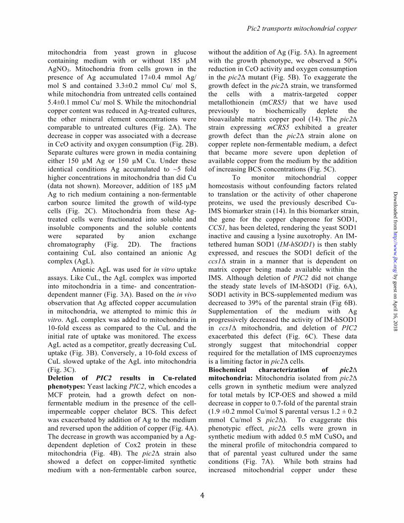
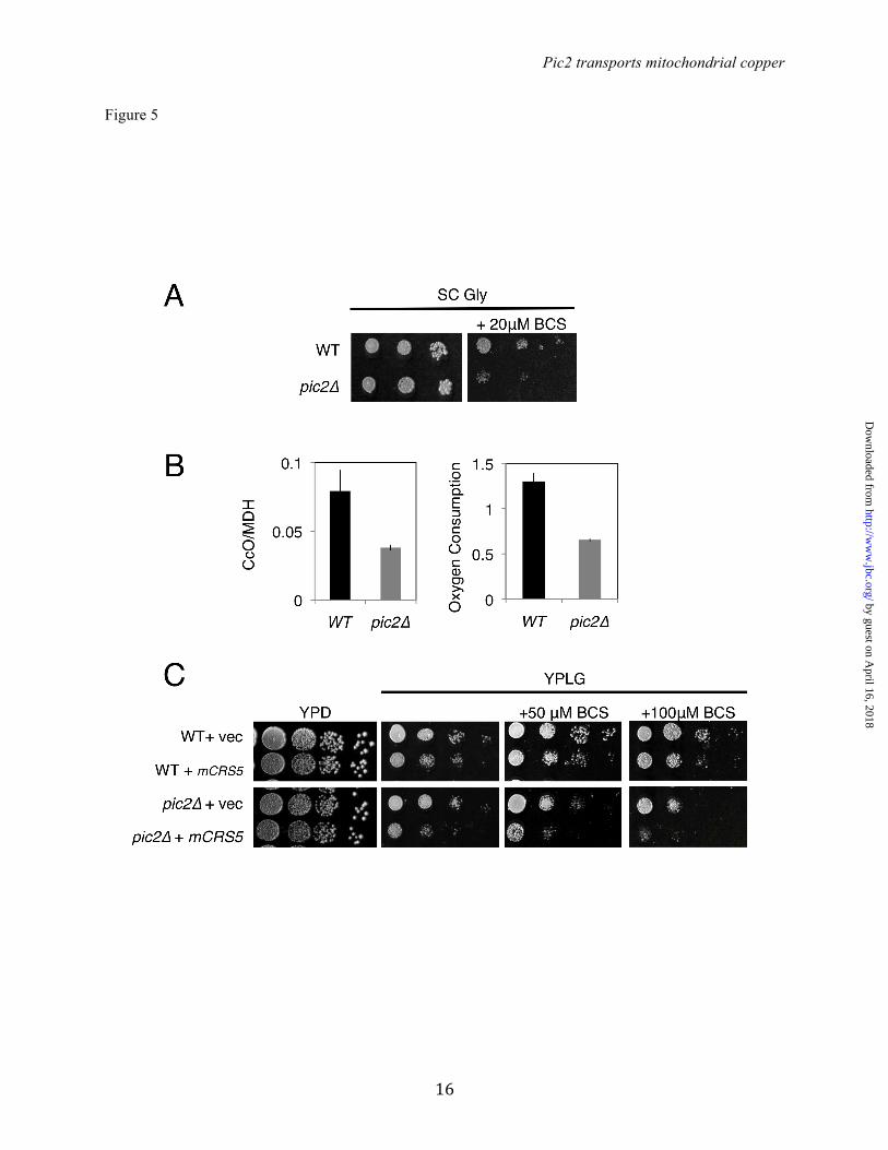
without the addition of Ag (Fig. 5A). In agreement with the growth phenotype, we observed a 50% reduction in CcO activity and oxygen consumption in the pic2∆ mutant (Fig. 5B). To exaggerate the growth defect in the pic2∆ strain, we transformed the cells with a matrix-targeted copper metallothionein (mCRS5) that we have used previously to biochemically deplete the bioavailable matrix copper pool (14). The pic2∆ strain expressing mCRS5 exhibited a greater growth defect than the pic2∆ strain alone on copper replete non-fermentable medium, a defect that became more severe upon depletion of available copper from the medium by the addition of increasing BCS concentrations (Fig. 5C). To monitor mitochondrial copper homeostasis without confounding factors related to translation or the activity of other chaperone proteins, we used the previously described Cu-IMS biomarker strain (14). In this biomarker strain, the gene for the copper chaperone for SOD1, CCS1, has been deleted, rendering the yeast SOD1 inactive and causing a lysine auxotrophy. An IM-tethered human SOD1 (IM-hSOD1) is then stably expressed, and rescues the SOD1 deficit of the ccs1∆ strain in a manner that is dependent on matrix copper being made available within the IMS. Although deletion of PIC2 did not change the steady state levels of IM-hSOD1 (Fig. 6A), SOD1 activity in BCS-supplemented medium was decreased to 39% of the parental strain (Fig 6B). Supplementation of the medium with Ag progressively decreased the activity of IM-hSOD1 in ccs1∆ mitochondria, and deletion of PIC2 exacerbated this defect (Fig. 6C). These data strongly suggest that mitochondrial copper required for the metallation of IMS cuproenzymes is a limiting factor in pic2∆ cells. Biochemical characterization of pic2∆ mitochondria: Mitochondria isolated from pic2∆ cells grown in synthetic medium were analyzed for total metals by ICP-OES and showed a mild decrease in copper to 0.7-fold of the parental strain (1.9 ±0.2 mmol Cu/mol S parental versus 1.2 ± 0.2 mmol Cu/mol S pic2∆). To exaggerate this phenotypic effect, pic2Δ cells were grown in synthetic medium with added 0.5 mM CuSO4 and the mineral profile of mitochondria compared to that of parental yeast cultured under the same conditions (Fig. 7A). While both strains had increased mitochondrial copper under these
by guest on April 16, 2018
http://ww
w.jbc.org/
Dow
nloaded from

Pic2 transports mitochondrial copper
5
conditions, the pic2Δ mitochondria accumulated less Cu than the parental strain (~0.4-fold). There was no observable change in the content of P, Fe, Mn, Ca, Mg but a ~1.5 fold increase in Zn and K. These data suggested a specific defect in copper uptake in pic2∆ mitochondria (Fig. 7A).
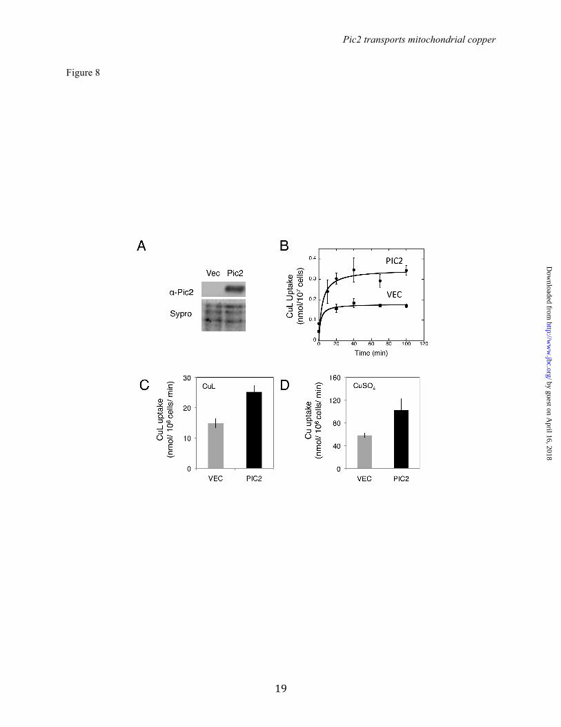
Intact mitochondria from pic2∆ cells were incubated with purified CuL complex and assayed for uptake. Initial rates of CuL uptake were decreased in pic2∆ mitochondria relative to those from parental cells (Fig. 7B). Moreover, pic2∆ mitochondria showed lower maximal rates of uptake. This change in saturation suggests a decreased capacity for CuL uptake in the pic2∆ strain, consistent with the lower total copper accumulation. This uptake defect was evident across a range of CuL concentrations (Fig. 7B). Because Pic2 has been previously identified as a secondary phosphate carrier, mitochondria from pic2∆ cells were assayed for phosphate uptake using an established swelling assay (30). Consistent with previous observations, no defect in phosphate uptake was detected in pic2∆ mitochondria (data not shown). Heterologous expression of Pic2: To account for possible indirect effects of other proteins contributing to the observed defects in mitochondrial copper uptake, we cloned PIC2 into a nisin-inducible vector for expression in the bacterium Lactococcus lactis. MCF proteins expressed in L. lactis are folded correctly in the cytoplasmic membrane and transport can be assayed directly in whole cells (31). The presence of Pic2 in L. lactis induced with nisin was confirmed by Western blot using a Pic2-specific antibody (Fig. 8A). Expression of PIC2 enhanced CuL uptake into L. lactis compared to cells that carried the empty vector (Fig. 8B and C). Addition of a ten-fold excess of phosphate in the form of potassium phosphate buffer did not significantly inhibit copper uptake in the PIC2-expressing cells (data not shown). PIC2 expression in L. lactis enhanced the cellular uptake of CuSO4 (Fig. 8D) and Cu-acetonitrile (data not shown), but did not enhance the uptake of Fe presented as FeSO4 (data not shown). DISCUSSION
Assembly of fully functional CcO depends on a non-proteinaceous pool of labile copper in the mitochondrial matrix (14). Here, we identify Pic2
as the first protein involved in matrix copper import. Yeast cells lacking PIC2 have copper-related growth defects and a deficit in total mitochondrial copper content. Intact mitochondria from these cells show decreased rates of CuL uptake in vitro, and heterologous expression of Pic2 in L. lactis supports copper uptake. Taken together, these data support a novel role for Pic2 in mitochondrial copper homeostasis in S. cerevisiae.
Total copper in the mitochondria of S. cerevisiae is approximately 50 and 200 µM under fermentative and respiratory growth conditions, respectively (32). Previously, we reported that matrix copper content increases at least 6-fold in response to copper salt supplementation in the medium (11). Therefore the in vitro estimation of 15 µM for half-maximal transport of copper in wild-type mitochondria is likely biologically relevant.
The data presented here suggest that Pic2 mediates CuL import, and under normal physiologic conditions we believe CuL to be the major substrate for uptake of the metal ion into the organelle. Apo-ligand appears to be underrepresented in the matrix, suggesting that only the CuL complex is transported across the IM (14). We therefore use CuL as the source of copper in most of our assays, and this complex is extremely stable under all conditions we have tested, including boiling and organic extraction (14). However, we cannot exclude the possibility that copper dissociates from the ligand for its transport across the IM, and rebinds the ligand upon entry into the matrix.
MCF proteins are predicted to contain three alpha helices that fold over into a tripartite structure with six membrane-spanning domains (33). Within these helices are conserved [P]X[DE]XX[RK] motifs which are proposed to narrow the pore of the carrier and form salt bridges to lock it in a state that is closed to the matrix (15,33,34). Substrate binding disrupts these salt bridges and allows for translocation, changing the carrier from an IMS facing state to a matrix facing state. Analysis of the sequence of MCF proteins to identify the salt bridges formed within these motifs has been used to predict whether particular family members act as uniporters or strict exchangers (34). The Pic2 salt bridge network in the IMS-facing conformation appears to be stronger than it is in the matrix-facing
by guest on April 16, 2018
http://ww
w.jbc.org/
Dow
nloaded from
Pic2 transports mitochondrial copper
6
conformation, consistent with the idea that Pic2 is capable of uniport transport (34). According to the proposed mechanism, it is the greater relative strength of the IMS salt bridge network that drives the protein to revert back to this original conformation after substrate translocation (34). Further experiments with isolated Pic2 will be required to assess the residues required for CuL transport and the exact mechanism of transport.
The phenotypes observed here are most consistent with Pic2 acting as an importer of CuL, supporting the steady-state level of CuL in the matrix that is required to activate subsequent transport by an exporter. The expression of matrix-targeted copper competitors does not affect the total levels of copper, but does decrease the available CuL and this induces respiratory defects (14). It is assumed that these competitor copper-binding proteins are able to out-compete the ligand complex for copper binding in the matrix. In pic2∆ this phenotype is exaggerated, presumably because decreased CuL import shifts the equilibrium in favor of the competitor protein(s) whose expression remains constant. To date, we cannot exclude the possibility that Pic2 is capable of bidirectional transport. Analysis of mitochondria from MCF deletion mutants grown under copper supplemented conditions did not reveal an obvious candidate that significantly over-accumulated copper (data not shown). Therefore any potential mechanism by which copper import and export may be linked remains unknown.
Defects in transport of ionic Cu and Fe were previously observed in sub-mitochondrial particles prepared from mutants of the high affinity Fe-transporters Mrs3 and Mrs4 (16). Similarly, we observe that Pic2 in L. lactis can transport CuSO4. Although free copper should not be encountered in the IMS during normal physiologic conditions, this result could explain previous observations of copper accumulation in mitochondria from cells with compromised copper homeostasis. Deletion of ACE1, a copper regulated transcription factor, in yeast prevents the increase in metallothionein expression normally seen in response to copper supplementation. In this mutant
mitochondrial copper is expanded more dramatically than it is in the parental strain with active Ace1 (11). This increase in copper not only expands the soluble matrix pool but also leads to the accumulation of an insoluble fraction within this mitochondrial compartment, the identity of which has not been investigated. Similarly, mitochondria isolated from a rat model of Wilson disease show accumulation of large quantities of insoluble copper that leads to severe membrane damage (35). It is possible that once copper is no longer bound to the CuL, it can react indiscriminately with various mitochondrial components. Pic2 has previously been implicated in phosphate transport in yeast as multi-copy PIC2 could reverse a mir1∆pic2∆ growth defect on a non-fermentable carbon source, and rescue phosphate transport in mir1∆pic2∆ mitochondria as assayed by mitochondrial swelling (30,36). PIC2 has also been identified as a high copy suppressor of the mitochondrial K+/H+ exchanger mutant (mdm38∆) of yeast (37). The authors concluded that PIC2 was suppressing the defect via rescue of proton motive force rather that K+ transport. Interestingly, MRS3 has also been implicated in transport of ionic copper, and also acts as a suppressor of the K+/H+ exchanger mutant (37). Whether the role of Pic2 in phosphate and potassium transport have any affect on copper, or vice versa, remains to be investigated.
The cumulative in vivo and in vitro data argue that Pic2 is a major component of the mitochondrial copper homeostasis machinery. This initial discovery provides a straightforward genetic strategy whereby this null strain can be used to identify additional transporters involved in organellar copper handling. It may also prove to be a useful tool in defining the pathways involved in synthesis of the ligand that binds Cu, as a pic2∆ strain should be sensitized to modest changes in total CuL content.
by guest on April 16, 2018
http://ww
w.jbc.org/
Dow
nloaded from

Pic2 transports mitochondrial copper
7
REFERENCES 1. Cobine, P. A., Pierrel, F., and Winge, D. R. (2006) Copper trafficking to the mitochondrion and
assembly of copper metalloenzymes. BBA-Mol Cell Res 1763, 759-772 2. Huffman, D. L., and O'Halloran, T. V. (2001) Function, structure, and mechanism of intracellular
copper trafficking proteins. Annu Rev Biochem 70, 677-701 3. Dancis, A., Yuan, D. S., Haile, D., Askwith, C., Eide, D., Moehle, C., Kaplan, J., and Klausner, R.
D. (1994) Molecular characterization of a copper transport protein in S. cerevisiae: an unexpected role for copper in iron transport. Cell 76, 393-402
4. Reddi, A. R., and Culotta, V. C. (2013) SOD1 Integrates Signals from Oxygen and Glucose to Repress Respiration. Cell 152, 224-235
5. Tsukihara, T., Aoyama, H., Yamashita, E., Tomizaki, T., Yamaguchi, H., Shinzawa-Itoh, K., Nakashima, R., Yaono, R., and Yoshikawa, S. (1996) The whole structure of the 13-subunit oxidized cytochrome c oxidase at 2.8 A. Science 272, 1136-1144
6. Pufahl, R. A., Singer, C. P., Peariso, K. L., Lin, S. J., Schmidt, P. J., Fahrni, C. J., Culotta, V. C., Penner-Hahn, J. E., and O'Halloran, T. V. (1997) Metal ion chaperone function of the soluble Cu(I) receptor Atx1. Science 278, 853-856
7. Furukawa, Y., Torres, A. S., and O'Halloran, T. V. (2004) Oxygen-induced maturation of SOD1: a key role for disulfide formation by the copper chaperone CCS. EMBO J 23, 2872-2881
8. Horng, Y. C., Cobine, P. A., Maxfield, A. B., Carr, H. S., and Winge, D. R. (2004) Specific copper transfer from the Cox17 metallochaperone to both Sco1 and Cox11 in the assembly of yeast cytochrome C oxidase. J Biol Chem 279, 35334-35340
9. Banci, L., Bertini, I., Ciofi-Baffoni, S., Hadjiloi, T., Martinelli, M., and Palumaa, P. (2008) Mitochondrial copper(I) transfer from Cox17 to Sco1 is coupled to electron transfer. P Natl Acad Sci USA 105, 6803-6808
10. Horn, D., Al-Ali, H., and Barrientos, A. (2008) Cmc1p is a conserved mitochondrial twin CX9C protein involved in cytochrome c oxidase biogenesis. Molecular and cellular biology 28, 4354-4364
11. Cobine, P. A., Ojeda, L. D., Rigby, K. M., and Winge, D. R. (2004) Yeast contain a non-proteinaceous pool of copper in the mitochondrial matrix. J Biol Chem 279, 14447-14455
12. Yang, L., McRae, R., Henary, M. M., Patel, R., Lai, B., Vogt, S., and Fahrni, C. J. (2005) Imaging of the intracellular topography of copper with a fluorescent sensor and by synchrotron x-ray fluorescence microscopy. P Natl Acad Sci USA 102, 11179-11184
13. Dodani, S. C., Leary, S. C., Cobine, P. A., Winge, D. R., and Chang, C. J. (2011) A targetable fluorescent sensor reveals that copper-deficient SCO1 and SCO2 patient cells prioritize mitochondrial copper homeostasis. J Am Chem Soc 133, 8606-8616
14. Cobine, P. A., Pierrel, F., Bestwick, M. L., and Winge, D. R. (2006) Mitochondrial matrix copper complex used in metallation of cytochrome oxidase and superoxide dismutase. J Biol Chem 281, 36552-36559
15. Kunji, E. R., and Robinson, A. J. (2006) The conserved substrate binding site of mitochondrial carriers. Biochimica et biophysica acta 1757, 1237-1248
16. Froschauer, E. M., Schweyen, R. J., and Wiesenberger, G. (2009) The yeast mitochondrial carrier proteins Mrs3p/Mrs4p mediate iron transport across the inner mitochondrial membrane. Biochimica et biophysica acta 1788, 1044-1050
17. Muhlenhoff, U., Stadler, J. A., Richhardt, N., Seubert, A., Eickhorst, T., Schweyen, R. J., Lill, R., and Wiesenberger, G. (2003) A specific role of the yeast mitochondrial carriers MRS3/4p in mitochondrial iron acquisition under iron-limiting conditions. J Biol Chem 278, 40612-40620
18. Shaw, G. C., Cope, J. J., Li, L., Corson, K., Hersey, C., Ackermann, G. E., Gwynn, B., Lambert, A. J., Wingert, R. A., Traver, D., Trede, N. S., Barut, B. A., Zhou, Y., Minet, E., Donovan, A.,
by guest on April 16, 2018
http://ww
w.jbc.org/
Dow
nloaded from
Pic2 transports mitochondrial copper
8
Brownlie, A., Balzan, R., Weiss, M. J., Peters, L. L., Kaplan, J., Zon, L. I., and Paw, B. H. (2006) Mitoferrin is essential for erythroid iron assimilation. Nature 440, 96-100
19. Wang, Y., Langer, N. B., Shaw, G. C., Yang, G., Li, L., Kaplan, J., Paw, B. H., and Bloomer, J. R. (2011) Abnormal mitoferrin-1 expression in patients with erythropoietic protoporphyria. Exp Hematol 39, 784-794
20. Chen, W., Paradkar, P. N., Li, L., Pierce, E. L., Langer, N. B., Takahashi-Makise, N., Hyde, B. B., Shirihai, O. S., Ward, D. M., Kaplan, J., and Paw, B. H. (2009) Abcb10 physically interacts with mitoferrin-1 (Slc25a37) to enhance its stability and function in the erythroid mitochondria. P Natl Acad Sci USA 106, 16263-16268
21. Paradkar, P. N., Zumbrennen, K. B., Paw, B. H., Ward, D. M., and Kaplan, J. (2009) Regulation of mitochondrial iron import through differential turnover of mitoferrin 1 and mitoferrin 2. Molecular and cellular biology 29, 1007-1016
22. Nilsson, R., Schultz, I. J., Pierce, E. L., Soltis, K. A., Naranuntarat, A., Ward, D. M., Baughman, J. M., Paradkar, P. N., Kingsley, P. D., Culotta, V. C., Kaplan, J., Palis, J., Paw, B. H., and Mootha, V. K. (2009) Discovery of genes essential for heme biosynthesis through large-scale gene expression analysis. Cell Metab 10, 119-130
23. Yang, M., Cobine, P. A., Molik, S., Naranuntarat, A., Lill, R., Winge, D. R., and Culotta, V. C. (2006) The effects of mitochondrial iron homeostasis on cofactor specificity of superoxide dismutase 2. EMBO J 25, 1775-1783
24. Gordon, D. M., Lyver, E. R., Lesuisse, E., Dancis, A., and Pain, D. (2006) GTP in the mitochondrial matrix plays a crucial role in organellar iron homoeostasis. Biochem J 400, 163-168
25. Yoon, H., Zhang, Y., Pain, J., Lyver, E. R., Lesuisse, E., Pain, D., and Dancis, A. (2011) Rim2, pyrimidine nucleotide exchanger, is needed for iron utilization in mitochondria. Biochem J
26. Lin, H., Li, L., Jia, X., Ward, D. M., and Kaplan, J. (2011) Genetic and biochemical analysis of high iron toxicity in yeast: iron toxicity is due to the accumulation of cytosolic iron and occurs under both aerobic and anaerobic conditions. J Biol Chem 286, 3851-3862
27. Tong, A. H., and Boone, C. (2006) Synthetic genetic array analysis in Saccharomyces cerevisiae. Methods Mol Biol 313, 171-192
28. Jin, Y. H., Dunlap, P. E., McBride, S. J., Al-Refai, H., Bushel, P. R., and Freedman, J. H. (2008) Global transcriptome and deletome profiles of yeast exposed to transition metals. PLoS Genet 4, e1000053
29. Zatulovskiy, E. A., Skvortsov, A. N., Rusconi, P., Ilyechova, E. Y., Babich, P. S., Tsymbalenko, N. V., Broggini, M., and Puchkova, L. V. (2012) Serum depletion of holo-ceruloplasmin induced by silver ions in vivo reduces uptake of cisplatin. Journal of inorganic biochemistry 116, 88-96
30. Hamel, P., Saint-Georges, Y., de Pinto, B., Lachacinski, N., Altamura, N., and Dujardin, G. (2004) Redundancy in the function of mitochondrial phosphate transport in Saccharomyces cerevisiae and Arabidopsis thaliana. Mol Microbiol 51, 307-317
31. Kunji, E. R., Chan, K. W., Slotboom, D. J., Floyd, S., O'Connor, R., and Monne, M. (2005) Eukaryotic membrane protein overproduction in Lactococcus lactis. Curr Opin Biotechnol 16, 546-551
32. Garber Morales, J., Holmes-Hampton, G. P., Miao, R., Guo, Y., Munck, E., and Lindahl, P. A. (2010) Biophysical characterization of iron in mitochondria isolated from respiring and fermenting yeast. Biochemistry 49, 5436-5444
33. Pebay-Peyroula, E., Dahout-Gonzalez, C., Kahn, R., Trezeguet, V., Lauquin, G. J., and Brandolin, G. (2003) Structure of mitochondrial ADP/ATP carrier in complex with carboxyatractyloside. Nature 426, 39-44
34. Robinson, A. J., Overy, C., and Kunji, E. R. (2008) The mechanism of transport by mitochondrial carriers based on analysis of symmetry. P Natl Acad Sci USA 105, 17766-17771
35. Zischka, H., Lichtmannegger, J., Schmitt, S., Jagemann, N., Schulz, S., Wartini, D., Jennen, L., Rust, C., Larochette, N., Galluzzi, L., Chajes, V., Bandow, N., Gilles, V. S., DiSpirito, A. A.,
by guest on April 16, 2018
http://ww
w.jbc.org/
Dow
nloaded from

Pic2 transports mitochondrial copper
9
Esposito, I., Goettlicher, M., Summer, K. H., and Kroemer, G. (2011) Liver mitochondrial membrane crosslinking and destruction in a rat model of Wilson disease. J Clin Invest 121, 1508-1518
36. Takabatake, R., Siddique, A. B., Kouchi, H., Izui, K., and Hata, S. (2001) Characterization of a Saccharomyces cerevisiae gene that encodes a mitochondrial phosphate transporter-like protein. J Biochem 129, 827-833
37. Zotova, L., Aleschko, M., Sponder, G., Baumgartner, R., Reipert, S., Prinz, M., Schweyen, R. J., and Nowikovsky, K. (2010) Novel components of an active mitochondrial K(+)/H(+) exchange. J Biol Chem 285, 14399-14414
by guest on April 16, 2018
http://ww
w.jbc.org/
Dow
nloaded from

Pic2 transports mitochondrial copper
10
ACKNOWLEDGEMENTS: We would like to thank Shamima Nasrin, Winston Smith, Brigitte Meyers and Hilliary Street for contributions towards creating necessary reagents and collecting data. FOOTNOTES: * This work was supported by National Science Foundation grant MCB 1158497 (to P.A.C), Saskatchewan Health Research Foundation establishment and Natural Sciences and Engineering Research Discovery grants (to S.C.L) and National Institutes of Health GM083292 (to D.R.W). 4. Abbreviations used: BCS: bathocuproine disulfonic, CCCP: carbonylcyanide m-chlorophenylhydrazone, CcO Cytochrome c oxidase, ICP-OES: Inductively coupled plasma optical emission spectroscopy, IMS: intermembrane space, IM: inner membrane, MCF: Mitochondrial carrier family FIGURE LEGENDS Figure 1: In vitro CuL uptake in purified mitochondria. A) Intact mitochondria were incubated with purified CuL then isolated by centrifugation and Cu was measured by ICP-OES. Fraction of maximal uptake is plotted versus incubation time for an average of three independent experiments with wild-type mitochondria. Error bars represent standard deviation. B) Initial rates of uptake across a range of CuL concentrations. Data is fit by a hyperbolic curve. Inset is initial rate of uptake over a 0-50 µM range of CuL, and is fitted by a linear regression. C) Initial rate of CuL uptake measured by ICP-OES in intact mitochondria, mitoplasts (MP) prepared by hypotonic lysis, mitochondria incubated with the uncoupler CCCP or mitochondria incubated at 4oC. The averages with standard deviation are shown for three independent experiments. Figure 2: Ag accumulates in yeast mitochondria. A) Mineral element content of purified mitochondria from BY4741 yeast cultured in the absence or presence of 185 µM Ag was quantified by ICP-OES, and expressed as a ratio (i.e. +Ag/-Ag). The averages with standard deviation of three independent cultures and mitochondrial preparations are shown. B) CcO activity of purified mitochondria from A) and oxygen consumption of whole cells cultured in YP-galactose medium with 0 or 185 µM Ag added. C) Serial dilutions of BY4741 yeast grown on rich medium with a fermentable carbon source (glucose; YPD) or with a non-fermentable carbon source (lactate-glycerol; YPLG) in the presence of the cell-impermeable copper chelator BCS and increasing Ag concentrations. The growth defect was reversed by adding Cu to the BCS- and Ag-supplemented plates. D) Anion exchange fractionation of soluble contents from purified mitochondria isolated from cultures grown in 185 µM Ag. Cu and Ag were measured by ICP-OES, and are shown as solid line with closed squares for Cu and dashed line with diamonds for Ag.
Figure 3: In vitro AgL uptake into purified mitochondria. A) Intact mitochondria were incubated with purified AgL, then isolated by centrifugation and Ag measured by ICP-OES. Initial rates of uptake were plotted against a range of AgL concentrations. A hyperbolic curve is fitted. B) Initial rate of CuL uptake at 10 µM CuL measured by ICP-OES in intact mitochondria in the presence of 100 µM AgL (n=5). C) Initial rate of AgL uptake with 10 µM AgL measured by ICP-OES in intact mitochondria in the presence of 100 µM CuL (n=5). Figure 4: Ag related growth phenotypes of pic2∆ yeast strains. A) Serial dilutions of BY4741 and pic2∆ strains grown on rich medium with a fermentable carbon source (glucose; YPD) or with a non-fermentable carbon source (lactate-glycerol; YPLG) in the presence of the cell-impermeable copper chelator BCS and increasing Ag concentrations. The growth defect was reversed by adding Cu to the BCS- and Ag-supplemented plates. B) Western blots for Cox2 in mitochondria isolated from cells grown in rich medium with a fermentable carbon source (top) or in identical medium supplemented with 150 µM Ag (bottom). Porin served as an internal loading control.
by guest on April 16, 2018
http://ww
w.jbc.org/
Dow
nloaded from

Pic2 transports mitochondrial copper
11
Figure 5: Growth phenotypes of pic2∆ yeast strains under copper depletion. A) Serial dilutions of BY4741 and pic2∆ strains grown on synthetic medium with a non-fermentable carbon source (glycerol; SC Gly) or in SC Gly with 20 µM BCS. B) CcO activity in mitochondria isolated from parental and pic2∆ strains grown in synthetic medium containing galactose as a carbon source, and oxygen consumption, read as % air/OD600/s of whole cells from each strain grown in galactose-containing rich medium (n=3). C) Serial dilutions of parental and pic2∆ strains transformed with either empty vector (vec) or mCRS5 on rich medium with a fermentable carbon source (YPD), non-fermentable carbon source (YPLG) or YPLG with limited copper availability (+ 50 µM BCS, + 100 µM BCS). Figure 6: Deletion of PIC2 limits hSOD1 activity in a ccs1∆::IMhSOD1 reporter strain. A) Western blot analysis of mitochondrial extracts from ccs1∆::IMhSOD and ccs1∆::IMhSOD pic2Δ strains, probed with human SOD1 antibody and porin as an internal loading control. B) Activity of SOD1 in isolated mitochondria from cells grown in synthetic media with fermentable carbon source supplemented with 50 µM BCS as measured by xanthine oxidase/tetrazolium salt assay (n=3). There was no detectable SOD1 activity in mitochondria from ccs1Δ. C) Activity of SOD1 in isolated mitochondria from cells grown in synthetic media with a fermentable carbon source supplemented with increasing Ag concentrations (20, 40 and 80 µM). Figure 7: Total mineral element profile and uptake in mitochondria from pic2Δ. A) Overall mineral profile of purified intact mitochondria from pic2∆ cells assayed by ICP-OES and compared to that of parental cells. Both strains were grown in medium containing 500 µM Cu. The value for each mineral represents the average of ICP-OES analysis of 10 independent mitochondrial preparations and bars represent the standard error of the mean. B) Isolated mitochondria from parental or pic2∆ cells assayed for in vitro uptake of the CuL. Initial rates of uptake are plotted versus variable CuL concentrations. The line is fit by a linear regression. Figure 8. Pic2 expressed in Lactococcus lactis. A) Uptake of the CuL by intact cells transformed with an empty vector (VEC) or PIC2 (PIC2) incubated at room temperature over time with 20 µM CuL (n=4). Error bars represent standard error of the mean. Data is fit by a hyperbolic curve. B) CuL uptake in cells with either VEC or PIC2 incubated with 10 µM CuL (n=3). Error bars represent standard error of the mean. C) Cu uptake in cells with either VEC or PIC2 incubated with 10 µM CuSO4 (n=3). Error bars represent standard error of the mean.
by guest on April 16, 2018
http://ww
w.jbc.org/
Dow
nloaded from

Pic2 transports mitochondrial copper
12
Figure 1
by guest on April 16, 2018
http://ww
w.jbc.org/
Dow
nloaded from

Pic2 transports mitochondrial copper
13
Figure 2
by guest on April 16, 2018
http://ww
w.jbc.org/
Dow
nloaded from

Pic2 transports mitochondrial copper
14
Figure 3
by guest on April 16, 2018
http://ww
w.jbc.org/
Dow
nloaded from

Pic2 transports mitochondrial copper
15
Figure 4
by guest on April 16, 2018
http://ww
w.jbc.org/
Dow
nloaded from
Pic2 transports mitochondrial copper
16
Figure 5
by guest on April 16, 2018
http://ww
w.jbc.org/
Dow
nloaded from

Pic2 transports mitochondrial copper
17
Figure 6
by guest on April 16, 2018
http://ww
w.jbc.org/
Dow
nloaded from

Pic2 transports mitochondrial copper
18
Figure 7
by guest on April 16, 2018
http://ww
w.jbc.org/
Dow
nloaded from
Pic2 transports mitochondrial copper
19
Figure 8
by guest on April 16, 2018
http://ww
w.jbc.org/
Dow
nloaded from

Katherine E. Vest, Scot C. Leary, Dennis R. Winge and Paul A. Cobineby Pic2, a Mitochondrial Carrier Family Protein
is MediatedSaccharomyces cerevisiaeCopper Import into the Mitochondrial Matrix in
published online July 11, 2013J. Biol. Chem.
10.1074/jbc.M113.470674Access the most updated version of this article at doi:
Alerts:
When a correction for this article is posted•
When this article is cited•
to choose from all of JBC's e-mail alertsClick here
by guest on April 16, 2018
http://ww
w.jbc.org/
Dow
nloaded from