mitochondrial protein import: specific recognition and ... · mitochondrial protein import:...
TRANSCRIPT
J. Membrane Biol. 135, 191-207 (1993) The Journal of
Membrane Biology �9 Springer-Verlag New York Inc. 1993
Topical Review
Mitochondrial Protein Import: Specific Recognit ion and Membrane Translocation of Preproteins
Michael Kiebler I, Karin Becker I, Nikolaus Pfanner 2, Walter Neupert I Ilnstitut fiir Physiologische Chemie der Universitfit Mfinchen, Goethestr. 33, 80336 Mfinchen 2, B.R.D. 2Biochemisches Institut der Universitfit Freiburg, Hermann-Herder- Str. 7, 79104 Freiburg/Breisgau, B.R.D.
Received: 20 April 1993
I. Introduction
Mitochondria are double-membrane bounded, energy-converting organelles within eukaryotic cells. They synthesize ATP by oxidative phosphory- lation. Important progress in understanding their function was made when an isolation procedure for intact mitochondria (Hogeboom, Schneider & Pa- lade, 1948) was developed. Time-lapse microcine- matography (Johnson, Walsh & Chen, 1980; Tzago- loft, 1982) revealed that these organelles are remarkably mobile and able to constantly change their shape and are continuously dividing and fusing. Their motility is apparently due to an association with the cytoskeleton which may determine the unique orientation and distribution of mitochondria in various cell types.
Mitochondria are not synthesized de novo; they arise by growth and division of pre-existing organelles (Luck, 1963, 1965). Mitochondria con- tain DNA (Schatz, Haslbrunner & Tuppy, 1964) and a complete system to carry out DNA replica- tion, transcription, and protein synthesis (McLean et al., 1958; Tzagoloff, 1982); however, only a small number of proteins are encoded by mitochondrial DNA, namely a few subunits of the respiratory chain and the ATP synthase, and depending on the organism, one or a few proteins required for the expression of mitochondrial genes. All other mitochondrial proteins are encoded by nuclear genes, and thus have to be imported into the
Key words: Mitochondrial receptor complex--General insertion pore (GIP)--ATP-dependent translocation--MOM22--Topol- ogy of the components of the receptor complex
Correspondence to: W. Neupert
mitochondria and sorted to the four subcompart- ments: the outer and the inner membranes, the intermembrane space and the matrix (Attardi & Schatz, 1988; Hartl et al., 1989).
Precursor forms of mitochondrial nuclear en- coded proteins contain targeting and sorting signals that are instrumental for directing them to these vari- ous subcompartments. Targeting signals can either be present as cleavable amino-terminal signal se- quences or signals present within the mature parts of precursor proteins. For all those precursors destined for the outer membrane and many of the inner membrane, the signals are not cleaved but are pres- ent in the mature protein sequence. Submitochon- drial sorting signals are often found in amino-termi- nal presequences containing a stretch of hydropho- bic amino acids of variable length.
On the other hand, mitochondria contain com- ponents which recognize mitochondrial targeting signals in a specific manner and facilitate the translo- cation of the precursors across the mitochondrial membranes. Thus, targeting of newly synthesized proteins to mitochondria seems to follow a general principle of intracellular protein traffic, that is the presence of signal sequences on the proteins to be transported across membranes and the existence of complementary structures on the surface of organ- elles which recognize the preproteins belonging to the respective cellular subcompartment (Milstein et al., 1972; Blobel & Dobberstein, 1975).
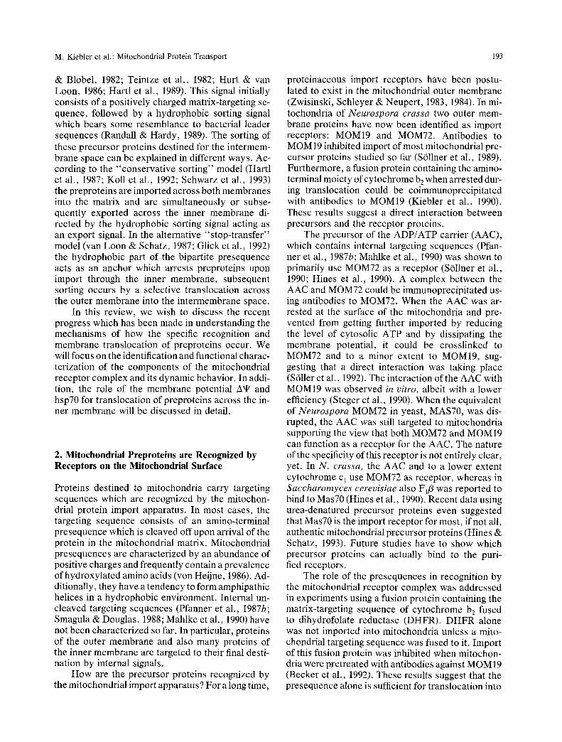
We will first summarize the principal pathways of mitochondrial preproteins from the cytosol to their final destination where they become assembled into functional complexes (Figs. 1 and 2) and then we will focus on specific aspects of recognition and transmembrane transfer of precursors.
Precursor proteins are stabilized in the cytosol

192 M. Kiebler et al.: Mitochondrial Protein Transport
Cytoso,
OM
IMS
Matrix
f
# +A~
Processing B f f ~ Peptidase l ~ , ++~.
~ Chaperones +ATP
|
I Trans,ocationl
Chaperones ' +ATP
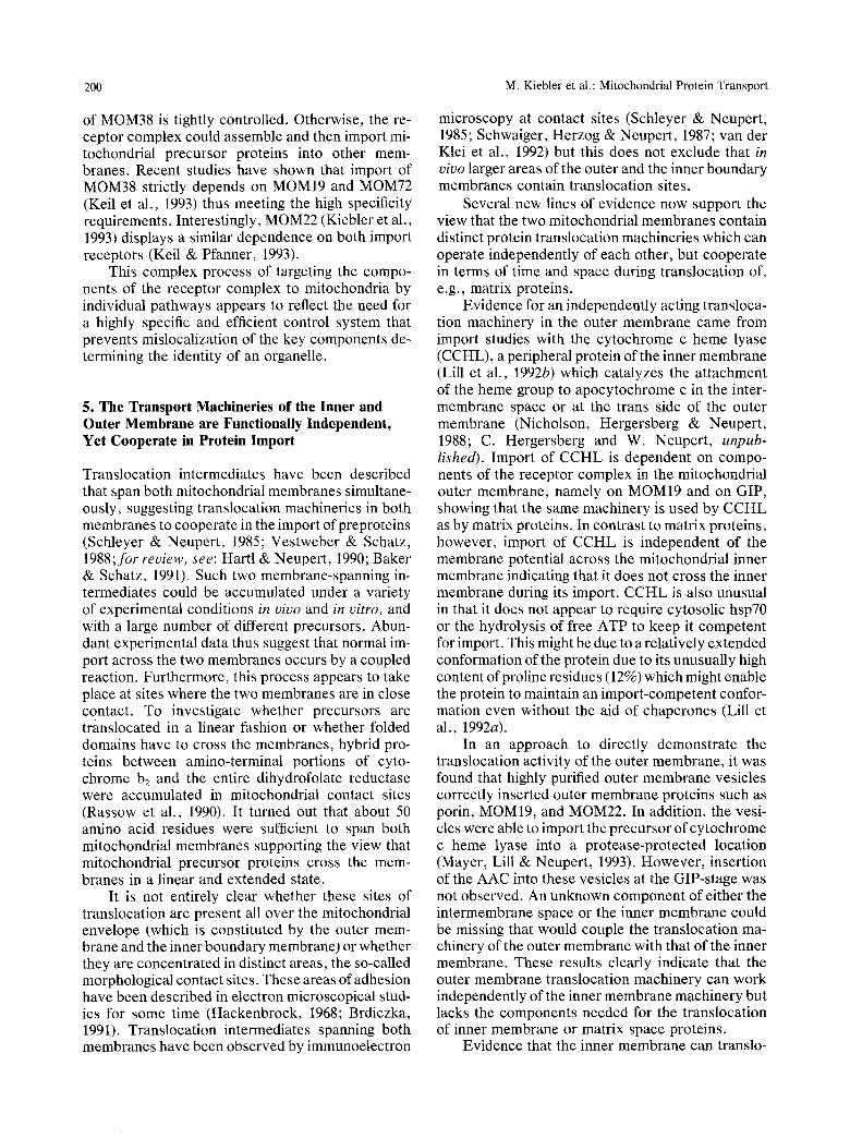
Fig. I. Schematic overview on import of preproteins into mito- chondria. Precursor proteins are recognized by the mitochondrial import receptors (R) and subsequently inserted into the outer membrane (OM) by the general insertion pore (GIP). Then precur- sors are transferred across the intermembrane space (IMS) to the translocation complex (MIM) of the inner membrane (IM) and enter the matrix.
in an import-competent, unfolded state by heat- shock proteins of the hsp70 class in an ATP-depen- dent process (for review, see Rothman, 1989). Other factors interacting with the presequences ap- pear to be also important. The presequences are recognized by receptors on the surface of the outer membrane. Precursor proteins then become inserted into a transport pore in the outer membrane (Pfanner et al., 1987a). Since competition studies revealed that many precursor proteins use the same pore for insertion into the outer membrane, this was termed general insertion pore GIP (Pfaller et al., 1988; Pfan- ner, Hartl & Neupert, 1988).
Precursor proteins destined for the inner mem- brane, for the matrix and some precursors destined for the intermembrane space, have to be translo- cated through or inserted into the inner membrane. This occurs at translocation contact sites where the outer and the inner membrane are in close contact (Pfanner et al., 1992). In vivo, more than 90% of the outer membrane appears to be in close contact with
a specific part of the inner membrane, termed inner boundary membrane (van der Klei, Veenhuis & Neupert, 1992).
Translocation of preproteins across the mito- chondrial inner membrane is dependent on a mem- brane potential A't r across the inner membrane and on ATP in the matrix. The precursor proteins in transit interact with mitochondrial heat-shock pro- teins of the hsp70 class (mt-hsp70) in the matrix in an ATP-dependent manner (Kang et al., 1990). It is assumed that binding and subsequent ATP-depen- dent release of hsp70 from the precursor provides at least part of the driving force for the translocation of preproteins through the two mitochondrial mem- branes. The mitochondrial processing peptidase consisting of the two related components a-MPP and fl-MPP removes the presequences (B6hni et al., 1980; Yaffe & Schatz, 1984; Hawlitschek et al., 1988; Jensen & Yaffe, 1988; Witte et al., 1988; Pollock et al., 1988; Schulte et al., 1989). Proteins finally lo- cated in the matrix are transferred from mt-hsp70 to the chaperonin hsp60 (Manning-Krieg, Scherer & Schatz, 1991), the mitochondrial homologue of the Escherichia coli GroEL, which mediates ATP- dependent folding and assembly (Cheng et al., 1989; Ostermann et al., 1989; Cheng, Hartl & Horwich, 1990). Hsp70 and hsp60 may act in concert with some helper proteins such as the recently discovered hspl0 (the GroES homologue; Hartman et al., 1990, 1992, 1993; Lubben et al., 1990) or not yet identified homologues of E. coli DnaJ and GrpE.
A number of mitochondrial precursors are known to become processed in a two-step mecha- nism. With the precursor of Fo-ATPase subunit 9, the second cleavage is also performed by MPP (Schmidt et al., 1984). For several other precursors, cleavage by MPP is followed by the removal of an octapeptide by an octapeptidyl-peptidase or mito- chondrial intermediate processing peptidase (MIP) which is located in the matrix (Kalousek, Hendrick & Rosenberg, 1988; Hendrick, Hodges & Rosen- berg, 1989; Isaya et al., 199I; Isaya, Kalousek & Rosenberg, 1992a,b; Kalousek, Isaya & Rosenberg, 1992). The sorting of some preproteins to the inter- membrane space involves another proteolytic step in addition to the one catalyzed by MPP. This pro- cessing is performed in the intermembrane space by the membrane-bound protease termed inner mem- brane peptidase 1 (IMP1, van Loon, Brfindli & Schatz, 1986; Hartl et al., 1987; Behrens, Michaelis & Pratje, 1991; A. Schneider et al., 1991). Interest- ingly, this protease shares sequence similarity with the E. coli leader peptidase supporting the endosym- biontic theory. Some of the intermembrane space precursor proteins contain a bipartite signal at their amino-terminus (Gasser et al., 1982; Kaput, Goltz
M. Kiebler et al.: Mitochondrial Protein Transport 193
& Blobel, 1982; Teintze et al., 1982; Hurt & van Loon, 1986; Hartl et al., 1989). This signal initially consists of a positively charged matrix-targeting se- quence, followed by a hydrophobic sorting signal which bears some resemblance to bacterial leader sequences (Randall & Hardy, 1989). The sorting of these precursor proteins destined for the intermem- brane space can be explained in different ways. Ac- cording to the "conservative sorting" model (Hartl et al., 1987; Koll et al., 1992; Schwarz et al., 1993) the preproteins are imported across both membranes into the matrix and are simultaneously or subse- quently exported across the inner membrane di- rected by the hydrophobic sorting signal acting as an export signal. In the alternative "stop-transfer" model (van Loon & Schatz, 1987; Glick et al., 1992) the hydrophobic part of the bipartite presequence acts as an anchor which arrests preproteins upon import through the inner membrane, subsequent sorting occurs by a selective translocation across the outer membrane into the intermembrane space.
In this review, we wish to discuss the recent progress which has been made in understanding the mechanisms of how the specific recognition and membrane translocation of preproteins occur. We will focus on the identification and functional charac- terization of the components of the mitochondrial receptor complex and its dynamic behavior. In addi- tion, the role of the membrane potential A't r and hsp70 for translocation of preproteins across the in- ner membrane will be discussed in detail.
2. Mitochondriai Preproteins are Recognized by Receptors on the Mitochondrial Surface
Proteins destined to mitochondria carry targeting sequences which are recognized by the mitochon- drial protein import apparatus. In most cases, the targeting sequence consists of an amino-terminal presequence which is cleaved off upon arrival of the protein in the mitochondrial matrix. Mitochondrial presequences are characterized by an abundance of positive charges and frequently contain a prevalence of hydroxylated amino acids (von Heijne, 1986). Ad- ditionally, they have a tendency to form amphipathic helices in a hydrophobic environment. Internal un- cleaved targeting sequences (Pfanner et al., 1987b; Smagula & Douglas, 1988; Mahlke et al., 1990) have not been characterized so far. In particular, proteins of the outer membrane and also many proteins of the inner membrane are targeted to their final desti- nation by internal signals.
How are the precursor proteins recognized by the mitochondrial import apparatus ? For a long time,
proteinaceous import receptors have been postu- lated to exist in the mitochondrial outer membrane (Zwisinski, Schleyer & Neupert, 1983, 1984). In mi- tochondria of Neurospora crassa two outer mem- brane proteins have now been identified as import receptors: MOMI9 and MOM72. Antibodies to MOM 19 inhibited import of most mitochondrial pre- cursor proteins studied so far (S611ner et al., 1989). Furthermore, a fusion protein containing the amino- terminal moiety ofcytochrome b 2 when arrested dur- ing translocation could be coimmunoprecipitated with antibodies to MOM19 (Kiebler et al., 1990). These results suggest a direct interaction between precursors and the receptor proteins.
The precursor of the ADP/ATP carrier (AAC), which contains internal targeting sequences (Pfan- ner et al., 1987b; Mahlke et al., 1990) was shown to primarily use MOM72 as a receptor (S611ner et al., 1990; Hines et al., 1990). A complex between the AAC and MOM72 could be immunoprecipitated us- ing antibodies to MOM72. When the AAC was ar- rested at the surface of the mitochondria and pre- vented from getting further imported by reducing the level of cytosolic ATP and by dissipating the membrane potential, it could be crosslinked to MOM72 and to a minor extent to MOM19, sug- gesting that a direct interaction was taking place (S611er et al., 1992). The interaction of the AAC with MOM19 was observed in vitro, albeit with a lower efficiency (Steger et al., 1990). When the equivalent of Neurospora MOM72 in yeast, MAS70, was dis- rupted, the AAC was still targeted to mitochondria supporting the view that both MOM72 and MOM19 can function as a receptor for the AAC. The nature of the specificity of this receptor is not entirely clear, yet. In N. crassa, the AAC and to a lower extent cytochrome cl use MOM72 as receptor, whereas in Saccharomyces cerevisiae also F1/3 was reported to bind to Mas70 (Hines et al., 1990). Recent data using urea-denatured precursor proteins even suggested that Mas70 is the import receptor for most, if not all, authentic mitochondrial precursor proteins (Hines & Schatz, 1993). Future studies have to show which precursor proteins can actually bind to the puri- fied receptors.
The role of the presequences in recognition by the mitochondrial receptor complex was addressed in experiments using a fusion protein containing the matrix-targeting sequence of cytochrome b2 fused to dihydrofolate reductase (DHFR). DHFR alone was not imported into mitochondria unless a mito- chondrial targeting sequence was fused to it. Import of this fusion protein was inhibited when mitochon- dria were pretreated with antibodies against MOM 19 (Becker et al., 1992). These results suggest that the presequence alone is sufficient for translocation into
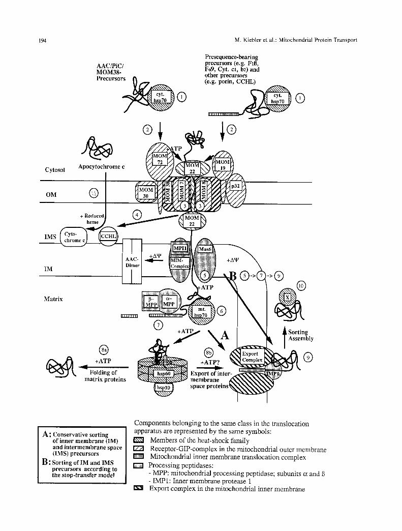
194 M. Kiebler et al.: Mitochondrial Protein Transport
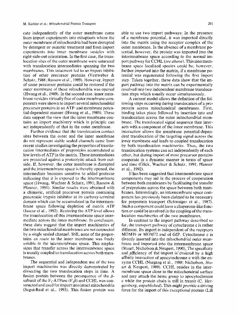
AAC/PiC/ MOM38- Precursor~ @
Cytos
OM
Presequence-bearing precursors (e.g. Fill, Fo9, Cyt. cl, b2)and other precursors (e.g. porin, CCHL)
I I I I I I I I|r;';::!;!;!;::!;r::;r:r:~
IMS
IM
Matrix
| ~ +ATP
Folding of matrix proteins
i ng mbly
G
A ~ Conservative sorting of inner membrane (IM) and intermemhrane space (IMS) precursors
B: Sorting of IM and IMS precursors according to the stop-transfer model
Components belonging to the same class in the translocation apparatus are represented by the same symbols:
Members of the heat-shock family [Z21 Receptor-GIP-complex in the mitochondrial outer membrane
Mitochondrial inner membrane translocation complex D Processing peptidases:
- MPP: mitochondrial processing peptidase; subunits ot and - IMP1: Inner membrane protease 1
I~ i Export complex in the mitochondrial inner membrane
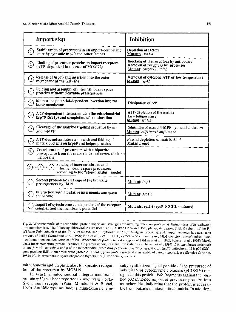
M. Kiebter et al.: Mitochondrial Protein Transport 195
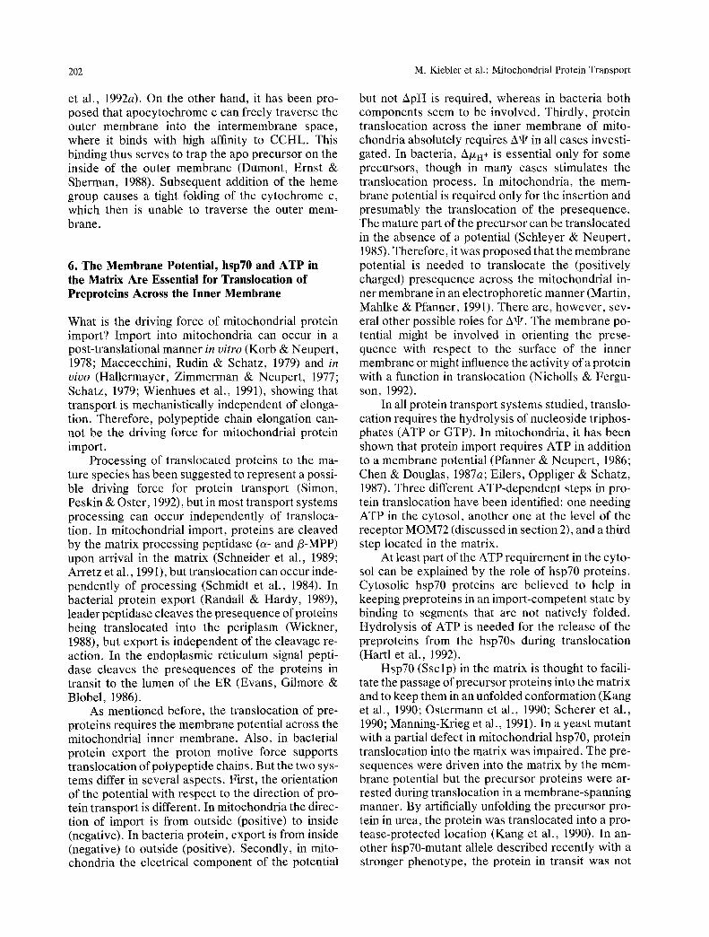
Import step
Stabilization of precursors in an import-competent state by cytosolic hsp70 and other factors
Binding of precursor proteins to import receptors (ATP-dependent in the case of MOM72)
i
Release of hsp70 and insertion into the outer membrane at the GIP-site
i
Folding and assembly of intermembrane space proteins without cleavable presequences
Membrane potential.dependent insertion into the inner membrane
@
�9
|
ATP-dependent interaction with the mitochondrial hsp70 (Ssclp) and completion of translocation
i
Cleavage of the matrix-targeting sequence by ~ - and g-MPP
ATP-dependent interaction with and folding of matrix proteins on hsp60 and helper proteins
Translocation of precursors with a bipartite presequence from the matrix into and across the inne membrane
Sorting of innermembrane and @ ' > @ ' > @ intermembrane space precursors
according to the "stop-transfer" model
Second proteolytic cleavage of the bipartite presequences by IMP1
Interaction with a putative intermembrane space chaperone
i i
Import of cytochrome c independent of the receptor complex and the membrane potential
Inhibition
Depletion of factors Mutants: ssal.4
ii
Blocking of the receptors by antibodies Removal of receptors by proteases Mutant: Amom72 , mirl
Removal of cytosolic ATP or low temperature Mutant: isp42
J
i
Dissipation of A~
ATP.depletion of the matrix Low temperature Mutant: sscl/2
Inhibition of c~ and B-MPP by metal chelators Mutant: mifl/masl mif2/mas2
J i
Partial depletion of matrix ATP Mutant: mif4
Mutant: impl
Mutant: scol ?
Mutants: cyt2-1; cyc3 (CCHL mutants)
<
Fig. 2. Working model of mitochondrial protein import and strategies for arresting precursor proteins at distinct steps of its pathways into mitochondria. The following abbreviations are used: AAC, ADP/ATP carrier; PiC, phosphate carrier; FIB,/3-subunit of the F I- ATPase; Fo9, subunit 9 of the Fo-ATPase; cyt. hsp70, cytosolic hsp70 (SSA1-4gene products); p32, import receptor in yeast, gene product of MIR1 (Murakami et al., 1990; Pain et al., 1990); CCHL, cytochrome c heine lyase; MIM complex, mitochondrial inner membrane translocation complex; MPI1, Mitochondrial protein import component 1 (Maarse et al., 1992; Scherer et al., 1992); Mas6, yeast inner membrane protein, required for protein import, essential for viability (R. Jensen et al., 1993); A~, membrane potential; a- and/3-MPP, subunits c~ and/3 of the mitochondrial processing peptidase (mifl/2 or masl/2); rot. hsp70, mitochondrial hsp70 (SSC1 gene product; IMP1, inner membrane protease 1; Scolp, yeast protein involved in assembly of cytochrome oxidase (Schulze & R6del, 1988); IC, interrnembrane space chaperone (hypothetical). For details, see text.
mitochondria and, in particular, for specific recogni- tion of the precursor by MOMI9.
In yeast, a mitochondrial integral membrane protein (p32) has been repor ted to function as a puta- tive import receptor (Pain, Murakami & Blobel, 1990). Anti-idiotypic antibodies, mimicking a chemi-
cally synthesized signal peptide of the precursor of subunit IV of cy tochrome c oxidase (pCOXIV) rec- ognized this protein. Fab fragments against the puri- fied p32 inhibited import of precursor proteins into mitochondria, indicating that the protein is accessi- ble from outside in intact mitochondria. In addition,
196 M. Kiebler et al.: Mitochondrial Protein Transport
p32 was localized to the outer membrane at contact sites by immunoelectron microscopy (Pain et al., 1990). When p32 was disrupted in yeast, haploid cells were unable to grow in nonfermentable carbon sources indicating that p32 is important but not es- sential for mitochondrial function and biogenesis. The mutant mitochondria showed a diminished im- port efficiency for precursor proteins. Unexpect- edly, the primary sequence of p32 (Murakami, Blo- bel & Pain, 1990) was identical to a protein identified as the mitochondrial phosphate carrier, an inner membrane protein (Gu6rin et al., 1990; Phelps, Schobert & Wohlrab, 1991). More recently, it was shown that purified p32 binds to mitochondrial pre- sequences, but not to signal sequences required for protein translocation into chloroplasts or endo- plasmatic reticulum (Murakami, Blobel & Pain, 1993). It remains to be investigated whether p32 is exclusively located in the inner membrane or if it has a dual localization in both the inner and the outer membrane and also, if p32 functions as a signal- sequence binding subunit of a protein-conducting channel in the outer membrane or as a phosphate translocator in the inner membrane.
To identify putative signal-sequence receptors, synthetic peptides derived from presequences of precursor proteins were used. These were either radiolabeled and cross-linked to mitochondrial pro- teins or used as ligands on an affinity chromatogra- phy column. By these procedures, several prese- quence-binding proteins were identified. In one report, a 28-kD protein of mitochondria from rabbit heart, rat liver, bovine adrenal cortex and S. pombe could be crosslinked (Font et al., 1991). This protein was suggested to be located at the outer side of the inner membrane. In other reports, two proteins of 29 and 52 kD could be purified from rat liver mito- chondria, and antibodies raised against these pro- teins inhibited import of precursor proteins into mi- tochondria (Ono & Tuboi, 1990a, 1991). Future studies have to show what the precise role of these signal sequence receptors is in protein translocation across the mitochondrial membranes.
Mitochondrial import receptors can be proteo- lyrically removed from the surface of mitochondria by treatment with proteases. Import into these mito- chondria could still occur albeit with low efficiency (Pfaller, Pfanner & Neupert, 1989). This "bypass import" showed the characteristics of authentic mi- tochondrial protein import except for receptor speci- ficity. Import of a chloroplast preprotein into mito- chondria, which occurred with a low efficiency in intact mitochondria, was not inhibited by trypsiniz- ing mitochondria prior to import. These results sug- gested that the function of the mitochondrial protein import receptors may be explained by conferring
specificity and increasing the efficiency of the process.
An example of a protein that enters the mito- chondrial import pathway at a post-receptor stage is the precursor to subunit Va of the yeast cytochrome oxidase (Miller & Cumsky, 1991). Its import occurs independently of protease-sensitive receptors in the outer membrane. It is not known with which compo- nent it interacts first, but it has been claimed that the protein enters at the stage of GIP. COXVa further proceeds into the mitochondrial matrix at the trans- location contact sites. It will be interesting to see which property enables COXVa to bypass the prote- ase-sensitive receptors.
The movement of the AAC from a receptor- bound stage to the GIP requires ATP (Pfanner & Neupert, 1986; 1987). A recent study provided some information on the molecular nature of this ATP requirement by using the ATP analogue 8-azido- ATP which can be photocrosslinked to ATP-binding proteins. Interestingly, irradiation of mitochondria in the presence of 8-azido-ATP resulted in a strongly reduced insertion of the AAC into the outer mem- brane (M. Kiebler et al., submitted for publication). The major target of ATP photocrosslinking was MOM72, whereas the other components of the re- ceptor complex were not labeled. Under the condi- tions used, mitochondrial hsp70 was not labeled, excluding that interference with hsp70 caused the inhibition of the import of AAC. These results sug- gest that MOM72 functions in an ATP-dependent manner with a possible role for ATP-hydrolysis in the release of the preprotein or the recycling of the receptor.
An interesting question is whether receptors can bind presequences directly or whether additional cy- tosolic factors are necessary for mediating this bind- ing reaction. If so, this would resemble the situation in the endoplasmic reticulum (ER), where the signal recognition particle (SRP) targets the precursor pro- teins to the ER membrane (Walter, Gilmore & Blobel, 1984). In the ER, the precursors are not directly recognized by the import receptors, but in- stead, the SRP is recognized by its cognate receptor and transfers the bound protein to the translocation channel in the ER membrane (Ogg et al., 1992).
Several studies have suggested a requirement of cytosolic cofactors in mitochondrial protein import (see Becker et al., 1992 for review). Two possible major functions for cofactors were proposed: (i) a chaperone-like function for preventing misfolding and preserving a transport-competent conformation of preproteins (Ohta & Schatz, 1984; Pelham, 1986; Chen& Douglas, 1987b; Chirico, Waters & Blobel, 1988; Deshaies et al., 1988; H. Murakami et al., 1988; Rothman, 1989; Neupert et al., 1990; Murakami &
M. Kiebler et al.: Mitochondrial Protein Transport 197
Mori, 1990; Sheffield, Shore & Randall, 1990) and (ii) a direct involvement in the targeting of precur- sors. The first function is apparently fulfilled by cyto- solic heat-shock proteins (e.g., hsp70), which bind to preproteins and keep them in an import-competent conformation (H. Murakami et al., 1988; Hartl, Mar- tin & Neupert, 1992).
A role of putative presequence-binding factors in targeting mitochondrial precursor proteins to mi- tochondria has been repeatedly suggested (K. Mura- kami et al., 1988; Ono & Tuboi, 1988; Murakami & Mori, 1990). These factors were identified by frac- tionating rabbit reticulocyte lysate and analyzing the fractions with regard to their ability to confer import competence to a precursor protein. A 50-kD protein called PBF (presequence binding factor) was found to be required for the import of precursors with a cleavable presequence into rat liver mitochondria. PBF was isolated due to its ability to form a complex with a purified mitochondrial precursor protein, namely pOTC (pre-ornithine transcarbamylase). This binding was inhibited by the corresponding pre- peptide, suggesting that the factor indeed binds to the presequence (K. Murakami et al., 1988; Mura- kami & Mori, 1990; Murakami et al., 1992). A 28- kD protein which stimulated import into mitochon- dria of a peptide corresponding to the presequence of pOTC was purified from rabbit reticulocyte lysate (Ono & Tuboi, 1990b). This protein was also re- quired for the binding of the presequence peptide to liposomes constituted of lecithin and the partially purified mitochondrial import receptor (Ono & Tu- boi, 1988, 1990a). Another cytosolic factor, mito- condrial import stimulating factor (MSF), was re- ported to enhance the import of the precursor to the matrix-localized protein adrenodoxin. The protein is a heterodimer of 30 and 32 kD subunits. It may promote depolymerization of the oligomeric precur- sor and unfolding in an ATP-dependent manner (Hachiya et al., 1993).
To investigate whether cytosolic factors are es- sential for import or whether their function can be circumvented, a fusion protein was overexpressed in E. coli and purified to apparent homogeneity that consisted of the amino-terminal part of cytochrome b 2 fused to F1/3 (Becker et al., 1992). The purified protein was denatured in urea and therefore its im- port was independent of cytosolic hsp70 (Eilers & Schatz, 1986; Pfanner et al., 1990). This protein was added to mitochondria which had been extensively washed in buffer containing high salt to remove any attached cytosolic factors. Import of the purified protein into these mitochondria was independent of the addition of cytosolic factors and could not be further stimulated by the addition of reticulocyte lysate. The import of the fusion protein was inhibited
by antibodies to MOM19 and showed all other char- acteristics of the authentic mitochondrial protein im- port. Therefore, it was concluded that protein tar- geting to mitochondria can occur independently of the addition of cytosolic recognition factors (Becket et al., 1992). This is in apparent contrast to the re- sults mentioned above concerning the identification ofpresequence binding factors. Clearly, studies with intact cells are required to fully understand the role of cytosolic factors in mitochondrial targeting.
3. A Protein Complex in the Mitochondrial Outer Membrane Contains the Receptors and a General Insertion Pore
After binding to the mitochondrial outer membrane receptors, the precursor proteins become inserted into and translocated across the mitochondrial outer membrane. An important step towards a molecular understanding of mitochondrial protein uptake was the establishment of methods to arrest translocation at certain stages and thus generate translocation in- termediates (Pfanner & Neupert, 1987; Hartl et al., 1989; Planner et al., 1990). In particular, analysis of the import of the ADP/ATP carrier provided useful information on the consecutive steps of transloca- tion. In contrast to the receptor-bound intermediate of the AAC precursor the intermediate at the GIP- stage was found to be resistant to proteases added to intact mitochondria and to require ATP for its formation. AAC arrested at the GIP could be ex- tracted by protein denaturants, but not by detergents suggesting the existence of a proteinaceous pore as had already been proposed earlier (Blobel, 1980).
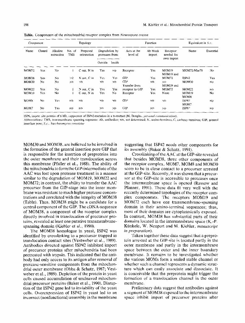
What could be the molecular nature of these proteinaceous pores? In N. crassa (Kiebler et al., 1990) and in S. cerevisiae (Moczko et al., 1992), the two import receptors MOM19 and MOM72 are found in a high molecular weight complex in the mitochondrial outer membrane. In both organisms, the complex contains at least five additional proteins of 7, 8, 22, 30 and 38 kD (Table), termed MOM7, MOM8, MOM22, MOM30 and MOM38 (Kiebler et al., 1990; Moczko et al., 1992; S611ner et al., 1992). This mitochondrial receptor complex is thought to represent the translocation machinery of the outer membrane organized in a cooperating multi-subunit complex as found in other biological systems (A1- berts & Miake-Lye, 1992). Interestingly, the AAC precursor only copurifies with the receptor complex if it is arrested at the GIP-site, but not if it is arrested at the receptor level or if it is fully imported and assembled, indicating that the GIP-site copurifies with the receptor complex (Kiebler et al., 1990).
Four of these proteins, MOM7, MOM8,
198 M. K i e b l e r et al . : M i t o c h o n d r i a l P r o t e i n T r a n s p o r t
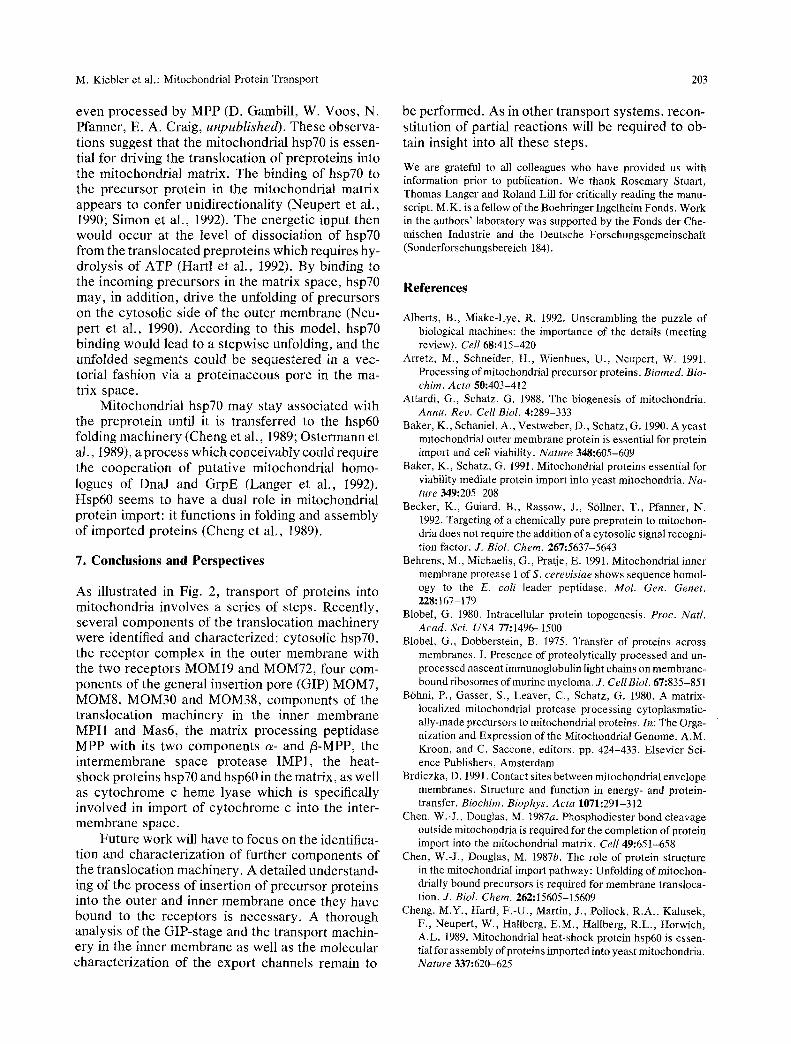
T a b l e . C o m p o n e n t s o f t he m i t o c h o n d r i a l r e c e p t o r c o m p l e x f r o m Neurospora crassa
Component Topology Function Equivalent in S.c.
Name Cloned Alkaline No. of Proposed Degradation by Acts at the Ab block Receptor Name Essential extraction TMS orientation proteases from: level of import needed for
own import
Outside Inside
MOM72 Yes No 1 C out, N in Yes ND Receptor Yes MOMI9 MOM72/Mas70 No MOM19 and
MOM38 Yes No >2 N out, C in Yes Yes GIP Yes MOM72 ISP42 Yes MOM30 No No ND ND ND ND GIP ND ND MOM30 NI)
Transfer from MOM19 and MOM22 Yes No 1 N out, C in Yes Yes receptor to GIP Yes MOM72 MOM22 ND MOM19 Yes No 1 C out, N in Yes No Receptor Yes None MOM19 No
MOM8 MOM8 No Yes ND ND ND ND GIP ND ND ISP6? ND
MOM7 MOM7 No Yes ND ND ND ND GIP ND ND ISP6? ND
ISP6, import site protein of 6 kD, suppressor of ISP42-mutation in a ts-mutant (M. Douglas, personal communication). Abbreviations: TMS, t ransmembrane spanning segments; Ab, antibodies; ND, not determined; N, amino-terminus; C, carboxy-terminus; GIP, general insertion pore; S.c., Saccharomyces cerevisiae
MOM30 and MOM38, are believed to be involved in the formation of the general insertion pore GIP that is responsible for the insertion of preproteins into the outer membrane and their translocation across this membrane (Pfaller et al., 1988). The ability of the mitochondria to form the GIP-intermediate of the AAC was lost upon protease treatment in a manner similar to the degradation of MOM19, MOM22 and MOM72; in contrast, the ability to transfer the AAC precursor from the GIP-stage into the inner mem- brane was resistant to much higher protease concen- trations and correlated with the integrity of MOM38 (Table). Thus, MOM38 might be a candidate for a central component of the GIP. The cDNA-sequence of MOM38, a component of the receptor complex directly involved in translocation of precursor pro- teins, revealed at least one putative transmembrane- spanning domain (Kiebler et al., 1990).
The MOM38 homologue in yeast, ISP42 was identified by crosslinking to a precursor trapped in translocation contact sites (Vestweber et al., 1989). Antibodies directed against ISP42 inhibited import of precursor proteins after mitochondria had been pretreated with trypsin. This indicated that the anti- body had only access to its antigen after removal of protease-sensitive components from the mitochon- drial outer membrane (Ohba & Schatz, 1987; Vest- weber et al., 1989). Depletion of the protein in yeast cells caused accumulation of uncleaved mitochon- drial precursor proteins (Baker et al., 1990). Disrup- tion of the ISP42 gene led to inviability of the yeast cells. Overexpression of ISP42 in yeast led to an incorrect (nonfunctional) assembly in the membrane
suggesting that ISP42 needs other components for its assembly (Baker & Schatz, 1991).
Crosslinking of the AAC at the GIP-site revealed that besides MOM38, three other components of the receptor complex, MOM7, MOM8 and MOM30 seem to be in close contact to a precursor arrested at the GIP-site. Recently, it was shown that a precur- sor at the GIP-site is accessible to proteases once the intermembrane space is opened (Rassow and Pfanner, 1991). These data fit very well with the recently determined topologies of the receptor com- plex components. The receptors MOM19 and MOM72 each have one transmembrane-spanning domain in their amino-terminal sequences; thus, most of their domains are cytoplasmically exposed. In contrast, MOM38 has substantial parts of their domains located in the intermembrane space (K.-P. Kt~nkele, W. Neupert and M. Kiebler, manuscript in preparation).
Taken together these data suggest that a prepro- tein arrested at the GIP-site is located partly in the outer membrane and partly in the intermembrane space between the outer and the inner boundary membrane. It remains to be investigated whether the various MOMs form a sealed stable channel or whether such a channel represents a dynamic struc- ture which can easily associate and dissociate. It is conceivable that the preprotein might trigger the formation of a translocation channel in the outer membrane.
Preliminary data suggest that antibodies against a segment of MOM38 exposed to the intermembrane space inhibit import of precursor proteins after
M. Kiebler et al.: Mitochondrial Protein Transport 199
swelling of mitochondria (K.-P. Ktinkele, W. Neup- ert and M. Kiebler, manuscript in preparation). Thus, parts of MOM38 in the intermembrane space may be essential for the GIP function. It is tempting to speculate that this part might be responsible for mediating the contact to a component of the translo- cation machinery of the inner membrane once the preprotein has been inserted into the outer mem- brane pore. In addition, these observations point to a direct involvement of MOM38 in the insertion process.
The function of MOM22, the seventh of the so far identified components of the receptor complex has recently been investigated in some detail. MOM22 exposes its amino-terminal half to the cyto- sol, this is followed by a transmembrane-spanning segment and by a carboxy-terminal portion in the intermembrane space. Remarkably, the amino-ter- minal domain of MOM22 contains a cluster of 18 negatively charged amino acid residues. MOM22 ap- pears to play a central role in the import of precursor proteins (Kiebler et al., 1993). Antibodies directed against the amino-terminal domain inhibited the im- port of practically all precursors tested. The current hypothesis is that MOM22 functions after the initial binding of precursors to the receptors and that both receptors transfer the precursor proteins to MOM22. The negatively charged cytosolic domain of MOM22 may then facilitate the insertion of the positively charged signal sequences into the outer membrane.
Yeast mitochondria contain a receptor/import complex with a similar protein composition as found in N. crassa mitochondria (Moczko et al., 1992). So far, three of the components of this yeast complex have been identified. ISP42, the yeast homologue of MOM38 (Vestweber et al., 1989; Baker et al., 1990) and MAS70, the homologue of MOM72 (Steger et al., 1990; Hines et al., 1990) and MOM19/MAS22 (Moczko et al., 1993). It may be expected that the two complexes exhibit similar properties. In fact, N. crassa MOM19 coexpressed in yeast efficiently assembled into the yeast complex. The possibility to isolate chemical amounts of the yeast complex should allow the identification and characterization of the remaining components.
Interestingly, the receptor complex varies in its composition with different isolation procedures. Coimmunoprecipitation with the anti-MOM19 anti- body identifies all known components of the original receptor complex. In contrast, the anti-MOM72 anti- body only coimmunoprecipitates MOM7, MOM8 and part of MOM38 (M. Moczko and N. Pfanner unpublished). Yet different subcomplexes are de- tected with antibodies directed against MOM22 or MOM38 (M. Kiebler, unpublished). These data sug- gest the existence of distinct receptor subcom-
plexes. Future studies have to show which particular functions these subcomplexes fulfill during protein import.
In a different approach to understand the physi- cal nature of the insertion pores in the mitochondrial outer membrane electrophysical methods have been applied. There is growing evidence that in the outer membrane in addition to the voltage-dependent anion channels (VDAC) or porin (Colombini, 1979; for review, see Manella, 1992) other channels might exist that function in protein import (Chich et al., 1991). These voltage-activated channels are blocked by synthetic peptides representing amino-terminal presequences of mitochondrial precursors. Interest- ingly, recent evidence also suggests that proteins are translocated across the ER (Simon & Blobel, 1991) and the E. coli membrane (Simon & Blobel, 1992) through aqueous translocation pores. It re- mains to be investigated whether the channels identi- fied with electrophysiological techniques represent the translocation pores responsible for importing precursor proteins into isolated mitochondria.
4. Components of the Mitochondrial Receptor Complex Are Targeted to Mitochondria by Differing Pathways
As the receptors on the mitochondria determine which proteins are imported and thereby contribute to define the identity of the mitochondria one may ask how the receptors themselves are targeted to this organelle. The precursor of MOM72 has been shown to use MOM19 as receptor (S611ner et al., 1990). The precursor of MOM 19, however, does not require any of the known receptors or protease-ac- cessible surface components. This raises the inter- esting question of how mitochondria control the spe- cific assembly ofMOM19. A mistargeting of MOM19 to other organelles might allow the other compo- nents of the receptor complex to assemble with MOM19 and eventually lead to the import of mito- chondrial precursor proteins into the wrong organ- elle. Since MOM19 is associated with MOM38 in the mitochondrial receptor complex, this interaction might provide the specificity of MOM 19 import into mitochondria. In fact, when the precursor of N. crassa MOM19 was imported after prebinding of anti-MOM38/ISP42 antibodies to yeast mitochon- dria, the import of MOM19 was inhibited (H. Schnei- der et al., 1991). We speculate that the receptor- independent association of MOM19 with MOM38 represents an evolutionary remnant form of mito- chondrial protein import allowing specific import without the requirement of surface receptors. The ability of MOM38 to target MOM19 into the mito- chondrial outer membrane requires that mistargeting
200 M. Kiebler et al.: Mitochondrial Protein Transport
of MOM38 is tightly controlled. Otherwise, the re- ceptor complex could assemble and then import mi- tochondrial precursor proteins into other mem- branes. Recent studies have shown that import of MOM38 strictly depends on MOM19 and MOM72 (Keil et al., 1993) thus meeting the high specificity requirements. Interestingly, MOM22 (Kiebler et al., 1993) displays a similar dependence on both import receptors (Keil & Pfanner, 1993).
This complex process of targeting the compo- nents of the receptor complex to mitochondria by individual pathways appears to reflect the need for a highly specific and efficient control system that prevents mislocalization of the key components de- termining the identity of an organelle.
5. The Transport Machineries of the Inner and Outer Membrane are Functionally Independent, Yet Cooperate in Protein Import
Translocation intermediates have been described that span both mitochondrial membranes simultane- ously, suggesting translocation machineries in both membranes to cooperate in the import of preproteins (Schleyer & Neupert, 1985; Vestweber & Schatz, 1988;for review, see: Hartl & Neupert, 1990; Baker & Schatz, 1991). Such two membrane-spanning in- termediates could be accumulated under a variety of experimental conditions in vivo and in vitro, and with a large number of different precursors. Abun- dant experimental data thus suggest that normal im- port across the two membranes occurs by a coupled reaction. Furthermore, this process appears to take place at sites where the two membranes are in close contact. To investigate whether precursors are translocated in a linear fashion or whether folded domains have to cross the membranes, hybrid pro- teins between amino-terminal portions of cyto- chrome b2 and the entire dihydrofolate reductase were accumulated in mitochondrial contact sites (Rassow et al., 1990). It turned out that about 50 amino acid residues were sufficient to span both mitochondrial membranes supporting the view that mitochondrial precursor proteins cross the mem- branes in a linear and extended state.
It is not entirely clear whether these sites of translocation are present all over the mitochondrial envelope (which is constituted by the outer mem- brane and the inner boundary membrane) or whether they are concentrated in distinct areas, the so-called morphological contact sites. These areas of adhesion have been described in electron microscopical stud- ies for some time (Hackenbrock, 1968; Brdiczka, 1991). Translocation intermediates spanning both membranes have been observed by immunoelectron
microscopy at contact sites (Schleyer & Neupert, 1985; Schwaiger, Herzog & Neupert, 1987; van der Klei et al., 1992) but this does not exclude that in vivo larger areas of the outer and the inner boundary membranes contain translocation sites.
Several new lines of evidence now support the view that the two mitochondrial membranes contain distinct protein translocation machineries which can operate independently of each other, but cooperate in terms of time and space during translocation of, e.g., matrix proteins.
Evidence for an independently acting transloca- tion machinery in the outer membrane came from import studies with the cytochrome c heme lyase (CCHL), a peripheral protein of the inner membrane (Lill et al., 1992b) which catalyzes the attachment of the heine group to apocytochrome c in the inter- membrane space or at the trans side of the outer membrane (Nicholson, Hergersberg & Neupert, 1988; C. Hergersberg and W. Neupert, unpub- lished). Import of CCHL is dependent on compo- nents of the receptor complex in the mitochondrial outer membrane, namely on MOM19 and on GIP, showing that the same machinery is used by CCHL as by matrix proteins. In contrast to matrix proteins, however, import of CCHL is independent of the membrane potential across the mitochondrial inner membrane indicating that it does not cross the inner membrane during its import. CCHL is also unusual in that it does not appear to require cytosolic hsp70 or the hydrolysis of free ATP to keep it competent for import. This might be due to a relatively extended conformation of the protein due to its unusually high content ofproline residues (12%) which might enable the protein to maintain an import-competent confor- mation even without the aid of chaperones (Lill et al., 1992a).
In an approach to directly demonstrate the translocation activity of the outer membrane, it was found that highly purified outer membrane vesicles correctly inserted outer membrane proteins such as porin, MOM19, and MOM22. In addition, the vesi- cles were able to import the precursor of cytochrome c heme lyase into a protease-protected location (Mayer, Lill & Neupert, 1993). However, insertion of the AAC into these vesicles at the GIP-stage was not observed. An unknown component of either the intermembrane space or the inner membrane could be missing that would couple the translocation ma- chinery of the outer membrane with that of the inner membrane. These results clearly indicate that the outer membrane translocation machinery can work independently of the inner membrane machinery but lacks the components needed for the translocation of inner membrane or matrix space proteins.
Evidence that the inner membrane can translo-
M. Kiebler et al.: Mitochondrial Protein Transport 201
cate independently of the outer membrane came from import experiments into mitoplasts where the outer membrane of mitochondria had been disrupted by detergent or osmotic treatment and from import experiments into inner membrane vesicles with right-side-out orientation. In the first case, the trans- location sites of the outer membrane were saturated with translocation intermediates spanning the two membranes. This treatment led to an import inhibi- tion of other precursor proteins (Vestweber & Schatz, 1988; Rassow et al., 1989). However, import of some precursor proteins could be restored if the outer membrane of these mitochondria was opened (Hwang et al., 1989). In the second case, inner mem- brane vesicles virtually free of outer membrane com- ponents were shown to import several mitochondrial precursor proteins in an ATP- and membrane poten- tial-dependent manner (Hwang et al., 1989). These data support the view that the inner membrane con- tains an import machinery which in principle can act independently of that in the outer membrane.
Further evidence that the translocation contact sites between the outer and the inner membrane do not represent stable sealed channels came from recent studies investigating the properties 0ftranslo- cation intermediates of preproteins accumulated at low levels of ATP in the matrix. These intermediates are protected against a proteolytic attack from out- side, If, however, the outer membrane is disrupted and the intermembrane space is thereby opened, the intermediate becomes sensitive to addedprotease indicating that it is exposed to the intermembrane space .(Hwang, Wachter & Schatz, 1991; Rassow & Pfanner, 1991). Similar results were obtained with a chimeric, artificial precursor protein containing pancreatic trypsin inhibitor at its carboxy-terminal domain which can be accumulated in the intermem- brane space following depletion of matrix ATP (Jascur et al., 1992). Restoring the ATP level allows the translocation of this intermembrane space inter- mediate across the inner membrane. In conclusion, these data suggest that the import machineries of the two mitochondrial membranes are not connected by a single sealed channel. Still, none of the prepro- reins en route to the inner membrane was freely soluble in the intermembrane space. This empha- sizes that transfer across the intermembrane !space is usually colapled to translocati0n across both mem- branes.
The sequential and independent use of the two import machineries was directly demonstrated by dissecting the two translocation steps in time. A fusion protein between the presequence of the/3- subunit of the F1-ATPase (F1/3) and CCHL was con- structed and used for import into intact mitochondria (Segui-Real et al., 1993), This fusion protein was
able to use two import pathways: In the presence of a membrane potential, it was imported directly into the matrix using the receptor complex of the outer membrane. In the absence of a membrane po- tential, however, the protein was imported into the intermembrane space according to the normal im- port pathway for CCHL (see above). This intermem- brane space localized species could be, however, further imported into the matrix, if a membrane po- tential was regenerated following the first import step. Taken together, these data show that the im- port pathway into the matrix can be experimentally resolved into two independent membrane transloca- tion steps which usually occur simultaneously.
A current model allows the definition of the fol- lowing steps occurring during translocation of a pre- protein across mitochondrial membranes. First, binding takes place followed by insertion into and translocation across the outer mit0chondrial mem- brane. The translocated signal sequence then inter- acts with a component of the inner membrane. This interaction allows the membrane potential-depen- dent translocation of the targeting signal across the inner membrane and leads to coupled translocation by both translocation machineries. Thus, the two translocation systems can act independently of each other, but during import of most precursor proteins cooperate in a dynamic manner in terms of space and time (Glick, Wachter & Schatz, 1991; Pfanner et al., 1992).
I thas been suggested that intermembrane space components may aid in the process of cooperation between both membranes by facilitating the transfer of preproteins across the space between both mem- branes. Interestingly, an intermembrane space cOm- ponent ha s previously been claimed to be necessary for preprotein transport (Schwaiger et al., 1987). Such a component could have a chaperone-like func- tion or could be involved in the coupling of the trans- location machineries of the two membranes.
In contrast to the import pathway described so far, the transport pathway of cytochrome c is rather different. Its import is independent of the receptors MOM19 or MOM72 and of GIP. Cytochrome c is directly inserted into the mitochondrial outer mem- brane and imported into the intermembrane space (Stuart, Nicholson &NeuperL 1990), The specificity andi:efficiency of th6qtnpb~rt ~is ~ dns~re~- by a high affihky interaction of apocytochrome c with the en- zyme CCHL (Nargang et al., 1988; Nicholson, Stu- art & Neupert, 1989). CCHL resides in the inter- membrane space close to the mitochondrial surface and may attach the heme group to apocytochrome c while the protein chain is still in transit (C. Her- gersberg, unpublished). This might provide a driving force for the import of this exceptional protein (Lill
202 M. Kiebler et al.: Mitochondrial Protein Transport
et al., 1992a). On the other hand, it has been pro- posed that apocytochrome c can freely traverse the outer membrane into the intermembrane space, where it binds with high affinity to CCHL. This binding thus serves to trap the apo precursor on the inside of the outer membrane (Dumont, Ernst & Sherman, 1988). Subsequent addition of the heine group causes a tight folding of the cytochrome c, which then is unable to traverse the outer mem- brane.
6. The Membrane Potential, hsp70 and ATP in the Matrix Are Essential for Transiocation of Preproteins Across the Inner Membrane
What is the driving force of mitochondrial protein import? Import into mitochondria can occur in a post-translational manner in vitro (Korb & Neupert, 1978; Maccecchini, Rudin & Schatz, 1979) and in vivo (Hallermayer, Zimmerman & Neupert, 1977; Schatz, 1979; Wienhues et al., 1991), showing that transport is mechanistically independent of elonga- tion. Therefore, polypeptide chain elongation can- not be the driving force for mitochondrial protein import.
Processing of translocated proteins to the ma- ture species has been suggested to represent a possi- ble driving force for protein transport (Simon, Peskin & Oster, 1992), but in most transport systems processing can occur independently of transloca- tion. In mitochondrial import, proteins are cleaved by the matrix processing peptidase (a- and/3-MPP) upon arrival in the matrix (Schneider et al., 1989; Arretz et al., 1991), but translocation can occur inde- pendently of processing (Schmidt et al., 1984). In bacterial protein export (Randall & Hardy, 1989), leader peptidase cleaves the presequence of proteins being translocated into the periplasm (Wickner, 1988), but export is independent of the cleavage re- action. In the endoplasmic reticulum signal pepti- dase cleaves the presequences of the proteins in transit to the lumen of the ER (Evans, Gilmore & Blobel, 1986).
As mentioned before, the translocation of pre- proteins requires the membrane potential across the mitochondrial inner membrane. Also, in bacterial protein export the proton motive force supports translocation of polypeptide chains. But the two sys- tems differ in several aspects. First, the orientation of the potential with respect to the direction of pro- tein transport is different. In mitochondria the direc- tion of import is from outside (positive) to inside (negative). In bacteria protein, export is from inside (negative) to outside (positive). Secondly, in mito- chondria the electrical component of the potential
but not 2xpH is required, whereas in bacteria both components seem to be involved. Thirdly, protein translocation across the inner membrane of mito- chondria absolutely requires A,tt in all cases investi- gated. In bacteria, A/xH+ is essential only for some precursors, though in many cases stimulates the translocation process. In mitochondria, the mem- brane potential is required only for the insertion and presumably the translocation of the presequence. The mature part of the precursor can be translocated in the absence of a potential (Schleyer & Neupert, 1985). Therefore, it was proposed that the membrane potential is needed to translocate the (positively charged) presequence across the mitochondrial in- ner membrane in an electrophoretic manner (Martin, Mahlke & Planner, 1991). There are, however, sev- eral other possible roles for A~F. The membrane po- tential might be involved in orienting the prese- quence with respect to the surface of the inner membrane or might influence the activity of a protein with a function in translocation (Nicholls & Fergu- son, 1992).
In all protein transport systems studied, translo- cation requires the hydrolysis of nucleoside triphos- phates (ATP or GTP). In mitochondria, it has been shown that protein import requires ATP in addition to a membrane potential (Planner & Neupert, 1986; Chen& Douglas, 1987a; Eilers, Oppliger & Schatz, 1987). Three different ATP-dependent steps in pro- tein translocation have been identified: one needing ATP in the cytosol, another one at the level of the receptor MOM72 (discussed in section 2), and a third step located in the matrix.
At least part of the ATP requirement in the cyto- sol can be explained by the role of hsp70 proteins. Cytosolic hsp70 proteins are believed to help in keeping preproteins in an import-competent state by binding to segments that are not natively folded. Hydrolysis of ATP is needed for the release of the preproteins from the hsp70s during translocation (Hartl et al., 1992).
Hsp70 (Ssclp) in the matrix is thought to facili- tate the passage of precursor proteins into the matrix and to keep them in an unfolded conformation (Kang et al., 1990; Ostermann et al., 1990; Scherer et al., 1990; Manning-Krieg et al., 1991). In a yeast mutant with a partial defect in mitochondrial hsp70, protein translocation into the matrix was impaired. The pre- sequences were driven into the matrix by the mem- brane potential but the precursor proteins were ar- rested during translocation in a membrane-spanning manner. By artificially unfolding the precursor pro- tein in urea, the protein was translocated into a pro- tease-protected location (Kang et al., 1990). In an- other hsp70-mutant allele described recently with a stronger phenotype, the protein in transit was not
M. Kiebler et al.: Mitochondrial Protein Transport 203
even processed by MPP (D. Gambill , W. Voos, N. Planner, E. A. Craig, u n p u b l i s h e d ) . These observa- tions suggest that the mitochondrial hsp70 is essen- tial for driving the t ranslocat ion of preproteins into the mitochondrial matrix. The binding of hsp70 to the p recursor protein in the mitochondrial matrix appears to confer unidirectionality (Neuper t et al., 1990; Simon et al., 1992). The energetic input then would occur at the level of dissociation of hsp70 from the t ranslocated preprote ins which requires hy- drolysis of ATP (Hartl et al., 1992). By binding to the incoming precursors in the matrix space, hsp70 may, in addition, drive the unfolding of precursors on the cytosol ic side of the outer membrane (Neu- pert et al., 1990). According to this model , hsp70 binding would lead to a stepwise unfolding, and the unfolded segments could be sequestered in a vec- torial fashion via a prote inaceous pore in the ma- trix space.
Mitochondrial hsp70 may stay associated with the preprote in until it is t ransferred to the hsp60 folding machinery (Cheng et al., 1989; Os termann et al., 1989), a process which conceivably could require the cooperat ion of putat ive mitochondrial homo- logues of DnaJ and GrpE (Langer et al., 1992). Hsp60 seems to have a dual role in mitochondrial protein import: it functions in folding and assembly of impor ted proteins (Cheng et al., 1989).
7. Conclusions and Perspectives
As illustrated in Fig. 2, t ranspor t of proteins into mitochondria involves a series of steps. Recently, several components of the translocation machinery were identified and characterized: cytosolic hsp70, the receptor complex in the outer membrane with the two receptors M O M I 9 and MOM72, four com- ponents of the general insertion pore (GIP) MOM7, MOM8, MOM30 and MOM38, components of the translocation machinery in the inner membrane MPI1 and Mas6, the matrix processing peptidase MPP with its two components a- and/3-MPP, the in te rmembrane space protease I M P I , the heat- shock proteins hsp70 and hsp60 in the matrix, as well as cy tochrome c heine lyase which is specifically involved in import of cy tochrome c into the inter- membrane space.
Future work will have to focus on the identifica- tion and character izat ion of further components of the t ranslocat ion machinery. A detailed understand- ing of the process of insertion of precursor proteins into the outer and inner membrane once they have bound to the receptors is necessary. A thorough analysis of the GIP-s tage and the t ransport machin- ery in the inner membrane as well as the molecular character izat ion of the export channels remain to
be performed. As in other t ransport systems, recon- stitution of partial react ions will be required to ob- tain insight into all these steps.
We are grateful to all colleagues who have provided us with information prior to publication. We thank Rosemary Stuart, Thomas Langer and Roland Lill for critically reading the manu- script. M.K. is a fellow of the Boehringer Ingelheim Fonds. Work in the authors' laboratory was supported by the Fonds der Che- mischen Industrie and the Deutsche Forschungsgemeinschaft (Sonderforschungsbereich 184).
References
Alberts, B., Miake-Lye, R. 1992. Unscrambling the puzzle of biological machines: the importance of the details (meeting review). Cell 68:415-420
Arretz, M., Schneider, H., Wienhues, U., Neapert, W. 1991. Processing of mitochondrial precursor proteins. Biomed. Bio- chim. Acta 50:403-412
Attardi, G., Schatz, G. 1988. The biogenesis of mitochondria. Annu. Rev. Cell Biol. 4:289-333
Baker, K., Schaniel, A., Vestweber, D., Schatz, G. 1990. A yeast mitochondrial outer membrane protein is essential for protein import and cell viability. Nature 348:605-609
Baker, K., Schatz, G. 1991. Mitochondrial proteins essential for viability mediate protein import into yeast mitochondria. Na- ture 349:205-208
Becker, K., Guiard, B., Rassow, J., S611ner, T., Pfanner, N. 1992. Targeting of a chemically pure preprotein to mitochon- dria does not require the addition of a cytosolic signal recogni- tion factor. J. Biol. Chem. 267:5637-5643
Behrens, M., Michaelis, G., Pratje, E. 1991. Mitochondrial inner membrane protease 1 of S. cerevisiae shows sequence homol- ogy to the E. coli leader peptidase. Mol. Gem Genet. 228:167-179
Blobel, G. 1980. Intracellular protein topogenesis. Proc. Natl. Acad. Sci. USA 77:1496-1500
Blobel, G., Dobberstein, B. 1975. Transfer of proteins across membranes. I. Presence of proteolytically processed and un- processed nascent immunoglobulin light chains on membrane- bound ribosomes of murine myeloma. J. Cell Biol. 67:835-851
B6hni, P., Gasser, S., Leaver, C., Schatz, G. 1980. A matrix- localized mitochondrial protease processing cytoplasmatic- ally-made precursors to mitochondrial proteins. In: The Orga- nization and Expression of the Mitochondrial Genome. A.M. Kroon, and C. Saccone, editors, pp. 424-433. Elsevier Sci- ence Publishers, Amsterdam
Brdiczka, D. 1991. Contact sites between mitochondrial envelope membranes. Structure and function in energy- and protein- transfer. Biochim. Biophys. Acta 1071:291-312
Chen. W.-J., Douglas, M. 1987a. Phosphodiester bond cleavage outside mitochondria is required for the completion of protein import into the mitochondrial matrix. Cell 49:651-658
Chert, W.-J., Douglas, M, 1987b. The role of protein structure in the mitochondrial import pathway: Unfolding of mitochon- drially bound precursors is required for membrane transloca- tion. J. Biol. Chem. 262:15605-15609
Cheng, M.Y., Hartl, F.-U., Martin, J., Pollock, R.A., Kalusek, F., Neupert, W., Hallberg, E.M., Hallberg, R.L., Horwich, A.L. 1989. Mitochondrial heat-shock protein hsp60 is essen- tial for assembly of proteins imported into yeast mitochondria. Nature 337:620-625
204 M. Kiebler et al.: Mitochondrial Protein Transport
Cheng, M.Y., Hartl, F.-U., Horwich, A.L. 1990. The mitochon- drial chaperonin hsp60 is required for its own assembly. Na- ture 348:455-458
Chich, J.-F., Goldschmidt, D., Thieffry, M., Henry, J.-P. 1991. A peptide-sensitive channel of large conductance is localized on mitochondrial outer membrane. Eur. J. Biochem. 196:29-36
Chirico, W.J., Waters, M.G., Blobel, G. 1988.70K heat shock related proteins stimulate protein translocation into micro- somes. Nature 322:805-810
Colombini, M. 1979. A candidate for the permeability pathway of the outer mitochondrial membrane. Nature 279:643-645
Deshaies, R., Koch, B., Werner-Washburne, M., Craig, E., Schekman, R. 1988. A subfamily of stress proteins facilitates translocation of secretory and mitochondrial precursor poly- peptides. Nature 332:800-805
Dumont, M.E., Ernst, J.F., Sherman, F. 1988. Coupling of heme attachment to import of cytochrome c into yeast mitochon- dria. J. Biol. Chem. 263:15928-15937
Eilers, M., Schatz, G. 1986. Binding of a specific ligand inhibits import of a purified precursor protein into mitochondria. Na- ture 322:228-232
Eilers, M., Oppliger, W., Schatz, G. 1987. Both ATP and an energized inner membrane are required to import a purified precursor protein into mitochondria. EMBO J. 6:1073-1077
Evans, E.A., Gilmore, R., Blobel, G. 1986. Purification of micro- somal signal peptidase as a complex. Proc. Natl. Acad. Sci. USA 83:581-585
Font, B., Goldschmidt, D., Chich, J.F., Thieffry, M., Henry, J.P., Gautheron, D.C. 1991. A 28 kDa mitochondrial protein is radiolabelled by crosslinking with a ~2~I-labelled presequence. FEBS Lett. 279:105-109
Gasser, S.M., Ohashi, A., Daum, G., Brhni, P.C., Gibson, J., Reid, G.A., Yonetari, T., Schatz, G. 1982. Imported mito- chondrial proteins cytochrome b2 and ct are processed in two steps. Proc. Natl. Acad. Sci. USA 79:267-271
Glick, B., Wachter, C,, Schatz, G. 199l. Protein import into mitochondria: two systems acting in tandem? Trends Cell Biol. 1:99-103
Glick, B., Brandt, A., Cunningham, K., MiJller, S., Hallberg, R.L., Schatz, G. 1992. Cytochromes c~ and b2 are sorted to the intermembrane space of yeast mitochondria by a stop- transfer mechanism. Cell 69:809-822
Gurrin, B., Bukusoglu, C., Rakotomanana, F., Wohlrab, H. 1990. Mitochondrial phosphate transport. NEM insensitivity correlates with absence of beef heart-like Cys42 from the S.c. phosphate transport proteins. J. Biol. Chem. 265:19736-19741
Hackenbrock, C.R. 1968. Chemical and physical fixation of iso- lated mitochondria in low-energy and high-energy states. Proc. Natl. Acad. Sci. USA 61:598-605
Hachiya, N., Alam, R., Sakasegawa, N., Sakaguchi, M., Mihara, N., Omura, T. 1993. A mitochondrial import factor purified from rat liver cytosol is an ATP-dependent conformational modulator for precursor proteins. EMBO J. 12:1579-1586
Hallermayer, G., Zimmermann, R., Neupert, W. 1977. Kinetic studies on the transport of cytoplasmically synthesized pro- teins into the mitochondria in intact cells of Neurospora crassa. Eur. J. Biochem. 81:523-532
Hartl, F.-U., Ostermann, J., Guiard, B., Neupert, W. 1987. Suc- cessive translocation into and out of the mitochondrial matrix: targeting of proteins to the intermembrane space by a bipartite signal peptide. Cell 51:1027-103
Hartl, F.-U., Planner, N., Nicholson, D.W., Neupert, W. 1989.
Mitochondrial protein import. Biochim. Biophys. Acta 988:1-45
Hartl, F.-U., Neupert, W. 1990. Protein sorting to mitochondria: evolutionary conservations of folding and assembly. Sci- ence 247:930-938
Hartl, F.-U., Martin, J., Neupert, W. 1992. Protein folding in the cell: the role of molecular chaperones Hsp70 and Hsp60. Annu. Rev. Biophys. Biomol. Struct. 21:293-322
Hartman, D., Dougan, D., Hoogenraad, N., Hoj, P.B. 1990. Heat shock proteins of barley mitochondria and chloroplasts: Identification of organellar hsp 10 and hsp 12: Putative chaper- onin 10 homologues. FEBS Lett. 305:147-150
Hartman, D., Hoogenraad, N., Condron, R., Hoj P.B. 1992. Identification of mammalian 10-kDa heat shock protein, a mitochondrial chaperonin 10 homologue essential for assisted folding of trimeric ornithine transcarbamoylase in vitro. Proe. Natl. Acad. Sci. USA 89:3394-3398
Hartman, D., Surin, B.P., Dixon, N.E., Hoogenraad, N., Hoj, P.B. 1993. Substoichiometric amounts of the molecular chap- erones GroEL and GroES prevent thermal denaturation and aggregation of mammalian mitochondrial malate dehydroge- nase in vitro. Proc. Natl. Acad. Sci. USA 90:2276-2280
Hawlitschek, G., Schneider, H., Schmidt, B., Tropschug, M., Hartl, F.-U., Neupert, W. 1988. Mitochondrial protein im- port: identification of processing peptidase and of PEP, a processing enhancing protein. Cell 53:795-806
Hendrick, J.P., Hodges, P.E., Rosenberg, L.E. 1989. Survey of amino-terminal proteolytic cleavage sites in mitochondrial precursor proteins: leader peptides cleaved by two matrix proteases share a three amino acid motif. Proc. Natl. Acad. Sci. USA 86:4056-4060
Hines, V., Brandt, A., Griffith, G., Horstmann, H., BrOltsch, H., Schatz, G. 1990. Protein import into yeast mitochondria is accelerated by the outer membrane protein MAS70. EMBO J. 9:3191-3200
Hines, V., Schatz, G. 1993. Precursor binding to yeast mitochon- dria: a general role for the outer membrane protein Mas70p. J. Biol. Chem. 268:449-454
Hogeboom, G.H., Schneider, W.C., Palade, G.E. 1948. Cyto- chemical studies of mammalian tissue. I. Isolation of intact mitochondria from rat liver; some biochemical properties of mitochondria and submicroscopic particulate material. J. Biol. Chem. 172:619-635
Hurt, E.C., van Loon, A.P.G.M. 1986. How proteins find mito- chondria and intramitochondrial compartments. Trends Bio- chem. Sci. 11:204-207
Hwang, S., Jascur, T., Vestweber, D., Pon, L., Schatz, G. 1989. Disrupted yeast mitochondria can import precursor proteins directly through their inner membrane. J. Cell. Biol. 109:487-493
Hwang, S.T., Wachter, C., Schatz, G. 1991. Protein import into the yeast mitochondrial matrix: a new translocational interme- diate between the two mitochondrial membranes. J. Biol. Chem. 266:21083-21089
Isaya, G., Kalousek, F., Fenton, W.A., Rosenberg, L.E. 1991. Cleavage of precursors by the mitochondrial processing pepti- dase requires a compatible mature protein or an intermediate octapeptide. J. Cell Biol. 113:65-76
Isaya, G., Kalousek, F., Rosenberg, L.E. 1992a. Sequence analy- sis of rat mitochondrial intermediate peptidase: similarity to zinc metallopeptidases and a putative yeast homologue. Proc. Natl. Acad. Sci. USA 89:8317-8321
Isaya, G., Kalousek, F., Rosenberg, L.E. 1992b. Amino-terminal
M. Kiebler et al.: Mitochondrial Protein Transport 205
octapeptides function as recognition signals for the mitochon- drial intermediate peptidase. J. Biol. Chem. 267:7904-7910
Jascur, T., Goldenberg, D.P., Vestweber, D., Schatz, G. 1992. Sequential translocation of an artificial precursor protein across the two mitochondrial membranes. J. Biol. Chem. 267:13636-13641
Jensen, R.E., Yaffe, G. 1988. Import of proteins into mitochon- dria: the nuclear MAS2 gene encodes a component of the processing protease that is homologous to the MAS 1-encoded subunit. EMBO J. 7:3863-3871
Jensen, R.E., Emtage, J.L.T., Ryan, K., Kalish, J. 1993. Mas6 encodes an essential inner membrane protein required for mitochondrial protein import. J. Cellular Biochem. Suppl. 17C:21
Johnson, L.V., Walsh, M.L., Chen, L.B. 1980. Localization of mitochondria in living cells with rhodamine 123. Proc. Natl. Acad. Sci. USA 77:990-994
Kalousek, F., Hendrick, J.P., Rosenberg, L.E. 1988. Two mito- chondrial matrix processing peptidases act sequentially in the processing of mammalian matrix enzymes. Proc. Natl. Acad. Sci. USA 85:7536-7540
Kalousek, F., Isaya, G., Rosenberg, L.E. 1992. Rat liver mito- chondrial intermediate peptidase (MIP): purification and ini- tial characterization. EMBO J. 11:2803-2809
Kang, P.J., Ostermann, J., Shilling, J., Neupert, W., Craig, E.A., Pfanner, N. 1990. Requirement for hsp70 in the mitochondrial matrix for translocation and folding of precursor proteins. Nature 348: 137-143
Kaput, J., Goltz, S., Blobel, G. 1982. Nucleotide sequence of the yeast nuclear gene for cytochrome c peroxidase precursor: functional implications of the presequence for protein trans- port into mitochondria. J. Biol. Chem. 257:15054-15058
Keil, P., Pfanner, N. 1993. Insertion of MOM22 into the mito- chondrial outer membrane strictly depends on surface recep- tors. FEBS Lett. 321:197-200
Keil, P., Weinzierl, A., Kiebler, M., Dietmeier, K.A., Srllner, T., Pfanner, N. 1993. Biogenesis of the mitochondrialreceptor complex: two receptors are required for binding of MOM38 to the outer membrane surface. J. Biol. Chem. (in press)
Kiebler, M., Keil, P., Schneider, H., van der Klei, I.J., Pfanner, N., Neupert, W. 1993. The mitochondrial receptor complex: a central role of MOM22 in mediating transfer of preproteins from receptors to the general insertion pore. Cell 74: (in press)
Kiebler, M., Pfaller, R., SSllner, T., Griffiths, G., Horstmann, H., Planner, N., Neupert, W. 1990. Identification of a mito- chondrial receptor complex required for recognition and mem- brane insertion of precursor proteins. Nature 348:610-616
Koll, H., Guiard, B.J.R., Ostermann, J., Horwich, A.L., Neu- pert, W., Hartl, F.-U. 1992. Antifolding activity of hsp60 couples protein import into the mitochondrial matrix with export to the intermembrane space. Cell 68:1163-1175
Korb, H., Neupert, W. 1978. Biogenesis of cytochrome c in Neurospora crassa. Eur. J. Biochem. 91:609-620
Langer, T., Lu, C., Echols, H., Flanagan, J., Hayer-Hartl, M.K., Hartl, F.-U. 1992. Successive action of DnaK (Hsp70), DnaJ and GroEL (Hsp60) along the pathway of chaperone-assisted protein folding. Nature 356:683-689
Lill, R., Hergersberg, C., Schneider, H., Srllner, T., Stuart, R., Neupert, W. 1992a. General and exceptional pathways of protein import into the sub-mitochondrial compartments. In: Membrane Biogenesis and Protein Targeting. W. Neupert and R. Lill, editors. Elsevier Science Publishers, Amsterdam
Lill, R., Stuart, R.A., Drygas, M.E., Nargang, F.E., Neupert, W. 1992b. Import of cytochrome c heme lyase into mitochon-
dria: a novel pathway into the intermembrane space. EMBO J. 11:449-456
Lubben, T., Gatenby, A., Donaldson, G., Lorimer, G., Vitanen, P. 1990. Identification of a GroES-like chaperonin in mito- chondria that facilitates protein folding. Proc. Natl. Acad. Sci. USA 87:7683-7687
Luck, D.J.L. 1963. Formation of mitochondria in Neurospora crassa. A quantitative radioautographic study. J. Cell. Biol. 16:483-499
Luck, D.J.L. 1965. Formation of mitochondria in Neurospora crassa. A study based on mitochondrial density changes. J. Cell. Biol. 24:461-470
Maarse, A.C., Blom, J., Grivell, L.A., Meijer, M. 1992. MPI1, an essential gene encoding a mitochondrial membrane protein, is possibly involved in protein import into yeast mitochondria. EMBO J. 11:3619-3628
Maccecchini, M.-L., Rudin, Y., Schatz, G. 1979. Transport of proteins across the mitochondrial outer membrane: a precur- sor form of the cytoplasmically made intermembrane enzyme cytochrome c peroxidase. J. Biol. Chem. 254:7468-7471
Mahlke, K., Pfanner, N., Martin, J., Horwich, A.L., Hartl, F.-U., Neupert, W. 1990. Sorting pathways of mitochondrial inner membrane proteins. Eur. J. Biochem. 192:551-555
Mannella, C.A. 1992. The "ins" and "outs" of mitochondrial membrane channels. Trends Biochem. Sci. 17:315-320
Manning-Krieg, U,C., Scherer, P.E., Schatz, G. 1991. Sequential action of mitochondrial chaperones in protein import into mitochondria. EMBO J. 10:3273-3280
Martin, J., Mahlke, K., Pfanner, N. 1991. Role of an energized inner membrane in mitochondrial protein import. J. Biol. Chem. 266:18051-18057
Mayer, A., Lill, R., Neupert, W. 1993. Translocation and inser- tion of precursor proteins into isolated outer membranes of mitochondria. J. Cell Biol. 121:2233-2243
McLean, J.R., Cohn, G.L., Brandt, I.K., Simpson, M.V. 1958. Incorporation of labeled amino acids into the protein of muscle and liver mitochondria. J. Biol. Chem. 233:657-663
Miller, B.R., Cumsky, M.G. 1991. An unusual mitochondrial import pathway for the precursor to yeast cytochrome c oxi- dase subunit Va. J. Cell. Biol. 112:833-841
Milstein, C., Brownlee, G.G., Harrison, T.M., Mathews, M.D. 1972. A possible precursor of immunoglobulin light chains. Nature 239:117-20
Moczko, M., Dietmeier, K.A., Srllner, T., Segui-Real, B., Steger, H.F., Neupert, W., Pfanner, N. 1992. Identification of the mitochondrial receptor complex in S. cerevisiae. FEBS Lett. 310:265-268
Moczko, M., Gfirtner, F., Pfanner, N. 1993. The protein import receptor MOM19 of yeast mitochondria. FEBS Lett. 326:251-254
Murakami, H., Pain, D., Blobel, G. 1988.70K heat-shock related protein is one of at least two distinct cytosolic factors stim- ulating protein import into mitochondria. J. Cell Biol. 107:2051-2057
Murakami, H., Blobel, G., Pain, D. 1990. Isolation and character- ization of the gene for a yeast mitochondrial import receptor. Nature 347:488-491
Murakami, K., Amaya, Y., Takigushi, M., Ebina, Y., Mori, M. 1988. Reconstitution of mitochondrial protein transport with purified ornithine carbamoyltransferase precursor expressed in E. coli. J. Biol. Chem. 263:18437-18442
Murakami, K., Mori, M. 1990. Purified presequence binding fac- tor (PBF) forms an import-competent complex with a purified mitochondrial precursor protein. EMBO J. 9:3201-3208
206 M. Kiebler et al.: Mitochondrial Protein Transport
Murakami, K., Tanase, S., Morino, Y., Mori, M. 1992. Prese- quence binding factor-dependent and -independent import of proteins into mitochondria. J. Biol. Chem. 267:13119-13122
Murakami, H., Blobel, G., Pain, D. 1993. Signal-sequence region of mitochondrial precursor proteins binds to mitochondrial import receptor. Proc. Natl. Acad. Sci. USA 90:3258-3262
Nargang, F.E., Drygas, M.E., Kwong, P.L., Nicholson, D.W., Neupert, W. 1988. A mutant of N. crassa deficient in cyto- chrome c heine lyase activity cannot import cytochrome c into mitochondria. J. Biol. Chem. 263:9388-9394
Neupert, W., Hartl, F.-U., Craig, E., Neupert, W., Pfanner, N. 1990. How do polypeptides cross the mitochondrial mem- branes? Cell 63:447-450
Nicholls, D., Ferguson, S. 1992. Transport of macromolecules across membranes. In: Bioenergetics 2. Academic, New York
Nicholson, D.W., Hergersberg, C., Neupert, W. 1988. Role of cytochrome c heine lyase in the import of cytochrome c into mitochondria. J. Biol. Chem. 263:19034-19042
Nicholson, D.W., Stuart, R.A., Neupert, W. 1989. Biogenesis of cytochrome c1: role of cytochrome cl heme lyase and of the two proteolytic processing steps during import into mito- chondria. J. Biol. Chem. 264:10156-10168
Ogg, S.C., Nunnary, J.M., Miller, J.D., Walter P. 1992. The role of GTP in protein targeting to the endoplasmic reticulum. In: Membrane Biogenesis and Protein Targeting, W. Neupert and R. Lill, editors, Elsevier Science Publishers, Amsterdam
Ohba, M., Schatz, G. 1987. Disruption of the outer membrane restores protein import to trypsin-treated yeast mitochondria. EMBO J. 6:2117-2122
Ohta, S., Schatz, G. 1984. A purified precursor polypeptide re- quires a cytosolic protein fraction for import into mitochon- dria. EMBO J. 3:651-657
Ono, H., Tuboi, S. 1988. The cytosolic factor required for import of precursors of mitochondrial proteins into mitochondria. J. Biol. Chem. 263:3188-3193
Ono, H., Tuboi, S. 1990a. Purification of the putative import- receptor for the precursor of the mitochondrial protein. J. Biochem. 107:840-845
Ono, H., Tuboi, S. 1990b. Purification and identification of a cytosolic factor required for import of precursors of mitochon- drial proteins into mitochondria. Arch. Biochem. Biophys. 280:299-304
Ono, H., Tuboi, S. 1991. Purification of 52 kDa protein: a putative component of the import machinery for the mitochondrial protein-precursor in rat liver. Biochem. Biophys. Res. Com- mun. 180:450-454
Ostermann, J., Horwich, A.L., Neupert, W., Hartl, F.-U. 1989. Protein folding in mitochondria requires complex formation with hsp60 and ATP hydrolysis. Nature 341:125-130
Ostermann, J., Voos, W., Kang, P.J., Craig, E.A., Neupert, W., Pfanner, N. 1990. Precursor proteins in transit through the mitochondrial contact sites interact with hsp70 in the matrix. FEBS Lett. 277:281-284
Pain, D., Murakami, H., Blobel, G. 1990. Identification of a receptor for protein import into mitochondria. Nature 347:444-449
Pelham, H. 1986. Speculations on the functions of the major heat shock and glucose-related proteins. Cell 46:959-961
Pfaller, R., Steger, H.F., Rassow, J., Pfanner, N., Neupert, W. 1988. Import pathways of precursor proteins into mitochon- dria: multiple receptor sites are followed by a common inser- tion site. J. Cell. Biol. 107:2483-2490
Pfaller, R., Pfanner, N., Neupert, W. 1989. Mitochondrial protein
import: Bypass of proteinaceous surface receptors can occur with low specificity and efficiency. J. Biol. Chem. 264:34-39
Pfanner, N., Hartl, F.-U., Guiard, B., Neupert, W. 1987a. Mito- chondrial precursor proteins are imported through a hydro- philic membrane environment. Eur. J. Biochem. 169:289-293
Pfanner, N., Hartl, F.-U., Neupert, W. 1988. Import of proteins into mitochondria: a multi-step process. Eur. J. Biochem. 175:205-212
Pfanner, N., Hoeben, P., Tropschug, M., Neupert, W. 1987b. The carboxyterminal two-thirds of the AI~P/ATP carrier poly- peptide contains sufficient information to direct translocation into mitochondria. J. Biol. Chem. 262:14851-14854
Pfanner, N., Neupert, W. 1986. Transport of FI-ATPase subunit /3 into mitochondria depends on both a membrane potential and nucleoside triphosphates. FEBS Lett. 209:152-156
Pfanner, N., Neupert, W. 1987. Distinct steps in the import of the ADP/ATP carrier into mitochondria. J. Biol. Chem. 262:7528-7536
Pfanner, N., Rassow, J., Guiard, B., S611ner, T., Hartl, F.-U., Neupert, W. 1990. Energy requirements for unfolding and membrane translocation of precursor proteins during import into mitochondria. J. Biol. Chem. 265:16324-16329
Pfanner, N., Rassow, J., van der Klei, I., Neupert, W. 1992. A dynamic model of the mitochondrial protein import machin- ery. Cell 68:999-1002
Phelps, A., Schobert, C.T., Wohlrab, H. 1991. Cloning and char- acterization of the mitochondrial phosphate transport protein gene from the yeast S. cerevisiae. Biochemistly 30:248-252
Pollock, R.A., Hartl, F.-U., Cheng, M.Y., Ostermann, J., Hor- wich, A.L., Neupert, W. 1988. The processing peptidase of yeast mitochondria: the two co-operating components MPP and PEP are structurally related. EMBO J. 7:3493-3500
Randall, L.L., Hardy, S.J.S. 1989. Unity in function in the ab- sence of consensus in sequence: role of leader peptides in export. Science 243:1156-1159
Rassow, J., Guiard, B., Wienhues, U., Herzog, V., Hartl, F.-U., Neupert, W. 1989. Translocation arrest by reversible folding of a precursor protein imported into mitochondria. A means to quantitate translocation contact sites. J. Cell. Biol. 109:1421-1428
Rassow, J., Hartl, F.-U., Guiard, B., Pfanner, N., Neupert, W. 1990. Polypeptides traverse the mitochondrial envelope in an extended state. FEBS Lett. 275:190-194
Rassow, J., Planner, N. 1991. Mitochondrial preproteins en route from the outer membrane to the inner membrane are exposed to the intermembrane space. FEBS Lett. 293:85-88
Rothman, J. 1989. Polypeptide chain binding proteins: catalysis of protein folding and related processes in cells. Cell 59:591-601
Schatz, G. 1979. How mitochondria import proteins from the cytoplasm. FEBS Lett. 103:203-211
Schatz, G., Haslbrunner, E., Tuppy, H. 1964. Deoxyribonucleic acid associated with yeast mitochondria. Biochem. Biophys. Res. Commun. 15:127-132
Scherer, P.E., Krieg, U.C., Hwang, S.T., Vestweber, D., Schatz, G. 1990. A precursor protein partly translocated into yeast mitochondria is bound to a 70 kD mitochondrial stress protein. EMBO J. 9:4315-4322
Scherer, P.E., Manning-Krieg, U.C., Jen6, P., Schatz, G., Horst, M. 1992. Identification of a 45-kDa protein at the protein import site of the yeast mitochondrial inner membrane. Proc. Natl. Acad. Sci. USA 89:11930-11934
Schleyer, M., Neupert, W. 1985. Transport of proteins into mito- chondria: translocational intermediates spanning contact sites between outer and inner membranes. Cell 43:339-350
M. Kiebler et al.: Mitochondrial Protein Transport 207
Schmidt, B., Wachter, E., Seebald, W., Neupert, W. 1984. Pro- cessing ofNeurospora crassa: two step cleavage of imported ATPase subunit 9. Eur. J. Biochem. 144:581-588
Schneider, A., Behrens, M., Scherer, P., Pratje, E., Mi- chaelis, G., Schatz, G. 1991. Inner membrane protease I, an enzyme mediating intramitochondrial protein sorting in yeast. EMBO J. 10:247-254
Schneider, H., Arretz, M., Wachter, E., Neupert, W. 1989. Ma- trix processing peptidase of mitochondria: structure-function relationships. J. Biol. Chem. 265:9881-9887
Schneider, H., S611ner, T., Dietmeier, K., Eckerskorn, C., Lottpeich, F., Tr~ilzsch, B., Neupert, W., Pfanner, N. 1991. Targeting of the master receptor MOM19 to mitochondria. Science 254"- 1659-1662
Schulze, M., R6del, G. 1988. SCO1, a yeast nuclear gene essential for accumulation of mitochondrial cytochrome c oxidase sub- unit II. Mol. Gen. Genet. 211:492-498
Schwaiger, M., Herzog, V., Neupert, W. 1987. Characterization of translocation contact sites involved in the import of mito- chondrial proteins. J. Cell. Biol. 105:235-246
Schwarz, E., Seytter, T., Guiard, B., Neupert, W. 1993. Tar- geting of cytochrome b2 into the mitochondrial intermembrane space: specific recognition of the sorting signal. EMBO J. 12:2295-2302
Schulte, U., Arretz, M., Schneider, H., Tropschug, M., Wachter, E., Neupert, W., Weiss, H. 1989. A family of mitochondrial proteins involved in bioenergetics and biogenesis. Nature 339:147-149
Segui-Real, B., Kispal, G., Lill, R., Neupert, W. 1993. Functional independence of the protein translocation machineries in mito- chondrial outer and inner membranes: Passage of preproteins through the intermembrane space. EMBO J. 12:2211-2218
Sheffield, W.P., Shore, G.C., Randall, S.K. 1990. Mitochondrial protein import: effects of 70-kd hsp70 on polypeptide folding, aggregation and import competence. J. Biol. Chem. 265" 11069-11076
Simon, S.M., Blobel, G. 1991. A protein-conducting channel in the endoplasmatic reticulum. Cell 65:371-380
Simon, S.M., Blobel, G. 1992. Signal peptides open protein- conducting channels in E. coli. Cell 69:677-684
Simon, S.M., Peskin, C.S., Oster, G.F. 1992. What drives the translocation of proteins? Proc. Natl. Acad. Sci. USA 89:3770-3774
Smagula, C.S., Douglas, M.G. 1988. ADP/ATP carrier of S. cere- visiae contains a mitochondrial import signal between amino acids 72 and 111. J. Cell. Biochem. 36:323-328
S611ner, T., Griffiths, G., Pfaller, R., Pfanner, N., Neupert, W. 1989. MOM 19, an import receptor for mitochondriaI precursor proteins. Cell 59:1061-1070
S611ner, T., Pfaller, R., Griffiths, G., Pfanner, N., Neupert, W. 1990. A mitochondrial import receptor for the ATP/ADP car- tier. Cell 62:107-115
S611ner, T., Rassow, J., Wiedmann, M., Schlossmann, J., Keil, P., Neupert, W., Pfanner, N. 1992. Mapping of the protein import machinery in the mitochondrial outer membrane by crosslinking of translocation intermediates. Nature 355:84-87
Steger, H., S611ner, T., Kiebler, M., Dietmeier, K., Triilzsch, K., Tropschug, M., Neupert, W., Pfanner, N. 1990. Import of ADP/ATP carrier into mitochondria: two receptors act in parallel. J. Cell Biol. 111:2353-2363
Stuart, R.A., Nicholson, D.W., Neupert, W. 1990. Early steps in mitochondrial protein import: receptor functions can be substituted by the membrane insertion activity of apocyto- chrome c. Cell 60:31-49
Teintze, M., Slaughter, M., Weiss, H., Neupert, W. 1982. Bio- genesis of mitochondrial nbiquinol : cytochrome c reductase (cytochrome bcl complex). J. Biol. Chem. 257:10364-10371
TzagoIoff, A. 1982. Mitochondria. In: Cellular Organelles. P. Siekevitz, series editor. Plenum, New York
van der Klei, I.J., Veenhuis, M., Neupert, W. 1992. A morpholog- ical view on mitochondrial protein targeting. Microscopy Res. Techniques (in press)
van Loon, A.P.G.M., Brfmdli, A., Schatz, G. 1986. The prese- quences of two imported mitochondrial proteins contain infor- mation for intracellular and intramitochondrial sorting. Cell 44:801-812
van Loon, A.P.G.M., Schatz, G. 1987. Transport of proteins to the inner membrane: the "sorting" domain of the cytochrome c~ presequence is a stop-transfer sequence for the mitochon- drial inner membrane. EMBO J. 6:2441-2448
Vestweber, D., Brunner, K., Baker, A., Schatz, G. 1989. A 42K outer membrane protein is a component of the yeast mitochondrial import site. Nature 341"205-209
Vestweber, D., Schatz, G. 1988. A chimeric mitochondrial pre- cursor protein with internal disulfide bridges blocks import of authentic precursors into mitochondria and allows quantita- tion of import sites. J. Cell Biol. 107:2037-2043
von Heijne, G. 1986. Mitochondrial targeting sequences may form amphiphilic helices. EMBO J. 5:1335-1342
Walter, P., Gilmore, R., Blobel, G. 1984. Protein translocation across the endoplasmic reticulum. Cell 38:5-8
Wickner, W. 1988. Mechanisms of membrane assembly: general lessons from the study of MI3 coat protein and E. coli leader peptidase. Biochemistry 27:1081-1086
Wienhues, U., Becket, K., Schleyer, M., Guiard, B., Tropschug, M., Horwich, A.L., Pfanner, K., Neupert, W. 1991. Protein folding causes an arrest of preprotein translocation in mito- chondria in vivo. J. Cell Biol. 115:1601-1609
Witte, C., Jensen, R.E., Yaffe, M.P., Schatz, G. 1988. MAS1, a gene essential for yeast mitochondrial assembly, encodes a subunit of the mitochondrial processing protease. EMBO J. 7:1439-1447
Yaffe, M.P., Schatz, G. 1984. Two nuclear mutations that block mitochondfial protein import in yeast. Proc. Natl. Acad. Sci. USA 81:4819-4823
Zwisinski, C., Schleyer, M., Neupert, W. 1983. Transfer of pro- teins into mitochondria: precursor of the ADP/ATP carrier binds to receptor sites on isolated mitochondria. J. Biol. Chem. 258:4071-4074
Zwisinski, C., Schleyer, M., Neupert, W. 1984. Proteinaceous receptors for the import of mitochondrial precursor proteins. J. Biol. Chem. 259:7850-7856