isolation and characterisation of the intermembrane … · isolation and characterisation of the...
TRANSCRIPT

Isolation and characterisation of the intermembrane space components of the
mitochondrial TIM22 protein import machinery of Neurospora crassa
Dissertation zur Erlangung des doktorgrades
der Fakultät für Biologie der Ludwig-Maximilians-Universität München
von Andreja Vasiljev aus Subotica,
Serbien und Montenegro
München, 2004
Mündliche Prüfung am: 15.11.2004
Sondergutachter: Herr Prof. Dr.Dr. Walter Neupert
1. Gutachter: Herr Prof. Dr. Reinhold Herrmann
2. Gutachter: Herr PD Dr. Enrico Schleiff

To my grandmothers, Jelena and Marija

I
Table of Contents 1. Introduction ....................................................................................1
1.1. Mitochondrial protein translocation machineries........................................................ 1 1.1.1. Mitochondrial structure and function................................................................. 1 1.1.2. Protein translocation in mitochondria of N. crassa and S. cerevisiae................ 4
1.2.2.1. Targeting of preproteins to mitochondria.......................................................... 4 1.2.2.2. Translocases of the outer mitochondrial membrane ......................................... 6 1.2.2.3. Translocases of the inner mitochondrial membrane ......................................... 8
1.2. Zinc fingers ............................................................................................................... 13 1.3. Aims of the present study.......................................................................................... 17
2. Material and methods ...................................................................18 2.1. Molecular biology methods....................................................................................... 18
2.1.1. PCR (polymerase chain reaction)..................................................................... 18 2.1.2. DNA purification and analysis ......................................................................... 19
2.1.2.1. Analytical and preparative gel electrophoresis ............................................... 19 2.1.2.2. DNA concentration measurement ................................................................... 19
2.1.3. Cloning of DNA fragments .............................................................................. 20 2.1.3.1. Enzymatic manipulation of DNA: restriction and ligation reactions .............. 20 2.1.3.2. Preparation and transformation of E. coli competent cells ............................. 20
2.1.4. E. coli strains used............................................................................................ 21 2.1.5. Small and large scale isolation of plasmid DNA from E. coli ......................... 21 2.1.6. Plasmids and genomic library clones used....................................................... 22 2.1.7. Cloning strategies............................................................................................. 23 2.1.8. S. cerevisiae strains used.................................................................................. 26 2.1.9. Preparation of yeast DNA ................................................................................ 26 2.1.10. N. crassa strains used ....................................................................................... 27 2.1.11. Screening of N. crassa cosmid libraries........................................................... 27 2.1.12. Southern blot .................................................................................................... 29 2.1.13. Screening of clones through in situ colony-blotting ........................................ 29
2.2. Cell biology methods ................................................................................................ 29 2.2.1. E. coli: Media and culture ................................................................................ 29 2.2.2. N. crassa: Media and culture............................................................................ 30
2.2.2.1. Media and solutions for N. crassa................................................................... 30 2.2.2.2. Growth of N. crassa hyphae............................................................................ 32 2.2.2.3. Transformation of N. crassa............................................................................ 34
2.2.3. Isolation of mitochondria from N. crassa hyphae............................................ 35 2.2.4. Crude isolation of mitochondria from N. crassa (“mini” prep) ....................... 36 2.2.5. S. cerevisiae: Culture and Media...................................................................... 37
2.2.5.1. Media for S.cerevisiae..................................................................................... 37 2.2.5.2. S. cerevisiae growth ........................................................................................ 37 2.2.5.3. Transformation of S. cerevisiae (lithium acetate method) .............................. 37
2.2.6. Dilution assay................................................................................................... 38 2.2.7. Isolation of mitochondria from S. cerevisiae ................................................... 38 2.2.8. Isolation of crude mitochondria from S. cerevisiae ......................................... 39
2.3. Biochemical methods ................................................................................................ 39 2.3.1. Electrophoretic methods................................................................................... 39
2.3.1.1. SDS-Polyacrylamide gel electrophoresis (SDS-PAGE) ................................. 39 2.3.1.2. High Tris-urea SDS-Polyacrylamide gel electrophoresis............................... 40 2.3.1.3. Blue-Native gel electrophoresis (BNGE)........................................................ 40

II
2.3.1.4. 2D Blue-Native gel electrophoresis (BNGE).................................................. 41 2.3.1.5. Coomassie blue staining of SDS gels.............................................................. 42 2.3.1.6. Silver staining of SDS gels.............................................................................. 42 2.3.1.7. Transfer of proteins to nitrocellulose/PVDF membrane (Western-Blot)........ 43
2.3.2. Protein concentration determination ................................................................ 43 2.3.3. Protein quantification by autoradiography and phosphorimaging ................... 44 2.3.4. Synthesis of radioactively labelled proteins in vitro ........................................ 44 2.3.5. Import of preproteins into isolated mitochondria............................................. 45 2.3.6. Generation of mitoplasts (“swelling”).............................................................. 46 2.3.7. Trichloroacetic acid (TCA) precipitation of proteins....................................... 46 2.3.8. Ammonium sulphate precipitation of proteins................................................. 46 2.3.9. Carbonate extraction ........................................................................................ 47 2.3.10. Expression and purification of proteins ........................................................... 47
2.3.10.1 Purification of recombinant proteins expressed in E. coli ............................. 47 2.3.10.2 Purification of immunoglobulin G (IgG) ....................................................... 48 2.3.10.3 Purification of Tim9·Tim10 complex from N. crassa mitochondria ............. 48
2.3.11. Gel filtration ..................................................................................................... 49 2.3.12. Digitonin fractionation ..................................................................................... 50 2.3.13. Thin layer chromatography (TLC) for determination of detergent traces in
protein preparations.......................................................................................... 50 2.3.14. Chemical cross-linking..................................................................................... 51 2.3.15. Screening of peptide libraries with the purified Tim9·Tim10 complex........... 51 2.3.16. Pull-down assay................................................................................................ 52 2.3.17. In-gel digestion of proteins for sequencing...................................................... 53
2.4. Immunological methods............................................................................................ 54 2.4.1. Generation of specific antibodies against N. crassa Tim9 and Tim10 proteins
in rabbits........................................................................................................... 54 2.4.2. Affinity purification of antibodies against Tim9 and Tim10 proteins ............. 55 2.4.3. Immunodecoration ........................................................................................... 56 2.4.4. Immunoprecipitation and co-immunoprecipitation.......................................... 56
3. Results ..........................................................................................58 3.1. Identification of the N. crassa tim9 and tim10 genes ................................................ 58
3.1.1. Identification of the N. crassa tim10 gene ....................................................... 58 3.1.2. Identification of the N. crassa tim9 gene ......................................................... 59 3.1.3. Tim9 is an essential protein in N. crassa ......................................................... 63
3.2. The Tim9 and Tim10 proteins form a heterooligomeric complex in the intermembrane space of mitochondria ...................................................................... 64
3.3. Isolation of the Tim9·Tim10 complex....................................................................... 67 3.4. Structural organization of the purified Tim9·Tim10 complex .................................. 70 3.5. Initial trials for the crystallisation of the Tim9·Tim10 complex ............................... 72 3.6. The influence of zinc on the integrity of the purified Tim9·Tim10 complex ........... 72 3.7. The purified Tim9·Tim10 complex is functional in binding its substrate proteins... 74 3.8. Identification of the sequences in protein substrates recognised by the Tim9·Tim10
complex ..................................................................................................................... 76 3.9. Tim23 protein is a substrate of the Tim9·Tim10 complex in N. crassa .................... 81 3.10. The TOM core complex and the Tim9·Tim10 complex are sufficient for the import
of the ADP/ATP carrier across the outer membrane of mitochondria ...................... 82 4. Discussion ....................................................................................85
4.1. Structural organization of the N. crassa Tim9·Tim10 complex ................................ 85 4.2. Zinc binding and the redox state of Tim9 and Tim10 proteins ................................. 86 4.3. Import of small Tim proteins across the outer membrane ........................................ 89

III
4.4. Function of the small Tim proteins ........................................................................... 90 4.4.1. N. crassa Tim9·Tim10 complex - the mode of substrate recognition.............. 91 4.4.2. Tim23 - a novel Tim9·Tim10 complex substrate in N. crassa......................... 93 4.4.3. The minimal machinery for the translocation of AAC across membranes ...... 94 4.4.4. Potential involvement of the small Tim proteins in the biogenesis of the β-
barrel proteins................................................................................................... 95 5. Summary ......................................................................................97 6. Abbreviations ...............................................................................98 7. References ..................................................................................101
1
1. Introduction
1.1. Mitochondrial protein translocation machineries
1.1.1. Mitochondrial structure and function
Mitochondria are semi-autonomous intracellular organelles of eukaryotic organisms.
They have essential roles in the iron-sulfur cluster biogenesis (Mühlenhoff and Lill, 2000),
and in the production of ATP (the main cellular energy-transducing molecule) by the means
of oxidative phosphorylation (Mitchell, 1979; Schägger, 2002; Kadenbach, 2003).
Furthermore, they perform functions related to the cell stress response and programmed cell-
death (Hengartner, 2000; Zamzami and Kroemer, 2001; Newmeyer and Ferguson-Miller,
2003), as well as aging (Finkel and Holbrook, 2000; de Souza-Pinto and Bohr, 2002). They
are also important for the maintenance of cellular Ca2+ homeostasis (Rizzuto et al., 1992;
Pozzan and Rizzuto, 2000; Orrenius et al., 2003; Parekh, 2003). Moreover, oxidative
decarboxylation of pyruvate, reactions of the citric/tricarboxylic acid cycle, certain steps of
the urea cycle and the biosynthesis of haem and metabolites such as amino acids and lipids
take place in mitochondria (Voet and Voet, 1995).
Mitochondrial functions are affected in various genetically inherited diseases (Ohta,
2003; Zeviani and Carelli, 2003). Mitochondrial morphology and abundance in the cell
depend on the type of organism, type of cell and the metabolic/physiological state of the cell.
Mitochondria differ in size, which ranges from less than 1 µm, to more than 10 µm. They can
be ovoid, bean-shaped or spherical, thread-like, elongated tubules, or highly branched nets
(Frey and Mannella, 2000). Their morphology is maintained through balanced fusion and
fission events which take place throughout the cell cycle (Nunnari et al., 1997). Even their
position in the cell can vary depending on the metabolic, energetic and various other cellular
requirements and environmental conditions. They manoeuvre around through the association
with cytoskeletal elements and linger in the vicinity of high energy consumption sites.
It is important to note that no de novo synthesis of the organelle occurs. Instead, these
organelles continuously grow throughout the cell cycle, and the daughter cells inherit a
portion of them upon cell division (Yoon and McNiven, 2001). Mitochondria probably arose
monophyletically from a single α-proteobacterial ancestor that underwent symbiotic fusion

2
with a nucleus-containing eukaryotic host resembling extant amitochondriate protists (Gray et
al., 1999; Emelyanov, 2003). This event took place approximately 1.5-2.0 billion years ago.
During evolution the ancestral endosymbiotic genome was significantly reduced, with most of
the genes being lost or transferred to the nucleus of the host organism (Herrmann, 2003).
Nowadays mitochondria contain rather small genomes (mtDNA nucleoids), that code for a
handful of proteins and some of its RNA species, while most of the genes required for
supporting its activity are located in the nucleus.
Mitochondria contain two membranes: the outer membrane which is the physical
barrier separating the mitochondrion from the cytoplasm, and the convoluted inner membrane,
physically dividing the intermembrane space from the dense matrix (Figure 1), adapted from
Frey and Manella, 2000).
Due to its highly convoluted character, the inner mitochondrial membrane can
constitute up to one third of the total cellular membrane content, carrying more than one fifth
of the total mitochondrial protein. Two distinct inner membrane sub-regions can be
distinguished: the inner boundary membrane closely apposed to the outer membrane, and the
cristae membrane which represent invaginations of the inner membrane that are projecting
into the matrix. Cristae membranes also show very rich shape variations, ranging from
tubular, lamellar to triangle-shaped. The morphology of cristae membrane changes as well,
with the differential mitochondrial activity (Reichert and Neupert, 2002). Outer and the inner
membranes do not only differ in their appearance, but also in their lipid composition,
permeability to various metabolites and integral membrane protein content, reflecting their
different, highly specialized functions.
Figure 1. Computer models generated from segmented 3D tomograms of a mitochondrion in chicken cerebellum. (A) The entire model showing all cristae in yellow, the inner boundary membrane in light blue, and the outer membrane in dark blue. (B) Outer membrane, inner boundary membrane and four representative cristae in different colors
3
.
The inner mitochondrial membrane contains components of the respiratory chain, ATP
synthase complex, protein translocation machineries and many metabolite transporters (in S.
cerevisiae 35 various members of the mitochondrial carrier family are present). It is
impermeable to polar molecules and ions, safekeeping the electrochemical proton gradient,
created by the action of the respiratory chain. The respiratory chain components pump protons
from the matrix into the intermembrane space, with pH and voltage differentials ensuing. In
the matrix, pH is more basic by about 0.4-1.4 pH units than in the intermembrane space and
the inner membrane’s surface facing the matrix is more negative than the one facing the
intermembrane space, giving rise to a voltage gradient of about 0.14 volts. The energy of the
described gradient is harnessed by the proton-transporting ATP synthase. This enzyme
complex produces ATP from ADP and the inorganic phosphate, as the protons released into
the mitochondrial matrix combine with reduced oxygen to form water.
The outer membrane is populated with highly abundant porins (voltage-dependent
anion channel (VDAC)) which form large aqueous channels in the lipid bilayer, components
of the protein translocation machinery, as well as proteins determining the organelle’s
morphology and mediating apoptosis. Due to the presence of porins, the outer membrane is
permeable to water, inorganic ions and metabolites of molecular weight smaller than 5 kDa.
The mitochondrial matrix is the site of a large number of metabolic processes, and
contains the mitochondrial genome (mtDNA) and special mitochondrial ribosomes.
Mitochondrial nucleoids are covalently closed, circular (with some exceptions in certain algae
and ciliates, where it is linear), multi-copy, double-stranded DNA molecules attached to the
inner membrane. They differ from nuclear DNA in base composition, higher density upon
separation by density gradient centrifugation and absence of histones. The mitochondrial
genetic code displays certain deviations from the universal genetic code. Mitochondrial genes
do not follow Mendelian rules of inheritance, being characterised by the non-mendelian
(cytosolic) inheritance (Alberts et al., 1994).
Mitochondrial protein synthesis generally differs from the cytosolic protein synthesis
in several aspects: (i) N-formylmethionine is the first amino acid incorporated in a
polypeptide chain, (ii) it is sensitive to antibiotics which inhibit bacterial protein synthesis and
(iii) its ribosomes are of the 74S sedimentation coefficient species in fungi and 60S in
metazoans. Although capable of sustaining their own translation, mitochondria do not possess
large enough genomes to accommodate their protein repertoire in full: mtDNA codes only for
eight of approximately 750 mitochondrial proteins identified in yeast (Sickmann et al., 2003),
and for 13 from more than a thousand proteins functioning in human mitochondria (Cotter et
4
al., 2004). On the whole, 20% of all cell proteins in eukaryotic cells are mitochondrial
proteins (Model et al., 2001).
The intermembrane space subcompartment harbours around 5% of total mitochondrial
proteins. Among those are the proteins involved in maintenance of mitochondrial morphology
(like Mgm1p; Herlan et al., 2003), electron transport along the respiratory chain (cytochrome
c; Alberts et al., 1994), apoptosis (Smac, AIF, cytochrome c; Newmeyer and Ferguson-Miller,
2003), protein translocation (small Tim proteins; Neupert, 1997), copper transport (Cox17p;
Beers et al., 1997) and iron sulfur cluster biogenesis (Erv1p, Lange et al., 2001).
1.1.2. Protein translocation in mitochondria of N. crassa and S. cerevisiae
1.2.2.1. Targeting of preproteins to mitochondria
Nuclear-encoded mitochondrial precursor proteins are synthesized in the cytosol on
free ribosomes. During synthesis, they are bound by the cytosolic chaperones of the Hsp70
family which help to keep them in an import competent, unfolded or partially folded state. In
mammals, mitochondrial import-stimulating factor (MSF) specifically recognizes and binds
the signal sequences of mitochondrial precursors and stimulates their binding to mitochondria
in an ATP dependent manner (Hachiya et al., 1994 and 1995). Although the majority of
mitochondrial preproteins are imported posttranslationally, evidence for cotranslational
import exists as well (Fujiki et al., 1993).
Mitochondrial precursor proteins contain targeting and sorting sequences that
determine the final destinations of proteins within mitochondria. Proteins destined for the
matrix generally contain N-terminal cleavable presequences. These N-terminal extensions are
rich in positively charged, hydrophobic and hydroxylated amino acid residues which form
amphipathic α-helical structures and their lengths vary between ca 12 and 70 amino acid
residues (von Heijne, 1986; von Heijne et al 1989; Roise, 1992; Roise and Schatz, 1988).
Proteins that are to be inserted into the inner membrane display great versatility in their
targeting signals (Table 1). Outer membrane proteins with single TMDs contain
mitochondrial targeting information in their hydrophobic anchors and the flanking positively
charged residues (Rapaport, 2002). The β-barrel proteins of the outer membrane possess
internal targeting signals with no consensus sequences identified up to date.

5
The targeting signals of the intermembrane space proteins can be grouped into at least
three classes. Class I consists of the N-terminal matrix-targeting sequences followed by the
hydrophobic sorting sequences (bipartite presequences related to the signals of bacterial and
eukaryotic secretory proteins), like those in cyt b2 (Glick et al., 1992a; Gärtner et al., 1995b).
In class II, the signal is confined to an internal, highly hydrophilic part of the molecule rich in
positively and negatively charged residues, like in cytochrome c heme lyase (CCHL, Lill et
al., 1992; Segui-Real et al.,1993; Diekert et al., 1999). In class III, represented by the small
Tim proteins, the targeting signal has not yet been clearly defined, but the cysteine residues
have been shown to be important for the import and assembly of a functional complex (Lutz
et al., 2003; Lu et al., 2004)
There are proteins which localise to two subcompartments of mitochondria, like the
Mcr1p which is found in the outer membrane, as well as in the intermembrane space (Hahne
et al., 1994), or Mgm1p, with the long isoform residing in the inner membrane and the short
one in the intermembrane space (Herlan et al., 2003). The targeting sequence of Mcr1p
closely resembles that of the outer and the inner membrane proteins with single TMDs, and
the one from Mgm1p consists of a presequence followed by two hydrophobic segments.
Table 1
Type of targeting signal Example Reference
Cleavable presequences combined with a
hydrophobic anchor located downstream
CoxVa
Gärtner et al., 1995a
Cleavable presequences together with a
downstream hydrophobic anchor,
combined with a cluster of charged amino
acids C-terminal to it
D-LDp Rojo et al., 1998
Internally positioned positively charged
presequence-like stretches, often preceded
by a TMD
BCS1p Fölsch et al., 1996;
Stan et al., 2003
Bipartite presequences cyt c1 Glick et al., 1992a
Multiple internal targeting signals
containing charged and non-charged parts
in proteins with modular structure*
metabolite carriers Kübrich et al., 1998;
Endres et al., 1999;
Wiedemann et al., 2001
* these signals, contained in each of modules and capable of functioning independently for
each module alone, exert a concerted action in vivo for highest import efficiency

6
Presequences which reach the matrix are, in the majority of cases, cleaved off by the
mitochondrial processing peptidase MPP (Hawlitschek et al., 1988; Gessert et al., 1994;
Gakh, Cavadini and Isaya, 2002) with a few exceptions, like the chaperonin 10 (Rospert et al.,
1993; Jarvis et al., 1995). A single cleavage by MPP is normally sufficient for the maturation
of most matrix and inner membrane protein precursors, with the exception of the octapeptide-
containing precursors that require two cleavages, sequentially carried out by MPP and
mitochondrial intermediate peptidase (MIP), also localized to the matrix (Isaya et al., 1991).
The bipartite presequences are however cleaved by the heterodimeric inner membrane
peptidase Imp1p-Imp2p (Nunnari et al., 1993).
1.2.2.2. Translocases of the outer mitochondrial membrane
The outer membrane of mitochondria contains two major protein complexes involved
in protein translocation, membrane insertion and assembly. All mitochondrial precursor
proteins described up to date are recognised first by the components of the TOM complex
(translocase of the outer mitochondrial membrane; Rapaport, 2002 and Paschen and Neupert,
2001). The TOM holo complex consists of the channel forming Tom40 subunit, three small
Tom proteins Tom5, Tom6 and Tom7, and three receptor proteins, Tom22, Tom20 and
Tom70 (Figure 2, Künkele et al., 1998a). When purified without the receptor subunits Tom20
and Tom70, it is referred to as the TOM core complex, or the GIP (general import pore;
Pfanner and Geissler, 2001). Both N. crassa and S. cerevisiae TOM complexes contain all
these subunits.
Receptors of the TOM complex show differential substrate recognition. Tom20 is
designated for binding presequence-carrying precursors, while Tom70 attends to the
mitochondrial carrier family members (Söllner et al., 1989 and 1990; Schlossmann et al.,
1994 and 1996; Brix et al., 1997; Komiya et al., 1997 and 1998). Tom22 binds all various
kinds of precursors (van Wilpe et al., 1999) and with the help of Tom5 (Dietmeier et al.,
1997) transfers them to the Tom40 which is most probably present in six copies per GIP
complex. A pair of Tom40 molecules builds a channel with a pore diameter of ca 22 Å
(Künkele et al., 1998b; Schwartz et al., 1999), the size being sufficient to accomodate two α-
helices. The channel has specific substrate-binding sites as well (Rapaport et al., 1997; Hill et
al., 1998). Aside from Tom20, the cytosolic domain of Tom22, as well as parts of Tom5, all
contain negatively charged, succeeding binding sites for the positively charged presequences.
This “acid chain” of negatively charged patches across the outer membrane, was proposed to

7
drive the translocation of presequence-containing substrates from the cis to the trans side of
the TOM complex (Komiya, 1998).
Figure 2. Import pathways in N. crassa mitochondria.
Meanwhile, it has been shown that the hydrophobic interactions also take place in the
process of translocation across the outer membrane (Brix et al., 1997 and 1999; Abe et al.,
2000; Meisinger et al., 2001). Understanding of the translocation process across the outer
membrane has therefore seen the acid chain hypothesis being recasted as the binding chain
hypothesis that encompasses all different types of non-covalent interactions (Pfanner and
Geissler, 2001).
An intriguing property of certain TOM complex members, namely Tom70 and
Tom20, is a repetitive, degenerate motif of 34 amino acid residues, called the tetratricopeptide
repeat (TPR) (Steger et al., 1990; Iwahashi et al., 1997; Young et al., 2003). It is present in
the cytosol-exposed domains of these proteins. This motif is a protein interaction module,
often arranged in tandem arrays. It is found in unrelated proteins involved in quite diverse
cellular processes. From data collected with various TPR-containing proteins, a common
design seems to emerge. The module is usually structured into two anti-parallel α-helices,
such that tandem arrays of TPR motifs generate a right-handed helical structure. This
structure forms an amphipathic channel that should accommodate complementary regions of
the binding partner proteins. It is therefore conceivable that the TPR motif has a vital role in
8
binding incoming precursors by the mentioned mitochondrial import receptors (Abe et al.,
2000).
At last, Tom6 and Tom7 proteins are involved in regulating the stability of the TOM
complex (Dekker et al., 1998).
The TOM complex is involved in transport of all nuclear-encoded mitochondrial
proteins, regardless of their final destination within the organelle. It can insert proteins with α-
helical folds into the outer membrane. For the integration of the β-barrel outer membrane
proteins, the TOM complex cooperates with the other oligomeric outer membrane protein
machinery, the TOB complex (for topogenesis of mitochondrial outer membrane beta-barrel
proteins; Paschen et al., 2003). It is also known as the SAM complex (sorting and assembly
machinery; Wiedemann et al., 2003). Up to now, the complex has been characterized in S.
cerevisiae only. The TOB complex consists of the channel-forming subunit Tob55 (identified
in N. crassa and in S. cerevisiae; Paschen et al., 2003; Wiedemann et al., 2003), and Mas37
(Gratzer et al., 1995; Hachiya et al., 1995). The latter component has been identified only in
yeast thus far. This complex takes over the β-barrel precursor proteins from the TOM
complex, but the mechanism of their insertion into the outer membrane is not yet resolved.
1.2.2.3. Translocases of the inner mitochondrial membrane
Proteins of the inner mitochondrial membrane are of dual origin: there are some
encoded by the nuclear genes (for instance Tim17, Tim22, Tim23, Tim50, Tim54, Oxa1,
AAC, etc.) and others, encoded by the mtDNA (cytochrome oxidase subunits Cox I, II and
III, F0F1-ATPase subunits 6, 8 and 9, and apocytochrome b). Furthermore, subsets of
nuclearly encoded precursors destined for the inner membrane differ significantly in their
targeting signals. These facts make for their divergence as substrates of different inner
membrane translocases.
The inner mitochondrial membrane contains three translocase complexes for insertion
of precursor proteins encoded by the nuclear genes, all with different substrate specificities
(for reviews see Neupert, 1997; Paschen and Neupert, 2001; Pfanner and Geissler, 2001;
Jensen and Dunn, 2002). The TIM23 complex (for translocase of the inner mitochondrial
membrane) has been characterised in much detail in S. cerevisiae and in N. crassa. This
translocase is specialized for the precursor proteins which contain presequences. Substrates of
the TIM23 translocase are destined mainly for the matrix, some for the intermembrane space
and some for the inner membrane. The essential TIM23 translocase subunits embedded in the
inner mitochondrial membrane are: Tim14 (Mokranjac et al., 2003b), also termed Pam18
9
(from presequence translocase-associated motor; Truscott et al., 2003), Tim17 (Kübrich et al.,
1994), the channel-forming Tim23 protein (Ryan et al., 1993; Emtage et al., 1993; Kübrich et
al., 1994) and the Tim50 receptor subunit (Geissler et al., 2002; Yamamoto et al., 2002;
Mokranjac et al., 2003a). The import motor of the Tim23 translocase (Neupert and Brunner,
2002; Voos and Röttgers, 2002; Okamoto et al., 2002) is located in the matrix and it includes
the essential subunits MIA1 (Tim16, Pam16; Kozany et al., 2004; Frazier et al., 2004),
Tim44, mtHsp70 and Mge1 (Schneider et al., 1996; Voos et al., 1996; Horst et al., 1997). The
only membrane-anchored component of this motor is the Tim14 protein. The transmembrane
potential (∆ψ) and ATP are the general requirements for the productive action of the TIM23
translocase.
Two groups of the inner membrane proteins are exported from the matrix in a process
mediated by the Oxa1 and Mba1 translocases, described up to date in S. cerevisiae (Bauer et
al., 1994; Bonnefoy et al., 1994; Herrmann et al., 1997; Hell et al., 1997; Preuss et al., 2001)
and N. crassa (Nargang et al., 2002). The first group contains some presequence-carrying
proteins that are completely imported into the matrix from where they insert into the inner
membrane in an export process. This pathway resembles insertion reactions of polytopic
membrane proteins of bacterial origin and has been termed the conservative sorting pathway
(Stuart, 2002; Herrmann and Neupert, 2003). The other group of Oxa1 and Mba1 substrates is
composed of highly hydrophobic membrane proteins encoded by the mtDNA. During
mitochondrial evolution transfer of their genes to the nucleus might have been prevented,
because of their hydrophobic nature and the tendency to form unproductive aggregates in the
cytosol. Therefore, they need to be inserted into the inner membrane co-translationally, before
the aggregation takes effect.
Translocases mediating protein export from the matrix, Oxa1 and Mba1, overlap in
substrate specificity and function. However, both are capable of performing their roles
independently. The matrix-exposed C-terminus of Oxa1 forms an α-helical coiled-coil domain
that binds mitochondrial ribosomes (Szyrach et al., 2003) thereby tethering the precursor to
the site of its integration into the lipid bilayer. Oxa1 is evolutionarily conserved – its
homologues are found in mitochondria of all investigated species. Similarly, its homologues,
YidC protein in the bacterial inner membrane and Alb3 protein in the chloroplast thylakoid
membrane, mediate protein insertion into corresponding membranes (Kuhn et al., 2003).
Two homologues, shown to be involved in the export translocation process coupled to
assembly of the cytochrome oxidase, are the yeast Cox18 (Souza et al., 2000), and the Oxa2
protein of Neurospora crassa (Funes et al., 2004). Both proteins also bear significant degree
10
of homology to the Oxa1 protein, but lack the α-helical C-terminal ribosome-binding domain
characteristic of Oxa1.
While the TIM23 complex inserts inner membrane proteins which contain only one
TMD, the TIM22 complex is required for the insertion of all nuclear-encoded, inner
membrane integral proteins characterised by multiple TMDs and the absence of the
presequence. Metabolite carrier proteins are the major class of TIM22 substrates. They all
reside in the inner membrane of mitochondria and have an approximate molecular mass of 30
kDa. Their distinctive attribute is the modular structure: six α-helical TMDs are tandemly
organised in three related modules of ~100 amino acid residues (Figure 3), adapted from
Pebay-Peyroula et al., 2003).
Figure 3. Architecture of the ADP/ATP carrier. (A) A schematic diagram of the carrier secondary structure. Transmembrane helices, surface helices, intermembrane space loops and matrix loops are labelled H, h, C or M, respectively. Inside and outside designate the matrix and the intermembrane space of mitochondria, respectively. (B) A ribbon diagram viewing the carrier from the side. The structure is coloured according to the sequence blue (N terminus) to red (C terminus). Membrane boundaries are drawn in agreement with the hydrophobic segments of the helices. (C) View from the “inside” (matrix). Two cardiolipins are represented in black as ball and sticks. (D) View from the outside (intermembrane space).
11
These tandem repeats are interrelated in different proteins, and probably have similar
secondary structures: two transmembrane α-helices linked by an extensive hydrophilic region.
Some members of the family have been well studied (Palmieri et al., 2000), like the
ADP/ATP carrier, the phosphate carrier, dicarboxylate and tricarboxylate carriers, the
ornithine transporter, the folate transporter, the aspartate-glutamate transporter, the
oxoglutarate carrier, the uncoupling protein, and many others, while some still await
characterisation.
Other identified TIM22 translocase substrates include the Tim23, Tim17 and Tim22
proteins, all with four TMDs and possibly with secondary modular structure similar to that of
carriers. The Tim22 precursor preferentially utilises the Tom20 receptor (Kurz et al., 1999),
but subsequently diverges from the TIM23 translocase substrates in joining the carrier import
pathway. The Tim54 precursor also shows a peculiar behaviour on its import route, mirroring
that of Tim22: it uses the Tom70 receptor which recognises its internal targeting signal(s), but
then joins the pathway of the TIM23 complex substrates. In addition, Tim23 protein was
reported to be bound by the Tim8·Tim13 complex on its journey through the intermembrane
space when ∆ψ is dissipated (Paschen et al., 2000; Davis et al., 2000; Curran et al., 2002b;
Jensen and Dunn, 2002).
The TIM22 translocase encompasses several membrane-integrated subunits: Tim22
(Sirrenberg et al., 1996; Kerscher et al., 1997), Tim54 (Kerscher et al., 1997) and its only
non-essential and for yeast unique component Tim18 (Kerscher et al., 2000; Koehler et al.,
2000). Additional members of the TIM22 translocase reside in the mitochondrial
intermembrane space. They are the small Tim proteins (Koehler, Merchant and Schatz, 1999).
In yeast, there have been five members of the small Tim protein family identified.
Homologues of Tim9 (Adam et al., 1999; Koehler et al., 1998b), Tim10 (Sirrenberg et al.,
1998), Tim8 (Davis et al., 2000; Paschen et al., 2000) and Tim13 protein (Davis et al., 2000;
Paschen et al., 2000) are generally found in all species under investigation regarding
mitochondrial TIM22 translocase. There exists one small Tim protein, Tim12 (Sirrenberg et
al., 1998, Koehler et al., 1998a), which features a unique fifth cysteine residue in its primary
sequence. The Tim9 and Tim10 proteins form one soluble heterohexameric complex in the
intermembrane space, and Tim8 and Tim13 another, the first one being leastwise ten times
more abundant. The main function of soluble small Tim complexes is to assist the transfer of
the TIM22 translocase substrates across the intermembrane space from the outer to the inner
membrane. Nonetheless, a small fraction of Tim9 and Tim10 proteins forms a 300 kDa

12
complex together with the membrane-associated Tim12 protein, and the membrane-integrated
components of the TIM22 complex.
All small Tim proteins contain the ‘twin CX3C’ motif, assumed to be involved in zinc
binding. This last premise found its grounds in experimental findings that recombinant MBP-
Tim10 and -Tim12 fusion proteins bind zinc, and that the interaction between Tim10 and
AAC is inhibited by metal chelators (Sirrenberg et al., 1998).
In yeast, Tim9, Tim10 and Tim12 proteins are essential, and Tim8 and Tim13 are not.
However, human Tim8 homologue has been implicated in the occurrence of a recessive X
chromosome-linked progressive neurodegenerative disorder. This rare disease is also known
as the deafness dystonia or Mohr-Tranebjaerg syndrome (DFN-1/MTS; Tranebjaerg et al.,
1995; Jin et al., 1996). It is caused by mutations in the DDP1 gene, resulting in a defective
assembly of the DDP1/TIMM8a-TIMM13 complex (Koehler et al., 1999). DDP1 is a
designation for the human Tim8 homologue (stands for deafness dystonia peptide). The
syndrome comprises various severe and progressive impairments, like the sensorineural
deafness, cortical blindness, mental retardation, paranoia, dysphagia and dystonia.
Import orchestrated by the TIM22 translocase is also reliant on ∆ψ.
The import of carrier proteins into mitochondria has been partitioned into several
stages (Kübrich et al., 1998; Endres et al., 1999; Ryan et al., 1999). Upon their synthesis in
the cytosol, carrier molecules reach the mitochondria bound to cytosolic chaperons Hsp70 and
MSF. This state is known as the stage I. At the outer membrane, each carrier module recruits
one dimer of Tom70p receptor molecules and is concomitantly released from the chaperones
in an ATP-dependent manner (stage II). Although Tom70 is proposed to be the major receptor
for carrier proteins, in tom70 null yeast strain the import of carriers resumes, albeit with
considerably reduced efficiency and involving the Tom20 receptor (Steger et al., 1990).
Tom70 protects the carrier precursors from aggregation and hands them over to the Tom40
protein which forms a pore. The modules are inserted into the channel in stage IIIa and they
are released from the outer membrane translocase through the action of the Tim9·Tim10
complex. In yeast, the Tim12 protein docks the soluble Tim9·Tim10 complex with bound
substrate to the Tim22·Tim54 complex (stage IIIb). The Tim22 protein receives the precursor
proteins and releases them into the inner membrane (stage IV) in a process which is strictly
dependent on the presence of ∆ψ. It is conceivable that the TIM22 translocase assembles
carrier dimers in the inner membrane (stage V), but no proof has been offered hitherto to back
this speculation.

13
1.2. Zinc fingers
In all organisms zinc is an essential element, a fact first established for eukaryotic
plants in 1869 (Raulin, 1869). It is the second most abundant trace metal found in eukaryotes.
If one subtracts the amount of iron present in haemoglobin, zinc becomes the most abundant
trace metal in humans. The adult human body contains up to 3 g of ionic zinc (Berg et al.,
1996). It occurs naturally as the divalent cation and has no redox activity under physiological
conditions. Indispensable for growth, development and differentiation, it also exerts very
important roles in the immune response, suppression of apoptosis, inhibition of cell
transformation and in antioxidation.
Over the past 60 years more than 300 different enzymes have been identified involving
zinc in the catalytic process. In addition, there exist hundreds of proteins in which zinc
stabilizes certain structural motifs and/or plays a regulatory role (Cox et al., 2000). Zinc is
commonly coordinated to proteins via the thiol moieties of cysteine residues or the imidazole
group of histidine residues, but other ligands, such as glutamate and aspartate residues, have
also been identified (Lippard et al., 1994).
Four different primary types of zinc sites exist: structural, catalytic, cocatalytic and the
protein interface site (Auld, 2001). In catalytic sites zinc is coordinated by any three N, O and
S donors and one water molecule. Predominant amino acid ligands of these sites are the
histidine residues. Structural sites contain no bound water, and cysteine is the most common
amino acid found in them. Cocatalytic sites comprise 2-3 closely spaced metals, two being
bridged by a side chain of Asp, Glu or His, or by a water molecule. These sites do not contain
cysteine residues. Zinc ligands can also be provided by interfaces of two protein subunits
forming a complex. These sites are usually grouped together with catalytic or structural types.
The first of many zinc-based protein motifs, termed the “zinc finger”, was identified in
a transcription factor TFIIIA of Xenopus, less than 20 years ago (Miller et al., 1985). It is
estimated that the zinc finger transcription factors alone encompass about 2-3% of proteins
encoded in the human genome (Maret, 2000; Matthews et al., 2002). Zinc finger modules are
small metal-binding domains found in nuclear hormone receptors, many gene regulatory
proteins participating in transcriptional and translational processes, proteins involved in
maintenance of metal ion homeostasis, peroxisomal biogenesis and signal transduction
pathways, proteins with regulatory roles in apoptosis, proteins necessary for viral
pathogenicity, chaperones and proteins which bind lipids (Laity et al., 2001; Saurin et al.,
14
1996). They perform their functions through binding to lipids, DNA, RNA and/or other
proteins.
There exist numerous families of zinc finger proteins (well-characterized are currently
fourteen) that contain multiple cysteine and/or histidine residues. Proteins are classified into
different zinc finger families based on their different properties regarding:
• nature and arrangement of zinc-binding sites (in the simplest example CCCC,
CCHC or CCHH variations, with different lengths of amino acid spacers
between zinc-coordinating cysteine and histidine residues),
• subcellular localisation (nuclear, cytoplasmic, organellar),
• function of the protein, and
• protein folding patterns (most common one being the ββα fold of the classical
zinc finger) (Wolfe et al., 1999, Berg et al., 1997).
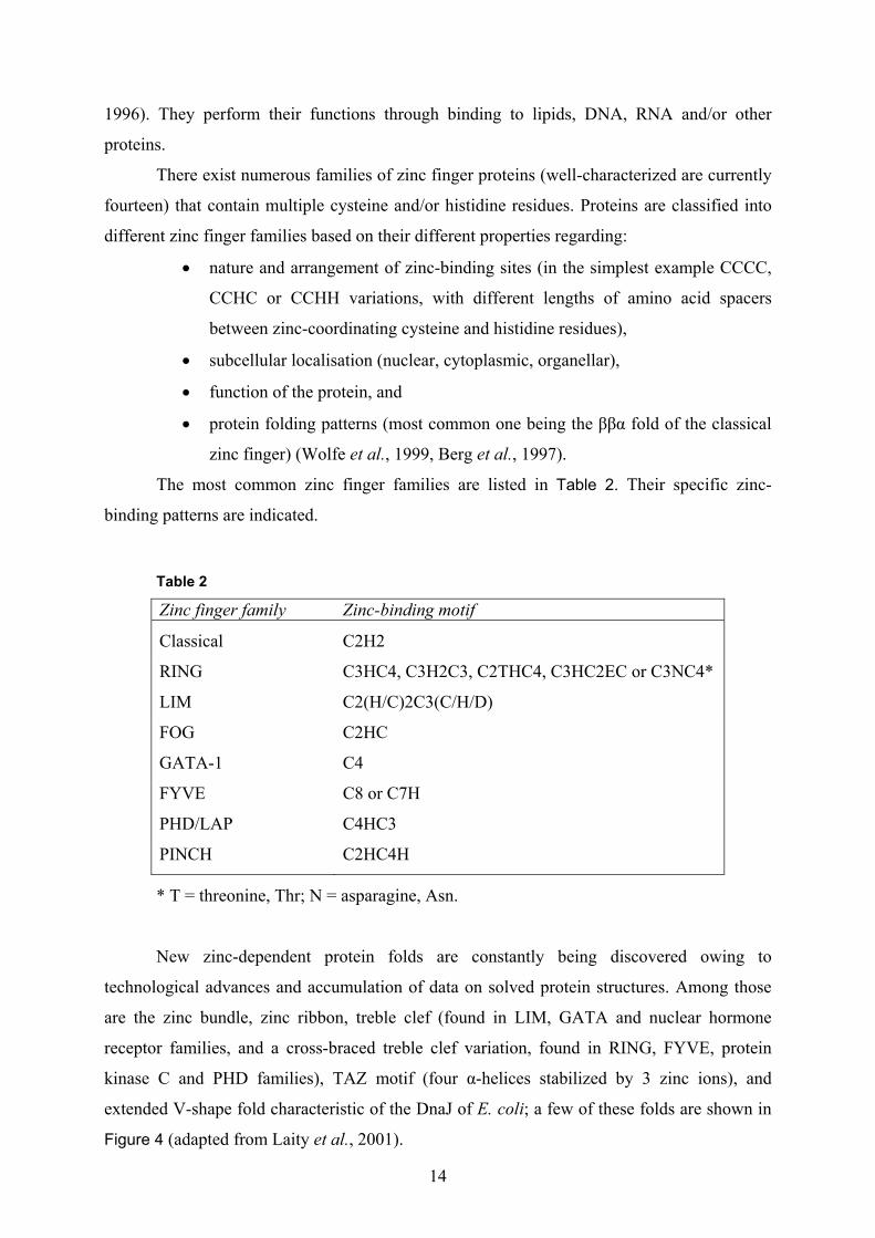
The most common zinc finger families are listed in Table 2. Their specific zinc-
binding patterns are indicated.
Table 2
Zinc finger family Zinc-binding motif
Classical
C2H2
RING C3HC4, C3H2C3, C2THC4, C3HC2EC or C3NC4*
LIM C2(H/C)2C3(C/H/D)
FOG C2HC
GATA-1 C4
FYVE C8 or C7H
PHD/LAP C4HC3
PINCH C2HC4H
* T = threonine, Thr; N = asparagine, Asn.
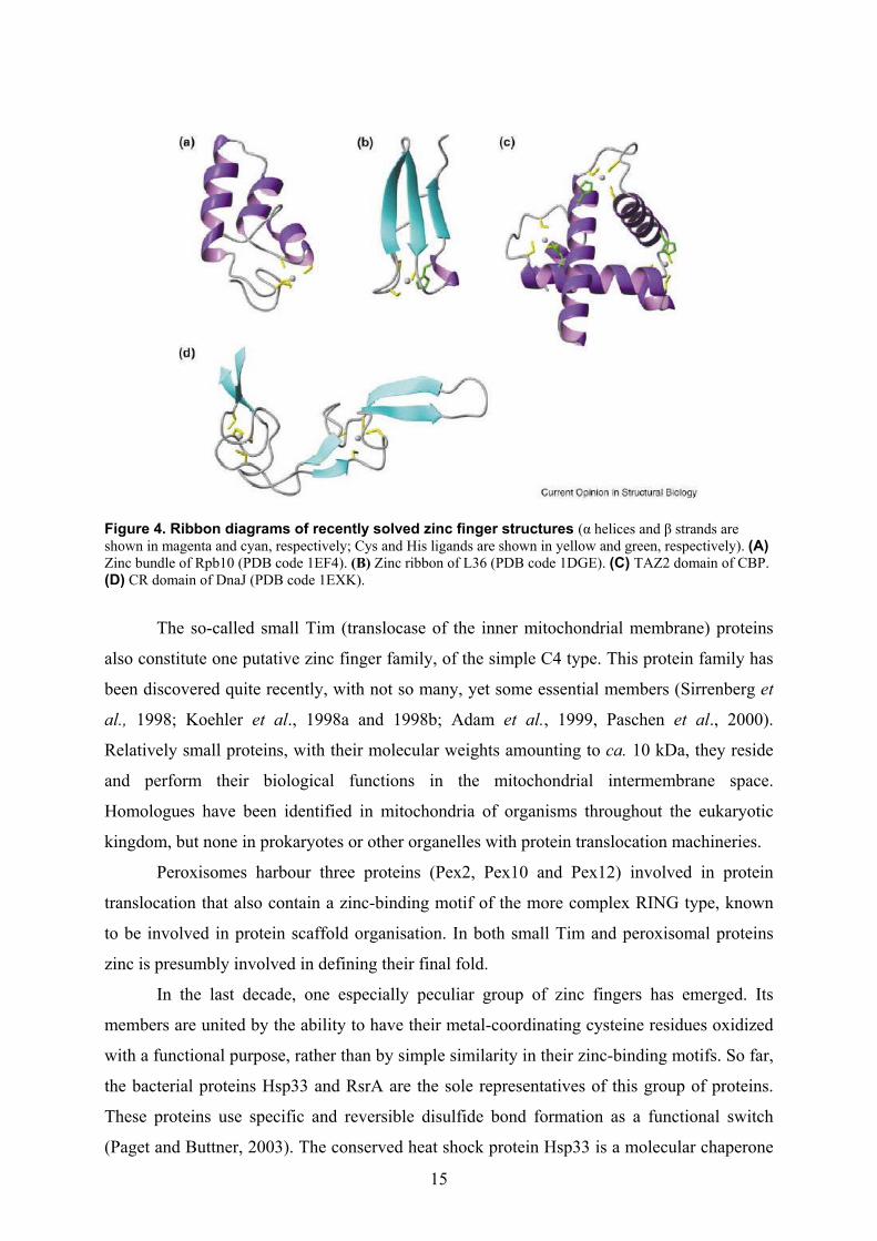
New zinc-dependent protein folds are constantly being discovered owing to
technological advances and accumulation of data on solved protein structures. Among those
are the zinc bundle, zinc ribbon, treble clef (found in LIM, GATA and nuclear hormone
receptor families, and a cross-braced treble clef variation, found in RING, FYVE, protein
kinase C and PHD families), TAZ motif (four α-helices stabilized by 3 zinc ions), and
extended V-shape fold characteristic of the DnaJ of E. coli; a few of these folds are shown in
Figure 4 (adapted from Laity et al., 2001).
15
Figure 4. Ribbon diagrams of recently solved zinc finger structures (α helices and β strands are shown in magenta and cyan, respectively; Cys and His ligands are shown in yellow and green, respectively). (A) Zinc bundle of Rpb10 (PDB code 1EF4). (B) Zinc ribbon of L36 (PDB code 1DGE). (C) TAZ2 domain of CBP. (D) CR domain of DnaJ (PDB code 1EXK).
The so-called small Tim (translocase of the inner mitochondrial membrane) proteins
also constitute one putative zinc finger family, of the simple C4 type. This protein family has
been discovered quite recently, with not so many, yet some essential members (Sirrenberg et
al., 1998; Koehler et al., 1998a and 1998b; Adam et al., 1999, Paschen et al., 2000).
Relatively small proteins, with their molecular weights amounting to ca. 10 kDa, they reside
and perform their biological functions in the mitochondrial intermembrane space.
Homologues have been identified in mitochondria of organisms throughout the eukaryotic
kingdom, but none in prokaryotes or other organelles with protein translocation machineries.
Peroxisomes harbour three proteins (Pex2, Pex10 and Pex12) involved in protein
translocation that also contain a zinc-binding motif of the more complex RING type, known
to be involved in protein scaffold organisation. In both small Tim and peroxisomal proteins
zinc is presumbly involved in defining their final fold.
In the last decade, one especially peculiar group of zinc fingers has emerged. Its
members are united by the ability to have their metal-coordinating cysteine residues oxidized
with a functional purpose, rather than by simple similarity in their zinc-binding motifs. So far,
the bacterial proteins Hsp33 and RsrA are the sole representatives of this group of proteins.
These proteins use specific and reversible disulfide bond formation as a functional switch
(Paget and Buttner, 2003). The conserved heat shock protein Hsp33 is a molecular chaperone

16
with a highly sophisticated mode of regulation. On the transcriptional level, its gene is under
heat shock control, whereas on the posttranslational level Hsp33 protein stands under
oxidative stress control. The redox sensor in Hsp33 is a four cysteine center that coordinates
zinc under reducing, i.e. inactivating conditions and that forms two intramolecular disulfide
bonds under oxidizing, i.e. activating conditions. As an oxidized dimer, Hsp33 is fully active
in refolding proteins. Its activity appears to specifically protect proteins and cells from the
otherwise deleterious effects of the oxidative stress. RsrA is another bacterial redox-sensitive,
zinc-containing protein. It is a σR-specific anti-σ factor, comprising seven cysteines in its
sequence. It binds the σR factor under reducing conditions, preventing it from activating
transcription of its target genes. Disulfide stress induces formation of one disulfide bond in
RsrA, causing it to release σR factor which then activates transcription of more than 30 genes
and operons. One of the gene products reduces the oxidized RsrA, thereby restoring its σR-
binding ability, and shutting off σR-dependent transcription, closing this physiological loop.
By these means, cycling between reduced, zinc-bound and the oxidized states, featuring at
least one disulfide bond, these zinc finger proteins are distinguished as key players in cellular
responses to oxidative stress and in the overall thiol-disulfide redox balance.

17
1.3. Aims of the present study
The objective of this study was to establish the existence of homologues of TIM9 and
TIM10 genes in N. crassa. This model system was then to be used to investigate the structural
and functional features of N. crassa Tim9 and Tim10 proteins.
For the structural analyses to commence, specific requirements had to be met. Initial
efforts were therefore directed towards isolation and purification of the N. crassa Tim9·Tim10
complex, in large quantities and of supreme purity. Certain properties of the purified
Tim9·Tim10 complex that would constitute demands of any structural and for that matter also
functional study attempt, had to be determined: its oligomeric state, potential zinc-binding
ability, CD spectra, and functionality.
As to the functional characterization of the complex, this study aimed at elucidating
two crucial aspects regarding the process of translocation of precursor proteins across the
outer mitochondrial membrane mediated by the Tim9·Tim10 complex. In particular, those
were: (i) the mechanism of substrate recognition by the Tim9·Tim10 complex, and (ii) the
sufficiency of the Tim9·Tim10 complex and the TOM complex for the transfer of AAC
precursor across the outer membrane. To answer the first of two questions, a screen of peptide
libraries covering the primary sequences of putative substrates of the Tim9·Tim10 complex
was performed. To resolve the second dilemma, the approach made use of a reconstituted
system of the Tim9·Tim10 complex and the TOM complex.
18
2. Material and methods
2.1. Molecular biology methods
2.1.1. PCR (polymerase chain reaction)
DNA sequences were amplified through the polymerase chain reaction (PCR) using
thermostable DNA polymerase, as described previously (Sambrook et al., 1989). DNA
polymerases used were: Taq (isolated from Thermus aquaticus), and Pfu (isolated from
Pyrococcus furiosus). Taq DNA polymerase has no proofreading ability, and therefore Pfu-
polymerase was used when the PCR product needed to be used for subsequent cloning.
PCR mix (total volume of 100 µl) contained: 1-2 U DNA polymerase (Taq-
polymerase and/or Pfu-polymerase), 10 µl 10x PCR-buffer (1% Triton X-100, 500 mM KCl,
15 mM MgCl2, 100 mM Tris·HCl, pH 8.8), 2 µl [10 mM] dNTPs, 2 µl [50 µM] primers and
200 ng plasmid DNA template or 1 µg genomic DNA template.
The following program, with different modifications regarding primer annealing
temperatures and length of elongation, was used:
1) 94ºC, 5 min, nuclease inactivation and complete DNA denaturation;
2) 30-40 cycles of: DNA amplification:
94ºC, 1 min DNA denaturation;
45-65º C, 1 min annealing of primers;
72ºC, 1-6 min
new DNA synthesis (extension);
duration of this step depends on
the length of DNA fragment to be
amplified;
Taq-polymerase: 1 min/1 kb
Pfu-polymerase: 2.5 min/1 kb;
3) 72ºC, 5-20 min completion of the last reaction.
Annealing temperature for primers was calculated by arithmetically adding the number
of A and T nucleotides (in primer’s sequence), multiplied by two, to the number of G and C
nucleotides multiplied by 4 (and only for that part of primer which anneals with the template
fully). Regions such as the restriction sites and possible Kozak sequences (Kozak, 1977 and
2003) contained in them were not taken into account, since they do not anneal. For a pair of

19
primers, temperature that is 5 degrees lower than the lowest calculated annealing temperature
of the two primers was chosen. In some cases I also tested two additional temperature values
(± 5ºC), to avoid occurance of possible non-specific PCR products.
2.1.2. DNA purification and analysis
2.1.2.1. Analytical and preparative gel electrophoresis
DNA fragments were separated according to their molecular weight through
electrophoresis in horizontal 0.8-3% (w/v) agarose gels; the fragments ranged in size from
0.05 to 10 kb. Lower agarose percentage gels were used for separating larger DNA fragments,
and higher agarose content gels for separating small DNA fragments. Agarose solutions were
made by dissolving the desired amount of agarose in Tris-acetate-EDTA buffer (TAE),
containing 1 mM EDTA and 40 mM Tris·acetate, pH 8.0, in a microwave oven. Ethidium
bromide was added to a final concentration of 0.5 µg/ml (it allows visualization of DNA when
the gel is exposed to UV light on a transilluminator). The agarose was stored at 65 ºC until
use.
The samples were loaded onto gels in a loading buffer containing 6% (v/v) glycerol,
0.01% bromphenolblue and 0.01% xylencyanol. The electrophoresis was performed at RT in
TAE buffer, with voltage set to U=60-70 mV. Commercially available molecular weight
marker was used in each run.
DNA fragments to be further processed were excised from the gel with a clean scalpel
and the DNA extracted from the gel using the “Gel extraction kit” protocol (Qiagen).
Extracted DNA was routinely stored at –20ºC.
2.1.2.2. DNA concentration measurement
For DNA concentration measurements, the absorption of DNA solutions was
measured at 260 nm. An OD of 1.0 corresponds to a concentration of 50 µg/ml of double
stranded DNA, 33 µg/ml mono stranded DNA, 40 µg/ml RNA or 20 µg/ml oligonucleotides.

20
2.1.3. Cloning of DNA fragments
2.1.3.1. Enzymatic manipulation of DNA: restriction and ligation reactions
Digestion of DNA with restriction endonucleases
Plasmid DNA was digested with up to 5 U of specific restriction endonuclease enzyme
for 1 µg of DNA. For preparative purposes up to 3 µg of DNA was digested in a 60 µl
reaction volume, while for analytical ones, much smaller amounts in a 20 µl reaction volume
were used. The buffer, incubation time (0.5-3 h) and temperature (usually 37ºC) of the
reactions were chosen according to the manufacturer’s recommendations. The obtained
digested fragments were analyzed by agarose gel electrophoresis. For preparative purposes,
desired DNA fragments were extracted from gels using Qiagen’s “Gel extraction kit”, and
used for ligation reactions.
In those cases where plasmid DNA was cut with a single restriction enzyme, it was
treated with calf intestinal alkaline phosphatase (CIP). This enzyme prevents vector’s
recircularization, through removal of its 5’-phosphate groups on linearised molecules.
Digested vector DNA (10 µg for instance) was incubated in 100 µl reaction with 10 µl 10x
CIP buffer (10 mM ZnCl2, 10 mM MgCl2, 100 mM spermidin, 0.5 M Tris·HCl, pH 9.0) and
0.1-0.5 units of alkaline phosphatase, for 30 min at 37ºC. The enzyme was inactivated
through heating to 65ºC for 20 min in the presence of 5 mM EDTA, and separated from the
DNA through agarose gel electrophoresis. DNA of interest was then extracted form the gel.
Ligation
Linearized DNA vector (50-200 ng) and a 5 fold molar excess of DNA fragment to be
inserted, were incubated in a 10 µl reaction with 1 µl of 10x ligation buffer (10 mM MgCl2,
5% (w/v) PEG-8000, 1 mM DTT, 1 mM ATP, 50 mM Tris·HCl, pH 7.6), and 0.5 µl (1 U) T4-
DNA ligase (Gibco-BRL). Reactions were performed at 14ºC for 16 h and 0.5-1 µl of this
mixture was used for E. coli cells transformation.
2.1.3.2. Preparation and transformation of E. coli competent cells
Preparation of competent cells
A small culture, usually 25 ml of LBamp-medium, inoculated with a single colony of
the corresponding E. coli strain (MH-1 or XL-1 Blue), was grown overnight at 37ºC under
moderate shaking conditions. The following day, 1 l of liquid LBamp-medium was inoculated
21
with the overnight culture. The bacterial cells were grown further until they reached the
logarithmic growth phase (OD578 ~ 0.5). Then they were incubated on ice for 30 min,
harvested by centrifugation (4,400 x g, 5 min, 4ºC) and washed sequentially with 400 ml, 200
ml, and 50 ml of 10% (v/v) glycerol. The competent cells were finally resuspended in 500 µl
10% (v/v) glycerol, aliquoted, and stored at –80ºC.
Transformation of competent cells through electroporation
To 40-60 µl of E. coli competent cells 0.5-1 µl of the ligation reaction mixture was
added on ice. The cells were transferred to an ice-cold cuvette and the cuvette introduced into
the electroporation Gene Pulser apparatus (BioRad) (settings: U=2.5 kV, R=400 Ω, C=25µF;
time constant obtained τ was 7-8 ms). After a brief application of high electric voltage to the
cells, the suspension was diluted with 1 ml of LB-medium, and incubated for 30-60 min at
37ºC under moderate shaking conditions (140 rpm), to allow cell recovery. The transformed
cells were harvested by centrifugation (10,000 x g, 15 sec, RT) and resuspended in a small
volume (up to 150 µl) of LB-medium. The cells were plated on LB-medium plates with
ampicillin and incubated overnight at 37ºC.
2.1.4. E. coli strains used
Strain Genotype Reference
XL1-Blue
supE44, hsdR17, recA1, endA1, gyrA96, thi-1,
relA1, lac-, F’[proAB+, lacIq lacZ∆M15, Tn10(tetr)]
commercially available
from Stratagene
MH1 MC1061 derivative; araD139 lacX74 galU galK
hsr hsm+ strA
Casadaban and Cohen,
1980
2.1.5. Small and large scale isolation of plasmid DNA from E. coli
Small scale preparation of plasmid DNA was performed according to a published
procedure (Birnboim and Doly, 1979), through alkaline lysis. Small volume of LB-medium
(2-5 ml) containing the appropriate antibiotic (ampicillin in majority of cases) was inoculated
with a single bacterial colony picked out from a Petri dish, and incubated overnight at 37ºC,
while shaking (140 rpm). The next day bacteria were harvested by centrifugation (8,000 x g,
30 sec, RT) and the resulting pellet resuspended in 300 µl of buffer E1 (10 mM EDTA, 50
mM Tris·HCl, pH 8.0) containing 100 mg/ml RNase. Cell lysis followed, through the addition
of 300 µl of buffer E2 (0.2 M NaOH, 1% SDS). Samples were mixed by inverting the tubes 5

22
times and incubated 5 min at RT. Neutralization was accomplished by adding 300 µl of buffer
E3 (3.1 M K-acetate, pH 5.5) and mixing the samples immediately afterwards, by inverting
the tubes 5 times. They were then centrifuged (10,000 x g, 10 min, 2ºC), the supernatant
transferred to new tubes and the DNA was precipitated through the addition of 600 µl of 96%
isopropanol. Samples were then centrifuged again (10,000 x g, 40 min, 2ºC), washed with
85% cold ethanol, dried at RT, resuspended in 20-30 µl water and stored at –20ºC.
Large scale preparation of plasmid DNA (up to 0.5 mg) was performed using a
“Jetstar” Midi-Kit (Genomed). LB-medium (50 ml) supplemented with ampicillin (or any
other required antibiotic) was inoculated with bacteria carrying the plasmid to be isolated, and
incubated overnight at 37ºC, while shaking at 140 rpm. Cells were harvested the next day by
centrifugation (3,000 x g, 10 min, RT or 4ºC) and resuspended in 4 ml of buffer E1. Cell lysis
was performed by adding 4 ml of buffer E2 and inverting the tubes 5 times; they were left for
5 min at RT. After neutralization by adding 4.4 ml of buffer E3, samples were centrifuged
(17,418 x g, 10 min, 4ºC), and the supernatants immediately applied onto an anion-exchange
column, previously equilibrated with 10 ml of buffer E4 (0.15% (v/v) Triton X-100, 0.6 M
NaCl, 100 mM Na-acetate, pH 5.0). The column was washed with 20 ml of buffer E5 (0.8 M
NaCl, 100 mM Na-acetate, pH 5.0) and the plasmid eluted into Corex tubes by adding 5 ml
buffer E6 (1.25 M NaCl, 100 mM Tris·HCl, pH 8.5). DNA was precipitated through the
addition of 3 ml of 96% isopropanol and one centrifugation step (12,000 x g, 30 min, 4ºC). It
was then washed with 5 ml of 70% ethanol, re-centrifuged, and dried at RT. DNA was finally
resuspended in up to 150 µl of ddH2O, and the concentration was measured, before freezing it
at –20ºC.
When a clone was propagated for the first time, 500 µl of the overnight culture was
removed and added to 500 µl of sterilized solution of 50% LB medium mixed with 50%
glycerol. It was then frozen at –80ºC, and stored as a glycerol stock for future propagation of
the same clone.
2.1.6. Plasmids and genomic library clones used
Plasmid Reference
pGEM4·NcAAC
Endres et al., 1999
pGEM4·AAC2 Lawson et al., 1988
pGEM4·NcTim23 Mokranjac, PhD thesis
pGEM4·NcTim10 This thesis
pGEM4·NcTim10his9 This thesis

23
pGEM4·NcTim9, clones 1, 3, and 5 This thesis
pGEM4·NcTim9his9, clones 1, 3, and 5 This thesis
pGEM4·Su9(1-69)-DHFR Gaume et al., 1998
pMalcRI·NcTim10, clones M2, M4, X2, X5* This thesis
pMalcRI·NcTim9, clones 3 and 31 This thesis
pCB1179·Pm·tim10his9, clones 11, and 19 This thesis
pCB1179·Pm·tim10his9·1kb, clones 3 and 8 This thesis
pQE30·NcTim10, clones M5, M6, X8, X9* This thesis
Cosmid Reference
pMOcosX#X20:A12 This thesis
pMOcosX#X25:B10 This thesis
pMOcosX#X12:C6 This thesis
* M=MH1; X=XL-1 bacterial clones
2.1.7. Cloning strategies
Constructs cloned for in vitro transcription and translation of mitochondrial
preproteins comprised of cDNAs of relevant genes inserted into pGEM4 vector (Promega).
Constructs for raising the antibodies consisted of cDNAs inserted into pMalcRI vector (NE
Biolabs), creating maltose-binding protein (MBP) fusion proteins. Alternatively, cDNA was
cloned into pQE30 vector (Qiagen), creating a his-tagged version of the gene of interest. For
the expression of proteins in N. crassa wt background, genes encoding Tim9 and Tim10
proteins were cloned into pCB1179 vector. All plasmids were first transformed into E. coli
XL-1 or MH1 strains for amplification and stock maintenance, and subsequently into S.
cerevisiae or N. crassa cells.
pGEM4·NcTim10
The following primers were used:
N-terminal primer (containing a BamHI cutting site), called BamTIM10:
5’- AAT AAT GGA TCC ATG TTC GGA CTC GGC AGG -3’,
C-terminal primer (containing a SalI cutting site), called TIM10Sal:
5’- AAT AAT GTC GAC TTA CAT GCC GAA GCC ACC -3’.
N. crassa cDNA(−) and cDNA(+) libraries (2.5 µl/50 µl PCR reaction) were used as
templates. Three different annealing temperatures were tested till unspecific PCR products

24
were eliminated. (cDNA libraries marked plus and minus were obtained from Neurospora
grown in the presence or absence of chloramphenicol, respectively).
The same primers were used to screen the genomic DNA library of N. crassa (1 µl of its
1:100 dilution/50 µl PCR reaction), and a 500 bp fragment contained in cosmids
pMOcosX#X12:C6, X20:A12 and X25:B10 was identified.
pGEM4·NcTim10his9
The following primers were used:
N-terminal primer, BamTIM10,
C-terminal primer (containing a XbaI cutting site), TIM10HisXba:
5’- TTT TTC TAG ATT AGT GAT GGT GAT GGT GGT GAT GGT GGT GCA TGC CGA
AGC CAC CTC CAC C-3’.
N. crassa cDNA(−) and cDNA(+) libraries were used as templates.
pGEM4·NcTim9
Region homologous to that of S. cerevisiae TIM9 gene was identified in a screen of N. crassa
database, and primers for screening N. crassa cDNA and genomic libraries constructed.
Positions of the starting methionine, as well as that of two introns were predicted, based upon
the identification of the intron flanking sequences most commonly found in N. crassa, in the
region corresponding to the tim9 N. crassa gene locus; these sequences are: G G T A77/G
A50/C G T76/C; C T A/G A C; A56/T T52/C A G G40 (numbers indicate the incidence with
which the nucleotide is found in genes containing introns analyzed so far).
For screening the genomic N. crassa library, following primers were used:
N-terminal primer (containing an EcoRI cutting site), EcoMDGT9ge:
5’- CCG GAA TTC ATG GAT GGG TAA GCA AGA GAG-3’,
C-terminal primer (containing a HindIII cutting site), ATTHindT9:
5’- TTC CCA AGC TTT TAC CGC CTC TGC ATC TCA GC -3’.
N. crassa genomic library (1 µl of its 1:100 dilution/50 µl PCR reaction) was used as a
template.
For screening of the cDNA(−) and cDNA(+) libraries, following primers were used:
N-terminal primer (containing an EcoRI cutting site), EcoT9cDNA:
5’- CCG GAA TTC AAA TCG ACA ACA ATG GAT GGG -3’,

25
C-terminal primer ATTHindT9.
N. crassa cDNA(−) and cDNA(+) libraries (2.5 µl/50 µl PCR reaction) were used as
templates.
Upon comparison of sequenced cDNA (apprx. 300 bp) and genomic DNA (apprx. 800 bp)
products of the PCR screens, the predictions of intron positions were verified.
pMalcRI·NcTim10
Same primers as for pGEM4·NcTim10 were used.
pMalcRI·NcTim9, clones 3 and 31
Same primers as for pGEM4·NcTim9 were used.
pCB1179·Pm·tim10his9
The following primers were used:
N-terminal primer (containing an EcoRI cutting site), EcoTIM10P:
5’- TTT TGA ATT CCG CTC GGG CCG TTG TCT GC -3’,
C-terminal primer, TIM10HisXba.
Cosmids pMOcosX#X20:A12, pMOcosX#X25:B10 and pMOcosX#X12:C6 were used as
templates.
pCB1179·Pm·tim10his9·1kb
The following primers were used to amplify the region 1 kb downstream from the tim10 gene:
N-terminal primer (containing a XbaI cutting site), T10Xba1kb:
5’- TTT TCT AGA TTT TTT TGG ATT ACT GGA ACG G -3’,
C-terminal primer (containing a SacII cutting site), T10Sac1kb:
5’- AAA CCG CGG CAG GAT CCA CAT ACC CGG -3’.
As templates cosmids pMOcosX#X20:A12, pMOcosX#X25:B10 and pMOcosX#X12:C6
were used. The resulting PCR product was inserted behind tim10 promotor region and the
tim10 gene in the plasmid pCB1179·Pm·tim10his9, using marked restriction sites.
pCB1179·P2·tim10his9·1kb
To amplify a bigger promoter region of tim10 together with tim10 gene and to add a his-tag to
it, the following primers were used:
26
N-terminal primer (containing an EcoRI cutting site), EcoTIM10P2:
5’- GGG AGT AGA TGA ATT CAT TAT TGC -3’,
C-terminal primer, TIM10HisXba.
Cosmids pMOcosX#X20:A12, pMOcosX#X25:B10 and pMOcosX#X12:C6 were used as
templates.
Resulting PCR products were cut with corresponding enzymes and exchanged against
Pm·tim10his9 fragment in pCB1179·Pm·tim10his9·1kb construct.
pQE30·NcTim10
Same primers as for pGEM4·NcTim10 were used.
2.1.8. S. cerevisiae strains used
Strain Genotype
D273-10B
ATCC 246557, Mat α, Mal (rho+)
W303-1A/-1B Mat a/α, ade2-1 ura3-1 his3-11 trp1-1 leu2-3 leu2-112 can1-
100; isogenic with RS 190 (ATCC 208354)
W334-a Mat a, leu2 ura3-52
BY 4743
Mat a/α, his3∆1/his3∆1 leu2∆0/leu2∆0 ura3∆0/ura3∆0
met15/MET15∆0 lys2∆0/LYS2
∆tom70 tom70::KANmx3, Mat a/α, his3∆1/his3∆1 leu2∆0/leu2∆0
ura3∆0/ura3∆0 met15/MET15∆0 lys2∆0/LYS2
EJ11-6 mrs11::HIS3 ade8 trp1 leu2 [pMRS11::URA3-CEN]
tim10-1 (807 1B) Koehler et al., 1998
YPH501 ade2-101 his3-∆200 leu2-∆1 ura3-52 trp1-∆63 lys2-801
2.1.9. Preparation of yeast DNA
Isolation of yeast DNA was performed as described previously by Rose et al., 1990.
YPD-medium (5 ml) was inoculated with S. cerevisiae cells and incubated overnight at 30ºC,
while shaking (140 rpm). Cells were harvested by centrifugation, washed with 25 ml of water,
and resuspended in 200 µl of breaking buffer (2% Triton-X100, 1% SDS, 100 mM NaCl, 1
mM EDTA, 10 mM Tris·HCl, pH 8.0). Phenol/chloroform/isoamyl alcohol (25:24:1) mix
(200µl) and 0.3 g glass beads were added, and the samples vortexed for 2 min. The probes
27
were then centrifuged (36,670 x g, 5 min, RT) and the supernatant (the aqueous phase)
transferred to new tubes. DNA was precipitated by adding 2.5 vol. of 100% ethanol. Samples
were incubated for 10 min at –20°C, centrifuged (36,670 x g, 10 min, 2°C), and washed with
70% ethanol. Pellets were dried at RT, resuspended in 20 µl H2O and stored at –20°C.
2.1.10. N. crassa strains used
Strain Description Source
74-OR23-1VA
wt
Fungal Genetic Stock Center #2489
TA2-1 contains tim10his9 This thesis
TA2-14 contains tim10his9 This thesis
TA2-14-31/2 contains tim10his9 This thesis
2.1.11. Screening of N. crassa cosmid libraries
N. crassa genomic cosmid libraries screened in this study were prepared by Dejana
Mokranjac (Mokranjac, PhD thesis, 2004). The cosmid library pMOcosX, screened for N.
crassa tim10 gene, comprises of 25 microtiter plates labeled pMOcosX#X1-25, each with 96
clones of the Neurospora genomic library (clones are labeled in a way standard for any
microtiter plate, with the plate number preceeding the number of the clone; for example
pMOcosX#X1:A1). Every microtiter plate has a corresponding 11 x 7 cm nylon membrane,
created through a colony-hybridization method. Shortly, the colonies are lysed in situ upon
replicating microtiter plates onto membranes, and the cellular debris washed off, leaving
DNA bound to the mebranes (Dembowski, PhD thesis, 2001). Furhermore, all 96 clones from
every plate are “pooled together” into 25 cultures, 25 midi-preps of DNA are made, and a
single dot created for each of 25 plates on one 5 x 10 cm membrane. This method for creation
of genomic libraries is referred to as the dot-blot method (Dembowski, PhD thesis, 2001).
This particular membrane is the first membrane screened (later on referred to as the “primary”
one), allowing identification of membranes corresponding to specific microtiterplates
containing the clones of interest, which are to be screened in the second round.
In order to make a probe for screening a genomic library, PCR was performed using a
PCR DIG (digoxigenin) Probe Synthesis Kit (Roche). Digoxigenin is a steroid, used to label
probes in a PCR reaction. The labeled probe can then be easily detected with commercially
available antibodies against digoxigenin. PCR reaction mixture (150 µl) contained: 110.25 µl
ddH2O, 15 µl 10x PCR buffer with MgCl2, 15 µl 10x PCR DIG synthesis mix (dNTPs: dATP,
dCTP, dGTP, 2mM each, and 1.3 mM dTTP and 0.7 mM DIG-11-dUTP), 3.75 µl [20 pM]

28
primer BamTIM10, 3.75 µl [20 pM] primer TIM10Sal, 2.25 µl enzyme mix and 1 µl genomic
library as template. A control PCR with regular dNTP mix was performed as well. Conditions
used were: initial denaturation: 94ºC for 5 min; 40 cycles of: 94ºC for 1 min, 60ºC for 1 min,
72ºC for 1 min; and the final elongation: 72ºC for 5 min. Expected PCR product size for
genomic tim10 clone is 500 bp, and with the label circa 600 bp. PCR product (1 µl ) was run
on a 2% agarose gel and the expected shift in size noted. The remaining 149 µl was heated to
94ºC for 5 min, and then cooled instantly by placing the probe in ice-cold water. It was then
added to 35 ml (the volume is usually estimated based on the band intensity seen on 2%
agarose gel) of standard hybridizing solution, containing 5x SSC (1x SSC: 150 mM NaCl, 15
mM Na-citrate, pH 7.0), 50% formamide, 0.1% Na-laurylsarcosin, 0.02% SDS and 2%
blocking reagent.
The “primary” cosmid library membrane was preincubated for 2 h with the standard
hybridizing solution at 42ºC, and then overnight at 42ºC with the generated probe, to allow
for the hybridization between the digoxigenin-labeled probe and corresponding clones on the
membrane to take place. The next day, solution with digoxigenin-labeled probe was poured
off, and the membrane washed twice for 5 min in 2x SSC with 0.1% SDS solution at RT, and
twice for 15 min with 0.1x SSC with 0.1% SDS solution at a higher temperature (circa 60ºC).
Membrane was then incubated for 30 sec in P1 solution (150 mM NaCl, 0.3% Tween 20, 100
mM maleic acid, pH7.5) at RT, 30 min in 1x blocking solution (1% (w/v) blocking reagent in
P1 buffer) at RT, and then left for 1 h at RT with αDIG-AP conjugate (an antibody against
digoxigenin coupled to alkaline phosphatase, whose chemiluminescent substrate is disodium
3-(4-methoxyspiro (1,2-dioxetane-3,2'-(5'-chloro) tricycle [3,3.1.13.7]decan-4-yl) phenyl
phosphate (CSPD), diluted 1:10,000 in blocking solution. It was subsequently washed, twice
for 15 min with P1 solution containing 0.03% Tween 20, and shortly twice with 10 ml of P3
buffer (100 mM NaCl, 100 mM Tris·HCl, pH 9.5). The membrane was placed between two
sheets of plastic folia, excess P3 solution removed, 200 µl of the substrate CSPD, in 20 ml of
solution P3 added, and the membrane incubated for 5 min at RT. Excess substrate was
removed, the membrane sealed completely and incubated further for 10-15 min at 37ºC. Films
were exposed for 1, 2, 3 and 4 hours.
To strip the membrane of the bound digoxigenin-labeled probe, it was shortly washed
in ddH2O, twice for 15 min in 0.2% NaOH, 1% (w/v) SDS at 37ºC, and 5 min in 2x SSC. The
membrane was then dried and stored for future use in a sealed plastic bag, or subjected
directly to further hybridization trials.

29
2.1.12. Southern blot
The agarose gel was incubated in 0.25 M HCl for 10 min to fragment the DNA
through depurinization. It was further incubated twice for 30 min in solution Southern I (1.5
M NaCl, 0.5 M NaOH) to denature the DNA, and then washed twice for 30 min in Southern II
solution (1.5 M NaCl, 1 M Tris·HCl, pH 7.4). Southern blot was assembled, according to
Southern, 1975, using nylon membranes (Pall-Gelman). Transfer buffer used was 2x SSC.
After 12 hours the membrane was washed in 6x SSC, dried and heated for 2 h at 120ºC.
2.1.13. Screening of clones through in situ colony-blotting
Clones were striken onto Petri dishes with appropriately supplemented LB medium,
and grown overnight at 37ºC. The next day, circularly cut nitrocellulose membranes were
gently pressed onto the Petri dishes, and after 2 min placed onto stacks of Whatmann 3MM
paper soaked in different solutions. Membranes were soaked first for 10 min in 10 % (w/v)
SDS solution, followed by 5 min in Southern I (denaturation), twice for 5 min in Southern II
(neutralization) and twice for 15 min in 2x SSC solution. They were then washed twice in
TBS, blocked in 5 % (w/v) skimmed milk in TBS solution and immunodecorated with
appropriate antibodies.
2.2. Cell biology methods
2.2.1. E. coli: Media and culture
Media for E. Coli
LB-medium: 0.5% (w/v) yeast extract, 1% (w/v) Bacto-Tryptone, 1% (w/v) NaCl;
LBamp-medium: LB-medium supplemented with 100 µg/ml of ampicillin.
Described media were used for preparing liquid cultures. To prepare plates with solid
media, 2% (w/v) bacto-agar was added to the liquid culture solution. Bacto-agar, glucose and
media were autoclaved separately (120ºC, 20-30 min), and the ampicillin was added after the
media had been chilled to 50ºC.

30
2.2.2. N. crassa: Media and culture
2.2.2.1. Media and solutions for N. crassa
Trace elements solution: 50 g citric acid x H2O, 50 g ZnSO4 x 7 H2O, 10 g Fe(NH4)
2SO4 x 6 H2O, 2.5 g CuSO4 x 5 H2O, 0.5 g MnSO4 x 1 H2O, 0.5 g H3BO3 (water free), 0.5 g
Na2MoO4 x 2 H2O, dissolved in 1 l of H2O. Chloroform (10 ml / 1 l) was added as a
preservative, and the solution stored at RT.
Biotin solution: 100 ml ethanol, 100 ml H2O, 20 mg biotin. It was stored at 4ºC.
50x Vogel’s minimal medium (Vogel’s salts; Vogel, 1964): 150 g Na3-citrate x 5 H2O,
250 g KH2PO4 (anhyd.), 100 g NH4NO3, 10 g MgSO4 x 7 H2O, 5 g CaCl2 x 2 H2O, 5 ml trace
elements-solution, 2.5 ml biotin solution, in 1 l of H2O; 5 ml of chloroform was added as
preservative, and the solution stored at RT.
Minimal medium: 2% (v/v) 50x Vogel’s minimal medium, 2% (w/v) glucose.
Complete medium (Vogel’s medium): 2% (v/v) 50x Vogel’s minimal medium, 1% (w/v)
sugar, 1% (w/v) glycerol, 0.2% (w/v) yeast extract, 0.1% (w/v) caseinhydrolysat.
Hygromycin stock solution: 50 mg/ml in water. It was stored at 4ºC.
Novozyme 234 stock solution: 5 mg/ml in 1 M sorbitol. It was filter sterilized and
stored in 2 ml aliquots at −80ºC.
Solutions for the transformation – all were prepared fresh a day before use:
Polyethylene glycol-Tris-Calcium (PTC) solution in water: 40% (w/v) PEG 4000, 50
mM CaCl2, 50 mM Tris·HCl, pH 8.0.
Sorbitol-Tris-Calcium (STC) solution: 1 M sorbitol, 50 mM CaCl2, 50 mM Tris·HCl,
pH 8.0.
31
pH was adjusted with 10 M NaOH to 8.0, prior to autoclaving. For both PTC and STC
solutions, filter sterilised CaCl2 was added to a final concentration of 50 mM after
autoclaving, to prevent its precipitation.
10x FGS additive (per 500 ml, in water): 2.5 g (w/v) fructose, 2.5 g (w/v) glucose,
100 g (w/v) sorbose.
Heparin stock solution: freshly made 5 mg/ml in STC; it can also be stored at −80ºC.
Spermidin stock solution:
50 mM in water. It was stored at −20ºC.
Top (regeneration) agar: 10 ml (v/v) 50x Vogel’s minimal medium, 91 g (w/v)
sorbitol, 14 g (w/v) agar; the mixture was filled up with water to 450 ml, sterilized and 50 ml
of prewarmed 10x FGS solution added. It was then split into 7-8 ml aliquots in 15 ml sterile
Falcon tubes and stored at 4ºC. The prepared agar was prewarmed to 50ºC before use.
Bottom agar: 10 ml (v/v) 50x Vogel’s minimal medium, 7.5 g (w/v) agar, 440 ml
water. It was sterilised and cooled till 50-60ºC. Prior to pouring the bottom agar into Petri
dishes (25 ml per standard plate), 50 ml of prewarmed 10x FGS additive and 2 ml of 50
mg/ml hygromycin stock solution (final concentration 200 µg/ml) were added. Hygromycin
was added for the selection of strains generated in the scope of this thesis. Plates were left to
dry and were stored at 4ºC.
Solutions for the microconidition:
Iodoacetic acid (IAA) stock solution: 208 mg IAA was dissolved in 10 ml of water to
obtain a 100 mM stock solution. It was filter sterilized, and always used fresh.
Synthetic crossing (SC) medium (Westergard and Mitchell, 1947): 1 g (w/v) KNO3, 0.7
g (w/v) K2HPO4, 0.5 g (w/v) KH2PO4, 0.5 g (w/v) MgSO4 x 7 H2O, 0.1 g (w/v) NaCl, 0.1 g
(w/v) CaCl2, 0.1 ml (v/v) trace element stock solution, 0.1 ml (v/v) biotin stock solution,
brought to 1 l with water; pH was adjusted to 6.5 and the solution stored at 4ºC.
This medium was used for standard crosses, with various carbon sources, 1% (w/v)
sucrose for instance.
32
For microconidiation slants, 2% (w/v) agar and 0.5% (w/v) sucrose were dissolved in
1:10 dilution of SC medium by heating - autoclaving for 20 min at 120ºC. When the solution
was cooled to 50-60ºC, 1% (v/v) iodoacetic acid was added. Medium was distributed to glass
tubes (16 x 150 mm), which were plugged with cotton, slanted and left to dry; they were
stored up to 1 week at 4ºC.
Sorbose medium: 0.03 % (w/v) glucose, 2% (v/v) Vogel’s minimal medium, 2% (w/v)
agar, 1% (w/v) sorbose. Medium was autoclaved 15 min at 110ºC, cooled to 50-60ºC, and for
TA2-14-32 strain hygromycin added to a final concentration of 150 µg/ml, prior to pouring
plates.
SM buffer for isolation of N. crassa mitochondria: 250 mM sucrose, 10 mM
MOPS·KOH, pH 7.2.
2.2.2.2. Growth of N. crassa hyphae
N. crassa hyphae were grown as described previously by Davis and Serres (1970).
N. crassa silica stocks preparation
Metal screw-capped tubes (13 x 50 mm) were filled with 4 ml of silica gel (Merck
7733), and the silica was activated and dry-sterilized through incubation for 3 h at 180ºC.
A culture of the strain of interest was grown for 5 days at 30ºC in 250 ml flask
containing 40 ml of solidified medium. Skimmed milk (sterilized, 1% (w/v) in water, 10-20
ml) was poured carefully into the flask and the culture vortexed vigorously. The suspension of
conidia was filtered through cotton-cloth funnel into sterile tubes and centrifuged shortly
(10,000 x g, 15 sec, RT). Most of the supernatant was poured off, and the conidia were
resuspended in the remaining small volume of supernatant. Using a 200 µl pipette,
approximately 100 µl of air was taken in and then a 100 µl of conidial suspension. The tip was
inserted into the sterile silica gel and the suspension slowly released into it, while stirring to
disperse it more evenly into the gel. This procedure was repeated one more time. Tube’s lid
was loosely replaced and the silica stock dried in a desicator for 2-3 weeks at RT. Lid was
then tightly closed and the stock placed at –20ºC.
33
Obtaining the conidia
Conidia were grown in 250 ml Erlenmeyer flasks with approximately 25 ml of
solidified complete medium (Vogel’s medium supplemented with 2% (w/v) agar). A few
crystals of the silica gel-dried conidia, or conidia obtained from so-called primary cultures or
A-flasks (inoculated with silica stocks of the appropriate strains), were inoculated in the
centre of the flasks, and incubated for 6 and 3 days in the dark, respectively. This was
followed by 3 days incubation at RT in daylight, to stimulate conidia formation. To collect
conidia, 50 ml of sterile water was carefully poured into the Erlenmeyer, the suspension
vortexed and after the airborne conidia in the flask had settled, the suspension poured into a
new sterile flask. Concentration of the conidial suspension was determined by counting
conidia using a haemocytometer under the light microscope. The conidia were stored at 4ºC
(for 2-3 days maximally) before use.
Growth of hyphae
To isolate mitochondria from different N. crassa strains on a small scale, 1 l of liquid
medium (containing 930 ml water, 20 ml 50x Vogel’s minimal medium (Vogel, 1964) and 40
ml 1.4 M sucrose; all the components autoclaved separately) was inoculated with 10 ml of
conidial suspension (108 conidia/ml). The culture was incubated for 15 h at 25ºC, aerated with
sterile forced air and exposed to a visible and ultraviolet light source. The hyphae were
collected by filtration.
To obtain large quantities of mycelia, 100 l cultures (containing 2 kg (w/v) sucrose
and 2 l (v/v) 50x Vogel’s minimal medium) were inoculated with 1 l conidia (108conidia/ml)
and incubated for 24 h at 25ºC, under forced aeration and light and UV light exposure. The
hyphae were collected and after wet weight determination, used for isolation of mitochondria.
Growth of hyphae in minimal medium with 35S
For the isolation of N. crassa mitochondria containing radioactively labeled proteins,
the cultures were grown with Vogel’s salt solution, where MgSO4 was exchanged with 35S-
labeled sulfate (1.56 mCi/l culture). Additionaly, 10 mg/l of unlabeled MgSO4 was added to
promote hyphal growth. Cultures (8 l) were grown in the isotop-laboratory, with the
outcoming air being directed through two connected bottles filled with 1 M NaOH, in order to
precipitate any radioactive sulfate present in this air. The subsequent growth and
mitochondrial isolation were performed as described for non-radioactively labeled cultures.
34
2.2.2.3. Transformation of N. crassa
Preparation of spheroplasts
Supplemented Vogel’s medium (150 ml) was inoculated with 1-2 x 10⁹ conidia and
incubated for 4-6 hours at 30ºC. The progress of germination was periodically examined, and
when a level of 90% germination was achieved, germinated conidia were harvested in three
50 ml tubes via centrifugation (1,935 x g, 8 min, RT). Conidia were resuspended to a final
volume of 50 ml (in water), and the centrifugation step was repeated twice. Final pellet was
resuspended in 10 ml of 1 M sorbitol, transferred to a sterile 250 ml flask and 4 ml of 5 mg/ml
Novozyme 234 (cell-wall-digesting preparation from Trichoderma) in 1 M sorbitol added.
Suspension was then incubated for 30-60 min at 30ºC, while shaking (80-100 rpm). Degree of
spheroplasting was examined after 30 min (over-digestion decreases their viability). This was
done by placing 5 µl of the spheroplast suspension on a slide with cover slip. Water was
added to one side and swelling and bursting of spheroplasts were observed under the
microscope, which confirmed the loss of cell wall. After appropriate spheroplasting efficiency
was ascertained, spheroplast suspension was gently poured over into a 50 ml conical tube, the
flask rinsed with 1 M sorbitol which was added to the conical tube and the volume adjusted to
50 ml with the same sorbitol solution. Three centrifugation steps were performed, twice
bringing the volume to 30 ml with 1 M sorbitol and once with STC solution (484 x g, 10 min,
RT). Final pellet was resuspended in 7.5 ml of STC, 2 ml of PTC and 100 µl of DMSO, and
mixed gently but thoroughly. Aliquots were made, and stored at –80ºC. N. crassa spheroplasts
can be thawed and refrozen with no consequent lessening of their transformability.
Transformation of N. crassa – the classical method
An aliqout of spheroplasts was thawed and stored on ice, and 100 µl were added to a
mixture of 1-5 µg of plasmid DNA, 2 µl spermidin and 5 µl heparin stock solution. After 30
min incubation on ice 1 ml of PTC solution was added, the suspension mixed carefully and
incubated for 20 min at RT. The mixture was then transferred into pre-warmed top agar
(equilibrated in a water bath at 54ºC), and immediately poured onto plates with bottom agar.
Regeneration agar was left to solidify and the plates were then incubated at 30ºC for 2-3 days.
Within first 24 h at 30ºC, some transformants had already grown a few hyphae; after two days
they were visible to the naked eye, and within three days they were ready to be isolated with a
spear-point needle into 13 x 100 mm culture tubes with 1-2 ml of minimal medium,
supplemented appropriately (with hygromycin for instance). They were kept for three or more
days at 30ºC in the incubator, subcultured once or twice, and then used for inoculation of 10 x
35
75 mm tubes containing complete medium, which were utilized for the generation of stocks
(stored as conidial cultures at −20ºC). Typically, the efficiency of transformation was 10,000
transformants per µg of DNA used. In this way, the TA2-1 and TA2-14 strains were
generated. To eliminate the non-transformed nuclei which accompany the transformed ones
upon isolation, homokaryotic stable transformants were purified before use through a
microconidiation method.
Microconidiation:
Selective induction of microconidiation in mycelia is used to obtain uninucleate
microconidia for isolating homokaryotic derivatives. In present work it was employed to
derive the TA2-14-32 strain, expressing equal amounts of the non-tagged and the tagged
version of Tim10 protein. The procedure was done as described previously by Pandit and
Maheshwari, 1993. The cultures were initiated with trace quantities of macroconidia placed in
microconidiation slants, and kept for 7-12 days at 25 ºC in a room with day/night cycle.
Macroconidia distinguish themselves from microconidia in size, and therefore a microscopic
examination yields insight into microconidiation efficiency. Microconidia were harvested via
addition of 2.5 ml sterile water to the microconidiation tubes, vortexing twice for 30-60 sec
and filtration through a 5 µm filter (Millex®-SV, Durapor), which separated them from the
macroconidia. Microconidia were counted by a haemocytometer and 150-200 of them plated
on a Petri dish with sorbose medium. Plates were incubated at 34ºC for 2-3 days, and the new
homokaryotic strains isolated by a spear-point needle. They were further grown in tubes with
minimal medium, and later on in 10 x 75 mm tubes, as described previously for
heterokaryotic transformants.
2.2.3. Isolation of mitochondria from N. crassa hyphae
The method was done as described by Sebald et al., 1979. Hyphae grown as described
above were collected from an overnight culture using a filter paper-covered funnel with a
sieve connected to a vacuum pump. For every 10 g of wet weight hyphae 15 g of quartz sand
and 20 ml of SMP (buffer SM supplemented with 1 mM PMSF) are added to the mortar and
ground together for 1-2 min at 4ºC. The ground material is centrifuged twice (3,000 x g, 5
min, 4ºC) to get rid of cellular debrie, and the mitochondria sedimented (17,000 x g, 12 min,
4ºC). They were resuspended in 30 ml of SM buffer, sedimented again, resuspended in 0.5 ml
of SM buffer and the protein concentration determined. The mitochondria were used within 1
h of preparation, as freezing them ruptures their outer membrane.

36
For purposes of Tim9·Tim10 complex purifications, large scale preparation of
mitochondria were performed. Basically, the same protocol was followed, with some small
modifications. Hyphae from a 100 l culture (1-2 kg) were mixed with SMP buffer (2 l for
every 0.5 kg hyphae) and quartz sand (0.75 kg sand for every 0.5 kg hyphae), homogenized
for 2 min in a blender, and passed through a mill. The mitochondria were isolated from the
homogenate by differential centrifugation as above, but instead of two times, they were
centrifugated just once at 17,700 x g, for 50 min. After the last centrifugation step
mitochondria were resuspended in up to 150 ml of buffer containing 300 mM NaCl, 20 mM
imidazole, 10 % glycerol, 50 mM MOPS·NaOH, pH 8.0, the protein concentration determined
and 50 ml aliquots frozen at –20 or –80ºC .
2.2.4. Crude isolation of mitochondria from N. crassa (“mini” prep)
Hyphae from 18 ml overnight cultures were collected as described above and put into
2 ml tubes. SMP buffer (1 ml) and some quartz sand were added. Probes were subjected to
homogenization in a Ribolyser (Hybaid) device, for 45 sec, setting 6.5, and subsequently
centrifuged (1,000 x g, 10 min, 4ºC). Supernatants were transferred into new 1.5 ml tubes and
recentrifuged. From supernatants of this centrifugation step, mitochondria were isolated
through centrifugation (16,000 x g, 10 min, 4ºC). The pelleted mitochondria were dissolved in
a small volume of SMP buffer and analysed further.
An alternative way was to finely grind collected mycelia (see above), in liquid
nitrogen, with a small amount of quartz sand in a mortar. A certain amount of homogenized
cells was transferred into two 2 ml tubes (to the 1 ml mark), which were then immersed in
liquid nitrogen. SMP buffer (1 ml) was added to each probe and the samples shortly vortexed
at 4ºC. A 10 min centrifugation step followed (1,000 x g). Supernatants were transferred into
new tubes and mitochondria pelleted (10 min, 16,000 x g, 4ºC). Pellets were resuspended in
100 µl SM buffer and protein concentration determined. Probes were then analysed further.
37
2.2.5. S. cerevisiae: Culture and Media
2.2.5.1. Media for S.cerevisiae
YP-medium: 1% yeast extract, 2% peptone, pH 5.0 (adjusted with HCl);
YPD-medium: YP-medium supplemented with 2% glucose;
YPG-medium: YP-medium supplemented with 3% glycerol;
YPGal-medium: YP-medium supplemented with 2% galactose.
SC medium: 0.17% (w/v) yeast nitrogen base, 0.5% (w/v) ammonium sulphate, 1.5 g/l
“drop-out mix” powder (mix containing equal weight of all amino acids; for selecting one
auxothophic marker, the corresponding amino acid was left out ), 2% glucose or 2% galactose
or 3% glycerol as carbon source.
The described media were used for preparing liquid cultures. To prepare plates with
solid media, 2% w/v bacto-agar was added. Bacto-agar, glucose, and media were autoclaved
separately.
2.2.5.2. S. cerevisiae growth
S. cerevisiae growth was performed as described in Sambrook et al., (1989) in YPD
complete medium or, when a selection on the auxotrophic marker was necessary, on SD-
medium. The cells were incubated at 30ºC, under shaking conditions.
2.2.5.3. Transformation of S. cerevisiae (lithium acetate method)
The corresponding yeast strain was grown overnight in YPD-medium and diluted the
next morning in 50 ml medium, to an OD578 of 0.2. Cells were grown further, till they reached
an OD578 of 0.8. They were then transferred to a sterile centrifuge tube, and harvested by
centrifugation (1,000 x g, 3 min, RT). After washing with 25 ml of sterile water, cells were
harvested under the same conditions, resuspended in 1 ml 100 mM lithium acetate and
transferred to an Eppendorf tube. Sample was centrifuged again (7,500 x g, 15 sec, RT) and
the cells were resuspended in 400 µl 100 mM lithium acetate. For each transformation 50 µl

38
of the cell suspension was centrifuged (7,500 x g, 5 min, RT) and the supernatant removed.
The following mixture was added to the cells in this particular order: 240 µl PEG 3350 (50%
v/v), 36 µl 1 M lithium acetate, 5 µl single stranded salmon sperm DNA (10 mg/ml;
previously incubated for 5 min at 95ºC), 70 µl H2O containing 0.1-10 µg of DNA to be
transformed. The mixture was vortexed for 1 min and incubated for 30 min at 30ºC, with
moderate shaking (350-500 rpm), followed by another 20-25 min at 42ºC.
The cells were harvested by centrifugation (6,000-8,000 x g, 15 sec, RT), resuspended
in a small volume of sterile water (150 µl), and spread on plates with the appropriate selective
media. The plates were incubated for 2-4 days at 30ºC to recover transformants.
2.2.6. Dilution assay
To determine the growth characteristics of yeast strains, a dilutions assay was
performed. The strains were grown in liquid culture to the logarithmic phase. Equal amounts
of cells (0.5 OD578 units) from every culture were isolated, resuspended in 500 µl H2O, and a
series of 1:10 dilutions were made; 5 µl from each dilution was spotted on agarose plates. The
results could be seen after 2-4 days incubation at the appropriate temperature.
2.2.7. Isolation of mitochondria from S. cerevisiae
Mitochondria were isolated from S. cerevisiae cultures as described by Herrmann et
al., 1994. Yeast cells were grown to OD578 of 0.8-1.5, harvested by centrifugation (3,000 x g,
5 min, RT), washed with water and resuspended in a buffer containing 10 mM dithiotreitol
(DTT), 100 mM Tris, pH unadjusted, to a concentration of 0.5 g/ml (2 ml of buffer is added
for every gram of cell wet weight). Cell suspension was incubated for 15 min at 30ºC with
moderate shaking, followed by the repeated centrifugation step and resuspended in 100 ml of
1.2 M sorbitol. To digest the cell wall and obtain spheroplasts, the cells were resuspended to a
concentration of 0.15 g/ml in zymolyase buffer (1.2 M sorbitol, 20 mM KH2PO4·KOH, pH
7.4) and incubated with 4 mg zymolyase/g wet weight, for 30-60 min at 30ºC, under moderate
shaking conditions.
The efficiency of cell wall digestion (spheroplasts generation) was tested after the first
30 minutes: 50 µl of cell suspension was diluted with 2 ml of water or with 2 ml of 1.2 M
sorbitol. Formation of spheroplasts was complete when the OD of the water dilution
amounted to 10-20% of the OD of the sorbitol dilution (the solution of spheroplasts in pure
39
water is clear, because spheroplasts burst under these conditions). All subsequent steps were
performed at 4ºC.
The spheroplasts were isolated by centrifugation (3,000 x g, 5 min, 4ºC), resuspended
(0.15 g/ml) in buffer for homogenization (0.6 M sorbitol, 1 mM EDTA, 0.2% (w/v) BSA, 1
mM PMSF, 10 mM Tris·HCl, pH 7.4), and dounced 10 times in a cooled douncer
(homogenisor) on ice. Cell debris and intact cells were sedimented by a centrifugation step
performed twice (2,000 x g, 5 min, 4ºC). The supernatant was centrifuged (17,400 x g, 12
min, 4ºC) to pellet down mitochondria. Sedimented mitochondria were resuspended in SEM
buffer (250 mM sucrose, 1 mM EDTA, 10 mM MOPS·KOH, pH 7.4), and separated from cell
remnants through two centrifugation steps (2,000 x g, 5 min, 4ºC). Finally, mitochondria were
resuspended in a small volume of the SEM buffer to a concentration of 10 mg/ml protein,
aliquoted, frozen in liquid nitrogen, and stored at – 80ºC till use.
2.2.8. Isolation of crude mitochondria from S. cerevisiae
Cells corresponding to 20 OD units were harvested by centrifugation (3,000 x g, 5
min, RT) and washed with water. The cells were resuspended in 300 µl SHK80 (SH buffer
with 80 mM KCl) or SMNa100 buffer (SM buffer containing 100 mM NaCl) with 1 mM
PMSF, and 0.3 g glass beads (diameter 0.3 mm) were added. The samples were vortexed four
times 30 sec each, with 30 sec breaks in between (during this break the samples were
incubated on ice). After centrifugation (1,000 x g, 3 min, 4ºC), the supernatant was transferred
to a new tube and the protein concentration measured. The desired amount of protein (25-150
µg) was pelleted by centrifugation (10,000 x g, 10 min, 4ºC). The crude mitochondrial pellets
were resuspended in 25 µl 2x sample buffer, shaken for 10 min at RT, and analyzed by SDS-
PAGE.
2.3. Biochemical methods
2.3.1. Electrophoretic methods
2.3.1.1. SDS-Polyacrylamide gel electrophoresis (SDS-PAGE)
The proteins were separated under denaturing conditions via one-dimensional vertical
slab SDS-polyacrylamide gel electrophoresis (SDS-PAGE), as described (Laemmli, 1970).
The concentrations of acrylamide and bis-acrylamide in the separating gel were chosen
40
considering the molecular size of proteins to be separated. Glass plates of 160 x 140 mm and
spacers of 1 mm thickness were used. The samples were dissolved in 25-60 µl sample buffer
and if required, incubated at 95ºC for 3 min before loading. The electrophoresis was
performed at 25-30 mA for 2.5 h for large gels and at 150 V for 1 h for minigels (BioRad).
Protein molecular weight standards that contain protein bands at 116, 66, 45, 35, 25, 18 and
14 kDa (Peqlab), and at 17, 14, 10, 8, 6 and 2.5 kDa (Pharmacia) were used.
Buffers for SDS-PAGE:
Stacking gel: 5% (w/v) acrylamide, 0.03% (w/v) bis-acrylamide, 0.05% (w/v) APS,
0.25% (v/v) TEMED, 60 mM Tris·HCl, pH 6.8.
Separating gel: 10-16% (w/v) acrylamide, 0.07-0.3% (w/v) bis-acrylamide, 0.1%
(w/v) SDS, 0.05% (w/v) APS, 0.05% (v/v) TEMED, 385 mM Tris·HCl, pH 8.8.
Bottom gel: 2% (w/v) agar in running buffer.
Sample buffer: 2% (w/v) SDS, 10% glycerol, 0.02% (w/v) bromphenolblue, 5% β-
mercaptoethanol, 60 mM Tris·HCl, pH 6.8.
Running buffer: 384 mM glycine, 0.1% (w/v) SDS, 50 mM Tris·HCl, pH 8.3.
2.3.1.2. High Tris-urea SDS-Polyacrylamide gel electrophoresis
The electrophoresis was performed at 35 mA, for 2 h 40 min.
Buffers for high tris-urea SDS-PAGE:
Stacking gel: 5% (w/v) acrylamide, 0.07% (w/v) bisacrylamide, 6 M urea, 0.1% (w/v)
SDS, 0.5% (w/v) APS, 0.25% (v/v) TEMED, 125 mM Tris·HCl, pH 6.8.
Separating gel: 19% (w/v) acrylamide, 0.25% (v/v) bisacrylamide, 6 M urea, 0.1%
(w/v) SDS, 0.05% (w/v) APS, 0.25% (v/v) TEMED, 0.75 M Tris·HCl, pH 8.8.
Bottom gel: as for regular SDS-PAGE
Sample buffer: as for regular SDS-PAGE
Running buffer: 50 mM Tris base, 200 mM glycin, 0.1 % SDS, pH 8.0.
2.3.1.3. Blue-Native gel electrophoresis (BNGE)
Blue-Native gel electrophoresis (BNGE) was used for the separation of proteins under
non-denaturing (native) conditions as described (Schägger, 1991). Mitochondria were
41
solubilized at 4ºC in a desired buffer (20 µl) containing the appropriate amount of detergent,
for up to 30 min in the overhead shaker. Most frequently used solubilization buffers contained
50 mM NaCl, 1 mM PMSF, 50 mM Na-phosphate·HCl, pH 8.0, with either 0.1-1% digitonin,
0.5-1% Triton X-100 or other detergent. A clarifying spin (20 min, 90.700 x g, 2ºC) followed.
Glycerol was added to the protein samples to a final concentration of 10% (v/v), 3 µl (v/v) of
10x loading dye was added, and volume of all samples adjusted to 30 µl. They were then
loaded onto 6%-13% or 6%-16.5% acrylamide gradient gels.
Buffers for BNGE:
Acrylamide 48%/Bis-acrylamide 1.5%
Acrylamide 30%/Bis-acrylamide 0.2%
Gel buffer: 1,5 M 6-amino-n-caproic acid, 0,15 M bis Tris·HCl, pH 7.0.
Sample buffer: 0.5% Coomassie-Brilliant-blue G250, 50 mM 6-amino-n-caproic acid,
10 mM bis-Tris·HCl, pH 7.0.
Cathode-Buffer: 50 mM Tricine, 15 mM bis-Tris, pH 7.0, with or without 0.02%
Coomassie-Brilliant-blue G250.
Anode-Buffer: 50 mM bis-Tris·HCl, pH 7.0.
Gel was prepared and used the same day. Samples were usually neither heated (unless
experiment requires otherwise), nor frozen. Electrophoresis was performed at 4ºC. In the first
1 h of electrophoresis, the gel was run with cathode buffer containing the Coomassie blue,
voltage set to 100 V and amperage to 15 mA. Voltage was then increased to 500 V, and after
the blue front had migrated to two thirds of the separation distance, the cathode buffer was
replaced by the cathode buffer without the Coomassie-Brilliant-blue. Marker proteins used are
thyreoglobulin (660 kDa), apoferitin (440 kDa), bovine serum albumine (BSA, monomer: 66
kDa, dimer: 132 kDa, trimer: 198 kDa) and alcohol dehydrogenase (ADH, monomer: 50 kDa,
dimer: 100 kDa, trimer: 150 kDa).
2.3.1.4. 2D Blue-Native gel electrophoresis (BNGE)
The first dimension separation by BNGE was performed, protein lanes were cut out
using a scalpel, and incubated twice for 5 minutes in 5 ml of sample buffer with β-
mercaptoethanol. Excess solution was then soaked using Whatman filter paper and the cut
lanes positioned onto glass plates (horizontally, in the area meant for the stacking gel) to be

42
assembled for running SDS-Gel electrophoresis of the second dimension. Upon plates
assembly, bottom gel was poured, and shortly afterwards the separating one as well. Before
pouring the stacking gel, spacers were inserted on both sides of the lane to be subjected to the
second dimension run, one to load mitochondria proteins dissolved in sample buffer, second
one for the marker proteins. The stacking gel was, however, prepared with two fold the
amount of APS and three fold the amount of TEMED used usually. Further separation of
proteins is as for the regular SDS-PAGE.
2.3.1.5. Coomassie blue staining of SDS gels
After SDS-PAGE, and removal of the bottom and stacking gels, separating gel was
stained at RT for 30 min (or longer), with a solution containing 30% (v/v) methanol, 10%
(v/v) acetic acid, and 0.1 (w/v) Coomassie-Brilliant-blue R250. The gel was then destained
with destaining solution (30% (v/v) methanol, 10% (v/v) acetic acid) until the protein bands
appeared against a clear background. The protein bands-background contrast was enhanced
through post-destain 15 minutes incubation with 7% (v/v) acetic acid solution (RT). This step
was followed by further 10-15 minutes incubation in 50% (v/v) methanol solution in water.
The gel was dried overnight between two gel-drying-films (Promega) or placed onto two
sheets of Whatman paper, covered with plastic wrap and dried for 1-2 h in a gel dryer at 80ºC.
2.3.1.6. Silver staining of SDS gels
Silver staining of gels was performed according to the published procedure (Bloom et
al., 1987). Separating gel was incubated for 30-60 min in a fixation solution containing 50%
(v/v) methanol, 12% (v/v) acetic acid, and 50 µl 37% HCHO per 100 ml. It was then washed
twice for 10 min with 50% (v/v) ethanol, once for 1 min in 0.02% (w/v) Na2S2O3 x 5H2O and
three times for 20 sec with water. The gel was then incubated for 15 min in 0.2% (w/v)
AgNO3 with 75 µl of 37% HCHO per 100 ml, and twice for 20 sec with water. It was finally
developed with a solution containing 6% (w/v) Na2CO3, 2 ml 0.02% (w/v) Na2S2O3 x 5H2O
and 50 µl 37% HCHO, for 1 to 15 minutes, depending on the desired intensity of staining.
Subsequently, it was washed with water (20 sec), incubated for 15 min in 50 mM EDTA,
washed once again with water and dried. The whole procedure was performed at RT, and
solutions containing formaldehyde and silver-nitrate prepared fresh (the latter was kept in
dark until use).
43
2.3.1.7. Transfer of proteins to nitrocellulose/PVDF membrane (Western-
Blot)
Proteins separated by SDS-PAGE were transferred onto nitrocellulose membranes
using a modified semi-dry method (Towbin, 1979; Kyhse-Anderson, 1984), whereas those
analysed by BNGE to PVDF membranes.
PVDF membrane was activated before blotting through short incubation (up to 5 min)
in methanol. It was then thoroughly washed, first with water, and then 5 min (or longer) with
transfer buffer. Prior to blotting, separating part of the blue native gel, freed from the stacking
and bottom gel parts, was equilibrated in blotting (transfer) buffer (20 mM Tris base (pH
unadjusted), 150 mM glycine, 20% (v/v) methanol, 0.02% (w/v) SDS), by shaking for 15
minutes at RT or 4ºC. The nitrocellulose membrane, on the other hand, was incubated for
three minutes in water and subsequently in transfer buffer prior to the blotting procedure. A
respective membrane was placed onto transfer-buffer soaked three sheets of Whatman 3MM
filter paper lying on the graphite anode electrode, the gel placed on top. It was then covered
with another three soaked filter papers, and finally with the cathode graphite electrode. The
transfer was performed at 1.5 mA/cm2 for 1.5 h (for our gels of dimensions of apprx. 14 cm x
9 cm, it translates to 200 mA for 1 h 30 min). Electrotransfer onto PVDF membranes was
generally performed at 220 mA for 1 h, and at 4ºC.
To verify transfer efficiency, and to visualize and label the marker proteins’ bands, the
nitrocellulose membranes were reversibly stained with Ponceau S solution (0.2% (w/v)
Ponceau S in 3% (w/v) TCA), and the PVDF membranes with Coomassie solution. The
PVDF membrane was then destained with destaining solution or simply with methanol, which
was then removed by excessive washing with TBS buffer (150 mM NaCl, 10 mM Tris·HCl,
pH 7.5). The membranes were then immunodecorated, or the radioactive material visualized
by autoradiography.
2.3.2. Protein concentration determination
Protein concentrations were determined by the Bradford assay (Bradford, 1976).
Protein solutions (1-10 µl) were diluted with 1 ml of 1:5 dilution of commercially available
BioRad Bradford reagent and incubated for 10 min at RT. The absorbance was measured at
595 nm using a 1 cm-path length microcuvette. Protein concentration was calculated
44
according to the standard curve, obtained using the commercially available IgG proteins
(BioRad).
2.3.3. Protein quantification by autoradiography and phosphorimaging
Dry nitrocellulose or PVDF membranes, with radiolabeled proteins blotted onto them,
were exposed to Röntgen films (autoradiography). The films were scanned and the intensity
of bands of interest quantified (densitometry). Alternatively, the membranes were exposed to
phosphor imaging plates and the intensity of the bands was determined using our
Phosphorimager (Fuji BAS 1500, TINA 2.0 software).
2.3.4. Synthesis of radioactively labelled proteins in vitro
Transcription/translation in a cell-free system
For in vitro synthesis of 35S labelled proteins, constructs cloned into pGEM4
(Promega) plasmids first have to be transcribed into mRNA using SP6-RNA-polymerase
(Melton, 1984; Sambrook, 1989). Transcription mixture (100 µl) contained: 20 µl 5x
transcription buffer (200 mM Tris·HCl, 50 mM MgCl2, 10 mM spermidine, pH 7.5), 10 µl 0.1
M DTT, 4 µl RNasin (40 U/µl), 20 µl 2.5 mM rNTP, 5.2 µl 2.5 mM m7G(5’)ppp(5’)G and
10-20 µg DNA. After addition of 3 µl of SP6-Polymerase (25 U/ml) the reaction mixture was
incubated for 1 h at 37ºC. The RNA was precipitated by adding 10 µl of 10 M LiCl and 300
µl of absolute ethanol, centrifuged, and subsequently washed with 70% ethanol. After ethanol
had evaporated, RNA was resuspended in H2O (supplemented with 1 µl of RNasin), aliquoted
and kept at –80ºC.
Translation was performed the following way: a mix containing 25 µl RNA, 3.5 µl
amino acid mix (without methionine), 7 µl 15 mM Mg-acetate, 12 µl 35S (10 mCi/ml) and 100
µl rabbit reticulocyte lysate (Promega) was incubated for 1 h at 30ºC. Rection was made 5
mM cold methionine and 250 mM sucrose, the probe was then centrifuged (90,700 x g, 1 h,
2ºC) to pellet down ribosomes, and 12 µl aliquots of the supernatant were frozen at –80ºC.
TNT coupled reticulocyte lysate system
This system combines transcription and translation in the same reaction mixture. TNT
mix (50µl) contained: 25µl TNT rabbit reticulocyte lysate (Promega), 2 µl TNT reaction
buffer, 1 µl TNT SP6 RNA polymerase, 1 µl amino acid mix without methionine, 2 µl 35S

45
methionine (10 mCi/ml), 1 µl RNasin ribonuclease inhibitor (40 U/µl) and 2 µl DNA template
(0.5 µg/µl). The reaction was incubated for 90 min at 30ºC, and further treatment was as
described for the translation reaction alone.
2.3.5. Import of preproteins into isolated mitochondria
Import of radiolabelled precursor proteins into N. crassa and S. cerevisiae
mitochondria was performed in either F5 import buffer (0.03-0.25% (w/v) BSA, 250 mM
sucrose, 80 mM KCl, 5 mM MgCl2, 2 mM ATP, 2.5 mM NADH, 10 mM MOPS·KOH, pH
7.2) or SI buffer (0.03% BSA (w/v), 0.6 M sorbitol, 80 mM KCl, 10 mM Mg-acetate, 2 mM
K-phosphate, 2.5 mM MnCl2, 2 mM ATP, 2.5 mM NADH, 50 mM HEPES·KOH pH 7.2),
respectively. EDTA was not included in the import buffers, due to the potential zinc-binding
capacity of the Tim9 and Tim10 proteins.
Import reactions were performed at various temperatures and time periods. In some
cases the mitochondria and mitoplasts were protease treated before or after the import
reaction. For removal of import receptors cytosolic domains before performing the import
reaction, trypsin was used at a final concentration of 20 µg/ml, and the incubation was
performed for 10 min on ice. It was then inhibited through the addition of 20x weight excess
of soybean trypsin inhibitor, and the import performed as usual without further changes.
Protease treatment after the import reaction was performed by incubation with proteinase K
(PK) or trypsin for 15 min on ice. For mitochondrial protein concentrations of 1 and 0.1
mg/ml, final protease concentrations of 250 µg/ml and 50 µg/ml, respectively, were used.
Proteinase K was inhibited through the addition of PMSF (1 mM), and trypsin was inhibited
as desribed above. At the end of import reactions mitochondria were sedimented (12,000 x g,
10 min, 4ºC) and TCA precipitated or directly solubilized in sample buffer. Imported and
bound proteins were analyzed by SDS-PAGE, autoradiography and phosphor imaging (Fuji
BAS 1500, TINA software).
In some import reactions, the radiolabelled precursor proteins were preincubated with
chemical amounts of Tim9·Tim10 complex for 10 min on ice.
To dissipate mitochondrial membrane potential prior to the import reaction, several
different reagents were used: either 1 µM valinomycin, or 20 µM oligomycin, or 8 µM
antimycin, or 1-50 µM CCCP, or a mixture of the first three reagents, termed the AVO mix,
was used. All these reagents were made as 100x stock solutions in ethanol, which were kept at
–20ºC.

46
For ATP depletion, a separate 10 min (RT) pre-incubation of both mitochondria and
lysate with 25 U/ml of apyrase was applied. An ATP-regenerating system included: creatin
kinase (final concentration 1 mg/ml) and creatin phosphate (final concentration 10 mM);
sometimes, succinate and malate (both at 2.5 mM end concentration) were added, too.
2.3.6. Generation of mitoplasts (“swelling”)
Mitochondria resuspended in SI buffer were diluted 10 times with buffer containing 1
mM ATP (empirically shown to increase the swelling efficiency), 20 mM HEPES·KOH, pH
7.2 and incubated on ice for 30 min. The mitoplasts were pelleted (17,400 x g, 10 min, 2ºC),
and analysed by SDS-PAGE. To estimate the efficiency of outer membrane disruption,
immunodecorations with antibodies against soluble proteins of the intermembrane space were
performed. It is important to note that some intermembrane space proteins are associated with
the inner membrane and can be extracted only by adding salt.
2.3.7. Trichloroacetic acid (TCA) precipitation of proteins
Proteins from aqueous solutions were precipitated by adding TCA to a final
concentration of 12%. The samples were incubated for 30 min on ice or at –20ºC, and then
centrifuged (30,000 x g, 20 min, 2ºC). The precipitated proteins were washed with acetone
(kept at –20ºC), and re-centrifuged (30,000 x g, 10 min, 2ºC). Protein pellet was shortly dried
at RT and dissolved in sample buffer.
2.3.8. Ammonium sulphate precipitation of proteins
Aqueous protein solutions were mixed at 4ºC with 2 volumes of saturated solution of
ammonium sulphate, and incubated on ice for 30 min. This was followed by centrifugation
(30,000 x g, 10 min, 4ºC). The supernatants were discarded, and the pellets containing
precipitated proteins dissolved in the desired buffer. To denature proteins, a buffer containing
7 M urea, 50 mM TCEP, 30 mM Tris·HCl, pH 7.5 was used.
Saturated ammonium sulphate solution was prepared by dissolving 76.7 g of
ammonium sulphate in 100 mM Tris·HCl pH 7.0 and chilling it to 4ºC, to allow ammonium
sulphate crystals to sediment.
47
2.3.9. Carbonate extraction
Carbonate extraction (Fujiki et al., 1982) was used to investigate whether a protein is
soluble or inserted into a membrane. For this purpose, pelleted mitochondria were usually
resuspended in 50 µl of SH (0.6 M sorbitol, 20 mM HEPES·KOH pH 7.2) or any other
appropriate buffer, and the same volume of 0.2 M Na2CO3 (pH 11.5) was added.
Alternatively, pelleted mitochondria were resuspended directly in 100 µl of 0.1 M Na2CO3
(pH 11.5). The alkaline solution was incubated for 30 min on ice. That was followed by a
centrifugation step (183,254 x g, 30 min, 2ºC). Supernatant containing soluble proteins was
TCA precipitated, whereas the carbonate extraction pellet containing integral membrane
proteins was resuspended directly in sample buffer for electrophoresis. The samples were then
analyzed by SDS-PAGE.
2.3.10. Expression and purification of proteins
2.3.10.1 Purification of recombinant proteins expressed in E. coli
Purification of recombinant maltose binding protein (MBP, MW=42 kDa) fusion
proteins MBP-Tim9 and MBP-Tim10 out of E. coli was done as described before (Guan et
al., 1987). The MH1 or XL-1 Blue E. coli colony containing the MBP-fusion protein cloned
into pMalcRI vector was innoculated for overnight growth (37ºC, 140 rpm) in up to 20 ml of
liquid LBamp-medium. The next day, 5 ml of the overnight culture was diluted into 250 ml of
LBamp-medium. This culture was then further incubated till it reached an OD600 of 0.5. At this
stage, 1-2 ml were removed from the culture (uninduced-cells control), the cells were pelleted
(10.000 x g, 15 sec, RT) and resuspended in 100 µl of sample buffer/ OD unit/ ml. The rest
was induced by adding isopropyl-β,D-thiogalactoside (IPTG) to a final concentration of 1
mM. Bacteria were grown further for 2-3 hours, OD600 measured again, and 1-2 ml taken for
the induced-cells sample, which was treated as described above.
Cells were then harvested by centrifugation (3,000 x g, 10 min, 4ºC), washed with
H2O, resuspended in 15 ml of column buffer (200 mM NaCl, 1 mM EDTA, 1 mM EGTA, 10
mM β-mercaptoethanol, 1 mM PMSF, 20 mM HEPES·NaOH, pH 7.4). To degrade the cell
walls and to obtain spheroplasts lysozyme was added to a final concentration of 1 mg/ml and
the suspension incubated for 30 min at 0ºC, while rolling. The spheroplasts were sonicated 10
times for 12 sec, with 48 sec breaks in between, on ice, utilizing Branson sonicator 450
(settings: timer: hold; output control: 4; duty cycle: 80 %). A column was packed with 5-7 ml

48
of amylose resin, washed with several column volumes (CV) of water, and then with 7 CV of
column buffer. The sonicated suspension was centrifuged (39,000 x g, 25 min, 4ºC) and the
supernatant applied onto the equilibrated amylose column with a flow rate of 0.2 ml/min.
Flow-through was collected, column washed with 7 CV of column buffer and the bound
proteins eluted with up to 20 ml of elution buffer (10 mM maltose in column buffer).
Fractions of 1 ml volume were collected, protein concentration determined and the fractions
frozen at –80ºC until further use.
A slightly modified protocol was used to purify the his6Tim10 recombinant protein. Ni-
NTA (Qiagen) column was used instead of an amylose one. For cell lysis and loading the
column, buffer A containing 300 mM NaCl, 0.01 mg/ml lysozyme, 10 mM imidazole, 10%
glycerol, 1 mM PMSF, 50 mM Na2HPO4·NaOH, pH 8.0 was used. For washing and eluting
the column, buffer A with 20 mM imidazole and no lysozyme, and buffer A without lysozyme
and with 300 mM imidazole were used, respectively.
2.3.10.2 Purification of immunoglobulin G (IgG)
Desired antiserum (4 ml) was centrifuged (20,000 x g, 20 min, 4ºC) and the white film
of aggregated lipids that forms on the surface removed with a pipette-tip. The antiserum was
diluted with 10 ml of buffer A (100 mM KPi, pH 8.5), filtered, and loaded onto a 5 ml Protein
A Superose column (Amersham Pharmacia) equilibrated with buffer A. After washing the
column with 5 CV of buffer A, bound IgGs were eluted with buffer B (100 mM citric acid,
pH 3.0). The eluate was immediately neutralized with 2 ml of 2 M Tris·HCl, pH 8.0, dialyzed
overnight against 5 l of H2O and lyophilized. The IgGs were resuspended in 10 mM
MOPS·KOH, pH 7.2 to a final protein concentration of 10-50 mg/ml, aliquoted, and stored at
–20ºC.
2.3.10.3 Purification of Tim9·Tim10 complex from N. crassa mitochondria
Mitochondria isolated from N. crassa strain TA2-14-32 were resuspended to a final
concentration of 10 mg/ml in solubilization buffer (300 mM NaCl, 20 mM imidazole, 10%
glycerol, 1% TX-100, 1 mM TCEP, 1 mM PMSF, 50 mM MOPS·NaOH, pH 8.0), and
incubated for 1 h at 4ºC, while mixing. The unsolubilized material was sedimented (15,900 x
g, 30 min, 4ºC), the supernatant filtered through filter paper and loaded onto a Superflow Ni-
NTA agarose (Qiagen) column (2 ml Ni-NTA beads per 10 g mitochondrial protein) at a flow
rate of 5.5 ml/min, using the Äkta-Prime unit (Pharmacia). The column was then washed with

49
6 CV of high salt wash buffer (essentially same as the solubilization buffer, but with 40 mM
imidazole and no TX-100 and PMSF), and with 14 CV of low salt wash buffer (10 mM K+-
acetate, 40 mM imidazole, 10% glycerol, 1 mM TCEP, 50 mM Tris·acetate, pH 8.0), at a 2
ml/min flow-rate. The column was eluted with 3-4 CV of 300 mM imidazole, 10% glycerol, 1
mM TCEP, 25 µM Zn2+-acetate, 20 mM Tris·acetate, pH 7.5, at a flow-rate of 1 ml/min.
Fractions containing the highest protein concentration were pooled together and
loaded onto a 1 ml Resource Q or Q Sepharose ion-exchanger column, flow-rate 0.5 ml/min,
using Äkta-Purifier (Pharmacia). The column was washed with 2 CV of buffer A containing
20 mM Tris·acetate pH 7.5, 10 % glycerol, 1 mM TCEP, 25 µM Zn++-acetate, and eluted
with 4 CV of buffer A supplemented with 1 M K+-acetate, applying the step-gradient. Flow-
through fractions of this column contained the Tim9·Tim10 complex, while the contaminants
eluted only with high salt elution buffer. Fractions containing Tim9·Tim10 complex were
pooled together, dialyzed against 20 mM Tris·acetate, pH 7.5, 1 mM TCEP, 25 µM Zn++-
acetate, using 3-15 ml Slide-A-Lyzer® Cassettes (Pierce) and concentrated using omega
Microsep® 10K or omega Macrosep® 10K centrifugal devices – concentrators (PALL).
Protein concentration was determined, and a small amount of diluted probe analyzed for
purity via high-Tris urea SDS-PAGE, followed by Coomassie or silver staining of the gel(s).
Tim9·Tim10 complex was stored at 4ºC till use.
2.3.11. Gel filtration
Superdex 75 HR 10/30 column (Amersham Pharmacia) was used to perform size-
exclusion chromatography of the purified N. crassa Tim9·Tim10 complex (up to 40 µg), and
Superose 12 HR 10/30 and Superose 6 HR 10/30 columns (both Amersham Pharmacia) for
the separation of detergent-solubilized mitochondria (1 mg). The buffers used were: buffer A,
containing 0.5% (w/v) digitonin, 50 mM NaCl, 10% (v/v) glycerol, 10 mM imidazole and 50
mM Na2HPO4·NaOH, pH 8.0; buffer B, comprising 0.5% ß-dodecyl maltoside, 50 mM NaCl,
2.5 mM MgCl2, 1 mM EDTA, 10% (v/v) glycerol, 20 mM HEPES·, pH 7.4, or buffer C: 20
mM HEPES, 200 mM K-acetate, 10% (v/v) glycerol, 1 mM TCEP and 25 µM Zn-acetate.
Mitochondria were solubilized at a final concentration of 0.5 mg/ml, for 30 min at 4ºC, in
buffers A or B containing 1 mM PMSF. The unsolubilized material was pelleted (90,700 x g,
20 min, 2ºC) and the supernatant loaded onto a Superose12 or Superose 6 size-exclusion
chromatography column connected to the Äkta-Purifier (Pharmacia).
Tim9·Tim10 complex was loaded in buffers A or C, but not containing detergent, nor
PMSF. Flow rate was set to 0.2 ml/min, detergent concentration in the buffer reduced (0.05%

50
(w/v) digitonin or ß-dodecyl maltoside) and 0.25 or 0.5 ml fractions collected. They were
TCA precipitated and submitted to SDS-PAGE, Western blot analysis and immunodecoration.
Standard markers used were: 25 µg thyreoglobulin (660 kDa), 25 µg BSA (66 kDa), 50 µg
alcohol dehydrogenase (50 kDa), 100 µg carboanhydrase (29 kDa) and 200 µg cytochrome c
(12.5 kDa). All marker protein solutions were ultracentrifuged (20 min, 90,700 x g, 2ºC) prior
to performing gel filtration runs. Following SDS-PAGE analysis, fractions containing markers
were stained with Coomassie blue.
2.3.12. Digitonin fractionation
The method allows selective solubilization of the outer mitochondrial membrane,
while keeping the inner one intact. Essentially, it was performed as described (Rojo, 1998),
with minor modifications. Mitochondria (125 µg, at a final concentration of ~5 µg/µl
mitochondrial protein), in either SM, SMK80 or SMNa300, were incubated with proteinase K
(end concentration 250 µg/ml) in same buffers with various end concentrations of digitonin.
As a control, PK was added to mitochondria fully solubilized with 0.1% (w/v) SDS. The
samples were incubated for 15 min on ice, diluted with 96 µl of buffer without PK, and
incubated further on ice for 30 min. Finally, TCA precipitation was performed. Samples were
analyzed by SDS-PAGE, blotting and immunodecorations.
2.3.13. Thin layer chromatography (TLC) for determination of detergent traces in protein preparations
Glass plates coated with a thin layer of a solid adsorbent (silica, SiO2) were used.
Samples of 15 µl buffer (1 mM TCEP, 25 µM Zn++-acetate, 20 mM Tris·acetate, pH 7.5)
containing various detergent concentrations (0.005-1% (v/v) TX-100) were deposited onto
plates, and the plates were left to dry. Sepharose Q elution fractions containing purified N.
crassa Tim9·Tim10 complex (5 µl containing up to 35 µg total protein), Ni-NTA elution
fractions containing purified Tim9·Tim10 complex used as the load for the Sepharose Q
column (5 µl as well) and some of Ni-NTA load (15 µl corresponding to 150 µg total protein)
were spotted onto a second TLC plate. The TLC plates were then placed for less than 1 h in a
shallow pool (of approximately 0.5 cm) of running solution (60:39:1 of CHCl3 : methanol :
water) in a developing chamber. When the solvent reached the top of the plates, the plates
were removed from the developing chamber, dried, and the separated components of the
mixtures visualized by placing the plate in iodine vapor.

51
2.3.14. Chemical cross-linking
Cross-linking of 35S-labeled precursors of AAC, Su9(1-69)DHFR and Tim23 was
performed by adding 100-500 µM of various cross-linkers (most frequently used was m-
maleimidobenzoyl-N-hydroxysuccinimide ester (MBS)) during incubation of the precursor
with de-energized mitochondria in F5/0.03% (w/v) BSA buffer. After 30 min at 15°C excess
cross-linker was quenched by the addition of glycine pH 8.8 to a final concentration of 100
mM, mitochondria solubilised and immunoprecipitation performed. Antibodies against N.
crassa Tim9, Tim10, Tim8 and the preimmune serum of the rabbit producing antibodies
against Tim9 protein were used, and the outcome analysed through SDS-PAGE, blotting and
autoradiography.
For ascertaining the oligomeric state of the purified Tim9·Tim10 complex, cross-
linking experiments with glutaraldehyde were performed. Various amounts of the purified
Tim9·Tim10 complex (ranging from 0.1 to 30 µg total protein per reaction) were incubated at
25°C with different amounts of glutaraldehyde (0.03-0.1%) for different time periods (1, 3, 9
and 30 min), in a reaction volume of 40.5 µl. Excess cross-linker was quenched as above. As
a negative control, denatured protein complex in buffer containing 0.5% SDS was subjected
to the same procedure. Samples were then analysed by SDS-PAGE, blotted onto a
nitrocellulose membrane, and immunodecorated with antibodies against the histidine-tag,
Tim9 and Tim10 proteins.
To investigate the influence of zinc on Tim9·Tim10 complex oligomeric state
maintenance, same experiments were performed in buffers containing either chelating
reagents (o-Phe, EDTA and EGTA, alone or in combination, to a final concentration of up to
10 mM each) or a sulfhydryl reagent N-ethylmaleimide (NEM), alone or in combination with
EDTA, both used at 5 mM final concentrations.
2.3.15. Screening of peptide libraries with the purified Tim9·Tim10 complex
Cellulose-bound peptide libraries covering complete primary structures of S.
cerevisiaeAAC2, N. crassa Tim17, N. crassa Tim22, N. crassa Tim23, N. crassa Flx1 and H.
sapiens UCP1 were prepared by automated spot synthesis by Jerini AG, Berlin (Frank, 1992;
Kramer and Schneider-Mergener, 1998). Peptides of 13 amino acid residues and with an
overlap of 10 residues, corresponding to the sequences of indicated proteins, were C-
terminally linked to the cellulose membrane via a (β-Ala)2 spacer. Screening of peptide

52
libraries with purified Tim9·Tim10 complex was done as described before (Brix et al., 1999).
Dry membrane was incubated once in methanol and three times in washing buffer (100 mM
KCl, 30 mM Tris·HCl, pH 7.6) at RT for 10 min. For analysis of protein binding activity, the
membrane was incubated with 20 ml of 150 nM solution of the isolated Tim9·Tim10complex
in binding buffer (100 mM KCl, 5% (w/v) sucrose, 0.05% (v/v) Tween 20, 0.5% (w/v) BSA,
and 30 mM Tris·HCl, pH 7.6) for 1 h at 25ºC, while shaking.
After washing (3 min, RT) the peptide-bound Tim9·Tim10 proteins were transferred
onto a polyvinylidene difluoride (PVDF) membrane using cathode buffer (75 mM Tris base,
120 mM 6-aminohexanoic acid, and 0.01% SDS) and anode buffers AI and AII (AI
containing 90 mM Tris base and AII containing 300 mM Tris base, pH unadjusted). Blotting
procedure was similar to the one described above, with some differences: three Whatman
3MM filter papers were soaked in AII and positioned onto anode graphite electrode of the
blotting chamber, another three in AI and put on top of first three, followed by the PVDF
membrane washed in AI. This membrane was then covered with the one containing peptide
libraries (peptides facing the PVDF membrane) and incubated in cathode buffer, followed by
another three filter paper sheets soaked in the cathode buffer as well, which were then covered
with the cathode graphite electrode. Semi-dry blotting conditions used were: 1mA/cm2, 2 h
(for our membrane dimensions of 13cm x 8cm, this translates to 104 mA). Bound Tim9 and
Tim10 proteins were detected with antibodies against the corresponding proteins. Obtained
data was analyzed by scanning laser densitometry and quantified utilizing TINA software.
For regeneration of the peptide library, the membrane was washed for 10 min in 0.5%
SDS in TBS solution, three times shortly with water, and finally twice for 10 min with TBS.
2.3.16. Pull-down assay
To demonstrate a direct interaction between the Tim9·Tim10 complex and its
substrates, a pull-down assay was used. Eppendorf tubes to be used for this experiment were
precoated with 0.1% BSA in 10 mM Tris·HCl pH 7.5, at RT, for 1-10 min, while shaking.
Protein A sepharose beads were washed with up to 5 volumes of each water, 10 mM
Tris·HCl pH 7.5, and F5/0.03% (w/v) BSA. They were then split into required number of
tubes and antibodies against Tim9, Fis1p and Tim9 preimmune serum were added and
incubated with Protein A Sepharose beads for 1.5 hours. Purified Tim9·Tim10 complex (90
µg) was incubated for 30 min on ice with 3 µg of purified either AAC2 or MBP-Fis1(1-98) as
a control protein, in F5/0.03% BSA buffer, in the presence of 1 mM ATP and 0.1% TX-100.
Similarly, 40 µg of the Tim9·Tim10 complex were incubated for 1 min at RT with 20 µl of
53
radioactively labeled precursors of AAC2, N. crassa AAC and Su9(1-69)DHFR, in the same
buffer. Samples were then diluted 1:150 and 1:300 with F5/0.03% (w/v) BSA/0.05% TX-100
buffer, for non-labeled and 35S-labeled Tim9·Tim10 substrates respectively, to a final volume
of 1.5 ml. Each probe was then split (3 x 450 µl) into tubes with Protein A Sepharose beads
with prebound preimmune serum, Tim9 or Fis1p antibodies. From each sample 45 µl was
kept as 10% input. Binding proceeded for 3 h at 4ºC in an overhead roller, and the beads were
then sequentially washed with F5 with 0.03% (w/v) BSA and 0.05% TX-100, F5 with 0.05%
TX-100 and 10 mM Tris·HCl pH 7.5 buffer, for 3 min at 4ºC. Elution was performed with 2x
sample buffer for 3 min at 95ºC. The bound material was analyzed by SDS-PAGE and
immunodecoration or autoradiography.
2.3.17. In-gel digestion of proteins for sequencing
The method was used to identify Tim9 protein in the purified Tim9·Tim10 complex, in
collaboration with Dr Lutz Eichacker.
Digestion buffer: 15 µl (25 µg/250 µl 1 mM HCl), 40 µl H218O, 10 µl H2O, 50 µl 0.1
M NH4HCO3, 5 µl 1% (w/v) CaCl2, making up a total volume of 120 µl.
15 µl of [1 µg/µl] Resource Q flow-through fraction, containing the purified
Tim9·Tim10 complex, was loaded on a high Tris-urea gel and electrophoresis was performed.
The gel was stained with Coomassie blue and washed twice for 10 min with water. The
protein bands of interest (of molecular weights 10 and 12 kDa), and a piece of gel of the same
size in an area not containing proteins (control), were excised with a clean scalpel, cut into 1 x
1 mm cubes, and transferred into 1.5 ml eppendorf tubes. Gel particles were then washed for 5
min with 100-150 µl of water, a mixture of water and CH3CN (v/v) of 1:1, and a 1:1 mixture
of 0.1 M NH4HCO3:CH3CN (all solutions removed via vacuum application).
The gel pieces were then shrunk with 300 µl acetonitrile, and dried in a vacuum
centrifuge. They were further swollen in 150 µl (enough to cover them) of 10 mM DTT/0.1 M
NH4HCO3, by incubating them for 45 min at 56ºC. The gel particles were subsequently
washed twice with acetonitrile and the same volume of 55 mM iodoacetamid/0.1 M
NH4HCO3 solution added (iodoacetamid was freshly weighed), and an incubation of 30 min
at RT in dark followed. They were then shortly washed, once with 0.1 M NH4HCO3, and once
with acetonitrile. Then they were shrunk again with acetonitrile. If necessary, the washing
cycle was repeated, until Coomassie stain was completely removed.
54
After drying the gel particles completely, they were rehydrated in a digestion buffer at
4ºC. After 10-15 min, some more of digestion buffer was added, because it got absorbed.
Incubation for 45 min at 4ºC insued, the remaining supernatant removed, and replaced with 5-
10 µl of the same buffer without trypsin. The gel pieces were shaken for further 3 h or
overnight at 37ºC. The peptides were then extracted from the gel. The gel pieces were incubated for 15
min at 37ºC, while shaking, with 25 µl of 25 mM NH4HCO3 added. The gel pieces were spun
down and 1-2 volumes of acetonitrile added. Another 15 min-incubation at 37ºC followed,
and after the gel pieces had been spun down, the supernatant was collected; 40-50 µl of 5%
(v/v) formic acid was added and the probe incubated for 15 min at 37ºC, with shaking. The
gel was spun down, the supernatant not removed, and 1-2 volumes of acetonitrile added,
followed by shaking for 15 min at 37ºC. The gel particles were spun down and the extracts
pooled together. Sample was then dried in a speed vac centrifuge, and dissolved in 5% (v/v)
formic acid in ddH2O, final volume 10 µl. A GC column equilibrated with 400 µl methanol
and subsequently with 5% (v/v) formic acid was loaded with the sample, and washed with 5%
(v/v) formic acid. It was then eluted with 1.4 µl of the 60% (v/v) methanol, 5% (v/v) formic
acid solution. The eluate was transferred into the needle for the mass spectrometry (MS) and
subjected to the MS analysis. The analysis was further kindly perfomed and the results
analysed by the members of Dr Lutz Eichacker’s group.
2.4. Immunological methods
2.4.1. Generation of specific antibodies against N. crassa Tim9 and Tim10 proteins in rabbits
N. crassa intronless versions of tim9 and tim10 genes were cloned into pMalcRI
vector, yielding MBP-tim9 and MBP-tim10 fusion constructs, which were transformed into
MH1 and XL-1 E. coli cells. The expressed fusion proteins were purified and separated from
remaining contaminants via SDS-PAGE. The gels were blotted and the bands, corresponding
in size to fusion constructs, excised from the nitrocellulose membranes. Excised protein bands
(the amount of total protein apprx. 200 µg) were dissolved in 200 µl DMSO by vortexing for
circa 3 min. TiterMax adjuvant (200 µl) was added and the emulsion injected subcutaneously
into rabbits in their neck area. For all subsequent injections, that took place every four weeks,
TiterMax adjuvant was replaced with Freundsches one. The rabbits were bled 10-12 days

55
after each injection cycle except for the first one. Approximately 10-30 ml of blood was bled
each time, and it was left at RT to coagulate, when it was centrifuged twice (5 min at 3,000xg
and 15 min at 20,000xg, RT), and the resulting antiserum (supernatant of centrifugation)
heated to 56ºC for 20 min. It was then aliquoted and frozen at −20ºC.
2.4.2. Affinity purification of antibodies against Tim9 and Tim10 proteins
Since the antisera contained antibodies recognizing unrelated antigens (the so-called
cross-reactivity), affinity purification had to be performed. For that purpose, the MBP-fusion
proteins that served as antigens were coupled to the activated Sepharose. PD10 column
(Amersham Pharmacia Biotech) was equilibrated with 50 ml of 0.5 M NaCl, 0.1 M NaHCO3,
pH 8.3 (the “coupling” buffer). An amylose-column elution fraction of MBP-fusion protein
(Tim9 or Tim10), containing not more than 9 mg of protein, was loaded onto this PD10
column, and 0.5 ml fractions were collected. Protein concentration was determined and three
peak fractions pooled together. CNBr-activated SepharoseTM 4B (Amersham Biosciences)
(0.3 g) was added onto and mixed with 10 ml of 1 mM HCl, pH 2.8 poured into a glass, and
left for 2-5 min for the sepharose to swell. The suspension was mixed again and poured onto a
sintered glass filter, connected to a vacuum-pump for washing with 100 ml of 1 mM HCl. The
matrix was transferred into a disposable 10 ml plastic column (Biorad), drained from HCl and
the outlet was closed with the supplied cap. Fraction containing MBP-fusion protein was
added, the column closed with parafilm and incubated for 1 h at RT, while gently rolling. It
was then positioned into a standard stand, the content allowed to settle, and the outlet opened.
Unbound material was collected, the column washed with 6 ml of coupling buffer, and
drained completely, before adding 6 ml of 1 M ethanolamine pH 8.0. When 4 ml of
ethanolamine buffer have passed through, the outlet was closed and the column left for 2 h at
RT. It was drained, washed in three cycles with 6 ml of 0.5 M NaCl, 0.1 M Na-acetate, pH 4.0
and 0.5 M NaCl, 0.1 M Tris·HCl, pH 8.0 and left overnight with 3 ml of 0.05% NaN3 in
water.
The day after, column was equilibrated with 10 ml of 10 mM Tris·HCl, pH 7.5, and 6
ml of the antiserum to be affinity purified, premixed with 24 ml of 10 mM Tris·HCl, pH 7.5,
150 µl of 200 mM PMSF, 15 µl of 1 M o-Phe and 30 µl of 1 mg/ml leupeptin, loaded onto it.
The flow-through was reloaded twice. The column was washed with 10 ml of 10 mM
Tris·HCl, pH 7.5, and then with 10 ml of 0.5 M NaCl, 10 mM Tris·HCl, pH 7.5.
56
For the elution, column is subjected to alternating pH through application of following
buffers, in given order (10 ml each): 10 mM Na-citrate, pH 4.0, 100 mM glycine·HCl, pH 2.5,
and 100 mM Na-phosphate, pH 11.5. Fractions of 1 ml are collected and neutralized
immediately, with 200 µl 1 M Tris·HCl, pH 8.8, in the case of first two buffers, and with 100
µl glycine, pH 2.2 in the case of the phosphate one. The column was equilibrated to alkaline
pH with 10 ml of 10 mM Tris·HCl, pH 8.8, between the glycine and phosphate buffer
elutions. It was washed with 10 mM Tris·HCl, pH 7.5, and left in 0.05% NaN3 at 4ºC.
From each of the affinity purified antisera, their “load”, flow-through, wash and
elution fractions 1-3, diluted 1:250 in 5% (w/v) skimmed milk in TBS, were checked by
immunodecoration. From this decoration the set of elution fractions containing the desired
purified antibody (citrate, glycin or phosphate) were identified, and all ten in that set were
then tested as described.
2.4.3. Immunodecoration
Proteins blotted onto nitrocellulose or PVDF membranes were visualized by
immunodecoration. Non-specific binding sites were blocked by incubating the membrane
with either 5% (w/v) skimmed milk in TBS buffer, 3% BSA (w/v) in TBS buffer or NET-
gelatin solution (150 mM NaCl, 5 mM EDTA, 0.05% TX-100, 0.25% (w/v) gelatine, 50 mM
Tris·HCl, pH 7.5), at RT for 1 h. Then the membranes were incubated with specific primary
antibody (1:200 to 1:1.000 dilutions in one of the above mentioned solutions) for 1 h at RT, or
overnight at 4ºC. The membrane was then washed 3 times (each wash lasts 5-10 min), with
TBS, TBS/0.05% (w/v) Triton X-100 and again with TBS, and incubated for 1 h with
horseradish peroxidase coupled to secondary goat anti-rabbit-IgG or anti-mouse-IgG (diluted
1:10.000 in one of the above solutions). The membrane was again washed (as already
described) and treated with ECL reagents: luminol (2.5 mM 3-aminophtalhydrazide and 0.4
mM p-cumaric acid in 0.1 M Tris·HCl, pH 8.5), diluted 1:1 with 0.018% (v/v) H2O2 (in 0.1 M
Tris·HCl, pH 8.5). The luminescence reactions were detected with Röntgen films (Fuji
NewRX).
2.4.4. Immunoprecipitation and co-immunoprecipitation
Eppendorf cups were precoated with BSA by washing them for 5 min at RT with 0.1%
BSA in 10 mM Tris·HCl, pH 7.5. Desired amount of Protein A SepharoseTM CL-4B
(Amersham Biosciences) beads slurry was washed with water, followed by 0.1% BSA in 10
57
mM Tris·HCl pH 7.5, and then with desired buffer. Appropriate amounts of antiseras (25-100
µl per 25 µl of beads), and required buffers are added (so that the final volume is no less than
500 µl), and incubates for at least 3 h at 4ºC, while rotating the cups overhead. The beads
were then washed from the unbound antiserum.
Mitochondrial protein samples to be co-immunoprecipitated (final protein
concentration 2.5 mg/ml) were dissolved in lysis buffer (0.05% BSA, 0.5% digitonin, 50 mM
NaCl, 1 mM PMSF, 25 µM Zn-acetate and 50 mM Na-phosphate·HCl, pH 8.0), and incubated
1 h at 4ºC while gently shaking. Mitochondria with radioactively imported and cross-linked
proteins were solubilized at a final protein concentration 2 mg/ml in 100 mM NaCl, 2 mM
PMSF, 1% SDS, 50 mM Na-phosphate·HCl, pH 8.0, and incubated for 15 min at RT, while
vigorously shaking. The latter samples were subsequently diluted 20 times with a TBS buffer
that contains 0.2% TX-100. After a clarifying spin (90.700xg, 20 min, 2ºC), the supernatants
were added to antibodies pre-coupled to Protein A Sepharose beads. The samples were
incubated further at 4ºC for additional 3 h, while rotating overhead, the beads were then
washed with 500 µl lysis buffer with no PMSF, then with lysis buffer with decreasing amount
of detergent (0.1% digitonin, 0.05% TX-100) and no BSA, and finally with 500 µl of 10 mM
Tris·HCl pH 7.4 (3 min each wash, rotating overhead, 4ºC). Bound proteins were eluted with
2x Laemmli sample buffer via incubation for 3 min at 95ºC or 10 min at 56ºC, and analyzed
by SDS-PAGE.

58
3. Results
3.1. Identification of the N. crassa tim9 and tim10 genes
3.1.1. Identification of the N. crassa tim10 gene
The amino acid sequence of the N. crassa Tim10 protein was determined based on a
BLAST search using the sequence of the S. cerevisiae Tim10 protein as a query.
A
B
32NcTIM10 36ScTIM10 32AtTIM10 32DmTIM10
....MFGLGRPQPTSAEKIAAVENELKVVAEMHSRMMSFLGFGGGQPQLSSQQKIQAAEAELDLVTDMFNKL....MASPIPVGVTKEQAFSMAQTEMEYRVELFNKL....MALPQISTADQAKLQLMQEMEIEMMSDLYNRM
M
MMAA
GGSPP
GGPPQQT
T
SSA
A
EQQ
KKIIF
AAAA
EEQE
EEEE
LLMI
DEE
VV
M
EDED
MMLL
FFY
NNN
RKKR
MLLM
68NcTIM10 72ScTIM10 68AtTIM10 68DmTIM10
VKICTLKCIDKSYREGDLSKGESVCLDRCAAKFFETVNNCYKKCINTSYSEGELNKNESSCLDRCVAKYFETAQTCFNKCVDKRYKEAELNMGENSCIDRCVSKYWQVTNACHKKCIPPRYSESELGKGEMVCIDRCVAKYLDI
VVN
N
CCCC
K
K
KKKK
CCCC
IIVI
DND
K
KRR
YYYY
S
S
EEEE
GGAS
DEEE
LLLL
NN
KK
K
G
GG
EEEE
SSSS
CCCC
LLII
DDDD
RRRR
CCCC
VVV
AA
A
KKKK
FYYY
FFWL
EEQD
TT
90NcTIM10 93ScTIM10 83AtTIM10 92DmTIM10
HQKISDQLQKETQARGGGGFGM..........NVQVGENMQKMGQSFNAAGKF...........NGMVGQLLS........AGKPPV.........HEKIGKKLT........AMFMQDEELMKKMSS
HNNH
K
K
IVVI
SGGG
LMLL
QQKK
GAAA
GGG
F
F
Figure 5. The tim10 gene of N. crassa. (A) Structure of the N.crassa tim10 gene. (B) Protein sequence alignment. The sequences of N. crassa Tim10 protein with homologues from other organisms are presented (Nc, Neurospora crassa; Sc, Saccharomyces cerevisiae; At, Arabidopsis thaliana; Dm, Drosophila melanogaster). Identical residues occurring in all organisms are indicated in darker violet, 75% similar in light blue and 50% similar in pink.
59
Primers were constructed based on sequences obtained from the data base
(http://www.broad.mit.edu/annotation/fungi/ neurospora/), and the intronless version of the
tim10 gene of around 300 bp was PCR-amplified using N. crassa cDNA(−) library as a
template. When cDNA(+) library was used as the template, yield of the PCR product was
much smaller.
Using these same primers, the N. crassa genomic library was screened and a 500 bp
PCR product was obtained. Another PCR reaction, with the same primers and digoxigenin-
labeled uracyl nucleotides was performed. A digoxigenin-labeled product of slightly increased
molecular weight (circa 600 bp) was obtained. This probe was used to screen the N. crassa
cosmid library (pMOcosX) and three cosmids containing the tim10 gene were identified:
X20:A12, X25:B10 and X12:C6.
Using these cosmids as templates, the tim10 gene was amplified by PCR, inserted into
pCB1179 vector and sequenced. This revealed that the gene is interrupted by one intron (gene
structure is given in Figure 5A). The DNA sequence was deposited in the Genebank
(GENBANK accession number AF343077). The N. crassa Tim10 protein comprises 90
amino acid residues and has 44% identity to the yeast Tim10p (Figure 5B).
The intronless version of the gene was further cloned into pGEM4 vector for the
synthesis of radioactively labelled Tim10 precursor. Moreover, his- and MBP-tagged Tim10
proteins were expressed in E.coli, purified and injected as antigens into rabbits. Serum
obtained from the rabbit injected with MBP-Tim10 fusion construct was found to decorate
Tim10 in the mitochondrial preparations isolated from the N. crassa mycelium.
3.1.2. Identification of the N. crassa tim9 gene
In the early stages of this study, the full genome sequence of N. crassa was not yet
available. Thus, the initial BLAST searches performed with the sequence of Tim9 protein
from yeast as bait were unsuccessful in identifying the sequence of the N. crassa Tim9
protein. To tackle this task, other approaches were attempted.
The 35S-labeled N. crassa wild type mitochondria were isolated, solubilised in buffers
containing different detergents, and a coimmunoprecipitation with an antibody against Tim10
protein was performed (Figure 6). Upon solubilization with TX-100, a wide range of proteins
of variable molecular weight coimmunoprecipitated with the Tim10 protein. The pattern of
protein bands was however indistinguishable for the preimmune and the Tim10 antisera, aside
from the intensity gain of the band equal in size to Tim10 protein.The two most prominent

60
protein bands were detected by autoradiography in probes solubilized with SDS (control), one
of 10 kDa and the other of 20 kDa, presumed to correspond to the Tim10 protein, and a
protein of 20 kDa of unknown function, rich in histidine residues. The unspecific cross-
reactions of the Tim10 antiserum imposed serious obstacles to identifying the Tim9 protein
via this aproach.
Figure 6. Coimmunoprecipitation of the 35S-labeled wt N. crassa mitochondria. Antibodies against Tim10 protein and the corresponding preimmune serum were used (RT, room temperature; PIS, preimmune serum). Marked temperatures describe conditions used for elution; all probes were first eluted at RT, followed by elution at 50ºC.
As an alternative method to identify the Tim9 protein, a heterokaryotic strain
comprising the histidine-tagged version of Tim10 protein in addition to the wild type protein
was constructed (for details see section 3.3). The TA2-1 transformant was selected for further
experiments among a range of different transformants. Mitochondria isolated from this strain
were solubilized in the sodium-phosphate buffer containing 1% TX-100, and a pull-down
assay with the Ni-NTA beads was performed. The bound fractions contained the Tim10
protein migrating at 10 kDa, and the Tim10his9 protein at 12 kDa. No additional band in the
small molecular weight range that would correspond to the Tim9 protein was detected (Figure
7, lane 1). Similar results were obtained when mitochondria were solubilized in various other
buffers (data not shown), or when the Ni-NTA elution fractions of the aforementioned
purification were run on the long high-Tris urea gel with better resolution of the low
molecular weight range (data not shown).
To exclude the possibility that the Tim9 protein migrates together with the Tim10his9
protein on high-Tris urea gels, the Ni-NTA purification procedure was modified, by adding

61
one elution step with 8 M urea, prior to the elution with imidazole. Urea denatures the
proteins, but it does not destabilize the interaction of the histidine tag with the column matrix.
This allowed the dissociation of all proteins without a histidine-tag from the column. No band
of about 12 kDa, which would correspond to a protein released by urea was detected (Figure
7, lane 2).
Figure 7. Purification of the Tim10·Tim10his9 complex. Elution of the Ni-NTA columns was preformed under native (control, lane 1), and denaturing conditions (lane2). After elution with 8 M urea, the same Ni-NTA column was subjected to the elution with 300 mM imidazole (lane 3).
To test whether the N. crassa Tim9 protein is “hidden” on the gels by co-migrating
with the Tim10 protein, the 10 kDa protein band was digested with trypsin and the tryptic
fragments were sequenced. Indeed, several peptides, not belonging to the Tim10 protein, were
identified (data not shown). Combining the sequences of these peptides yielded a stretch of
amino acid residues long enough to perform a BLAST search of the meanwhile extended
database of N. crassa genome.
The segment identified in this search, allowed the identification of a DNA sequence
corresponding to the region of the alleged tim9 gene. Inspection of a larger DNA segment
around the identified region allowed the construction of the primers for tim9 exons, making an
educated guess regarding the position of introns. Simultaneous screens of the N. crassa cDNA
and genomic libraries produced the PCR products of the expected size which confirmed the
presumed gene organization of tim9. Meantime, sequencing of the N. crassa genome was
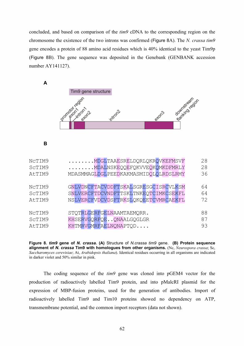
62
concluded, and based on comparison of the tim9 cDNA to the corresponding region on the
chromosome the existence of the two introns was confirmed (Figure 8A). The N. crassa tim9
gene encodes a protein of 88 amino acid residues which is 40% identical to the yeast Tim9p
(Figure 8B). The gene sequence was deposited in the Genebank (GENBANK accession
number AY141127).
A
B
28NcTIM9 28ScTIM9 36AtTIM9
........MDGLTAAESRELDQRLQKRQVKEFMSVF
........MDALNSKEQQEFQKVVEQKQMKDFMRLYMDASMMAGLDGLPEEDKAKMASMIDQLQLRDSLRMY
MML
DDD
GAG
LLL
EED
EE Q
Q
QQQ
KKR
EDD
FFMMLRR
FYY
64NcTIM9 64ScTIM9 72AtTIM9
GNLVDNCFTACVDDFTSKALSGRESGCISRCVLKSMSNLVERCFTDCVNDFTTSKLTNKEQTCIMKCSEKFLNSLVERCFVDCVDSFTRKSLQKQEETCVMRCAEKFL
NNLLL
VVV
DEERR
CCC
FFF
TTDD
CCC
VVV
DND
DDFFF
TTT
K
K
LLL
EEETT
CCC
IIVMM
RKR
CCCEE
KKKFF
MLL
88NcTIM9 87ScTIM9 93AtTIM9
STQTRLGERFGELNAAMTAEMQRR.KHSERVGQRFQE..QNAALGQGLGRKHTMRVGMRFAELNQNAPTQD....KKHH
RRR
LVV
GGG
RRR
FFF
EEE
L
L
N
NQQNNAA
Figure 8. tim9 gene of N. crassa. (A) Structure of N.crassa tim9 gene. (B) Protein sequence alignment of N. crassa Tim9 with homologues from other organisms. (Nc, Neurospora crassa; Sc, Saccharomyces cerevisiae; At, Arabidopsis thaliana). Identical residues occurring in all organisms are indicated in darker violet and 50% similar in pink.
The coding sequence of the tim9 gene was cloned into pGEM4 vector for the
production of radioactively labelled Tim9 protein, and into pMalcRI plasmid for the
expression of MBP-fusion proteins, used for the generation of antibodies. Import of
radioactively labelled Tim9 and Tim10 proteins showed no dependency on ATP,
transmembrane potential, and the common import receptors (data not shown).

63
3.1.3. Tim9 is an essential protein in N. crassa
Since TIM9 was shown to be an essential gene in yeast (Koehler et al., 1998b; Adam
et al., 1999), our research collaborator Frank Nargang and his coworkers used the sheltered
repeat-induced point mutation (RIP) procedure to generate the tim9 mutants in N. crassa. The
product of the procedure was a heterokaryotic strain with the tim9 gene in one type of nucleus
inactivated by RIP, and a wild type copy of the gene in the other (Figure 9A).
Figure 9. (A) Sheltered heterokaryon containing the tim9RIP mutant. The box depictes the heterokaryon, circles represent genetically distinct nuclei which make up the heterokaryon. Nucleus 1 contains only the nonfunctional RIPed version of tim9, whereas nucleus 2 contains a wt copy of the gene. (B) Level of Tim9 protein is decreased in cultures where the nucleus containing the RIPed gene is increased in number. The sheltered heterokaryon (panel A) and an acriflavine resistant control strain (Host III) were grown in the presence of acriflavine and tryptophan and mitochondria were isolated. Mitochondrial proteins were separated by SDS-PAGE and blotted to nitrocellulose. The blot was decorated with antibodies against the indicated proteins. (C) Scoring of single colonies from the heterokaryotic strain decribed in A. (ade, adenine; trp, tryptofan).
Growth of the heterokaryon in the presence of acriflavine and tryptophan resulted in
the RIPed nucleus gaining numerical superiority, since it provided the drug resistance and the
addition of tryptophan attended to its nutritional requirements. Under these conditions the
cells showed a slowed growth rate. The level of Tim9 protein was drastically decreased in the
mitochondria of the heterokaryotic strain, whereas the level of the Tim10 protein was slightly
reduced in these cells (Figure 9B).
In order to determine whether tim9 is an essential gene in N. crassa, conidiaspores
produced by the heterokaryon were streaked onto medium containing all nutritional

64
requirements of both nuclei in the strain (Figure 9A). Testing of nutritional requirements of
individual colonies produced from these conidia disclosed the tim9RIP nucleus as inviable
(Figure 9C). To confirm that the effects of RIP are to be attributed to the tim9 gene, the
sheltered heterokaryon was transformed with a bleomycin resistance plasmid containing a
wild type copy of the tim9 gene. Viable tryptophan-requiring homokaryotic strains were
recovered (data not shown). Therefore, tim9 is an essential gene in N. crassa.
The tim10 gene has proven an unsuitable candidate for the RIP mutagenesis because
of the proximity of tim10 to a potential tRNA-synthetase gene and the size of the duplication
required to act as a RIP substrate.
3.2. The Tim9 and Tim10 proteins form a heterooligomeric complex in
the intermembrane space of mitochondria
To investigate the subcellular localization of the N. crassa Tim9 and Tim10 proteins,
subcellular fractionation by differential centrifugation of cellular homogenate was performed.
Both proteins were found to be exclusively localised in the mitochondria (data not shown).
Their intramitochondrial location was further examined via the digitonin fractionation
procedure (Hartl et al., 1986).
Figure 10. Tim9 and Tim10 localize to the mitochondrial intermembrane space. Mitochondria were subfractionated in the presence of PK (0.25 µg/µl) by adding increasing amounts of the detergent (0.025-0.4%). The samples were TCA precipitated and analysed by SDS-PAGE and immunodecoration with the antibodies against indicated proteins (F26, typical N-terminal fragment of Tom40, indicative of opening of the outer membrane).

65
The isolated mitochondria of N. crassa were treated with increasing amounts of the
mild detergent digitonin. Upon this treatment solubilization of the outer membrane occurred
first, followed by that of the inner membrane. Fractionation was performed in the presence of
proteinase K to monitor for the degree of accessibility of various proteins. The degradation of
both Tim9 and Tim10 proteins occurred at the same digitonin concentrations as that of one
representative of the intermembrane space proteins, like CCHL, and of the N- and C-termini
of Tom40 which are also in the intermembrane space (Künkele et al., 1998) (Figure 10). This
indicates that Tim9 and Tim10 proteins of N. crassa reside in the mitochondrial
intermembrane space. A proteinase K-resistant core structure observed in both Tim9 and
Tim10 proteins is suggestive of a tight fold of this domain (Figure 10).
Figure 11. Analysis of the Tim9·Tim10 complex by BNGE. Mitochondria were solubilized in a buffer containing 0.5 % (w/v) digitonin and subjected to analysis via BNGE (with the marked amount of total mitochondrial protein loaded per lane), and immunodecoration with antibodies against either Tim9 or Tim10 protein. Molecular weight of ADH used as a marker protein is indicated on the righthand side.
To ascertain the oligomeric state of the Tim9 and Tim10 proteins, mitochondria were
solubilized in a buffer containing either 0.5% digitonin or 0.5% ß-dodecyl maltoside (DDM),
and subjected to the analysis by blue native gel electrophoresis (BNGE). Both proteins were
present in a 70-80 kDa complex as revealed by the decoration with antibodies against both
Tim9 and Tim10 proteins (Figure 11).
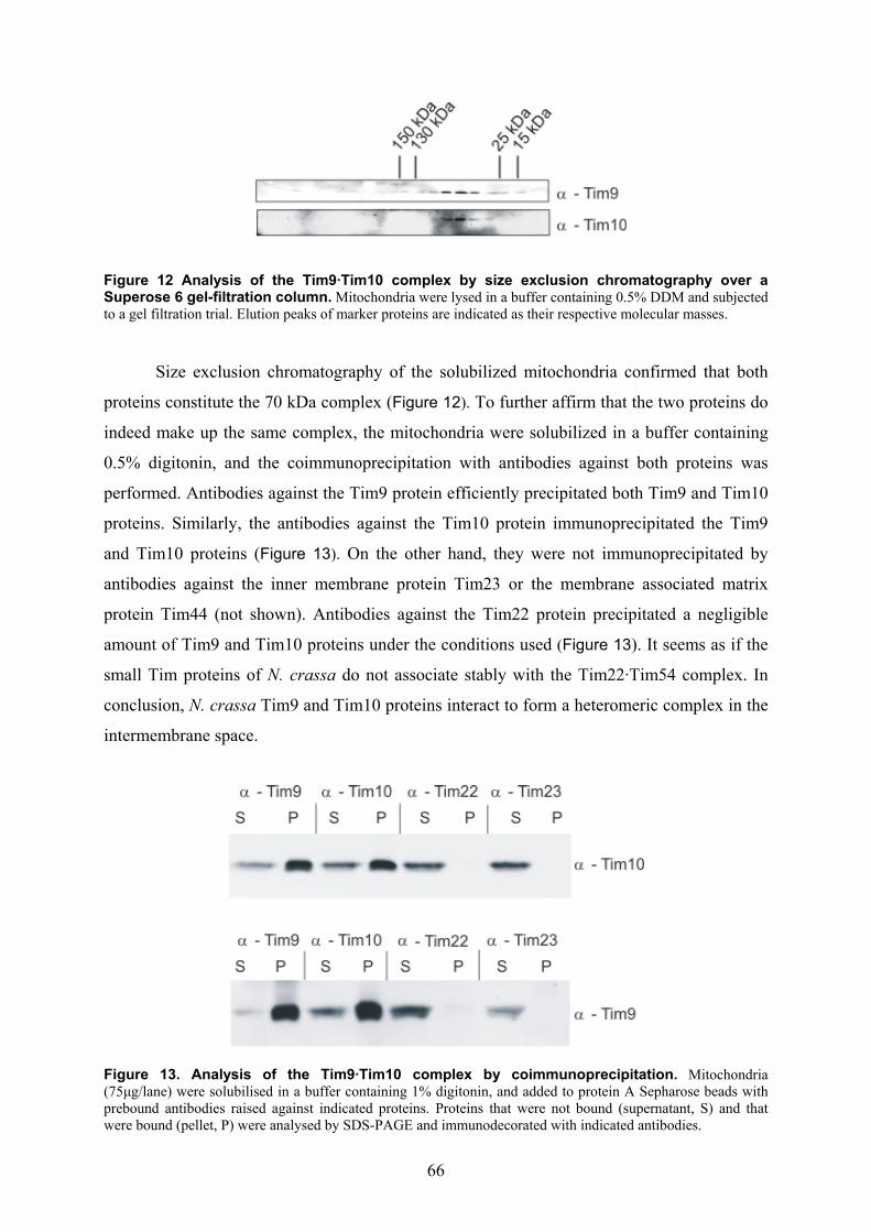
66
Figure 12 Analysis of the Tim9·Tim10 complex by size exclusion chromatography over a Superose 6 gel-filtration column. Mitochondria were lysed in a buffer containing 0.5% DDM and subjected to a gel filtration trial. Elution peaks of marker proteins are indicated as their respective molecular masses.
Size exclusion chromatography of the solubilized mitochondria confirmed that both
proteins constitute the 70 kDa complex (Figure 12). To further affirm that the two proteins do
indeed make up the same complex, the mitochondria were solubilized in a buffer containing
0.5% digitonin, and the coimmunoprecipitation with antibodies against both proteins was
performed. Antibodies against the Tim9 protein efficiently precipitated both Tim9 and Tim10
proteins. Similarly, the antibodies against the Tim10 protein immunoprecipitated the Tim9
and Tim10 proteins (Figure 13). On the other hand, they were not immunoprecipitated by
antibodies against the inner membrane protein Tim23 or the membrane associated matrix
protein Tim44 (not shown). Antibodies against the Tim22 protein precipitated a negligible
amount of Tim9 and Tim10 proteins under the conditions used (Figure 13). It seems as if the
small Tim proteins of N. crassa do not associate stably with the Tim22·Tim54 complex. In
conclusion, N. crassa Tim9 and Tim10 proteins interact to form a heteromeric complex in the
intermembrane space.
Figure 13. Analysis of the Tim9·Tim10 complex by coimmunoprecipitation. Mitochondria (75µg/lane) were solubilised in a buffer containing 1% digitonin, and added to protein A Sepharose beads with prebound antibodies raised against indicated proteins. Proteins that were not bound (supernatant, S) and that were bound (pellet, P) were analysed by SDS-PAGE and immunodecorated with indicated antibodies.
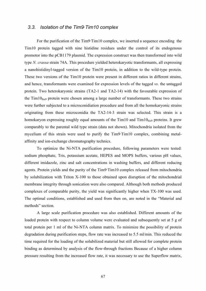
67
3.3. Isolation of the Tim9·Tim10 complex
For the purification of the Tim9·Tim10 complex, we inserted a sequence encoding the
Tim10 protein tagged with nine histidine residues under the control of its endogenous
promotor into the pCB1179 plasmid. The expression construct was then transformed into wild
type N. crassa strain 74A. This procedure yielded heterokaryotic transformants, all expressing
a nanohistidinyl-tagged version of the Tim10 protein, in addition to the wild-type protein.
These two versions of the Tim10 protein were present in different ratios in different strains,
and hence, transformants were examined for expression levels of the tagged vs. the untagged
protein. Two heterokaryonic strains (TA2-1 and TA2-14) with the favourable expression of
the Tim10his9 protein were chosen among a large number of transformants. These two strains
were further subjected to a microconidiation procedure and from all the homokaryonic strains
originating from these microconidia the TA2-14-3 strain was selected. This strain is a
homokaryon expressing roughly equal amounts of the Tim10 and Tim10his9 proteins. It grew
comparably to the parental wild type strain (data not shown). Mitochondria isolated from the
mycelium of this strain were used to purify the Tim9·Tim10 complex, combining metal-
affinity and ion-exchange chromatography technics.
To optimize the Ni-NTA purification procedure, following parameters were tested:
sodium phosphate, Tris, potassium acetate, HEPES and MOPS buffers, various pH values,
different imidazole, zinc and salt concentrations in washing buffers, and different reducing
agents. Protein yields and the purity of the Tim9·Tim10 complex released from mitochondria
by solubilization with Triton X-100 to those obtained upon disruption of the mitochondrial
membrane integrity through sonication were also compared. Although both methods produced
complexes of comparable purity, the yield was significantly higher when TX-100 was used.
The optimal conditions, established and used from then on, are noted in the “Material and
methods” section.
A large scale purification procedure was also established. Different amounts of the
loaded protein with respect to column volume were evaluated and subsequently set at 5 g of
total protein per 1 ml of the Ni-NTA column matrix. To minimize the possibility of protein
degradation during purification steps, flow rate was increased to 5.5 ml/min. This reduced the
time required for the loading of the solubilized material but still allowed for complete protein
binding as determined by analysis of the flow-through fractions Because of a higher column
pressure resulting from the increased flow rate, it was necessary to use the Superflow matrix,
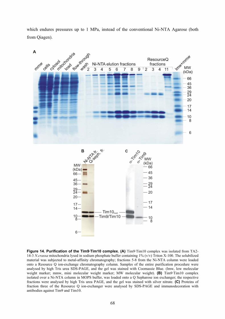
68
which endures pressures up to 1 MPa, instead of the conventional Ni-NTA Agarose (both
from Qiagen).
Figure 14. Purification of the Tim9·Tim10 complex. (A) Tim9·Tim10 complex was isolated from TA2-14-3 N.crassa mitochondria lysed in sodium phosphate buffer containing 1% (v/v) Triton X-100. The solubilized material was subjected to metal-affinity chromatography; fractions 5-8 from the Ni-NTA column were loaded onto a Resource Q ion-exchange chromatography column. Samples of the entire purification procedure were analysed by high Tris urea SDS-PAGE, and the gel was stained with Coomassie Blue. (lmw, low molecular weight marker; mmw, mini molecular weight marker; MW molecular weight). (B) Tim9·Tim10 complex isolated over a Ni-NTA column in MOPS buffer, was loaded onto a Q Sepharose ion exchanger; the respective fractions were analysed by high Tris urea PAGE, and the gel was stained with silver nitrate. (C) Proteins of fraction three of the Resource Q ion-exchanger were analysed by SDS-PAGE and immunodecoration with antibodies against Tim9 and Tim10.

69
The Tim9·Tim10 complex eluted from the Ni-NTA column contained several
contaminating proteins (Figure 14A). Therefore, we went on to investigate possibilities for
segregating the complex from these protein contaminants. The size exclusion chromatography
turned out to be an undependable tool. The loss in protein amount due to weak, yet
undeniably present, unspecific interactions of the Tim9·Tim10 complex with the gel filtration
column’s matrices, overshaddowed the overall modest improvements gained in purity-level.
The ion exchange chromatography was tested next. Fractions eluted from a Ni-NTA column
were pooled together and subjected to either cation- or anion-exchange chromatographies
(Figure 14A). The cation-exchange chromatography was found to be inadequate, since the
Tim9·Tim10 complex could not be eluted with high salt buffers at all pH values tested (pH 6-
10, at half-unit increments). The anion-exchangers, on the contrary, showed excellent
potential for improving the Tim9·Tim10 complex purification procedure. When using the pH
values from 6.0 till 8.0, the complex was in the flow-through of the anion-exchange column,
while the contaminants remained bound. The only exception was pH 6.0, where some
contaminants were also found in the flow-through. At pH values equal to and higher than 9.0,
Tim9·Tim10 complex was bound to the ion-exchanger. However, for all subsequent
purifications more physiological pH values were selected, closer to that of the mitochondrial
intermembrane space (pH 7.5-8.0). Replacing the Resource Q with Q Sepharose anion
exchanger enabled me to substantially decrease both protein amount losses, caused by
unspecific binding to the ion-exchanger matrix, and the amount of contaminating proteins
(compare A to B in Figure 14).
The protein bands of the purified complex were identified by mass spectroscopy (data
not shown) and immunodecoration (Figure 14C).
To essay whether there are some residual TX-100 traces in the samples containing the
Tim9·Tim10 complex, I have performed a thin-layer chromatography (TLC) of these probes.
Every step of the purification procedure was monitored via this method, which can detect TX-
100 concentrations as low as 0.005% (v/v). Minor amounts of TX-100 were found in the
washing fractions of the Ni-NTA column. In the Q Sepharose flow-through fractions, dialysed
against buffers without TX-100, the detergent was completely eliminated (data not shown).
To syllogise, N. crassa Tim9 and Tim10 proteins are the sole constituents of the purified
complex.

70
3.4. Structural organization of the purified Tim9·Tim10 complex
To prove the authenticity of the Tim9·Tim10 complex purified from mitochondria
expressing Tim10his9, it was compared to the complex set free from the wild type
mitochondria. Both complexes displayed an apparent molecular weight of 70-80 kDa when
analyzed by BNGE (Figure 15). The identical results were obtained using antibodies against
Tim9 and Tim10 proteins, confirming that the 70-80 kDa complex contains both proteins.
Thus, the purified complex possesses the same composition as the native one.
Figure 15. N. crassa Tim9 and Tim10 proteins form a hetero-hexamer. Mitochondria solubilized in 0.5% digitonin and the purified Tim9·Tim10 complex were analyzed by blue native gel electrophoresis, followed by immunodecoration with antibodies against Tim9.
To study the oligomeric organization of the purified Tim9·Tim10 complex, the
chemical cross-linking assay was used. The purified complex was incubated with
glutaraldehyde, and the cross-linking products analyzed by high-tris urea/SDS-PAGE and
immunostaining with antibodies against either Tim9 or Tim10 proteins (Figure 16).
In the absence of glutaraldehyde, the Tim9 and Tim10 proteins were found to be
monomers. The bands corresponding to the oligomeric species (dimer to hexamer) were
observed when the complex was treated with the cross-linker (Figure 16A). As a control, no
cross-linking adducts were observed when the cross-linking was performed in the presence of
0.5% SDS (Figure 16B). Upon decoration with the antibodies against the Tim10 protein, at
least two different X-linking species of a dimer product could be more readily observed
(Figure 16A, right panel). The same bands were observed upon decorations with the antibody

71
against the histidine-tag (data not shown). Thus, the cross-linking adduct corresponding to the
higher molecular weight dimer contains the Tim10his9 protein. This suggests that this variation
of the Tim10 protein is also a part of the oligomeric structure.
In conclusion, the isolated Tim9·Tim10 complex is a heterohexamer of an
approximate molecular weight of 70-80 kDa, equivalent in size and oligomeric state to the
native complex.
A
B
Figure 16. The Tim9 and Tim10 proteins form a heterohexamer. (A) The purified Tim9·Tim10 complex was incubated with glutaraldehyde, at indicated concentrations, for indicated time periods. Cross-linked products were analyzed by SDS-PAGE, followed by immunodecoration with antibodies against Tim9. (M, monomer; H, hexamer). (B) The purified complex was treated as above; in some of the samples, as control, the same X-linking procedure was performed in the presence of 0.5% SDS. After high Tris-urea PAGE, the immunodecoration was performed using antibodies against Tim10.
72
3.5. Initial trials for the crystallisation of the Tim9·Tim10 complex
One of the initial goals was to obtain a three-dimensional structure of the Tim9·Tim10
complex. To investigate the disposition of the complex to form ordered crystals, we have
attempted to measure circular dichroism (CD) spectra of the purified complex. However, the
complex adhered to the glass of the cuvette of the CD spectropolarimeter and subsequently
partially aggregated. Therefore, we were unable to obtain any conclusive data on the
secondary structure of the purified Tim9·Tim10 complex. Nevertheless, we probed the ability
of the pure fractions of the isolated Tim9·Tim10 complex to crystalise. Using the hanging
drop vapour diffusion crystallisation method (Crystal ScreenTM kit, Hampton Research, CA)
we tested various crystallisation conditions. As none of the tested crystallisation conditions
yielded crystals of the Tim9·Tim10 complex, we abandoned this line of research.
3.6. The influence of zinc on the integrity of the purified Tim9·Tim10
complex
It was previously reported that the yeast Tim10p as a recombinant MBP-Tim10p
fusion protein binds Zn2+ (Sirrenberg et al., 1998). To determine if Zn2+ is genuinly essential
for the oligomeric state of the purified N. crassa Tim9·Tim10 complex, the ability of the
complex to refold in the presence of reducing reagents or metal chelators was examined. The
treated samples were analysed using BNGE technique. When incubated at low temperatures,
the complex proved stable in the presence of metal chelator EDTA or the reducing agent
TCEP (Figure 17A). When it was heated to 95°C for 10 min in the presence or absence of
EDTA and left to cool on ice, it refolded to its native structure. In contrast, the presence of the
reducing reagent TCEP during the procedure led to its dissociation, demonstrated by the
disappearance of the bands corresponding to the complex and detection of the monomeric
Tim9 and Tim10 proteins (Figure 17A).
As a control, dimerization of the cytosolic domain of the yeast Tom70p which
contains three cysteine residues was analysed. The dimer resumed its native conformation
after heat denaturation in the presence of both EDTA and TCEP (Figure 17B). Thus, the
observed effect of the reducing agent on the stability of the Tim9·Tim10 complex is not a
common feature of every protein containing multiple cysteine residues. These results argue
that the complex purified from mitochondria isolated under non-reducing conditions does not
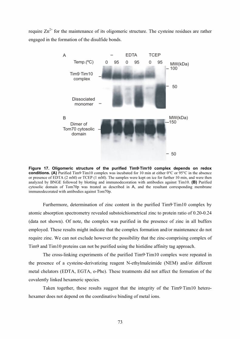
73
require Zn2+ for the maintenance of its oligomeric structure. The cysteine residues are rather
engaged in the formation of the disulfide bonds.
Figure 17. Oligomeric structure of the purified Tim9·Tim10 complex depends on redox conditions. (A) Purified Tim9·Tim10 complex was incubated for 10 min at either 0°C or 95°C in the absence or presence of EDTA (2 mM) or TCEP (1 mM). The samples were kept on ice for further 10 min, and were then analyzed by BNGE followed by blotting and immunodecoration with antibodies against Tim10. (B) Purified cytosolic domain of Tom70p was treated as described in A, and the resultant corresponding membrane immunodecorated with antibodies against Tom70p.
Furthermore, determination of zinc content in the purified Tim9·Tim10 complex by
atomic absorption spectrometry revealed substoichiometrical zinc to protein ratio of 0.20-0.24
(data not shown). Of note, the complex was purified in the presence of zinc in all buffers
employed. These results might indicate that the complex formation and/or maintenance do not
require zinc. We can not exclude however the possibility that the zinc-comprising complex of
Tim9 and Tim10 proteins can not be purified using the histidine affinity tag approach.
The cross-linking experiments of the purified Tim9·Tim10 complex were repeated in
the presence of a cysteine-derivatizing reagent N-ethylmaleimide (NEM) and/or different
metal chelators (EDTA, EGTA, o-Phe). These treatments did not affect the formation of the
covalently linked hexameric species.
Taken together, these results suggest that the integrity of the Tim9·Tim10 hetero-
hexamer does not depend on the coordinative binding of metal ions.
74
3.7. The purified Tim9·Tim10 complex is functional in binding its
substrate proteins
Studies of the yeast Tim9p and Tim10p proteins suggested that they are generally
involved in import of the carrier proteins into the mitochondrial inner membrane (Sirrenberg
et al., 1998; Koehler et al., 1998a and 1998b). To assert the functionality of the purified N.
crassa Tim9·Tim10 complex, its ability to interact with its physiological substrate, the
precursor of ADP/ATP carrier (AAC), was examined. For that purpose, the ability of excess
amounts of the purified complex to compete the in vitro import of radiolabelled precursors of
AAC2p, and of control pSu9(1-69)DHFR proteins, was tested. The Tim9·Tim10 complex of
N. crassa inhibited the import of the carrier protein almost entirely, while the import of the
matrix-destined protein was not affected (Figure 18A). A slight increase in the import of
pSu9(1-69)DHFR precursor in the presence of N. crassa Tim9·Tim10 complex did not result
from the partial loss of the outer membrane integrity, since the intermembrane space proteins
D-lactate dehydrogenase and the Tim13p protein were not degraded by the added protease,
demonstrated through immunodecorations (data not shown).
The Tom70p protein was found to be the main receptor for the AAC precursor. In the
absence of the Tom70p receptor, AAC2p is imported via a considerably less efficient bypass
pathway (Hines et al., 1990). Therefore, the significant inhibition in the AAC import observed
in our experiments could have resulted from the possible interaction of the N. crassa
Tim9·Tim10 complex with the Tom70p receptor. Furthermore, the addition of the purified
antibodies against N. crassa Tom70 protein to the isolated N. crassa mitochondria prior to the
in vitro import significantly reduced the import of the radioactively labelled AAC2p
precursor, but neither of the yeast Tim13p nor the N. crassa Tim10 proteins (data not shown).
However, import into isolated yeast mitochondria from a strain lacking this receptor protein
was also competed by the N. crassa Tim9·Tim10 complex (Figure 18B).
Additional evidence for a direct interaction between the N. crassa Tim9·Tim10
complex and its substrate came from the immunoprecipitation experiments. Significant
amounts of the in vitro synthesized, radioactively labelled AAC protein, preincubated with the
N. crassa Tim9·Tim10 complex, were immunoprecipitated with the antibody against the N.
crassa Tim9 protein (data not shown). No specific precipitation of control protein pSu9(1-
69)DHFR was observed. Moreover, upon incubation of the complex with a mixture of porin
and AAC2p isolated from yeast cells (Panneels, 2003), the antibodies against N. crassa Tim9
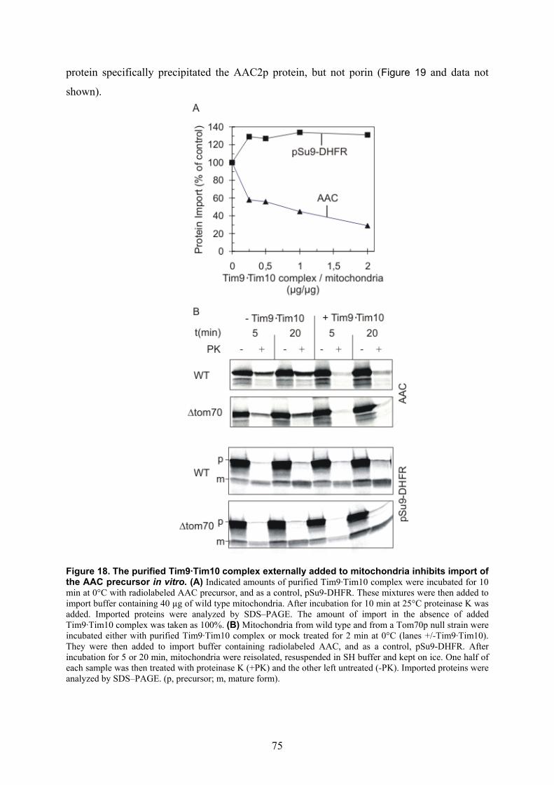
75
protein specifically precipitated the AAC2p protein, but not porin (Figure 19 and data not
shown).
Figure 18. The purified Tim9·Tim10 complex externally added to mitochondria inhibits import of the AAC precursor in vitro. (A) Indicated amounts of purified Tim9·Tim10 complex were incubated for 10 min at 0°C with radiolabeled AAC precursor, and as a control, pSu9-DHFR. These mixtures were then added to import buffer containing 40 µg of wild type mitochondria. After incubation for 10 min at 25°C proteinase K was added. Imported proteins were analyzed by SDS–PAGE. The amount of import in the absence of added Tim9·Tim10 complex was taken as 100%. (B) Mitochondria from wild type and from a Tom70p null strain were incubated either with purified Tim9·Tim10 complex or mock treated for 2 min at 0°C (lanes +/-Tim9·Tim10). They were then added to import buffer containing radiolabeled AAC, and as a control, pSu9-DHFR. After incubation for 5 or 20 min, mitochondria were reisolated, resuspended in SH buffer and kept on ice. One half of each sample was then treated with proteinase K (+PK) and the other left untreated (-PK). Imported proteins were analyzed by SDS–PAGE. (p, precursor; m, mature form).
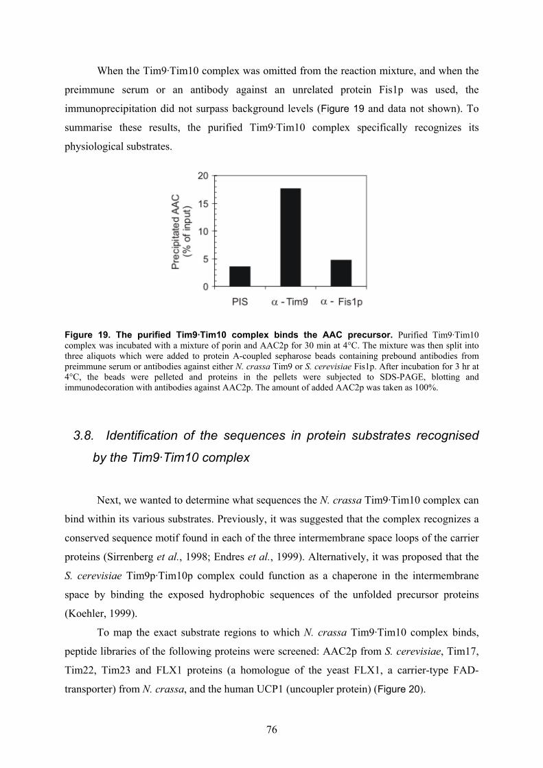
76
When the Tim9·Tim10 complex was omitted from the reaction mixture, and when the
preimmune serum or an antibody against an unrelated protein Fis1p was used, the
immunoprecipitation did not surpass background levels (Figure 19 and data not shown). To
summarise these results, the purified Tim9·Tim10 complex specifically recognizes its
physiological substrates.
Figure 19. The purified Tim9·Tim10 complex binds the AAC precursor. Purified Tim9·Tim10 complex was incubated with a mixture of porin and AAC2p for 30 min at 4°C. The mixture was then split into three aliquots which were added to protein A-coupled sepharose beads containing prebound antibodies from preimmune serum or antibodies against either N. crassa Tim9 or S. cerevisiae Fis1p. After incubation for 3 hr at 4°C, the beads were pelleted and proteins in the pellets were subjected to SDS-PAGE, blotting and immunodecoration with antibodies against AAC2p. The amount of added AAC2p was taken as 100%.
3.8. Identification of the sequences in protein substrates recognised
by the Tim9·Tim10 complex
Next, we wanted to determine what sequences the N. crassa Tim9·Tim10 complex can
bind within its various substrates. Previously, it was suggested that the complex recognizes a
conserved sequence motif found in each of the three intermembrane space loops of the carrier
proteins (Sirrenberg et al., 1998; Endres et al., 1999). Alternatively, it was proposed that the
S. cerevisiae Tim9p·Tim10p complex could function as a chaperone in the intermembrane
space by binding the exposed hydrophobic sequences of the unfolded precursor proteins
(Koehler, 1999).
To map the exact substrate regions to which N. crassa Tim9·Tim10 complex binds,
peptide libraries of the following proteins were screened: AAC2p from S. cerevisiae, Tim17,
Tim22, Tim23 and FLX1 proteins (a homologue of the yeast FLX1, a carrier-type FAD-
transporter) from N. crassa, and the human UCP1 (uncoupler protein) (Figure 20).

77
Figure 20. Substrate recognition by the Tim9·Tim10 complex. A peptide library on a cellulose membrane, corresponding to the indicated proteins and control peptides, was incubated with purified Tim9·Tim10 complex. Bound proteins were blotted to PVDF membranes and decorated with antibodies against Tim9. Binding was quantified by scanning densitometry of three independent experiments. The intensity of the strongest spot from each experiment was set to 100. Below each peptide, it is indicated whether its sequence covers a transmembrane domain (TMD), a loop or if its sequence stretches over both types of structure.

78
These librarires commonly comprise of 13mer peptides covering the whole primary
sequence of a given protein, with 10 residues-overlaps. The yeast carrier was chosen to allow
the comparison with experiments performed with the yeast small Tim proteins (Curran et al.,
2002a). The radioactively labelled precursor of S. cerevisiae AAC2p was efficiently imported
into the isolated N. crassa mitochondria (data not shown), supporting combining of
translocons of different taxonomic groups as feasible. Control peptides covering
transmembrane domains of various non-mitochondrial proteins were also included in the
screen (origin and sequences of control peptides are given in Figure 20). All peptides were
spotted onto one membrane allowing direct comparison of different proteins.
Analysis of the binding sequences of the N. crassa Tim9·Tim10 complex revealed a
strong general inclination towards peptides corresponding to transmembrane domains (TMD)
and their flanking regions. Among the analyzed proteins, the highest affinity was observed
towards the AAC2p protein (Figure 20). The strongest interaction was with the peptides
covering parts of its fourth TMD, followed by the TMDs 1, 3 and 5.
Binding to Tim22 protein was relatively weak and it occurred mainly in regions
covering its TMDs, rather than the intermembrane space loops. Interaction of the Tim9·Tim10
complex with the human Ucp1 was less restricted to TMDs, yet the highest affinity observed
was still towards the putative TMDs 1 and 2.
One unexpected finding of the screen was the significant binding of the complex to
two segments of the N. crassa Tim23 protein (Figure 20). A possible interaction of the yeast
Tim9p and Tim10p proteins with the yeast Tim23p protein is still under debate (Leuenberger
et al., 1999; Davis et al., 2000). Peptides covering segments of the N. crassa Tim23 protein
identified in the screen to be bound by the purified Tim9·Tim10 complex with considerable
affinity were the putative intermembrane space domain (peptides 24-27, residues 70-91) and
the interface between the intermembrane space domain and the first predicted TMD (peptides
33-37, residues 97-121) (Figure 20). Both regions are highly conserved in the Tim23 proteins
from different organisms (Figure 21).
Binding to the hydrophobic non-mitochondrial control peptides was weak (Figure 20).
Moreover, from 20 peptides displaying the highest affinity in both AAC2p (Figure 22) and
FLX1 (data not shown), nineteen comprised at least one charged residue within their
sequence. The Tim9·Tim10 complex displayed the highest affinity prevalently towards
peptides covering interface regions between the membrane spanning segments and the soluble
loops (Figure 22) and not those covering midsections of the putative TMDs.
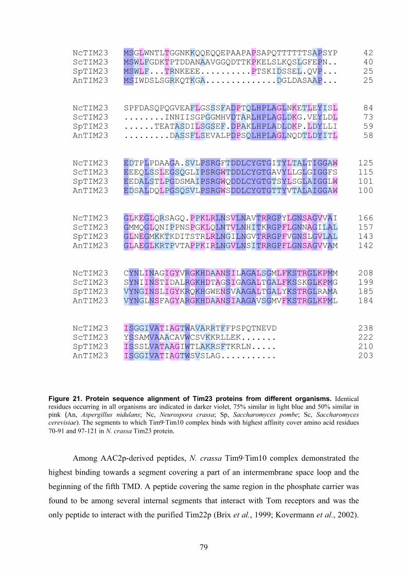
79
42NcTIM23 40ScTIM23 25SpTIM23 25AnTIM23
MMMM
SSSS
GWWI
LLLW
WFFD
NG.S
TD.L
LK.S
TTTG
GPRR
GTNK
NDKQ
KDET
KAEK
QNEG
QA.A
EA..
QV..
QG..
EG..
PQ..
AD..
AT..
PT..
AK..
PPP.
SKT.
AES.
PLK.
QSI.
TLDD
TKSG
TQSL
TSED
TLLA
TG.S
SFQA
AEVA
PPPP
SN..
Y...
P...
84NcTIM23 73ScTIM23 59SpTIM23 58AnTIM23
S...
P...
F...
D...
A...
S...
Q.T.
P.E.
QIA.
GNTD
VNAA
EISS
AIDS
FSIF
LGLL
GPSS
SGGE
SGSV
SMEA
FHFL
AV.P
DDDD
PTPP
TAAS
QRKQ
LLLL
HHHH
PPPP
LLLL
AAAA
GGDG
LLLL
NDDN
KKKQ
EGPD
T..T
LVLL
EEDD
YYYY
ILLI
SDLT
LLIL
125NcTIM23 115ScTIM23 101SpTIM23 100AnTIM23
EEEE
DEED
TEDS
PQAA
LLLL
PSSD
DSTQ
ALLL
AEPP
GGGG
ASDS
.QSQ
SGMS
VLAV
LIIL
PPPP
SSSS
RRRR
GGGG
FWWW
TTQS
DDDD
DDDD
LLLL
CCCC
YYYY
GGGG
TTTT
GGGG
IATT
TVST
YYYY
LLLV
TLST
AGGA
LLLL
TGAA
IIII
GGGG
GGGG
AFLA
WSWW
166NcTIM23 157ScTIM23 143SpTIM23 142AnTIM23
GGGG
LMLL
KMNA
EQEE
GGGG
LLML
QQKK
RNKR
SITT
APKP
GPDV
QNIT
.STA
PPSP
PGTP
KKRK
LLLI
RQRR
LLLL
NNNN
STGG
VVIV
LLLL
NNNN
AHGS
VIVI
TTTT
RKRR
RRRR
GGGG
PPPP
YFFF
LLVL
GGGG
NNNN
SNSS
AALA
GGGG
VIVV
VLLV
AAAA
ILLM
208NcTIM23 199ScTIM23 185SpTIM23 184AnTIM23
CSVV
YYYY
NNNN
LIGG
IIIL
NNNN
ASSS
GTLF
IIIA
GDGG
YAYY
VLKA
RRRR
GGQG
KKKK
HHHH
DDGD
ATWA
AAEA
NGNN
SSSS
IIVI
LGAA
AAAA
GGGG
AAAA
LLLV
STTS
GGGG
MAAM
LLLV
FFYF
KKKK
SSSS
TSTT
RKRR
GGGG
LLLL
KKRK
PPAP
MMMM
MGAL
238NcTIM23 222ScTIM23 210SpTIM23 203AnTIM23
IYII
SSSS
GSSG
GASG
IMLI
VVVV
AAAA
TATT
IAAI
ACAA
GAGG
TVIT
WWWW
ACTS
VSLV
AVAS
RKKL
RKRA
TRSG
FLF.
FLT.
PEK.
SKR.
P.L.
Q.N.
T...
N...
E...
V...
D...
Figure 21. Protein sequence alignment of Tim23 proteins from different organisms. Identical residues occurring in all organisms are indicated in darker violet, 75% similar in light blue and 50% similar in pink (An, Aspergillus nidulans; Nc, Neurospora crassa; Sp, Saccharomyces pombe; Sc, Saccharomyces cerevisiae). The segments to which Tim9·Tim10 complex binds with highest affinity cover amino acid residues 70-91 and 97-121 in N. crassa Tim23 protein.
Among AAC2p-derived peptides, N. crassa Tim9·Tim10 complex demonstrated the
highest binding towards a segment covering a part of an intermembrane space loop and the
beginning of the fifth TMD. A peptide covering the same region in the phosphate carrier was
found to be among several internal segments that interact with Tom receptors and was the
only peptide to interact with the purified Tim22p (Brix et al., 1999; Kovermann et al., 2002).
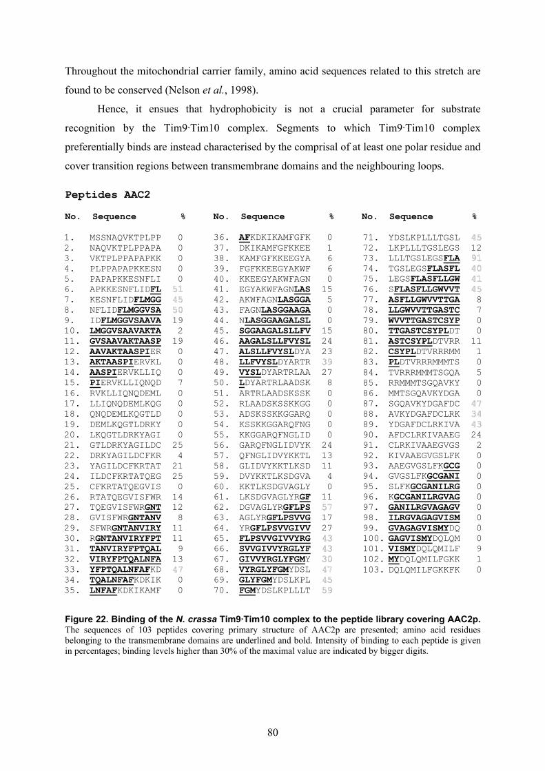
80
Throughout the mitochondrial carrier family, amino acid sequences related to this stretch are
found to be conserved (Nelson et al., 1998).
Hence, it ensues that hydrophobicity is not a crucial parameter for substrate
recognition by the Tim9·Tim10 complex. Segments to which Tim9·Tim10 complex
preferentially binds are instead characterised by the comprisal of at least one polar residue and
cover transition regions between transmembrane domains and the neighbouring loops. Peptides AAC2 No. Sequence % No. Sequence % No. Sequence % 1. MSSNAQVKTPLPP 0 2. NAQVKTPLPPAPA 0 3. VKTPLPPAPAPKK 0 4. PLPPAPAPKKESN 0 5. PAPAPKKESNFLI 0 6. APKKESNFLIDFL 51 7. KESNFLIDFLMGG 45 8. NFLIDFLMGGVSA 50 9. IDFLMGGVSAAVA 19 10. LMGGVSAAVAKTA 2 11. GVSAAVAKTAASP 19 12. AAVAKTAASPIER 0 13. AKTAASPIERVKL 0 14. AASPIERVKLLIQ 0 15. PIERVKLLIQNQD 7 16. RVKLLIQNQDEML 0 17. LLIQNQDEMLKQG 0 18. QNQDEMLKQGTLD 0 19. DEMLKQGTLDRKY 0 20. LKQGTLDRKYAGI 0 21. GTLDRKYAGILDC 25 22. DRKYAGILDCFKR 4 23. YAGILDCFKRTAT 21 24. ILDCFKRTATQEG 25 25. CFKRTATQEGVIS 0 26. RTATQEGVISFWR 14 27. TQEGVISFWRGNT 12 28. GVISFWRGNTANV 8 29. SFWRGNTANVIRY 11 30. RGNTANVIRYFPT 11 31. TANVIRYFPTQAL 9 32. VIRYFPTQALNFA 13 33. YFPTQALNFAFKD 47 34. TQALNFAFKDKIK 0 35. LNFAFKDKIKAMF 0
36. AFKDKIKAMFGFK 0 37. DKIKAMFGFKKEE 1 38. KAMFGFKKEEGYA 6 39. FGFKKEEGYAKWF 6 40. KKEEGYAKWFAGN 0 41. EGYAKWFAGNLAS 15 42. AKWFAGNLASGGA 5 43. FAGNLASGGAAGA 0 44. NLASGGAAGALSL 0 45. SGGAAGALSLLFV 15 46. AAGALSLLFVYSL 24 47. ALSLLFVYSLDYA 23 48. LLFVYSLDYARTR 39 49. VYSLDYARTRLAA 27 50. LDYARTRLAADSK 8 51. ARTRLAADSKSSK 0 52. RLAADSKSSKKGG 0 53. ADSKSSKKGGARQ 0 54. KSSKKGGARQFNG 0 55. KKGGARQFNGLID 0 56. GARQFNGLIDVYK 24 57. QFNGLIDVYKKTL 13 58. GLIDVYKKTLKSD 11 59. DVYKKTLKSDGVA 4 60. KKTLKSDGVAGLY 0 61. LKSDGVAGLYRGF 11 62. DGVAGLYRGFLPS 57 63. AGLYRGFLPSVVG 17 64. YRGFLPSVVGIVV 27 65. FLPSVVGIVVYRG 43 66. SVVGIVVYRGLYF 43 67. GIVVYRGLYFGMY 30 68. VYRGLYFGMYDSL 47 69. GLYFGMYDSLKPL 45 70. FGMYDSLKPLLLT 59
71. YDSLKPLLLTGSL 45 72. LKPLLLTGSLEGS 12 73. LLLTGSLEGSFLA 91 74. TGSLEGSFLASFL 40 75. LEGSFLASFLLGW 41 76. SFLASFLLGWVVT 45 77. ASFLLGWVVTTGA 8 78. LLGWVVTTGASTC 7 79. WVVTTGASTCSYP 0 80. TTGASTCSYPLDT 0 81. ASTCSYPLDTVRR 11 82. CSYPLDTVRRRMM 1 83. PLDTVRRRMMMTS 0 84. TVRRRMMMTSGQA 5 85. RRMMMTSGQAVKY 0 86. MMTSGQAVKYDGA 0 87. SGQAVKYDGAFDC 47 88. AVKYDGAFDCLRK 34 89. YDGAFDCLRKIVA 43 90. AFDCLRKIVAAEG 24 91. CLRKIVAAEGVGS 2 92. KIVAAEGVGSLFK 0 93. AAEGVGSLFKGCG 0 94. GVGSLFKGCGANI 0 95. SLFKGCGANILRG 0 96. KGCGANILRGVAG 0 97. GANILRGVAGAGV 0 98. ILRGVAGAGVISM 0 99. GVAGAGVISMYDQ 0 100. GAGVISMYDQLQM 0 101. VISMYDQLQMILF 9 102. MYDQLQMILFGKK 1 103. DQLQMILFGKKFK 0
Figure 22. Binding of the N. crassa Tim9·Tim10 complex to the peptide library covering AAC2p. The sequences of 103 peptides covering primary structure of AAC2p are presented; amino acid residues belonging to the transmembrane domains are underlined and bold. Intensity of binding to each peptide is given in percentages; binding levels higher than 30% of the maximal value are indicated by bigger digits.

81
3.9. Tim23 protein is a substrate of the Tim9·Tim10 complex in N.
crassa
To substantiate the proposed interaction of the Tim9·Tim10 complex with the Tim23
precursor protein, we used the chemical cross-linking approach. When the radiolabelled
precursor of Tim23 was incubated with de-energized mitochondria, an import intermediate
was formed. It was accessible to external protease, but partially translocated into the
intermembrane space, thereby being equivalent to the well defined stage III intermediate of
the AAC precursor (Pfanner and Neupert, 1987). Under these conditions, the addition of the
bifunctional cross-linking reagent m-maleimidobenzoyl-N-hydroxysuccinimide ester (MBS)
resulted in the formation of the distinct cross-linking adducts that specifically
immunoprecipitated with the antibodies against Tim9, Tim10 and Tim8 proteins (Figure 23).
The precursor bound to mitochondria was degraded by the externally added proteinase K, as
opposed to the cross-linking adduct which was inaccessible to it.
Figure 23. The Tim23 protein is in the vicinity of Tim9, Tim10 and Tim8 proteins on its mitochondrial translocation pathway. The radiolabelled Tim23 precursor was incubated with de-energized mitochondria in the presence or absence of the chemical cross-linker MBS for 30 min at 15°C. Reactions were then quenched by the addition of glycine (pH 8.8) to a final concentration f 100 mM, and one half of the sample was treated with proteinase K. The mitochondria of all four samples were pelleted and subjected to SDS-PAGE. Another sample treated with MBS but not with PK (+MBS, -PK) was subjected to immunoprecipitation with antibodies against the indicated TIM proteins or with preimmune serum. The immunoprecipitates were solubilized in sample buffer and analyzed by SDS-PAGE and autoradiography.
These results indicate that the Tim23 precursor molecules associated with the
Tim9·Tim10 complex were translocated across the outer mitochondrial membrane. Thus,
while crossing the intermembrane space on its way into the inner membrane the precursor of
N. crassa Tim23 protein interacts with both Tim9·Tim10 and Tim8·Tim13 complexes.
82
3.10. The TOM core complex and the Tim9·Tim10 complex are
sufficient for the import of the ADP/ATP carrier across the outer
membrane of mitochondria
The most competent way to certify the utter sufficiency of certain components in a
particular translocation process across biological membranes in vitro is to use the
reconstituted system. We employed the reconstituted in vitro system to examine if some as yet
unknown components in the internal compartments of mitochondria are required for
translocating the AAC precursor across the mitochondrial outer membrane. Detergent
solutions of the purified TOM core complex (Ahting et al., 1999) and E. coli lipids were
combined, the detergent was removed by polystyrene beads and thereby the proteoliposomes
were formed. In certain cases, the enzyme lactate dehydrogenase (LDH) was also enclosed in
the vesicles and its activity was used to essay the pore forming ability of the reconstituted
TOM complex. When the substrates of LDH, NADH and pyruvate, were added to vesicles
containing either the reconstituted TOM core complex or porin (a control pore forming
protein), free influx of the externally added LDH-substrates was observed (data not shown)
(Künkele et al., 1998a). The Tim9·Tim10 complex was added to some of the reconstitution
reactions, resulting in its enclosure within the proteoliposomes. In Figure 24A the protein
composition of the lipid vesicles containing the TOM core complex alone or in combination
with the enclosed Tim9·Tim10 complex is depicted.
Translocation of the radiolabelled carrier precursor across the membranes of
proteoliposomes was assessed through resistance to the externally added protease. Some
unspecific association of the AAC protein with the pure lipid vesicles was observed, due to
overall hydrophobic nature of the carrier protein (Figure 24B, lanes marked -PK). With the
TOM core or the Tim9·Tim10 complex reconstituted into vesicles separately, the amount of
the protease-protected carrier protein was negligible (Figure 24B, lanes 2 and 8, respectively).
However, the amount of protected material increased by about four fold to levels similar to
those observed with intact mitochondria, when vesicles containing both the TOM complex
and the Tim9·Tim10 complex were used (Figure 24B, lanes 4 and 12). The specificity of this
observation was supported by several control experiments:
i. import of the carrier precursor was competed out with the externally added
Tim9·Tim10 complex (Figure 24B, lane 6);

83
ii. complete degradation of the imported AAC precursor by proteinase K (PK) occurred
upon solubilization of the proteoliposomes by Triton-X100 (not shown);
iii. the degree of protection of the AAC precursor against PK was of the background level
(data not shown), when porin was reconstituted into lipid vesicles containing the
enclosed Tim9·Tim10 complex.
In conclusion, the TOM core complex and the Tim9·Tim10 complex meet all the
requirements for the productive transfer of the ADP/ATP carrier across the outer membrane
of mitochondria.
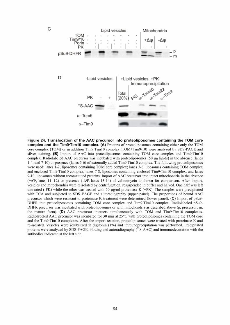
84
Figure 24. Translocation of the AAC precursor into proteoliposomes containing the TOM core complex and the Tim9·Tim10 complex. (A) Proteins of proteoliposomes containing either only the TOM core complex (TOM) or in addition Tim9·Tim10 complex (TOM+Tim9/10) were analyzed by SDS-PAGE and silver staining. (B) Import of AAC into proteoliposomes containing TOM core complex and Tim9·Tim10 complex. Radiolabeled AAC precursor was incubated with proteoliposomes (50 µg lipids) in the absence (lanes 1-4, and 7-10) or presence (lanes 5-6) of externally added Tim9·Tim10 complex. The following proteoliposomes were used: lanes 1-2, liposomes containing TOM core complex; lanes 3-6, liposomes containing TOM complex and enclosed Tim9·Tim10 complex; lanes 7-8, liposomes containing enclosed Tim9·Tim10 complex; and lanes 9-10, liposomes without reconstituted proteins. Import of AAC precursor into intact mitochondria in the absence (+∆Ψ, lanes 11−12) or presence (-∆Ψ, lanes 13-14) of valinomycin is shown for comparison. After import, vesicles and mitochondria were reisolated by centrifugation, resuspended in buffer and halved. One half was left untreated (-PK) while the other was treated with 50 µg/ml proteinase K (+PK). The samples were precipitated with TCA and subjected to SDS–PAGE and autoradiography (upper panel). The proportions of bound AAC precursor which were resistant to proteinase K treatment were determined (lower panel). (C) Import of pSu9-DHFR into proteoliposomes containing TOM core complex and Tim9·Tim10 complex. Radiolabeled pSu9-DHFR precursor was incubated with proteoliposomes or with mitochondria as described above (p, precursor; m, the mature form). (D) AAC precursor interacts simultaneously with TOM and Tim9·Tim10 complexes. Radiolabeled AAC precursor was incubated for 30 min at 25°C with proteoliposomes containing the TOM core and the Tim9·Tim10 complexes. After the import reaction, proteolipisomes were treated with proteinase K and re-isolated. Vesicles were solubilized in digitonin (1%) and immunoprecipitation was performed. Precipitated proteins were analyzed by SDS-PAGE, blotting and autoradiography (35S-AAC) and immunodecoration with the antibodies indicated at the left side.
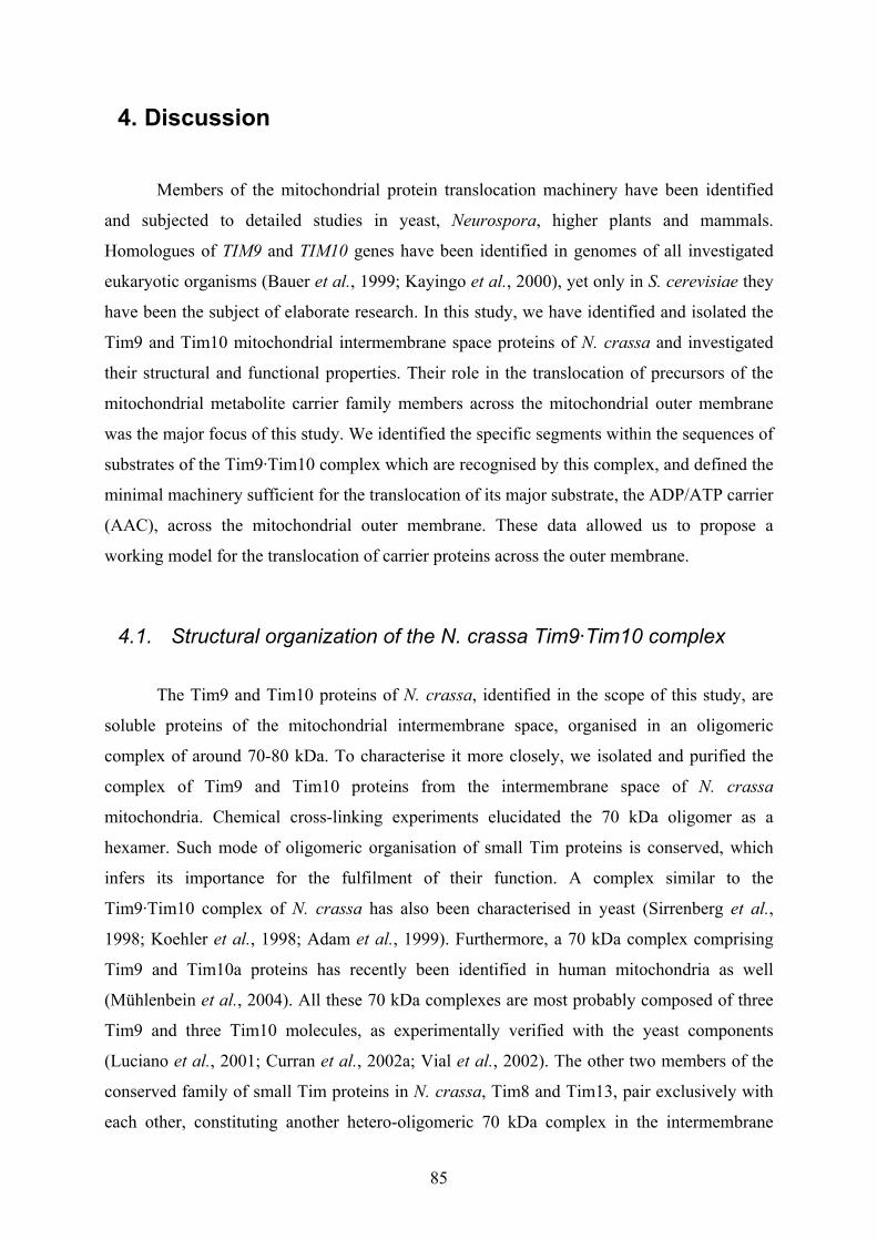
85
4. Discussion
Members of the mitochondrial protein translocation machinery have been identified
and subjected to detailed studies in yeast, Neurospora, higher plants and mammals.
Homologues of TIM9 and TIM10 genes have been identified in genomes of all investigated
eukaryotic organisms (Bauer et al., 1999; Kayingo et al., 2000), yet only in S. cerevisiae they
have been the subject of elaborate research. In this study, we have identified and isolated the
Tim9 and Tim10 mitochondrial intermembrane space proteins of N. crassa and investigated
their structural and functional properties. Their role in the translocation of precursors of the
mitochondrial metabolite carrier family members across the mitochondrial outer membrane
was the major focus of this study. We identified the specific segments within the sequences of
substrates of the Tim9·Tim10 complex which are recognised by this complex, and defined the
minimal machinery sufficient for the translocation of its major substrate, the ADP/ATP carrier
(AAC), across the mitochondrial outer membrane. These data allowed us to propose a
working model for the translocation of carrier proteins across the outer membrane.
4.1. Structural organization of the N. crassa Tim9·Tim10 complex
The Tim9 and Tim10 proteins of N. crassa, identified in the scope of this study, are
soluble proteins of the mitochondrial intermembrane space, organised in an oligomeric
complex of around 70-80 kDa. To characterise it more closely, we isolated and purified the
complex of Tim9 and Tim10 proteins from the intermembrane space of N. crassa
mitochondria. Chemical cross-linking experiments elucidated the 70 kDa oligomer as a
hexamer. Such mode of oligomeric organisation of small Tim proteins is conserved, which
infers its importance for the fulfilment of their function. A complex similar to the
Tim9·Tim10 complex of N. crassa has also been characterised in yeast (Sirrenberg et al.,
1998; Koehler et al., 1998; Adam et al., 1999). Furthermore, a 70 kDa complex comprising
Tim9 and Tim10a proteins has recently been identified in human mitochondria as well
(Mühlenbein et al., 2004). All these 70 kDa complexes are most probably composed of three
Tim9 and three Tim10 molecules, as experimentally verified with the yeast components
(Luciano et al., 2001; Curran et al., 2002a; Vial et al., 2002). The other two members of the
conserved family of small Tim proteins in N. crassa, Tim8 and Tim13, pair exclusively with
each other, constituting another hetero-oligomeric 70 kDa complex in the intermembrane

86
space (Hoppins et al., 2004). Yeast and human counterparts of these two proteins are also
characterised by a similar mode of structural arrangement (Leuenberger et al., 1999; Koehler
et al., 1999; Paschen et al., 2000; Rothbauer et al., 2001).
Figure 25. Hypothetical model of the secondary structure of the Tim10 protein. (Program used to predict the secondary structure of the Tim10 protein can be found on the web site: http://www.expasy.org/tools/#secondary).
The purified Tim9·Tim10 complex seems to be quite stable under variety of different
conditions, like high salt concentrations, sonication, and relatively high detergent amounts,
and even to be able to refold into a hexamer upon exposure to extreme temperatures (95ºC).
This, together with the relatively simple secondary structure predictions for the small Tim
proteins (Figure 25), and the potential stabilization of the ensuing 3D structure through zinc
binding, led us to attempt to obtain crystals and solve the crystal structure of the Tim9·Tim10
complex. Attempts to confirm the secondary structure predictions through CD spectrometry
failed due to the unspecific adherence to equipment and consequential aggregation of the
Tim9 and Tim10 proteins. Also, under all different conditions of crystallization screens which
we tested, we did not observe crystal formation.
4.2. Zinc binding and the redox state of Tim9 and Tim10 proteins
The mitochondrial intermembrane space is considered to be in a complete redox
equilibrium with the reducing cytoplasm. Therefore, the small Tim proteins were also
assumed to be in a reduced state. Due to the presence of two CX3C motives in their primary
sequence, which are conserved in all identified small Tim proteins (Sirrenberg et al., 1998;
Koehler et al., 1998; Adam et al., 1999, Koehler et al., 1999; Leuenberger et al., 1999;
Paschen et al., 2000; Mühlenbein et al., 2004), they were suggested to bind zinc.
87
However, although we included zinc in all steps of the purification procedure, only
substoichiometric zinc to protein ratios in the purified N. crassa Tim9·Tim10 complex were
measured. Moreover, formation of their hexameric cross-linking species was neither affected
by the treatment with a variety of metal chelators, nor with the cysteine alkylating reagents.
We observed the inhibition of refolding of the complex into hexameric structures in the
presence of a reducing reagent after the heat-treatment. These data strongly pointed towards
the possibility that the cysteines of the purified Tim9·Tim10 complex were in an oxidized
state. This however, might also be the result of an inability to reverse their oxidation which
could have occurred during the mitochondrial isolation procedure, despite the presence of the
reducing reagent throughout the subsequent purification procedure of the Tim9·Tim10
complex.
In recent publications (Curran et al., 2002a; Curran et al., 2002b; Lu et al., 2004a; Lu
et al., 2004b), the same four cysteine residues of the purified yeast recombinant
Tim9p·Tim10p and Tim8p·Tim13p complexes have been found to be involved in
intramolecular disulfide bond formation. In short, the authors purified the recombinant
Tim9p·Tim10p and Tim8p·Tim13p complexes and employed the thiol-trapping method for
ascertaining the redox status of the cysteins of small Tim proteins. When the cysteine residues
were reduced, they were available for the alkylation reaction to take place, and they therefore
showed decreased electrophoretic mobility. The used alkylating reagent (4-acetamido-4’-
maleimidylstilbene-2,2’-disulfonic acid, AMS) adds 0.5 kDa to the molecular weight of each
thiol group (which amounts to 2 kDa per small Tim protein). As controls, they pretreated
some samples with either hydrogen peroxide or a reducing reagent. They found the subunits
of their purified complexes to migrate as the same molecular weight species as the proteins
from the hydrogen peroxide-pretreated control sample. Moreover, one other research group
(Lu et al., 2004a) has even shown that once oxidized, cysteines of the Tim9p and Tim10p
proteins are buried in the structure beyond the reach of a reducing reagent, i.e., they are no
longer eligible for reduction. They argued that both reduced and oxidized forms of the
Tim9p·Tim10p complex have a certain physiological relevance, and that in vivo, the structure
of the complex may be redox dependent. In the reducing environment of the cytoplasm, and
on their route to mitochondria, yeast small Tims are reduced and largely unstructured. Upon
their import into mitochondria, intramolecular bonds are proposed to be formed, which induce
conformational changes of monomeric Tim proteins into assembly-competent forms. So far,
there have been no reports of the purified zinc-containing native complexes of the small Tim
proteins, from yeast and Neurospora (Curran et al., 2002a, Vasiljev et al., 2004). The purified
88
complexes of the recombinant yeast small Tim proteins without a tag or with a GST tag, from
the groups of Koehler (Curran et al., 2002a and 2002b) and Tokatlidis (Lu et al., 2004a and
2004b), did not demonstrate any zinc-binding either.
For the yeast Tim13p protein, Lutz et al. (2003) have on the other hand shown that it
is largely reduced in vivo. This time, the aforementioned thiol-trapping approach was applied
to yeast spheroplasts isolated under nitrogen atmosphere. Virtually all endogenous Tim13p
was found to be modified by the AMS reagent, and hence to be in a reduced state. However, a
counterclaim has recently been made from Lu et al. (2004b), as they reported the small Tim
proteins to be fully oxidized in the TCA-precipitated whole yeast cell extracts.
Zinc-binding trials with the MBP-fusion proteins of yeast small Tim proteins have
shown that the zinc ions were found bound to small Tim proteins only when the zinc was
present throughout the purification procedure (Sirrenberg et al., 1998; Rothbauer et al., 2001;
Lutz et al., 2003).
Taken together, the question whether the small Tim proteins are zinc-binding proteins
is still an open one. Aside from the small Tim proteins, there exist no other zinc fingers of the
C4 type characterised by similar spacings of exactly three amino acid residues in both metal-
chelating pairs of cysteines. Zinc finger proteins that exceptionally contain one CX3C motif
are extremely rare (for instance ribosomal protein L24E (Grishin, 2001)). The overwhelming
majority of the zinc finger proteins are characterised by the CX2C patterns, or with numbers
of residues in the intercysteine spacer exceeding three. The reported dissociation constants Kd
for eukaryotic structural zinc-binding domains are quite consistent, regardless of whether the
site comprises CCHH, CCHC or CCCC pattern. They all fall into the range of 10-11 to 10-9 M
for naturally occurring zinc sites (Payne et al., 2003). This means that most zinc finger
proteins bind their zinc ions relatively tightly, and that once folded they are very stable
(Luciano et al., 2001). There exists a special group of bacterial zinc finger proteins, like
Hsp33 and RsrA (Paget and Buttner, 2003), which use specific and reversible disulfide bond
formation as functional switches. For these two proteins, it has been shown that both the zinc-
bound reduced and the zinc-free oxidized forms have distinct roles in the cell. In Hsp33, the
C4 motif, which tightly binds one Zn2+ ion, blocks the dimerization surface of this protein,
keeping the protein in its inactive, monomeric state. It has been empirically shown that zinc
ion enhances the proteolytic stability of this protein and that the zinc-loaded protein can be
activated through hydrogen peroxide treatment much faster than the zinc-free reduced Hsp33
protein. In small Tim proteins, zinc also stabilises the reduced state against proteolysis,
without significantly affecting the secondary structure (Lu et al., 2004a). Ultimately, zinc

89
increases the reactivity of the cysteine residues in Hsp33 by as yet unknown means, whereas
in RsrA zinc has been shown to protect the cysteines from oxidation. Clearly, more
experiments are required to resolve the question of the classification of small Tim proteins as
actual zinc finger proteins.
4.3. Import of small Tim proteins across the outer membrane
The import of Tim9 and Tim10 precursors into mitochondrial intermembrane space
was also investigated in the course of this study. The results of experiments concerning import
requirements of small Tim proteins of N. crassa corroborated that, for their import, they do
not require (i) the known import receptors, (ii) ATP, and (iii) the transmembrane potential
across the inner mitochondrial membrane.
These results are in accordance with the general observations regarding the import of
proteins into the intermembrane space. The yeast small Tim proteins were imported in vitro
into mitochondria isolated from ∆tom20 and ∆tom70 strains as efficiently as into wt yeast
mitochondria (Kurz et al., 1999; Lutz et al., 2003). Removal of these receptors through
trypsin-treatment of isolated yeast mitochondria prior to the import, does not affect their
import into the intermembrane space (Kurz et al., 1999; Lutz et al., 2003).
Import of the radioactively labelled small Tim proteins into mitochondria tends to
show an overall low efficiency. This might be the result of their improper partial folding
outside the mitochondria. Lutz et al. (2003) have proposed that zinc acquisition might
stabilize the folding on the trans side of the TOM complex and trap the small Tim proteins in
the intermembrane space, thereby providing the driving force for their translocation across the
outer membrane. This model is in agreement with the general observation, that in zinc finger
proteins, zinc binding results in the folding and the enhanced rigidity of the protein (Berg and
Godwin, 1997). However, not all metalloproteins have their folding coupled to their cofactor
acquisition. One extreme example are the blue copper proteins (Nar et al., 1992), completely
folded even in the absence of metal. Their metal-binding sites are fully preorganized and the
metal binds with little structural change.
Somewhat similar mechanism of import into the intermembrane space was reported
for the apocytochrome c (Diekert et al., 2001). Previously, it was believed that this protein
might undergo spontaneous insertion into lipid bilayers (Snel et al., 1994), since it interacts
with negatively charged phospholipids at the outer surface of the mitochondrial outer
90
membrane, and does not exert requirements for the import receptors either (Mayer et al.,
1995). In a reconstituted system containing the proteoliposomes with the TOM complex and
with the enclosed antibodies against apocytochrome c in them, it was shown that this
precursor did indeed use the TOM pore. Surprisingly, saturating the TOM complex with a
matrix-destined precursor did not affect translocation of the apocytochrome c across the
membrane of such vesicles (Diekert et al., 2001). The authors proposed a model where the
acquisition of haem by the apocytochrome c in a process mediated by the cytochrome c heme
lyase (CCHL) provides the driving force which shifts the equilibrium towards the imported
species, i.e. the holocytochrome c.
For CCHL itself, trans side receptors have not yet been identified. This intermembrane
space protein also does not require ATP, transmembrane potential and import receptors for
import, and it also does not carry a charged presequence (Lill et al., 1992). Its import signal is
rather located in the third quarter of the protein sequence (Diekert et al., 1999). It was
suggested that association of CCHL with either alleged components of the intermembrane
space, high affinity binding sites within Tom40p, or the surface of the inner mitochondrial
membrane, provides the requisite means for its translocation across the outer membrane.
Lu et al. (2004b) advocate another mechanism for the import of the small Tim proteins
across the mitochondrial outer membrane. Their model relies on the oxidative folding of small
Tim proteins in the mitochondria. It involves import of the subunits in a cysteine-reduced and
unfolded state, followed by the folding into an assembly-competent structure maintained by
the intramolecular disulfide bonding, and assembly of the oxidised, zinc-devoid subunits into
a functional complex. An enzyme in the intermembrane space, Erv1p (Lange et al., 2001),
might be responsible for the disulfide bond formation in small Tim proteins.
In conclusion, the driving force for the import of small Tim proteins could be the
interaction with a protein in the intermembrane space which (i) delivers the zinc ion to them,
or (ii) introduces the disulfide bond formation in them, thereby promoting their folding.
4.4. Function of the small Tim proteins
The yeast Tim10p and Tim12p proteins were initially identified as suppressors of a
defect in mitochondrial RNA splicing (Jarosch et al., 1995 and 1997). Later on, it was
established that these essential proteins, together with the Tim9p protein, are all involved in
the import of the metabolite carrier precursor proteins (Sirrenberg et al., 1998; Koehler et al.,
1998; Adam et al., 1999). The ADP/ATP carrier (AAC) is the most abundant member of the

91
carrier family. Detailed in vitro import studies of its import pathway revealed that AAC could
be arrested at several distinct stages during import (Kübrich et al., 1998; Endres et al., 1999;
Ryan et al., 1999). Addition of the AAC preprotein (stage I) to isolated mitochondria in the
absence of ATP leads to its arrest at the outer membrane (termed stage II) where it is bound to
the receptor Tom70p. Each of the modules of the AAC recruits one Tom70p dimer
(Wiedemann et al., 2001). The AAC precursor can then be chased across the outer membrane
by the addition of ATP. In the absence of the transmembrane potential across the inner
mitochondrial membrane, it is arrested at the inner face of the TOM machinery (stage III).
Recently, it has been shown that the carrier inserts into the TOM pore in a loop conformation
(Wiedemann et al., 2001), in stage IIIa. It is subsequently released from the TOM complex
into the intermembrane space in an event mediated by the Tim9p·Tim10p complex (stage IIIb,
Koehler et al., 1998; Adam et al., 1999). This, however, still does not imply that a soluble
AAC-Tim9·Tim10 import intermediate exists. The complex of small Tim proteins might
insure that the AAC does not aggregate in the transition through the intermembrane space
compartment, thereby exerting chaperone-like behaviour (Vial et al., 2002). The transition
from stage II to stage IIIa, and from IIIa to IIIb requires a fully functional Tim9p·Tim10p
complex (Truscott et al., 2002). In the fully energized mitochondria, the AAC is inserted into
the inner membrane through the action of the TIM22 translocase with the involvement of the
Tim12p protein (stage IV, Sirrenberg et al., 1996; Kerscher et al., 1997 and 2000; Koehler et
al., 1998 and 2000), where it assembles into its dimeric form (stage V).
4.4.1. N. crassa Tim9·Tim10 complex - the mode of substrate recognition
The ability of Tim9p and Tim10p proteins to facilitate the translocation of the carrier
proteins across membranes (Kübrich et al., 1998; Endres et al., 1999; Ryan et al., 1999),
brought up a question of the nature of the interactions of the Tim9p·Tim10p complex with
their various substrates. It was suggested that the complex recognizes a conserved, carrier-
signature sequence motif found in each of the three matrix loops of carrier proteins
(Sirrenberg et al., 1998; Endres et al., 1999). However, some of the substrates of the
Tim9·Tim10 complexes lack sequence similarity to the carrier proteins, making the sequence-
specific recognition an unlikely option. Alternatively, it was proposed that the Tim9p·Tim10p
complex could function as a chaperone in the intermembrane space by binding exposed
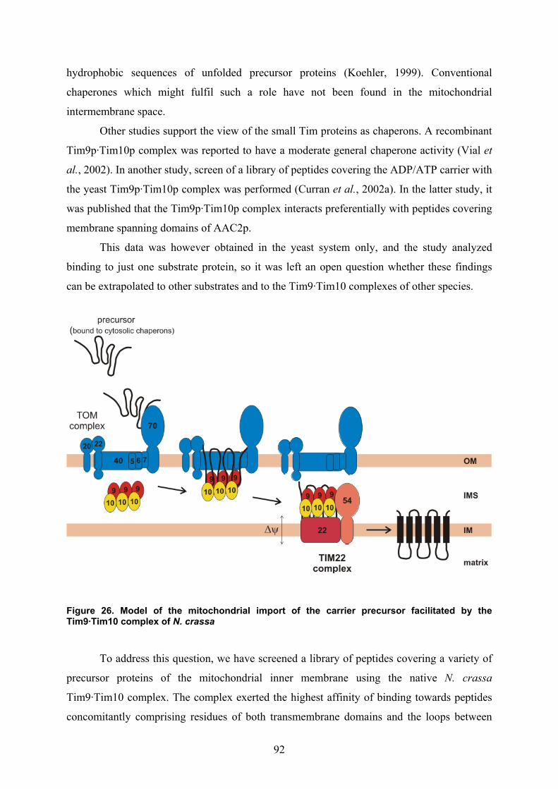
92
hydrophobic sequences of unfolded precursor proteins (Koehler, 1999). Conventional
chaperones which might fulfil such a role have not been found in the mitochondrial
intermembrane space.
Other studies support the view of the small Tim proteins as chaperons. A recombinant
Tim9p·Tim10p complex was reported to have a moderate general chaperone activity (Vial et
al., 2002). In another study, screen of a library of peptides covering the ADP/ATP carrier with
the yeast Tim9p·Tim10p complex was performed (Curran et al., 2002a). In the latter study, it
was published that the Tim9p·Tim10p complex interacts preferentially with peptides covering
membrane spanning domains of AAC2p.
This data was however obtained in the yeast system only, and the study analyzed
binding to just one substrate protein, so it was left an open question whether these findings
can be extrapolated to other substrates and to the Tim9·Tim10 complexes of other species.
Figure 26. Model of the mitochondrial import of the carrier precursor facilitated by the Tim9·Tim10 complex of N. crassa
To address this question, we have screened a library of peptides covering a variety of
precursor proteins of the mitochondrial inner membrane using the native N. crassa
Tim9·Tim10 complex. The complex exerted the highest affinity of binding towards peptides
concomitantly comprising residues of both transmembrane domains and the loops between

93
them. Advantages of overwhelming binding to such regions over exclusive binding to the
membrane-spanning segments are evident. Such mode of binding allows the trapping of
segments which emerge into the intermembrane space from the TOM complex, while most of
the precursor is still protected from this aqueous environment by remaining in the TOM
complex. In this manner, the transmembrane domains are able to undergo transfer from the
TOM to the Tim9·Tim10 complex without having their hydrophobic stretches exposed to the
intermembrane space (Figure 26).
Furthermore, the model of the carrier translocation involving a soluble intermembrane
space intermediate can be considered unlikely for following reasons. Up to date no such
soluble import intermediate of Tim9p·Tim10p and AAC2p has been identified in and isolated
from the mitochondrial intermembrane space. This further suggests that a translocation
contact site might form, where the outer and inner membrane come together and allow the
carrier precursor to slip from the TOM pore into the Tim22p protein channel (Kovermann et
al., 2002). This proposition is consistent with the N. crassa Tim9·Tim10 recognition pattern
and binding to the parts of the carrier protein which are also left unconcealed when the carrier
is finally integrated into the lipid bilayer of the inner membrane (Pebay-Peyroula et al., 2003).
These segments are most probably concealed during import as well, by being buried inside the
TOM channel and/or the TIM22 translocase. Binding to domains along the ends of the
hydrophobic segments of the carrier would in this way prevent any retrograde movement
along its import pathway, simultaneously supporting the vectorial progress towards the
completion of import.
4.4.2. Tim23 - a novel Tim9·Tim10 complex substrate in N. crassa
The Tim23p protein inserts into the inner membrane with the help of the membrane-
embedded TIM22 complex (Kerscher et al., 1997; Kaldi et al., 1998; Leuenberger et al.,
1999). Upon in vitro import of the Tim23p precursor into the isolated de-energised
mitochondria, the major cross-linking species of the Tim23p precursors to Tim8p and Tim13p
proteins form, and very little if any, cross-linking products to the Tim9p and Tim10p proteins
(Leuenberger et al., 1999; Paschen et al., 2000). Interestingly, a weak interaction of the
Tim9p·Tim10p complex with the COOH-terminal hydrophobic domain of Tim23p was
reported from another research team. They suggested that the Tim9p·Tim10p complex might
play an important role in the import of Tim23p precursor (Davis et al., 2000; Jensen and
Dunn, 2002). In this study, the Tim23 protein was identified as another substrate of the N.
94
crassa Tim9·Tim10 complex. Results of both our peptide screen and the cross-linking
experiments in N. crassa revealed the existence of an interaction between Tim23 and the
native Tim9·Tim10 complex. This interaction of the Tim23 precursor with the Tim9·Tim10
complex might occur concomitantly with its interaction with the Tim8·Tim13 complex, or at a
later stage of its import pathway.
In the peptide screens made by Koehler group using the yeast model system, it was
suggested that peptides representing Tim23p were bound by the Tim8p·Tim13p complex, but
not the Tim9p·Tim10p complex (Curran et al., 2002a; Curran et al., 2002b). A plausible
explanation of these differences between Neurospora and yeast might be the absence of
Tim12p in N. crassa. An essential protein in yeast, it facilitates the transfer of the substrate
proteins from the Tim9p·Tim10p to the TIM22 complex (Koehler et al., 1998a; Sirrenberg et
al., 1998), but it has not been found in any organism other than S. cerevisiae. In N. crassa a
similar function may be fulfilled by the Tim9·Tim10 complex itself.
4.4.3. The minimal machinery for the translocation of AAC across membranes
Recent studies have indicated the demand for Tim9p and Tim10p proteins in the
process of the carrier preprotein release from the TOM complex and its insertion into the
inner membrane (Luciano et al., 2001; Truscott et al., 2002). Yet, the explicit facts of whether
they accomplish the precursor transfer across the outer membrane with the help of additional,
yet unknown component(s) or whether the Tim9p·Tim10p complex and the TOM complex are
sufficient for mediating this process were not dealt with. To address this issue, we
reconstituted the TOM core complex into lipid vesicles with the purified N. crassa
Tim9·Tim10 complex entrapped, and performed import of the ADP/ATP carrier precursor.
Following this line of research, we made several observations. First, the TOM core
complex (lacking import receptors) can facilitate the transfer of the AAC precursor to the
internal side of the outer membrane. Whereas in the absence of the Tim9·Tim10 complex
parts of the AAC precursor molecule are still exposed to the cytosol, the presence of a
functional Tim9·Tim10 complex at the internal side of the outer membrane was sufficient to
pull the AAC precursor further across the outer membrane. Evidently, the TOM core complex
and the Tim9·Tim10 complex form the minimal machinery for the translocation of the
ADP/ATP carrier across the mitochondrial outer membrane.
95
The observation that the AAC is found still associated with the receptors Tom70p and
Tom20p when the function of Tim10p protein is impaired, support our conclusion
(Wiedemann et al., 2001; Truscott et al., 2002). In other words, whereas the initial insertion
of AAC into the import channel is independent of the Tim9p·Tim10p complex, completion of
the translocation process across the outer membrane, including the release from the TOM
complex, requires a functional Tim9p·Tim10p complex. It appears that neither proteins in the
mitochondrial inner membrane or the intermembrane space, nor structural elements like the
import contact sites are required for this translocation step. Rather, the interaction of the small
Tim proteins with the incoming precursor is sufficient to prevent its retrograde sliding out of
the translocation machinery and to ensure a vectorial movement along the import pathway.
4.4.4. Potential involvement of the small Tim proteins in the biogenesis of the β-barrel proteins
Recently, certain reports have been published suggesting an additional, though non-
essential function of the small Tim proteins, both in N. crassa and S. cerevisiae. Their role in
the biogenesis of β-barrel proteins of the mitochondrial outer membrane has been reported
(Hoppins et al., 2004; Wiedemann et al., 2004).
Hoppins et al. (2004) have isolated tim8 and tim13 mutants in N. crassa and have
shown that mitochondria lacking the Tim8·Tim13 complex were mildly impaired in the
import of Tom40 and porin, which are both β-barrel proteins. Their cross-linking studies
demonstrated the existence of an interaction between Tom40 precursor and the Tim8·Tim13
complex. The reported cross-linking products formed exclusively in the initial import stages.
Also, formation of an early assembly intermediate of the Tom40 precursor was slower in the
mitochondria lacking the Tim8·Tim13 complex, than in the wild type mitochondria.
Wiedemann et al. (2004) investigated the yeast mitochondria with defects in small Tim
proteins and also found them impaired in the early stages of the Tom40p assembly pathway.
Deficiency in the Tim9p·Tim10p complex seemed to have a more profound effect on the
biogenesis of the mitochondrial β-barrel proteins, than the one of the impaired Tim8p·Tim13p
complex.
However, all the observed effects were very moderate. Furthermore, the endogenous
levels of β-barrel proteins of the mitochondrial outer membrane were not affected. There were
also no phenotypic repercussions observed.

96
Using our reconstituted system, we could not detect any protease protected porin
species upon its incubation with the proteoliposomes with the Tim9·Tim10 complex
entrapped. This was not surprising since the TOB complex was not present in our trials.
Nevertheless, this system might prove useful for acquiring further insights into the biogenesis
of β-barrel proteins of the mitochondrial outer membrane.

97
5. Summary
Mitochondria are essential cellular organelles of eukaryotic organisms, which import
most of their proteinaceous constituents from the cytoplasm. Two mitochondrial membranes
contain different translocation machineries which are involved in the import and proper
sorting of mitochondrial precursor proteins. The TIM22 translocase in the inner mitochondrial
membrane mediates the import of polytopic proteins into this membrane. In addition to the
membrane integrated components Tim22 and Tim54, the TIM22 translocase possesses
components in the intermembrane space, termed Tim9 and Tim10.
In the present study, the tim9 and tim10 genes of the TIM22 translocase of N. crassa
were identified. The structural and functional characteristics of the corresponding gene
products, the Tim9 and Tim10 proteins, were examined. Tim9 was demonstrated to be an
essential protein. The Tim9 and Tim10 proteins were shown to build a 70-80 kDa
heterohexameric complex in the mitochondrial intermembrane space. The isolated
Tim9·Tim10 complex had the same oligomeric structure as the native one, and it proved fully
functional in interacting in vitro with its physiological substrate, the ADP/ATP carrier (AAC).
Peptide library screens were performed to determine the structural determinants of the
substrates that are recognised by the Tim9·Tim10 complex. Efficient binding to the regions
covering residues of the hydrophobic membrane spanning domains and of the connecting
hydrophilic loops was observed. In this way, Tim9 and Tim10 proteins interact with their
substrates, while the hydrophobic regions of the substrates are still present in the TOM
complex and thereby protected from the aqueous environment of the intermembrane space
compartment. Furthermore, when enclosed into proteoliposomes containing the reconstituted
TOM complex, Tim9·Tim10 complex specifically promoted the translocation of the AAC
precursor. Hence, the Tim9·Tim10 complex and the TOM complex are both necessary and
sufficient to facilitate translocation of carrier proteins across the outer mitochondrial
membrane. Finally, peptide screens and chemical cross-linking experiments were used to
identify the precursor of N. crassa Tim23 protein as a novel substrate of the Tim9·Tim10
complex.
98
6. Abbreviations
AAC ADP/ATP carrier
Ac acetate
ADH alcohol dehydrogenase
ADP adenosine diphosphate
amp ampicillin
APS ammonium peroxodisulfate
ATP adenosine triphosphate
ATPase adenosine triphosphatase
β-ME β-mercaptoethanol
BSA bovine serum albumin
BNG(E) blue native gel (electrophoresis)
Ci Curie
CCCP carbonyl cyanide m-chlorphenylhydrazone
CCHL cytochrome c haem lyase
cDNA complementary DNA
CIP calf intestinal alkaline phosphatase
CNBr cyanogen bromide
CSPD 3-(4-methoxyspiro (1,2-dioxetane-3,2'-(5'-chloro) tricycle
[3,3.1.13.7]decan-4-yl) phenyl phosphate, disodium salt
CV column volume
DDM n-dodecyl-β-maltopyranosid
DHFR dihydrofolate reductase
DMSO dimethylsulfoxid
dNTP deoxyribonucleoside triphosphate
DNA desoxyribonucleic acid
DTT dithiotreitol
∆ψ membrane potential
E. coli Escherichia coli
EDTA ethylendiamine tetraacetate
ER endoplasmatic reticulum
GIP general import pore
HEPES N-2 hydroxyl piperazine-N`-2-ethane sulphonic acid

99
Hsp heat shock protein
IgG immunoglobulin G
IM inner membrane
IMS intermembrane space
IP immunoprecipitation
IPTG isopropyl-β,D-thiogalactopyranoside
KAN kanamycin
kDa kilodalton
LB Luria Bertani
m7G(5`)ppp(5`)G 7-methylguanosine triphospate
MBP maltose-binding protein
MBS m-maleimidobenzoyl-N-hydroxysuccinimide ester
MOPS N-morpholinopropane sulphonic acid
MPP mitochondrial processing peptidase
MSF mitochondrial import stimulating factor
MTS matrix targeting signal
MW molecular weight
N- amino-
N. crassa Neurospora crassa
NADH nicotine amide adenine dinucleotide
NADPH nicotine amide adenine dinucleotide phosphate
Ni-NTA nickel-nitrilotriacetic acid
ODx optical density at x nm
OM outer membrane
OMVs outer membrane vesicles
PAGE polyacrylamide gel electrophoresis
PAS Protein A Sepharose
PCR polymerase chain reaction
PEG polyethylene glycol
PIS preimmune serum
PK Proteinase K
PMSF phenylmethylsulfonyfluoride
PVDF polyvinylidene difluoride
RIP repeat-induced point mutation

100
RNase ribonuclease
RNasin ribonuclease inhibitor
RT room temperature
S. cerevisiae Saccharomyces cerevisiae
SDS sodium dodecyl sulfate
STI soybean trypsin inhibitor
TBS Tris-buffered saline
TCA trichloroacetic acid
TCEP Tris-(2-carboxyethyl) phopshine
TEMED N,N,N‘,N‘-tetramethylene diamine
TIM translocase of the inner mitochondrial membrane
TLC thin layer chromatography
TOM translocase of the outer mitochondrial membrane
Tris tris-(hydroxymethyl)-aminomethane
TX-100 Triton X-100
Vol. volumes
v/v volume per volume
w/v weight per weight
wt wild type
101
7. References Abe, Y., Shodai, T., Muto, T., Mihara, K., Torii, H., Nishikawa, S., Endo, T. and D. Kohda
(2000). Structural basis of presequence recognition by the mitochondrial protein import
receptor Tom20.Cell 100, 551-60.
Adam A, Endres M, Sirrenberg C, Lottspeich F, Neupert W and M. Brunner (1999). Tim9, a
new component of the TIM22.54 translocase in mitochondria. EMBO J. 18 (2), 313-319.
Ahting, U., Thun, C., Hegerl, R., Typke, D., Nargang, F.E., Neupert, W. and S. Nussberger
(1999). The TOM core complex: The general protein import pore of the outer membrane of
mitochondria. J. Cell. Biol. 147, 959-968.
Alberts, B., Bray, D., Lewis, J., Raff, M., Roberts, K. and J.D. Watson (1994). Molecular
biology of the cell. Third edition. Garland Publishing, Inc., New York & London.
Arnold, I., Fölsch, H., Neupert, W. and R.A. Stuart (1998). Two distinct and independent
mitochondrial targeting signals functions in the sorting of an inner membrane protein,
cytochrome c1. J. Biol. Chem. 273, 1469-1476.
Auld D.S. (2001). Zinc coordination sphere in biochemical zinc sites. Biometals 14, 271-313.
Bauer, M., Behrens, M., Esser, K., Michaelis, G. and E. Pratje (1994). PET1402, a nuclear
gene required for proteolytic processing of cytochrome oxidase subunit 2 in yeast. Mol. Gen.
Genet. 245, 272-278.
Bauer, M.F., Gempel, K., Reichert, A.S., Rappold, G.A., Lichtner, P., Gerbitz, K.D., Neupert,
W., Brunner M. And S. Hofmann (1999). Genetic and structural characterization of the
human mitochondrial inner membrane translocase. J Mol Biol 289, 69-82.
Beers, J., Glerum, D.M. and A. Tzagoloff (1997). Purification, characterization, and
localization of yeast Cox17p, a mitochondrial copper shuttle. J Biol Chem 272, 33191-6.
Berg, J.M. and H.A. Godwin (1997). Lessons from zinc-binding peptides. Annu Rev Biophys
Biomol Struct 26, 357-371.
102
Birnboim, H.C. and J. Doly (1979). A rapid alkaline extraction procedure for screening
recombinant plasmid DNA. Nucleic Acids Res 7, 1513-1523.
Bloom, H., H. Beier and H.S. Gross (1987). Improved silver staining of plant protein, RNA
and DNA in polyacrylamide gels. Electrophoresis 8: 93-99.
Bonnefoy, N., Chalvet, F., Hamel, P., Slominski, P.P. and G. Dujardin (1994). OXA1, a
Saccharomyces cerevisiae nuclear gene whose sequence is conserved from prokaryotes to
eukaryotes controls cytochrome oxidase biogenesis. J. Mol. Biol., 239, 201–12.
Bradford, M.M. (1976). A rapid and sensitive method for the quantitation of microgram
quantities of protein utilizing the principle of protein-dye binding. Anal. Biochem. 72, 248-
254.
Brix, J., Dietmeier, K. and N. Pfanner (1997). Differential recognition of preproteins by the
purified cytosolic domains of the mitochondrial import receptors Tom20, Tom22 and Tom70.
J. Biol. Chem. 272, 20730-20735.
Brix, J., Rudiger, S., Bukau, B., Schneider-Mergener, J. and N. Pfanner (1999). Distribution
of binding sequences for the mitochondrial import receptors Tom20, Tom22, and Tom70 in a
presequence-carrying preprotein and a non-cleavable preprotein. J. Biol. Chem. 274, 16522-
16530.
Casadaban, M.J. and S.N. Cohen (1980). Analysis of gene control signals by DNA fusion and
cloning in E. coli. J. Mol. Biol. 138, 179-207.
Chacinska, A., Rehling, P., Guiard, B., Frazier, A.E., Schulze-Specking, A., Pfanner, N.,
Voos, W. and C. Meisinger (2003). Mitochondrial translocation contact sites: separation of
dynamic and stabilizing elements in formation of a TOM-TIM-preprotein supercomplex.
EMBO J 22, 5370-81.
Cotter, D., Guda, P., Fahy, E. and S. Subramaniam (2004). MitoProteome: mitochondrial
protein sequence database and annotation system. Nucleic Acids Res. 32, database issue:
D463-7.
103
Cox, E.H. and G.L. McLendon (2000). Zinc-dependent protein folding. Current opinion in
chemical Biology 4, 162-165.
Curran, S.P., Leuenberger, D., Oppliger, W. and C.M. Koehler (2002a). The Tim9p-Tim10p
complex binds to the transmembrane domains of the ADP/ATP carrier. EMBO J. 21, 942-953.
Curran, S.P., Leuenberger, D., Schmidt, E. and C.M. Koehler (2002b). The role of the Tim8p-
Tim13p complex in a conserved import pathway for mitochondrial polytopic inner membrane
proteins. J. Cell. Biol. 158, 1017-1027.
Davis, R.H. and F.J. De Serres (1970). Genetic and microbiological research techniques for
Neurospora crassa. Methods Enzymol. 17, 79-143.
Davis, A.J., Sepuri, N.B., Holder, J., Johnson, A.E. and R.E. Jensen (2000). Two
intermembrane space TIM complexes interact with different domains of Tim23p during its
import into mitochondria. J. Cell. Biol. 150, 1271-82.
Davis, R. H. (2000). Neurospora: contributions of a model organism. Oxford Press, New
York, N.Y.
Dekker, P.J.T., Ryan, M.T., Brix, J., Müller, H., Hönlinger, A. and N. Pfanner (1998).
Preprotein translocase of the outer mitochondrial membrane: Molecular dissection and
assembly of the general import pore complex. Mol. Cell. Biol. 18, 6515-6524.
de Souza-Pinto, N.C. and V.A. Bohr (2002). The mitochondrial theory of aging: involvement
of mitochondrial DNA damage and repair. Int Rev Neurobiol 53, 519-34.
Diekert, K., Kispal, G., Guiard, B. and R. Lill (1999). An internal targeting signal directing
proteins into the mitochondrial intermembrane space. Proc. Natl. Acad. Sci. USA 96, 11752-
57.
Diekert, K., de Kroon, A.I., Ahting, U., Niggemeyer, B., Neupert, W., de Kruijff, B. And R.
Lill (2001). Apocytochrome c requires the TOM complex for translocation across the
mitochondrial outer membrane. EMBO J 20, 5626-35.
104
Dietmeier, K., Honlinger, A., Bomer, U., Dekker, P.J., Eckerskorn, C., Lottspeich, F.,
Kubrich, M. and N. Pfanner (1997). Tom5 functionally links mitochondrial preprotein
receptors to the general import pore. Nature 388, 195-200.
Ebbole, D. and Sachs, MS (1990). A rapid and simple method for isolation of Neurospora
crassa homokaryons using microconidia. Fungal Genetics Newsletter 37, 17-18.
V.V. Emelyanov (2003). Mitochondrial connection to the origin of the eukaryotic cell. Eur. J.
Biochem. 270, 1699-1618.
Emtage, J.L. and R.E. Jensen (1993). MAS6 encodes an essential inner membrane component
of the yeast mitochondrial protein import pathway. J. Cell. Biol. 122, 1003-1012.
Endres, M., Neupert, W. and M. Brunner (1999). Transport of the ADP/ATP carrier of
mitochondria from the TOM complex to the TIM22.54 complex. EMBO J. 18, 3214-3221.
Finkel, T. and N.J. Holbrook (2000). Oxidants, oxidative stress and the biology of ageing.
Nature 408, 239-47.
Fölsch, H., Guiard, B., Neupert, W. and R.A. Stuart (1996). Internal targeting signal of the
BCS1 protein: a novel mechanism of import into mitochondria. EMBO J. 15, 479-87.
Frazier, A.E., Dudek, J., Guiard, B., Voos, W., Li, Y., Lind, M., Meisinger, C., Geissler, A.,
Sickmann, A., Meyer, H.E., Bilanchone, V., Cumsky, M.G., Truscott, K.N., Pfanner, N. and
P. Rehling (2004). Pam16 has an essential role in the mitochondrial protein import motor. Nat
Struct Mol Biol 11, 226-33.
Frank, R. (1992). Spot synthesis: An easy technique for the positionally addressable, parallel
chemical synthesis on a membrane support. Tetrahedron 48, 9217-9232.
Frey, T.G. and C.A. Mannella (2000). The internal structure of mitochondria. Trends
Biochem. Sci. 25, 319-324.

105
Fujiki, M. and K. Verner (1993). Coupling of cytosolic protein synthesis and mitochondrial
protein import in yeast. Evidence for cotranslational import in vivo. J. Biol. Chem. 268 (3),
1914-20.
Fujiki, Y., Fowler, S., Shio, H., Hubbard, A.L., & Lazarow, P.B. (1982). Isolation of
intracellular membranes by means of sodium carbonate treatment: Application to endoplasmic
reticulum. J. Cell Biol. 93, 97-102.
Funes, S., Nargang, F.E., Neupert, W. and J.M. Herrmann (2004). The Oxa2 protein of
Neurospora crassa plays a critical role in the biogenesis of cytochrome oxidase and defines a
ubiquitous subbranch of the Oxa1/YidC/Alb3 protein family. Mol Biol Cell.
Gakh, O., Cavadini, P. and G. Isaya (2002). Mitochondrial processing peptidases. Biochim
Biophys Acta 1592(1), 63-77.
Gaume, B., Klaus, C., Ungermann, C., Guiard, B., Neupert, W. and M. Brunner (1998).
Unfolding of preproteins upon import into mitochondria. EMBO J. 17, 6497-6507.
Gärtner, F., Voos, W., Querol, A., Miller, B.R., Craig, E.A., Cumsky, M.G. and N. Pfanner
(1995a). Mitochondrial import of subunit Va of cytochrome c oxidase characterized with
yeast mutants. J Biol Chem. 270, 3788-95.
Gärtner, F., Bomer, U., Guiard, B. and N. Pfanner (1995b). The sorting signal of cytochrome
b2 promotes early divergence from the general mitochondrial import pathway and restricts the
unfoldase activity of matrix Hsp70. EMBO J. 14, 6043-6057.
Geissler, A., Chacinska, A., Truscott, K.N., Wiedemann, N., Brandner, K., Sickmann, A.,
Meyer, H.E., Meisinger, C., Pfanner, N. and P. Rehling (2002). The mitochondrial
presequence translocase. An essential role of Tim50 in directing preproteins to the import
channel. Cell 111, 507-518.
Gessert, S.F., Kim, J.H., Nargang, F.E., and R.L. Weiss (1994). A polyprotein precursor of
two mitochondrial enzymes in Neurospora crassa. Gene structure and precursor processing. J
Biol Chem 269, 8189-203

106
Glick, B.S., Brandt, A., Cunningham, K., Muller, S., Hallberg, R.L. and G. Schatz (1992a).
Cytochromes c1 and b2 are sorted to the intermembrane space of yeast mitochondria by a
stop-transfer mechanism. Cell 69(5), 809-22.
Glick, B.S., Beasley, E.M. and G. Schatz (1992b). Protein sorting in mitochondria. Trends
Biochem. Sci. 17, 453-459.
Gratzer, S., Lithgow, T., Bauer, R.E., Lamping, E., Paltauf, F., Kohlwein, S.D., Haucke, V.,
Junne, T., Schatz, G. and M. Horst (1995). Mas37p, a novel receptor subunit for protein
import into mitochondria. J. Cell. Biol. 129, 25-34.
Gray, M.W., Burger, G. and B. Franz Lang (1999). Mitochondrial evolution. Science 283,
1476-1481.
N.V. Grishin (2001). Treble clef finger--a functionally diverse zinc-binding structural motif.
Nucleic Acids Res 29, 1703-14.
Guan,C., Li, P., Riggs, P.D. and Inouye, H. (1987). Vectors that facilitate the expression and
purification of foreign peptides in Esherichia coli by fusion to maltose-binding proten. Gene
67, 21-30.
Hachiya, N., Komiya, T., Alam, R. Iwahashi, J., Sakaguchi, M., Omura, T., and K. Mihara
(1994). MSF, a novel cytoplasmic chaperone which functions in precursor targeting to
mitochondria. EMBO J. 13(21), 5146-54.
Hachiya, N., Mihara, K., Suda, K., Horst, M., Schatz, G. and T. Lithgow (1995).
Reconstitution of the initial steps of mitochondrial protein import. Nature 376, 705-709.
Hahne, K., Haucke, V., Ramage, L. and G. Schatz (1994). Incomplete arrest in the outer
membrane sorts NADH-cytochrome b5 reductase to two different submitochondrial
compartments. Cell 79, 829-839.
107
Hartl, F.U., Schmidt, B., Wachter, E., Weiss, H. and W. Neupert (1986). Transport into
mitochondria and intramitochondrial sorting of the Fe/S protein of ubiquinol-cytochrome c
reductase. Cell 47, 939-951.
Hawlitschek, G., Schneider, H., Schmidt, B., Tropschug, M., Hartl, F.U. and W. Neupert
(1988). Mitochondrial protein import: identification of processing peptidase and of PEP, a
processing enhancing protein. Cell 53, 759-806.
Hell, K., Herrmann, J., Pratje, E., Neupert, W. and R.A. Stuart (1997). Oxa1p mediates the
export of the N- and C-termini of pCoxII from the mitochondrial matrix to the intermembrane
space. FEBS Lett. 418, 367-370.
Hengartner, M.O. (2000). The biochemistry of apoptosis. Nature 407, 770 – 776.
Herlan, M., Vogel, F., Bornhovd, C., Neupert, W. and A.S. Reichert (2003). Processing of
Mgm1 by the rhomboid-type protease Pcp1 is required for maintenance of mitochondrial
morphology and of mitochondrial DNA. J Biol Chem. 278, 27781-8.
Herrmann, J.M., Stuart, R.A., Craig, E.A. and W. Neupert (1994). Mitochondrial heat shock
protein 70, a molecular chaperone for proteins encoded by mitochondrial DNA. J. Cell. Biol.
127, 893-902.
Herrmann, J.M., Neupert, W. and R.A. Stuart (1997). Insertion into the mitochondrial inner
membrane of a polytopic protein, the nuclear-encoded Oxa1p. EMBO J. 16, 2217-26.
Herrmann, J.M. and W. Neupert (2003). Protein insertion into the inner membrane of
mitochondria. IUBMB Life 55, 219-25.
Herrmann, J.M. (2003). Converting bacteria to organelles: evolution of mitochondrial protein
sorting. Trends in microbiology 11, 74-79.
Hill, K., Model, K., Ryan, M.T., Dietmeier, K., Martin, F., Wagner, R. and N. Pfanner (1998).
Tom40 forms the hydrophilic channel of the mitochondrial import pore for preproteins.
Nature 395, 516-521.
108
Hines, V., Brandt, A., Griffiths, G., Horstmann, H., Brütsch, H. and G. Schatz (1990). Protein
import into yeast mitochondria is accelerated by the outer membrane protein MAS70. EMBO
J. 9, 3191-3200.
Hofmann, S., Rothbauer, U., Muhlenbein, N., Neupert, W., Gerbitz, K.D., Brunner, M. And
M.F. Bauer (2002). The C66W mutation in the deafness dystonia peptide 1 (DDP1) affects the
formation of functional DDP1.TIM13 complexes in the mitochondrial intermembrane space. J
Biol Chem. 277, 23287-93.
Hoppins, S.C. and F.E. Nargang (2004). The Tim8-Tim13 complex in Neurospora crassa
functions in the assembly of proteins into both mitochondrial membranes. J Biol Chem.
Horst, M., Oppliger, W., Feifel, B., Schatz, G. and B.S. Glick (1997). The mitochondrial
protein import motor: dissociation of mitochondrial hsp70 from its membrane anchor requires
ATP binding rather than ATP hydrolysis. Protein Sci. 5, 759-767.
Isaya, G., Kalousek, F., Fenton, W.A., and L.E. Rosenberg (1991). Cleavage of precursors by
the mitochondrial processing peptidase requires a compatible mature protein or an
intermediate octapeptide. J Cell Biol. 113, 65-76.
Iwahashi, J., Yamazaki, S., Komiya, T., Nomura, N., Nishikawa, S., Endo, T. and K. Mihara
(1997). Analysis of the functional domain of the rat liver mitochondrial import receptor
Tom20. J Biol Chem 272, 18467-72.
Jarosch, E., Tuller, G., Daum, G., Waldherr, M., Voskova, A., and R.J. Schweyen (1995).
Mrs5p, an essential protein of the mitochondrial intermembrane space, affects protein import
into yeast mitochondria. J Biol Chem 271, 17219-25.
Jarosch, E., Rodel, G., and R.J. Schweyen (1997). A soluble 12-kDa protein of the
mitochondrial intermembrane space, Mrs11p, is essential for mitochondrial biogenesis and
viability of yeast cells. Mol Gen Genet 255, 157-65.

109
Jarvis, J.A., Ryan, M.T., Hoogenraad, N.J., Craik, D.J. and P.B. Hoj (1995). Solution
structure of the acetylated and noncleavable mitochondrial targeting signal of rat chaperonin
10. J. Biol. Chem. 270, 1323-1331.
Jensen R.E. and C.D. Dunn (2002). Protein import into and across the mitochondrial inner
membrane: role of the TIM23 and TIM22 translocons. Biochim Biophys Acta 1592, 25-34
Jin, H., May, M., Tranebjaerg, L., Kendall, E., Fontan, G., Jackson, J., Subramony, S.H.,
Arena, F., Lubs, H., Smith, S. et al. (1996). A novel X-linked gene, DDP, shows mutations in
families with deafness (DFN-1), dystonia, mental deficiency and blindness. Nat. Genet., 14,
177–80.
B. Kadenbach (2003). Intrinsic and extrinsic uncoupling of oxidative phosphorylation.
Biochim Biophys Acta. 1604, 77-94.
Kayingo, G., Potier, S., Hohmann, S. and B.A. Prior (2000). Isolation and characterization of
the TIM10 homologue from the yeast Pichia sorbitophila: a putative component of the
mitochondrial protein import system. Yeast 16, 589-96.
Kerscher, O., Holder, J., Srinivasan, M., Leung, R.S. and R.E. Jensen (1997). The Tim54p-
Tim22p complex mediates insertion of proteins into the mitochondrial inner membrane. J.
Cell. Biol. 139, 1663-1675.
Kerscher, O., Sepuri, N.B. and R.E. Jensen (2000). Tim18p is a new component of the
Tim54p-Tim22p translocon in the mitochondrial inner membrane. Mol Biol Cell, 11, 103-16.
Koehler, C.M., Jarosch, E., Tokatlidis, K., Schmid, K., Schweyen, R.J. and G. Schatz (1998a).
Import of mitochondrial carriers mediated by essential proteins of the intermembrane space.
Science 279, 369-73.
Koehler, C.M., Merchant, S., Oppliger, W., Schmid, K., Jarosch, E., Dolfini, L., Junne, T.,
Schatz, G. and K. Tokatlidis (1998b). Tim9p, an essential partner subunit of Tim10p for the
import of mitochondrial carrier proteins. EMBO J. 17, 6477-6486.
110
Koehler, C.M., Leuenberger, D., Merchant, S., Renold, A., Junne, T. and G. Schatz (1999).
Human deafness dystonia syndrome is a mitochondrial disease. Proc Natl Acad Sci U S A 96,
2141-6.
Koehler, C.M., Merchant, S. and G. Schatz (1999). How membrane proteins travel across the
mitochondrial intermembrane space. Trends Biochem. Sci. 24, 428-432.
Koehler, C.M. (2000). Protein translocation pathways of the mitochondrion. FEBS Lett. 476,
27-31.
Koehler, C.M., Murphy, M.P., Bally, N.A., Leuenberger, D., Oppliger, W., Dolfini, L., Junne,
T., Schatz, G. and E. Or (2000). Tim18p, a new subunit of the TIM22 complex that mediates
insertion of imported proteins into the yeast mitochondrial inner membrane. Mol Cell Biol 20,
1187-93.
Koehler, C.M. (2004). The small Tim proteins and the twin Cx3C motif. Trends in Biochem
Sci 29, 1-4.
Komiya, T., Sakaguchi, M. and M. Mihara (1996). Cytoplasmic chaperones determine the
targeting pathway of precursor proteins to mitochondria. EMBO J. 15, 399-407.
Komiya, T., Rospert, S., Schatz, G. and K. Mihara (1997). Binding of mitochondrial
precursor. proteins to the cytoplasmic domains of the import receptors Tom70 and Tom20 is
determined by cytoplasmic chaperones. EMBO J. 16, 4267-4275.
Komiya, T., Rospert, S., Koehler, C.M., Looser, R., Schatz, G. and K. Mihara (1998).
Interaction of mitochondrial targeting signals with acidic receptor domains along the protein
import pathway: Evidence for the "Acidic Chain" hypothesis. EMBO J. 17, 3886-3898.
Kovermann, P., Truscott, K.N., Guiard, B., Rehling, P., Sepuri, N.B., Muller, H., Jensen,
R.E., Wagner, R. and N. Pfanner (2002). Tim22, the essential core of the mitochondrial
protein insertion complex, forms a voltage-activated and signal-gated channel. Mol Cell. 9,
363-73.

111
Kozak, M. (1977). Nucleotide sequences of 5V-terminal ribosome-protected initiation regions
from two reovirus messages. Nature 269, 390–394.
Kozak, M. (2003). Alternative ways to think about mRNA sequences and proteins that appear
to promote internal initation of translation. Gene 318, 1-23.
Kozany, C., Mokranjac, D., Sichting, M., Neupert, W. and K. Hell (2004). Tim16 is the J-
domain-like containing cochaperone essential for the Tim23 protein translocase. Nature
structural biology 11, 234-41.
Kramer, A. and J. Schneider-Mergener (1998). Synthesis and screening of peptide libraries on
continuous cellulose membrane supports. Methods Mol. Biol. 87, 25-39.
Kuhn, A., Stuart, R., Henry, R. and R.E. Dalbey (2003). The Alb3/Oxa1/YidC protein family:
membrane-localized chaperones facilitating membrane protein insertion? Trends Cell Biol 13,
510-6.
Kurz, M., Martin, H., Rassow, J., Pfanner, N. and M.T. Ryan (1999). Biogenesis of Tim
proteins of the mitochondrial carrier import pathway: differential targeting mechanisms and
crossing over with the main import pathway. Mol Biol Cell. 10, 2461-74.
Kübrich, M., Keil, P, Rassow, J., Dekker, P.J., Blom, J., Meijer, M. and N. Pfanner (1994).
The polytopic mitochondrial inner membrane proteins MIM17 and MIM23 operate at the
same preprotein import site. FEBS Lett. 349, 222-228.
Kübrich, M., Rassow, J., Voos, W., Pfanner, N. and A. Honlinger (1998). The import route of
ADP/ATP carrier into mitochondria separates from the general import pathway of cleavable
preproteins at the trans side of the outer membrane. J. Biol.Chem. 273, 16374-81.
Künkele, K.P., Heins, S., Dembowski, M., Nargang, F.E., Benz, R., Thieffry, M., Walz, J.,
Lill, R., Nussberger, S. and W. Neupert (1998a). The Preprotein Translocation Channel of the
Outer Membrane of Mitochondria. Cell 93, 1009-1019.
112
Künkele, K.P., Juin, P., Pompa, C., Nargang, F.E., Henry, J.P., Neupert, W., Lill, R. and M.
Thieffry (1998b). The isolated complex of the translocase of the outer membrane of
mitochondria. Characterization of the cation-selective and voltage-gated preprotein-
conducting pore. J. Biol. Chem. 273, 31032-31039.
Kyhse-Andersen J (1984) Electroblotting from multiple gels: A simple apparatus without
buffer tank for rapid transfer of proteins to nitrocellulose membrans. J Biochem Biophys
Methods 10, 203-209.
U.K. Laemmli (1970). Cleavage of structural proteins during the assembly of the head of
bacteriophage T4. Nature 227, 680-685.
Laity, J.H., Lee, B.M., Wright, P.E. (2001). Zinc finger proteins: new insights into structural
and functional diversity. Current opinion in structural biology 11, 39-46.
Lang, B.F., Gray, M.W. and G. Burger (1999). Mitochondrial genome evolution and the
origin of eukaryotes. Annu. Rev. Genet. 33, 351-397.
Lange, H., Lisowsky, T., Gerber, J., Mühlenhoff, U., Kispal, G. and R. Lill R. (2001). An
essential function of the mitochondrial sulfhydryl oxidase Erv1p/ALR in the maturation of
cytosolic Fe/S proteins. EMBO Rep 2, 715-20.
Lawson, J.E. and M.G. Douglas (1988). Separate genes encode functionally equivalent
ADP/ATP carrier proteins in Saccharomyces cerevisiae. Isolation and analysis of AAC2. J
Biol Chem. 263, 14812-8.
Lee, C.M., Sedman, J., Neupert, W. and R.A. Stuart (1999). The DNA helicase, Hmi1p, is
transported into mitochondria by a C-terminal cleavable targeting signal. J. Biol. Chem. 274,
20937-20942.
Leuenberger, D., Bally, N.A., Schatz, G. and C.M. Koehler (1999). Different import pathways
through the mitochondrial intermembrane space for inner membrane proteins. EMBO J. 18,
4816-22.
113
Lill, R., Stuart, R.A., Drygas, M.E., Nargang, F.E. and W. Neupert (1992). Import of
cytochrome c heme lyase into mitochondria: a novel pathway into the intermembrane space.
EMBO J. 11, 449-56.
Lippard, S.J. and J.M. Berg (1994). Principles of Bioinorganic Chemistry. University science
books, Mill Valley, CA.
Lu, H., Golovanov, A.P., Alcock, F., Grossmann, G.J., Allen, S., Lian, L.Y. and K. Tokatlidis
(2004a). The structrural basis of the TIM10 chaperone assembly. J Biol Chem
Lu, H., Allen, S., Wardleworth, L., Savory, P. and K. Tokatlidis (2004b). Functional TIM10
chaperone assembly is redox-regulated in vivo. J Biol Chem 279, 18952-8.
Luciano, P., Vial, S., Vergnolle, M.A., Dyall, S.D., Robinson, D.R. and K. Tokatlidis (2001).
Functional reconstitution of the import of the yeast ADP/ATP carrier mediated by the TIM10
complex. EMBO J 20, 4099-106.
Lutz, T., Neupert, W. and J. Herrmann (2003). Import of small Tim proteins into the
mitochondrial intermembrane space. EMBO J. 22 (17), 4400-8.
Maret W. (2000). Optical methods for measuring zinc binding and release, zinc coordination
environments in zinc finger proteins, and redox sensitivity and activity of zinc-bound thiols.
Anal.Biochem. 146, 230-231.
Matthews, J.M. and M. Sunde (2002). Zinc fingers – folds for many occasions. IUBMB Life
54, 351-5.
Mayer, A., Neupert ,W. and R. Lill (1995). Translocation of apocytochrome c across the outer
membrane of mitochondria. J. Biol. Chem. 270, 12390-12397.
Meisinger, C., Ryan, M.T., Hill, K., Model, K., Lim, J.H., Sickmann, A., Muller, H., Meyer,
H.E., Wagner, R. and N. Pfanner (2001). Protein import channel of the outer mitochondrial
membrane: a highly stable Tom40-Tom22 core structure differentially interacts with
preproteins, small tom proteins, and import receptors. Mol Cell Biol. 21, 2337-48.
114
Melton, D.A., Krieg, P.A., Rebagliati, M.R., Maniatis, T., Zinn, K. and M.R. Green (1984).
Efficient in vitro synthesis of biologically active RNA and RNA hybridization probes from
plasmids containing a bacteriophage SP6 promoter. Nucleic Acids Res. 12, 7035-7056.
Miller, J., McLachlan, A.D. and A. Klug (1985). Repetitive zinc-binding domains in the
protein transcription factor IIIA from Xenopus oocytes. EMBO J 4, 1609-14.
P. Mitchell (1979). Keilin's respiratory chain concept and its chemiosmotic consequences.
Science 206, 1148-59.
Model, K., Meisinger, C., Prinz, T., Wiedemann, N., Truscott, K.N., Pfanner, N. and M.T.
Ryan (2001). Multistep assembly of the protein import channel of the mitochondrial outer
membrane. Nat Struct Biol 8, 361-70.
Mokranjac, D., Paschen, S.A., Kozany, C., Prokisch, H., Hoppins, S., Nargang, F.E., Neupert,
W. and K. Hell (2003a). Tim50, a novel component of the TIM23 preprotein translocase of
mitochondria. EMBO J. 22, 816-25.
Mokranjac, D., Sichting, M., Neupert, W. and K. Hell (2003b). Tim14, a novel key
component of the import motor of the TIM23 protein translocase of mitochondria. EMBO J.
22, 4945-56.
Mühlenbein, N., Hofmann, S., Rothbauer, U. and M.F. Bauer (2004). Organization and
function of the small tim complexes acting along the import pathway of metabolite carriers
into mammalian mitochondria. J Biol Chem.
Mühlenhoff U. and R. Lill (2000). Biogenesis of iron-sulfur proteins in eukaryotes: a novel
task of mitochondria that is inherited from bacteria. Biochim. Biophys. Acta 1459 (2-3), 370-
382.
Nar, H., Messerschmidt, A., Huber, R., van de Kamp, M. and G.W. Canters (1992). Crystal
structure of Pseudomonas aeruginosa apo-azurin at 1.85 A resolution. FEBS Lett 306, 119-24.

115
Nargang, F.E., Preuss, M., Neupert, W. and J.M. Herrmann (2002). The Oxa1 protein forms a
homooligomeric complex and is an essential part of the mitochondrial export translocase in
Neurospora crassa. J Biol Chem. 12, 12846-53.
Neupert, W. (1997). Protein import into mitochondria. Annu. Rev. Biochem. 66, 863-917.
Neupert, W. and M. Brunner (2002). The protein import motor of mitochondria. Nat. Rev.
Mol. Cell. Biol. 3, 555-565.
Newmeyer, D.D. and S. Ferguson-Miller (2003). Mitochondria: releasing power for life and
unleashing the machineries of death. Cell 112, 481-90.
Nunnari, J., Fox, T.D. and P. Walter (1993). A mitochondrial protease with two catalytic
subunits of nonoverlapping specificities. Science 262, 1997-2004.
Nunnari J., Marshall, W. F., Straight, A., Murray, A., Sedat, J. W. and P. Walter (1997).
Mitochondrial transmission during mating in Saccharomyces cerevisiae is determined by
mitochondrial fusion and fission and the intramitochondrial segregation of mitochondrial
DNA. Mol. Biol. Cell 8, 1233–1242.
Ohta, S. (2003) A multi-functional organelle mitochondrion is involved in cell death,
proliferation and disease. Curr Med Chem. 10, 2485-94.
Okamoto, K., Brinker, A., Paschen, S.A., Moarefi, I., Hayer-Hartl, M., Neupert, W. and M.
Brunner (2002). The protein import motor of mitochondria: a targeted molecular ratchet
driving unfolding and translocation. EMBO J. 21, 3659-3671.
Orrenius, S., Zhivotovsky, B. and P. Nicotera (2003). Regulation of cell death: the calcium-
apoptosis link. Nat Rev Mol Cell Biol. 4,552-65.
Paget, M.S. and M.J. Buttner (2003). Thiol-based regulatory switches. Annu Rev Genet 37,
91-121.
116
Palmieri, L., Runswick, M.J., Fiermonte, G., Walker, J.E. and F. Palmieri (2000). Yeast
mitochondrial carriers: bacterial expression, biochemical identification and metabolic
significance. J Bioenerg Biomembr 32, 67-77.
Pandit, A., and R. Maheshwari (1993). A simple method of obtaining pure microconidia in
Neurospora crassa. Fungal Genet. Newslett. 40: 64–65.
Panneels, V., Schussler, U., Costagliola, S. and I. Sinning (2003). Choline head groups
stabilize the matrix loop regions of the ATP/ADP carrier ScAAC2. Biochem Biophys Res
Commun 300, 65-74.
A.B. Parekh (2003). Mitochondrial regulation of intracellular Ca2+ signaling: more than just
simple Ca2+ buffers. News Physiol Sci 18, 252-6.
Paschen, S.A., Rothbauer, U., Kaldi, K., Bauer, M.F., Neupert,W. and M. Brunner (2000).
The role of the TIM8-13 complex in the import of Tim23 into mitochondria. EMBO J. 19,
6392-6400.
Paschen, S.A. and W. Neupert (2001). Protein import into mitochondria. IUBMB Life 52, 101-
112.
Paschen, S.A., Waizenegger, T., Stan, T., Preuss, M., Cyrklaff, M., Hell, K., Rapaport, D.,
and W. Neupert (2003). Evolutionary conservation of biogenesis of beta-barrel membrane
proteins. Nature 426, 862-6.
Payne, J.C., Rous, B.W., Tenderholt, A.L. and H.A. Godwin (2003). Spectroscopic
determination of the binding affinity of zinc to the DNA-binding domains of nuclear hormone
receptors. Biochemistry 42, 14214-24.
Pebay-Peyroula, E., Dahout-Gonzalez, C., Kahn, R., Trezeguet, V., Lauquin, G.J. and G.
Brandolin (2003). Structure of mitochondrial ADP/ATP carrier in complex with
carboxyatractyloside. Nature 426, 39-44.

117
Pelham, H.R. and R.J. Jackson (1976). An efficient mRNA-dependent translation system from
reticulocyte lysates. Eur. J. Biochem. 67, 247-256.
Pfanner, N., Hoeben, P., Tropschug, M. and W. Neupert. (1987). The carboxyl-terminal two-
thirds of the ADP/ATP carrier polypeptide contains sufficient information to direct
translocation into mitochondria. J. Biol. Chem. 262, 14851-14854.
Pfanner, N., and A. Geissler (2001). Versatility of the mitochondrial protein import
machinery. Nat. Rev. Mol. Cell. Biol. 2, 339-349.
Pozzan T. and R. Rizzuto (2000). The renaissance of mitochondrial calcium transport. Eur J
Biochem 267, 5269-73.
Preuss, M., Leonhard, K., Hell, K., Stuart, R.A., Neupert, W. and J.M. Herrmann (2001).
Mba1, a novel component of the mitochondrial protein export machinery of the yeast
Saccharomyces cerevisiae. J Cell Biol. 153, 1085-96.
Rapaport, D., Neupert, W. and R. Lill (1997). Mitochondrial protein import. Tom40 plays a
major role in targeting and translocation of preproteins by forming a specific binding site for
the presequence. J. Biol. Chem. 272, 18725-18731.
Rapaport, D. (2002). Biogenesis of the mitochondrial TOM complex. Trends Biochem Sci. 27,
191-197.
J. Raulin, (1869). Etudes chimiques sur la végétation. Ann Sci Natl Bot Biol Végét 11, 93.
Reichert, A.S. and W. Neupert (2002). Contact sites between the outer and inner membrane of
mitochondria-role in protein transport. Biochim. Biophys. Acta 1592, 41-49.
Rizzuto, R., Simpson, A.W., Brini, M. and T. Pozzan (1992). Rapid changes of mitochondrial
Ca2+ revealed by specifically targeted recombinant aequorin. Nature 358, 325-7. Erratum in:
Nature 360, 768.
118
Roise, D. (1992). Interaction of a synthetic mitochondrial presequence with isolated yeast
mitochondria: mechanism of binding and kinetics of import. Proc. Natl. Acad. Sci. USA 89,
608-612.
Roise, D. and G. Schatz (1988). Mitochondrial presequences. J. Biol. Chem. 263, 4509-4511.
Roesch, K., Curran, S.P., Tranebjaerg, L. and C.M. Koehler (2002). Human deafness dystonia
syndrome is caused by a defect in assembly of the DDP1/TIMM8a-TIMM13 complex.
Hum Mol Genet. 11, 477-86.
Rojo, E.E., Guiard, B., Neupert, W. and R.A. Stuart (1998). Sorting of D-lactate
dehydrogenase to the inner membrane of mitochondria. Analysis of topogenic signal and
energetic requirements. J Biol Chem. 273, 8040-7.
Rothbauer, U., Hofmann, S., Muhlenbein, N., Paschen, S.A., Gerbitz, K.D., Neupert, W.,
Brunner, M. And M.F. Bauer (2001). Role of the deafness dystonia peptide 1 (DDP1) in
import of human Tim23 into the inner membrane of mitochondria. J Biol Chem. 276, 37327-
34.
Rose, M.D., Winston, F. and P. Hieter (1990). Methods in Yeast Genetics. CSH Laboratory
Press. Cold Spring Harbor.
Rospert, S., Junne, T., Glick, B.S. and G. Schatz (1993). Cloning and disruption of the gene
encoding yeast mitochondrial chaperonin 10, the homolog of E. coli groES. FEBS Lett. 335,
358-60.
Ryan, K.R. and R.E. Jensen (1993). Mas6p can be cross-linked to an arrested precursor and
interacts with other proteins during mitochondrial protein import. J. Biol. Chem. 268, 23743-
23746.
Ryan, M.T., Muller, H. and N. Pfanner. (1999). Functional staging of ADP/ATP carrier
translocation across the outer mitochondrial membrane. J. Biol. Chem. 274, 20619-20627.

119
Sambrook, J., Fritsch, E.F. and T. Maniatis (1989). Molecular cloning. CSH Laboratory
Press. Cold Spring Harbor.
Saurin, A.J., Borden, K.L.B., Boddy, M.N. and P.S. Freemont (1996). Does this have a
familiar RING? Trends Biochem. Sci. 21, 208-14.
Sebald, W., Neupert, W. and H. Weiss (1979). Preparation of Neurospora crassa
mitochondria. Methods Enzymol 55, 144-8.
Segui-Real, B., Kispal, G., Lill, R. and W. Neupert (1993). Functional independence of the
protein translocation machineries in mitochondrial outer and inner membranes: passage of
preproteins through the intermembrane space. EMBO J. 12, 2211-8.
Schagger, H. and G. von Jagow (1991). Blue native electrophoresis for isolation of membrane
protein complexes in enzymatically active form. Anal. Biochem. 199, 223-231.
H. Schagger (2002). Respiratory chain supercomplexes of mitochondria and bacteria. Biochim
Biophys Acta 10;1555, 154-9.
Schlossmann, J., Dietmeier, K., Pfanner, N. and W. Neupert (1994). Specific recognition of
mitochondrial preproteins by the cytosolic domain of the import receptor MOM72. J. Biol.
Chem. 269, 11893-11901.
Schlossmann, J., Lill, R., Neupert, W. and D.A. Court (1996). Tom71, a novel homologue of
the mitochondrial preprotein receptor Tom70. J. Biol. Chem. 271, 17890-17895.
Schneider, H.C., Westermann, B., Neupert, W. and M. Brunner (1996). The nucleotide
exchange factor MGE exerts a key function in the ATP-dependent cycle of mt-Hsp70-Tim44
interaction driving mitochondrial protein import. EMBO J. 15, 5796-803.
Schwartz, M.P. and A. Matouschek (1999). The dimensions of the protein import channels in
the outer and inner mitochondrial membranes. Proc. Natl. Acad. Sci. USA 96, 13086-13090.

120
Sebald, W., Werner, S. and H. Weiss (1979). Biogenesis of mitochondrial membrane proteins
in Neurospora crassa. Methods Enzymol. 56, 50-58.
Segui-Real, B., Kispal, G., Lill, R. and W. Neupert (1993). Functional independence of the
protein translocation machineries in mitochondrial outer and inner membranes: passage of
preproteins through the intermembrane space. EMBO J. 12, 2211-8.
Shi, Y., Beger, R.D. adn J.M. Berg (1993). Metal binding properties of single amino acid
deletion mutants of zinc finger peptides: studies using cobalt(II) as a spectroscopic probe.
Biophys J 64, 749-53.
Sickmann, A., Reinders, J., Wagner, Y., Joppich, C., Zahedi, R., Meyer, H.E., Schonfisch, B.,
Perschil, I., Chacinska, A., Guiard, B., Rehling, P., Pfanner, N. and C. Meisinger (2003). The
proteome of Saccharomyces cerevisiae mitochondria. Proc Natl Acad Sci U S A 100, 13207-
12.
Sirrenberg, C., Bauer, M.F., Guiard, B., Neupert, W. and M. Brunner (1996). Import of carrier
proteins into the mitochondrial inner membrane mediated by Tim22. Nature 384, 582-585.
Sirrenberg, C., Endres, M., Fölsch, H., Stuart, R.A., Neupert, W. and M. Brunner (1998).
Carrier protein import into mitochondria mediated by the intermembrane proteins
Tim10/Mrs11 and Tim12/Mrs5. Nature 391, 912-915.
Snel, M.M., de Kruijff, B. and D. Marsh (1994). Membrane location of spin-labeled
apocytochrome c and cytochrome c determined by paramagnetic relaxation agents.
Biochemistry 33, 11150-7.
Söllner, T., Griffiths, G., Pfaller, R., Pfanner, N. and W. Neupert (1989). MOM19, an import
receptor for mitochondrial precursor proteins. Cell 59, 1061-1070.
Söllner, T., Pfaller, R., Griffiths, G., Pfanner, N. and W. Neupert (1990). A mitochondrial
import receptor for the ADP/ATP carrier. Cell 62, 107-115.

121
Southern, E. (1975). Detection of specific sequences among DNA fragments separated by gel-
electrophoresis. J. Mol. Biol. 98, 503.
Souza, R.L., Green-Willms, N.S., Fox, T.D., Tzagoloff, A. and F.G. Nobrega (2000). Cloning
and characterization of COX18, a Saccharomyces cerevisiae PET gene required for the
assembly of cytochrome oxidase. J Biol Chem 275,14898-902.
Stan, T., Ahting, U., Dembowski, M., Künkele, K.P., Nussberger, S., Neupert, W. and D.
Rapaport (2000). Recognition of preproteins by the isolated TOM complex of mitochondria.
EMBO J. 19, 4895-4902.
Steger, H.F., Sollner, T., Kiebler, M., Dietmeier, K.A., Pfaller, R., Trulzsch, K.S., Tropschug,
M., Neupert, W. and N. Pfanner (1990). Import of ADP/ATP carrier into mitochondria: two
receptors act in parallel. J Cell Biol. 111, 2353-63.
Steiner, H., Zollner, A., Haid, A., Neupert, W. and R. Lill (1995). Biogenesis of
mitochondrial heme lyases in yeast. Import and folding in the intermembrane space. J Biol
Chem 270, 22842-9.
R. Stuart (2002). Insertion of proteins into the inner membrane of mitochondria: the role of
the Oxa1 complex. Biochim Biophys Acta 1592, 79-87.
Szyrach, G., Ott, M., Bonnefoy, N., Neupert, W. and J.M. Herrmann (2003). Ribosome
binding to the Oxa1 complex facilitates co-translational protein insertion in mitochondria.
EMBO J. 22, 6448-57.
Towbin, H., Staehelin, T. and J. Gordon (1979). Electrophoretic transfer of proteins from
polyacrylamide gels to nitrocellulose sheets: procedure and some applications. Proc. Natl.
Acad. Sci. USA. 76, 4350-4.
Tranebjaerg, L., Schwartz, C., Eriksen, H., Andreasson, S., Ponjavic, V., Dahl, A., Stevenson,
R.E., May, M., Arena, F., Barker, D. et al. (1995). A new X linked recessive deafness
syndrome with blindness, dystonia, fractures, and mental deficiency is linked to Xq22. J.
Med. Genet., 32, 257–63.
122
Truscott, K.N., Kovermann, P., Geissler, A., Merlin, A., Meijer, M., Driessen, A.J., Rassow,
J., Pfanner, N. and R. Wagner (2001). A presequence- and voltage-sensitive channel of the
mitochondrial preprotein translocase formed by Tim23. Nat. Struct. Biol. 8, 1074-1082.
Truscott, K.N., Wiedemann, N., Rehling, P., Muller, H., Meisinger, C., Pfanner, N.and B.
Guiard (2002). Mitochondrial import of the ADP/ATP carrier: the essential TIM complex of
the intermembrane space is required for precursor release from the TOM complex.
Mol Cell Biol. 22, 7780-9.
Truscott, K.N., Voos, W., Frazier, A.E., Lind, M., Li, Y., Geissler, A., Dudek, J., Muller, H.,
Sickmann, A., Meyer, H.E., Meisinger, C., Guiard, B., Rehling, P., and N. Pfanner (2003). A
J-protein is an essential subunit of the presequence translocase-associated protein import
motor of mitochondria. J Cell Biol 163, 707-13.
van Wilpe, S., Ryan, M.T., Hill, K., Maarse, A.C., Meisinger, C., Brix, J., Dekker, P.J.,
Moczko, M., Wagner, R., Meijer, M., Guiard, B., Honlinger, A. and N. Pfanner (1999).
Tom22 is a multifunctional organizer of the mitochondrial preprotein translocase. Nature 401,
485-9.
Vial, S., Lu, H., Allen, S., Savory, P., Thornton, D., Sheehan, J., and K. Tokatlidis (2002).
Assembly of Tim9 and Tim10 into a functional chaperone. J Biol Chem. 277, 36100-8.
Voet and Voet (1995). Biochemistry. J.Willey
Vogel, H.J. (1964). Distribution of lysine pathways among fungi: evolutionary implications.
The Americ. Nat. 98.
von Heijne, G. (1986). Mitochondrial targeting sequences may form amphiphilic helices.
EMBO J. 5, 1335-1342.
Voos, W. and K. Röttgers (2002). Molecular chaperons as essential mediators of
mitochondrial biogenesis. Biochim Biophys Acta 1592, 51-62.
123
Voos, W., von Ahsen, O., Muller, H., Guiard, B., Rassow, J. and N. Pfanner (1996).
Differential requirement for the mitochondrial Hsp70-Tim44 complex in unfolding and
translocation of preproteins. EMBO J. 15, 2668-77.
Wiedemann, N., Pfanner, N. and M.T. Ryan (2001). The three modules of ADP/ATP carrier
cooperate in receptor recruitment and translocation into mitochondria. EMBO J. 20, 951-960.
Wiedemann, N., Kozjak, V., Chacinska, A., Schonfisch, B., Rospert, S., Ryan, M.T., Pfanner,
N., and C. Meisinger (2003). Machinery for protein sorting and assembly in the mitochondrial
outer membrane. Nature 424, 565-71.
Wolfe, S.A., Nekludova, L. and C.O. Pabo (1999). DNA recognition by Cys2His2 Zinc finger
proteins. Annu. Rev. Biophys. Biomol. Struct. 3, 3183-3212.
Yamamoto, H., Esaki, M., Kanamori, T., Tamura, Y., Nishikawa, S. and T. Endo (2002).
Tim50 Is a Subunit of the TIM23 Complex that Links Protein Translocation across the Outer
and Inner Mitochondrial Membranes. Cell 111, 19-28.
Yoon, Y. and M.A. McNiven. (2001). Mitochondrial division: New partners in membrane
pinching Curr. Biol. 11, 67-70.
Young, J.C., Hoogenraad, N. J. and F.U. Hartl (2003). Molecular Chaperones Hsp90 and
Hsp70 Deliver Preproteins to the Mitochondrial Import Receptor Tom70. Cell. 112, 41-50.
Zamzami, N. and G. Kroemer (2001). The mitochondrion in apoptosis: how Pandora's box
opens. Nat. Rev. Mol. Cell. Biol. 2, 67-71.
Zeviani M. and V. Carelli (2003). Mitochondrial disorders. Curr Opin Neurol. 16,585-94.

124
Publications resulting from this thesis
Vasiljev, A., Ahting, U., Nargang, F.E., Go, N.E., Habib, S.J., Kozany, C., Panneels, V.,
Sinning, I., Prokisch, H., Neupert, W., Nussberger, S. and D. Rapaport (2004). “Reconstituted
TOM core complex and Tim9/Tim10 complex of mitochondria are sufficient for translocation
of the ADP/ATP carrier across membranes”. Mol Biol Cell. 15, 1445-58.
Habib, S.J., Vasiljev, A., Neupert, W. and D. Rapaport (2003). “Multiple functions of tail-
anchor domains of mitochondrial outer membrane proteins”. FEBS Lett 555, 511-5.

125
Acknowledgements
In appreciation of all the special people who have taught me quite a few things about science,
and life
First and foremost, I would like to express my gratitude to Prof. Dr.Dr. Walter Neupert
for his expert scientific guidance. From Prof. Neupert I have learned a great deal about proper
posing of scientific questions and discussing of different points of view in a productive
manner. I am very appreciative of the thought-provoking conversations we have had
concerning the major research guidelines throughout my PhD.
Another individual who made a special contribution to the completion of this thesis is
Dr. Doron Rapaport. I am especially grateful to him for making the decisive turnaround in
defining the final framework for the manuscript and for the detailed comments and
suggestions on experiments as well. I would also like to thank both Prof. Neupert and Doron
for their support, understanding and kind advices, sciencewise, and otherwise.
I would also like to thank Prof. Dr. Reinhold Herrmann for kindly accepting to act as
my mentor at the Faculty of Biology, Ludwig-Maximilians-Universität in Munich.
This work has greatly benefited from the collaboration with Dr. Frank Nargang –
thank you so much for making the tim9RIP strain! Stephan Nussberger and Uwe Ahting
deserve special thanks for their generous contribution to our common publication. To Dr. Lutz
Eichacker and his group members I am thankful for performing the MS analysis of Tim9 and
Tim10 proteins. I would also like to thank Dr. Irmgard Sinning and Valerie Paneels for their
contributions to the article.
I am thankful to Dr. Holger Prokisch for what he taught me about Neurospora crassa.
I am in great debt to Dejana Mokranjac for many good advices on protein purification, to
Christian Kozany for discussing the crosstalk points of our projects and Marc Preuss for
freezing together with me and the Äkta Prime in the cold room.
As with any publication or thesis, there are many individuals who work “behind the
scenes” providing technical support. A very special thank-you goes to Frau Döge, Frau Farsen
and Frau Hauck, for their extraordinary commitment to steering all of us students clear of the
bureaucratic chaos, making it possible for us to fully devote ourselves to scientific problems.
Heiko Germeroth and Marica Malesic must also be individually recognized for their
contributions to this thesis through their excellent technical assistance. I am also extremely
grateful to all the members of the Werkstatt and the Spülküche for their cooperative efforts.

126
I would not have reached the final stage of my PhD without Tina Stan and Shukry
James Habib – thank you for being good-hearted friends, for your positive attitudes and
always friendly faces. To Tina, Shukry, Thomas, Macek and Jelena - one big thank-you for
fostering a stress-free working environment. Many thanks to Tom Lutz and Gregor Szyrach
for being there for me in times of frustration, to Stefan Paschen for valuable thought exchange
sessions and to Kirsten Heiss (alias Kiki) for solving all computer problems.
My deepest appreciation goes to the most important people in my life, Oliver, my
parents Zdenka and Stevan and my brothers Dejan and Vanja, for their undivided love and
continuous support which have accompanied me to the successful closure of this chapter of
life.

127
Curriculum Vitae
Andreja Vasiljev
Citizenship: Serbia and Montenegro Marital Status: single
January 5th, 1974 Born in Subotica, Yugoslavia
1980-1988 “10.Oktobar” primary school, Subotica
1981-1989 Music School, Subotica
1988-1992 “Svetozar Markovic” Gymnasium, Subotica
May 1992 Graduated from the “Svetozar Markovic” Gymnasium, Subotica
1992-1999 Studies at the Biological faculty, University of Belgrade, Belgrade, Serbia and Montenegro
1998-1999 Diploma Thesis done at the Institute of Molecular Genetics and Genetic Engineering, Laboratory for Molecular Haematology Mentor: Professor Dr. Zvezdana Popovic Title of the diploma work: “Analysis of normal and b/b rat fibroblast mRNA by Differential Display-PCR technique”
Since March 2000 PhD thesis in biology at the Institute for Physiological Chemistry, Ludwig-Maximilians-Universtität, Munich, Germany Mentor: Professor Dr. Dr. Walter Neupert