isolation and characterisation of bacteriophages...
TRANSCRIPT
ISOLATION AND CHARACTERISATION OF BACTERIOPHAGES
AGAINST Shigella flexneri
By
SOO ZHENG MAY
A project report submitted to the Department of Biomedical Science
Faculty of Science
Universiti Tunku Abdul Rahman
in partial fulfilment of the requirements for the degree of
Bachelor of Science (Hons) Biomedical Science
May 2013
ii
ABSTRACT
ISOLATION AND CHARACTERISATION OF BACTERIOPHAGES
AGAINST Shigella flexneri
Soo Zheng May
Bacteriophages are viruses that parasitise on bacteria. Bacteriophage is studied
widely to improve the safety of foods and prevent food borne diseases of bacterial
aetiology, as well as to reduce the use of antibiotics in livestock. Besides,
therapeutic applications of bacteriophage in human and the usage of phages in
veterinary medicine and agriculture are also highly assessed. Hence, the present
study aimed to isolate and to characterise Shigella flexneri specific bacteriophage.
Bacteriophages were isolated from various environmental water samples. These
environmental phages were then selected based on the size and clarity of their
formed plaques. The selected bacteriophage particles were further enriched and
purified via PEG precipitation method. Host range of these bacteriophages was
determined by spot test on selected bacteria panel. Lastly, the genomes of these
bacteriophages were characterised through restriction enzyme digestion analysis.
The 8 isolated bacteriophages were found to be able to lyse the Shigella flexneri.
However, among the 8 isolates, there was 1 isolate designated as D2, was found
to demonstrate lytic activity against ETEC and EPEC. Two bacteriophages, C2
and D1, were also found to have lysogenic effects towards E. coli. Based on

iii
restriction enzyme digestion analysis using EcoRI, it was found that B1, D1, D2,
E1 and E2 phages showed similar restriction patterns, thus, they are most
probably belonged to the same strain. D2 phage was selected for further
restriction enzyme characterisation. After digestion with BamHI, NotI, Sall, XbaI
and SacI enzymes, only SacI, SaII and BamHI enzymes able to produce the
desired fragment size of 600 bp to 1000 bp suitable for cloning. Further
identification and characterisation of the isolated bacteriophages should be carried
out to determine the suitability of these bacteriophages for therapeutic application.

iv
ACKNOWLEDGEMENTS
First of all, I would like to express my sincere gratitude and appreciation to my
supervisor, Dr Tan Gim Cheong, for his patience and supervision throughout this
final year project. This work would not be completed without his expert guidance
and mentorship.
I would also like to extend my appreciation to the post-graduate student, Miss Sia
Siew Chuiang for her invaluable support and advice during the working of this
project. Additionally, special thanks to all the lab assistants who had been a great
help by providing the materials and equipments I needed.
Besides, I would like to take this opportunity to thank my group members for
their valuable idea and suggestions throughout the project development.
Last but not least, I would like to extend my heartiest appreciation to my friends
and family for their unfailing love and encouragement. Because of all of you, I am
able to complete the research successfully.

v
DECLARATION
I hereby declare that the project report is based on my original work except for
quotations and citations which have been duly acknowledged. I also declare that it
has not been previously or concurrently submitted for any other degree at UTAR
or other institutions.
___________________
SOO ZHENG MAY

vi
APPROVAL SHEET
This project report entitled “ISOLATION AND CHARACTERISATION OF
BACTERIOPHAGES AGAINST Shigella flexneri” was prepared by SOO
ZHENG MAY and submitted as partial fulfilment of the requirements for the
degree of Bachelor of Science (Hons) Biomedical Science at Universiti Tunku
Abdul Rahman.
Approved by:
_____________________
(Dr. TAN GIM CHEONG) Date: __________________
Supervisor
Department of Biomedical Science
Faculty of Science
Universiti Tunku Abdul Rahman
vii
UNIVERSITI TUNKU ABDUL RAHMAN
FACULTY OF SCIENCE
Date: ___________________
SUBMISSION OF FINAL YEAR PROJECT
It is hereby certified that SOO ZHENG MAY (ID No: 09ADB03712) has
completed this final year project entitled “ISOLATION AND
CHARACTERISATION OF BACTERIOPHAGES AGAINST Shigella
flexneri” under the supervision of DR. TAN GIM CHEONG (Supervisor) from
the Department of Biomedical Science, Faculty of Science.
I understand that University will upload softcopy of my final year project in pdf
format into UTAR Institutional Repository, which may be made accessible to
UTAR community and public.
Yours truly,
______________________
(SOO ZHENG MAY)
viii
TABLE OF CONTENTS
Page
ABSTRACT ii
ACKNOWLEDGEMENTS iv
DECLARATION v
APPROVAL SHEET vi
PERMISSION SHEET vii
TABLE OF CONTENTS viii
LIST OF TABLES xi
LIST OF FIGURES xii
LIST OF ABBREVIATIONS xiii
CHAPTER
1.0 INTRODUCTION 1
2.0 LITERATURE REVIEW 4
2.1 History of Bacteriophage Discovery 4
2.2 Bacteriophage in General 5
2.2.1 Morphology of Bacteriophages 5
2.2.2 Genomes of Bacteriophages 8
2.2.3 Life Cycle of Bacteriophages 9
2.3 Phage Ecology 11
2.4 Applications of Bacteriophages 13
2.4.1 Phage Display 13
2.4.2 Phage Typing 14
2.4.3 Phage Therapy 16
2.5 Shigella flexneri 18
2.6 Epidemiological study of Shigella flexneri infection 20
ix
3.0 MATERIALS AND METHODS 22
3.1 Bacterial strains 22
3.2 Chemicals, Reagents and Equipments 23
3.3 Preparation of Culture Media, Reagents and
Solutions 25
3.3.1 Preparation of LB agar 25
3.3.2 Preparation of LB broth 25
3.3.3 TBE buffer (pH 8.3) 26
3.3.4 Agarose gel (1%) 26
3.3.5 Ethanol (70%) 26
3.3.6 Mixture of 20% PEG 6000 and 10% NaCI 27
3.3.7 Phosphate Buffered Saline (PBS) 27
3.3.8 Sodium Acetate (3M) 27
3.4 Methods 28
3.4.1 Overview of Research Methodology 28
3.4.2 Isolation of Bacteriophages 29
3.4.3 Amplification and Purification of Bacteriophages 31
3.4.4 Host Specificity Test 32
3.4.5 Molecular Characterisation 33
4.0 RESULTS 36
4.1 Isolation of Bacteriophages 36
4.1.1 Sample Collection Sites 36
4.1.2 Double Agar Overlay Plaque Assay 38
4.2 Characterisation of Isolated Bacteriophages 40
4.2.1 Host Specificity Test 40
4.2.2 Phage DNA Extraction 43
4.2.3 Restriction Digestion of Phage DNA 44
5.0 DISCUSSION 48
5.1 Isolation of Bacteriophages 48

x
5.2 Host Specificity Test 52
5.3 Molecular Characterisation 55
5.3.1 Phage DNA Extraction 55
5.3.2 Restriction Digestion of Phage DNA 56
5.4 Limitations and Further Study 58
6.0 CONCLUSIONS 62
REFERENCES 64
APPENDICES 75
xi
LIST OF TABLES
Table Page
3.1 List of chemicals and reagents 23
3.2 List of equipments and laboratory wares 24
3.3 Restriction enzyme digestion reaction for phage DNA 35
3.4 Heat inactivation of restriction enzymes 35
4.1 Morphology and size of the isolated plaques 40
4.2 Host range spectrums of the isolated bacteriophages 41
against different bacterial strains
5.1 Restriction endonucleases with rare-cutting frequency in 57
genomes

xii
LIST OF FIGURES
Figure Page
2.1 Basic morphologies of different families of prokaryote viruses 6
2.2 Basic morphologies of the three families of Caudovirales 7
2.3 Phage morphologies and genome sizes 9
2.4 Replication cycles of lytic and lysogenic bacteriophages 11
2.5 Resistance pattern in Shigella flexneri from 2008 to 2010 20
2.6 Incidence of different subtypes of Shigella from 1998 to 21
2009 in (A) Europe-America and (B) Asia-Africa
3.1 Overall work flow 28
4.1 Sample collection sites in Kampar area 37
4.2 Plaques formation for water samples A, B, C, D and E 39
4.3 Host range determination of phages 42
4.4 Phage DNA extracted using phenol-chloroform method 43
4.5 Restriction digestion profile of DNA digested with EcoRI 45
4.6 Restriction digestion profile of D2 phage DNA with SalI, 46
BamHI, NotI and XbaI
4.7 Restriction digestion profile of D2 phage DNA with SacI 47

xiii
LIST OF ABBREVIATIONS
BSA bovine serum albumin
DNA deoxyribonucleic acid
dsDNA double stranded deoxyribonucleic acid
dsRNA double stranded ribonucleic acid
EDTA ethylenediaminetetraacetic acid
EFSA European Food Safety Authority
EMBL-EBI European Molecular Biology Laboratory-European
Bioinformatics Institute
HCI hydrochloric acid
ICTV International Committee on the Taxonomy of Viruses
LB Luria-Bertani
NaCI Sodium chloride
NaOH sodium hydroxide
OD optical density
PAIs pathogenicity islands
PBS phosphate buffered saline
PCR polymerase chain reaction
PEG polyethylene glycol
pH potential of hydrogen
RNA ribonucleic acid
SRL Shigella resistance locus

xiv
ssDNA single stranded deoxyribonucleic acid
ssRNA single stranded ribonucleic acid
TBE Tris-Borate-EDTA
TTP tail tube protein
UV ultraviolet
WHO World Health Organisation
µg microgramme
µL microlitre
µM micrometre
bp base pair
kbp kilobase pair
L litre
M molar
nm nanometre
rpm revolutions per minute
xg relative centrifugal force

CHAPTER 1
INTRODUCTION
Bacteriophages are viruses that specifically target and infect bacterial cells. They
are the most abundant living entities on earth. They are found predominantly in
area where their hosts live, for example, sewage, soil, deep thermal vents and
natural bodies of water. They play an important role in regulating the microbial
balance in every ecosystem due to their high level of specificity, long-term
survivability and ability to reproduce rapidly in suitable host (Guttman et al.,
2005). Like all viruses, bacteriophages are obligate intracellular parasite that
absolute depend on specific host bacterium for reproduction. Each phage particle
contains lipoprotein coat or capsid that encloses the nucleic acid genome.
Although they carry all the genetic information for their replication in a
susceptible host, they have no machinery to generate energy and no ribosomes to
make proteins. Therefore, they replicate inside a host cell with the aid of host
biosynthetic machinery (Carlton 1999).
Shigella flexneri is a human intestinal pathogen that is transmitted by the faecal-
oral route. Consumption of food contaminated with these bacteria causes
shigellosis within 12-48 hours (Anany et al., 2011). Upon infection, an individual
will develop severe abdominal cramps, fever and diarrhoea, which turn to
bacillary dysentery or shigellosis after a period of time. S. flexneri causes
2
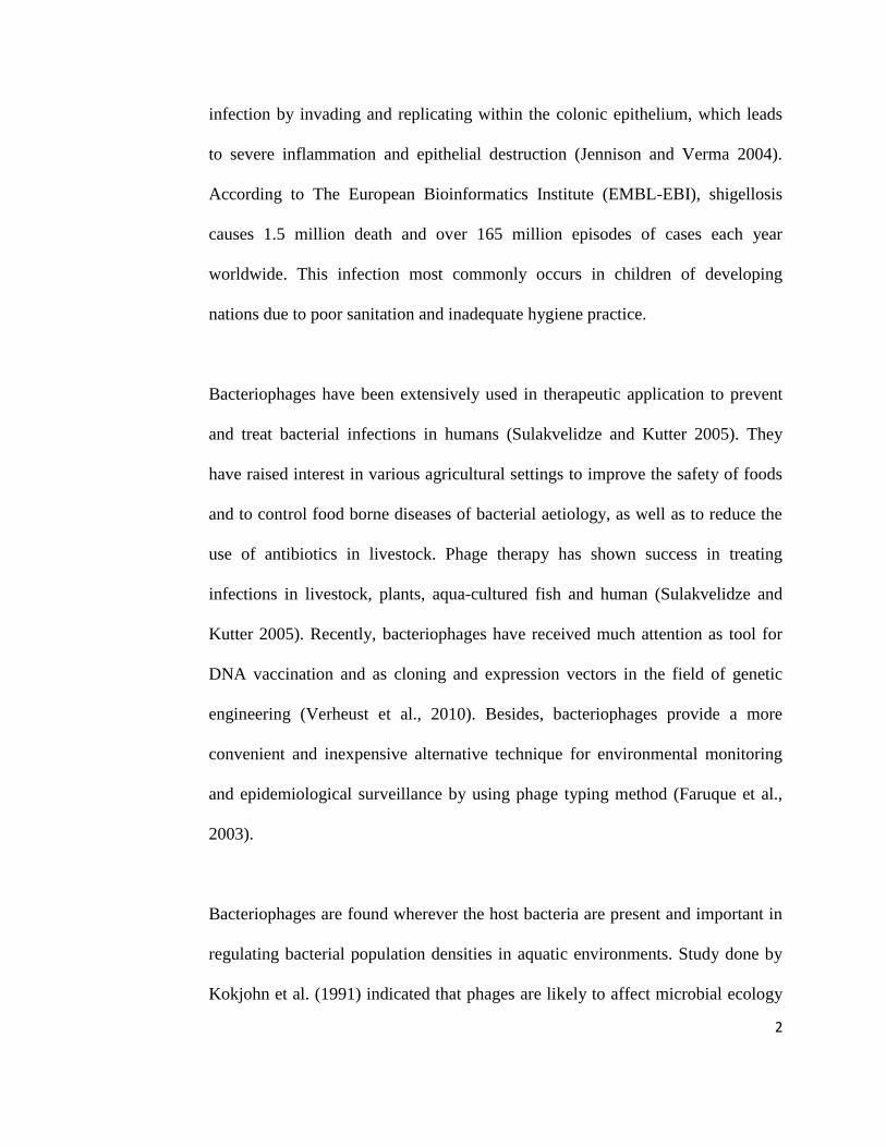
infection by invading and replicating within the colonic epithelium, which leads
to severe inflammation and epithelial destruction (Jennison and Verma 2004).
According to The European Bioinformatics Institute (EMBL-EBI), shigellosis
causes 1.5 million death and over 165 million episodes of cases each year
worldwide. This infection most commonly occurs in children of developing
nations due to poor sanitation and inadequate hygiene practice.
Bacteriophages have been extensively used in therapeutic application to prevent
and treat bacterial infections in humans (Sulakvelidze and Kutter 2005). They
have raised interest in various agricultural settings to improve the safety of foods
and to control food borne diseases of bacterial aetiology, as well as to reduce the
use of antibiotics in livestock. Phage therapy has shown success in treating
infections in livestock, plants, aqua-cultured fish and human (Sulakvelidze and
Kutter 2005). Recently, bacteriophages have received much attention as tool for
DNA vaccination and as cloning and expression vectors in the field of genetic
engineering (Verheust et al., 2010). Besides, bacteriophages provide a more
convenient and inexpensive alternative technique for environmental monitoring
and epidemiological surveillance by using phage typing method (Faruque et al.,
2003).
Bacteriophages are found wherever the host bacteria are present and important in
regulating bacterial population densities in aquatic environments. Study done by
Kokjohn et al. (1991) indicated that phages are likely to affect microbial ecology
3
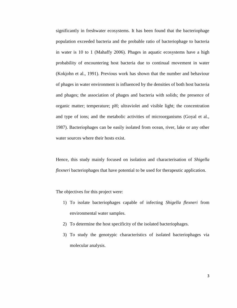
significantly in freshwater ecosystems. It has been found that the bacteriophage
population exceeded bacteria and the probable ratio of bacteriophage to bacteria
in water is 10 to 1 (Mahaffy 2006). Phages in aquatic ecosystems have a high
probability of encountering host bacteria due to continual movement in water
(Kokjohn et al., 1991). Previous work has shown that the number and behaviour
of phages in water environment is influenced by the densities of both host bacteria
and phages; the association of phages and bacteria with solids; the presence of
organic matter; temperature; pH; ultraviolet and visible light; the concentration
and type of ions; and the metabolic activities of microorganisms (Goyal et al.,
1987). Bacteriophages can be easily isolated from ocean, river, lake or any other
water sources where their hosts exist.
Hence, this study mainly focused on isolation and characterisation of Shigella
flexneri bacteriophages that have potential to be used for therapeutic application.
The objectives for this project were:
1) To isolate bacteriophages capable of infecting Shigella flexneri from
environmental water samples.
2) To determine the host specificity of the isolated bacteriophages.
3) To study the genotypic characteristics of isolated bacteriophages via
molecular analysis.

4
CHAPTER 2
LITERATURE REVIEW
2.1 History of Bacteriophage Discovery
Bacteriophage was first discovered by a British bacteriologist, Ernest Hankin in
1896. He reported the presence of bactericidal activity against Vibrio cholerae in
the water of the Ganges and Jumna rivers in India. He proposed that a heat labile
unknown substance which passed through fine porcelain filters was responsible
for preventing the spread of cholera epidemics. Two years later, Gamaleya, the
Russian bacteriologist observed similar phenomenon while working with Bacillus
subtilis. Almost 20 years after Hankin’s observation, Frederick Twort, a
medically trained bacteriologist from England reported similar cases and
hypothesised that it was most probably caused by virus. However, Twort did not
pursue his discovery due to various reasons such as financial difficulties. Two
years later of Twort discovery, Felix d’ Herelle, a French Canadian microbiologist
at the Pasteur institute in Paris reported the same observation. Felix d’ Herelle
proposed that it was virus that parasitised bacteria based on the appearance of
small, clear zones in the lawn of bacterial culture and the lysis in liquid culture.
He was initially named it as taches, then taches vierges and later plaques. Felix d’
Herelle officially named it as “bacteriophage” that formed from “bacteria” and
“phagein” which means phages “eat” or “devour” bacteria (Sulakvelidze et al.,
2001). He called phages as “exogenous agents of immunity” due to their function

5
as therapeutic and prophylactic agents in eradicating various types of infectious
disease (Sulakvelidze et al., 2001; Summers 2005).
2.2 Bacteriophage in General
2.2.1 Morphology of Bacteriophages
Basically, bacteriophages are classified into 1 order, 13 families and 31 genera
(Ackermann 2005). The International Committee on the Taxonomy of Viruses
(ICTV) classifies the phages into 13 families according to the nature of phage
nucleic acid and overall virion morphology traits (Figure 2.1). Most of the
characterised phages (95%) are in the order of Caudovirales or tailed dsDNA
phages. The three main families comprising the Caudovirales are distinguished by
their distinct tail morphologies: 60% of the phages with long and flexible tails are
Siphoviridae; 25% with double-layered, contractile tails is known as Myoviridae;
and 15% with short, stubby tails is called Podoviridae (Guttman et al., 2005).
Polyhedral, filamentous and pleomorphic phages only comprise less than 4% of
the studied phages. They are grouped into 10 families and sometimes include only
a single genus and species (Ackermann 2005).

6
Figure 2.1: Basic morphologies of different families of prokaryote viruses
(Adapted from Hyman and Abedon 2009).
All Caudovirales have heads with icosahedral (20 sides/12 vertices) symmetry or
elongated derivatives that are assembled from multiple copies of specific protein.
The size of the phage heads varies in diameter between 45 and 100 nm, depending
on the size of phage genome packaged during head assembly. The corners of the
icosahedral head are made up of pentamers of capsid proteins, and the remaining
sides are made up of hexamers of the same or a similar protein (Guttman et al.,
2005).
The portal protein connecting the head and tail structures is important for the
infection cycle. This connector which is located at the vertex of the phage head
has a homo-oligomeric structure that participates in the morphogenesis of new
progeny. This protein controls the entry of the DNA and assembly of the tail to

7
the immature head. During infection, it undergoes conformational change and
allows the penetration of DNA from the viral core into the bacterial cell
(Valpuesta et al., 2000).
The tail, base plates, spikes or terminal fibers play an important role in the
attachment of host and delivery of the phage DNA from the head into the host cell
cytoplasm. Long phage tails may be contractile, as seen in the phage T2 of
Myoviridae family, or non-contractile, as shown in the phage λ of Siphoviridae
family. During infection, the overall shape of the non-contractile tail remains
unchanged while for contractile tails, the tail sheath contracts, allowing the central
tail tube protein (TTP) to penetrate both the outer cell membrane and cell wall
(Leiman et al., 2004). The short-tailed Podoviridae, for example T7 phage, form
an extensible tail from the ejected internal core proteins, providing a channel for
the translocation of phage genome into the cell (Molineux 2001). Figure 2.2
shows the morphologies of the three families of Caudovirales.
Figure 2.2: Basic morphologies of the three families of Caudovirales. From left
to right, schematic drawings of Siphoviridae, Myoviridae and Podoviridae
(Adapted from Ceyssens 2009).

8
2.2.2 Genome of Bacteriophages
Majority of the phages contain dsDNA, but there are also small proportion phage
groups with ssDNA, ssRNA or dsRNA (Ackermann 2005). Studies done by
Abedon (2011) on the differentiation of bacteriophages into four genome size
categories: very small (single stranded RNA phages), small (single-stranded DNA
phages), medium (lipid-containing, double-stranded DNA, tailless phages) and
large (double-stranded DNA, tailed phages). As shown in Figure 2.3, very small
phages belong to the members of family Leviridae with genome size that range in
3.5 to 4 kb. Small phages, have genomes that are slightly larger than the members
of family Leviridae, which range from 4.5 kb up to about 9 kb. Medium-sized
phages have genomes that range in size from approximately 9 to 15 kb. At last,
the larger genome tailed phages, members of virus order Caudovirales, have
genomes with 16 kb or greater. Phages with larger genome size have higher gene
number, greater infectivity and virion sophistication, higher possibility of gene
acquisition through horizontal gene transfer as well as additional genetic
redundancy both within and between genomes (Abedon 2011).

9
Figure 2.3: Phage morphologies and genome sizes (Adapted from Hyman and
Abedon 2012).
2.2.3 Life Cycle of Bacteriophages
Bacteriophages may undergo lytic (virulent) cycle or lysogenic (temperate) cycle.
Lysis of the infected host bacterial cell is caused by two mechanisms: lysis from
within and lysis from without (European Food Safety Authority [EFSA], 2009).
For the mechanism of lysis from within, lysis of the host cell is generally due to
the phage replication. The phage virion adsorbs to the surface of a host bacterial
cell and inserts its genetic material into the host cell by injection with contractile
tails or forming pore on the cell wall. When it has successfully entered the cell,
10
the phage undergoes replication process with the aid of host biosynthetic
machinery. The new copies of phage progeny are then liberated with the help of
holin, a hydrophobic polypeptide that creates pores in the cell membrane, which
causes lysis to the host cell. For lysis from without, the host cell lysed without
phage replication process. This occurs through changes in the membrane electric
potential and activity of wall degrading enzymes with large number of phage
particles attach to the cell (EFSA, 2009).
Another alternative method of bacteriophage propagation is lysogenic cycle. In
this process, progeny phages are not produced and the bacterial host cell is not
lysed. This is because the phage is able to produce a repressor protein that stops
the synthesis of phage enzymes and proteins needed in lytic cycle. If the repressor
protein stops producing or is inactivated, an enzyme encoded by the prophage will
excise the viral DNA from bacterial chromosome. The excised DNA will enter
the lytic cycle that causes death in bacterial cell (Todar 2012). Additionally, the
repressor also protects the host bacterium from infection by other phages
(Guttman et al., 2005). In the presence of repressor protein, bacteriophage DNA is
incorporated into the host chromosome via site-specific recombination but
sometimes maintained as a plasmid. This prophage remains in this condition
indefinitely and replicates together with host chromosome whenever the cell
duplicates its chromosomal DNA during normal cell division (Guttman et al.,
2005; Todar 2012). Lysogenic conversion frequently occurred in lysogenised cell
where the host cell may exhibit new characteristics such as virulence factors and
11
antibiotic resistance when the temperate bacteriophages harbour other genes that
are expressed during lysogeny (Guttman et al., 2005; EFSA, 2009). Previous
studies showed that phage gamma and C1 encode the virulence factors of
Corynebacterium diphtheria and Clostridium botulinum, causing the host to
possess pathogenic phenotype (Freeman 1951; Barksdale and Arden 1974).
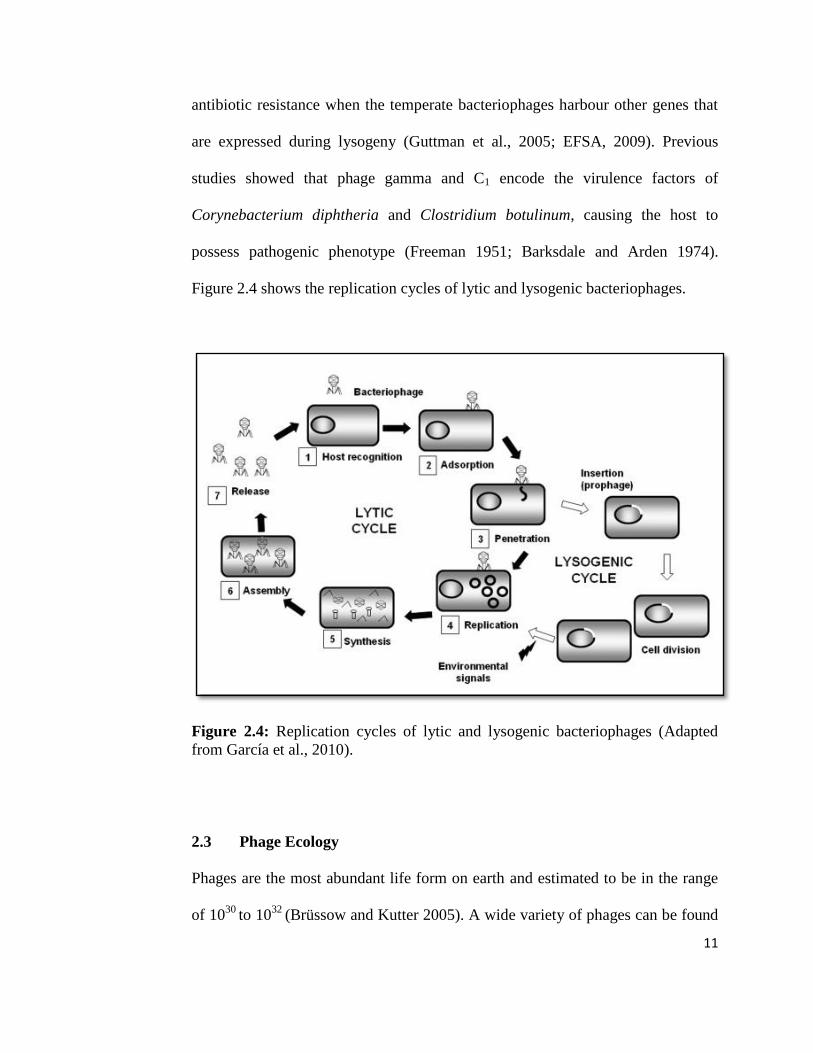
Figure 2.4 shows the replication cycles of lytic and lysogenic bacteriophages.
Figure 2.4: Replication cycles of lytic and lysogenic bacteriophages (Adapted
from García et al., 2010).
2.3 Phage Ecology
Phages are the most abundant life form on earth and estimated to be in the range
of 1030
to 1032
(Brüssow and Kutter 2005). A wide variety of phages can be found

12
in water environments (Goyal et al., 1987). Random sequencing of viral DNA had
estimated 400 to 7000 various phage types to be contained in two uncultured 100-
litre marine water (Breitbart et al., 2002). Besides, phages have been isolated from
mammalian faeces (O’Flynn et al., 2004) and environmental sources including
soil (Ashelford et al., 2003), water (Bergh et al., 1989) and sewage sludge (Carey-
Smith et al., 2006; Oliveira et al., 2009).
The production and distribution of phage are dependent on host concentration.
The phage populations can grow faster when there is greater density of
susceptible bacteria. Generally, the virus-to-bacterium ratio falls between 3 and
10, depending on the nutrient level (Brüssow and Kutter 2005). The consequence
of phage lysis not only reduces the productivity of bacterial populations, but also
delays the ecosystem nutrient cycling and energy flow (Abedon 2006).
Studies by Jensen et al. (2006) showed the correlation of populations of bacteria
and phage with cholera outbreak. They proposed that the severity of the outbreak
is determined by the density of phage in the reservoir. Increase of phage predation
will reduce the bacterial density, which ultimately ends the outbreak by returning
the bacterial populations to pre-outbreak stage. Therefore, outbreak occurs when
there is reduction of phage density. In short, introduction of vibriophage can
reduce the duration and severity of cholera outbreak.
13
The 4 key steps in the phage lytic life cycle are correlated to the phage ecology.
Firstly, the extracellular search for host bacteria is limited by diffusion rates and
determined by the density of the host populations. Secondly, a phage adsorption
step occurs after phage-host collision, which involves reversible phage binding,
irreversible attachment and transfer of genome into the host. Thirdly, the infection
step where the phage genome is replicated and phage particles are synthesised,
resulting the production of phage progeny. Lastly, for temperate phage, the phage
DNA is integrated into the host chromosome and replicates with it or replicates
synchronously as a plasmid (Brüssow and Kutter 2005).
2.4 Applications of Bacteriophages
2.4.1 Phage display
The development of phage display enables the synthesis of novel polypeptides
with high binding affinity to a particular substrate. In this system, the DNA that
encodes the polypeptide is cloned as gene fusions with the phage coat protein
genes. The desired protein is then incorporated into the mature phage particle and
expressed on the surface of the phage (Rees and Loessner 2005). Libraries of
phage particles can also be used for the screening and identification of clones
expressing peptides that are highly specific and possess desired binding
characteristics (Rees and Loessner 2005; Haq 2012).
14
This approach has been extensively used for medical or pharmaceutical
applications. Phage display has led to the revolution of phage antibody technology
where phages were used to display antibody molecule with specific antigen-
binding domain that can be used for affinity selection process (Rader and Barbas
1997). This antibody-derived peptide has been successfully developed as
therapeutic agent that serves the function of agonist and antagonist in receptor-
ligand interaction. Research work done by Dickerson et al. (2005) had showed the
effectiveness of this method in the treatment of cocaine addiction where
bacteriophage displaying cocaine-sequestering antibodies was used to block the
action of cocaine in the brain.
Furthermore, phage display can be applied for detection assay to detect for the
biological threat agents. This assay utilises phage-borne peptide as diagnostic
detector or probe that specifically binds to bacterium, spore, virus and toxin. This
technique has proved to be successful in the identification of various types of
viruses (Petrenko and Vodyanoy 2003), detection of Bacillus spores (Zhou et al.,
2002) and differentiation of Candida species in clinical samples (Bliss et al.,
2003).
2.4.2 Phage Typing
Phage typing is a procedure for characterising and identifying bacterial strains by
their reaction (susceptibility or resistance) to various known strains of phages. It

15
is a relatively rapid, simple and inexpensive method for the typing of bacteria
(Hagens and Loessner 2007). The host specificity of phages is a useful tool for the
classification of bacteria and the detection of pathogenic bacteria (Clark and
March 2006). Many phages are highly specific for the receptors on host cell
surface and only receptors with similar structure and configuration can interact
with the respective phage.
Phage typing is easy and convenient to be performed with large number of
bacterial isolates can be analysed at the same time. For example, Escherichia coli
O157:H7 has been successfully subdivided into 66 different phage types (Frost et
al., 1993). Phages are not only specific towards species of bacteria, but also
strains of bacteria, permitting typing beyond the level of species (Welkos et al.,
1974). According to the work done by Pruneda and Farmer (1977), phage typing
method appears to be more sensitive than colicin typing and antibiograms in
differentiating bacterial strains.
Susceptibility to infection by a particular phage enables the phenotypic
differentiation of strains and identification of the strain that causes an outbreak of
disease. This property can also be employed for epidemiological investigations to
trace the causative agent responsible for the infection. Faruque et al. (2003) had
successfully isolated phage SF-9 that has epidemiological applications in tracing
and monitoring the presence of Shigella dysenteriae type 1 from environmental
waters in Bangladesh. This method has proved to be useful in predicting
16
outbreaks and the spread of shigellosis, which occurs as epidemics in many
developing countries. Besides, this system provides reliable, sensitive and fast
results for epidemiologists in the surveillance of outbreaks (Pruneda and Farmer
1977).
2.4.3 Phage Therapy
Phage therapy is the therapeutic use of bacteriophages to treat pathogenic
bacterial infections. Lytic phages are preferred for the biocontrol of pathogenic
bacteria since they are highly specific and very effective in lysing targeted
pathogenic bacteria. Furthermore, lytic phages do not contain integrase genes on
their genomes, therefore, they are unable to coexist with the host and carry
virulent genes from one host to another (Brüssow 2005).
Phage therapy is an alternative for the antibiotics treatment of bacterial infection.
Unlike broad-spectrum antibiotics, phages are specific to their prokaryotic host
cell and less commonly illicit resistance in non-host cells (Sulakvelidze and
Kutter 2005). Additionally, phages are not susceptible to the onset of bacterial
resistance due to its ability to evolve with the host (Sulakvelidze and Kutter
2005). Moreover, phage therapy is not affected by antibiotic resistance of bacteria
where it is effective for the treatment of multidrug resistant strains infection. It
was showed that Shigella phages were successfully used as a prophylaxis of
bacterial dysentery (Babalova et al., 1968). Anpilov and Prokudin (1984) reported

17
that the incidence of dysentery in the phage-treated group was ten fold less than
that occurring in phage-untreated group.
Phage therapy has more advantages compared to antibiotics that are in clinical use
due to its specificity towards targeted bacteria. In addition, this therapy is
harmless to the eukaryotic host undergoing therapy and beneficial bacteria such as
normal flora in gut, so, reduce the chances of opportunistic infections (Carlton
1999). Another advantage is that bacteriophage usually replicates at the site of
infection and they are self-limiting. They are only able to self-replicate with the
presence of susceptible bacterial pathogens (Carlton 1999).
Humans are continuously exposed to bacteriophage because phages are the
simplest and most abundant organisms on earth that present as part of both
gastrointestinal and environmental ecosystems (Carlton 1999; Sulakvelidze et al.,
2001). Hence, it is suggested that phage therapy is well tolerated by humans,
providing little or no side effects to human (Hausler 2007; Sulakvelidze et al.,
2001).
Phages having lytic activity against a wide range of bacterial strains are preferred
to be used as therapeutic agent (Verma et al., 2013). These wide host range
bacteriophages are potential candidate for phage therapy due to their broad
antimicrobial range which could potentially treat a wide range of infections.
However, precautions need to be taken to avoid cross-interaction and lysis of
18
normal microbiota. Besides, amplification of bacteriophages in non-pathogenic
alternative host bacteria improves the safety of bacteriophage application in
treatment of infections (Bielke et al, 2007).
2.5 Shigella flexneri
Shigella flexneri is highly infectious and causes communicable bacterial
diarrhoeas, shigellosis. This disease is commonly transmitted via faecal-oral
route. Dose response studies done at the University of Maryland have shown that
S. flexneri needs as few as 100 cells to cause disease in adult volunteers (DuPont
1989). This phenomenon is associated with the ability of S. flexneri to express
acid resistance genes that enable this enteric pathogen to survive the acidic
environment of the human stomach (Small et al., 1994; Jennison and Verma
2007).
When S. flexneri invades the colonic mucosa of humans, it begins to multiply
intracellularly, spread from cell to cell and disseminate throughout the mucosal
epithelial cells. The inflammatory response of the host subsequently destroys the
colonic epithelial layer, causing abscesses and ulceration in the superficial layer
of the colonic mucosa. Damage of the epithelial layer produces the clinical
symptoms of shigellosis ranging from watery diarrhoea to classic dysentery
characterised by fever, violent intestinal cramps and eventually discharge of

19
mucopurulent and bloody stools characteristic of bacillary dysentery (Philpott et
al., 2000).
Numerous virulence genes have been identified in Shigella flexneri, which termed
as pathogenicity islands (PAIs). PAIs are only present in pathogenic strains and
absent or limited distribution in less pathogenic strains. Three chromosomal PAIs
have been located on the S. flexneri chromosome, participate in the pathogenic
process directly or contribute to survival in the environments encountered during
infection. These three PAIs are involved in virulence such as enterotoxin,
aerobactin operon and Shigella resistance locus (SRL) encoding resistance
determinants to streptomycin, ampicillin, chloramphenicol and tetracycline (Moss
1999; Al-Hasani et al., 2001; Turner 2001). The genome of S. flexneri serotype
2a, the most prevalent serotype that causes bacillary dysentery, has found to
contain a number of bacteriophage-related genes conferred by temperate
bacteriophages, which responsible for the O-antigen modification in S. flexneri
(Allison and Verma 2000).
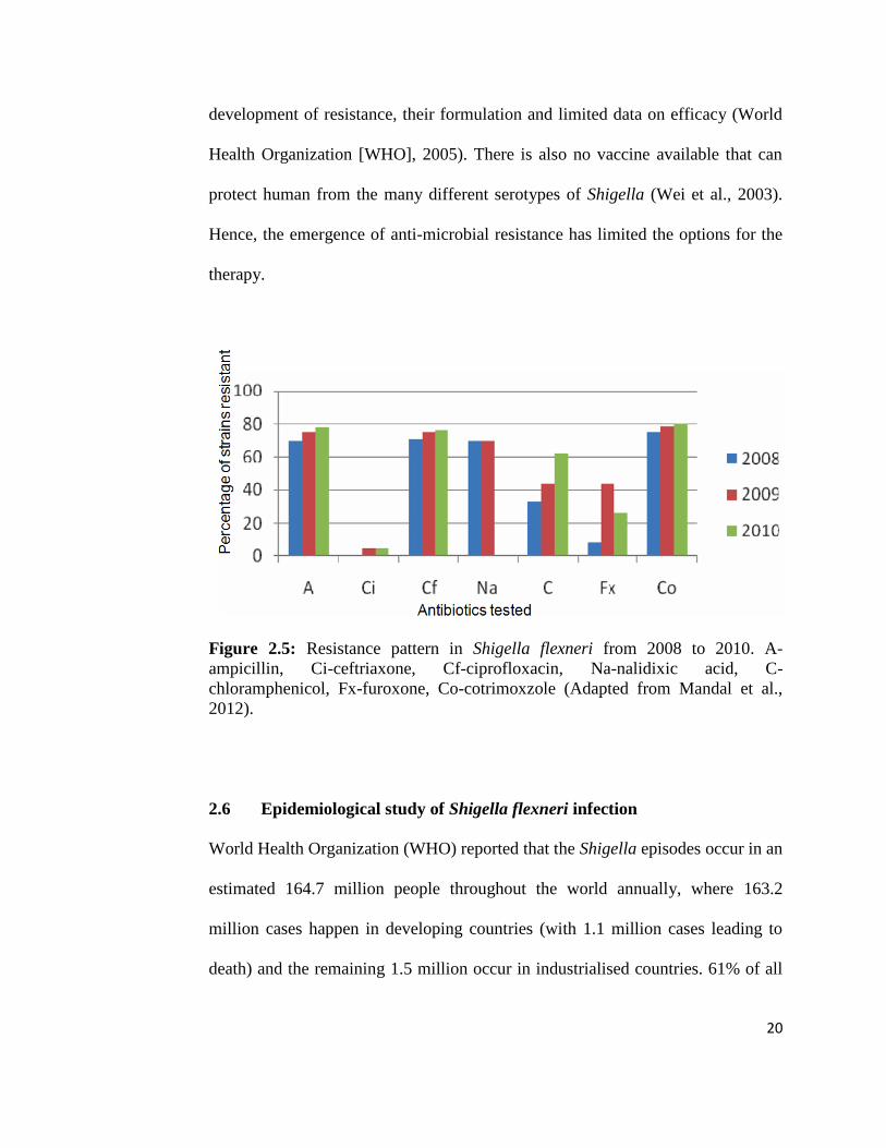
Study done by Mandal et al. (2012) highlighted the increasing antimicrobial
resistance in Shigella flexneri in recent years. Ciprofloxacin has been
recommended by WHO as the drug of choice for effective treatment of multidrug-
resistant strains of Shigella. However, as shown in Figure 2.5, S. flexneri is
gaining resistance towards this drug. Unfortunately, the alternative drugs such as
pivmecillinam, ceftriaxone and azithromycin are limited by their high cost, rapid
20
development of resistance, their formulation and limited data on efficacy (World
Health Organization [WHO], 2005). There is also no vaccine available that can
protect human from the many different serotypes of Shigella (Wei et al., 2003).
Hence, the emergence of anti-microbial resistance has limited the options for the
therapy.
Figure 2.5: Resistance pattern in Shigella flexneri from 2008 to 2010. A-
ampicillin, Ci-ceftriaxone, Cf-ciprofloxacin, Na-nalidixic acid, C-
chloramphenicol, Fx-furoxone, Co-cotrimoxzole (Adapted from Mandal et al.,
2012).
2.6 Epidemiological study of Shigella flexneri infection
World Health Organization (WHO) reported that the Shigella episodes occur in an
estimated 164.7 million people throughout the world annually, where 163.2
million cases happen in developing countries (with 1.1 million cases leading to
death) and the remaining 1.5 million occur in industrialised countries. 61% of all
21
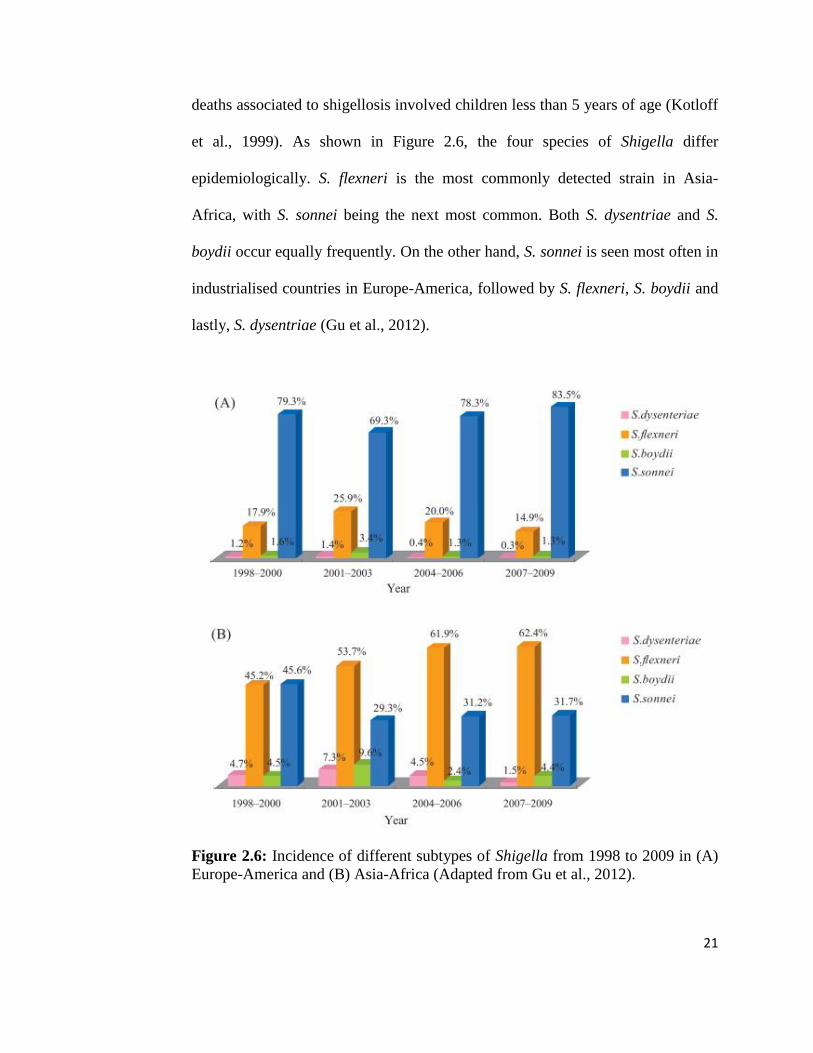
deaths associated to shigellosis involved children less than 5 years of age (Kotloff
et al., 1999). As shown in Figure 2.6, the four species of Shigella differ
epidemiologically. S. flexneri is the most commonly detected strain in Asia-
Africa, with S. sonnei being the next most common. Both S. dysentriae and S.
boydii occur equally frequently. On the other hand, S. sonnei is seen most often in
industrialised countries in Europe-America, followed by S. flexneri, S. boydii and
lastly, S. dysentriae (Gu et al., 2012).
Figure 2.6: Incidence of different subtypes of Shigella from 1998 to 2009 in (A)
Europe-America and (B) Asia-Africa (Adapted from Gu et al., 2012).
22
CHAPTER 3
MATERIALS AND METHODS
3.1 Bacterial Strains
This study included 8 clinical strains of bacteria (Shigella flexneri, Shigella
sonnei, Shigella dysenteriae, enterotoxigenic and enteropathogenic Escherichia
coli, Staphylococcus aureus and Klebsiella pneumonia). All of them were clinical
isolates from Hospital Universiti Science Malaysia (HUSM). The bacteria were
grown on Luria Bertani (LB) agar pH 7.0 by streaking and incubating at 37oC for
16-18 h. The plates were stored at 4oC after incubation. Exponential growing
bacteria culture was used in all the tests in this study. This was done by
inoculating one colony of bacteria in LB broth and incubated at 37oC with
constant agitation (200 rpm) until mid-log phase (OD600 = 0.4-0.6) was reached.
23
3.2 Chemicals, Reagents and Equipments
All the chemicals and reagents used in this study are listed in Table 3.1.
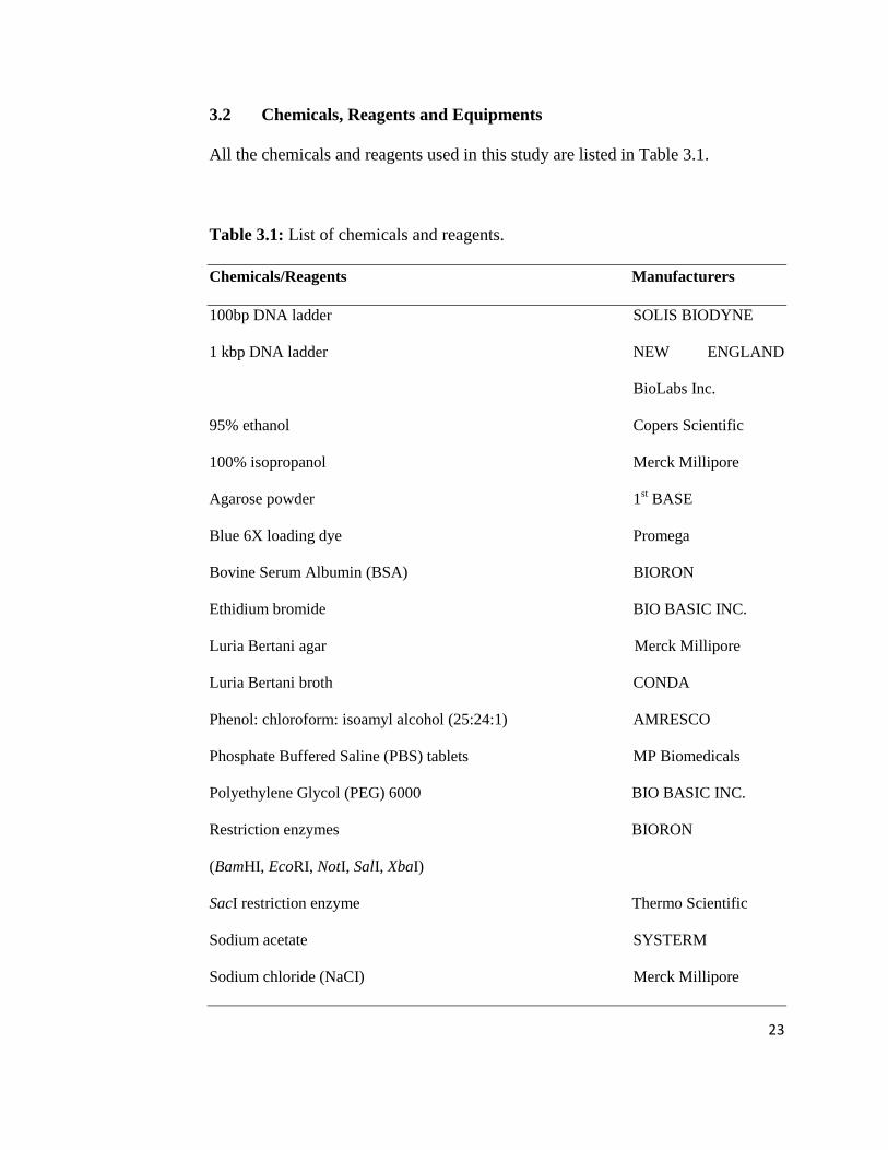
Table 3.1: List of chemicals and reagents.
Chemicals/Reagents Manufacturers
100bp DNA ladder SOLIS BIODYNE
1 kbp DNA ladder NEW ENGLAND
BioLabs Inc.
95% ethanol Copers Scientific
100% isopropanol Merck Millipore
Agarose powder 1st BASE
Blue 6X loading dye Promega
Bovine Serum Albumin (BSA) BIORON
Ethidium bromide BIO BASIC INC.
Luria Bertani agar Merck Millipore
Luria Bertani broth CONDA
Phenol: chloroform: isoamyl alcohol (25:24:1) AMRESCO
Phosphate Buffered Saline (PBS) tablets MP Biomedicals
Polyethylene Glycol (PEG) 6000 BIO BASIC INC.
Restriction enzymes BIORON
(BamHI, EcoRI, NotI, SalI, XbaI)
SacI restriction enzyme Thermo Scientific
Sodium acetate SYSTERM
Sodium chloride (NaCI) Merck Millipore
24
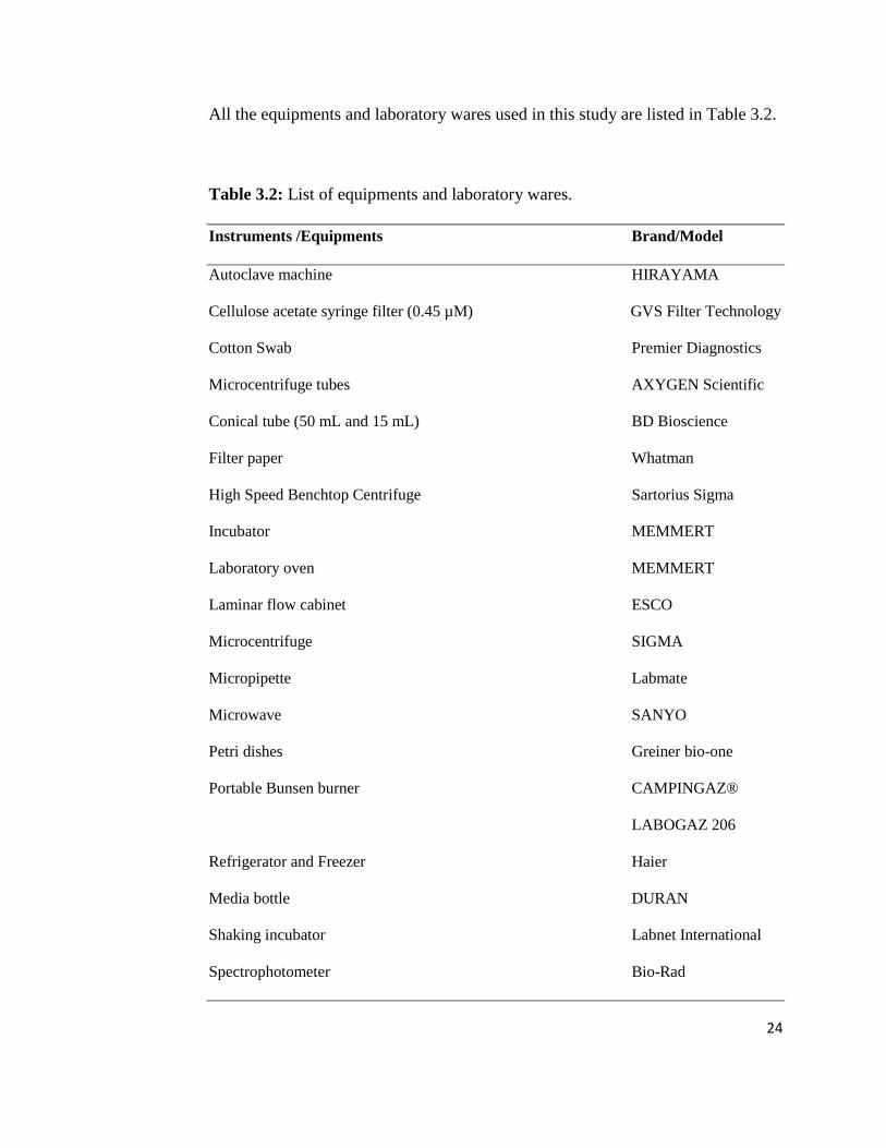
All the equipments and laboratory wares used in this study are listed in Table 3.2.
Table 3.2: List of equipments and laboratory wares.
Instruments /Equipments Brand/Model
Autoclave machine HIRAYAMA
Cellulose acetate syringe filter (0.45 µM) GVS Filter Technology
Cotton Swab Premier Diagnostics
Microcentrifuge tubes AXYGEN Scientific
Conical tube (50 mL and 15 mL) BD Bioscience
Filter paper Whatman
High Speed Benchtop Centrifuge Sartorius Sigma
Incubator MEMMERT
Laboratory oven MEMMERT
Laminar flow cabinet ESCO
Microcentrifuge SIGMA
Micropipette Labmate
Microwave SANYO
Petri dishes Greiner bio-one
Portable Bunsen burner CAMPINGAZ®
LABOGAZ 206
Refrigerator and Freezer Haier
Media bottle DURAN
Shaking incubator Labnet International
Spectrophotometer Bio-Rad
25
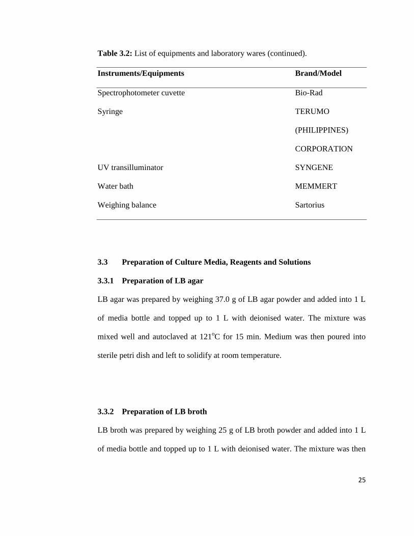
Table 3.2: List of equipments and laboratory wares (continued).
Instruments/Equipments Brand/Model
Spectrophotometer cuvette Bio-Rad
Syringe TERUMO
(PHILIPPINES)
CORPORATION
UV transilluminator SYNGENE
Water bath MEMMERT
Weighing balance Sartorius
3.3 Preparation of Culture Media, Reagents and Solutions
3.3.1 Preparation of LB agar
LB agar was prepared by weighing 37.0 g of LB agar powder and added into 1 L
of media bottle and topped up to 1 L with deionised water. The mixture was
mixed well and autoclaved at 121oC for 15 min. Medium was then poured into
sterile petri dish and left to solidify at room temperature.
3.3.2 Preparation of LB broth
LB broth was prepared by weighing 25 g of LB broth powder and added into 1 L
of media bottle and topped up to 1 L with deionised water. The mixture was then

26
mixed well and autoclaved at 121oC for 15 min. The medium was allowed to cool
down at room temperature.
3.3.3 TBE buffer (pH 8.3)
Tris-Borate-EDTA buffer was prepared by mixing 108 g of Tris Base and 55 g of
boric acid in 900 mL of deionised water. Then, 40 mL of EDTA (0.5 M) was
poured into the mixture. The mixture was measured and adjusted to pH 8.3 with 1
M NaOH and 1 M HCI. The solution was subsequently topped up to 1 L to
prepare the solution of 10X TBE buffer. To prepare working solution of 1X TBE
buffer, 100 mL of TBE buffer was diluted with 900 mL of deionised water.
3.3.4 Agarose gel (1.0%)
0.20 g of agarose powder was dissolved in 20 mL of 1X TBE buffer by
microwave heating. The melted agarose was poured into the cassett and left to
solidify at room temperature.
3.3.5 Ethanol (70%)
737 mL of 95% ethanol was topped up to 1 L with deionised water to prepare
70% ethanol.
27
3.3.6 Mixture of 20% PEG 6000 and 10% NaCI
10 g of PEG 6000 and 5 g of NaCI were dissolved in 50 mL of deionised water.
The mixture was then filter sterilised.
3.3.7 Phosphate Buffered Saline (PBS)
Phosphate buffered saline was prepared by dissolving 1 tablet of PBS in 100 mL
of deionised water and sent to autoclaved at 121oC for 15 min..
3.3.8 Sodium Acetate (3 M)
Sodium acetate was prepared by dissolving 12.3 g of sodium acetate powder in 50
mL of deionised water and sent to autoclaved at 121oC for 15 min.

28
3.4 Methods
3.4.1 Overview of Research Methodology
Figure 3.1: Overall work flow.
Bacteriophage Sample Collection
General Enrichment
Double Agar Overlay Plaque
Assay
Phage Selection and Amplificaton
Concentration and Purification of Bacteriophages
Host Specificity Test
DNA Extraction Agarose Gel
Electrophoresis
Restriction Digestion of Phage
DNA

29
3.4.2 Isolation of Bacteriophages
3.4.2.1 Bacteriophage Sample Collection
Water samples were collected from Kampar area including rivers, drains, ponds
and lakes. These samples were collected in sterile 50 mL conical tubes. The water
at collection site was mixed thoroughly and the sediments were collected together
with the overlying water.
3.4.2.2 General Enrichment
A procedure of bacteriophage enrichment method was performed on the collected
samples (Twest and Kropinski 2009). The samples were centrifuged at 6000 rpm
for 10 min to remove large particulates and bacteria. 40 mL of the supernatant
were decanted into a new sterile conical tube. The tubes were inoculated with 1
mL of broth culture of Shigella flexneri host bacterium (OD600 = 0.4-0.6) and
mixed thoroughly by inversion. The tubes were incubated at 37oC in static
condition to allow specific phage enrichment. After 24 h incubation, the contents
of the tubes were centrifuged at 9,000 rpm for 10 min. The supernatant of the
water sample was filtered slowly through the 0.45 µM cellulose acetate syringe
filter to a 1.5 mL microcentrifuge tube. The filtered samples were then
centrifuged at 14,000 xg for 15 min. The supernatant were carefully transferred to
a new sterile tube and stored at 4oC.

30
3.4.2.3 Double Agar Overlay Plaque Assay
This assay was adapted from a modified procedure of the double agar layered
method (Kropinski et al., 2009). A row of 4 sterile microcentrifuge tubes was set
up. Each of them was numbered with the appropriate sequential 100-fold dilutions
from 100 X to 100 M. 990 µL of Luria Bertani (LB) broth that serves as diluents
was added aseptically to each tube. 10 µL of undiluted phage was added to the
first tube and mixed well. 10 µL of broth was then transferred from first tube to
the second tube in the series. No phage was added to the control tube. Only the
undiluted, third and fourth phage preparations (1 M and 100 M dilutions
respectively) were tested for plaque assay. A plate of bacterial control without
phage was prepared as negative control.
The Shigella flexneri host bacterium was grown in 5 mL of LB broth with shaking
at 200 rpm, 37oC until it reached log phase (OD600 = 0.4-0.6). LB bottom agar
plates containing 1.5% agar were prepared.
Two millilitre of log phase Shigella flexneri was transferred into a sterile 15 mL
conical tube. 200 µL of the selected dilution of phages was added immediately.
The tubes were preincubated at 37oC for 20 min to allow phage adsorption onto
host bacterium. After preincubation, the tubes were added with 2 mL of molten
(45oC) soft agar (0.75%) that was previously prepared from 1.5% of LB agar.
The tubes were gently mixed by inversion for a few times and the contents were
poured on Petri dishes containing LB bottom agar. The plates were swirled in

31
circles to spread the mixture evenly over the plates. The plates were left at room
temperature for 10-15 min until the soft agar has solidified and the Petri dishes
were incubated at 37oC for 16-18 h. The following day, the plates were checked
and observed for the plaque formation. The plaques formed in each plate were
enumerated.
3.4.3 Amplification and Purification of Bacteriophages
3.4.3.1 Phage Selection and Amplification
The exponential growing culture of Shigella flexneri was inoculated into a
microcentrifuge tube. Phage plaques were selected based on size and clarity using
a 100 µL pipette. The pipette tip was inserted carefully into centre of a discrete
plaque and a plug of soft agar containing the bacteriophages was removed out.
The phage-containing plug was added into the inoculated tube. The tubes were
incubated at 37oC for 24 h or more. The phage suspension was centrifuged at
14,000 xg for 15 min. The supernatant was transferred to fresh, sterile
microcentrifuge tube for further analysis.
3.4.3.2 Concentration and Purification of Bacteriophages
The phages were concentrated and purified based on the precipitation with
polyethylene glycol (PEG) 6000 (Boulanger 2009). The Shigella flexneri host
bacterium was cultured in 50 mL of LB broth with constant agitation (200 rpm) at

32
37oC until OD600 of 0.4–0.6. The isolated phage was then added into the bacteria
culture and agitated overnight at 37oC, 200 rpm. The bacteria-bacteriophage
mixture was centrifuged at 9,000 rpm for 15 min to remove bacterial debris. 40
mL of phage-containing supernatant was transferred into a new 50 mL conical
tube and 10 mL of the PEG/NaCI was added. The mixture was mixed by
inversion and incubated on ice for at least 1 h in order to precipitate phage
particles. The precipitated phage was collected by centrifugation at 9,000 rpm,
4oC for 30 min and the supernatant was carefully discarded. The conical tube was
turned over to drain away the remaining fluid from the pellet for 5 min. The phage
pellet was resuspended in 500 µL phosphate buffered saline (PBS) and transferred
to a sterile microcentrifuge tube. The phage particles were separated from co-
precipitated bacterial debris by centrifugation at 11,600 xg for 10 min. The
supernatant was then collected into a sterile microcentrifuge tube.
3.4.4 Host Specificity Test
Bacterial susceptibility to bacteriophage was assayed based on the spot test
method (Raya and Hébert 2009) with modification. The host ranges of the
isolated phages were determined using Shigella sonnei, Shigella dysenteriae,
Shigella flexneri, enterotoxigenic and enteropathogenic Escherichia coli,
Staphylococcus aureus and Klebsiella pneumonia. Briefly, the bacteria strains
were cultured in LB broth at 37oC with constant agitation (200 rpm). A sterile
swab was moistened with the indicator cells and spread on the surface of LB agar.

33
The plate was previously marked with grids to allow identification of each
inoculum. A sterile filter paper disk was placed on each grid and 10 µL of each
phage suspension was spotted on the disk, dried and incubated for 16-18 h at
37oC. The host range of phages was determined based on susceptibility of
bacterial strains by observing lytic zone formed in inoculation area.
3.4.5 Molecular Characterisation
3.4.5.1 DNA Extraction
Extraction of nucleic acid was conducted according to the method of Sambrook
and Russell (2001) with minor modifications. The DNA of phage particles were
extracted by mixing 500 µL of the purified phage with 500 µL of phenol:
chloroform: isoamyl alcohol (25:24:1). The mixture was then centrifuged for 10
min at 14,000 xg to separate the phases. The top aqueous phase was transferred to
a new fresh 1.5 mL microcentrifuge tube and 500 µL of 100% cold isopropanol
and 50 µL of 3 M sodium acetate were added into it. The DNA was left to
precipitate at room temperature for 20 min. After incubation at room temperature,
they were centrifuged at 14,000 xg for 15 min at 4oC. The DNA pellet was
washed twice with 70% ethanol and air dried. The DNA was resuspended with 50
µL distilled water and stored at -20oC.

34
3.4.5.2 Agarose Gel Electrophoresis
The extracted phage genomic DNA was analysed using agarose gel
electrophoresis. Three microlitre of the DNA was mixed with 0.6 µL of Blue 6x
Loading Dye and loaded into wells of a 1.0% agarose gel in 1X Tris-Borate-
EDTA (TBE). The gel was subjected to electrophoresis at 80 V for 45 min until
the dye front was near to the bottom of the gel. After electrophoresis, the gel was
stained in 1 µg/mL of ethidium bromide for 10 min, followed by destaining with
distilled water for a few seconds. The gel was then viewed using UV
transilluminator to visualise the presence of DNA bands.
3.4.5.3 Restriction Digestion of Phage DNA
Six restriction enzymes were used to digest phage genomic DNA. The chosen
enzymes were BamHI, EcoRI, NotI, SacI, SalI and XbaI. The reaction mixture
was prepared (Table 3.3) and mixed gently before allowed for incubation at 37oC
for 3 h. The restriction enzyme was then inactivated (Table 3.4) by incubating the
reaction mixture in water bath. The digested DNA was separated by agarose gel
electrophoresis process. The restriction patterns were visualised by
transillumination with UV light after staining with ethidium bromide.

35
Table 3.3: Restriction enzyme digestion reaction for phage DNA.
Components per reaction
Phage DNA 5.0 µL
Restriction enzyme 0.5 µL
Restriction enzyme buffer 1.0 µL
BSA 0.1 µL
PCR water 3.4 µL
Total 10.0 µL
Table 3.4: Heat inactivation of restriction enzymes used in this study (Adapted
from Thermo Scientific, 2012; BIORON, 2013).
Restriction enzymes Temperature (oC) Time (min)
BamHI 80 20
EcoRI 65 20
NotI 65 20
SacI 65 20
SalI 65 20
XbaI 65 20

36
CHAPTER 4
RESULTS
4.1 Isolation of Bacteriophages
4.1.1 Sample Collection Sites
Sample A was collected from the river that was polluted by solid waste disposal
which was flowing freely. The water sample collected was clear, with some
sediment present. The river for sample collection site B was shallower as
compared to site A and the river was flowing in slow motion. The water appeared
to be clean and clear from sediments. The water sample from sample collection
site C which was a drain was also clear with little sediment. The water in the drain
was not flowing and was in static condition. Water sample D which was from a
pond was greenish in colour with abundant amount of algae floating on the
surface of water. The water was not flowing due to the confined area. The water
in sampling site E which was from the lake was calm and stagnant. A large
amount of algae was also present in this sample. The water in site E appeared to
be the most turbid among all the samples collected. The sites of all the sample
collection are shown in Figure 4.1.

37
Figure 4.1: Sample collection sites in Kampar area. (A) River near to Old Town
residential area, (B) River connecting Old Town and New Town, (C) Drain beside
KTAR, (D) Pond outside Station A, and (E) Westlake.
A B
C D
E

38
4.1.2 Double Agar Overlay Plaque Assay
All the plates showed positive for the isolation of bacteriophages. Round, clear
and transparent plaques were observed in all plates, indicating that the samples
collected from different sites contained bacteriophages that were infectious
against Shigella flexneri. The phages produced clear, medium-sized (1.0-3.0 mm
in diameter) plaques with well-defined edges in bacterial lawn, showing that the
isolated phages have lytic effect against Shigella flexneri. Different types of
bacteriophages were found in each plate based on the formation of plaques with
different degree of transparency and sizes. The plaque count in sample collection
sites A and B were 22 and 17 respectively while the plaques produced in C, D and
E were too numerous to be enumerated. These different morphologies of plaques
were isolated from each plate for further study. Figure 4.2 shows the plaques
formation for water samples A, B, C, D and E. Table 4.1 shows the size and
morphology of the isolated plaques.

39
Figure 4.2: Plaques formation for water samples A, B, C, D and E.
A B
C D
E

40
Table 4.1: Morphology and size of the isolated plaques.
Bacteriophage Sampling site Plaque Size Plaque
isolates (mm) Morphology
A1 A 1.0 clear
B1 B 1.5 clear
C1 C 2.2 clear
C2 C 2.0 clear
D1 D 3.0 clear
D2 D 2.0 clear
E1 E 2.0 clear
E2 E 1.0 clear
4.2 Characterisation of Isolated Bacteriophages
4.2.1 Host Specificity Test
The host range of the 8 isolated bacteriophages were determined using Shigella
sonnei, Shigella dysenteriae, enterotoxigenic and enteropathogenic Escherichia
coli, Staphylococcus aureus and Klebsiella pneumonia (Table 4.2). Only C2, D1
and D2 bacteriophages possessed broad host range. Other bacteriophages have
narrow host ranges. D2 phage was lytic against 2 out of the 6 bacteria tested
where clear zone was produced on the enteropathogenic and enterotoxigenic E.
coli pathotypes tested. C2 and D1 phages produced faint clear zone in EPEC and
ETEC bacterial lawn respectively. Meanwhile, other phages showed negative
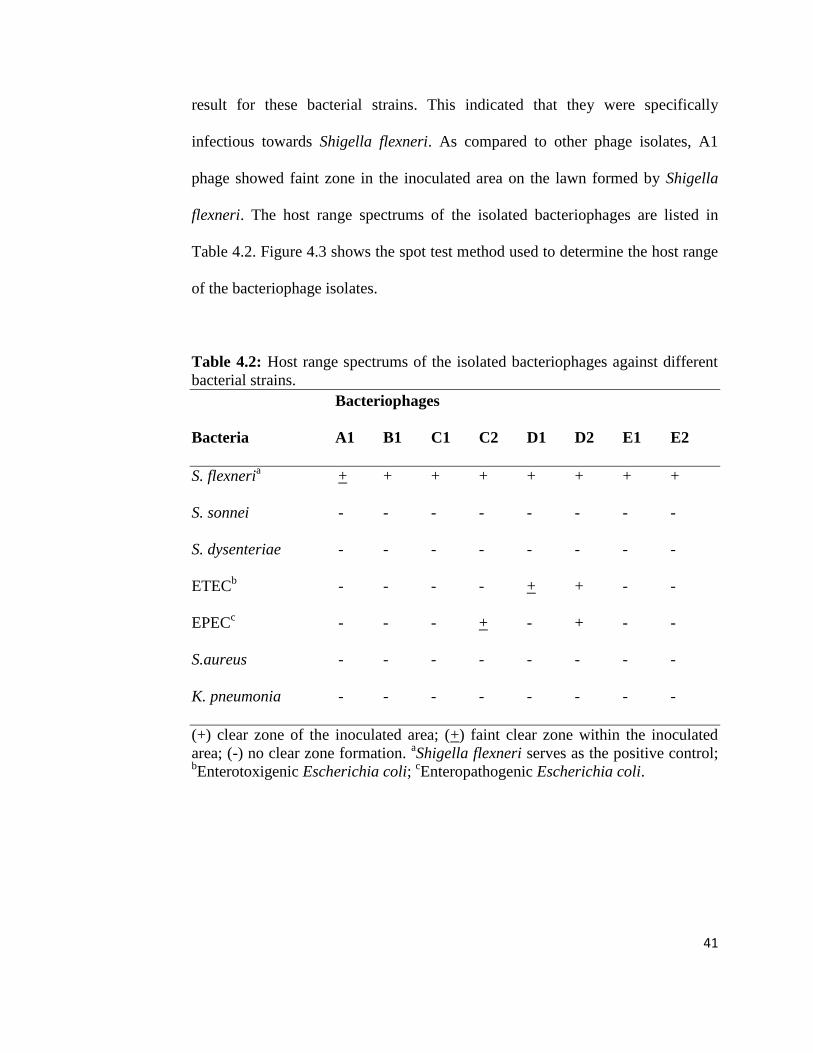
41
result for these bacterial strains. This indicated that they were specifically
infectious towards Shigella flexneri. As compared to other phage isolates, A1
phage showed faint zone in the inoculated area on the lawn formed by Shigella
flexneri. The host range spectrums of the isolated bacteriophages are listed in
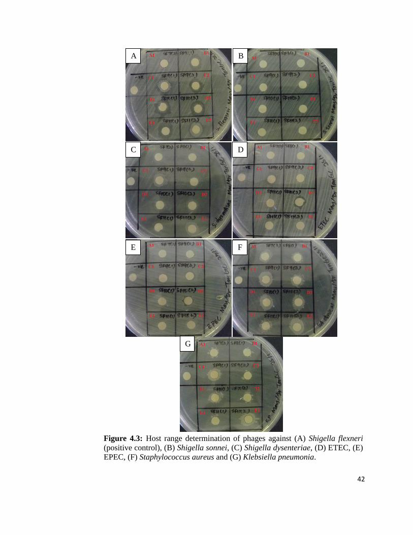
Table 4.2. Figure 4.3 shows the spot test method used to determine the host range
of the bacteriophage isolates.
Table 4.2: Host range spectrums of the isolated bacteriophages against different
bacterial strains.
Bacteriophages
Bacteria A1 B1 C1 C2 D1 D2 E1 E2
S. flexneria + + + + + + + +
S. sonnei - - - - - - - -
S. dysenteriae - - - - - - - -
ETECb
- - - - + + - -
EPECc
- - - + - + - -
S.aureus - - - - - - - -
K. pneumonia - - - - - - - -
(+) clear zone of the inoculated area; (+) faint clear zone within the inoculated
area; (-) no clear zone formation. aShigella flexneri serves as the positive control;
bEnterotoxigenic Escherichia coli;
cEnteropathogenic Escherichia coli.
42
A B
C D
E F
G
Figure 4.3: Host range determination of phages against (A) Shigella flexneri
(positive control), (B) Shigella sonnei, (C) Shigella dysenteriae, (D) ETEC, (E)
EPEC, (F) Staphylococcus aureus and (G) Klebsiella pneumonia.

43
Figure 4.4: Phage DNA extracted using phenol-chloroform method. Lane M: 100
bp ladder; Lane 1: A1 phage; Lane 2: B1 phage; Lane 3: C1 phage; Lane 4: C2
phage; Lane 5: D1 phage; Lane 6: D2 phage; Lane 7: E1 phage; Lane 8: E2
phage.
4.2.2 Phage DNA Extraction
The phage genome was successfully extracted using phenol-chloroform method.
As shown in Figure 4.4, all the phage genome sizes were above 3,000 bp. The
bands for Lane 2, 6 and 8 were very clear, indicating higher concentration of
phage DNA extracted from B1, D2 and E2 phages. The yield and purity of the
phage DNA were high and there was no contamination of bacterial genomic
DNA.
M 1 2 3 4 5 6 7 8
3,000
2,000
1,500
1,000
800 700 600
500
400
300
200
100
(bp)
44
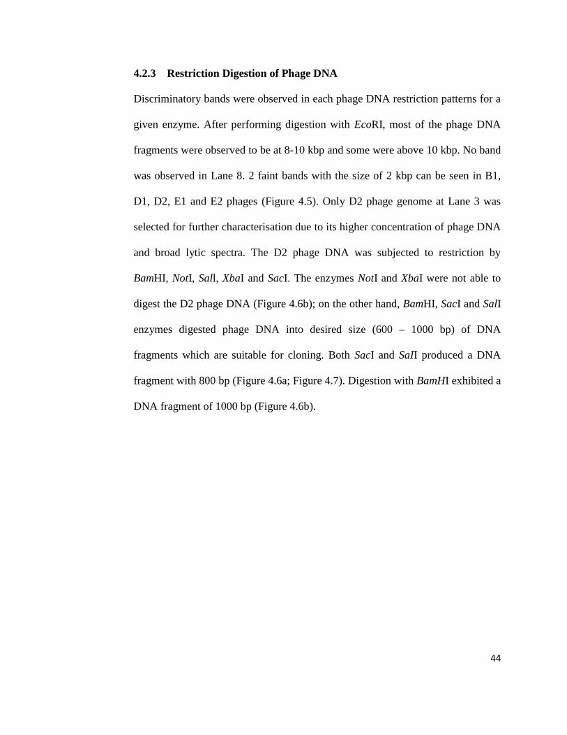
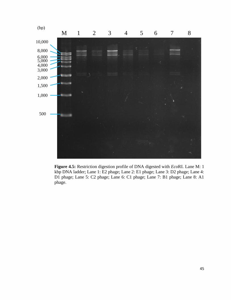
4.2.3 Restriction Digestion of Phage DNA
Discriminatory bands were observed in each phage DNA restriction patterns for a
given enzyme. After performing digestion with EcoRI, most of the phage DNA
fragments were observed to be at 8-10 kbp and some were above 10 kbp. No band
was observed in Lane 8. 2 faint bands with the size of 2 kbp can be seen in B1,
D1, D2, E1 and E2 phages (Figure 4.5). Only D2 phage genome at Lane 3 was
selected for further characterisation due to its higher concentration of phage DNA
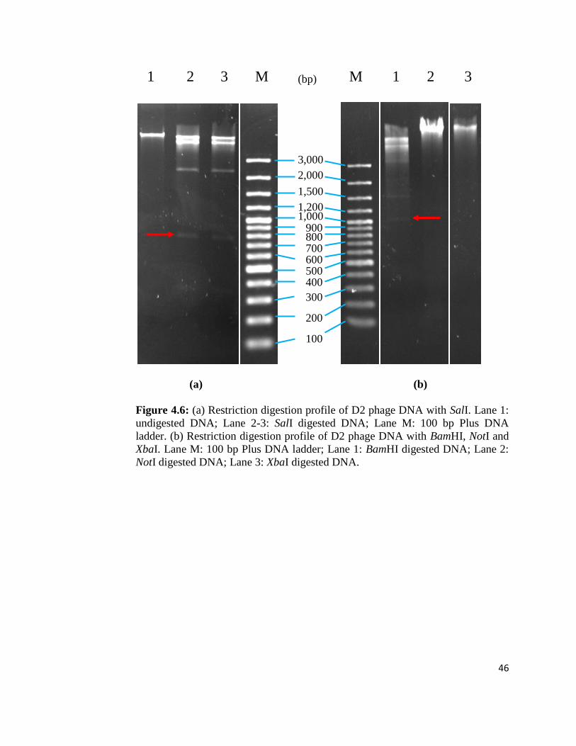
and broad lytic spectra. The D2 phage DNA was subjected to restriction by
BamHI, NotI, Sall, XbaI and SacI. The enzymes NotI and XbaI were not able to
digest the D2 phage DNA (Figure 4.6b); on the other hand, BamHI, SacI and SalI
enzymes digested phage DNA into desired size (600 – 1000 bp) of DNA
fragments which are suitable for cloning. Both SacI and SaII produced a DNA
fragment with 800 bp (Figure 4.6a; Figure 4.7). Digestion with BamHI exhibited a
DNA fragment of 1000 bp (Figure 4.6b).
45
Figure 4.5: Restriction digestion profile of DNA digested with EcoRI. Lane M: 1
kbp DNA ladder; Lane 1: E2 phage; Lane 2: E1 phage; Lane 3: D2 phage; Lane 4:
D1 phage; Lane 5: C2 phage; Lane 6: C1 phage; Lane 7: B1 phage; Lane 8: A1
phage.
M 1 2 3 4 5 6 7 8
10,000
8,000
6,000 5,000 4,000 3,000
2,000
1,500
1,000
500
(bp)
46
1 2 3 M M 1 2 3
(a) (b)
Figure 4.6: (a) Restriction digestion profile of D2 phage DNA with SalI. Lane 1:
undigested DNA; Lane 2-3: SalI digested DNA; Lane M: 100 bp Plus DNA
ladder. (b) Restriction digestion profile of D2 phage DNA with BamHI, NotI and
XbaI. Lane M: 100 bp Plus DNA ladder; Lane 1: BamHI digested DNA; Lane 2:
NotI digested DNA; Lane 3: XbaI digested DNA.
3,000
2,000
1,500
1,200 1,000
900 800 700 600 500 400
300
200
100
(bp)

47
M 1
Figure 4.7: Restriction digestion profile of D2 phage DNA with SacI. Lane M:
100 bp DNA ladder; Lane 1: SacI digested DNA.
3,000
2,000
1,500
1,000
800 700 600 500 400 300
200
100
(bp)
48
CHAPTER 5
DISCUSSION
5.1 Isolation of Bacteriophages
All the water samples were collected together with the sediment because virus
present in higher number in bulk sediment compared to pore water (Drake et al.,
1998). Most viruses will be adsorbed to the sediments in particle rich environment
and only a small portion of them will be found in pore water. Once viruses are
adsorbed to sediment, they are immobilised, which may lead to accumulation and
concentration of viruses in the sediments (De Flora et al., 1975). Therefore, in
order to isolate bacteriophage successfully, sediments which include both the
particle-adsorbed virus and virus in pore water were collected in this study.
Many phages were isolated from sample collection sites C, D and E and the
number of plaques formed were too numerous to be enumerated. Phages isolated
from sample collection site A were observed to be lower in number (22 plaques),
followed by B with the least number (17 plaques). Both the water samples A and
B were collected from river. The flowing water in the river might constantly
dilute the phage with fresh water source, hence, leads to lower number of phages
isolated. In contrast, the water in lake, drain and pond remains stagnant, therefore,
higher number of phages will be concentrated in this region. Apart from that,
bacteria in fresh flowing water environments in sampling sites A and B may be

49
concentrated at the surface of solids rather than in the overlaying water, thereby
lowering down the number of planktonic bacteria needed for replication of phages
(Goyal et al., 1987). The number of phages will then be reduced. Furthermore, A
and B sites may have insufficient food source or nutrient required for growth of
Shigella flexneri. Abundant food source for bacteria propagation in water samples
from sites C, D and E might serve as rich bacterial pool that provides the
necessary host for the phage to grow.
Sunlight, specifically ultraviolet (UV) light damages viral genomic element
beyond repair, thus, causes viral decay in aquatic environment (Kirchman 2012).
Sunlight inactivation was significant down to a depth of 200 m. UV-A (320 to
400 nm) has the greatest effect (Murray and Jackson 1993). The penetration of
sunlight depends on the water clarity and depth (Davies-Colley et al., 1994). The
sampling sites A and B are believed to have higher penetration of sunlight due to
the clear and fresh flowing water in shallow river, which directly exposes the
phages to sunlight. The UV light may inactivate the phages present in these sites.
In accordance to research work done by Chandra et al. (2011), direct sun radiation
is deleterious to the phage survival. Sunlight has inactivation effect on various
faecal indicator bacteria and bacteriophages (Sinton et al., 1999; Sinton et al.,
2002). Phage inactivation in sunlight-exposed environment causes damage to both
the capsid and nucleic acid genome (Sinton et al., 1999). The UV in sunlight can
cause 5% loss in viable phage per hour for surface water due to the formation of
thymine dimers (Wommack et al., 1996).
50
In addition, the concentration of dissolved oxygen (DO) will be reduced. As water
temperature increases, the water’s capacity to hold oxygen decreases (United
States Environmental Protection Agency [EPA], 2012). Low level of dissolved
oxygen can decrease bacterial propagation. Additionally, high temperature will
also decrease infectivity of phages (Kinnunen 1978). Thus, low number of phages
can be isolated from sampling sites A and B.
Unlike samples A and B, the water samples taken from sample collection sites D
and E contain abundant amount of algae floating in it. The algae have symbiotic
relationship with the bacteria. Photosynthetic algae convert large amount of
carbon dioxide into oxygen, thereby releasing more oxygen for the growth and
survival of bacteria. Bacteria respiration in turn provides carbon dioxide needed
for microalgae photosynthesis (Su 2012). The concentration of dissolved oxygen
increases during algal photosynthesis. The high concentration of dissolved oxygen
promotes the growth of bacteria. Other than that, large amount of algae also
provide an adsorption site for phage which protects the phage from inactivating
factor in environment such as sunlight. As a result, higher phage count can be
obtained from sample collection sites D and E.
The density of both phages and their host bacteria are important criteria for the
multiplication of phages. Phage multiplication depends on host bacterial growth
(Uchiyama et al., 2007). At low level of host bacteria in A and B sites, a phage
has low chance of encountering a susceptible host, hence, productive infection

51
may not occur. A successful phage replication generally requires at least 104 host
bacteria per mL (Goyal et al., 1987). Hence, it can be deduced that low density of
host bacteria will lead to low chance of isolation of phages. Besides, the presence
of Shigella flexneri phages in water may also used as a possible indicator of the
presence of host bacterium. Therefore, it can be known that the sample collection
sites C, D and E may have more bacteria than A and B sites according to the
higher number of phages isolated. Drain, pond and lake of sampling site C, D and
E are more prone to contamination by human activity. So, the existence of S.
flexneri bacterium in these environmental waters may possibly caused by faecal
contamination.
Bacteriophages infective to the Shigella flexneri can be isolated from all the water
samples. The most probable reason is using the S. flexneri as enrichment cultures
for phage isolation. When S. flexneri is added into the environmental sample, the
phage that is infective towards the bacteria will outnumber other phages that have
limited or no infectious activity. Therefore, it is easier to isolate the phages for
certain bacterial strains that are being introduced in enrichment cultures
(Stenholm et al., 2008).
Based on the plaque morphology obtained in plaque assay, all the phage isolates
produced clear plaques, indicating all bacteria in the plaque zone are being lysed.
Thus, these phages were known to be lytic where large numbers of phage progeny
were released to cause lysis (rupture) of the host cell. Furthermore, the phages did

52
not give plaques which were uniform in size. The heterogeneity of plaque size
exhibited by these phages is an indication of more than one phage type present in
the water sample. This is because a slowly replicating phage or one which
produces low number of infective progeny particles, will tend to form a smaller
plaque compared to the rapidly replicating phage (Irving et al., 1990). Another
factor that affects the plaque size is the physical size of the phage. A small phage
will diffuse through the semi-solid agar more easily and quickly than the large
one, therefore, larger plaque can be formed (Irving et al., 1990). This is the
preliminary test to distinguish or relate the different phages on the basis on their
plaque morphology.
5.2 Host Specificity Test
The 8 isolated bacteriophages were examined for their ability to lyse multiple host
species. It was determined that D2 phage has broader host range compared to
other phage isolates. This phage has the ability to lyse other bacterial strains
which includes enterotoxigenic Escherichia coli (ETEC) and enteropathogenic
Escherichia coli (EPEC). Besides, C2 and D1 phages produced a faint zone on the
lawn of EPEC and ETEC respectively. Clear zone produced by D2 phage
demonstrates complete lysis, whereas faint zone from C2 and D1 phages indicates
minimal lysis. The clarity of plaques showed that D2 phage has a stronger lytic
capability compared to the other two phages. This test illustrated that C2, D1 and
D2 phages can infect more than one bacteria genus even though most of the

53
phages display host specificity to a particular bacterial species and strain. This
phenomenon may due to the relaxed host specificity of bacteriophages, thus,
allowing productive interaction with several prey genus (Jensen et al., 1998).
These phages that are able to infect various bacterial host genus can easily
encounter a susceptible prey and replicate in it. On the other hand, the inability of
A1, B1, C1, E1 and E2 phages to lyse strains of bacteria of other genera confirms
their generic specificity.
C2, D1 and D2 phages were infectious towards Escherichia genus because of the
common characteristics shared between Shigella strains and members of the
genus Escherichia. Their genetic relatedness suggested that Shigella belongs to
the diverse species Escherichia coli. Shigella is phylogenetically similar to E.
coli, indicating Shigella flexneri and E. coli are closely related. Shigella and E.
coli are shown to be genomically indistinguishable at the species level in DNA
hybridization studies. These studies have proved that Shigella species and
members of E. coli are belonged to the common ancestor (Jin et al., 2002; Lan
and Reeves 2002; Wei et al., 2003). Therefore, the host range of the phages shows
a correlation with the degree of relatedness where those bacterial strain closely
related to Shigella flexneri are generally susceptible to the Shigella phages, while
the more distantly related species are resistant to these phages.
D2 phage is a potential candidate for therapeutic application due to its broad host
range and strong lytic capability. These properties are desirable for the biocontrol

54
of pathogenic bacterial infections or foodstuff treatment without pre-analysis of
sensitivity of target host cell to a given bacteriophages (Bielke et al., 2007).
Hence, much attention was given for D2 phage in present study.
López-Cuevas et al. (2011) hypothesised that the differences of host range might
be due to environmental origin of bacteria tested in which a loss of bacteriophage
receptors may have happened as a result of antagonistic co-evolution between
bacterium and bacteriophage. Apart from that, it could also be due to bacterial
receptor mutation or degradation caused by restriction or modification of the
resistance bacterial system which prevents the adsorption of phage to host
bacteria.
The standard method of bacteriophage enrichment was used in this project to
increase the number of Shigella flexneri that serves as the host for specific phage
to multiply, hence, phages can be easily isolated from the sample. However, this
method may favour bacteriophages to possess a more limited host range (Jensen
et al., 1998). Jensen et al. (1998) suggested that probability to isolate broad host
range bacteriophage can be increased by using two bacterial host species in
isolation protocol instead of one. Cross infection between S. flexneri phage and
Escherichia coli host may provide an alternate host for the multiplication of
phage. So, if E. coli and S. flexneri bacteria are added together for bacteriophage
enrichment, the probability of phage isolation will increase with the presence of
multiple susceptible hosts. The potential to infect multiple hosts would maximise
55
opportunities for effective phage multiplication since the phage is more likely to
encounter suitable prey.
The C2, D1 and D2 isolated phages in this study are most likely indigenous phage
of Escherichia coli. In other words, they are possibly E. coli phages which able to
co-infect Shigella flexneri bacterium due to their broad lytic spectrum. E. coli
with shorter doubling time (20 min) is more abundant in the environmental setting
compared to S. flexneri (doubling time, about 40 min) (Maloy et al., 1994;
Lucchini et al., 2005). Phage replicates faster on the more abundant, rapidly
growing host population, where new hosts can be found more easily. This applied
for the concept of “killing the winner populations” (Thingstad and Lignell 1997).
Thus, isolation of these phages could not conclude the presence of S. flexneri in
the water sample since E. coli can be used as the host for these phages to
replicate.
5.3 Molecular Characterisation
5.3.1 Phage DNA Extraction
This method was done to verify extraction yield and absence of bacterial genomic
DNA. The gel image shows that the phage DNA was successfully extracted using
phenol chloroform method and it was free of genomic contamination. It can be
observed that the genome size of all the bacteriophages were above 3,000 bp. The
banding pattern was clear showing high concentration of phage DNA extracted.

56
By visualising the band pattern, it was shown that the concentration and purity of
the extracted DNA are high and suitable to proceed for restriction digestion
analysis. This also demonstrates that polyethylene glycol (PEG) 6000 used for the
purification and concentration of phage particles was useful and effective in
obtaining highly purified phage preparations. This method is independent of
phage concentration where phages can be concentrated even with low titer lysates.
It is also fast which provides a 100-fold phage concentration after low speed
centrifugation with little loss of infectivity (Boulanger 2009). As a result, both
PEG 6000 and phenol chloroform method have proved to produce high yields and
purity DNA, hence, these methods are reliable to be used for future research work
or molecular study.
5.3.2 Restriction Digestion of Phage DNA
Differences between phages were confirmed by comparison between the
restriction endonuclease cleavage patterns of the phage DNA. Based on the
digestion pattern by EcoRI, all the phages have similar restriction patterns
especially B1, D1, D2, E1 and E2 phages with an extra band at 2,000 bp. This
indicated that they are possibly the same bacteriophage which belonged to the
same strain. However, these phages presented different host ranges. This
demonstrates that similarity in genotypic characteristics does not imply similarity
in other phenotypic characteristics of the phages. Likewise, similar host range
does not imply that phages are genetically related. This observation is supported
57
by Stenholm et al. (2008) that showed no indication of a relation between lytic
potential and the genome sizes or the morphological characteristics of the phages
tested. The most probable factor is the differences in phage receptor properties
between host strains. Surprisingly, no band can be seen in Lane 8 loaded with A1
phage. This phenomenon might due to the contamination of DNase that causes
degradation of phage DNA.
The D2 phage digestion pattern by BamHI was not very clear, probably due to
overlapped bands. The inability to digest phage DNA by NotI and XbaI enzymes
could be due to the absence of target sites for the restriction enzyme tested
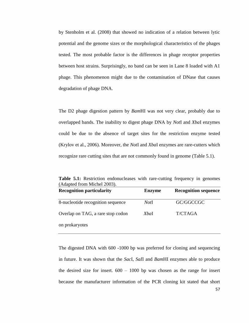
(Krylov et al., 2006). Moreover, the NotI and XbaI enzymes are rare-cutters which
recognize rare cutting sites that are not commonly found in genome (Table 5.1).
Table 5.1: Restriction endonucleases with rare-cutting frequency in genomes
(Adapted from Michel 2003).
Recognition particularity Enzyme Recognition sequence
8-nucleotide recognition sequence NotI GC/GGCCGC
Overlap on TAG, a rare stop codon XbaI T/CTAGA
on prokaryotes
The digested DNA with 600 -1000 bp was preferred for cloning and sequencing
in future. It was shown that the SacI, SaII and BamHI enzymes able to produce
the desired size for insert. 600 – 1000 bp was chosen as the range for insert
because the manufacturer information of the PCR cloning kit stated that short

58
DNA fragments (<1 kb) are cloned with a higher efficiency compared to long one
(Thermo Scientific 2012). The efficiency of cloning depends on insert size.
Larger inserts are amenable to cloning in high-copy number vectors, however, at a
lower efficiency (Invitrogen Corporation 2013). In addition, inserts with the same
size as vector will compete equally with other vector molecules for ligation. So, if
the inserts are smaller than the vector molecules, they will outcompete other
vectors that exist (Nybo 2011). 600 bp was decided as the lower limit because
insert size that is too small will reduce specificity and accuracy of sequence
identification. Short fragments contain less sequence information, hence, it is hard
to be identified and compared. A few more sequence added may contain the key
information that define and represent the feature of a particular phage.
5.4 Limitations and Further Study
The major problem encountered was the high contamination risk obtained from
environmental sample. There is wide variety of bacteria present in the
environmental sample despite the desired phage and host bacteria. This unrelated
bacteria or contaminant can affect the accuracy and reliability of result. The
growth of this contaminants cause difficulty in the inspection of plaque formation.
This poses problem for the isolation and selection of plaque. Centrifugation and
filtration through 0.45 µM cellulose acetate syringe filter were not sufficient to
eliminate the bacterial debris and particulates completely. Instead, chloroform can
59
be added to eliminate all viable bacterial cells so that the phage obtained is more
purified (Jensen et al., 1998).
As shown in the plaque assay, the phage titre for the sample collection sites C, D
and E was too high. The plaques formed were found to be close to each other and
they were nearly joined together. When the number of plaques is too numerous,
there is high possibility to pick up the neighbouring plaques. The chances of
contamination by other phages will therefore increase. To overcome this problem,
more serial dilution should be carried out to dilute the phage to achieve low
plaque count, preferably 30 – 300 pfu (plaque forming units) (Panec and Katz
2003). In addition, phages can also be plated repeatedly to assure similar phage
plaque parameters are obtained.
There were only 6 bacterial strains used for the host specificity test in this study.
This limited number of bacterial strains was insufficient to determine the host
range of phages. For successful therapeutic application of bacterial infection, a
group of broad host range phages are needed in order to cover a possible broad
spectrum of potential pathogenic host strains involved in an outbreak of disease.
Hence, more bacterial strains from Shigella species or even other genera,
particularly antibiotic resistant strains, should be included in this test to help in the
selection of phage candidate for phage therapy.

60
Apart from that, use of only one restriction enzyme, EcoRI, in this study was
inadequate to distinguish and relate the isolated phages. In future, more restriction
enzymes should be used in restriction endonuclease analysis to determine the
genetic variability of the isolated phages and to improve the reliability of the
result. Molecular analysis such as cloning and sequencing can also be used to
identify the phage of interest. Molecular analysis normally produces more rapid,
sensitive and reproducible result.
In addition, the banding pattern obtained for restriction analysis was not clear and
the bands were extremely thin because of low concentration of DNA used for the
analysis. The faint bands cause difficulty in the estimation of size. Perhaps more
DNA can be used in future to obtain a clear and strong band. Moreover, the
phages isolated in this study were found to be above 3000 bp. However, the actual
genome size of the phage was not known due to the small size marker used. In
future, larger size marker should be used to estimate the size of the phage.
Detailed characterisation of phage properties is a prerequisite for evaluating the
potential of phages as biocontrol agent. The newly isolated phages need to be
examined in detail especially when they are intended to be used for therapeutic
application. Therefore, more characterisation tests should be carried out in future
to analyse the isolated phages. These include visualisation of phage morphology
using transmission electron microscope. Based on morphological characteristics,
the phages can then be classified to families following the guidelines of the

61
International Committee on Taxonomy of Viruses. Besides, one-step phage
growth curve can be performed to determine the lytic ability, adsorption time,
latent period, exponential growth and burst size of the phages. This method is
useful to study the properties of phages which are going to be used as controller
of a pathogenic host. The heat and pH stability of phages can also be tested to
study the adaptability of the phages in adverse environmental conditions. In
addition, sodium dodecyl sulfate-polyacryamide gel electrophoresis (SDS-PAGE)
profile could be carried out to analyse the protein composition of the phages. This
protein profile provides additional information to differentiate the isolated phages.
The D2 phage isolated in the present study may be an ideal therapeutic phage
candidate due to its broad host specificity and strong infectious ability. However,
more identification and characterisation tests need to be done to examine the
ability of this newly isolated phage in overcoming outbreaks and spread of
shigellosis. Same as drug, its form and mode of action have to be known in detail.
One of the effective methods is genomic analysis that provides assessment for the
safety of phage by examining the presence or absence of undesirable genes such
as antibiotic resistance and virulence factor. Other than that, the therapeutic
effectiveness of the phage can be tested in vivo because the bacteriocidal
effectiveness is different in vitro and in vivo.

62
CHAPTER 6
CONCLUSION
In conclusion, all of the bacteriophage isolates collected from Kampar area were
able to infect Shigella flexneri. All the 8 isolated phages (A1, B1, C1, C2, D1, D2,
E1 and E2) are virulent phages which produced round, clear and medium sized
(1.0-3.0 mm in diameter) plaques. Sample collection sites C, D and E have higher
plaque count compared to A and E due to the environmental factor. It has shown
that sampling sites C, D and E provide favourable condition for the growth of
phage. Based on the number of phages isolated, higher number of host bacteria
population was present in C, D and E than A and B.
In addition, C2, D1 and D2 bacteriophages possessed broad spectrum of
infectious activity while other bacteriophages have narrow host range. D2 phage
was able to lyse enteropathogenic E. coli (EPEC) and enterotoxigenic E. coli
(ETEC) while C2 and D1 phages have lysogenic effect against EPEC and ETEC
respectively. Thus, these bacteriophages were able to infect bacterial strains of
other genus. Conversely, the inability of A1, B1, C1, E1 and E2 phages to lyse
strains of bacteria of other genera confirms their generic specificity.
Based on the digestion pattern by EcoRI, B1, D1, D2, E1 and E2 phages were
found to have similar restriction patterns. This indicated that they are most

63
probably belonged to the similar phage lineages. However, these bacteriophages
have different host specificity. This demonstrates that similarity in genotypic
characteristics does not reflect similar phenotypic characteristics among the
phages. Moreover, SacI, SalI and BamHI enzymes were able to digest phage
DNA into desirable size of DNA fragments (600 – 1000 bp) that are required for
cloning and sequencing in future.
D2 phage isolated in this study was suggested to be a potential candidate for
therapeutic application due to its broad host range and strong lytic capability.
Nevertheless, more identification and characterisation tests should be done to
confirm this hypothesis.

64
REFERENCES
Abedon, S.T., 2006. Phage ecology. In: Calender, R. (eds.). The Bacteriophages.
New York: Oxford University Press, pp. 37 - 46.
Abedon, S.T., 2011. Size does matter-distinguishing bacteriophages by genome
length (and ‘breadth’). Microbiology Australia, 32(2), pp. 90 – 91.
Ackermann, H.W., 2005. Bacteriophage classification. In: Kutter, E. and
Sulakvelidze, A. (eds.). Bacteriophages: Biology and Applications. Florida: CRC
Press, pp. 67 – 89.
Al-Hasani, K. et al., 2001. Genetic organization of the she pathogenicity island in
Shigella flexneri 2a. Microbial Pathogenesis, 30(1), pp. 1 – 8.
Allison, G.E. and Verma, N.K., 2000. Serotype-converting bacteriophages and O-
antigen modification in Shigella flexneri. Trends in Microbiology, 8(1). pp. 17 –
23.
Anany, H. et al., 2011. A Shigella boydii bacteriophage which resembles
Salmonella phage Vil. Virology Journal, 8, pp. 242.
Anpilov, L.I. and Prokudin, A.A., 1984. Preventive effectiveness of dried
polyvalent Shigella bacteriophage in organized collective farms. Voen Med Zh, 5,
pp. 39 – 40.
Ashelford, K.E., Martin, J.D. and John, C.F., 2003. Elevated abundance of
bacteriophage infecting bacteria in soil. Applied and Environmental
Microbiology, 69(1), pp. 285 – 289.
Babalova, E.G. et al., 1968. Preventive value of dried dysentery bacteriophage. Zh
Mikrobiol Epidermiol Immunobiol, 45(2), pp. 143-145.

65
Barksdale, L. and Arden, S.B., 1974. Persisting bacteriophage infections,
lysogeny, and phage conversions. Annual Review of Microbiology, 28, pp. 265 –
300.
Bergh, Ø. et al., 1989. High abundance of viruses found in aquatic environments.
Nature, 340, pp. 467 – 468.
Bielke, L., 2007. Salmonella host range of bacteriophages that infect multiple
genera. Poultry Science, 86, pp. 2536 – 2540.
BIORON, 2013. Restriction endonucleases [Online]. Available at:
http://www.bioron.net/en/products/restriction-endonucleases/ [Accessed: 2 April
2013].
Bliss, J.M. et al., 2003. Differentiation of Candida albicans and Candida
dubliniensis by using recombinant human antibody single-chain variable
fragments specific for hyphae. Journal of Clinical Microbiology, 41(3), pp. 1152
– 1160.
Boulanger, P., 2009. Purification of bacteriophages and SDS-PAGE analysis of
phage structural proteins from ghost particles. In: Clokie, M.R.J. and Kropinski,
A.M. (eds.). Bacteriophages: Methods and Protocols, Volume 2: Molecular and
Applied Aspects. United Kingdom: Humana Press, pp. 227 - 238.
Breitbart, M. et al., 2002. Genomic analysis of uncultured marine viral
communities. Proceedings of the National Academy of Sciences of the United
States of America, 99(22), pp. 14250 – 14255.
Brüssow, H. and Kutter, E., 2005. Phage ecology. In: Kutter, E. and Sulakvelidze,
A. (eds.). Bacteriophages Biology and Application. Florida: CRC Press, pp. 129 –
163.
Brüssow, H., 2005. Phage therapy: the Escherichia coli experience. Microbiology,
151(7), pp. 2133 – 2140.
66
Carey-Smith, G.V. et al., 2006. Isolation and characterization of bacteriophages
infecting Salmonella spp. Federation of European Microbiological Societies, 258,
pp. 182 – 186.
Carlton., R.M., 1999. Phage therapy: past history and future prospects. Archivum
Immunologiae et Therapiae Experimentalis, 47, pp. 267 – 274.
Ceyssens, P.J., 2009. Isolation and characterisation of lytic bacteriophages
infecting Pseudomonas aeruginosa. PhD thesis, Katholieke Universiteit Leuven,
Belgium.
Chandra, M. et al., 2011. Isolation of a bacteriophage against Salmonella Dublin
and determination of its physical resistance under varied in vitro conditions.
African Journal of Microbiology Research, 5(15), pp. 2044 – 2047.
Clark, J.R. and March, J.B., 2006. Bacteriophages and biotechnology: vaccines,
gene therapy and antibacterials. Trends in Biotechnology, 24(5), pp. 212 – 218.
Davies-Colley, R., Bell, R.G. and Donnison, A.M., 1994. Sunlight inactivation of
enterocci and fecal coliforms in sewage effluent diluted in seawater. Applied and
Environmental Microbiology, 60(6), pp. 2049 – 2058.
De Flora, S., De Renzi, G.P. and Badolati, G., 1975. Detection of animal viruses
in coastal seawater and sediments. Applied Microbiology, 30(3), pp. 472 – 475.
Dickerson, T.J., Kaufmann, G.F. and Janda, K.D., 2005. Bacteriophage-mediated
protein delivery into the central nervous system and its application in
immunopharmacotherapy. Expect Opinion on Biological Therapy, 5(6), pp. 773 –
781.
Drake, L.A. et al., 1998. Vertical profiles of virus-like particles and bacteria in the
water column and sediments of Chesapeake Bay, USA. Aquatic Microbial
Ecology, 16, pp. 17 – 25.
67
DuPont, H.L. et al., 1989. Inoculum size in shigellosis and implications for
expected mode of transmission. The Journal of Infectious Disease, 159(6), pp.
1126 – 1128.
European Food Safety Authority, 2009. The use and mode of action of
bacteriophages in food production. EFSA Journal, 1076, pp. 1 – 26.
European Molecular Biology Laboratory-European Bioinformatics Institute,
2013. Bacteria Genomes – Shigella flexneri [Online]. Available at:
http://www.ebi.ac.uk/2can/genomes/bacteria/Shigella_flexneri.html [Accessed: 17
March 2013].
Faruque, S.M. et al., 2003. Shigella dysenteriae type 1-specific bacteriophage
from environmental waters in Bangladesh. Applied and Environmental
Microbiology, 69(12), pp. 7028 – 7031.
Freeman, V.J., 1951. Studies on the virulence of bacteriophage-infected strains of
Corynebacterium diphtheria. Journal of Bacteriology, 61, pp. 765 – 688.
Frost, J.A. et al., 1993. Phage typing of Vero-cytotoxin (VT) producing
Escherichia coli O157 isolated in the United Kingdom. Epidemiology and
Infection, 103(1), pp. 73 – 81.
García, P. et al., 2010. Food biopreservation: promising strategies using
bacteriocins, bacteriophages and endolysins. Trends in Food Science &
Technology, 21(8), pp. 373 – 382.
Goyal, S.M., Gerba, C.P. and Bitton, G., 1987. Phage Ecology, 1st ed. New York:
John Wiley and Sons.
Gu, B. et al., 2012. Comparison of the prevalence and changing resistance to
nalidixic acid and ciprofloxacin of Shigella between Europe-America and Asia-
Africa form 1998 to 2009. International Journal of Antimicrobial Agents, 40(1),
pp. 9 – 17.

68
Guttman, B., Raya, R. and Kutter, E., 2005. Basic phage biology. In: Kutter, E.
and Sulakvelidze, A. (eds.). Bacteriophages: Biology and Applications. Florida:
CRC Press, pp. 29 – 66.
Hagens, S. and Loessner, M.J., 2007. Application of bacteriophages for detection
and control of foodborne pathogens. Applied Microbiology and Biotechnology,
76(3), pp. 513 – 519.
Haq, et al., 2012. Bacteriophages and their implications on future biotechnology:
a review. Virology Journal, 9, pp. 9.
Hausler, T., 2007. Viruses vs. superbugs: a solution to the antibiotics crisis?.
London: Macmillan.
Hyman, P. and Abedon, S.T., 2009. Bacteriophage (overview). In: Encyclopedia
of Microbiology. Oxford: Elsevier, pp. 322 – 338.
Hyman, P. and Abedon, S.T., 2012. Smaller fleas: viruses of microorganisms.
Hindawi Publishing Corporation, doi: 10.6064/2012/734023.
Invitrogen Corporation, 2013. PCR cloning considerations [Online]. Available at:
http://tools.invitrogen.com/content/sfs/appendix/Cloning_Trans/PCR%20Cloning
%20Considerations.pdf [Accessed: 20 March 2013].
Irving, T.E., Barnes, M. and Ellis, N., 1990. Surrogate viral indicators.
Buckinghamshire: Water Research Centre.
Jennison, A.V. and Verma, N.K., 2004. Shigella flexneri infection: pathogenesis
and vaccine development. FEMS Microbiology Reviews, 28 (1), pp. 43-58.
Jennison, A.V. and Verma, N.K., 2007. The acid-resistance pathways of Shigella
flexneri 2457T. Microbiology, 153, pp. 2593 – 2602.
69
Jensen, E.C. et al., 1998. Prevalence of borad-host-range lytic bacteriophaegs of
Sphaerotilus natans, Escherichia coli, and Pseudomonas aeruginosa. Applied and
Environmental Microbiology, 64(2), pp. 575 – 580.
Jensen, M.A., Faruque, S.M., Mekalanos, J.J. and Levin, B.R., 2006. Modelling
the role of bacteriophage in the control of cholera outbreaks. Proceedings of the
National Academy of Sciences of the United States of America, 103(12), pp. 4652
– 4657.
Jin, Q. et al., 2002. Genome sequence of Shigella flexneri 21: insights into
pathogenicity through comparison with genomes of Escherichia coli K12 and
O157. Nucleic acid Research, 30(20), pp. 4432 – 4441.
Kinnunen, K., 1978. Tracing water movement by means of Escherichia coli
bacteriophages. Finland: Water Research Institute.
Kirchman, D.L., 2012. Ecology of viruses. In: Processes in Microbial Ecology.
New York: Oxford University Press, pp. 137 – 155.
Kokjohn, T.A., Sayler, G.S. and Miller, R.V., 1991. Attachment and replication of
Pseudomonas aeruginosa bacteriophages under conditions simulating aquatic
environments. Journal of General Microbiology, 137, pp. 661 – 666.
Kotloff, K.L. et al., 1999. Global burden of Shigella infections: implications for
vaccine development and implementation of control strategies. Bulletin of the
World Health Organization, 77(8), pp. 651 – 666.
Kropinski, A.M., 2009. Enumeration of bacteriophages by double agar overlay
plaque assay. In: Clokie, M.R.J. and Kropinski, A.M. (eds.). Bacteriophages:
Methods and Protocols, Volume 1: Isolation, Characterization and Interactions.
United Kingdom: Humana Press, pp. 69 - 76.
Krylov, V. et al., 2006. Ambivalent bacteriophages of different species active on
Escherichia coli K12 and Salmonella sp. strains. Russian Journal of Genetics,
42(2), pp. 106 – 114.
70
Lan, R. and Reeves, P.R., 2002. Escherichia coli in disguise: molecular origins of
Shigella. Microbes and Infection, 4, pp. 1125 – 1132.
Leiman, P.G. et al., 2004. Three-dimensional rearrangement of proteins in the tail
of bacteriophage T4 on infection of its host. Cell Press, 118, pp. 419 – 429.
López-Cuevas, O. et al., 2011. Characterization of bacteriophages with a lytic
effect on various Salmonella serotypes and Escherichia coli O157:H7. NRC
Research Press, 57, pp. 1042 – 1051.
Lucchini, S. et al., 2005. Transcriptional adaptation of Shigella flexneri during
infection of macrophages and epithelial cells: insights into the strategies of a
cytosolic bacterial pathogen. Infection and Immunity, 73(1), pp. 88 – 102.
Mahaffy, J.M., 2006. Modeling marine phage ecology [Online]. Available at:
http://www-rohan.sdsu.edu/~jmahaffy/docs/ubc_phage1_05.pdf [Accessed: 17
March 2013].
Maloy, S.R., Cronan, J.E. and Freifelder, D.M., 1994. Bacteria. In: Microbial
Genetics. 2nd ed. Sudbury: Jones and Bartlett Publishers, pp. 67 – 80.
Mandal, J. et al., 2012. The recent trends of shigellosis: a JIPMER perspective.
Journal of Clinical and Diagnostic Research, 6(9), pp. 1474 – 1477.
Michel, J.G., 2003. Genomes, mapping of bacterial. In: The Desk Encyclopedia of
Microbiology. California: Elsevier Academic Press, pp. 537 – 546.
Molineux, I.J., 2001. No syringes please, ejection of phage T7 DNA from the
virion is enzyme driven. Molecular Microbiology, 40(1), pp. 1 – 8.
Moss, J.E. et al., 1999. The SelC-associated SHI-2 pathogenicity island of
Shigella flexneri. Molecular Microbiology, 33, pp. 74 – 83.

71
Murray, A.G. and Jackson, G.A., 1993. Viral dynamics II: a model of the
interaction of ultraviolet light and mixing processes on virus survival in seawater.
Marine Ecology Progress Series, 102, pp. 105 – 114.
Nybo, K., 2011. Cloning efficiency. BioTechniques, 51(5), pp. 305 – 307.
O’Flynn, G. et al., 2004. Evaluation of a cocktail of three bacteriophages for
biocontrol of Escherichia coli O157:H7. Applied and Environmental
Microbiology, 70(6), pp. 3417 – 3424.
Oliveira, A. et al., 2009. Isolation and characterization of bacteriophages for avian
pathogenic E. coli strains. Journal of Applied Microbiology, 106, pp. 1919 –
1927.
Panec, M. and Katz, D.S., 2013. Plaque assay protocols [Online]. Available at:
http://www.microbelibrary.org/component/resource/laboratory-test/3073-plaque-
assay-protocols [Accessed: 2 April 2013].
Petrenko, V.A. and Vodyanoy, V.J., 2003. Phage display for detection of
biological threat agents. Journal of Microbiological Methods, 53, pp. 253 – 262.
Philpott, D.J., Edgeworth, J.D. and Sansonetti, P.J., 2000. The pathogenesis of
Shigella flexneri infection: lessons from in vitro and in vivo studies. The Royal
Society, 355, pp. 575 – 586.
Pruneda, R.C. and Farmer, J.J., 1977. Bacteriophage typing of Shigella sonnei.
Journal of Clinical Microbiology, 5(1), pp. 66 – 74.
Rader, C. and Barbas, C.F., 1997. Phage display of combinatorial antibody
libraries. Current Opinion in Biotechnology, 8(4), pp. 503 – 508.
Raya, R.R. and Hébert, E.M., 2009. Isolation of phage via induction of lysogens.
In: Clokie, M.R.J. and Kropinski, A.M. (eds.). Bacteriophages: Methods and
Protocols, Volume 1: Isolation, Characterization and Interactions. United
Kingdom: Humana Press, pp. 23 - 32.

72
Rees, C.E.D. and Loessner, M.J., 2005. Phage for the detection of pathogenic
bacteria. In: Kutter, E. and Sulakvelidze, A. (eds.). Bacteriophages: Biology and
Application. Florida: CRC Press, pp. 267 – 284.
Sambrook, J. and Russell, D.W., 2001. Purification of nucleic acids by extraction
with phenol:chloroform. In: Molecular Cloning. 3rd ed. New York: Cold Spring
Harbor Laboratory Press.
Sinton, L.W. et al., 2002. Sunlight inactivation of fecal indicator bacteria and
bacteriophages from waste stabilization pond effluent in fresh and saline waters.
Applied and Environmental Microbiology, 68(3), pp. 1122 – 1131.
Sinton, L.W., Finlay, R.K. and Lynch, P.A., 1999. Sunlight inactivation of fecal
bacteriophages and bacteria in sewage-polluted seawater. Applied and
Environmental Microbiology, 65(8), pp. 3605 – 3613.
Small, P. et al., 1994. Acid and base resistance in Escherichia coli and Shigella
flexneri: role of rpoS and growth pH. Journal of Bacteriology, 176(6), pp. 1729 –
1737.
Stenholm, A.R., Dalsgaard, I. and Middelboe, M., 2008. Isolation and
characterization of bacteriophages infecting the fish pathogen Flavobacterium
psychrophilum. Applied and Environmental Microbiology, 74(13), pp. 4070 –
4078.
Su, Y., 2012. Settleable algal-bacterial culture for municipal wastewater
treatment. PhD thesis, University Lüneburg, Germany.
Sulakvelidze, A. and Kutter, E., 2005. Bacteriophage therapy in humans. In:
Bacteriophages: Biology and Applications. Florida: CRC Press, pp. 381 – 436.
Sulakvelidze, A., Alavidze, Z. and Jr, J.G.M, 2001. Bacteriophage therapy.
Antimicrobial Agents and Chemotherapy, 45(3), pp. 649 – 659.

73
Summers, W.C., 2005. Bacteriophage research: early history. In: Kutter, E. and
Sulakvelidze, A. (eds.). Bacteriophages: Biology and Applications. Florida: CRC
Press, pp. 5 – 27.
Thermo Scientific, 2012. CloneJET PCR cloning kit [Online]. Available at:
http://www.thermoscientificbio.com/uploadedFiles/Resources/k1230-product-
information.pdf [Accessed: 20 March 2013].
Thermo Scientific, 2012. SacI [Online]. Available at:
http://www.thermoscientificbio.com/restriction-enzymes/saci/ [Accessed: 2 April
2013].
Thingstad, T.F. and Lignell, R., 1997. Theoretical models for the control of
bacterial growth rate, abundance, diversity and carbon demand. Aquatic Microbial
Ecology, 13, pp. 19 – 27.
Todar, K., 2012. Bacteriophage [Online]. Available at:
http://textbookofbacteriology.net/phage_2.html [Accessed: 13 March 2013].
Turner, S.A. et al., 2001. Nested deletions of the SRL pathogenicity island of
Shigella flexneri 2a. Journal of Bacteriology, 183, pp. 5535 – 5543.
Twest, R.V. and Kropinski, A.M., 2009. Bacteriophage enrichment from water
and soil. In: Clokie, M.R.J. and Kropinski, A.M. (eds.). Bacteriophages: Methods
and Protocols, Volume 1: Isolation, Characterization and Interactions. United
Kingdom: Humana Press, pp. 15 - 21.
Uchiyama, J. et al., 2007. Isolation and characterization of a novel Enterococcus
faecalis bacteriophage ϕEF24C as a therapeutic candidate. Federation of
European Microbiological Societies, 278, pp. 200 – 206.
United States Environmental Protection Agency, 2012. Turbidity [Online].
Available at: http://water.epa.gov/type/rsl/monitoring/vms55.cfm [Accessed: 18
March 2013].

74
Valpuesta, J.M. et al., 2000. Structural analysis of the bacteriophage T3 head-to-
tail connector. Journal of Structural Biology, 131(2). pp. 146 – 155.
Verheust, C. et al., 2010. Contained use of bacteriophages: risk assessment and
biosafety recommendations. Applied Biosafety, 15 (1), pp. 32 – 44.
Verma, H., 2013. Isolation and partial characterization of lytic phage against
Salmonella Abortusequi. Veterinary World, 6(2), pp. 72 – 75.
Wei, J. et al., 2003. Complete genomic sequence and comparative genomics of
Shigella flexneri serotype 2a strain 2457T. Infection and Immunity, 71(5), pp.
2775 – 2786.
Welkos, S., Schreiber, M. and Baer, H., 1974. Identification of Salmonella with
the O-1 bacteriophage. Applied Microbiology, 28(4), pp. 618 – 622.
Wommack, K.E. et al., 1996. Effects of sunlight on bacteriophage viability and
structure. Applied and Environmental Microbiology, 62(4), pp. 1336 – 1341.
World Health Organization, 2005. Guidelines for the control of shigellosis,
including epidemics due to Shigella dysenteriae type 1. Switzerland: WHO Press.
Zhou, B., Wirsching, P. and Janda, K.D., 2002. Human antibodies against spores
of the genus Bacillus: A model study for detection of and protection against
anthrax and the bioterrorist threat. Proceedings of the National Academy of
Sciences of the United States of America, 99(8), pp. 5241 – 5.
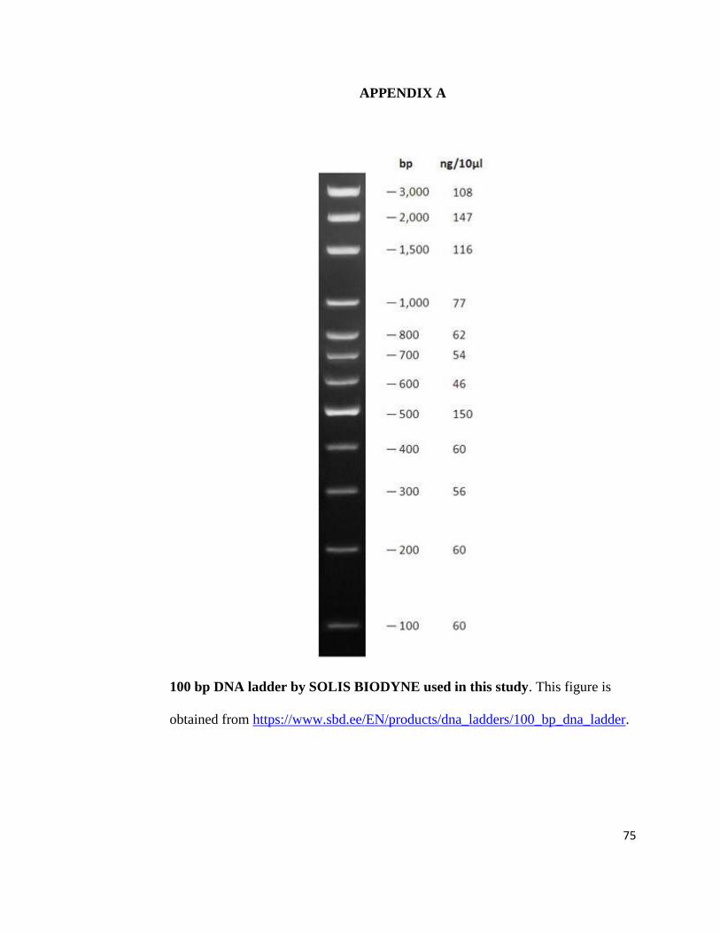
75
APPENDIX A
100 bp DNA ladder by SOLIS BIODYNE used in this study. This figure is
obtained from https://www.sbd.ee/EN/products/dna_ladders/100_bp_dna_ladder.

76
APPENDIX B
1 kbp DNA ladder by NEW ENGLAND Bio Labs Inc. used in this study. This
figure is obtained from
http://66.155.211.155/nebecomm/products_intl/productN3232.asp.