mitochondrial protein import receptors in kinetoplastids ... · mitochondrial protein import...
TRANSCRIPT
ARTICLE
Received 8 Sep 2014 | Accepted 13 Feb 2015 | Published 26 Mar 2015
Mitochondrial protein import receptors inKinetoplastids reveal convergent evolution overlarge phylogenetic distancesJan Mani1, Silvia Desy1, Moritz Niemann1, Astrid Chanfon1, Silke Oeljeklaus2, Mascha Pusnik1,w, Oliver Schmidt3,w,
Carolin Gerbeth3, Chris Meisinger3, Bettina Warscheid2 & Andre Schneider1
Mitochondrial protein import is essential for all eukaryotes and mediated by hetero-
oligomeric protein translocases thought to be conserved within all eukaryotes. We have
identified and analysed the function and architecture of the non-conventional outer
membrane (OM) protein translocase in the early diverging eukaryote Trypanosoma brucei.
It consists of six subunits that show no obvious homology to translocase components of
other species. Two subunits are import receptors that have a unique topology and unique
protein domains and thus evolved independently of the prototype receptors Tom20 and
Tom70. Our study suggests that protein import receptors were recruited to the core of the
OM translocase after the divergence of the major eukaryotic supergroups. Moreover, it links
the evolutionary history of mitochondrial protein import receptors to the origin of the
eukaryotic supergroups.
DOI: 10.1038/ncomms7646 OPEN
1 Department of Chemistry and Biochemistry, University of Bern, Freiestrasse 3, Bern CH-3012, Switzerland. 2 Department of Biochemistry and FunctionalProteomics, Faculty of Biology and BIOSS Centre for Biological Signalling Studies, University of Freiburg, Freiburg 79104, Germany. 3 Institut fur Biochemie undMolekularbiologie, ZBMZ and BIOSS Centre for Biological Signalling Studies, Universitat Freiburg, Freiburg 79104, Germany. w Present addresses: HES-SOValais, Institute of Life Technologies, CH-1950 Sion, Switzerland (M.P.); Innsbruck Medical University, Innsbruck Biocenter, Division of Cell Biology, Innrain 80,A-6020 Innsbruck, Austria (O.S.). Correspondence and requests for materials should be addressed to A.S. (email: [email protected]).
NATURE COMMUNICATIONS | 6:6646 | DOI: 10.1038/ncomms7646 | www.nature.com/naturecommunications 1
& 2015 Macmillan Publishers Limited. All rights reserved.
The origin of eukaryotes is tightly linked to a singleendosymbiotic event between a probably prokaryotic hostcell and an a-proteobacterium that subsequently was
converted into a mitochondrion. At the heart of this organello-genesis lays the evolution of a protein import system1–3. Only thepresence of such a system allowed the ancestor of themitochondrion to profit from proteins whose genes it hadtransferred to the genome of the host cell. Today 495% of allmitochondrial proteins are imported into the organelle, a processthat is mediated by translocases in the outer and innermembranes4,5. The hetero-oligomeric translocase of the outermembrane (TOM) is of special interest since it is at the interfacebetween the organelle and the cytosol. Essentially all importedproteins irrespective of their final intramitochondrial localizationrequire TOM to be translocated across the OM. TOM consists of(i) the pore-forming b-barrel protein Tom40, (ii) the TOMcomplex organizer Tom22 that also functions as a secondaryreceptor6, (iii) the three small proteins Tom5, Tom6 and Tom7,which function in the regulation of TOM complex assembly and(iv) the receptor subunits Tom20 and Tom707. The latter two aresignal-anchored proteins with a single tetratricopeptide repeat(TPR) in the case of Tom20 or 11 TPR motifs in the case ofTom70 and have in part overlapping functions. While Tom20primarily binds to the presequence of precursor proteins, Tom70has a preference to bind hydrophobic internal targetingsequences8,9 .
The machinery and the mechanism of mitochondrial proteinimport have been analysed in great detail. However, with thenotable exception of plants10, these studies are phylogeneticallybiased, because they essentially have been performed only infungi, which belong to the eukaryotic supergroup of theOpisthokonts. Bioinformatic analyses indicate that the corecomponents Tom40 and Tom22 might be present in alleukaryotes2. However, whether and to which extent the otherTom subunits, including the Tom20 and Tom70 receptors, areconserved is unclear11,12.
The parasitic protozoan Trypanosoma brucei, a representativeof the supergroup of the Excavates, is an excellent, experimentallyhighly accessible model system to identify and investigatediverged features of the mitochondrial protein importmachinery13. In contrast to most other eukaryotes,bioinformatic analyses failed to identify any orthologues ofTOM complex subunits in the trypanosomal genome13. Thus, itneeded a biochemical approach to identify the import pore theonly known component of the trypanosomal OM translocase14.It consists of a b-barrel protein that as Tom40 can be groupedinto the mitochondrial porin protein family15–17. However,it also shows similarities to the Omp85-like protein familyof bacterial protein translocases. A direct electrophysiologicalcomparison of the recombinant trypanosomal proteinwith recombinant Tom40 showed that the former—unlikeTom40—shares physical features with Omp85-like plastid andbacterial pores18. Thus, the protein was initially termed archaicTOM (ATOM)14,18. To keep the nomenclature consistent withother systems such as yeast and plants, we decided to renameATOM to ATOM40, the number indicating the approximatemolecular weight of the protein. The term ATOM without anumber will now stand for the entire ATOM complex includingall of its subunits.
ATOM40 migrates in a high molecular weight complex ofB700 kDa when analysed by blue native polyacrylamide electro-phoresis (BN–PAGE)14. In the present study we have identifiedand functionally analysed the ATOM complex subunits. Inaddition, we have delineated the architecture of the ATOMcomplex. We show that it consists of at least six subunits. Two ofthem are novel protein import receptors with overlapping
substrate specificities that evolved independently from Tom70and Tom20 of fungi and humans.
ResultsIdentification of ATOM complex subunits. Using a quantitativeproteomics approach, we have recently shown that the trypano-somal mitochondrial OM proteome consists of 82 proteins19.To identify which of these proteins are candidates for ATOMcomplex subunits immunoprecipitations (IPs) were performed.Mitochondria were gradient-purified from cells expressinghaemagglutinin (HA)-tagged ATOM40, solubilized by digitoninand subjected to IPs using anti-HA antibodies. The experimentwas performed in duplicate and IPs using wild-type mitochondrialacking tagged ATOM40 served as controls. Mass spectrometricanalysis identified 49 proteins that reproducibly and specificallyco-purified with HA-tagged ATOM40 under denaturing elutionconditions (Supplementary Data 1). Furthermore, a variation ofthe same IP experiment was done in which the elution wasperformed under native conditions using an excess of HApeptide. Subsequently, the protein complexes present in theeluates were separated by BN–PAGE. Gel lanes were cut intoequal slices and analysed by mass spectrometry, which resulted inthe identification of 17 proteins that were co-enriched with HA-tagged ATOM40 (Supplementary Data 2). The intersection of thethree data sets (‘OM proteome’/‘IP’/‘IPþBN–PAGE’) containedATOM40 and five additional proteins, which were consideredprime candidates for ATOM complex subunits (Fig. 1a).
According to their predicted molecular weight the candidateproteins were termed ATOM69, ATOM46, ATOM14, ATOM12and ATOM11. They are well conserved among Kinetoplastids(Supplementary Table 1). However, with the exception ofATOM14, which shows some limited similarity to Tom22,homology search programs such as (PSI)-BLAST or HHPred20
failed to identify homologous proteins in other organisms exceptfor proteins that contain shared conserved domains (see below).
To verify that the five candidates indeed are ATOM complexsubunits, we performed reciprocal IPs (Supplementary Fig. 1). Tothat end the five candidates were tagged at their N- and C-terminiusing the c-Myc epitope. In all cases, IPs of HA-tagged ATOM40pulled down the c-Myc-tagged candidate proteins and vice versa.Except for ATOM46, where only the C-terminally tagged versionwas mitochondrially localized, it did not matter on which side theproteins were tagged. Furthermore, immunofluorescence analysisand digitonin-based enrichment of crude mitochondrial fractionsindicated that all five candidates exclusively localize to mito-chondria (Fig. 1b,c).
All ATOM complex subunit candidates show similar relativeabundances in the mitochondrial proteome, as might be expectedfor proteins that form a hetero-oligomeric complex (Fig. 1d).Many mitochondrial proteins such as cytochrome c oxidase (Cox)and alternative oxidase (TAO) are stage specifically regulated21.Mitochondrial protein import, however, is constitutively active.In line with this all putative ATOM complex subunits showedsimilar and relatively minor changes in abundance between thetwo life cycle stages. The higher amounts of the proteins observedin the insect form is consistent with the larger size of themitochondrion in this stage (Fig. 1e)22.
We also tested a number of the 12 proteins that were onlypresent in the intersection of the two data sets ‘OM proteome’and ‘IP’ (Fig. 1a and Supplementary Fig. 1). Neither of theseproteins fulfilled all the criteria defined for ATOM complexsubunits that are discussed above. Moreover, we recentlydescribed, pATOM36, an essential mitochondrial OM proteinthat is implicated in the import of a subset of mitochondrialproteins and loosely associated with ATOM40 (ref. 23). However,
ARTICLE NATURE COMMUNICATIONS | DOI: 10.1038/ncomms7646
2 NATURE COMMUNICATIONS | 6:6646 | DOI: 10.1038/ncomms7646 | www.nature.com/naturecommunications
& 2015 Macmillan Publishers Limited. All rights reserved.
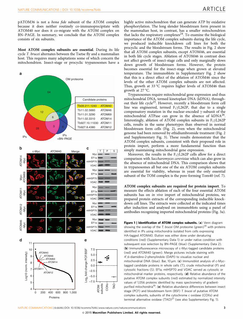
pATOM36 is not a bona fide subunit of the ATOM complexbecause it does neither routinely co-immunoprecipitate withATOM40 nor does it co-migrate with the ATOM complex onBN–PAGE. In summary, we conclude that the ATOM complexconsists of six subunits.
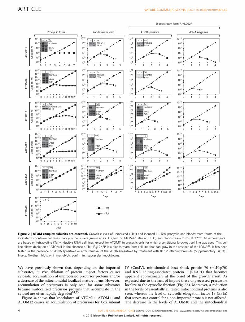
Most ATOM complex subunits are essential. During its lifecycle T. brucei alternates between the Tsetse fly and a mammalianhost. This requires many adaptations some of which concern themitochondrion. Insect-stage or procyclic trypanosomes have a
highly active mitochondrion that can generate ATP by oxidativephosphorylation. The long slender bloodstream form present inthe mammalian host, in contrast, has a smaller mitochondrionthat lacks the respiratory complexes24. To examine the biologicalimportance of the ATOM complex subunits during the life cycle,we produced inducible knockdown cell lines for both theprocyclic and the bloodstream forms. The results in Fig. 2 showthat all ATOM complex subunits, except ATOM46, are essentialin both life cycle stages. Ablation of ATOM46 in contrast doesnot affect growth of insect-stage cells and only marginally slowsdown growth of bloodstream forms. However, the proteinbecomes essential for the insect-stage when grown at elevatedtemperature. The immunoblots in Supplementary Fig. 2 showthat this is a direct effect of the ablation of ATOM46 since thelevels of the other ATOM complex subunits are not affected.Thus, growth at 33 �C requires higher levels of ATOM46 thangrowth at 27 �C.
Trypanosomes require mitochondrial gene expression and thusmitochondrial DNA, termed kinetoplast DNA (kDNA), through-out their life cycle25. However, recently a bloodstream form cellline was engineered, termed F1gL262P, that due to a singlecompensatory mutation in the nuclear-encoded g-subunit of themitochondrial ATPase can grow in the absence of kDNA26.Interestingly, ablation of ATOM complex subunits in F1gL262Pcells, results in the same phenotypes than observed in normalbloodstream form cells (Fig. 2), even when the mitochondrialgenome had been removed by ethidiumbromide treatment (Fig. 2and Supplementary Fig. 3). These results demonstrate that theATOM complex subunits, consistent with their proposed role inprotein import, perform a more fundamental function thansimply maintaining mitochondrial gene expression.
Moreover, the results in the F1gL262P cells allow for a directcomparison with Saccharomyces cerevisiae which can also grow inthe absence of mitochondrial DNA. This comparison shows thatin trypanosomes all but one of the six ATOM complex subunitsare essential for viability, whereas in yeast the only essentialsubunit of the TOM complex is the pore-forming Tom40 (ref. 7).
ATOM complex subunits are required for protein import. Tomeasure the effects ablation of each of the four essential ATOMsubunits has on in vivo import of mitochondrial proteins, weprepared protein extracts of the corresponding inducible knock-down cell lines. The extracts were collected at the indicated timesafter induction and analysed on immunoblots using a panel ofantibodies recognizing imported mitochondrial proteins (Fig. 3a).
OM proteome
Candidate proteins
Tb09.211.1240
Tb927.10.11030
Tb927.8.4380 ATOM12
ATOM11
ATOM14
ATOM69
ATOM46
ATOM40
Tb11.02.3310
Tb11.01.3290
Tb11.02.5660
64
12
28
6
3
0
8
IPs IPs+BN–PAGE
c-Myc
10
9
8
7
6
5
40 200
ATOM11
ATOM12
ATOM14ATOM69
ATOM46ATOM40
400 600 800 1,000
–7.5
7.5
–5.0
5.0
–2.5
2.5
0.0
ATO
Msu
buni
ts
CO
Xs
TAO
Log 2
fold
cha
nge
PC
F/B
SF
Proteins
11
ATO
M14
-Myc
ATO
M11
-Myc
ATO
M12
-Myc
ATO
M46
-Myc
Log1
0 (n
orm
. int
ensi
ty)
Myc
-AT
OM
69
ATOM40Myc
Myc
EF1a
mtHSP70
VDAC
EF1a
mtHSP70
VDAC
Myc
EF1amtHSP70
VDAC
MycEF1a
mtHSP70
VDAC
MycEF1a
mtHSP70VDAC
T P SMerge
Figure 1 | Identification of ATOM complex subunits. (a) Venn diagram
showing the overlap of the T. brucei OM proteome (green)19 with proteins
identified in IPs using mitochondria isolated from cells expressing
HA-tagged ATOM40. Elution was either done under denaturing
conditions (red) (Supplementary Data 1) or under native condition with
subsequent size selection by BN–PAGE (blue) (Supplementary Data 2).
(b) Immunofluorescence microscopy of c-Myc-tagged candidate proteins
(red) and ATOM40 (green). Merge pictures include staining with
40 ,6-diamidino-2-phenylindole (DAPI) to visualize nuclear and
mitochondrial DNA (blue). Bar, 10mm. (c) Immunoblot analysis of c-Myc-
tagged candidate proteins in whole cells (T), crude mitochondrial (P) and
cytosolic fractions (S). EF1a, mtHSP70 and VDAC served as cytosolic or
mitochondrial marker proteins, respectively. (d) Relative abundance of the
putative ATOM complex subunits (red) estimated by normalized intensity
values of 1,056 proteins identified by mass spectrometry of gradient-
purified mitochondria19. (e) Relative abundance differences between insect
stage (PCF) and bloodstream form (BSF) T. brucei of putative ATOM
complex subunits, subunits of the cytochrome c oxidase (COXs) and
terminal alternative oxidase (TAO)21 (see also Supplementary Fig. 1).
NATURE COMMUNICATIONS | DOI: 10.1038/ncomms7646 ARTICLE
NATURE COMMUNICATIONS | 6:6646 | DOI: 10.1038/ncomms7646 | www.nature.com/naturecommunications 3
& 2015 Macmillan Publishers Limited. All rights reserved.
We have previously shown that, depending on the importedsubstrates, in vivo ablation of protein import factors causescytosolic accumulation of unprocessed precursor proteins and/ora decrease of the mitochondrial localized mature forms. However,accumulation of precursors is only seen for some substratesbecause mislocalized precursor proteins that accumulate in thecytosol are often rapidly degraded14,23.
Figure 3a shows that knockdown of ATOM14, ATOM11 andATOM12 causes an accumulation of precursors for Cox subunit
IV (CoxIV), mitochondrial heat shock protein 70 (mtHsp70)and RNA editing-associated protein 1 (REAP1) that becomesapparent approximately at the onset of the growth arrest. Asexpected due to the lack of import these unprocessed precursorslocalize to the cytosolic fraction (Fig. 3b). Moreover, a reductionin the levels of essentially all tested mitochondrial proteins is alsoseen, whereas the level of cytosolic elongation factor 1a (EF1a)that serves as a control for a non-imported protein is not affected.The decrease in the levels of ATOM40 and the mitochondrial
1011
1010
109
108
107
106
0 1
– + Tet – + Tet
– + Tet
–– ++ TetTet
EF1aATOM11-Myc
EF1aATOM69
EF1aATOM14
– + Tet
EF1aATOM14
– + Tet
EF1aATOM69
Northern
rRNAEtBr
– + Tet
Northern
rRNAEtBr
Northern
rRNAEtBr
– + Tet
Northern
rRNAEtBr
– + Tet
Northern
rRNAEtBr
– + Tet
Northern
rRNAEtBr
– + Tet
Northern
rRNAEtBr
– + Tet
Northern
rRNAEtBr
– + Tet
Northern
rRNAEtBr
– + TetNorthern
rRNAEtBr
2 3 4 5 0 1 2 3 4 0 1 2 3 4 0 1 2 3 4
0 1 2 3 40 1 2 3 4
0 1 2 3 4
0 1 2 3 4
0 1 2 3 4
5
0 1 2 3 4
Days Days Days
Days
– Tet
+ Tet
5
0 1 2 3 4 5
†
†
† †
†††
†
0 1 2 3 4 5
0 1 2 3 4 5
6 7
0 1 2 3 4 5 6 7
0 1 2 3 4 5 6 7 8 9 10 11
0 1 2 3 4 5 6 7 8 9 10 11
0 1 2 3 4 5 6 7 8 9 10 110 1 2 3 4 5 6 7 8 9 101112
0 1 2 3 4 5 6 7 8 9
0 1 2 3
33 °C
4 5 6 7 8 9
10
0 1 2 3 4 5 6 7 8 9 10
11
1010
109
108
107
106
105
1010
109
108
107
106
105
1010
109
108
107
106
105
1010
109
108
107
106
105
1010
109
108
107
106
105
1010
109
108
107
106
105
1010
109
108
107
106
105
1010
109
108
107
106
105
1010
109
108
107
106
105
1010
109
108
107
106
105
1010
1011
109
108
107
106
105
109
108
107
106
105
104
101110121013
1010
109
108
107
106
101110121013
1010
109
108
107
106
1011
1012
1010
109
108
107
106
1011
1010
109
108
107
106
10111012
1012
10141013
1010 1010
109
108
108
107
106
106
104
1012
1014
1010
108
106
104
1011
1012
1010
109
108
107
106
Procyclic form
ATO
M14
Cel
ls p
er m
lC
ells
per
ml
Cel
ls p
er m
lC
ells
per
ml
Cel
ls p
er m
lC
ells
per
ml
ATO
M69
ATO
M11
ATO
M12
ATO
M46
ATO
M46
Bloodstream form
Bloodstream form F1γ L262P
kDNA positive kDNA negative
Figure 2 | ATOM complex subunits are essential. Growth curves of uninduced (-Tet) and induced (þTet) procyclic and bloodstream forms of the
indicated knockdown cell lines. Procyclic cells were grown at 27 �C (and for ATOM46 also at 33 �C) and bloodstream forms at 37 �C. All experiments
are based on tetracycline (Tet)-inducible RNAi cell lines, except for ATOM11 in procyclic cells for which a conditional knockout cell line was used. This cell
line allows depletion of ATOM11 in the absence of Tet. F1gL262P is a bloodstream form cell line that can grow in the absence of the kDNA26. It has been
tested in the presence of kDNA (positive) or after removal of the kDNA (negative) by treatment with 10 nM ethidiumbromide (Supplementary Fig. 3).
Insets, Northern blots or immunoblots confirming successful knockdowns.
ARTICLE NATURE COMMUNICATIONS | DOI: 10.1038/ncomms7646
4 NATURE COMMUNICATIONS | 6:6646 | DOI: 10.1038/ncomms7646 | www.nature.com/naturecommunications
& 2015 Macmillan Publishers Limited. All rights reserved.
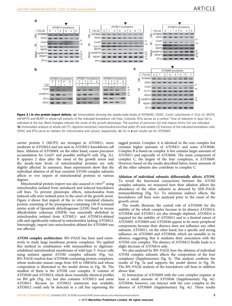
carrier protein 5 (MCP5) are strongest in ATOM11, moremoderate in ATOM14 and not seen in ATOM12 knockdown celllines. Ablation of ATOM69, on the other hand, causes precursoraccumulation for CoxIV and possibly mtHsp70 only (Fig. 3c).It appears 2 days after the onset of the growth arrest andthe steady-state levels of mitochondrial proteins are onlyslightly affected. In summary, these experiments show that theindividual ablation of all four essential ATOM complex subunitsaffects in vivo import of mitochondrial proteins to variousdegrees.
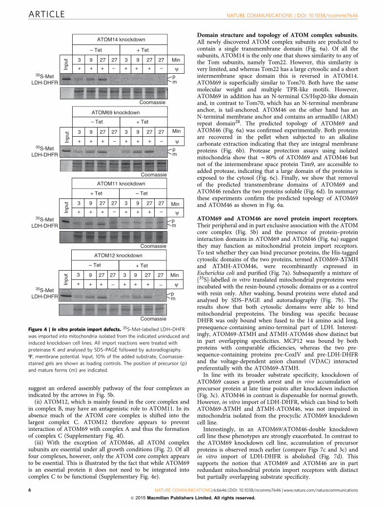
Mitochondrial protein import was also assayed in vitro27 usingmitochondria isolated from uninduced and induced knockdowncell lines. To prevent pleiotropic effects, mitochondria frominduced cells were isolated prior to the onset of the growth arrest.Figure 4 shows that import of the in vitro translated chimericprotein consisting of the presequence-containing 150 N-terminalamino acids of lipoamide dehydrogenase (LDH) fused to mousedihydrofolate reductase (DHFR) was essentially abolished inmitochondria isolated from ATOM11- and ATOM14-ablatedcells and significantly reduced in mitochondria lacking ATOM12.Interestingly, import into mitochondria ablated for ATOM69 wasnot affected.
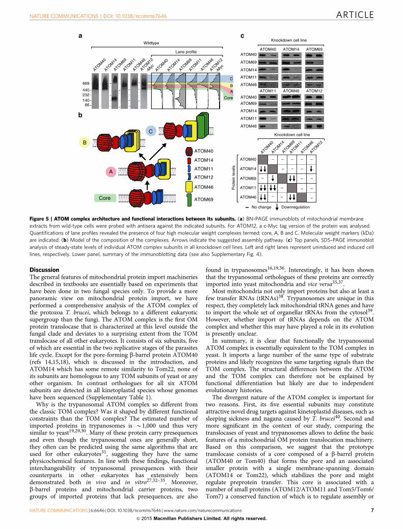
ATOM complex architecture. BN–PAGE has been used exten-sively to study large membrane protein complexes. We appliedthis method in combination with immunoblots to digitonin-solubilized mitochondrial membranes of wild-type T. brucei cellsusing antisera against ATOM complex subunits (Fig. 5a).BN–PAGE resolves four ATOM40-containing protein complexes,whose molecular masses range from 450 to 1000 kDa and whosecomposition is illustrated in the model shown in Fig. 5b. Thesmallest of them is the ATOM core complex. It consists ofATOM40 and ATOM14, which show essentially identical profileson BN gels (Fig. 5a), but also contains ATOM12 and someATOM11. Because no ATOM12 antiserum was available,ATOM12 could only be detected in a cell line expressing the
tagged protein. Complex A is identical to the core complex butcontains higher amounts of ATOM11 and some ATOM46.Complex B is based on complex A but contains larger amounts ofATOM11 and especially of ATOM46. The main component ofcomplex C, the largest of the four complexes, is ATOM69.However, based on the results described below, lower amounts ofall the other subunits also contribute to complex C.
Ablation of individual subunits differentially affects ATOM.To reveal the functional connections between the ATOMcomplex subunits, we measured how their ablation affects theabundance of the other subunits as detected by SDS–PAGEimmunoblotting (Fig. 5c). To minimize indirect effects, theknockdown cell lines were analysed prior to the onset of thegrowth arrest.
The results illustrate the central role of ATOM40 for thestability of the whole complex because in its absence ATOM14,ATOM46 and ATOM11 are also strongly depleted. ATOM14 isrequired for the stability of ATOM11 and to a limited extent ofATOM69. ATOM69 and ATOM46 appear to be more peripheralcomponents because their absence does not influence any othersubunits. ATOM11, on the other hand, has a specific and stronginfluence on ATOM69 and ATOM46, which are unstable in itsabsence, suggesting that it mediates their association with theATOM core complex. The absence of ATOM12 finally leads to aslight decrease of ATOM14 only.
We also analysed by BN–PAGE how the ablation of individualATOM complex subunits affects the composition of the fourcomplexes (Supplementary Fig. 4). This analysis confirms theresults of Fig. 5a and supports the model shown in Fig. 5b.The BN–PAGE analysis of the knockdown cell lines in additionshows that:
(i) Interaction of ATOM69 with the core complex requires atleast a small amount of ATOM46 (Supplementary Fig. 4e).ATOM46, however, can interact with the core complex in theabsence of ATOM69 (Supplementary Fig. 4c). These results
ATOM14 knockdown
ATOM14 knockdown ATOM11 knockdown ATOM12 knockdown
+Tet (d) 0 1
ATOM69 knockdown
2 3 4 5 6 7
TTet – + + +
CoxlVpmpmmtHSP70
EF1a
VDAC
T P S T+ + – –
T P S T– + + +
T P S
+Tet (d)
ATOM40
VDAC
CoxIV
MCP5
mtHSP70REAP1
EF1a
Mat
rixIM
OM
Cyt C1
0 1 2 3 4 5
ATOM11 knockdown ATOM12 knockdown
–Tet (d) +Tet (d)0 1 2 3 4 5 6 7 0 1 2 3 4 5
p
OM ATOM40
VDAC
CoxIV
Cyt C1MCP5
mtHSP70
REAP1
EF1a
ATOM40VDAC
CoxIV
Cyt C1MCP5
mtHSP70REAP1
EF1a
p
p
m
m
ATOM40
VDAC
CoxIV
Cyt C1
MCP5
mtHSP70
REAP1
EF1a
IMM
atrix
OM
IMM
atrix
OM
IMM
atrix
p
m
mpm
p
p
m
mpm
p
p
m
mpm
Figure 3 | In vivo protein import defects. (a) Immunoblots showing the steady-state levels of ATOM40, VDAC, CoxIV, cytochrome c1 (Cyt c1), MCP5,
mtHSP70 and REAP1 in whole-cell extracts of the indicated knockdown cell lines. Cytosolic EF1a serves as a control. Time of induction in days (d) is
indicated at the top. Black triangles indicate the onset of the growth phenotype. The position of precursor (p) and mature forms (m) are indicated.
(b) Immunoblot analysis of whole-cell (T), digitonin-extracted, mitochondria-enriched pellet (P) and soluble (S) fractions of the indicated knockdown cells.
VDAC and EF1a serve as markers for mitochondria and cytosol, respectively. (c) As in a but results are for ATOM69.
NATURE COMMUNICATIONS | DOI: 10.1038/ncomms7646 ARTICLE
NATURE COMMUNICATIONS | 6:6646 | DOI: 10.1038/ncomms7646 | www.nature.com/naturecommunications 5
& 2015 Macmillan Publishers Limited. All rights reserved.
suggest an ordered assembly pathway of the four complexes asindicated by the arrows in Fig. 5b.
(ii) ATOM12, which is mainly found in the core complex andin complex B, may have an antagonistic role to ATOM11. In itsabsence much of the ATOM core complex is shifted into thelargest complex C. ATOM12 therefore appears to preventinteraction of ATOM69 with complex A and thus the formationof complex C (Supplementary Fig. 4f).
(iii) With the exception of ATOM46, all ATOM complexsubunits are essential under all growth conditions (Fig. 2). Of allfour complexes, however, only the ATOM core complex appearsto be essential. This is illustrated by the fact that while ATOM69is an essential protein it does not need to be integrated intocomplex C to be functional (Supplementary Fig. 4e).
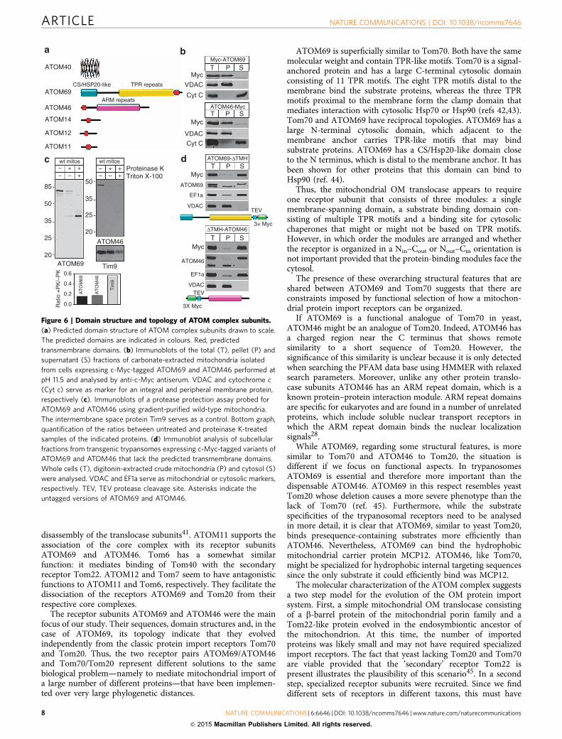
Domain structure and topology of ATOM complex subunits.All newly discovered ATOM complex subunits are predicted tocontain a single transmembrane domain (Fig. 6a). Of all thesubunits, ATOM14 is the only one that shows similarity to any ofthe Tom subunits, namely Tom22. However, this similarity isvery limited, and whereas Tom22 has a large cytosolic and a shortintermembrane space domain this is reversed in ATOM14.ATOM69 is superficially similar to Tom70. Both have the samemolecular weight and multiple TPR-like motifs. However,ATOM69 in addition has an N-terminal CS/Hsp20-like domainand, in contrast to Tom70, which has an N-terminal membraneanchor, is tail-anchored. ATOM46 on the other hand has anN-terminal membrane anchor and contains an armadillo (ARM)repeat domain28. The predicted topology of ATOM69 andATOM46 (Fig. 6a) was confirmed experimentally. Both proteinsare recovered in the pellet when subjected to an alkalinecarbonate extraction indicating that they are integral membraneproteins (Fig. 6b). Protease protection assays using isolatedmitochondria show that B80% of ATOM69 and ATOM46 butnot of the intermembrane space protein Tim9, are accessible toadded protease, indicating that a large domain of the proteins isexposed to the cytosol (Fig. 6c). Finally, we show that removalof the predicted transmembrane domains of ATOM69 andATOM46 renders the two proteins soluble (Fig. 6d). In summarythese experiments confirm the predicted topology of ATOM69and ATOM46 as shown in Fig. 6a.
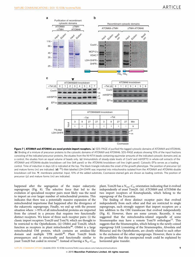
ATOM69 and ATOM46 are novel protein import receptors.Their peripheral and in part exclusive association with the ATOMcore complex (Fig. 5b) and the presence of protein–proteininteraction domains in ATOM69 and ATOM46 (Fig. 6a) suggestthey may function as mitochondrial protein import receptors.To test whether they can bind precursor proteins, the His-taggedcytosolic domains of the two proteins, termed ATOM69-DTMHand DTMH-ATOM46, were recombinantly expressed inEscherichia coli and purified (Fig. 7a). Subsequently a mixture of[35S]-labelled in vitro translated mitochondrial preproteins wereincubated with the resin-bound cytosolic domains or as a controlwith resin only. After washing, bound proteins were eluted andanalysed by SDS–PAGE and autoradiography (Fig. 7b). Theresults show that both cytosolic domains were able to bindmitochondrial preproteins. The binding was specific becauseDHFR was only bound when fused to the 14 amino acid long,presequence-containing amino-terminal part of LDH. Interest-ingly, ATOM69-DTMH and DTMH-ATOM46 show distinct butin part overlapping specificities. MCP12 was bound by bothproteins with comparable efficiencies, whereas the two pre-sequence-containing proteins pre-CoxIV and pre-LDH-DHFRand the voltage-dependent anion channel (VDAC) interactedpreferentially with the ATOM69-DTMH.
In line with its broader substrate specificity, knockdown ofATOM69 causes a growth arrest and in vivo accumulation ofprecursor protein at late time points after knockdown induction(Fig. 3c). ATOM46 in contrast is dispensable for normal growth.However, in vitro import of LDH-DHFR, which can bind to bothATOM69-DTMH and DTMH-ATOM46, was not impaired inmitochondria isolated from the procyclic ATOM69 knockdowncell line.
Interestingly, in an ATOM69/ATOM46-double knockdowncell line these phenotypes are strongly exacerbated. In contrast tothe ATOM69 knockdown cell line, accumulation of precursorproteins is observed much earlier (compare Figs 7c and 3c) andin vitro import of LDH-DHFR is abolished (Fig. 7d). Thissupports the notion that ATOM69 and ATOM46 are in partredundant mitochondrial protein import receptors with distinctbut partially overlapping substrate specificity.
ATOM14 knockdown
ATOM69 knockdown
ATOM11 knockdown
ATOM12 knockdown
– Tet + Tet
– Tet
3
+ + + – + + + – ψ
ψ
9 27 27 3 9 27 27 Min
Min
pm
pm
pm
pm
Coomassie
Coomassie
Coomassie
Inpu
t
35S-MetLDH-DHFR
35S-MetLDH-DHFR
35S-MetLDH-DHFR
35S-MetLDH-DHFR
+ Tet
– Tet
3
+ + + – + + + –
9 27 27 3 9 27 27
Inpu
t
ψMin3
+ + + – + + + –
9 27 27 3 9 27 27
ψ
Min3
+ + + – +
Coomassie
+ + –
9 27 27 3 9 27 27
Inpu
tIn
put
+ Tet
+ Tet – Tet
Figure 4 | In vitro protein import defects. 35S-Met-labelled LDH–DHFR
was imported into mitochondria isolated from the indicated uninduced and
induced knockdown cell lines. All import reactions were treated with
proteinase K and analysed by SDS–PAGE followed by autoradiography.
C, membrane potential. Input, 10% of the added substrate, Coomassie-
stained gels are shown as loading controls. The position of precursor (p)
and mature forms (m) are indicated.
ARTICLE NATURE COMMUNICATIONS | DOI: 10.1038/ncomms7646
6 NATURE COMMUNICATIONS | 6:6646 | DOI: 10.1038/ncomms7646 | www.nature.com/naturecommunications
& 2015 Macmillan Publishers Limited. All rights reserved.
DiscussionThe general features of mitochondrial protein import machineriesdescribed in textbooks are essentially based on experiments thathave been done in two fungal species only. To provide a morepanoramic view on mitochondrial protein import, we haveperformed a comprehensive analysis of the ATOM complex ofthe protozoa T. brucei, which belongs to a different eukaryoticsupergroup than the fungi. The ATOM complex is the first OMprotein translocase that is characterized at this level outside thefungal clade and deviates to a surprising extent from the TOMtranslocase of all other eukaryotes. It consists of six subunits, fiveof which are essential in the two replicative stages of the parasiteslife cycle. Except for the pore-forming b-barrel protein ATOM40(refs 14,15,18), which is discussed in the introduction, andATOM14 which has some remote similarity to Tom22, none ofits subunits are homologous to any TOM subunits of yeast or anyother organism. In contrast orthologues for all six ATOMsubunits are detected in all kinetoplastid species whose genomeshave been sequenced (Supplementary Table 1).
Why is the trypanosomal ATOM complex so different fromthe classic TOM complex? Was it shaped by different functionalconstraints than the TOM complex? The estimated number ofimported proteins in trypanosomes is B1,000 und thus verysimilar to yeast19,29,30. Many of these protein carry presequencesand even though the trypanosomal ones are generally short,they often can be predicted using the same algorithms that areused for other eukaryotes31, suggesting they have the samephysicochemical features. In line with these findings, functionalinterchangeability of trypanosomal presequences with theircounterparts in other eukaryotes has extensively beendemonstrated both in vivo and in vitro27,32–35. Moreover,b-barrel proteins and mitochondrial carrier proteins, twogroups of imported proteins that lack presequences, are also
found in trypanosomes16,19,36. Interestingly, it has been shownthat the trypanosomal orthologues of these proteins are correctlyimported into yeast mitochondria and vice versa35,37.
Most mitochondria not only import proteins but also at least afew transfer RNAs (tRNAs)38. Trypanosomes are unique in thisrespect, they completely lack mitochondrial tRNA genes and haveto import the whole set of organellar tRNAs from the cytosol39.However, whether import of tRNAs depends on the ATOMcomplex and whether this may have played a role in its evolutionis presently unclear.
In summary, it is clear that functionally the trypanosomalATOM complex is essentially equivalent to the TOM complex inyeast. It imports a large number of the same type of substrateproteins and likely recognizes the same targeting signals than theTOM complex. The structural differences between the ATOMand the TOM complex can therefore not be explained byfunctional differentiation but likely are due to independentevolutionary histories.
The divergent nature of the ATOM complex is important fortwo reasons. First, its five essential subunits may constituteattractive novel drug targets against kinetoplastid diseases, such assleeping sickness and nagana caused by T. brucei40. Second andmore significant in the context of our study, comparing thetranslocases of yeast and trypanosomes allows to define the basicfeatures of a mitochondrial OM protein translocation machinery.Based on this comparison, we suggest that the prototypetranslocase consists of a core composed of a b-barrel protein(ATOM40 or Tom40) that forms the pore and an associatedsmaller protein with a single membrane-spanning domain(ATOM14 or Tom22), which stabilizes the pore and mightregulate preprotein transfer. This core is associated with anumber of small proteins (ATOM12/ATOM11 and Tom5/Tom6/Tom7) a conserved function of which is to regulate assembly or
Wildtype
669
44023214066
Lane profile
C
BA
Core
ATOM40
ATOM40
ATOM14
ATOM14
ATOM69
ATOM69
ATOM11
ATOM11
ATOM46
ATOM46
ATOM12
-Myc
ATOM12
ATOM40
ATOM14
ATOM69
ATOM11
ATOM46
ATOM12
-Myc
ATOM40
ATOM14
ATOM69
ATOM11
ATOM46
ATOM40 – – – – –
–––
–
–
–
– –
–
––
–
ATOM40
C
B
A
Core
ATOM14
ATOM14
ATOM11
ATOM12 ATOM69
ATOM69
Pro
tein
leve
ls
ATOM11
ATOM46
ATOM46
No change Downregulation
ATOM40
ATOM69
ATOM14
ATOM11
ATOM46
ATOM11 ATOM46
ATOM40 ATOM14 ATOM69
Knockdown cell line
Knockdown cell line
ATOM12
Figure 5 | ATOM complex architecture and functional interactions between its subunits. (a) BN–PAGE immunoblots of mitochondrial membrane
extracts from wild-type cells were probed with antisera against the indicated subunits. For ATOM12, a c-Myc tag version of the protein was analysed.
Quantifications of lane profiles revealed the presence of four high molecular weight complexes termed: core, A, B and C. Molecular weight markers (kDa)
are indicated. (b) Model of the composition of the complexes. Arrows indicate the suggested assembly pathway. (c) Top panels, SDS–PAGE immunoblot
analysis of steady-state levels of individual ATOM complex subunits in all knockdown cell lines. Left and right lanes represent uninduced and induced cell
lines, respectively. Lower panel, summary of the immunoblotting data (see also Supplementary Fig. 4).
NATURE COMMUNICATIONS | DOI: 10.1038/ncomms7646 ARTICLE
NATURE COMMUNICATIONS | 6:6646 | DOI: 10.1038/ncomms7646 | www.nature.com/naturecommunications 7
& 2015 Macmillan Publishers Limited. All rights reserved.
disassembly of the translocase subunits41. ATOM11 supports theassociation of the core complex with its receptor subunitsATOM69 and ATOM46. Tom6 has a somewhat similarfunction: it mediates binding of Tom40 with the secondaryreceptor Tom22. ATOM12 and Tom7 seem to have antagonisticfunctions to ATOM11 and Tom6, respectively. They facilitate thedissociation of the receptors ATOM69 and Tom20 from theirrespective core complexes.
The receptor subunits ATOM69 and ATOM46 were the mainfocus of our study. Their sequences, domain structures and, in thecase of ATOM69, its topology indicate that they evolvedindependently from the classic protein import receptors Tom70and Tom20. Thus, the two receptor pairs ATOM69/ATOM46and Tom70/Tom20 represent different solutions to the samebiological problem—namely to mediate mitochondrial import ofa large number of different proteins—that have been implemen-ted over very large phylogenetic distances.
ATOM69 is superficially similar to Tom70. Both have the samemolecular weight and contain TPR-like motifs. Tom70 is a signal-anchored protein and has a large C-terminal cytosolic domainconsisting of 11 TPR motifs. The eight TPR motifs distal to themembrane bind the substrate proteins, whereas the three TPRmotifs proximal to the membrane form the clamp domain thatmediates interaction with cytosolic Hsp70 or Hsp90 (refs 42,43).Tom70 and ATOM69 have reciprocal topologies. ATOM69 has alarge N-terminal cytosolic domain, which adjacent to themembrane anchor carries TPR-like motifs that may bindsubstrate proteins. ATOM69 has a CS/Hsp20-like domain closeto the N terminus, which is distal to the membrane anchor. It hasbeen shown for other proteins that this domain can bind toHsp90 (ref. 44).
Thus, the mitochondrial OM translocase appears to requireone receptor subunit that consists of three modules: a singlemembrane-spanning domain, a substrate binding domain con-sisting of multiple TPR motifs and a binding site for cytosolicchaperones that might or might not be based on TPR motifs.However, in which order the modules are arranged and whetherthe receptor is organized in a Nin–Cout or Nout–Cin orientation isnot important provided that the protein-binding modules face thecytosol.
The presence of these overarching structural features that areshared between ATOM69 and Tom70 suggests that there areconstraints imposed by functional selection of how a mitochon-drial protein import receptors can be organized.
If ATOM69 is a functional analogue of Tom70 in yeast,ATOM46 might be an analogue of Tom20. Indeed, ATOM46 hasa charged region near the C terminus that shows remotesimilarity to a short sequence of Tom20. However, thesignificance of this similarity is unclear because it is only detectedwhen searching the PFAM data base using HMMER with relaxedsearch parameters. Moreover, unlike any other protein translo-case subunits ATOM46 has an ARM repeat domain, which is aknown protein–protein interaction module. ARM repeat domainsare specific for eukaryotes and are found in a number of unrelatedproteins, which include soluble nuclear transport receptors inwhich the ARM repeat domain binds the nuclear localizationsignals28.
While ATOM69, regarding some structural features, is moresimilar to Tom70 and ATOM46 to Tom20, the situation isdifferent if we focus on functional aspects. In trypanosomesATOM69 is essential and therefore more important than thedispensable ATOM46. ATOM69 in this respect resembles yeastTom20 whose deletion causes a more severe phenotype than thelack of Tom70 (ref. 45). Furthermore, while the substratespecificities of the trypanosomal receptors need to be analysedin more detail, it is clear that ATOM69, similar to yeast Tom20,binds presequence-containing substrates more efficiently thanATOM46. Nevertheless, ATOM69 can bind the hydrophobicmitochondrial carrier protein MCP12. ATOM46, like Tom70,might be specialized for hydrophobic internal targeting sequencessince the only substrate it could efficiently bind was MCP12.
The molecular characterization of the ATOM complex suggestsa two step model for the evolution of the OM protein importsystem. First, a simple mitochondrial OM translocase consistingof a b-barrel protein of the mitochondrial porin family and aTom22-like protein evolved in the endosymbiontic ancestor ofthe mitochondrion. At this time, the number of importedproteins was likely small and may not have required specializedimport receptors. The fact that yeast lacking Tom20 and Tom70are viable provided that the ‘secondary’ receptor Tom22 ispresent illustrates the plausibility of this scenario45. In a secondstep, specialized receptor subunits were recruited. Since we finddifferent sets of receptors in different taxons, this must have
–– – +
++ –– – +
++
ATOM40
CS/HSP20-like TPR repeats
ARM repeats
ATOM14
ATOM11
wt mitos
85
50
35
25
20
50
35
25
20
ATOM46
ATOM69 Tim9
0.0
0.2
0.4
AT
OM
69
AT
OM
46
Tim
9
Rat
io +
PK
/–P
K 0.6
wt mitosProteinase KTriton X-100
Myc-ATOM69
TMyc
VDAC
Cyt C
Myc
Myc
VDAC
Cyt C
P S
ATOM46-Myc
ATOM69-ΔTMH
ΔTMH-ATOM46
ATOM69
EF1a
VDAC
Myc
ATOM46*
*
EF1a
VDAC
TEV
3× Myc
TEV
3X Myc
T P S
T P S
T P S
ATOM12
ATOM69
ATOM46
Figure 6 | Domain structure and topology of ATOM complex subunits.
(a) Predicted domain structure of ATOM complex subunits drawn to scale.
The predicted domains are indicated in colours. Red, predicted
transmembrane domains. (b) Immunoblots of the total (T), pellet (P) and
supernatant (S) fractions of carbonate-extracted mitochondria isolated
from cells expressing c-Myc-tagged ATOM69 and ATOM46 performed at
pH 11.5 and analysed by anti-c-Myc antiserum. VDAC and cytochrome c
(Cyt c) serve as marker for an integral and peripheral membrane protein,
respectively (c). Immunoblots of a protease protection assay probed for
ATOM69 and ATOM46 using gradient-purified wild-type mitochondria.
The intermembrane space protein Tim9 serves as a control. Bottom graph,
quantification of the ratios between untreated and proteinase K-treated
samples of the indicated proteins. (d) Immunoblot analysis of subcellular
fractions from transgenic trypansomes expressing c-Myc-tagged variants of
ATOM69 and ATOM46 that lack the predicted transmembrane domains.
Whole cells (T), digitonin-extracted crude mitochondria (P) and cytosol (S)
were analysed. VDAC and EF1a serve as mitochondrial or cytosolic markers,
respectively. TEV, TEV protease cleavage site. Asterisks indicate the
untagged versions of ATOM69 and ATOM46.
ARTICLE NATURE COMMUNICATIONS | DOI: 10.1038/ncomms7646
8 NATURE COMMUNICATIONS | 6:6646 | DOI: 10.1038/ncomms7646 | www.nature.com/naturecommunications
& 2015 Macmillan Publishers Limited. All rights reserved.
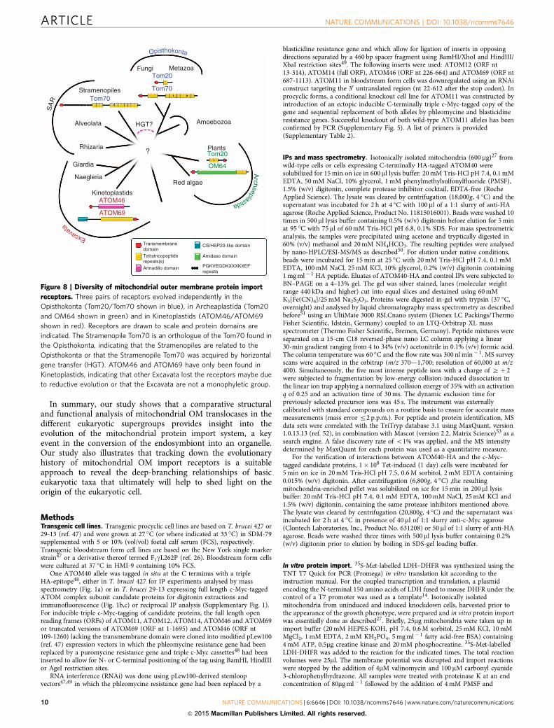
happened after the segregation of the major eukaryoticsupergroups (Fig. 8). The selective force that led to theevolution of specialized receptor pairs most likely was the needto import an ever larger number of mitochondrial proteins. Thisindicates that there was a potentially massive expansion of themitochondrial importome that happened after the divergence ofthe eukaryotic supergroups. Finally, we end up with the presentsituation where 495% of all mitochondrial proteins are importedfrom the cytosol in a process that requires two functionallydistinct receptors. We know of three such receptor pairs: (i) theclassic import receptors Tom20 and Tom70, which are thought tobe restricted to the Opisthokonts. (ii) OM64 and Tom20, whichfunction as receptors in plant mitochondria46. OM64 is a largemitochondrial OM protein, which contains an amidase-likedomain and multiple TPR motifs46. Plant Tom20 bindspresequences and is structurally and functionally similar toyeast Tom20 but coded in reverse12. Instead of having a Nin–Cout
plant, Tom20 has a Nout–Cin orientation indicating that it evolvedindependently of yeast Tom20. (iii) ATOM69 and ATOM46 thetwo import receptors of Kinetoplastids, which belong to thesupergroup of the Excavates.
The finding of three distinct receptor pairs that evolvedindependently from each other and that are restricted to singlesupergroups, each strongly support that import receptors are alate addition to the OM translocase that evolved independently(Fig. 8). However, there are some caveats. Recently, it wassuggested that the mitochondria-related organelle of someStramenopiles may have a remote Tom70 orthologue11. Thissuggests that the Stramenopiles, which belong to the newly coinedsupergroup SAR (consisting of the Stramenopiles, Alveolata andRhizaria) and the Opisthokonts, are closely related to each otherto the exclusion of the other supergroups. However, there is alsothe possibility that this unexpected result could be explained byhorizontal gene transfer.
Inpu
t
116
66
35
25
+Tet (d)
ATOM46&69 knockdown
ATOM46&69 knockdown
ATOM46 knockdown
0 1 2 3 4 5 6 7 +Tet (d) 0 1
His6 His6
2 3 4 5 6p
pm
m
CoxIV
mtHSP70
EF1a
35S-MetLDH-DHFR
CoxIV
mtHSP70
EF1a
–Tet
+ + + + + +
Coomassie
p
m
––
3 9 27 27 3 9 27 27 Min
ψInpu
t
+Tet
45
ATOM69-ΔTMH ATOM69-ΔTMH
ΔTMH-ATOM46 ΔTMH-ATOM46
Purification of recombinantcytosolic domains Recombinant cytosolic domains
Inpu
tElut
ion
Elution
Inpu
t
CoxIV
LDH-DHFR
DHFR
MCP12
VDAC
Inpu
tElut
ion Bead
ctrl Bea
d
ctrl
Elution
Figure 7 | ATOM69 and ATOM46 are novel protein import receptors. (a) SDS–PAGE of purified His-tagged cytosolic domains of ATOM69 and ATOM46.
(b) Binding of a mixture of precursor proteins to the cytosolic domains of ATOM69 and ATOM46. SDS–PAGE analysis showing 10% of the input fractions
consisting of the indicated precursor proteins, the eluates from the Ni-NTA beads containing equimolar amounts of the indicated cytosolic domains and, as
a control, the eluates from an equal volume of beads only. (c) Immunoblots of steady-state levels of CoxIV and mtHSP70 in whole-cell extracts of the
ATOM69 and ATOM46-double knockdown cell line (left panel) or the ATOM46 knockdown cell line (right panel). Cytosolic EF1a serves as a loading
control. Time of induction in days (d) is indicated at the top. The black triangle indicates the onset of the growth phenotype. The position of precursor (p)
and mature forms (m) are indicated. (d) 35S-Met-labelled LDH-DHFR was imported into mitochondria isolated from the ATOM69 and ATOM46-double
knockdown cell line. C, membrane potential. Input, 10% of the added substrate, Coomassie-stained gels are shown as loading controls. The position of
precursor (p) and mature forms (m) are indicated.
NATURE COMMUNICATIONS | DOI: 10.1038/ncomms7646 ARTICLE
NATURE COMMUNICATIONS | 6:6646 | DOI: 10.1038/ncomms7646 | www.nature.com/naturecommunications 9
& 2015 Macmillan Publishers Limited. All rights reserved.
In summary, our study shows that a comparative structuraland functional analysis of mitochondrial OM translocases in thedifferent eukaryotic supergroups provides insight into theevolution of the mitochondrial protein import system, a keyevent in the conversion of the endosymbiont into an organelle.Our study also illustrates that tracking down the evolutionaryhistory of mitochondrial OM import receptors is a suitableapproach to reveal the deep-branching relationships of basiceukaryotic taxa that ultimately will help to shed light on theorigin of the eukaryotic cell.
MethodsTransgenic cell lines. Transgenic procyclic cell lines are based on T. brucei 427 or29-13 (ref. 47) and were grown at 27 �C (or where indicated at 33 �C) in SDM-79supplemented with 5 or 10% (vol/vol) foetal calf serum (FCS), respectively.Transgenic bloodstream form cell lines are based on the New York single markerstrain47 or a derivative thereof termed F1gL262P (ref. 26). Bloodstream form cellswere cultured at 37 �C in HMI-9 containing 10% FCS.
One ATOM40 allele was tagged in situ at the C terminus with a tripleHA-epitope48, either in T. brucei 427 for IP experiments analysed by massspectrometry (Fig. 1a) or in T. brucei 29-13 expressing full length c-Myc-taggedATOM complex subunit candidate proteins for digitonin extractions andimmunofluorescence (Fig. 1b,c) or reciprocal IP analysis (Supplementary Fig. 1).For inducible triple c-Myc-tagging of candidate proteins, the full length openreading frames (ORFs) of ATOM11, ATOM12, ATOM14, ATOM46 and ATOM69or truncated versions of ATOM69 (ORF nt 1-1695) and ATOM46 (ORF nt109-1260) lacking the transmembrane domain were cloned into modified pLew100(ref. 47) expression vectors in which the phleomycine resistance gene had beenreplaced by a puromycine resistance gene and triple c-Myc cassettes48 had beeninserted to allow for N- or C-terminal positioning of the tag using BamHI, HindIIIor AgeI restriction sites.
RNA interference (RNAi) was done using pLew100-derived stemloopvectors47,49 in which the phleomycine resistance gene had been replaced by a
blasticidine resistance gene and which allow for ligation of inserts in opposingdirections separated by a 460 bp spacer fragment using BamHI/XhoI and HindIII/XbaI restriction sites49. The following inserts were used: ATOM12 (ORF nt13-314), ATOM14 (full ORF), ATOM46 (ORF nt 226-664) and ATOM69 (ORF nt687-1113). ATOM11 in bloodstream form cells was downregulated using an RNAiconstruct targeting the 30 untranslated region (nt 22-612 after the stop codon). Inprocyclic forms, a conditional knockout cell line for ATOM11 was constructed byintroduction of an ectopic inducible C-terminally triple c-Myc-tagged copy of thegene and sequential replacement of both alleles by phleomycine and blasticidineresistance genes. Successful knockout of both wild-type ATOM11 alleles has beenconfirmed by PCR (Supplementary Fig. 5). A list of primers is provided(Supplementary Table 2).
IPs and mass spectrometry. Isotonically isolated mitochondria (600 mg)27 fromwild-type cells or cells expressing C-terminally HA-tagged ATOM40 weresolubilized for 15 min on ice in 600ml lysis buffer: 20 mM Tris-HCl pH 7.4, 0.1 mMEDTA, 50 mM NaCl, 10% glycerol, 1 mM phenylmethylsulfonylfluoride (PMSF),1.5% (w/v) digitonin, complete protease inhibitor cocktail, EDTA-free (RocheApplied Science). The lysate was cleared by centrifugation (18,000g, 4 �C) and thesupernatant was incubated for 2 h at 4 �C with 100 ml of a 1:1 slurry of anti-HAagarose (Roche Applied Science, Product No. 11815016001). Beads were washed 10times in 500ml lysis buffer containing 0.5% (w/v) digitonin before elution for 5 minat 95 �C with 75ml of 60 mM Tris-HCl pH 6.8, 0.1% SDS. For mass spectrometricanalysis, the samples were precipitated using acetone and tryptically digested in60% (v/v) methanol and 20 mM NH4HCO3. The resulting peptides were analysedby nano-HPLC/ESI-MS/MS as described50. For elution under native conditions,beads were incubated for 15 min at 25 �C with 20 mM Tris-HCl pH 7.4, 0.1 mMEDTA, 100 mM NaCl, 25 mM KCl, 10% glycerol, 0.2% (w/v) digitonin containing1 mg ml� 1 HA peptide. Eluates of ATOM40-HA and control IPs were subjected toBN–PAGE on a 4–13% gel. The gel was silver stained, lanes (molecular weightrange 440 kDa and higher) cut into equal slices and destained using 60 mMK3[Fe(CN)6]/25 mM Na2S2O3. Proteins were digested in-gel with trypsin (37 �C,overnight) and analysed by liquid chromatography mass spectrometry as describedbefore51 using an UltiMate 3000 RSLCnano system (Dionex LC Packings/ThermoFisher Scientific, Idstein, Germany) coupled to an LTQ-Orbitrap XL massspectrometer (Thermo Fisher Scientific, Bremen, Germany). Peptide mixtures wereseparated on a 15-cm C18 reversed-phase nano LC column applying a linear30-min gradient ranging from 4 to 34% (v/v) acetonitrile in 0.1% (v/v) formic acid.The column temperature was 60 �C and the flow rate was 300 nl min� 1. MS surveyscans were acquired in the orbitrap (m/z 370—1,700; resolution of 60,000 at m/z400). Simultaneously, the five most intense peptide ions with a charge of Zþ 2were subjected to fragmentation by low-energy collision-induced dissociation inthe linear ion trap applying a normalized collision energy of 35% with an activationq of 0.25 and an activation time of 30 ms. The dynamic exclusion time forpreviously selected precursor ions was 45 s. The instrument was externallycalibrated with standard compounds on a routine basis to ensure for accurate massmeasurements (mass error r2 p.p.m.). For peptide and protein identification, MSdata sets were correlated with the TriTryp database 3.1 using MaxQuant, version1.0.13.13 (ref. 52), in combination with Mascot (version 2.2, Matrix Science)53 as asearch engine. A false discovery rate of o1% was applied, and the MS intensitydetermined by MaxQuant for each protein was used as a quantitative measure.
For the verification of interactions between ATOM40-HA and the c-Myc-tagged candidate proteins, 1� 108 Tet-induced (1 day) cells were incubated for5 min on ice in 20 mM Tris-HCl pH 7.5, 0.6 M sorbitol, 2 mM EDTA containing0.015% (w/v) digitonin. After centrifugation (6,800g, 4 �C) ,the resultingmitochondria-enriched pellet was solubilized on ice for 15 min in 200 ml lysisbuffer: 20 mM Tris-HCl pH 7.4, 0.1 mM EDTA, 100 mM NaCl, 25 mM KCl and1.5% (w/v) digitonin, containing the same protease inhibitors mentioned above.The lysate was cleared by centrifugation (20,800g, 4 �C) and the supernatant wasincubated for 2 h at 4 �C in presence of 40 ml of 1:1 slurry anti-c-Myc agarose(Clontech Laboratories, Inc., Product No. 631208) or 50 ml of 1:1 slurry of anti-HAagarose. Beads were washed three times with 500ml lysis buffer containing 0.2%(w/v) digitonin prior to elution by boiling in SDS-gel loading buffer.
In vitro protein import. 35S-Met-labelled LDH–DHFR was synthesized using theTNT T7 Quick for PCR (Promega) in vitro translation kit according to theinstruction manual. For the coupled transcription and translation, a plasmidencoding the N-terminal 150 amino acids of LDH fused to mouse DHFR under thecontrol of a T7 promoter was used as a template14. Isotonically isolatedmitochondria from uninduced and induced knockdown cells, harvested prior tothe appearance of the growth phenotype, were prepared and in vitro protein importwas essentially done as described27. Briefly, 25mg mitochondria were taken up inimport buffer (20 mM HEPES-KOH, pH 7.4, 0.6 M sorbitol, 25 mM KCl, 10 mMMgCl2, 1 mM EDTA, 2 mM KH2PO4, 5 mg ml� 1 fatty acid-free BSA) containing4 mM ATP, 0.5mg creatine kinase and 20 mM phosphocreatine. 35S-Met-labelledLDH-DHFR was added to the reaction for the indicated times. The total reactionvolumes were 25ml. The membrane potential was disrupted and import reactionswere stopped by the addition of 4mM valinomycin and 100 mM carbonyl cyanide3-chlorophenylhydrazone. All samples were treated with proteinase K at an endconcentration of 80mg ml� 1 followed by the addition of 4 mM PMSF and
Fungi
Stramenopiles
Alveolata HGT?
?
Amoebozoa
PlantsTom20
OM64
Rhizaria
Giardia
Naegleria
Kinetoplastids
Red algae
TransmembranedomainTetratricopeptiderepeats(s)Armadillo domain
CS/HSP20-like domain
Amidase domain
PGKVEGDKXXXKXEFrepeats
ATOM46
ATOM69
MetazoaTom20
Tom70
Tom70
SA
R
Excava
ta
Opisthokonta
Archaeplastida
Figure 8 | Diversity of mitochondrial outer membrane protein import
receptors. Three pairs of receptors evolved independently in the
Opisthokonta (Tom20/Tom70 shown in blue), in Archeaplastida (Tom20
and OM64 shown in green) and in Kinetoplastids (ATOM46/ATOM69
shown in red). Receptors are drawn to scale and protein domains are
indicated. The Stramenopile Tom70 is an orthologue of the Tom70 found in
the Opisthokonta, indicating that the Stramenopiles are related to the
Opisthokonta or that the Stramenopile Tom70 was acquired by horizontal
gene transfer (HGT). ATOM46 and ATOM69 have only been found in
Kinetoplastids, indicating that other Excavata lost the receptors maybe due
to reductive evolution or that the Excavata are not a monophyletic group.
ARTICLE NATURE COMMUNICATIONS | DOI: 10.1038/ncomms7646
10 NATURE COMMUNICATIONS | 6:6646 | DOI: 10.1038/ncomms7646 | www.nature.com/naturecommunications
& 2015 Macmillan Publishers Limited. All rights reserved.
reisolation of mitochondria by centrifugation. Full scans of autoradiographs andgels are shown in Supplementary Fig. 6.
Antibodies. Full length His6- or MBP-tagged ATOM11, ATOM14, ATOM46 andATOM69 were recombinantly expressed in E. coli. Proteins were either isolated byaffinity chromatography or purification of inclusion bodies. Purified proteins wereseparated on SDS–PAGE and the Coomassie-stained bands were cut out and usedto produce polyclonal rabbit antisera commercially (Eurogentec, Belgium). TheATOM14 serum was used at a dilution of 1:500. ATOM11, ATOM46 andATOM69 sera were subjected to affinity purification using the recombinant pro-teins. Purified antibodies were used at a dilution of 1:50. Other antibodies used inthis study were: mouse anti-c-Myc (Invitrogen, Product No. 132500, dilution1:2,000), rabbit anti-c-Myc (Bethyl Laboratories, Inc., Product No. A190-105 A,dilution 1:1,000), mouse anti-HA (Enzo Life Sciences AG, Product No. CO-MMS-101 R-1000, dilution 1:5,000) and mouse anti-EF1a (Merck Millipore, ProductNo. 05-235, dilution 1:10,000). Polyclonal rabbit anti-VDAC (dilution 1:1,000),anti-ATOM40 (dilution 1:1,000), anti-CoxIV (dilution 1:1,000) and anti Cytc1(dilution 1:1,000) were previously produced in our lab19. Mouse anti-REAP1(ref. 54; dilution 1:1,500), rabbit anti-mtHSP70 (ref. 55; 1:1,000) and rabbitanti-MCP5 (ref. 37; dilution 1:2,500) were kindly provided by S. H. Hajduk,R. Jensen and F. Voncken, respectively. Full scans of blots are shown inSupplementary Fig. 6.
Protease protection assay. Isotonically isolated mitochondria (25 mg each) wereresuspended in 20 mM Tris-HCl pH 7.2, 15 mM KH2PO4, 20 mM MgSO4, 0.6 Msorbitol in a total volume of 50 ml with the indicated additions of proteinase K(10 mg ml� 1) and 0.5% (v/v) Triton-X100 followed by incubation on ice for15 min. Reactions were stopped by adding PMSF at 5 mM and mitochondria werecentrifuged (6,800g, 4 �C), resuspended in SDS loading buffer and boiled. Full scansof blots are shown in Supplementary Fig. 6.
Carbonate extractions. Isotonically isolated mitochondria (100 mg each) wereresuspended in 160 ml of 100 mM Na2CO3 pH 11.5. About 80ml were removed andmixed with 40ml 3� SDS loading buffer and boiled to serve as the ‘total’ sample.The remaining 80ml was incubated on ice for 10 min and centrifuged (100,000g,4 �C, 10 min.). The pellet was resuspended in 80 ml of 100 mM Na2CO3. All sampleswere analysed by SDS–PAGE. Full scans of blots are shown in SupplementaryFig. 6.
Binding of precursors to cytosolic receptor domains. The cytosolic domains ofATOM69 (amino acids: 1–569) and of ATOM46 (amino acids: 37–419) were fusedto a C-terminal and N-terminal hexahistidine tag, respectively, expressed in E. coliand purified by Ni-affinity chromatography. The purity of the isolated protein wasassessed by SDS–PAGE (Fig. 7a).
The radioactive precursor proteins, pre-LDH (1-14)-DHFR, VDAC(Tb927.2.2510), pre-CoxIV (Tb927.1.4100) and MCP5 (Tb927.10.14810) weresynthesized as described for ‘in vitro protein import’ and mixed in a ratio yieldingequal radioactive intensities.
Binding assays were essentially done as described9. In short, for each reaction abead volume containing 0.5 nmol of the bound proteins or the same volume ofcontrol beads was used. The beads were washed three times with 450 ml of bindingbuffer (20 mM imidazole, 100 mM KCl, 10 mM MOPS-KOH pH 7.2, 1% (w/v) BSAand 0.5% (w/v) digitonin) and then resuspended in 93 ml of binding buffer. To eachreaction, 7 ml of precursor protein mix was added and incubated at 27 �C for40 min. The beads were washed three times with 450ml of 20 mM imidazole,100 mM KCl, 10 mM MOPS-KOH pH 7.2, 0.1% (w/v) digitonin and eluted withtwo times 100 ml of elution buffer (50 mM NaH2PO4, 300 mM NaCl, 500 mMimidazole pH 8) The eluted proteins were trichloroacetic acid (TCA) precipitatedand analysed by 14% SDS–PAGE. Full scans of autoradiographs and gels are shownin Supplementary Fig. 6.
Immunofluorescence microscopy. Expression of triple c-Myc-tagged ATOM14,ATOM69, ATOM11, ATOM12 and ATOM46 was induced for 24 h. Cells werefixed with 4% paraformaldehyde in PBS and permeabilized with 0.2% Triton-X100in PBS. Primary antibodies were mouse anti-c-Myc and rabbit anti-ATOM40(1:1,000) and secondary antibodies were goat anti-mouse IRDye680RD conjugated(LI-COR Biosciences, Product No. 926-68070, dilution 1:500) and goat anti-rabbitFITC conjugated (Sigma, Product No. F0382, dilution 1:100). Cells were postfixedin cold methanol and slides mounted with VectaShield containing 40 ,6.diamidino-2-phenylindole (DAPI) (Vector Laboratories, Product No. H-1200). Images wereacquired with a DFC360 FX monochrome camera (Leica Microsystrems) mountedon a DMI6000B microscope (Leica Microsystems). Images were analysed usingLAS AF software (Leica Microsystems).
Digitonin extractions. Plasmamembranes were lysed by resuspension of cells inSoTe buffer (20 mM Tris-HCl pH 7.5, 0.6 M sorbitol and 2 mM EDTA) containing0.015% (w/v) digitonin followed by differential centrifugation. This yielded a
mitochondria-enriched pellet fraction and a fraction enriched for cytosolicproteins49. Full scans of blots are shown in Supplementary Fig. 6.
Northern blotting. Total RNA was isolated using acid guanidinium thiocyanate–phenol–chloroform extraction56. RNA was separated on a 1% agarose gel in 20 mMMOPS buffer, pH7.0 containing 0.5% formaldehyde. Northern probes wereprepared from gel-purified PCR products corresponding to RNAi inserts describedabove and radioactively labelled using the Prime-a-Gene labelling system(Promega). Full scans of blots and gels are shown in Supplementary Fig. 6.
BN–PAGE. Mitochondrial membranes were solubilized in a buffer (20 mMTris-HCl pH 7.4, 50 mM NaCl, 10% glycerol and 0.1 mM EDTA) containing 1.5%(w/v) digitonin. Solubilized membrane extracts were cleared by centrifugationprior to separation on 4–13% gradient gels. To facilitate transfer of proteins tomembranes, gels were incubated in SDS–PAGE running buffer (25 mM Tris, 1 mMEDTA, 190 mM glycine, 0,05% (w/v) SDS) prior to Western blotting. Full scans ofblots are shown in Supplementary Fig. 6.
References1. Lithgow, T. & Schneider, A. Evolution of macromolecular import pathways in
mitochondria, hydrogenosomes and mitosomes. Philos. Trans. R. Soc. Lond. BBiol. Sci 365, 799–817 (2010).
2. Dolezal, P., Likic, V., Tachezy, J. & Lithgow, T. Evolution of the molecularmachines for protein import into mitochondria. Science 313, 314–318 (2006).
3. Hewitt, V., Alcock, F. & Lithgow, T. Minor modifications and majoradaptations: the evolution of molecular machines driving mitochondrialprotein import. Biochim. Biophys. Acta 1808, 947–954 (2011).
4. Schmidt, O., Pfanner, N. & Meisinger, C. Mitochondrial protein import: fromproteomics to functional mechanisms. Nat. Rev. Mol. Cell Biol. 11, 655–667(2010).
5. Neupert, W. & Herrmann, J. M. Translocation of proteins into mitochondria.Annu. Rev. Biochem. 76, 723–749 (2007).
6. Wilpe, S. V. et al. Tom22 is a multifunctional organizer of the mitochondrialpreprotein translocase. Nature 401, 485–489 (1999).
7. Chacinska, A., Koehler, C. M., Milenkovic, D., Lithgow, T. & Pfanner, N.Importing mitochondrial proteins: machineries and mechanisms. Cell 138,628–644 (2009).
8. Yamamoto, H. et al. Roles of Tom70 in import of presequence-containingmitochondrial proteins. J. Biol. Chem. 284, 31635–31646 (2009).
9. Brix, J., Dietmeier, K. & Pfanner, N. Differential recognition of preproteins bythe purified cytosolic domains of the mitochondrial import receptors Tom20,Tom22, and Tom70. J. Biol. Chem. 272, 20730–20735 (1997).
10. Murcha, M. W., Wang, Y., Narsai, R. & Whelan, J. The plant mitochondrialprotein import apparatus - The differences make it interesting. Biochim.Biophys. Acta 1840, 1233–1245 (2013).
11. Tsaousis, A. D. et al. A functional Tom70 in the human parasite Blastocystissp.: implications for the evolution of the mitochondrial import apparatus. Mol.Biol. Evol. 28, 781–791 (2011).
12. Perry, A. J., Hulett, J. M., Likic, V. A., Lithgow, T. & Gooley, P. R. Convergentevolution of receptors for protein import into mitochondria. Curr. Biol. 16,221–229 (2006).
13. Schneider, A., Bursac, D. & Lithgow, T. The direct route: a simplified pathwayfor protein import into the mitochondrion of trypanosomes. Trends Cell Biol.18, 12–18 (2008).
14. Pusnik, M. et al. Mitochondrial preprotein translocase of trypanosomatids has abacterial origin. Curr. Biol. 21, 1738–1743 (2011).
15. Zarsky, V., Tachezy, J. & Dolezal, P. Tom40 is likely common to allmitochondria. Curr. Biol. 22, R479–R481 (2012).
16. Pusnik, M. et al. The single mitochondrial porin of Trypanosoma brucei is themain metabolite transporter in the outer mitochondrial membrane. Mol. Biol.Evol. 26, 671–680 (2009).
17. Pusnik, M. et al. Response to Zarsky et al. Curr. Biol. 22, R481–R482 (2012).18. Harsman, A. et al. Bacterial origin of a mitochondrial outer membrane protein
translocase: New perspectives from comparative single channelelectrophysiology. J. Biol. Chem. 287, 31437–31445 (2012).
19. Niemann, M. et al. Mitochondrial outer membrane proteome of Trypanosomabrucei reveals novel factors required to maintain mitochondrial morphology.Mol. Cell. Proteomics 12, 515–528 (2013).
20. Soding, J., Biegert, A. & Lupas, A. N. The HHpred interactive server for proteinhomology detection and structure prediction. Nucleic Acids Res. 33,W244–W248 (2005).
21. Urbaniak, M. D., Guther, M. L. & Ferguson, M. A. Comparative SILACproteomic analysis of Trypanosoma brucei bloodstream and procyclic lifecyclestages. PLoS ONE 7, e36619 (2012).
22. Bohringer, S. & Hecker, H. Quantitative ultrastructural investigations of thelife cycle of Trypanosoma brucei: a morphometric analysis. J. Protozool. 22,463–467 (1975).
NATURE COMMUNICATIONS | DOI: 10.1038/ncomms7646 ARTICLE
NATURE COMMUNICATIONS | 6:6646 | DOI: 10.1038/ncomms7646 | www.nature.com/naturecommunications 11
& 2015 Macmillan Publishers Limited. All rights reserved.
23. Pusnik, M. et al. An essential novel component of the non-canonicalmitochondrial outer membrane protein import system of trypanosomatids.Mol. Biol. Cell 23, 3420–3428 (2012).
24. Fenn, K. & Matthews, K. R. The cell biology of Trypanosoma bruceidifferentiation. Curr. Opin. Microbiol. 10, 539–546 (2007).
25. Cristodero, M., Seebeck, T. & Schneider, A. Mitochondrial translation isessential in bloodstream forms of Trypanosoma brucei. Mol. Microbiol. 78,757–769 (2010).
26. Dean, S., Gould, M. K., Dewar, C. E. & Schnaufer, A. C. Single point mutationsin ATP synthase compensate for mitochondrial genome loss in trypanosomes.Proc. Natl Acad. Sci. USA 110, 14741–14746 (2013).
27. Hauser, R., Pypaert, M., Hausler, T., Horn, E. K. & Schneider, A. In vitro importof proteins into mitochondria of Trypanosoma brucei and Leishmaniatarentolae. J. Cell Sci. 109, 517–523 (1996).
28. Coates, J. C. Armadillo repeat proteins: beyond the animal kingdom. TrendsCell Biol. 13, 463–471 (2003).
29. Panigrahi, A. K. et al. A comprehensive analysis of Trypanosoma bruceimitochondrial proteome. Proteomics 9, 434–450 (2009).
30. Sickmann, A. et al. The proteome of Saccharomyces cerevisiae mitochondria.Proc. Natl Acad. Sci. USA 100, 13207–13212 (2003).
31. Pusnik, M., Small, I., Read, L. K., Fabbro, T. & Schneider, A. Pentatricopeptiderepeat proteins in Trypanosoma brucei function in mitochondrial ribosomes.Mol. Cell Biol. 27, 6876–6888 (2007).
32. Hausler, T., Stierhof, Y.-D., Blattner, J. & Clayton, C. Conservation ofmitochondrial targeting sequence function in mitochondrial andhydrogenosomal proteins from the early-branching eukaryotes Crithidia,Trypanosoma and Trichomonas. Eur. J. Cell Biol. 73, 240–251 (1997).
33. Long, S. et al. Ancestral roles of eukaryotic frataxin: mitochondrial frataxinfunction and heterologous expression of hydrogenosomal Trichomonashomologues in trypanosomes. Mol. Microbiol. 69, 94–109 (2008).
34. Long, S., Jirku, M., Ayala, F. J. & Lukes, J. Mitochondrial localization ofhuman frataxin is necessary but processing is not for rescuing frataxindeficiency in Trypanosoma brucei. Proc. Natl Acad. Sci. USA 105, 13468–13473(2008).
35. Eckers, E., Cyrklaff, M., Simpson, L. & Deponte, M. Mitochondrial proteinimport pathways are functionally conserved among eukaryotes despitecompositional diversity of the import machineries. Biol. Chem. 393, 513–524(2012).
36. Colasante, C., Diaz, P. P., Clayton, C. & Voncken, F. Mitochondrialcarrier family inventory of Trypanosoma brucei brucei: Identification,expression and subcellular localisation. Mol. Biochem. Parasitol. 167, 104–117(2009).
37. Pena-Diaz, P. et al. Functional characterization of TbMCP5, a conserved andessential ADP/ATP carrier present in the mitochondrion of the humanpathogen Trypanosoma brucei. J. Biol. Chem. 287, 41861–41874 (2012).
38. Schneider, A. Mitochondrial tRNA Import and Its Consequences forMitochondrial Translation. Annu. Rev. Biochem. 80, 1033–1053 (2011).
39. Tan, T. H. P., Pach, R., Crausaz, A., Ivens, A. & Schneider, A. tRNAs inTrypanosoma brucei: genomic organization, expression and mitochondrialimport. Mol. Cell Biol. 22, 3707–3717 (2002).
40. Luscher, A., de Koning, H. P. & Maser, P. Chemotherapeutic strategies againstTrypanosoma brucei: drug targets vs. drug targeting. Curr. Pharm. Des. 13,555–567 (2007).
41. Model, K. et al. Multistep assembly of the protein import channel of themitochondrial outer membrane. Nat. Struct. Biol. 8, 361–370 (2001).
42. Young, J. C., Hoogenraad, N. J. & Hartl, F. U. Molecular chaperones Hsp90 andHsp70 deliver preproteins to the mitochondrial import receptor Tom70. Cell112, 41–50 (2003).
43. Chan, N. C., Likic, V. A., Waller, R. F., Mulhern, T. D. & Lithgow, T. TheC-terminal TPR domain of Tom70 defines a family of mitochondrial proteinimport receptors found only in animals and fungi. J. Mol. Biol. 358, 1010–1022(2006).
44. Lee, Y. T. et al. Human Sgt1 binds HSP90 through the CHORD-Sgt1 domainand not the tetratricopeptide repeat domain. J. Biol. Chem. 279, 16511–16517(2004).
45. Lithgow, T., Junne, T., Wachter, C. & Schatz, G. Yeast mitochondria lacking thetwo import receptors Mas20p and Mas70p can efficiently and specificallyimport precursor proteins. J. Biol. Chem. 269, 15325–15330 (1994).
46. Duncan, O., Murcha, M. W. & Whelan, J. Unique components of the plantmitochondrial protein import apparatus. Biochim. Biophys. Acta 1833, 304–313(2013).
47. Wirtz, E., Leal, S., Ochatt, C. & Cross, G. A. A tightly regulated inducibleexpression system for conditional gene knock-outs and dominant-negativegenetics in Trypanosoma brucei. Mol. Biochem. Parasitol. 99, 89–101 (1999).
48. Oberholzer, M., Morand, S., Kunz, S. & Seebeck, T. A vector series for rapidPCR-mediated C-terminal in situ tagging of Trypanosoma brucei genes. Mol.Biochem. Parasitol. 145, 117–120 (2005).
49. Bochud-Allemann, N. & Schneider, A. Mitochondrial substrate levelphosphorylation is essential for growth of procyclic Trypanosoma brucei. J. Biol.Chem. 277, 32849–32854 (2002).
50. Gebert, N. et al. Dual function of Sdh3 in the respiratory chain and TIM22protein translocase of the mitochondrial inner membrane. Mol. Cell 44,811–818 (2011).
51. Oeljeklaus, S. et al. Identification of Core Components and TransientInteractors of the Peroxisomal Importomer by Dual-track SILAC Analysis.J. Proteome Res. 11, 2567–2580 (2012).
52. Cox, J. & Mann, M. MaxQuant enables high peptide identification rates,individualized p.p.b.-range mass accuracies and proteome-wide proteinquantification. Nat. Biotechnol. 26, 1367–1372 (2008).
53. Perkins, D. N., Pappin, D. J., Creasy, D. M. & Cottrell, J. S. Probability-basedprotein identification by searching sequence databases using mass spectrometrydata. Electrophoresis 20, 3551–3567 (1999).
54. Madison-Antenucci, S., Sabatini, R. S., Pollard, V. W. & Hajduk, S. L.Kinetoplastid RNA-editing-associated protein 1 (REAP-1): a novel editingcomplex protein with repetitive domains. EMBO J. 17, 6368–6376 (1998).
55. Povelones, M. L., Gluenz, E., Gull, K., Englund, P. T. & Jensen, R. E.Mitochondrial shape and function in trypanosomes requires the outermembrane protein, TbLok1. Mol. Microbiol. 87, 713–129 (2013).
56. Chomczyinski, P. & Sacchi, N. Single-step method of RNA isolation by acidguanidinium thiocyanate-phenol-chloroform extraction. Anal. Biochem. 162,156–159 (1987).
AcknowledgementsM.N. gratefully acknowledges a fellowship from the Peter und Traudl Engelhornfoundation. Research in the groups of C.M. and B.W. were funded by the DeutscheForschungsgemeinschaft and the Excellence Initiative of the German Federal &State Governments (EXC 294 BIOSS Centre for Biological Signalling Studies). Researchin the lab of A.S. was supported by grant 138355 of the Swiss National Foundation.
Author contributionsJ.M., S.D., M.N., O.S., M.P. and C.G. designed, performed and analysed all experimentsexcept for mass spectrometric analyses, which were done by S.O.; A.C. provided technicalsupport; A.S., B.W. and C.M. supervised the project; A.S. coordinated the entire projectand obtained the main source of funding; J.M. prepared the figures; A.S. and J.M. wroteand revised the manuscript.
Additional informationSupplementary Information accompanies this paper at http://www.nature.com/naturecommunications
Competing financial interests: The authors declare no competing financial interests.
Reprints and permission information is available online at http://npg.nature.com/reprintsandpermissions/
How to cite this article: Mani J. et al. Mitochondrial protein import receptors inKinetoplastids reveal convergent evolution over large phylogenetic distances. Nat.Commun. 6:6646 doi: 10.1038/ncomms7646 (2015).
This work is licensed under a Creative Commons Attribution 4.0International License. The images or other third party material in this
article are included in the article’s Creative Commons license, unless indicated otherwisein the credit line; if the material is not included under the Creative Commons license,users will need to obtain permission from the license holder to reproduce the material.To view a copy of this license, visit http://creativecommons.org/licenses/by/4.0/
ARTICLE NATURE COMMUNICATIONS | DOI: 10.1038/ncomms7646
12 NATURE COMMUNICATIONS | 6:6646 | DOI: 10.1038/ncomms7646 | www.nature.com/naturecommunications
& 2015 Macmillan Publishers Limited. All rights reserved.