wavelength dependence for the photoreactions dna … · · 2008-01-06wavelength dependence for...
TRANSCRIPT

3792 Biochemistry 1987, 26, 3792-3798
Cimino, G. D., Gamper, H. B., Isaacs, S. T., & Hearst, J. E.
Cimino, G. D., Shi, Y.-B., Hearst, J. E. (1986) Biochemistry
Deering, R. A., & Setlow, R. B. (1963) Biochim. Biophys. Acta 68, 526-534.
Fitzpatrick, J. G., Stern, R. S., & Parrish, J . A. (1982) in Psoriasis, Proceedings of the International Symposium, 3rd (Farber, E. M., Ed.) pp 149-156, Grune and Stratton, New York.
(1985) Annu. Rev. Biochem. 54, 1151-1193.
25, 301 3-3020.
Fujita, H. (1984) Photochem. Photobiol. 39, 835-839. Garrett-Wheeler, E., Lockard, R. E., & Kumar, A. (1 984)
Nucleic Acids Res. 12, 3405-3423. Gasparro, F. P., Saffran, W. A., Cantor, C. R., & Edelson,
R L. (1 984) Photochem. Photobiol. 40, 21 5-2 19. Isaacs, S. T., Shen, C.-K. J., Hearst, J . E., & Rapoport, H.
(1977) Biochemistry 16, 1058-1064. Isaacs, S. T., Chun, C., Hyde, J . E., Rapoport, H., & Hearst,
J. E. (1982) in Trends in Photobiology (Helene, C.,, Charlier, M., Monternay-Carestier, Th., & Laustriat, G., Eds.) pp 279-294, Plenum, New York.
Kanne, D., Straub, K., Hearst, J. E., & Rapoport, H. (1982) J . Am. Chem. SOC. 104, 6754-6764.
Kao, J. P.-Y. (1984) Ph.D. Thesis, University of California. Krainer, A,, & Maniatis, T. (1985) Cell (Cambridge, Mass.)
Land, E. J., & Truscott, T. G. (1979) Photochem. Photobiol.
Maniatis, T., Fritsch, E. F., & Sambrook, J. (1982) in Mo- lecular Cloning, pp 125-127, Cold Spring Harbor Labo- ratory, Cold Spring Harbor, NY.
42, 725-736.
29, 861-866.
Parrish, J. A., Stern, F. S., Pathak, M. A. & Fitzpatrick, J. B. (1982) in Science of Photomedicine (Regan, J. D., & Parrish, J. A,, Eds.) pp 595-623, Plenum, New York.
Parsons, B. J. (1980) Photochem. Photobiol. 32, 81 3-821. Peckler, S., Graves, B., Kanne, D., Rapoport, H., Hearst, J.
E., & Kim, S.-H. (1982) J . Mol. Biol. 162, 157-172. Rinke, J., Appel, B., Digweed, M., & Luhrmann, R. (1985)
J . Mol. Biol. 185, 721-731. Saffran, W. A. & Cantor, C. R. (1984) J . Mol. Biol. 178,
Setyono, B., & Pederson, T. (1984) J . Mol. Biol. 174,
Shi, Y., & Hearst, J. E. (1986) Biochemistry 25, 5895-5902. Shi, Y., & Hearst, J. E. (1987) Biochemistry (following paper
Song, P. S., & Tapley, K. J., Jr. (1979) Photochem. Photobiol.
Straub, K., Kanne, D., Hearst, J. E., & Rapoport, H. (1982)
Thompson, J. F., & Hearst, J . E. (1983) Cell (Cambridge,
Turner, S., & Noller, H. F. (1983) Biochemistry 22,
Van Houten, B., Gamper, H., Hearst, J. E., & Sancar, A. (1986a) J . Biol. Chem. 261, 14135-14141.
Van Houten, B., Gamper, H., Holbrook, S. R., Hearst, J . E., & Sancar, A. (1986b) Proc. Natl. Acad. Sci. U.S.A. 83,
Zhen, W.-P., Jeppesen, C., & Nielsen, P. E. (1986) Photo-
595-609.
28 5-295.
in this issue).
29, 1177-1197.
J . Am. Chem. SOC. 103, 2347-2355.
Mass.) 32, 1355-1365.
4 1 59-4 1 64.
8077-808 1.
chem. Photobiol. 44, 47-5 1.
Wavelength Dependence for the Photoreactions of DNA-Psoralen Monoadducts. 2. Photo-Cross-Linking of Monoadductst
Yun-bo Shi and John E. Hearst*
Received September 23, 1986; Revised Manuscript Received February 3, I987 Department of Chemistry, University of California. Berkeley, Berkeley, California 94720
ABSTRACT: The photoreactions of H M T [4’-(hydroxymethyl)-4,5’,8-trimethylpsoralen] monoadducts in double-stranded DNA have been studied with complementary oligonucleotides. The H M T was first attached to the thymidine residue in the oligonucleotide 5’-GAAGCTACGAGC-3’ as either a furan-side monoadduct or a pyrone-side monoadduct. The HMT-monoadducted oligonucleotide was then hybridized to the com- plementary oligonucleotide 5’-GCTCGTAGCTTC-3’ and irradiated with monochromatic light. In the case of the pyrone-side monoadducted oligonucleotide, photoreversal was the predominant reaction, and very little cross-link was formed a t all wavelengths. The course of the photoreaction of the double-stranded furan-side monoadducted oligonucleotide was dependent on the irradiation wavelength. At wavelengths below 3 13 nm, both photoreversal and photo-cross-linking occurred. A t wavelengths above 3 13 nm, pho- toreversal of the monoadduct could not be detected, and photo-cross-linking occurred efficiently with a quantum yield of 2.4 X lo-’.
%e use of psoralens (furocoumarins) in the medical and bioloigical fields is highly dependent upon the ability of these compounds to cross-link double-stranded nucleic acids through photoreactions with adjacent pyrimidine bases on opposite strands. The cross-link formation is well understood and occurs in three steps (Song & Tapley, 1979; Parson, 1980; Parson,
1980; Cimino et al., 1985). First, psoralens intercalate between base pairs of a double-stranded nucleic acid. Second, the intercalated psoralens photoreact with pyrimidine bases to form psoralen-pyrimidine monoadducts under UV (320-400 nm) irradiation. Finally, the monoadducts photoreact with adjacent pyrimidine bases by absorbing a second photon to form di- adducts that cross-link the two strands of the nucleic acid together. It has been well established that the furan-side monoadduct can be easily driven to a diadduct in a double-
tThis work was supported by NIH Grant GMI 1180. * Author to whom correspondence should be addressed.
0006-2960/87/0426-3792$01.50/0 0 1987 American Chemical Society

C R O S S - L I N K I N G 0 F D N A - P S O R A L E N M O N O A D D U CTS V O L . 2 6 , N O . 1 3 , 1 9 8 7 3793
stranded nucleic acid. Gasparro et al. (1984) have reported the action spectrum for the AMT [4’-(aminomethyl)-4,5’,8- trimethylpsoralen] cross-linking of pBR322 DNA in the wavelength region from 300 to 380 nm. They found that the wavelength dependence for cross-link formation correlates with the absorption spectrum of the furan-side monoadduct. Tessman et al. (1 985) have reported the photo-cross-linking of another furan-side monoadduct, thymidine-8-methoxy- psoralen furan-side monoadduct, in calf thymus DNA. They observed that the monoadduct could be converted to the di- adduct at 341.5 nm with a quantum yield of 2.8 X lo-,. The pyrone-side monoadducts do not absorb light above 320 nm, so they cannot be driven to diadducts with 320-380-11111 light.
In the preceding paper (Shi & Hearst, 1987), we reported the wavelength dependence for the photoreversals of DNA- HMT monoadducts [HMT: 4’-(hydroxymethyl)-4,5’&tri- methylpsoralen] * and isolated T-HMT monoadducts. We found that both the DNA-HMT and the T-HMT pyrone-side monoadducts are photoreversible at wavelengths below 334 nm, which is consistent with the absorption spectrum of the T-HMT pyrone-side monoadduct. The DNA-HMT and T-HMT furan-side monoadducts can be photoreversed at wavelengths up to 365 nm, which also correlates with the absorption spectrum of the T-HMT furan-side monoadduct. The quantum yield of the photoreversal of the furan-side monoadduct is, however, only 7 X at wavelengths above 285 nm, which is much smaller than the photo-cross-linking quantum yield of the thymidine-8-methoxypsoralen furan-side monoadduct at 341.5 nm as reported by Tessman et al. (1985). Here we report the complete action spectra for the photo- reactions of DNA-HMT monoadducts in a double-stranded helix. We observed that the DNA-HMT pyrone-side mo- noadduct yielded very little cross-link and was predominantly photoreversed upon UV irradiation in its absorption bands. In the case of the DNA-HMT furan-side monoadduct, UV irradiation at wavelengths 1 3 13.2 nm yields only cross-link with a quantum yield similar to that observed by Tessman et al. (1989, whereas UV irradiation at wavelengths 1302.2 nm yields both photo-cross-linking and photoreversal products.
MATERIALS AND METHODS
Materials. All materials were obtained as described in the preceding paper (Shi & Hearst, 1987).
Preparation of 5‘-End Labeled DNA Oligonucleotides. The DNA oligonucleotides used in this study are 5’-GAAGCT- ACGAGC-3’, 5’-GCTCGTAGCTTC-3’, and 5’- TCGTAGCT-3’, respectively called oligonucleotide A, B, and C. The furan-side DNA-HMT monoadduct, 5’-GAAGC[T- (HMT),,]ACGAGC-3’ (MFu-A), and the pyrone-side DNA- HMT monoadduct, 5’-GAAGC[T(HMT),,]ACGAGC-3’ (Mp,-A), were prepared as described in the preceding paper (Shi & Hearst, 1987). All unmodified and HMT-monoad- ducted oligonucleotides were 5’-phosphorylated. Oligo- nucleotides with 5’-OH were labeled with [ Y - ~ ~ P ] A T P and T4 polynucleotide kinase and subsequently chased with cold ATP (Maniatis et al., 1982; Shi & Hearst, 1986). The kinase exchange reaction was used to label oligonucleotides with
’ Abbreviations: HMT, 4’4 hydroxymethyl)-4,5’,8-trimethylpsoralen; EtOH, ethanol; ATP, adenosine 5’-triphosphate; EDTA, ethylenedi- aminetetraacetic acid; Tris, tris(hydroxymethy1)aminomethane; M,,, furan-side monoadduct; M,,, pyrone-side monoadduct; 5’-[T- (HMT)F,]-3’, thymidine-HMT furan-side monoadduct with H M T on the 3’-side of the thymidine: 5’-[T(HMT),,]-3’, thymidine-HMT py- rone-side monoadduct with H M T on the 3’-side of the thymidine; T- HMT-T, thymidine-HMT-thymidine diadduct.
. . A B
FIGIJRE 1 : Photoreaction kinetics of HMT monoadducts in double- stranded DNA.
5’-phosphate (Maniatis et al., 1982; Shi & Hearst, 1987). The concentrations of the unmodified and HMT-monoad-
ducted DNA oligonucleotides were determined by absorption measurements at 260 nm. The extinction coefficients for oligonucleotides A-C and MF,-A and Mp,-A are 1.2 X lo5, 1.1 X lo5, 7.4 X lo4, 1.3 X lo5, and 1.3 X lo5, respectively (Shi & Hearst, 1986).
Photo-Cross-Linking of the DNA-HMT Monoadducts. Monochromatic irradiations were done with the apparatus as described by Cimino et al. (1986). The bandwidth was maintained constant at 5.0 nm. The light intensity was monitored continuously with an in-line photoiode, which was calibrated by actinometry with K3Fe(C204)3. The photo- cross-linking of the DNA-HMT monoadducts, both MF,-A and M,,-A, was done in the presence of the cold 5’- phosphorylated complementary oligonucleotide B or C. The solutions were irradiated with monochromatic light in a stirred cuvette, which was covered with Parafilm, at 4 ‘C in 100 mM NaOAc, 10 mM MgCl,, and 0.1 mM EDTA, pH 6.0, or as otherwise indicated. A total of 16 pg of carrier tRNA was added to each sample after irradiation, and the sample was then concentrated in a Speedvac concentrator (Savant In- strument Inc.) to 400 pL (irradiation volume was 750 pL) and EtOH precipitated overnight (2.5 v/v EtOH) at -20 “C. The sample pellets were then dissolved in 8 M urea, 0.02% bro- mophenol blue, and 0.02% xylene cyano1 FF loading buffer and electrophoresed on a 20” polyacrylamide-7 M urea gel (40 cm X 40 cm X 0.05 cm, 35 W for 3-4 h). Band positions of the photoproducts and the reactants were located by au- toradiography. The bands were then excised and quantified by 32P Cerenkov counting with a Beckman LS-230 liquid scintillation counter.
Kinetics of the Photoreactions. The photoreactions of DNA-HMT monoadducts in double-stranded DNA are rel- atively complicated. Figure 1 shows the possible reactions of a double-stranded DNA formed between oligonucleotides A and B, with an HMT monoadduct attached to A. The initial reactions are the photoreversal and the photo-cross-linking of the monoadduct. Photo-cross-linking yields a cross-link that links A and B together. Upon absorbing a second photon, the cross-link can be photoreversed to yield a DNA-HMT mo- noadduct with the HMT attached to either B or A. photo- reversal of the monoadduct gives a complex with the HMT molecule intercalated in the double-stranded helix A:B. This complex can undergo either a photoreaction to yield a DNA- HMT monoadduct or a reversible dark dissociation to yield a free HMT molecule, which can be subsequently photodam- aged.
Instead of relying upon the complicated kinetic equations, we used initial rate analysis to obtain the rate constants for the photoreversal and photo-cross-linking of the monoadducts.

3794 BIOCHEMISTRY
334nn 302.2” 2S6nn --- I 2 a 4 5 a 7 e 9 K) I O 12 IS I. 15 IO
D { ;;; XL -
FIGURE 2 Photocross-linking of doublestranded M,-AB. V = 750 rL; I = I cm. Abbreviations: A = 5‘-GAAGCTACGAGC-3’: M,-A = 5‘-GAAGC[T(HMT),.]ACGAGC-3’; B = 5’- GCTCGTAGCTTC-3’; XL =cross-link formed ktween M,-A and B through the addition of the pyrone end of the HMT to the middle thymidine in B D,. D,, and D, = XL with DNA damage@) (sce text). (Lane I ) Dark control; lane 2-6) samples exposed to 2.37 X
photons at 334 nm, respectively. Light intensity was 1.58 X IO” photonsjs for lane 2 and 3.56 X 10” photons s for theother lanes. (Lanes 7-1 1) Samples exposed to 5.52 X 1.29 X IO”. 3.43 X IO1’, 1.03 X 10”. and 3.54 X 10” photons at 302.2 nm, respectively. Light intensity was 3.68 X IO” photonsjs for lane 7 and 8.58 X IO” photonsjs for theother lanes. (Lanes 12-16) Samples exposed to 1.49 X IOL6. 2.99 X 10l6. 1.97 X 2.49 X IO1’. and 7.97 X IO” photons at 266 nm. respectively. Light intensity was 1.66 X IO“ photonsjs.
The photoreaction kinetics at very low reactant concentrations can be analyzed as described by Cimino et al. (1 986) to obtain the following initial rate equations: -
5.28 X 10l6, 1.06 X IO’ \ , 3.20 X IO”, and 1.07 X
-d(C/@)/d(Iot) = k / ( V N o ) (1)
d(P/@)/d(Iot) = Ik/(VNo) (2)
k = 2.303~4 (3) where k, is the total photoreaction rate constant of the DNA-HMT monoadduct [L/(einstein.cm)] Io is the light intensity (photons/s), I,t is the irradiation dose (photons), V is the reaction volume (L), No is Avogardro’s number, L is the path length (cm), t is the extinction coefficient of the monoadduct: Pis the concentration of a product, k and 4 are the rate constant [L/einstein.cm)] and the quantum yield for the formation of P, respectively, and @ and C a r e the con- centrations (mol/L, or M) of the DNA-HMT monoadduct at time zero and at time t, respectively.
RESULTS Photo-Cross-Linking of 5‘-GAAGC[T(HMT),,]-
ACGAGC-3’. The photo-cross-linking of M,A (2 nM) was done in the presence of a IO-fold excess (20 nM) of its un- labeled complementary oligonucleotide B (5‘- GCTCGTAGCTTC-3’) at 4 OC in 100 mM NaOAc, IO mM MgCI,, and 0.1 mM EDTA, PH 6.0. Under these conditions, M,A and its complement B exist in the double-stranded state (Shi & Hearst. 1986). The photo-cross-linking samples were irradiated with monochromatic light followed by EtOH pre- cipitation in the presence of carrier tRNA. The samples were then electrophoresed on a 20% polyacrylamide gel to separate the photoproducts. An autoradiogram of the time course of the irradiation at three wavelengths is shown in Figure 2. With increasing dose, the amount of MFu-A decreased a t all wavelengths. Product formation is very dependent on the irradiation wavelength. The three wavelengths in the figure are in three different absorption bands of the isolated T-HMT furan-side monoadduct [see Figure 3 of the preceding paper (Shi & Hearst 1987)]. More photoreversal product (A) was
S H I A N D H E A R S T
produced at shorter wavelengths. No photorevenal occurred at 334 nm. More cross-link(s), which link(s) the oligo- nucleotides A and B together through the middle thymidine on each strand, was (were) produced a t longer wavelengths. The bands moving slower than the cross-link XL. i.e., D,, D,. D3, were identified as cross-links with photodamage(s) on oligonucleotide B and/or A (see below). At 334 nm, XL and D (including D,, D2, and D,) increased with increasing dose, whereas a t shorter wavelength@) XL and D increased first and then decreased with increasing dose. This is attributed to the competing photoreversal of the cross-links at the shorter wavelengths since it is known that cross-links can be photo- reversed at wavelengths 2313 nm (Cimino et al., 1986). It is also apparent that the damaged cross-link formation is dependent upon the wavelength and that more damaged bands were seen at 266 nm.
The identities of the photoproducts were tested by the following experiments. Unlabeled M,-A and 3zP-labeled B were irradiated under the same conditions as above and then analyzed by gel electrophoresis. Again, the cross-linking yielded XL, D,, D,, and D,. Upon photoreversal at 254 nm, these purified adducts yielded, as expected, MRjB and B as analyzed by gel electrophoresis. These and above results in- dicate that XL, D,, D,, and D, are DNA-HMT cross-links. In addition, photoreversal of D, and D, yielded bands running slightly slower than B. When similar photoreversal was performed on XL, D,. D,, and D, with labeled A strand, only A and M,-A were produced in all cross-links. These results suggest that there is photodamage on the B strand in D, and D,. But the possibility that damage on the A strand, especially in the case of D, where photoreversal yielded no damaged DNA when the label was on either A strand or B strand. cannot be ruled out because of the possibility of photoreversal of the damage a t 254 nm, especially pyrimidine dimers, and the limited resolution by gel electrophoresis. The mechanism of the damaged product formation is unknown. These products could be produced by direct absorption of the DNA bases or more probably by energy tranfer from the excited psoralen moiety to the pyrimidine bases, especially at wavelengths 2334 nm, where DNA bases have little absorption. It has been shown that the triplet states of the furan-side monoadducts of several psoralen derivatives with thymine as well as those of coumarin and 4‘.5‘-dihydropsoralen can be quenched by nucleic acid bases (Bensasson et al., 1980 Land & Truscott. 1979; Blais et at. 1985). suggesting that energy transfer be- tween these adducts and nucleic acid bases can occur.
The bands of the reactant and the products, such as those in Figure 2, were excised and quantified by 12P Cerenkov counting in order to determine the concentration of each reactant and product. The plots of the concentrations as a function of irradiation dose a t 302.2 and 334 nm are shown in Figure 3, where D stands for the total damaged cross-links running slower than XL on the gel. Again, it can be seen that only XL and D were produced a t 334 nm. whereas photo- reversal of MFu-A to yield A was a competing process at 302.2 nm. With increasing dose a t 302.2 nm, XL and D initially increased and then decreased due to the photoreversal of the cross-links. The production of A was sigmoidal. Initially A was produced only from the photoreversal of M,-A. As more XL and D were formed, a significant amount of A was pro- duced from the photoreversal of XL and D. since it is known that about 30% of the time photoreversal of cross-links at this wavelength occurs a t the furan end (Cimino et al., 1986), which in our case yields A. Consequently, the rate of A formation is increased. The initial rate constants for the

C R O S S - L I N K I N G 0 F D N A - P S O R A L E N M O N O A D D U C T S V O L . 2 6 , N O . 13, 1 9 8 7 3195
I I I
- \ \ \ \ \ \ \ - \
\ \ \ \ - \
k+XL \ \ I
MFU-A Crosslinking
--- - kRE
\ -
Table I: Initial Rate Constants of MF,-A Cross-Linkinga X = 248 nm X = 297 nm X = 334 nm
4 o c 25 OC, 4 “C 25 O C , 4 O C 25 O C , B B C B B C B B C
k‘ 2020 2530 2010 285 321 212 680 1050 529 kfXL 870 1090 740 266 280 173 680 1050 529 RE 1150 1440 1270 19 41 39 0 0 0 kD/kfXL 0.43 0.5 1 0 0.37 0.54 0 0.29 0.44 0
(I Abbreviations: MF,-A = 5’-GAAGC[T(HMT),]ACGAGC-3’; B = 5’-GCTCGTAGCTTC-3’; C = 5’-TCGTAGCT-3’. Irradiation buffer: 100 mM NaOAc, 10 mM MgCl,, and 0.1 mM EDTA, pH 6.0. Concentrations: [MF,-A] = 2 nM; [B] = 20 nM; [C] = 6 nM. k‘ is total photoreaction rate constant of MF,-A [L/(einstein.cm)]; kkL is total photo-cross-linking rate constant [L/(einstein.cm)]; kRE is photoreversal rate constant [L/ (einsteinan)]; kD is rate constant of damaged cross-link formation [L/(einstein.cm)]. Estimated precision in rate constants is f20%.
X = 334nm 0.8
\ 0.6 \
----A XL
0.2
0 2 4 6 8 IO 12 0 V .
0 I I I I I I I 1 5 IO 15 20 25 30 35 4 0
Dose ( x IO” photons 1 FIGURE 3: (A) C/C‘ vs. irradiation dose for the photo-cross-linking of MF,-A. All the irradiation conditions and abbreviations are identical with those described in Figure 2. The ratios of concentrations of the reactant (Mw-A) and the products (A, XL, and D) to the initial concentration of MF,-A plotted in this figure were determined by excising the bands from the photo-cross-linking gel and measuring the 32P counts of each band with a scintillation counter. The relative concentrations of A, XL, and D shown in (B) are the actual value for each product multiplied respectively by 2, 4, and 4 as indicated in the figure for the convenience of presentation.
photoreversal (ICRE) and for XL and D formation (kxL and kD, respectively) were determined from these plots. The experi- ments were repeated a t wavelengths ranging from 248 to 379 nm to obtain the action spectra for the photoreversal and photo-cross-linking. Figure 4 shows the initial rate constants for the photoreversal (kRE) and the total cross-link formation (kkL = kXL + k,) as a function of wavelength. Essentialy no initial photoreversal occurs at wavelengths 1 3 1 3 nm, although a detectable amount of A was produced after prolonged ir- radiation a t 313.2 nm, which was due to photoreversal of the cross-link (Cimino et al., 1986).
Two series of experiments were done to determine the de- pendence of the reaction rate on the stability of the double helix. In the first, the irradiation temperature was raised from 4 to 25 OC, and in the second, the oligonucleotide C was used as the complement of MF,-A. The photoreaction data were analyzed as described above, and the results are presented inTable I . When the irradiation temperature was changed from 4 to 25 “C without altering other conditions, the pho-
1 240 260 200 300 320 340 360
Wavelength (nm) FIGURE 4: Action spectra for the photoreversal and photo-cross-linking of T-HMT furan-side monoadduct in double-stranded MF,-A:B. khL is the initial rate constant for the total cross-link formation, and kRE is the initial rate constant for the formation of the unmodified oli- gonucleotide A, Le., the photoreversal. kRE is zero at wavelengths 2313 nm.
toreaction rate constants were slightly decreased, though the differences were not significant compared to the experimental error (see data in the first and second columns for each wavelength). The ratio of kD/khL a t 25 O C was also smaller than that a t 4 O C a t all wavelengths, indicating that fewer damaged cross-links (D) were produced a t the higher tem- perature. Data in the second and third columns for each wavelength show that the rate constants when C was used as the complement were smaller than those obtained with B as the complement. The results from these two series of ex- periments indicate that the photoreactions are less efficient in less stable helices. Also shown by these experiments is that the ratio of kD/khL was 0, or no damaged cross-links (D) were produced, a t all wavelengths when C was used as the com- plement.
To ensure that all MF,-A were in double-stranded state under the conditions used to obtain the action spectra shown in Figure 4, the salt concentration of the irradiation buffer and/or the DNA concentrations were increased, and the samples were irradiated at 334 nm and analyzed as described above. The results are shown in Table 11. Upon changing the MgCl, concentration in the irradiation buffer from 10 to 20 mM or increasing the DNA concentrations by a factor of five, or both, the photoreversal rate constants as well as the ratio of kD/ khL remained the same. Since these changes favor double-stranded DNA formation, the results indicate that

3796 B I O C H E M I S T R Y
Table 11: Initial Rate Constants at 334 nm and 4 OC0
S H I A N D H E A R S T
1 2 3 4 5
sample set I 2 3 4
[MF.-AI OM) 2 IO 2 10 [Bl (nM) 20 100 20 100 buffer B1 BI R2 R2 ~ ~ _. ~~
k‘ = k:, I050 I010 1040 IWO k d k k 0.44 0.44 0.37 0.42
‘Abbreviations: See Table 1. Buffers: BI = 100 mM NaOAe, 10 mM MgCI,. and 0.1 mM EDTA, pH 6 . 0 E2 = 100 mM NaOAc. 20 mM MgCI,. and 0.1 mM EDTA, pH 6.0. Estimated precision in rate COnStantS *20%.
under the original conditions all the MFu-A was double- stranded.
Tessman et al. (1985) have reported that a thymidine-8- methoxypsoralen furan-side monoadduct in double-stranded calf thymus DNA can be converted to a pyrone-side mono- adduct by direct photoisomerization upon irradiation at 341.5 nm. To test whether the T-HMT furan-side monoadduct in a double helix can photoisomerize to the pyrone-side mono- adduct, 5’-3*P-labeled oligonucleotide C was used as the complement of MFu-A in photoass-linking experiments. Any production of pyrone-side monoadducted C could be detected as a unique band on a polyacrylamide gel (Shi & Hearst, 1986). These experiments showed that no pyrone-side mo- noadducted C was produced a t 334 nm, although detectable amounts were generated a t 297 and 248 nm. Formation of Mb-C a t shorter wavelengths was probably due to the pho- toreversal of the cross-link formed in the forward cross-linking reaction. Therefore, it is concluded that the T-HMT furan- side monoadduct cannot undergo direct photoisomerization. The difference between this adduct and the thymidine-8- methoxypsoralen furan-side monoadduct is probably due to the nature of the two psoralen derivatives.
The reciprocity of the photoreactions was confirmed by exposing several identical samples to a constant light dose (1.08 X IO” photons) at 334 nm. The light intensity seen by each sample was varied by up to a factor to ten, and the irradiation time was varied correspondingly to maintain the constant dose. The photoreactions in all samples occurred to the same extent. They are, therefore, dependent only on the irradiation dose and not on the light intensity and are one-photon processes. The initial rate constants determined above should also be independent of light intensity.
The quantum yield of initial cross-link formation was es- timated on the basis of kkL and the extinction coefficient of the isolated T-HMT furan-side monoadduct [Figure 3 of the preceding paper (Shi & Heant 1987)l according to eq 3. The result is shown in Table 111. The quantum yield in the shorter wavelength region is slightly larger than that in the longer wavelength region. The quantum yield above 300 nm is 2.4 X which is similar to that reported by Tessman et al. (1985) for the cross-linking of thymidine-8-methoxyoralen furan-side monoadduct in calf thymus DNA. The uncertainty in quantum yield is not known due to the unknown effects that the DNA helix and bases may have on the absorption of the T-HMT furan-side monoadduct in the double-stranded MFu-AB helix, especially in the wavelength regions where
XL-
-- A- --”
M h - A -
FIGURE 5: Photo-cross-linking of double-stranded Mb-AB. V = 750 rL; I = I Em. Abbreviations: Mb-A = 5’-GAACC[T(HMT)b]- ACGAGC-3’; X L = crosslink formed between Mb-A and B through the addition of the furan end of the HMT to the middle thymidine in B. (Lane I ) Dark eontrol: (lanes 2-5) samples exposed to 2.33 X IO”. 7.0 X IO”, 2.10 X 10”. and 7.0 X photons at 297 nm. respectively. Light intensity was 5.83 X IO” photons/s.
DNA bases absorb. Energy transfer from excited DNA bases to the HMT group may also contribute to the cross-linking, which may explain the bigger quantum yield around 260 nm. Phofo-Cross-Linking of 5’-GAAGC[T(HMT)py]-
ACGACC-3’. The photo-cross-linking of Mb-A (2 nM) was performed under the same conditions used for M,,-A. All of the Mb-A should be double stranded under these conditions on the basis of the thermodynamic parameters for the dou- ble-helix formation by Mb-A and B (Shi & Hearst, 1986). The photo-cross-linking samples were treated as above, and an autoradiogram of the time courses of the irradiation at 297 nm is shown in Figure 5. With increasing irradiation dose, the amount of Me,-A decreased. The loss of Mps-A was predominantly photoreversal. The amount of XL formed was only a few percent, and it decreased after prolonged irradiation (compare lane 5 to lanes 2-4). When this XL was isolated and photoreversed at 254 nm, it gave Mb-A and A (data not shown), indicating that in XL the H M T is attached to A through its pyrone end and attached to B through its furan end and that the formation of XL was not due to contami- nation of Mb-A with MFu-A. The bands migrating slightly slower than A represent damaged DNA. These same bands were also observed in the photoreversal of MpY-A in its sin- gle-stranded state [see the preceding paper, (Shi & Hearst, 1987)]. The amount of material in each band was quantified by excising the band from the gel and counting it in a scin- tillation counter. The photoreaction rate constant(s) could then be determined. The experiments were repeated a t dif- ferent wavelengths in both absorption bands of the T-HMT pyrone-side monoadduct [see Figure 4 of the preceding paper (Shi & Hearst, 1987)j. It was found that photoreversal was the predominant reaction and that only a few percent of XL was formed at all wavelengths. Thus, the total photoreaction rate constant (i.e., the rate constant for the consumption of Mb-A) is approximately equal to the photoreversal rate constant of Me-A in the double-stranded helix. The photo-
Table 111: Quantum Yield of MF.-A Cross-Linkin@ h (nm)
248 266 281.7 289.5 297 302.2 313.2 334 365.4 0 (xlo-2) 5 8 4 2 I .2 I .4 1.7 2.4 4
‘Oligonucleotide B was used as the complement in the photoreaction (4 “C). See Table I for abbreviations and other reaction conditions.
C R O S S - L I N K I N G 0 F D N A - P S O R A L E N M O N O A D D U C T S V O L . 2 6 , N O . 1 3 , 1 9 8 7 3797
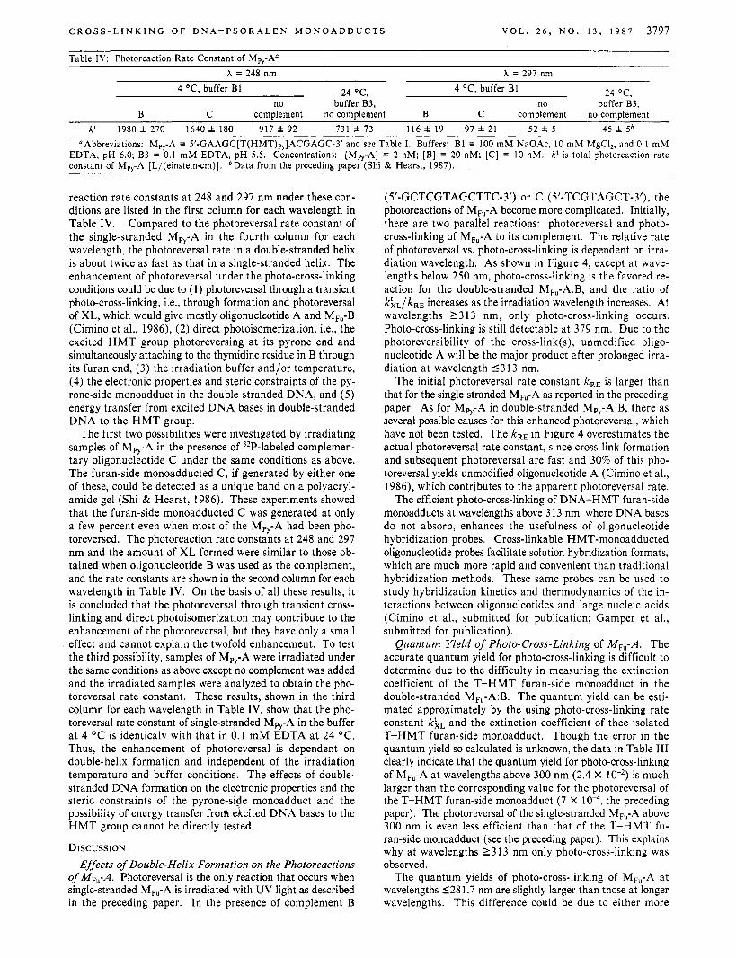
Table IV: Photoreaction Rate Constant of Mp,-An X = 248 nm X = 297 nm
24 ‘C , 4 OC, buffer B1 24 OC, 4 OC, buffer B1 no buffer B3, no buffer B3,
complement no complement B C complement no complement B C k‘ I980 f 270 1640 i 180 917 f 92 731 f 73 116f 19 97f21 52 f 5 45 f 56
OAbbreviations: Mpy-A = 5’-GAAGC[T(HMT),,]ACGAGC-3’ and see Table I. Buffers: B1 = 100 mM NaOAc, 10 mM MgCI,, and 0.1 mM EDTA, pH 6.0; B3 = 0.1 mM EDTA, pH 5.5. Concentrations: [Mpy-A] = 2 nM; [B] = 20 nM; [C] = 10 nM. k‘ is total photoreaction rate constant of Mpy-A [L/(einstein.cm)]. bData from the preceding paper (Shi & Hearst, 1987).
reaction rate constants a t 248 and 291 nm under these con- ditions are listed in the first column for each wavelength in Table IV. Compared to the photoreversal rate constant of the single-stranded MPy-A in the fourth column for each wavelength, the photoreversal rate in a double-stranded helix is about twice as fast as that in a single-stranded helix. The enhancement of photoreversal under the photo-cross-linking conditions could be due to (1) photoreversal through a transient photo-cross-linking, Le., through formation and photoreversal of XL, which would give mostly oligonucleotide A and Mw-B (Cimino et al., 1986), (2) direct photoisomerization, Le., the excited H M T group photoreversing a t its pyrone end and simultaneously attaching to the thymidine residue in B through its furan end, (3) the irradiation buffer and/or temperature, (4) the electronic properties and steric constraints of the py- rone-side monoadduct in the double-stranded DNA, and ( 5 ) energy transfer from excited DNA bases in double-stranded DNA to the H M T group.
The first two possibilities were investigated by irradiating samples of Mp,-A in the presence of 32P-labeled complemen- tary oligonucleotide C under the same conditions as above. The furan-side monoadducted C, if generated by either one of these, could be detected as a unique band on a polyacryl- amide gel (Shi & Hearst, 1986). These experiments showed that the furan-side monoadducted C was generated a t only a few percent even when most of the Mpy-A had been pho- toreversed. The photoreaction rate constants at 248 and 297 nm and the amount of XL formed were similar to those ob- tained when oligonucleotide B was used as the complement, and the rate constants are shown in the second column for each wavelength in Table IV. On the basis of all these results, it is concluded that the photoreversal through transient cross- linking and direct photoisomerization may contribute to the enhancement of the photoreversal, but they have only a small effect and cannot explain the twofold enhancement. To test the third possibility, samples of Mpy-A were irradiated under the same conditions as above except no complement was added and the irradiated samples were analyzed to obtain the pho- toreversal rate constant. These results, shown in the third column for each wavelength in Table IV, show that the pho- toreversal rate constant of single-stranded M,-A in the buffer a t 4 “C is identicaly with that in 0.1 m M EDTA a t 24 “C. Thus, the enhancement of photoreversal is dependent on double-helix formation and independent of the irradiation temperature and buffer conditions. The effects of double- stranded DNA formation on the electronic properties and the steric constraints of the pyrone-side monoadduct and the possibility of energy transfer frorh &cited DNA bases to the H M T group cannot be directly tested.
DISCUSSION Effects of Double-Helix Formation on the Photoreactions
of MF,-A. Photoreversal is the only reaction that occurs when single-stranded MF,-A is irradiated with UV light as described in the preceding paper. In the presence of complement B
(5’-GCTCGTAGCTTC-3’) or C (5’-TCGTAGCT-3’), the photoreactions of M,-A become more complicated. Initially, there are two parallel reactions: photoreversal and photo- cross-linking of MF,-A to its complement. The relative rate of photoreversal vs. photo-cross-linking is dependent on irra- diation wavelength. As shown in Figure 4, except at wave- lengths below 250 nm, photo-cross-linking is the favored re- action for the double-stranded MF,-A:B, and the ratio of khL/kRE increases as the irradiation wavelength increases. At wavelengths 1 3 13 nm, only photo-cross-linking occurs. Photo-cross-linking is still detectable a t 379 nm. Due to the photoreversibility of the cross-link(s), unmodified oligo- nucleotide A will be the major product after prolonged irra- diation a t wavelength 1313 nm.
The initial photoreversal rate constant kRE is larger than that for the single-stranded Mm-A as reported in the preceding paper. As for Mpy-A in double-stranded Mp,-A:B, there as several possible causes for this enhanced photoreversal, which have not been tested. The kRE in Figure 4 overestimates the actual photoreversal rate constant, since cross-link formation and subsequent photoreversal are fast and 30% of this pho- toreversal yields unmodified oligonucleotide A (Cimino et al., 1986), which contributes to the apparent photoreversal rate.
The efficient photo-cross-linking of DNA-HMT furan-side monoadducts at wavelengths above 313 nm, where DNA bases do not absorb, enhances the usefulness of oligonucleotide hybridization probes. Cross-linkable HMT-monoadducted oligonucleotide probes facilitate solution hybridization formats, which are much more rapid and convenient than traditional hybridization methods. These same probes can be used to study hybridization kinetics and thermodynamics of the in- teractions between oligonucleotides and large nucleic acids (Cimino et al., submitted for publication; Gamper et al., submitted for publication).
Quantum Yield of Photo-Cross-Linking of MF,-A. The accurate quantum yield for photo-cross-linking is difficult to determine due to the difficulty in measuring the extinction coefficient of the T-HMT furan-side monoadduct in the double-stranded MF,-A:B. The quantum yield can be esti- mated approximately by the using photo-cross-linking rate constant kkL and the extinction coefficient of thee isolated T-HMT furan-side monoadduct. Though the error in the quantum yield so calculated is unknown, the data in Table I11 clearly indicate that the quantum yield for photo-cross-linking of MF,-A a t wavelengths above 300 nm (2.4 X is much larger than the corresponding value for the photoreversal of the T-HMT furan-side monoadduct (7 X the preceding paper). The photoreversal of the single-stranded MF,-A above 300 nm is even less efficient than that of the T-HMT fu- ran-side monoadduct (see the preceding paper). This explains why a t wavelengths 2313 nm only photo-cross-linking was observed.
The quantum yields of photo-cross-linking of MF,-A a t wavelengths 1281.7 nm are slightly larger than those at longer wavelengths. This difference could be due to either more
3798 B I O C H E M I S T R Y S H I A N D H E A R S T
efficient cross-linking of the monoadduct at these wavelengths or energy transfer from excited DNA bases to the HMT group in the monoadduct, especially around 260 nm, where DNA bases absorb efficiently. At wavelengths above 300 nm, DNA bases absorb little light, and only the absorption of the T- HMT furan-side monoadduct in the helix contributes to the photo-cross-linking. The quantum yield at wavelengths above 300 nm is similar to that observed for the photo-cross-linking of the thymidine-8-methoxypsoralen furan-side monoadduct in calf thymus DNA (Tessman et al., 1985).
Enhanced Photoreversal of Mpy-A in Double-Stranded DNA. In contrast to M,,-A:B, UV irradiation of double- stranded Mpy-A:B results mostly in photoreversal of Mpy-A. Consequently, the photoreaction rate constant approximates the value of the photoreversal rate constant of Mp,-A in the double helix. Results show that photoreversal of M,-A in the double helix is more efficient than that in the single-stranded helix. Upon the formation of the double-stranded Mpy-A:B, the electronic and steric constraints of the T-HMT pyrone-side monoadduct may change, thereby affecting the photoreversal of the monoadduct. As described above, energy transfer from excited DNA bases in the double-stranded DNA to the HMT group may also play a role in the enhancement of photorev- ersal. Two other mechanisms, though having only a small effect as described above, can also contribute to the enhanced photoreversal. These are photoreversal through a transient cross-link formation and direct photoisomerization. In the first case, the cross-link is formed initially and then photoreversed. Approximately 70% of the photoreversal of the cross-link will produce the furan-side monoadducted B and unmodified ol- igonucleotide A (Cimino et al., 1986), consequently increasing the apparent photoreversal rate constant of Mpy-A. In the second case, the excited H M T group photoreverses at the pyrone end and simultaneously attaches to B through its furan end. This yields the same products as in the first case.
CONCLUSIONS In this paper we have shown that the photoreactions of the
HMT-monoadducted oligonucleotide in the double-stranded state are quite different from that in the single-stranded state. In the case of the pyrone-side monoadducted oligonucleotide,
Mpy-A, photoreversal is the predominant reaction even in a double helix, and very little cross-link is generated. The photoreversal of the monoadduct in the double-stranded DNA is enhanced compared to that in the single-stranded DNA. In contrast, UV irradiation of MF,-A in the double-stranded state produces predominantly cross-link, although the amount of cross-link decreases after prolonged irradiation at wavelengths 5313 nm due to photoreversal of the cross-link. At wave- lengths 1 3 13 nm, no photoreversal of MF,-A can be detected, and photo-cross-linking occurs with a quantum yield of 2.4 x 10⁻².
ACKNOWLEDGMENTS We thank Dr. G. D. Cimino, Dr. H. Gamper, and J. D.
Kahn for helpful discussions and critical comments on the manuscript.
REFERENCES Bensasson, R. V., Salet, C., Land, E. J., & Rushton, F. A. P.
(1980) Photochem. Photobiol. 31, 129-1 33. Blais, J. Ronfard-Haret, J . C., Vigny, P., Cadet, J., & Voi-
turiez, L. (1985) Photochem. Photobiol. 42, 599-602. Cimino, G. D., Gamper, H. B., Isaacs, S. T., & Hearst, J. E.
(1985) Annu. Rev. Biochem. 54, 1151-1 193. Cimino, G. D., Shi, Y.-B., & Hearst, J. E. ( 1 986) Biochem-
istry 25, 3013-3020. Gasparro, F. P., Saffran, W. A,, Cantor, C. R., & Edelson, R. L. (1 984) Photochem. Photobiol. 40, 2 15-2 19.
Land, E. J., & Truscott, T. G. (1979) Photochem. Photobiol.
Maniatis, T., Fritsch, E. F., & Sambrook, J. (1982) in Mo- lecular Cloning, pp 125-127, Cold Spring Harbor Labo- ratory, Cold Spring Harbor, NY.
Parsons, B. J. (1980) Photochem. Photobiol. 32, 8 13-82 1. Shi, Y., & Hearst, J. E. (1986) Biochemistry 25, 5895-5902. Shi, Y., & Hearst, J. E. (1987) Biochemistry (preceding paper
Song, P. S., & Tapley, K. J., Jr. (1979) Photochem. Photobiol.
Tessman, J. W., Isaacs, S. T., & Hearst, J. E. (1985) Bio-
29, 861-866.
in this issue).
29, 1177-1 197.
chemistry 24, 1669-1676.