vitamin d in the skin physiology and pathology · vitamin d in the skin physiology and pathology...
TRANSCRIPT
Review
Vitamin D in the skin physiology and pathologyAnna Piotrowska, Justyna Wierzbicka and Michał A. Żmijewski*
Department of Histology, Medical University of Gdańsk, Gdańsk, Poland
Vitamin D plays important, pleiotropic role in the main-tenance of global homeostasis. Its influence goes far beyond the regulation of calcium and phosphorus bal-ance, as diverse activities of vitamin D and its natural metabolites assure proper functioning of major human organs, including skin. Recently, we reviewed the current understanding of vitamin D impact on human health from historical perspective (Wierzbicka et al. (2014) The renaissance of vitamin D. Acta Biochim Pol 61: 679–686). This article focuses on its functions in the skin. The skin and its appendages, creates a platform connecting and protecting internal organs against, usually harmful, ex-ternal environment. It uppermost layer — epidermis in order to maintain a protective barrier undergoes a con-stant exchange of cornified keratinocytes layer. Its dis-turbance leads to development of serious skin disorders including psoriasis, vitiligo, atopic dermatitis and skin cancer. All of those dermatopathologies have a huge im-pact on modern societies, affecting not only the physi-cal, but also mental state of patients as well as their social status. Furthermore, multiple human systemic dis-eases (autoimmune, blood and digestive diseases) have skin manifestation, thus “condition of the skin” often re-flects the condition and pathological changes within the internal organs. In humans, the skin is the natural source of vitamin D, which is produced locally from 7-dehydro-cholesterol in photoreaction induced by ultraviolet B (UVB) radiation from the sun. It is also well established, that the process of proliferation and differentiation of keratinocytes is tightly regulated by calcium and the ac-tive form of vitamin D (1,25(OH)2D3). Thus, the skin phys-iology is inseparably connected with vitamin D produc-tion and activity. Unfortunately, UVB, which is required for vitamin D production, is also known as the main cause of a skin cancer, including melanoma. Here, we are going to review benefits of vitamin D and its analogues in the maintenance of epidermal barrier and its potential use in the treatment of common skin diseases.
Key words: vitamin D, vitamin D , cancer, skin, keratinocytes, vitamin D analogues
Received: 09 July, 2015; revised: 18 November, 2015; accepted: 29 November, 2015; available on-line: 28 January, 2016
INTRODUCTION
Vitamin D is considered as the oldest hormone on the earth, and there is no doubt, that this ancient molecule is inherently connected with well-being of almost every form of life, from phytoplankton to humans (Holick et al., 2007). Even though the vitamin D physiological role, most commonly, is attributed to maintenance of muscu-loskeletal system, the biological properties of this rela-
tively simple compound goes far beyond the regulation of calcium-phosphorus homeostasis (Wacker & Holick, 2013).
The major source of vitamin D is the exposure of epidermis to solar irradiation. In the photochemical reac-tion 7-dehydrocholesterol (7-DHC) is converted to vita-min D3 under UVB light (280–320 nm) in keratinocytes of the basal layer of epidermis (Bikle, 2012). After its release to the extracellular space, vitamin D3 is captured in the capillary bed by vitamin D binding protein (DBP) (Holick et al., 2007). Vitamin D produced in the skin or obtained with food is biologically inactive and requires two subsequent hydroxylations to gain its full hormonal activity (Holick et al., 2007; Holick, 2011). Initially, vita-min D3 is converted to 25-hydroxyvitamin D3 (25(OH)D3) in hepatocytes, by the key vitamin D 25-hydroxylase — CYP2R1 (Cheng et al., 2004; Strushkevich et al., 2008). 25(OH)D3 is the major metabolite of vitamin D, thus its serum level is widely used in clinic as the representation of vitamin D status (Holick, 2011). The second requisite hydroxylation occurs in the kidney, due to the action of another hydroxylase — CYP27B1 which results in for-mation of 1,25(OH)2D3, calcitriol (Takeyama et al., 1997). The level of vitamin D is tightly regulated by CYP24A1, which is 24-hydroxylase. Its activity leads to deactivation of 1,25(OH)2D3 or 25(OH)D3 and their subsequent re-moval with urine (Bikle, 2011). Finally, recently revealed alternative metabolic pathway for 7-DHC and vitamin D with major contribution of CYP450scc (CYP11A1) broadens the spectrum of naturally occurring vitamin D derivatives (see recent reviews for discussion: Slominski et al., 2013a–c; 2014b).
*e-mail: [email protected]: 1α,25(OH)2D3, calcitriol (1α,25-dihydroxyvitamin D3); 20-OH D3, 20-hydroxyvitamin D3; 25-OH D3, calcifediol (25-hy-droxyvitamin D3); 7-DHC, 7-dehydrocholesterol (provitamin D3, cholesta-5,7-dien-3β-ol); AD, atopic dermatitis; AMPs, antimicrobial peptides; BAK, Bcl-2 homologous antagonist/killer, a pro-apoptotic member of the Bcl-2 gene family; BAX, Bcl-2-associated X protein, a pro-apoptotic member of the Bcl-2 gene family; BCC, basal cell carcinoma; CaR, calcium receptor; CDKN1A, cyclin-dependent ki-nase inhibitor 1A; CMM, cutaneous malignant melanoma; CYP24 or 24OHase, 24-hydroxylase; CYP27A1 or 25OHase, 25-hydroxylase; CYP27B1 or 1αOHase, 1α-hydroxylase; CYP450scc, cytochrome P450scc, also known as CYP11A1; DBP, vitamin D-binding protein; DRIP/mediator-vitamin D receptor interacting protein; GADD45A, growth arrest and DNA-damage-inducible protein, alpha; IFN, in-terferon; IL, interleukin; JLS, juvenile localized scleroderma; MARRS receptor, membrane-associated rapid response to steroid binding protein (other names: ERp57, GRp58, Pdia3); MED, minimal erythe-mal dose; NFkappaB, nuclear factor kappa-light-chain-enhancer of activated B cells; PDIA3, protein disulfide-isomerase A3; PKC, pro-tein kinase C; PLC, phospholipases C; PUVA, psoralen and ultravio-let A radiation; SCC, squamous cell carcinoma; SNP, single-nucleo-tide polymorphism; SRC, steroid receptor coactivator; SSc, systemic sclerosis; TNFα, tumour necrosis factor; UVA/B, ultraviolet radiation A and B; VDR, vitamin D receptor; VDRE, vitamin D response ele-ments
Vol. 63, No 1/201617–29
http://dx.doi.org/10.18388/abp.2015_1104
18 2016A. Piotrowska and others
1,25(OH)2D3, a hormonal form of vitamin D3, may elicit rapid responses, which rely on nuclear receptor — VDR. Upon binding of 1,25(OH)2D3, VDR with its co-receptor RXR form a powerful transcription factor, that regulates expression of more than 3 000 target genes in human genome (Haussler et al., 2011). On the other hand, 1,25(OH)2D3 can bind to ER membrane-bound protein — PDIA3. As a result, multiple signal transduc-tion pathways are activated, leading to changes in intra-cellular calcium concentration (Haussler et al., 2011; Ne-mere et al., 2012).
Beyond the beneficial role of vitamin D3 in the regula-tion of calcium homeostasis, it is already known, that the potential of activated 1,25(OH)2D3 is much more diverse. The VDR is present not only in enterocytes or osteo-blasts, but in multiple immune cells, cells of parathyroid gland, keratinocytes and ovarian cells as well (DeLuca, 2004). It should be also emphasized, that vitamin D3 supplementation improves muscle performance and re-duces falls in vitamin D-deficient older adults (Garcia et al., 2011). Heath and Elovic reported, that nearly 90% of patients, who suffered from musculoskeletal pain, were vitamin D3 deficient (Heath & Elovic, 2006). It is already suggested that vitamin D3 might have an anabolic effect on skeletal muscles (Okuno et al., 2012). Furthermore, the growing body of evidence suggests also neuroprotec-tive role of vitamin D3. A few years ago Eyles and cow-orkers reported, that vitamin D receptor and 25(OH)D3-1α-hydroxylase (CYP27B1), are both present in the brain, mainly in the hypothalamus and the dopaminer-gic neurons of the substantia nigra (Eyles et al., 2005). It was also shown, that higher concentration of vitamin D3 in the bloodstream is associated with better performance on neuropsychiatric tests in the non-demented subset of patients suffering from Parkinson’s disease, especially concerning verbal fluency and verbal memory (Peterson et al., 2013). Recent study also suggests potential use of high-doses of vitamin D in prevention and treatment of preeclampsia in pregnancy, broadening the spectrum of its medical uses (Zabul et al., 2015).
Although the beneficial effect of vitamin D on human health is already well established, vitamin D deficiencies have become a global problem (Holick, 2006; Hossein-nezhad & Holick, 2013). Recent studies conducted in Poland also underlined this issue (Kmieć et al., 2014; Pludowski et al., 2014, Kmieć et al., 2015). Furthermore, seasonal changes in vitamin D level that were observed, pointed out the necessity of its proper supplementation, especially in the winter season (Pludowski et al., 2013, Kmieć et al., 2015).
The active form of vitamin D3 — calcitriol, was also shown to play an important role in cancer prevention. An antitumor activity of calcitriol is reflected in the in-hibition of growth, induction of differentiation and ap-optosis of cancer cells (Ylikomi et al., 2002; Deeb et al., 2007; Szyszka et al., 2012). Inhibition of growth due to vitamin D or its new analogues was reported for many cancer cell lines (Yuan et al., 2012; Chiang et al., 2013; Guo et al., 2013; Lundqvist et al., 2014; Thill et al., 2015; Wierzbicka et al., 2015). Indeed, the inverse association between the concentration of 25(OH)D in the circulat-ing serum and total cancer incidence and mortality was recently described (Yin et al, 2013), emphasizing the vital role of vitamin D in cancer prophylaxis.
Skin production is the efficient and natural way to compensate deficiency of vitamin D in the body (Hol-ick, 2007). Having in mind global precaution of the sun overexposure, as a known factor for the melanoma development, alternative sources of vitamin D are usu-
ally considered. Vitamin D may be introduced in diet, but only limited forms of food naturally contain it. The list is relatively short and includes fatty fish, cod liver oil and some mushrooms (Holick, 2011). Therefore in some countries, for instance in Great Britain or United States, due to the appreciation of vitamin D pleiotropic benefits for human health, a fortification of food with vitamin D is a strategy rather than an exception (Wacker & Holick, 2013). Unfortunately, this is still not a case in Poland and other countries, where proper food fortifi-cation with vitamin D in uncommon (Wierzbicka et al., 2014). In Poland, however, unequivocal and significantly extended legal regulations in this field are needed. At the moment, existing national legislations concerning public health issue require vitamin D fortification of margarine, butter and oils. Companies practice also fortification of milk and dairy products.
Human skin seems to be inseparably connected with vitamin D by being its source and a target as well. Here we are presenting a comprehensive overview of current knowledge concerning the impact of vitamin D and its analogues on skin physiology and pathology with poten-tial clinical implications.
VITAMIN D IN SKIN PHYSIOLOGY
Differentiation of keratinocytes
Keratinocytes forming epidermal layer of the skin are highly specialized cells designated to protect the organ-ism from the environmental factors. In order to accom-plish this goal keratinocytes produce keratin intermedi-ate filaments and secrete elements of cornified envelope (Eckert & Rorke, 1989). The epidermis is composed of keratinocytes arranged in four layers, which represent various stages of differentiation. The innermost stratum basale, which rests on the basal lamina, is composed of highly proliferating cells as well as epidermal progenitor cells that are designed to provide constituents for up-per differentiating layers. These basal cells are intercon-nected by an extensive, intracellular network of keratin filaments, mainly keratins K5 and K14 (Eichner et al., 1986). The cells of the spinous layer (stratum spinosum), which is situated above the basal layer, initiate the pro-duction of keratins K1 and K10, considered as indicators of more differentiated layers of the epidermis (Moll et al., 1982). Moreover, synthesis of involucrin (Warhol et al., 1985), which is one of the cornified envelope precursors, and the transglutaminases (Thacher & Rice, 1985; Eckert et al., 2014), responsible for cross-linking of these pre-cursors, also begins in the stratum spinosum. The process of keratinocytes’ differentiation escalates in the granular layer (stratum granulosum). The structural changes of the cells in this particular layer involve accumulation of spe-cific keratohyalin granules in the cytoplasm, filled with loricrin, involucrin and profilaggrin. The latter is the pre-cursor of filaggrin, which is believed to be involved in the process of keratin filaments aggregation (Dale et al., 1985). What is more, the cytoplasm of granular cells is enriched in lamellar bodies, glycolipids-filled structures, which contribute to the formation of water-impermeable barrier (Elias et al., 1988). Simultaneously, the nuclei of the cells undergo atrophy and eventually newly formed superficial cornified layer (stratum corneum) is composed of tightly packed, akaryotic cells that provide highly in-soluble barrier against water loss and invasion of patho-gens as well (Hennings & Holbrook, 1983).
Vol. 63 19Vitamin D in the skin physiology and pathology
Skin as a target of vitamin D
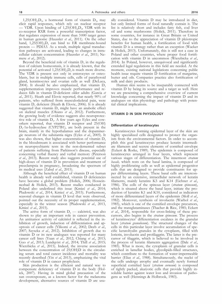
Vitamin D is produced endogenously in the skin. It was also demonstrated that keratinocytes produce abundant quantities of 1α,25(OH)2D3 from 25-OH D3 under the regulation of exogenous 1α,25(OH)2D3. Importantly, biologically active vitamin D3 produc-tion varies with the degree of the keratinocytes dif-ferentiation (Pillai et al., 1988). What is more, since keratinocytes express VDR, they are therefore able to respond to the fully active form of vitamin D3 — 1α,25(OH)2D3, which, together with calcium, is one of the most potent regulators of the epidermal differen-tiation (Bikle, 2004). In vivo calcium forms gradient in the epidermis (Fig. 1), with the lowest concentration in stratum basale and the highest in the stratum granu-losum (Menon et al., 1985). 1,25(OH)2D3 increases the expression of involucrin, transglutaminase, loricrin and filaggrin and potentiates calcium-induced differentia-tion of the keratinocytes at the level of both gene ex-pression and mRNA stability (Su et al., 1994). It also enhances formation of the cornified envelope (Bikle & Pillai, 1993). This phenomenon occurs, at least partially, due to the ability of the hormonal form of vitamin D3 to increase intracellular calcium levels via induction of the calcium receptor (CaR) and phospho-lipase C (PLC) (Pillai et al., 1995; Ratnam et al., 1999). On the other hand, 1α,25(OH)2D3 inhibits prolifera-tion of keratinocytes (Bikle, 2011). During the differ-entiation process of epidermal cells specific genes are sequentially turned on and off, due to the collective work of 1α,25(OH)2D3 and calcium, to fulfil the inher-ent specialization of keratinocytes (Bikle, 2004). For instance, keratinocytes of the basal layer express cy-tokeratins 5 and 14, which are replaced by cytokerat-ins 1, 10 and involucrin in the spinous layer (Bikle, 2004). Two distinct coactivators are involved in VDR transactivation during keratinocytes differentiation: interacting proteins (DRIP/mediator) and the p160 steroid receptor coactivator family (SRC/p160) (Bikle et al., 2003; Bikle et al., 2004). Interestingly, in pro-liferating keratinocytes, the VDR binds selectively to the DRIP/mediator complex of coactivators. During differentiation DRIP’s expression is decreasing and VDR switches partners in favour of SRC/p160 (Fig.
1). Both of discussed coactivators effectively poten-tiate vitamin D-induced transcription in proliferating cells (Oda et al., 2004). Knockdown of DRIP205 in epithelium causes increased keratinocytes proliferation (Oda, et al., 2007) and disturbs expression of keratins 1, 10, and involucrin (Hawker, et al., 2007). On the contrary, SRC3 knockdown decreases the production of glucosylceramide and the lamellar body formation (Oda, et al., 2009). These studies indicated, that both coactivators are differentially involved in vitamin D-induced differentiation of epidermal keratinocytes — SRC in regulation of terminal differentiation, whereas DRIP in regulation of proliferation and early keratino-cytes differentiation. Interestingly, it was also shown, that apart from coactivators, liganded VDR interacts reciprocally with β-catenin, which itself promotes pro-liferation rather than differentiation. This interaction results in enhanced expression of VDR-stimulated genes involved in the differentiation process, while genes involved in proliferation stimulated solely by β-catenin are repressed (Hu et al., 2014).
Another aspect of keratinocytes growth and differ-entiation is the formation of cell-cell junctions, which are essential for intercellular communication and there-fore for regulation of epithelial morphogenesis, growth, and differentiation (Klymkowsky & Parr, 1995). Now it is well known, that any impairment of intracellular junctions is closely associated with carcinogenesis, tu-mour progression, and metastasis (Frixen et al., 1991; Mbalaviele et al., 1996). Expression of E-cadherin and β-catenin (proteins forming intracellular junctions) is also decreased in skin malignant tumours, including basal cell carcinoma, squamous cell carcinoma and melanoma (Fuller et al., 1996; Seline et al., 1996; Takayama et al., 1996). One of the studies revealed, that 4-day incubation of human keratinocytes with 1α,25(OH)2D3 caused the assembly of adherens junctions, but not of desmosomes. The same study demonstrated that 1α,25(OH)2D3 may induce formation of intracellular junctions by protein ki-nase C (PKC) activation (Gniadecki et al., 1997). Thus, it is speculated that 1α,25(OH)2D3-induction of cell–cell junctions formation may be a novel, promising mecha-nism of the anti-neoplastic and anti-proliferative cancer treatment.
Figure 1. Regulation of keratinocytes functions, proliferation and differentiation by calcium and VDR with its co-activators .
20 2016A. Piotrowska and others
UV LIGHT — FRIEND OF FOE
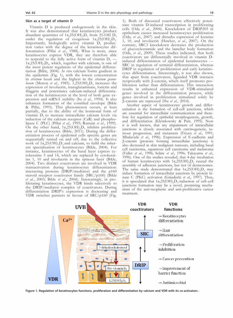
It is well established that sunlight (mainly ultraviolet type B (UVB)) is required for the efficient production of vitamin D. Paradoxically, the same solar radiation is considered as one of the most harmful factors for the skin. The UVB (280–320 nm) causes direct DNA and cell damage, thus contributes to the development of skin neoplasia. On the other hand, the UVA (320–400 nm) is mainly responsible for the skin aging. Thus, for the last decades physicians and scientists are warning against the potential danger concerning sunbathing (O’Leary et al., 2014). As a result, people all over the world avoid the sun, but meanwhile we are facing the global vitamin D deficiency (Ben-Shoshan, 2012), with noticeable outburst of rickets (Misra et al., 2008). Surprisingly, the number of new cases of melanoma and none melanoma skin cancer have constantly increased over the last years. The latter could be attributed to better diagnostic methodology, but it seems also, that simply avoiding the sun is not the solu-tion. Moreover, it is well established, that melanoma may develop in the unexposed area of the skin, such as soles and palms which is observed especially in individuals with dark complexion (Bataille, 2013). Recent studies also sug-gested that occupational exposure to the solar radiation is actually a protecting factor (Field et al., 2013), but one also has to take under consideration, that the history of sun-burns dramatically increases the probability of melanoma (Cust et al., 2011). It has to be underlined, that optimal skin photoproduction of vitamin D does not require ex-tensive sunbathing. As little as approximately 15 minutes long exposure of arms and legs in a sunny day (0.25–0.50 minimal erythemal dose (MED)) is sufficient to generate equivalent of about 2 000–4 000 IU of vitamin D (Plu-dowski et al., 2013). Moreover, it was estimated, that ex-posure to 1 MED results in production of around 20 000 units of vitamin D (Holick, 2004). Nevertheless, it is rec-ommended to expose the skin to the sun, but without ex-ceeding 1 MED, or just simply provide an equivalent of vitamin D by supplementation. The detailed recommenda-tions concerning the supplementation for different groups were published recently (Płudowski et al., 2013).
Interestingly, it is not possible to overdose vitamin D by sunbathing, because the excessive exposition to UV light leads to structural rearrangements of vitamin D and its subsequent photodegradation. The main prod-ucts are 5,6-transvitamin D3, and suprasterols I and II (Webb et al., 1989). Moreover, irradiation of 7-DHC and its analogues, called 5,7-dienes, may result in formation of 5,7,9(11)-trienes, which were described as a photo-sensitizing agents, thought to be responsible for gen-eration of reactive oxygen species (Chignell et al., 2006; Zmijewski et al., 2009). It is still unknown, whether vi-tamin D has any anti-oxidative or pro-oxidative proper-ties, because several groups presented contradictory re-sults and concepts. For instance, recent studies showed protective effects of 1α,25(OH)2D3 and its analogues against UVB-induced DNA damage (Gordon-Thomson et al., 2012; Slominski et al., 2015b), while previously de-scribed results suggested otherwise. It seems that several factors have to be taken under consideration. First, one has to differentiate between the effects of 1α,25(OH)2D3 on cellular homeostasis and the process of vitamin D production. The best studied effects of 1α,25(OH)2D3 require VDR activation and lead to alteration of gene expression, including several genes involved in reactive oxygen response and DNA repair (Moukayed & Grant, 2013). Vitamin D is also considered as a stimulator of melanogenesis, thus contributing to the skin protection against UV irradiation (see for discussion: Szyszka et al., 2012). Meanwhile, the photolysis of 7-DHC provides surprisingly large variety of by-products and alternative routes, including formation of 5,7,9(11)-trienes and oxi-dized derivatives (for instance as reported recently for short-side chain analogues of 7-DHC) (Zmijewski et al., 2009; Zmijewski et al., 2011; Slominski et al., 2013c–d). Although it requires further investigation, all of those compounds may not only possess similar activity to vita-min D, but also unique properties, including direct inter-action with reactive oxygen species generated as a result of UVB irradiation. Secondly, genomic and non-genomic effects has to be distinguished (see our recent review for discussion: Wierzbicka et al., 2014). Finally, the effect of vitamin D might be strongly dependent on cell type and
Figure 2. Effect of vitamin D deficiency on development of skin diseases and potential advantages of proper supplementation.
Vol. 63 21Vitamin D in the skin physiology and pathology
modulated by internal and external factors, pathological conditions or genetic background. Thus, it seems that in order to understand the complexity of vitamin D func-tion in the human skin, we have to go far beyond classi-cal pathways and structures, especially in relation to so-lar radiation (Slominski et al., 2013c, d; Slominski et al., 2014a; Slominski et al., 2014b). The possible interactions between UV radiation, skin carcinogenesis and vitamin D are summarised in Fig. 2.
UV light is not only the main factor in skin car-cinogenesis, but also strongly contributes to skin ag-ing, which could be defined as a slow process of flat-tening of both epidermis and dermis which results in deterioration of skin elasticity and its barrier function. It is caused by the decrease in the number of and the increase of heterogeneity of keratinocytes and melano-cytes as well as by decreased mitotic activity, migra-tion and differentiation of keratinocytes. Skin aging is associated with decline in DNA repair capability, mitochondrial dysfunction, destabilization of extracel-lular matrix, including disintegration of dermal colla-gen and elastic fibers, and overall deregulation of cel-lular metabolism. Interestingly, all of those could be associated with hormonal dysfunction (Zouboulis & Makrantonaki, 2011). It has to be stressed out that the vitamin D production capacity of the skin decreases with age, thus the supplementation in the elderly is especially recommended (Zouboulis & Makrantonaki, 2011). Several factors are considered to explain this phenomenon besides limited sun exposure and insuf-ficient dietary supplementation. For instance, the de-creased level of vitamin D precursor — 7-DHC was observed in elderly people, which also coincides with lower level of previtamin D in the skin. Thus, elder-ly population is more prone to vitamin D deficiency (Zouboulis & Makrantonaki, 2011).
Summarizing, sun is a friend, but it has to be used wisely. Its beneficial effects, exemplified by epidermal vitamin D production, may be overcome by melano-ma risk, especially in individuals with very light com-plexion, freckles and red hair, which, among others, are very strong contraindications for direct solar ex-posure.
VITAMIN D IN SKIN PATHOLOGY
Skin cancer
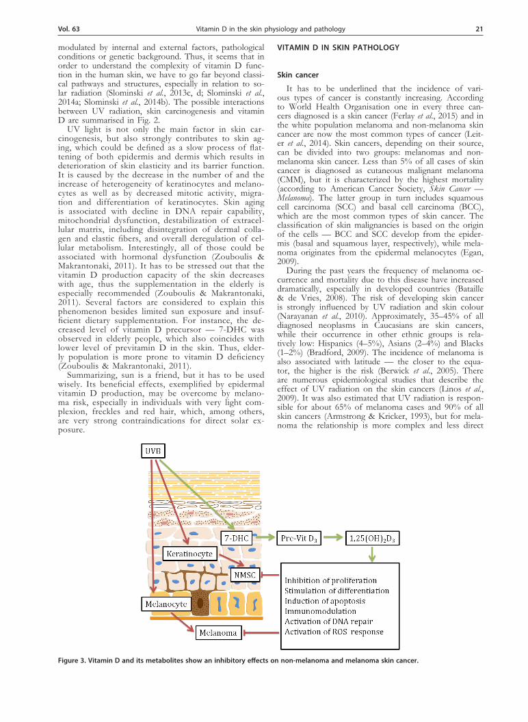
It has to be underlined that the incidence of vari-ous types of cancer is constantly increasing. According to World Health Organisation one in every three can-cers diagnosed is a skin cancer (Ferlay et al., 2015) and in the white population melanoma and non-melanoma skin cancer are now the most common types of cancer (Leit-er et al., 2014). Skin cancers, depending on their source, can be divided into two groups: melanomas and non-melanoma skin cancer. Less than 5% of all cases of skin cancer is diagnosed as cutaneous malignant melanoma (CMM), but it is characterized by the highest mortality (according to American Cancer Society, Skin Cancer — Melanoma). The latter group in turn includes squamous cell carcinoma (SCC) and basal cell carcinoma (BCC), which are the most common types of skin cancer. The classification of skin malignancies is based on the origin of the cells — BCC and SCC develop from the epider-mis (basal and squamous layer, respectively), while mela-noma originates from the epidermal melanocytes (Egan, 2009).
During the past years the frequency of melanoma oc-currence and mortality due to this disease have increased dramatically, especially in developed countries (Bataille & de Vries, 2008). The risk of developing skin cancer is strongly influenced by UV radiation and skin colour (Narayanan et al., 2010). Approximately, 35–45% of all diagnosed neoplasms in Caucasians are skin cancers, while their occurrence in other ethnic groups is rela-tively low: Hispanics (4–5%), Asians (2–4%) and Blacks (1–2%) (Bradford, 2009). The incidence of melanoma is also associated with latitude — the closer to the equa-tor, the higher is the risk (Berwick et al., 2005). There are numerous epidemiological studies that describe the effect of UV radiation on the skin cancers (Linos et al., 2009). It was also estimated that UV radiation is respon-sible for about 65% of melanoma cases and 90% of all skin cancers (Armstrong & Kricker, 1993), but for mela-noma the relationship is more complex and less direct
Figure 3. Vitamin D and its metabolites show an inhibitory effects on non-melanoma and melanoma skin cancer.
22 2016A. Piotrowska and others
than for non-melanoma skin cancer. The BCC and SCC are proved to arise from skin chronically exposed to sunlight, while melanoma can develop in regions of the body (palms, armpits and soles) with very limited con-tact with UV radiation (Bataille & de Vries, 2008; Egan, 2009), or even in internal organs such as oesophagus, trachea, bronchial tree, cervix, colon, urinary tract and central nervous system (Cummins et al., 2006; Brozyna et al., 2007).
Having in mind antiproliferative and immunosuppres-sive activity of vitamin D and its analogues, their use in treatment of several dermatopathies is currently under investigation (Fig. 3).
Melanoma
So far the most effective treatment for melanoma is surgical removal of the neoplastic lesions (Garbe et al., 2010). Therefore, early detection of melanoma in patient increases the chances for successful curing of this dis-ease. The significant mortality is caused by the ineffec-tiveness of standard treatment methods, particularly dur-ing the stage of metastasis (Dummer et al., 2008). Nota-ble advance in the treatment of this type of cancer was the introduction of targeted therapy with inhibitors for tyrosine kinase receptor KIT (Carvajal et al., 2011). Re-cent developments, including BRAF-inhibitors, as well as drugs targeting the MAPK pathway, bring new perspec-tives to the field, especially when used as a combined immunotherapy (Aris & Barrio, 2015). Nevertheless, seeking for new drugs is still required, since during the treatment many tumours acquire resistance to the thera-pies.
Studies conducted in the 70’s showed, that vitamin D can stimulate the activity of tyrosinase, the principal enzyme involved in melanin synthesis in cultured mel-anoma cell line (Oikawa & Nakayasu, 1974). Following studies confirmed the presence of the vitamin D recep-tor (VDR) in the primary melanoma tissue (Colston et al., 1981). Having in mind its antiproliferative properties, vitamin D and its analogues are potential candidates for melanoma treatment. In fact, it was shown that the me-tabolites of vitamin D inhibit proliferation and induce differentiation of melanoma cells expressing VDR (re-cent reviews: Szyszka et al., 2012; Slominski et al., 2014; Burns et al., 2015). Interestingly, it seems that not trans-formed melanocytes and keratinocytes are protected by 1,25(OH)2D3 and its analogues (Slominski et al., 2012). There is also evidence, that 1α,25(OH)2D3 promotes sur-vival of the cells and inhibits the tumour invasion and angiogenesis (Osborne & Hutchinson, 2002; De Haes et al., 2005). In the Gupta’s study, keratinocytes after the treatment with 1α,25(OH)2D3 and exposure to UV ra-diation were characterized by the increased survival and reduced amount of DNA damage (Gupta et al., 2007). Similar protective effect was observed in melanoma cells. Furthermore, VDR was shown to take part in ac-tivation of DNA repair mechanism. Thus, it seems that vitamin D is an important, physiologically relevant fac-tor preventing UV-induced carcinogenesis (Reichrath & Rass, 2014). It is not surprising, that a number of recent studies pointed out that the development of melanoma can be linked with vitamin D deficiency or defects in vitamin D signalling pathway (Reichrath & Nurnberg, 2009; Pinczewski & Slominski, 2010; Newton-Bishop et al., 2011). In humans, VDR gene has multiple splicing variants, which are likely to affect protein expression and activity of the VDR. In addition, over 1 000 polymorphic sites have been detected in VDR gene, some of which
have been correlated with increased risk of melanoma occurrence, its aggressiveness and prognosis (Gapska et al., 2009; Randerson-Moor et al., 2009), as well as with other pathologies (Malodobra-Mazur et al., 2012).
The best studied SNPs linked with melanoma devel-opment are: rs10735810 (T > A nucleotide change in the restriction site FokI), rs1544410 (G > A in the BsmI) and rs731236 (T > C TaqI). For instance, FokI f and BsmI b alleles have been identified as high-risk alleles (Hutchin-son et al., 2000; Li et al., 2007), while TaqI t allele as a protective one (Li et al., 2007). In addition, rs4516035 allele (change A > G in the EcoRV restriction site) has been identified as an exclusive risk allele for melanoma (Halsall et al., 2009). Similar conclusions were drawn about the influence of defects in the vitamin D signal-ling pathway on the development and progression of melanoma, mainly of the decrease in vitamin D receptor expression (Pinczewski & Slominski, 2010; Brozyna et al., 2011). It should be stressed that the analysis of VDR expression and polymorphism may serve as important prognostic factor for melanoma treatment with vitamin D and its analogues (see discussion in: Szyszka et al., 2012). What is more, recent studies proved that mela-nogenesis reduces the overall survival and disease-free survival in patients with melanoma at III and IV stage (Brozyna et al., 2013). In the light of these studies, it is also important to draw the attention to two other re-ports. The first of them demonstrated that reduced level or absence of VDR is associated with melanoma pro-gression (melanogenesis can suppress the expression of the receptor), resulting in deteriorated survival of mela-noma patients (Brozyna et al., 2011). Subsequent study showed that the expression of CYP27B1 was inversely proportional to the level of melanin in the melanoma cells in cultures, both in vivo and in vitro. Thus, reduced level of the enzyme involved in the synthesis of the ac-tive form of vitamin D (CYP27B1), correlates with mel-anoma phenotype, but has no effect on the survival of patients (Brozyna et al., 2013). Recent studies also pro-vided evidence that vitamin D and its analogues may be effective in melanoma treatment (Wasiewicz et al., 2015), especially recently tested low-calcemic analogues such as 20-OH D3 (Slominski et al., 2012) or analogues with pregnenolone-like side chain (21-hydroxypregnacalciferol) (Zmijewski et al., 2009; Zmijewski et al., 2011; Kim et al., 2012; Slominski et al., 2012; Tang et al., 2013; Slominski et al., 2015a).
Non-melanoma skin cancers
Similarly as in case of melanomas, there are number of studies concerning the role of vitamin D in basal cell carcinoma (BCC) and squamous cell carcinoma (SCC). Just like keratinocytes, BCC cells also express VDR, and furthermore, peripheral cells forming BCC tumours show even higher expression of VDR than neighbouring, un-affected epidermal cells (Kamradt et al., 2003; Mitschele et al., 2004). It was demonstrated that vitamin D sup-presses a key tumour pathway in BCCs development — hedgehog signalling pathway (Bijlsma et al., 2006; Tang et al., 2007). It was shown that VDR-knockout mice, after the exposure to a carcinogen, were more prone to BCCs skin tumours development than the wild type animals (Zinser et al., 2005). Another studies on mice showed that topical application of vitamin D3 reduces BCC cell proliferation and also inhibits the hedgehog signal-ling pathway, both in vitro and in vivo (Tang et al., 2007). 1α,25(OH)2D3 also inhibits the growth of SCCs in vivo as well as in vitro (Hershberger et al., 1999). Similar studies
Vol. 63 23Vitamin D in the skin physiology and pathology
on animals showed that mice lacking VDR and exposed to high and prolonged doses of UVB are predisposed to SCC tumour formation (Ellison et al., 2008). In addition, as in BCCs, topically applied 1α,25(OH)2D3 inhibits for-mation of chemically induced tumour in a dose-dependent manner (Wood et al., 1983; Hershberger et al., 1999). What is more, 20(OH)D3, 20,22(OH)D3 and 20,23(OH)D3, nov-el vitamin D3 analogues produced by P450scc, show pro-differentiation, anti-proliferative and anticancer properties (Zbytek et al., 2008; Janjetovic et al., 2009; Janjetovic et al., 2010; Slominski & Zmijewski et al., 2014a). Although the in vitro and animal studies suggested that vitamin D may prevent development of BCCs and SCCs, additional studies on humans are needed to assess the suitability of topical or oral vitamin D3 supplementation in chemopre-vention of non-melanoma skin cancers.
Psoriasis
Psoriasis is a chronic, inflammatory skin disease, af-fecting approximately 2% of the Western population (Burfield & Burden, 2013). The affliction in the most common form is characterized by well-demarcated red plaques, covered in scales, symmetrically affecting the extensor surfaces of the limbs and with a preference for the scalp (Slominski & Zbytek et al., 2013d; Nedoszytko et al., 2014; Stawczyk-Macieja et al., 2015).
It has to be emphasized, however, that the develop-ment of psoriasis may go far beyond the skin, resulting in the increased risk of psoriatic arthritis, inflammatory bowel disease, cardiovascular disease, depression and metabolic syndrome (Burfield & Burden, 2013). First-line therapy in psoriasis is the topical treatment with corti-costeroids and vitamin D analogues (Hsu et al., 2012; Slominski & Kim et al., 2013). Efficiency of the therapy is based on the fact, that keratinocytes as well as the lymphocytes infiltrating the lesions do express the vita-min D receptor (Provvedini et al., 1983; Burfield & Bur-den, 2013).
Interestingly, the first effect of vitamin D analogues treatment of psoriatic patient was noticed accidentally, when during 1α-hydroxyvitamin D3 therapy of osteo-porosis the unexpected remission of psoriatic lesions was observed (Morimoto & Kumahara, 1985). Over the following years the oral or topical administration of 1α,25(OH)2D3 as well as the topical treatment with 1,24(OH)2D3 were used, showing a significant improve-ment of the skin status in 70–80% of patients. What is more, the complete disappearance of any signs of psoria-sis was noticed in 20–25% of suffering patients (Nagpal et al., 2001). The topical administration of calcipotriol, another vitamin D analogue, is also practiced in psoriasis (Karthaus et al., 2014). It has to be emphasized, however, that the treatment of the disease depends on its sever-ity and location. When topical therapies fail or especially when psoriasis is extensive, the phototherapy should be managed. The best studied combination of psoralen and ultraviolet A radiation (PUVA) treatment has been used for decades to treat severe psoriasis, allowing patients to resume normal life activities. Unfortunately, PUVA was shown to increase the risk of skin cancer. Since then, the phototherapy using narrow-band UVB is the preferred option, highly effective at clearing psoriasis plaques (Bur-field & Burden, 2013). Interestingly, such treatment has to stimulate epidermal production of vitamin D, but this phenomenon and its implication to the healing process have to be studied in details, yet.
Although the pathogenesis of psoriasis is still not fully understood, it seems that development of psori-
atic plaques is mediated by Th1 cells and connected with hyperproliferation of keratinocytes. Thus, having in mind immunosuppressive and antiproliferative proper-ties of vitamin D-like compounds, such as calcipotriol, their successful introduction in the therapy of psoriasis is fully justified (Brown & Slatopolsky, 2008). Deregu-lation of the keratinocytes differentiation process ob-served in psoriasis also prevents the formation of the functional impermeable epidermal barrier. The applica-tion of 1α,25(OH)2D3 induces, among others, expres-sion of involucrin and transglutaminase and therefore stimulates the formation of cornified envelope (Pillai & Bikle, 1991). Moreover, VDR ligands inhibit expression of pro-inflammatory cytokines produced by T lympho-cytes, such as IL-2, IFN-γ, IL-6 and IL-8, which are re-sponsible for the exacerbation of the skin inflammation (Manolagas et al., 1985). Apart from that, 1α,25(OH)2D3 enhances expression of anti-inflammatory cytokine, IL-10, within the psoriatic lesions (Kang et al., 1998), as well as the expression of its receptor in keratinocytes (Michel et al., 1997). It seems that antigen presenting cells and dendritic cells in particular, also remain under modulatory influence of 1α,25(OH)2D3 and its analogues, as they inhibit differentiation, maturation, activation and the survival of these cells. Consequently, biological activity of vitamin D3 analogues leads also to suppres-sion of the T cell-mediated immune response (Penna & Adorini, 2000). Furthermore, it was also shown that cal-citriol as well as novel vitamin D3 derivatives, 20(OH)D3 and 20,23(OH)2D3, produced by Cyp450scc, inhibit the transcriptional activity of NFkappaB, a major inducer of inflammation, and therefore they may be effective therapeutic agents for inflammatory and hyperprolifera-tive skin disorders (Janjetovic et al., 2009; Janjetovic et al., 2010). Finally, it seems that epidermal production of vitamin D may be at least partially impaired in psoriatic skin, which may contribute to the aggravation of symp-toms. Recently, several single nucleotide polymorphisms (SNPs) in VDR and CYP24A1 genes were found to cor-relate with occurrence and severity of psoriasis, although it seems that effect of SNPs may be limited to the spe-cific ethnic groups, as it was suggested recently (Kostner et al., 2009).
Atopic dermatitis
Atopic dermatitis (AD) is a common chronic inflam-matory disease of the skin, clinically represented by pruri-tus and eczematous plaques. Furthermore, its occurrence not infrequently is a prelude to asthma or other allergic disorders (Boguniewicz & Leung, 2011; Mesquita Kde et al., 2013). Patients with advanced or persistent form of this disorder experience significant impairment of the quality of life (Beattie & Lewis-Jones, 2006). It should be emphasized that the incidence of AD has tripled since 1960, making this affliction a growing problem in devel-oping as well as in developed countries (Mesquita Kde et al., 2013). In the United States the occurrence is up to 20% in children and up to 3% in adults (Amestejani et al., 2012). Although the ethiopathogenesis of AD is very complex and therefore still not fully elucidated, it is connected to genetic predisposition, structural abnor-malities in the epidermis together with changes in the immune system and its interaction with environmental factors (Nedoszytko et al., 2014). It is worth pointing out that the incidence of the disease varies not only between countries, but surprisingly even between regions in the same country, showing the noticeable impact of factors, such as sun exposure or dietary habits, on manifestation
24 2016A. Piotrowska and others
of AD (Mesquita Kde et al., 2013). In addition, patients suffering from AD are prone to colonization by micro-organisms, especially Staphylococcus aureus and Herpes sim-plex virus (Mesquita Kde et al., 2013). It is estimated, that S. aureus in particular, is found in as many as 90% of patients with AD, leading to exacerbated, persistent skin inflammation. What is more, this pathogen can colonize healthy appearing skin in AD patients (Cho et al., 2001). It was shown, that vitamin D deficiency is a risk fac-tor of methicillin-resistant S. aureus infection (Matheson et al., 2010). Vitamin D is also recognized to be involved in the regulation of cutaneous production of antimicro-bial peptides (AMPs) in keratinocytes (Searing & Leung, 2010), which constitute the crucial component of the in-nate immune system for the defence against invading mi-croorganisms (Gschwandtner et al., 2014). It was already shown, that after oral supplementation with 4000 IU/d vitamin D for 21 days, lesional skin from AD patients showed a statistically significant increase in an expression of classic AMP — cathelicidin (Hata et al., 2008). It is not surprising, because the cathelicidin promoter pos-sesses putative vitamin D response element (Wang et al., 2004). Similar positive effect of vitamin D supplementa-tion versus placebo was observed in AD children (Sid-bury et al., 2008) and adults (age 14 and older) (Ameste-jani et al., 2012). In fact, the inverse relationship between the severity of atopic dermatitis and vitamin D levels was already shown (Peroni et al., 2011). VDR is considered in turn as a potent regulator of proinflammatory cytokines expression in keratinocytes, such as IL-6 or TNF-α. It is also involved in the inhibition of dendritic cells matura-tion. Polymorphism of VDR gene may therefore result in diminished responsiveness to vitamin D in inflamma-tory conditions (Heine et al., 2013). As it was stated pre-viously, in addition to AMPs expression, vitamin D also improves epidermal barrier function by modulation of the filaggrin and involucrin expression. Thus, proper vi-tamin D supplementation results in the improvement of AD patients skin status and subsequently in alleviation of the inflammatory cascade (Mutgi & Koo, 2013). This statement is supported by the observation that specific VDR haplotype is more frequent in patients with severe AD indeed (Heine et al., 2013). These encouraging re-sults point towards avoiding, at least, vitamin D hypovi-taminosis, which constitutes a risk factor for infections and chronic disorders.
Vitiligo
Vitiligo is an autoimmune pigmentary disorder, char-acterized by the destruction of melanocytes, cells respon-sible for pigment production, and clinically described by smooth, white patches within normally pigmented skin (Abu Tahir et al., 2010). The designation ‘vitiligo’ pre-sumably comes from Latin word ‘Vitilus’, that denotes ‘calf’, and was introduced in the first century A.D. by Roman physician, Celsus (Abu Tahir et al., 2010). The disorder tends to be associated with systemic autoim-mune conditions, such as hypo- and hyperthyroidism (Niepomniszcze & Amad, 2001), type I diabetes mel-litus, pernicious anaemia, rheumatoid arthritis and sys-temic lupus erythematosus (Sehgal et al., 1976; Montes et al., 2003; Adorini & Penna, 2008). Connection between vitamin D deficiency and some autoimmunological dis-eases was already reported, indicating this condition as an environmental trigger for the induction of autoim-munity (Saleh et al., 2013). It was shown recently, that serum concentration of 25-OH D3 was significantly low-er in patients with vitiligo than in the healthy controls
(p < 0.001). What is more, 70.5% of the patients with vitiligo were vitamin D deficient (25-OH D3<20ng.mL) (Aksu Cerman et al., 2014). Therefore, apart from topi-cal treatment with steroids, which are still first-line of therapy in most cases (Abu Tahir et al., 2010), combina-tion of PUVA (psoralen-UVA therapy) and vitamin D analogue calcipotriol is highly effective (Ameen et al., 2001). Vitamin D analogues as a monotherapy are not as effective as the topical corticosteroids, nevertheless their co-administration increases effectiveness of the treatment (Hossani-Madani & Halder, 2011). It was also reported that patients suffering from vitiligo, who display low lev-els of vitamin D, constitute a risk group for secondary forms of autoimmunity. Furthermore, insufficient vita-min D levels were associated with increasing Fitzpatrick phototype (Silverberg et al., 2010). Hence, monitoring of the serum levels of 25-OH D3 among patients with vitiligo, may be considered as a screening tool for the presence of coexisting autoimmunity. Still, further inves-tigations are required for the evaluation of vitamin D potential in vitiligo treatment.
Morphea
Scleroderma is a rare condition among children and adults. At least two typical categories of scleroderma could be distinguished: juvenile systemic sclerosis (SSc) and juvenile localized scleroderma (JLS). JLS, also referred to as ‘morphea’ in the literature, is char-acterized by inflammation and skin thickening with increased collagen deposition and sometimes also by subcutaneous tissue sclerosis (Nelson, 2001; Zulian et al., 2005; Zulian et al., 2006). Unfortunately, treatment of localized scleroderma is still very difficult and there is no accepted or proved therapeutic procedure for this disease. Initial studies showed that vitamin D may inhibit the proliferation of fibroblasts derived from skin of scleroderma patients, or even regulate the collagen gene expression (Boelsma et al., 1995). Yet, in clinical trials oral administration of 1,25(OH)2D3 showed mixed results. In adult patients with general-ized morphea after 3 to 7 months of treatment with 0.50 to 0.75 µg of 1,25(OH)2D3 daily, improvement of joint mobility and skin extensibility was observed (Hulshof et al., 1994). However, in more recent stud-ies oral 1,25(OH)2D3 did not demonstrate effective-ness among 20 adult patients with localized sclero-derma (Hulshof et al., 2000). On the other hand, in a study of seven children with morphea treated with oral 1,25(OH)2D3, five of them showed a good to ex-cellent reduction of their skin lesions without adverse effects. One of the patients showed an excellent re-sponse to the secondary therapy and one patient failed to respond to therapy with 1,25(OH)2D3 (Elst et al., 1999). Other studies focused on vitamin D analogue — calcipotriol — administered topically (0.005%). Af-ter 3-months trial, all 12 patients aged 12 to 38 years with active morphea or linear scleroderma showed sig-nificant skin improvement. No adverse effects were observed (Cunningham et al., 1998). Recently, novel vitamin D derivatives like 20(OH)D3 were tested for their antifibrotic activities. Both in vitro and in vivo studies showed that they may be a potential candi-dates for therapeutic agents to treat diseases like scle-roderma or fibrosis (Slominski et al., 2011; Slominski et al., 2013a). Abovementioned studies indicate, that 1,25(OH)2D3 and its analogues can be effective agents for treating localized scleroderma, but further clinical trials have to be conducted.
Vol. 63 25Vitamin D in the skin physiology and pathology
CONCLUSIONS
There is cumulative evidence that human epidermis is not only the natural source of vitamin D, but also the main regulator of skin physiology.
Overall status of vitamin D, as measured by serum concentration of 25-hydroxyvitamin D, or alternation in vitamin D response and metabolism, have strong impact on skin physiology, thus vitamin D analogues are cur-rently used or tested for potential therapy of multiple skin diseases. What is more, it seems that new vitamin D analogues with little or none impact on calcium level are promising candidates not only for topical treatment but for general applications in prophylaxis and treatment of multiple skin or systemic human disorders.
Acknolwedgements
Supported by grant of Polish Ministry of Science and Higher Education, project no. N405 623238 and N402 662840 (MAZ)
REFERENCES
Abu Tahir M, Pramod K, Ansari SH, Ali J (2010) Current reme-dies for vitiligo. Autoimmun Rev 9: 516–520. doi: 10.1016/j.au-trev.2010.02.013.
Adorini L, Penna G (2008) Control of autoimmune diseases by the vitamin D endocrine system. Nat Clin Pract Rheumatol 4: 404–412. doi: 10.1038/ncprheum0855.
Aksu Cerman A, Sarikaya Solak S, Kivanc Altunay I (2014) Vitamin D deficiency in alopecia areata. Br J Dermatol 170: 1299-304. doi: 10.1111/bjd.12980.
Ameen M, Exarchou V, Chu AC (2001) Topical calcipotriol as mono-therapy and in combination with psoralen plus ultraviolet A in the treatment of vitiligo. Br J Dermatol 145: 476–479. doi: 10.1111/j.1365-2133.2001.04381.
Aris M, Barrio MM. (2015) Combining immunotherapy with onco-gene-targeted therapy: a new road for melanoma treatment. Front Immunol 6: 1–17. doi: 10.3389/fimmu.2015.00046.
Amestejani M, Salehi BS, Vasigh M, Sobhkhiz A, Karami M, Alinia H, Kamrava SK, Shamspour N, Ghalehbaghi B, Behzadi AH (2012) Vitamin D supplementation in the treatment of atopic dermatitis: a clinical trial study. J Drugs Dermatol 11: 327–330.
Armstrong BK, Kricker A (1993) How much melanoma is caused by sun exposure? Melanoma Res 3: 395–401. http://dx.doi.org/10.1097/00008390-199311000-00002.
Bataille V, de Vries E (2008) Melanoma — Part 1: epidemiology, risk factors, and prevention. Bmj 337: 2249. http://dx.doi.org/10.1136/bmj.a2249.
Bataille V (2013) Sun exposure, sunbeds and sunscreens and melano-ma. What are the controversies? Curr Oncol Rep 15: 526–32. http://dx.doi.org/10.1007/s11912013-0342-4.
Beattie PE, Lewis-Jones MS (2006) A comparative study of impairment of quality of life in children with skin disease and children with oth-er chronic childhood diseases. Br J Dermatol 155: 145–151. http://dx.doi.org/10.1111/j.13652133.2006.07185.x.
Ben-Shoshan M (2012) Vitamin D deficiency/insufficiency and chal-lenges in developing global vitamin D fortification and supplemen-tation policy in adults. Int J Vitam Nutr Res 82: 237–259. http://dx.doi.org/10.1024/0300-9831/a000117.
Berwick M, Armstrong BK, Ben-Porat L, Fine J, Kricker A, Eberle C, Barnhill R (2005) Sun exposure and mortality from melanoma. J Natl Cancer Inst 97: 195–199. http://dx.doi.org/10.1093/jnci/dji019.
Bijlsma MF, Spek CA, Zivkovic D, van de Water S, Rezaee F, Pep-pelenbosch MP (2006) Repression of smoothened by patched-de-pendent (pro-)vitamin D3 secretion. PLoS Biol 4: 232. http://dx.doi.org/10.1371/journal.pbio.0040232.
Bikle DD, Pillai S (1993) Vitamin D, calcium, and epidermal differ-entiation. Endocr Rev 14: 3–19. http://dx.doi.org/10.1210/er.14.1.3.
Bikle DD, Tu CL, Xie Z, Oda Y (2003) Vitamin D regulated keratino-cyte differentiation: role of coactivators. J Cell Biochem 88: 290–295. http://dx.doi.org/10.1002/jcb.10339.
Bikle DD (2004) Vitamin D regulated keratinocyte differentiation. J Cell Biochem 92: 436–444. http://dx.doi.org/10.1002/jcb.20095.
Bikle DD, Oda Y, Xie Z (2004) Calcium and 1,25(OH)2D: interacting drivers of epidermal differentiation. J Steroid Biochem Mol Biol 89–90: 355–360. http://dx.doi.org/10.1016/j.jsbmb.2004.03.020.
Bikle DD (2011) Vitamin D metabolism and function in the skin. Mol Cell Endocrinol 347: 80–89. http://dx.doi.org/10.1016/j.mce.2011.05.017.
Bikle DD (2012) Vitamin D and the skin: Physiology and pathophysi-ology. Rev Endocr Metab Disord 13: 3–19. http://dx.doi.org/10.1007/s11154-011-9194-0.
Boelsma E, Pavel S, Ponec M (1995) Effects of calcitriol on fibroblasts derived from skin of scleroderma patients. Dermatology 191: 226–233. http://dx.doi.org/10.1159/000246550.
Boguniewicz M, Leung DY (2011) Atopic dermatitis: a disease of al-tered skin barrier and immune dysregulation. Immunol Rev 242: 233–246. http://dx.doi.org/10.1111/j.1600-065X.2011.01027.x.
Bradford PT (2009) Skin cancer in skin of color. Dermatol Nurs 21: 170–177, 206.
Brown AJ, Slatopolsky E (2008) Vitamin D analogs: therapeutic ap-plications and mechanisms for selectivity. Mol Aspects Med 29: 433–452. http://dx.doi.org/10.1016/j.mam.2008.04.001.
Brozyna AA, Zbytek B, Granese J, Carlson AJ, Ross J, Slominski AT (2007) Mechanism of UV-related carcinogenesis and its contribution to nevi/melanoma. Expert Rev Dermatol 2: 451–469. http://dx.doi.org/10.1586/17469872.2.4.451.
Brozyna AA, Jozwicki W, Janjetovic Z, Slominski AT (2011) Ex-pression of vitamin D receptor decreases during progression of pigmented skin lesions. Hum Pathol 42: 618–631. http://dx.doi.org/10.1016/j.humpath.2010.09.014.
Brozyna AA, Jozwicki W, Janjetovic Z, Slominski AT (2013) Expres-sion of the vitaimin D-activating enzyme 1 alpha-hydroxylase (CY-P27B1) decreases during melanoma progression. Human Pathology 44: 374–387. doi:10.1016/j.humpath.2012.03.031.
Burfield L, Burden AD (2013) Psoriasis. J R Coll Physicians Edinb 43: 334–338. doi: 10.4997/JRCPE.2013.414.
Burns EM, Elmets CA, Yusuf N (2015) Vitamin D and skin can-cer. Photochem Photobiol 91: 201–209. http://dx.doi.org/10.1111/php.12382.
Carvajal RD, Antonescu CR, Wolchok JD, Chapman PB, Roman RA, Teitcher J, Panageas KS, Busam KJ, Chmielowski B, Lutzky J, Pavlick AC, Fusco A, Cane L, Takebe N, Vemula S, Bouvier N, Bastian BC, Schwartz GK. (2011) KIT as a therapeutic target in metastatic melanoma. JAMA 305: 2327–2334. http://dx.doi.org/10.1001/jama.2011.746.
Cheng JB, Levine MA, Bell NH, Mangelsdorf DJ, Russell DW (2004) Genetic evidence that the human CYP2R1 enzyme is a key vitamin D 25-hydroxylase. Proc Natl Acad Sci 18: 7711–7715. http://dx.doi.org/10.1073/pnas.0402490101.
Chiang KC, Yeh CN, Hsu JT, Chen LW, Kuo SF, Sun CC, Huang CC, Pang JH, Flanagan JN, Takano M, Kittaka A, Juang HH, Yang SW, Chen TC (2013) MART-10, a novel vitamin D analog, inhibits head and neck squamous carcinoma cells growth through cell cycle arrest at G0/G1 with upregulation of p21 and p27 and downregula-tion of telomerase. J Steroid Biochem Mol Biol 138: 427–434. http://dx.doi.org/10.1016/j.jsbmb.2013.09.002.
Chignell CF, Kukielczak BM, Sik RH, Bilski PJ, He YY (2006) Ultravi-olet A sensitivity in Smith-Lemli-Opitz syndrome: Possible involve-ment of cholesta-5,7,9(11)-trien-3 beta-ol. Free Radic Biol Med 41: 339–346. http://dx.doi.org/10.1016/j.freeradbiomed.2006.04.021.
Cho SH, Strickland I, Boguniewicz M, Leung DY (2001) Fibronectin and fibrinogen contribute to the enhanced binding of Staphylo-coccus aureus to atopic skin. J Allergy Clin Immunol 108: 269–274. http://dx.doi.org/10.1067/mai.2001.117455.
Colston K, Colston MJ, Feldman D (1981) 1,25-dihydroxyvitamin D3 and malignant melanoma: the presence of receptors and inhibition of cell growth in culture. Endocrinology 108: 1083–1086.
Cummins DL, Cummins JM, Pantle H, Silverman MA, Leonard AL, Chanmugam A (2006) Cutaneous Malignant Melanoma. Mayo Clinic Proceedings 81: 500–507. http://dx.doi.org/10.4065/81.4.500.
Cunningham BB, Landells ID, Langman C, Sailer DE, Paller AS (1998) Topical calcipotriene for morphea/linear scleroderma. J Am Acad Dermatol 39: 211–215. http://dx.doi.org/10.1016/S0190-9622(98)70077-5.
Cust AE, Armstrong BK, Goumas C, Jenkins MA, Schmid H, Hopper JL, Kefford RF, Giles GG, Aitken JF, Mann GJ (2011) Sunbed use during adolescence and early adulthood is associated with increased risk of early-onset melanoma. Int J Cancer 128: 2425–2435. http://dx.doi.org/10.1002/ijc.25576.
Dale BA, Resing KA, Lonsdale-Eccles JD (1985) Filaggrin: a kera-tin filament associated protein. Ann N Y Acad Sci 455: 330–342. http://dx.doi.org/10.1111/j.1749-6632.1985.tb50420.x.
Deeb KK, Trump DL, Johnson CS (2007) Vitamin D signalling path-ways in cancer: potential for anticancer therapeutics. Nat Rev Cancer 7: 684–700. http://dx.doi.org/10.1038/nrc2196.
De Haes P, Garmyn M, Verstuyf A, De Clercq P, Vandewalle M, De-greef H, Vantieghem K, Bouillon R, Segaert S (2005) 1,25-Dihy-droxyvitamin D3 and analogues protect primary human keratino-cytes against UVB-induced DNA damage. J Photochem Photobiol B 78: 141–148. http://dx.doi.org/10.1016/j.jphotobiol.2004.09.010.
26 2016A. Piotrowska and others
DeLuca HF (2004) Overview of general physiologic features and func-tions of vitamin D. Am J Clin Nutr 80: 1689–1696.
Dummer R, Hauschild A, Jost L (2008) Cutaneous malignant melano-ma: ESMO clinical recommendations for diagnosis, treatment and follow-up. Ann Oncol 19: 86–88. http://dx.doi.org/10.1093/annonc/mdn100.
Eckert RL, Rorke EA (1989) Molecular biology of keratinocyte dif-ferentiation. Environ Health Perspect 80: 109–116. http://dx.doi.org/10.1289/ehp.8980109.
Eckert RL, Kaartinen MT, Nurminskaya M, Belkin AM, Colak G, Johnson GV, Mehta K (2014) Transglutaminase regulation of cell function. Physiol Rev 94: 383–417.
Egan KM (2009) Vitamin D and melanoma. Ann Epidemiol 19: 455–461. http://dx.doi.org/10.1152/physrev.00019.2013.
Eichner R, Sun TT, Aebi U (1986) The role of keratin subfamilies and keratin pairs in the formation of human epidermal intermedi-ate filaments. J Cell Biol 102: 1767–1777. http://dx.doi.org/10.1083/jcb.102.5.1767.
Elias PM, Menon GK, Grayson S, Brown BE (1988) Membrane struc-tural alterations in murine stratum corneum: relationship to the lo-calization of polar lipids and phospholipases. J Invest Dermatol 91: 3–10. http://dx.doi.org/10.1111/1523-1747.ep12463279.
Ellison TI, Smith MK, Gilliam AC, MacDonald PN (2008) Inactivation of the vitamin D receptor enhances susceptibility of murine skin to UV-induced tumorigenesis. J Invest Dermatol 128: 2508–2517. http://dx.doi.org/10.1038/jid.2008.131.
Elst EF, Van Suijlekom-Smit LW, Oranje AP (1999) Treatment of linear scleroderma with oral 1,25-dihydroxyvitamin D3 (calcitri-ol) in seven children. Pediatr Dermatol 16: 53–58. http://dx.doi.org/10.1046/j.1525-1470.1999.99016.x.
Eyles DW, Smith S, Kinobe R, Hewison M, McGrath JJ (2005) Distri-bution of the vitamin D receptor and 1 alpha-hydroxylase in human brain. J Chem Neuroanat 29: 21–30.
Ferlay J, Soerjomataram I, Dikshit R, Eser S, Mathers C, Rebelo M, Parkin DM, Forman D, Bray F (2015) Cancer incidence and mor-tality worldwide: sources, methods and major patterns in GLOBO-CAN 2012. Int J Cancer 136: 359–386. http://dx.doi.org/10.1002/ijc.29210.
Field S, Davies J, Bishop DT, Newton-Bishop JA (2013) Vitamin D and melanoma. Dermatoendocrinol 5: 121–129. http://dx.doi.org/10.4161/derm.25244.
Frixen UH, Behrens J, Sachs M, Eberle G, Voss B, Warda A, Löchner D, Birchmeier W (1991) E-cadherin-mediated cell-cell adhesion pre-vents invasiveness of human carcinoma cells. J Cell Biol 113: 173–185. http://dx.doi.org/10.1083/jcb.113.1.173.
Fuller LC, Allen MH, Montesu M, Barker JN, Macdonald DM (1996) Expression of E-cadherin in human epidermal non-mela-noma cutaneous tumours. Br J Dermatol 134: 28–32. http://dx.doi.org/10.1111/j.1365-2133.1996.tb07835.x.
Gapska P, Scott RJ, Serrano-Fernandez P, Mirecka A, Rassoud I, Gorski B, Cybulski C, Huzarski T, Byrski T, Nagay L, Maleszka R, Sulikowski M, Lubinski J, Debniak T (2009) Vitamin D recep-tor variants and the malignant melanoma risk: a population-based study. Cancer Epidemiol 33: 103–107. http://dx.doi.org/10.1016/j.canep.2009.06.006.
Garbe, C, Peris K, Hauschild A, Saiag P, Middleton M, Spatz A, Grob JJ, Malvehy J, Newton-Bishop J, Stratigos A, Pehamberger H, Egg-ermont A (2010) Diagnosis and treatment of melanoma: European consensus-based interdisciplinary guideline. Eur J Cancer 46(2):270-283. http://dx.doi.org/10.1016/j.ejca.2009.10.032.
Garcia LA, King KK, Ferrini MG, Norris KC, Artaza JN (2011) 1,25(OH)2vitamin D3 stimulates myogenic differentiation by inhib-iting cell proliferation and modulating the expression of promyo-genic growth factors and myostatin in C2C12 skeletal muscle cells. Endocrinology 152: 2976–2986. doi: 10.1210/en.2011-0159.
Gniadecki R, Gajkowska B, Hansen M (1997) 1,25-dihydroxyvitamin D3 stimulates the assembly of adherens junctions in keratinocytes: involvement of protein kinase C. Endocrinology 138: 2241–2248.
Gordon-Thomson C, Gupta R, Tongkao-on W, Ryan A, Halliday GM, Mason RS (2012) 1α,25 dihydroxyvitamin D3 enhances cellular defences against UV-induced oxidative and other forms of DNA damage in skin. Photochem Photobiol Sci 11: 1837–1847. doi: 10.1039/c2pp25202c.
Gschwandtner M, Zhong S, Tschachler A, Mlitz V, Karner S, Elbe-Burger A, Mildner M (2014) Fetal human keratinocytes pro-duce large amounts of antimicrobial peptides: involvement of his-tone-methylation processes. J Invest Dermatol 134: 2192–2201. http://dx.doi.org/10.1038/jid.2014.165.
Guo J, Ma Z, Ma Q, Wu Z, Fan P, Zhou X, Chen L, Zhou S, Goltz-man D, Miao D, Wu E (2013) 1, 25(OH)D inhibits hepatocellular carcinoma development through reducing secretion of inflammatory cytokines from immunocytes. Curr Med Chem 20: 4131–4141. doi: 10.2174/09298673113209990248.
Gupta R, Dixon KM, Deo SS, Holliday CJ, Slater M, Halliday GM, Reeve VE, Mason RS (2007) Photoprotection by 1,25 dihydroxyvi-tamin D3 is associated with an increase in p53 and a decrease in
nitric oxide products. J Invest Dermatol 127: 707–715. http://dx.doi.org/10.1038/sj.jid.5700597.
Halsall JA, Osborne JE, Epstein MP, Pringle JH, Hutchinson PE (2009) The unfavorable effect of the A allele of the vitamin D re-ceptor promoter polymorphism A-1012G has different mechanisms related to susceptibility and outcome of malignant melanoma. Der-matoendocrinol 1: 54–57. http://dx.doi.org/10.4161/derm.1.1.7674.
Hata TR, Kotol P, Jackson M, Nguyen M, Paik A, Udall D, Kanada K, Yamasaki K, Alexandrescu D, Gallo RL (2008) Administration of oral vitamin D induces cathelicidin production in atopic individu-als. J Allergy Clin Immunol 122: 829–831. http://dx.doi.org/10.1016/j.jaci.2008.08.020.
Haussler MR, Jurutka PW, Mizwicki M, Norman AW (2011) Vita-min D receptor (VDR)-mediated actions of 1α,25(OH) vitamin D: genomic and non-genomic mechanisms. Best Pract Res Clin Endocrinol Metab 25: 543–559. doi: 10.1016/j.beem.2011.05.010.
Hawker NP, Pennypacker SD, Chang SM, Bikle DD (2007) Regula-tion of human epidermal keratinocyte differentiation by the vitamin D receptor and its coactivators DRIP205, SRC2, and SRC3. J Invest Dermatol 127: 874–880. http://dx.doi.org/10.1038/sj.jid.5700624.
Heath KM, Elovic EP (2006) Vitamin D deficiency: implications in the rehabilitation setting. Am J Phys Med Rehabil 85: 916–923. http://dx.doi.org/10.1097/01.phm.0000242622.23195.61.
Heine G, Hoefer N, Franke A, Nothling U, Schumann RR, Hamann L, Worm M (2013) Association of vitamin D receptor gene poly-morphisms with severe atopic dermatitis in adults. Br J Dermatol 168: 855–858. http://dx.doi.org/10.1111/bjd.12077.
Hennings H, Holbrook KA (1983) Calcium regulation of cell-cell contact and differentiation of epidermal cells in culture. An ul-trastructural study. Exp Cell Res 143: 127–142. http://dx.doi.org/10.1016/0014-4827(83)90115-5.
Hershberger PA, Modzelewski RA, Shurin ZR, Rueger RM, Trump DL, Johnson CS (1999) 1,25-Dihydroxycholecalciferol (1,25-D3) in-hibits the growth of squamous cell carcinoma and down-modulates p21(Waf1/Cip1) in vitro and in vivo. Cancer Res 59: 2644–2649.
Holick MF (2004) Vitamin D: importance in the prevention of cancers, type 1 diabetes, heart disease, and osteoporosis. Am J Clin Nutr 79: 362–371.
Holick MF (2006) Resurrection of vitamin D deficiency and rickets. J Clin Invest 116: 2062–2072. http://dx.doi.org/10.1172/JCI29449.
Holick MF (2007) Vitamin D deficiency. N Engl J Med 19: 266–281. http://dx.doi.org/10.1056/NEJMra070553.
Holick MF, Chen TC, Lu Z, Sauter E (2007) Vitamin D and skin physiology: a D-lightful story. J Bone Miner Res 22: 28–33. http://dx.doi.org/10.1359/jbmr.07s211.
Holick MF (2011) Vitamin D: a d-lightful solution for health. J Investig Med 59: 872–880.
Hossani-Madani A, Halder R (2011) Treatment of vitiligo: advantages and disadvantages, indications for use and outcomes. G Ital Dermatol Venereol 146: 373–395.
Hossein-nezhad A, Holick MF (2013) Vitamin D for health: a global perspective. Mayo Clin Proc 88: 720–755. http://dx.doi.org/10.1016/j.mayocp.2013.05.011.
Hsu S, Papp KA, Lebwohl MG, Bagel J, Blauvelt A, Duffin KC, Crowley J, Eichenfield LF, Feldman SR, Fiorentino DF, Gelfand JM, Gottlieb AB, Jacobsen C, Kalb RE, Kavanaugh A, Korman NJ, Krueger GG, Michelon MA, Morison W, Ritchlin CT, Stein Gold L, Stone SP, Strober BE, Van Voorhees AS, Weiss SC, Wanat K (2012) Consensus guidelines for the management of plaque psoria-sis. Arch Dermatol 148: 95–102. http://dx.doi.org/10.1001/archder-matol.2011.1410.
Hu L, Bikle DD, Oda Y (2014) Reciprocal role of vitamin D receptor on β-catenin regulated keratinocyte proliferation and differentiation. J Steroid Biochem Mol Biol 144: 237–241. http://dx.doi.org/10.1016/j.jsbmb.2013.11.002.
Hulshof MM, Pavel S, Breedveld FC, Dijkmans BA, Vermeer BJ (1994) Oral calcitriol as a new therapeutic modality for generalized morphea. Arch Dermatol 130: 1290–1293. http://dx.doi.org/10.1001/archderm.1994.01690100074012.
Hulshof MM, Bouwes Bavinck JN, Bergman W, Masclee AA, Heick-endorff L, Breedveld FC, Dijkmans BA (2000) Double-blind, pla-cebo-controlled study of oral calcitriol for the treatment of local-ized and systemic scleroderma. J Am Acad Dermatol 43: 1017–1023. http://dx.doi.org/10.1067/mjd.2000.108369.
Hutchinson PE, Osborne JE, Lear JT, Smith AG, Bowers PW, Morris PN, Jones PW, York C, Strange RC, Fryer AA (2000) Vitamin D receptor polymorphisms are associated with altered prognosis in pa-tients with malignant melanoma. Clin Cancer Res 6: 498–504.
Janjetovic Z, Zmijewski MA, Tuckey RC, DeLeon DA, Nguyen MN, Pfeffer LM, Slominski AT (2009) 20-Hydroxycholecalciferol, prod-uct of vitamin D3 hydroxylation by P450scc, decreases NF-kappaB activity by increasing IkappaB alpha levels in human keratinocytes. PLoS One 4: e5988. doi: 10.1371/journal.pone.0005988.
Janjetovic Z, Tuckey RC, Nguyen MN, Thorpe EM, Jr., Slominski AT (2010) 20,23-dihydroxyvitamin D3, novel P450scc product, stimu-lates differentiation and inhibits proliferation and NF-kappaB activ-
Vol. 63 27Vitamin D in the skin physiology and pathology
ity in human keratinocytes. J Cell Physiol 223: 36–48. doi: 10.1002/jcp.21992.
Kamradt J, Rafi L, Mitschele T, Meineke V, Gartner BC, Wolfgang T, Holick MF, Reichrath J (2003) Analysis of the vitamin D system in cutaneous malignancies. Recent Results Cancer Res 164: 259–269. http://dx.doi.org/10.1007/978-3-642-55580-0_19.
Kang S, Yi S, Griffiths CE, Fancher L, Hamilton TA, Choi JH (1998) Calcipotriene-induced improvement in psoriasis is associated with reduced interleukin-8 and increased interleukin-10 levels within le-sions. Br J Dermatol 138: 77–83. http://dx.doi.org/10.1046/j.1365-2133.1998.02029.x.
Karthaus N, van Spriel AB, Looman MW, Chen S, Spilgies LM, Lieb-en L, Carmeliet G, Ansems M, Adema GJ (2014) Vitamin d con-trols murine and human plasmacytoid dendritic cell function. J Invest Dermatol 134: 1255–1264. http://dx.doi.org/10.1038/jid.2013.501.
Kim TK, Wang J, Janjetovic Z, Chen J, Tuckey RC, Nguyen MN, Tang EK, Miller D, Li W, Slominski AT (2012) Correlation be-tween secosteroid-induced vitamin D receptor activity in melanoma cells and computer-modeled receptor binding strength. Mol Cell En-docrinol 361: 143–152. http://dx.doi.org/10.1016/j.mce.2012.04.001.
Klymkowsky MW, Parr B (1995) The body language of cells: the inti-mate connection between cell adhesion and behavior. Cell 83: 5–8. http://dx.doi.org/10.1016/0092-8674(95)90226-0.
Kmieć P, Żmijewski M, Waszak P, Sworczak K, Lizakowska-Kmieć M (2014) Vitamin D deficiency during winter months among an adult, predominantly urban, population in Northern Poland. Endokrynol Pol 65: 105–113. doi: 10.5603/EP.2014.0015.
Kmieć P, Żmijewski M, Lizakowska-Kmieć M, Sworczak K (2015) Widespread vitamin D deficiency among adults from northern Po-land (54°N) after months of low and high natural UVB radiation. Endokrynol Pol 66: 30–38. doi: 10.5603/EP.2015.0006.
Kostner K, Denzer N, Muller CS, Klein R, Tilgen W, Reichrath J (2009) The relevance of vitamin D receptor (VDR) gene poly-morphisms for cancer: a review of the literature. Anticancer Res 29: 3511–3536.
Leiter U, Eigentler T, Garbe C (2014) Epidemiology of skin cancer. Adv Exp Med Biol 810: 120–140. http://dx.doi.org/10.1007/978-1-4939-0437-2_7.
Li C, Liu Z, Zhang Z, Strom SS, Gershenwald JE, Prieto VG, Lee JE, Ross MI, Mansfield PF, Cormier JN, Duvic M, Grimm EA, Wei Q (2007) Genetic variants of the vitamin D receptor gene alter risk of cutaneous melanoma. J Invest Dermatol 127: 276–280. http://dx.doi.org/10.1038/sj.jid.5700544.
Linos E, Swetter SM, Cockburn MG, Colditz GA, Clarke CA (2009) Increasing burden of melanoma in the United States. J Invest Derma-tol 129: 1666–1674. http://dx.doi.org/10.1038/jid.2008.423.
Lundqvist J, Yde CW, Lykkesfeldt AE (2014) 1α,25-dihydroxyvitamin D3 inhibits cell growth and NFκB signaling in tamoxifen-resist-ant breast cancer cells. Steroids 85: 30–35. doi: 10.1016/j.ster-oids.2014.04.001.
Malodobra-Mazur M, Paduch A, Lebioda A, Konopacka M, Rogolinski J, Szymczyk C, Wierzgon J, Maciejewski A, Chmielik E, Jonkisz A, Pontorak S, Dobosz T (2012) VDR gene single nucleotide polymor-phisms and their association with risk of oral cavity carcinoma. Acta Biochim Pol 59: 627–630.
Manolagas SC, Provvedini DM, Tsoukas CD (1985) Interactions of 1,25-dihydroxyvitamin D3 and the immune system. Mol Cell Endocri-nol 43: 113–122. http://dx.doi.org/10.1016/0303-7207(85)90074-7.
Matheson EM, Mainous AG 3rd, Hueston WJ, Diaz VA, Everett CJ (2010) Vitamin D and methicillin-resistant Staphylococcus au-reus nasal carriage. Scand J Infect Dis 42: 455–460. http://dx.doi.org/10.3109/00365541003602049.
Mbalaviele G, Dunstan CR, Sasaki A, Williams PJ, Mundy GR, Yoneda T (1996) E-cadherin expression in human breast cancer cells sup-presses the development of osteolytic bone metastases in an experi-mental metastasis model. Cancer Res 56: 4063–4070.
Menon GK, Grayson S, Elias PM (1985) Ionic calcium reservoirs in mammalian epidermis: ultrastructural localization by ion-cap-ture cytochemistry. J Invest Dermatol 84: 508–512. http://dx.doi.org/10.1111/1523-1747.ep12273485.
Mesquita Kde C, Igreja AC, Costa IM (2013) Atopic dermatitis and vitamin D: facts and controversies. An Bras Dermatol 88: 945–953. http://dx.doi.org/10.1590/abd1806-4841.20132660.
Michel G, Gailis A, Jarzebska-Deussen B, Muschen A, Mirmohammad-sadegh A, Ruzicka T (1997) 1,25-(OH)2-vitamin D3 and calcipotriol induce IL-10 receptor gene expression in human epidermal cells. Inflamm Res 46: 32–34.
Misra M, Pacaud D, Petryk A, Collett-Solberg PF, Kappy M (2008) Vitamin D deficiency in children and its management: review of current knowledge and recommendations. Pediatrics 122: 398–417. http://dx.doi.org/10.1542/peds.2007-1894.
Mitschele T, Diesel B, Friedrich M, Meineke V, Maas RM, Gartner BC, Kamradt J, Meese E, Tilgen W, Reichrath J (2004) Analysis of the vitamin D system in basal cell carcinomas (BCCs). Lab Invest 84: 693–702. http://dx.doi.org/10.1038/labinvest.3700096.
Moll R, Franke WW, Schiller DL, Geiger B, Krepler R (1982) The catalog of human cytokeratins: patterns of expression in normal epithelia, tumors and cultured cells. Cell 31: 11–24. http://dx.doi.org/10.1016/0092-8674(82)90400-7.
Montes LF, Pfister R, Elizalde F, Wilborn W (2003) Association of vitiligo with Sjogren’s syndrome. Acta Derm Venereol 83: 293.
Morimoto S, Kumahara Y (1985) A patient with psoriasis cured by 1 alpha-hydroxyvitamin D3. Med J Osaka Univ 35: 51–54.
Moukayed M, Grant WB (2013) Molecular link between vitamin D and cancer prevention. Nutrients 5: 3993–4021. http://dx.doi.org/10.3390/nu5103993
Mutgi K, Koo J (2013) Update on the role of systemic vitamin D in atopic dermatitis. Pediatr Dermatol 30: 303–307. http://dx.doi.org/10.1111/j.1525-1470.2012.01850.x.
Nagpal S, Lu J, Boehm MF (2001) Vitamin D analogs: mechanism of action and therapeutic applications. Curr Med Chem 8: 1661–1679. http://dx.doi.org/10.2174/0929867013371950.
Narayanan DL, Saladi RN, Fox JL (2010) Ultraviolet radiation and skin cancer. Int J Dermatol 49: 978–986. doi: 10.1111/j.1365-4632.2010.04474.x.
Nedoszytko B, Sokolowska-Wojdylo M, Ruckemann-Dziurdzinska K, Roszkiewicz J, Nowicki RJ (2014) Chemokines and cytokines net-work in the pathogenesis of the inflammatory skin diseases: atopic dermatitis, psoriasis and skin mastocytosis. Postepy Dermatol Alergol 31: 84–91. http://dx.doi.org/10.5114/pdia.2014.40920.
Nelson A (2001) Localized sclerodermas. In Textbook of pediatric rheuma-tology. Cassidy JT, Petty RE, eds, pp 535–544.W. B. Saunders, Phil-adelphia. http://dx.doi.org/10.1016/B978-1-4160-0246-8.50026-7.
Nemere I, Garbi N, Hammerling G, Hintze KJ (2012) Role of the 1,25D3-MARRS receptor in the 1,25(OH)2D3-stimulated uptake of calcium and phosphate in intestinal cells. Steroids 77: 897–902. doi: 10.1016/j.steroids.2012.04.002.
Newton-Bishop JA, Chang YM, Elliott F, Chan M, Leake S, Karpavi-cius B, Haynes S, Fitzgibbon E, Kukalizch K, Randerson-Moor J, Elder DE, Bishop DT, Barrett JH (2011) Relationship between sun exposure and melanoma risk for tumours in different body sites in a large case-control study in a temperate climate. Eur J Cancer 47: 732–741. http://dx.doi.org/10.1016/j.ejca.2010.10.008.
Niepomniszcze H, Amad RH (2001) Skin disorders and thyroid dis-eases. J Endocrinol Invest 24: 628–638. http://dx.doi.org/10.1007/BF03343905.
O’Leary RE, Diehl J, Levins PC (2014) Update on tanning: More risks, fewer benefits. J Am Acad Dermatol 70: 562–568. http://dx.doi.org/10.1016/j.jaad.2013.11.004.
Oda Y, Sihlbom C, Chalkley RJ, Huang L, Rachez C, Chang CP, Burl-ingame AL, Freedman LP, Bikle DD (2004) Two distinct coacti-vators, DRIP/mediator and SRC/p160, are differentially involved in VDR transactivation during keratinocyte differentiation. J Steroid Biochem Mol Biol 89–90: 273–276. http://dx.doi.org/10.1016/j.js-bmb.2004.03.106.
Oda Y, Ishikawa MH, Hawker NP, Yun QC, Bikle DD (2007) Dif-ferential role of two VDR coactivators, DRIP205 and SRC-3, in ke-ratinocyte proliferation and differentiation. J Steroid Biochem Mol Biol 103: 776–780. http://dx.doi.org/10.1016/j.jsbmb.2006.12.069.
Oda Y, Uchida Y, Moradian S ,Crumrine D, Elias PM, Bikle DD (2009) Vitamin D receptor and coactivators SRC2 and 3 regulate epidermis-specific sphingolipid production and permeability bar-rier formation. J Invest Dermatol 129: 1367–1378. http://dx.doi.org/10.1038/jid.2008.380.
Oikawa A, Nakayasu M (1974) Stimulation of melanogenesis in cul-tured melanoma cells by calciferols. FEBS Lett 42: 32–35. http://dx.doi.org/10.1016/0014-5793(74)80272-3.
Okuno H, Kishimoto KN, Hatori M, Itoi E (2012) 1α,25-dihydroxyvitamin D enhances fast-myosin heavy chain expression in differentiated C2C12 myoblasts. Cell Biol Int 36: 441–447. doi: 10.1042/CBI20100782.
Osborne JE, Hutchinson PE (2002) Vitamin D and systemic cancer: is this relevant to malignant melanoma? Br J Dermatol 147: 197–213. http://dx.doi.org/10.1046/j.1365-2133.2002.04960.x.
Penna G, Adorini L (2000) 1 Alpha,25-dihydroxyvitamin D3 inhib-its differentiation, maturation, activation, and survival of dendritic cells leading to impaired alloreactive T cell activation. J Immunol 164: 2405–2411. doi: 10.4049/ jimmunol.164.5.2405.
Peroni DG, Piacentini GL, Cametti E, Chinellato I, Boner AL (2011) Correlation between serum 25-hydroxyvitamin D levels and sever-ity of atopic dermatitis in children. Br J Dermatol 164: 1078–1082. http://dx.doi.org/10.1111/j.1365-2133.2010.10147.x.
Peterson AL, Murchison C, Zabetian C, Leverenz JB, Watson GS, Montine T, Carney N, Bowman GL, Edwards K, Quinn JF (2013) Memory, mood, and vitamin D in persons with Parkinson’s disease. J Parkinsons Dis 3: 547–555. doi: 10.3233/JPD-130206.
Pillai S, Bikle DD, Elias PM (1988) Vitamin D and epidermal differ-entiation: evidence for a role of endogenously produced vitamin D metabolites in keratinocyte differentiation. Skin Pharmacol 1: 149–160. http://dx.doi.org/10.1159/000210769.
28 2016A. Piotrowska and others
Pillai S, Bikle DD (1991) Role of intracellular-free calcium in the corni-fied envelope formation of keratinocytes: differences in the mode of action of extracellular calcium and 1,25 dihydroxyvitamin D3. J Cell Physiol 146: 94–100. http://dx.doi.org/10.1002/jcp.1041460113.
Pillai S, Bikle DD, Su MJ, Ratnam A, Abe J (1995) 1,25-Dihydroxyvi-tamin D3 upregulates the phosphatidylinositol signaling pathway in human keratinocytes by increasing phospholipase C levels. J Clin In-vest 96: 602–609. http://dx.doi.org/10.1172/JCI118075.
Pinczewski J, Slominski A (2010) The potential role of vitamin D in the progression of benign and malignant melanocytic neoplasms. Exp Dermatol 19: 860–864. http://dx.doi.org/10.1111/j.1600-0625.2010.01169.x.
Provvedini DM, Tsoukas CD, Deftos LJ, Manolagas SC (1983) 1,25-di-hydroxyvitamin D3 receptors in human leukocytes. Science 221: 1181–1183. http://dx.doi.org/10.1126/science.6310748.
Pludowski P, Karczmarewicz E, Bayer M, Carter G, Chlebna-Sokol D, Czech-Kowalska J, (2013) Practical guidelines for the supplementa-tion of vitamin D and the treatment of deficits in Central Europe — recommended vitamin D intakes in the general population and groups at risk of vitamin D deficiency. Endokrynol Pol 64: 319–327. http://dx.doi.org/10.5603/EP.2013.0012.
Pludowski P, Grant WB, Bhattoa HP, Bayer M, Povoroznyuk V, Ru-denka E, Ramanau H, Varbiro S, Rudenka A, Karczmarewicz E, Lorenc R, Czech-Kowalska J, Konstantynowicz J (2014) Vitamin d status in central europe. Int J Endocrinol 2014: 589587. http://dx.doi.org/10.1155/2014/589587.
Randerson-Moor JA, Taylor JC, Elliott F, Chang YM., Beswick S, Kukalizch K, Affleck P, Leake S, Haynes S, Karpavicius B, Mars-den J, Gerry E, Bale L, Bertram C, Field H, Barth JH, Silva Idos S, Swerdlow A, Kanetsky PA, Barrett JH, Bishop DT, Bishop JA (2009) Vitamin D receptor gene polymorphisms, serum 25-hy-droxyvitamin D levels, and melanoma: UK case-control compari-sons and a meta-analysis of published VDR data. Eur J Cancer 45: 3271–3281. http://dx.doi.org/10.1016/j.ejca.2009.06.011.
Ratnam AV, Bikle DD, Cho JK (1999) 1,25 dihydroxyvita-min D3 enhances the calcium response of keratinocytes. J Cell Physiol 178: 188–196. http://dx.doi.org/10.1002/(SICI)1097-4652(199902)178:2%3C188::AID-JCP8%3E3.0.CO;2-4.
Reichrath J, Nurnberg B (2009) Cutaneous vitamin D synthesis ver-sus skin cancer development: The Janus faces of solar UV-radi-ation. Dermatoendocrinol 1: 253–261. http://dx.doi.org/10.4161/derm.1.5.9707.
Reichrath J, Rass K (2014) Ultraviolet damage, DNA repair and vi-tamin D in nonmelanoma skin cancer and in malignant mela-noma: an update. Adv Exp Med Biol 810: 208–233. http://dx.doi.org/10.1007/978-1-4939-0437-2_12.
Saleh HM, Abdel Fattah NS, Hamza HT (2013) Evaluation of serum 25-hydroxyvitamin D levels in vitiligo patients with and without au-toimmune diseases. Photodermatol Photoimmunol Photomed 29: 34–40. http://dx.doi.org/10.1111/phpp.12016.
Searing DA, Leung DY (2010) Vitamin D in atopic dermatitis, asth-ma and allergic diseases. Immunol Allergy Clin North Am 30: 397–409. http://dx.doi.org/10.1016/j.iac.2010.05.005.
Sehgal VN, Rege VL, Mascarenhas F, Kharangate VN (1976) Clinical pattern of vitiligo amongst Indians. J Dermatol 3: 49–53. http://dx.doi.org/10.1111/j.1346-8138.1976.tb00971.x.
Seline PC, Norris DA, Horikawa T, Fujita M, Middleton MH, Morelli JG (1996) Expression of E and P-cadherin by melanoma cells de-creases in progressive melanomas and following ultraviolet radiation. J Invest Dermatol 106: 1320–1324. http://dx.doi.org/10.1111/1523-1747.ep12349048.
Sidbury R, Sullivan AF, Thadhani RI, Camargo CA Jr. (2008) Random-ized controlled trial of vitamin D supplementation for winter-relat-ed atopic dermatitis in Boston: a pilot study. In Br J Dermatol 159: 245–247. http://dx.doi.org/10.1111/j.1365-2133.2008.08601.x.
Silverberg JI, Silverberg AI, Malka E, Silverberg NB (2010) A pilot study assessing the role of 25 hydroxy vitamin D levels in patients with vitiligo vulgaris. J Am Acad Dermatol 62: 937–941. http://dx.doi.org/10.1016/j.jaad.2009.11.024.
Slominski A, Li W, Bhattacharya SK, Smith RA, Johnson PL, Chen J, Nelson K, Tuckey RC, Miller D, Jiao Y, Gu W, Postleth-waite AE (2011) Novel vitamin D analog 17,20S(OH)2pD and 17,20R(OH)2pD are noncalcemic and exhibit antifibrotic activity. J Invest Dermatol 131: 1167–1169. doi: 10.1038/jid.2010.425.
Slominski AT, Janjetovic Z, Kim TK, Wright AC, Grese LN, Riney SJ, Nguyen MN, Tuckey RC (2012) Novel vitamin D hydroxyde-rivatives inhibit melanoma growth and show differential effects on normal melanocytes. Anticancer Res 32: 3733–3742.
Slominski A, Janjetovic Z, Tuckey RC, Nguyen MN, Bhattacharya KG, Wang J, Li W, Jiao Y, Gu W, Brown M, Postlethwaite AE (2013a) 20S-Hydroxyvitamin D3, noncalcemic product of CYP11A1 action on vitamin D3, exhibits potent antifibrogenic activity in vivo. J Clin Endocrinol Metab 98: E298–E303. http://dx.doi.org/10.1210/jc.2012-3074.
Slominski A, Kim TK, Li W, Yi AK, Postlethwaite A, Tuckey RC (2013b) The role of CYP11A1 in the production of vitamin D me-
tabolites and their role in the regulation of epidermal functions. J Steroid Biochem Mol Biol 44: 28–39. http://dx.doi.org/10.1016/j.js-bmb.2013.10.012.
Slominski A, Kim TK, Zmijewski MA, Janjetovic Z, Li W, Chen J, Kusniatsova EI, Semak I, Postlethwaite A, Miller DD, Zjawiony JK, Tuckey RC (2013c) Novel vitamin D photoproducts and their precursors in the skin. Dermatoendocrinol 5: 7–19. http://dx.doi.org/10.4161/derm.23938.
Slominski A, Zbytek B, Nikolakis G, Manna PR, Skobowiat C, Zmi-jewski M, Li W, Janjetovic Z, Postlethwaite A, Zouboulis CC, Tuckey RC (2013d) Steroidogenesis in the skin: implications for local immune functions. J Steroid Biochem Mol Biol 137: 107–123. http://dx.doi.org/10.1016/j.jsbmb.2013.02.006.
Slominski AT, Zmijewski MA, Semak I, Zbytek B, Pisarchik A, Li W, Zjawiony J, Tuckey RC (2014a) Cytochromes p450 and skin can-cer: role of local endocrine pathways. Anticancer Agents Med Chem 14: 77–96. http://dx.doi.org/10.2174/18715206113139990308.
Slominski AT, Kim TK, Li W, Yi AK, Postlethwaite A, Tuckey RC (2014b) The role of CYP11A1 in the production of vitamin D me-tabolites and their role in the regulation of epidermal functions. J Steroid Biochem Mol Biol 144: 28–39. http://dx.doi.org/10.1016/j.js-bmb.2013.10.012.
Slominski AT, Brozyna A, Jozwicki W, Tuckey RC (2015a) Vitamin D as an adjuvant in melanoma therapy. Melanoma Manag 2: 1–4. http://dx.doi.org/10.2217/mmt.14.36.
Slominski AT, Janjetovic Z, Kim TK, Wasilewski P, Rosas S, Hanna S, Sayre RM, Dowdy JC, Li W, Tuckey RC (2015b) Novel non-cal-cemic secosteroids that are produced by human epidermal keratino-cytes protect against solar radiation. J Steroid Biochem Mol Biol 148: 52–63. http://dx.doi.org/10.1016/j.jsbmb.2015.01.014.
Stawczyk-Macieja M, Szczerkowska-Dobosz A, Rebala K, Purzyc-ka-Bohdan D (2015) Genetic background of skin barrier dysfunc-tion in the pathogenesis of psoriasis vulgaris. Postepy Dermatol Alergol 32: 123–126. http://dx.doi.org/10.5114/pdia.2014.44003.
Strushkevich N, Usanov SA, Plotnikov AN, Jones G, Park HW (2008) Structural analysis of CYP2R1 in complex with vitamin D3. J Mol Biol 27: 95–106. http://dx.doi.org/10.1016/j.jmb.2008.03.065.
Su MJ, Bikle DD, Mancianti ML, Pillai S (1994) 1,25-Dihydroxyvitamin D3 potentiates the keratinocyte response to calcium. J Biol Chem 269: 1472–14729.
Szyszka P, Zmijewski MA, Slominski AT (2012) New vitamin D an-alogs as potential therapeutics in melanoma. Expert Rev Anticancer Ther 12: 585–599. http://dx.doi.org/10.1586/era.12.40.
Takayama T, Shiozaki H, Shibamoto S, Oka H, Kimura Y, Tamura S, Inoue M, Monden T, Ito F, Monden M (1996) Beta-catenin expres-sion in human cancers. Am J Pathol 148: 39–46.
Takeyama K, Kitanaka S, Sato T, Kobori M, Yanagisawa J, Kato S (1997) 25-Hydroxyvitamin D3 1alpha-hydroxylase and vitamin D synthesis. Science 277: 1827–1830. doi: 10.1126/science.277.5333.1827
Tang JY, So PL, Epstein EH Jr. (2007) Novel Hedgehog pathway tar-gets against basal cell carcinoma. Toxicol Appl Pharmacol 224: 257–264. http://dx.doi.org/10.1016/j.taap.2006.12.011.
Tang EK, Chen J, Janjetovic Z, Tieu EW, Slominski AT, Li W, Tuckey RC (2013) Hydroxylation of CYP11A1-derived products of vitamin D3 metabolism by human and mouse CYP27B1. Drug Metab Dispos 41: 1112–1124. http://dx.doi.org/10.1124/dmd.113.050955.
Thacher SM, Rice RH (1985) Keratinocyte-specific transglutaminase of cultured human epidermal cells: relation to cross-linked envelope formation and terminal differentiation. Cell 40: 685–695. http://dx.doi.org/10.1016/0092-8674(85)90217-X.
Thill M, Woeste A, Reichert K, Fischer D, Rody A, Friedrich M, Köster F (2015) Vitamin D inhibits ovarian cancer cell line prolif-eration in combination with celecoxib and suppresses cyclooxyge-nase-2 expression. Anticancer Res 35: 1197–1203.
Wacker M, Holick MF (2013) Sunlight and Vitamin D: A global per-spective for health. Dermatoendocrinol 5: 51–108. http://dx.doi.org/10.4161/derm.24494.
Wang TT, Nestel FP, Bourdeau V, Nagai Y, Wang Q, Liao J, Tavera-Mendoza L, Lin R, Hanrahan JW, Mader S, White JH (2004) Cut-ting edge: 1,25-dihydroxyvitamin D3 is a direct inducer of antimi-crobial peptide gene expression. J Immunol 173: 2909–2912. http://dx.doi.org/10.4049/jimmunol.173.5.2909.
Warhol MJ, Roth J, Lucocq JM, Pinkus GS, Rice RH (1985) Immu-no-ultrastructural localization of involucrin in squamous epithelium and cultured keratinocytes. J Histochem Cytochem 33: 141–149. http://dx.doi.org/10.1177/33.2.2578499.
Wasiewicz T, Szyszka P, Cichorek M, Janjetovic Z, Tuckey RC, Slo-minski AT, Zmijewski MA (2015) Antitumor effects of vitamin D analogs on hamster and mouse melanoma cell lines in relation to melanin pigmentation. Int J Mol Sci 16: 6645–6667. http://dx.doi.org/10.3390/ijms16046645.
Webb AR, DeCosta BR, Holick MF (1989) Sunlight regulates the cu-taneous production of vitamin D3 by causing its photodegradation. J Clin Endocrinol Metab 68: 882–887. http://dx.doi.org/10.1210/jcem-68-5-882#sthash.vbSqTOav.dpuf
Vol. 63 29Vitamin D in the skin physiology and pathology
Wierzbicka J, Piotrowska A, Żmijewski MA (2014) The renaissance of vitamin D. Acta Biochim Pol 61: 679–686.
Wierzbicka JM, Binek A, Ahrends T, Nowacka JD, Szydlowska A, Turczyk L, Wasiewicz T, Wierzbicki PM, Sadej R, Tuckey RC, Slo-miski AT, Chybicki J, Adrych K, Kmiec Z, Zmijewski MA (2015) Differential antitumor effects of vitamin D analogues on colorec-tal carcinoma in culture. Int J Oncol 47: 1084–1096. http://dx.doi.org/10.3892/ijo.2015.3088.
Wood AW, Chang RL, Huang MT, Uskokovic M, Conney AH (1983) 1 alpha, 25-Dihydroxyvitamin D3 inhibits phorbol ester-dependent chemical carcinogenesis in mouse skin. Biochem Biophys Res Commun 116: 605–611. doi:10.1016/0006-291X(83)90567-3.
Yin L, Ordóñez-Mena JM, Chen T, Schöttker B, Arndt V, Brenner H (2013) Circulating 25-hydroxyvitamin D serum concentration and total cancer incidence and mortality: a systematic review and meta-analysis. Prev Med 57: 753–764. http://dx.doi.org/10.1016/j.ypmed.2013.08.026.
Ylikomi T, Laaksi I, Lou YR, Martikainen P, Miettinen S, Pennanen P, Purmonen S, Syvälä H, Vienonen A, Tuohimaa P (2002) Antiprolif-erative action of vitamin D. Vitam Horm 64: 357–406. http://dx.doi.org/10.1016/S0083-6729(02)64010-5.
Yuan L, Jiang R, Yang Y, Ding S, Deng H (2012) 1,25-Dihydroxyvi-tamin D3 inhibits growth of the breast cancer cell line MCF-7 and downregulates cytochrome P4501B1 through the COX-2/PGE2 pathway. Oncol Rep 28: 2131–2137. doi: 10.3892/or.2012.2031.
Zabul P, Wozniak M, Slominski AT, Preis K, Gorska M, Korozan M, Wieruszewski J, Zmijewski MA, Zabul E, Tuckey R, Kuban-Jan-kowska A, Mickiewicz W, Knap N. (2015) A proposed molecular mechanism of high-dose vitamin D3 supplementation in preven-tion and treatment of preeclampsia. Int J Mol Sci 16: 13043–13064. http://dx.doi.org/10.3390/ijms160613043.
Zbytek B, Janjetovic Z, Tuckey RC, Zmijewski MA, Sweatman TW, Jones E, Nguyen MN, Slominski AT (2008) 20-Hydroxyvitamin D3,
a product of vitamin D3 hydroxylation by cytochrome P450scc, stimulates keratinocyte differentiation. J Invest Dermatol 128: 2271–2280. http://dx.doi.org/10.1038/jid.2008.62.
Zinser GM, Suckow M, Welsh J (2005) Vitamin D receptor (VDR) ablation alters carcinogen-induced tumorigenesis in mammary gland, epidermis and lymphoid tissues. J Steroid Biochem Mol Biol 97: 153–164. http://dx.doi.org/10.1016/j.jsbmb.2005.06.024.
Zmijewski MA, Li W, Zjawiony JK, Sweatman TW, Chen J, Miller DD, Slominski AT (2009) Photo-conversion of two epimers (20R and 20S) of pregna-5,7-diene-3beta, 17alpha, 20-triol and their bio-activity in melanoma cells. Steroids 74: 218–228. doi: 10.1016/j.ster-oids.2008.10.017.
Zmijewski MA, Li W, Chen J, Kim TK, Zjawiony JK, Sweatman TW, Miller DD, Slominski AT (2011) Synthesis and photochemical transformation of 3β,21-dihydroxypregna-5,7-dien-20-one to novel secosteroids that show anti-melanoma activity. Steroids 76: 193–203. doi: 10.1016/j.steroids.2010.10.009.
Zouboulis CC, Makrantonaki E (2011) Clinical aspects and molecu-lar diagnostics of skin aging. Clin Dermatol 29: 3–14. http://dx.doi.org/10.1016/j.clindermatol.2010.07.001.
Zulian F, Vallongo C, Woo P, Russo R, Ruperto N, Harper J, Espada G, Corona F, Mukame M, Vesely R, Musiej-Nowakowska E, Cha-itow J, Ros J, Apaz MT, Gerloni V, Mazur-Zielinska H, Nielsen S, Ullman S, Horneff G, Wouters C, Martini G, Cimaz R, Laxer R, Athreya BH (2005) Localized scleroderma in childhood is not just a skin disease. Arthritis Rheum 52: 2873–2881. http://dx.doi.org/10.1002/art.21264.
Zulian F, Athreya BH, Laxer R, Nelson AM, Feitosa de Oliveira SK, Punaro MG, et al. (2006) Juvenile localized scleroderma: clinical and epidemiological features in 750 children. An international study. Rheumatology (Oxford) 45: 614–620. http://dx.doi.org/10.1093/rheu-matology/kei251.