ultrastructural features of rhodophyta from the … · comparative ultrastructure of some...
TRANSCRIPT

SSttuuddiiaa UUnniivveerrssiittaattiiss
Studia Universitatis “Vasile Goldiș”, Seria Ştiințele Vieţii (Life Sciences Series), vol. 18, 2008 © 2008 Vasile Goldis University Press http://www.studiauniversitatis.ro
159
UULLTTRRAASSTTRRUUCCTTUURRAALL FFEEAATTUURREESS OOFF RRHHOODDOOPPHHYYTTAA FFRROOMM TTHHEE
BBLLAACCKK SSEEAA CCOOAASSTT
Elena DOROFTEI*, Manuela ROTARU-STĂNCIC, Liliana MIRON Faculty of Natural and Agricultural Science, “Ovidius” University, Constantza, Romania
* Correspondence: Elena Doroftei, Faculty of Natural and Agricultural Science, “Ovidius” University, Constantza, no. 124 Mamaia Blv., Constantza, 900527, Romania, Tel. +40-(241)-614576-interior 39, email: [email protected] Received: march 2008; Published: may 2008
ABSTRACT. An important component of the oceanic and marine vegetation is represented by the algae, among which the macrophytes ones are an important link in the trophic net and an important biologic resource. The present paper reports the results of our research of the morphology and ultrastructure of Polysiphonia denudata (Dillw.) And Ceramium rubrum (Huds). And completes a series of papers on the comparative ultrastructure of some Chlorophyta, Rhodophyta and Phaeophyta from the Romanian Black Sea Coast. Strips of thalli were cut out of the algal thallus from different regions: basal, middle and apical region. In light microscopy we used fresh thalli sectioned and observed at a Biorom photonic microscope, without colorants. Also, thin sections from algal thalli were processed by the electron microscopic technique through transmission and were observed under the performing electronic microscope Philips CM120. In our light microscope observations the branched filamentous habit is characteristic for both species studied. At the ultrastructural level the most distinctive features comparatively analyzed are: possession of simple plastids with unstacked thylakoids, two layered chloroplast envelope, lack of chloroplasts ER, pit-connection or pit plug, floridean starch as the storage product, which lies freely in the cytoplasm, the thallus heaving unicleate cells, the nucleus being small and with one or two nucleoli and cell wall composed of cellulose with the fibrils randomly arranged. Keywords: algae, Polysiphonia denudata, Ceramium rubrum, thallus, chloroplast, thylakoid, floridean starch, pit plug INTRODUCTION
An important component of the oceanic and marine vegetation is represented by the algae, among which the macrophytes ones are an important link in the trophic net and an important biologic resource.
Red seaweeds have had a more diverse evolution than the green and the brown. Many species cannot stand desiccation and dominate the inter-tidal rock pools. Others tolerate desiccation, such as the purple laver which can often be seen stretched out like a dry black film over mussle beds on rocky beaches. Some red seaweed are epiphytes, these are plants that grow on other plants for physical support. In this case the epiphyte benefits from the host's buoyancy lifting it closer to the sunlight.
The red colour of the seaweeds is due to the larger amount of red phycoblin pigments overriding the green pigment chlorophyll. The pigments that colour it red have a purpose, enabling the seaweeds to photosynthesis light from a specific part of the light spectrum. Within the group of phycoblins two pigments are of importance phycoerythrin and phycocyanin. Phycoerythrin absorbs green, yellow and red light while phycocyanin absorbs blue, green and yellow light. These parts of the spectrum are the type of light that penetrates the deepest in sea water. The red pigments absorb the light but chlorophyll is still required to process it. This method allows red seaweed
to survive in low light conditions where green seaweeds could not. The light intensity has an affect on the red pigments which is reflected in the colour of the seaweeds. With high light levels the pigment starts to break down, the seaweeds becoming pink or even bleached white. With low levels pigment production is stepped up producing really red plants.
It is opportune to study the ultrastructure of the marine algae cells in order to make up a veridical data base, a theoretic aid for the identification of species (Bold, 1978; Van den Hoeck, 1997), action which represents the first step in the initiation of the different experiments absolutely necessary to the knowledge of algae physiology under the present conditions when the quantity and quality of the algal representatives is diminishing.
The present paper reports the results of our research of the morphology and ultrastructure of Polysiphonia denudata (Dillw.) Grev. Ex Harv. In Hook (fam. Rhodomelaceae, order Ceramiales, class Florideophyceae) and Ceramium rubrum (fam. Ceramiaceae, order Ceramiales, class Florideophyceae) and completes a series of papers on the comparative ultrastructure of some Chlorophyta, Rhodophyta and Phaeophyta from the Romanian Black Sea Coast (Bavaru et al., 1999, 2000; Doroftei et al., 2002; Doroftei et al., 2003; Doroftei et al., 2004; Doroftei et al., 2005; Doroftei et al., 2006a, 2006b).
SSttuuddiiaa UUnniivveerrssiittaattiiss
Studia Universitatis “Vasile Goldiş”, Seria Ştiințele Vieţii (Life Sciences Series), vol. 18, 2008 © 2008 Vasile Goldis University Press
http://www.studiauniversitatis.ro 160
The number of Rodophyta inhabiting the Romanian Black Sea Coast appeared to be a large one class, Rhodophyceae, two subclass Bangiophycidae and Florideophycidae, and eight orders: Goniotrichales, Bangiales, Acrochaetiales, Gelidiales, Cryptonemiales, Gigartinales, Rhodymeliales, Ceramiales, with 69 species observed on the Black Sea Coast (Bavaru et al., 1991). Morphology of the Rhodophyceae ranges from unicells (which are rare) to filamentous and pseudoparenchymatous forms. There is no truly parenchymatous growth, and the majority of species grow in a highly organized apical manner, with the exception of some Bangiophycidae. Some members of more advanced orders may exhibit intercalary growth (some Cryptonemiales and Ceramiales). MATERIALS AND METHODS
The experimental part of researches was accomplished during the period July 2005 – April 2007 at the laboratory of electronic microscopy of “Ovidius” University of Constantza, founded in 1998. The algal thalli were gathered from a rocky substratum, in an optimal physiological state, from the coast area of the beach “Trei papuci” and from Cazino area from Constantza city. They were transported in plastid bags, in refrigerating boxes and after the taxonomic identification they were processed by the electronomicroscopic technique through transmission (Hayat, 1972; Reynolds, 1963). Strips of thalli were cut out of the algal thallus from different regions: basal, middle and apical region. The samples were cut in the shape of thallus fragments of 2mm /2 mm and were prefixed in tampons of sodium cacodylate 0.1 A with glutaraldehyde 2.5%. After the prefixation the specimens were washed in tampon cacodylate 0.1 N and fixed in solution of osmium tetra-oxide 2%. The specimens were washed again in tampon cacodylate 0.1 N to remove the osmium excess and dehydrated in serial baths la alcohol of 30o, 50o, 70o, 90o, 95o, 100o 15 -30 min each. The first three baths are executed at 4oC and the rest were carried out at the room temperature. In order not to affect the cell osmolarity all the solutions were prepared with filtered and tindalized seawater.
After the dehydration the samples were kept overnight in a mixture of propylene oxide with epoxidic resins of the type Epon 812 and DMP-30 as a hardening agent in order to introduce the warm resin polymerization. Subsequently are placed in plastic capsules, covered with Epon 812 and then placed for polymerization in sterilizers at 67oC for 48-60 hours.
The semifine and ultrafine sections were obtained with the aid of an Ultracut-R ultramicrotome. The semifine sections are plucked out of the bath with a thin wire loop and put on a degreased port-object blade. The coloration is made applying on the blade 2-3 drops of the solution of toluidine blue, then the blades are set back in the thermostat at 60oc for 10-30 min, are washed in flowing water, then in acetone 100%, are rapidly passed through xilol, blotted, assembled and examined under the photonic microscope. The ultrafine sections of 400-600 Å are placed on metallic grills and double contrasted with
uranil acetate and lead citrate. The grills were observed under the performing electronic microscope Philips CM120 and the images obtained were processed by a video camera.
In light microscopy we used fresh thalli sectioned and observed at a Biorom photonic microscope, without colorants. RESULTS AND DISSCUSIONS
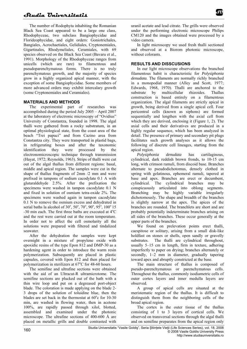
In our light microscope observations the branched filamentous habit is characteristic for Polysiphonia denudata. The filaments are normally richly branched in a monopodial manner (Alley and Scott, 1977; Edwards, 1968, 1970). Thalli are anchored to the substrate by multicellular rhizoides. Thallus construction is based entirely on a filamentous organization. The algal filaments are strictly apical in growth, being derived from a single apical cell. Four pericentral cells (known as siphons) are formed sequentially and lengthen with the axial cell from which they are derived, enclosing it (Figure 1, 2). The axial cells and their derivatives are produced in a highly regular sequence, which has been analyzed in detail. The presence of primary and secondary pit plugs facilitates such growth analyses as it allows the following of discrete cell lineages, starting from the apical region.
Polysiphonia denudata has cartilaginous, cylindrical, dark reddish brown fronds, to 10-15 cm long, with crimson ramuli, from discoid base. Branches alternate to pseudodichotomus, densely clothed in spring with gelatinous, ephemeral ramuli, tapered at base and apex. Branches are erect or decumbent, cylindrical. The cylindrical branches may be conspicuously articulated into oblong segments. Branching may be highly variable irregularly dichotomously. The shape and breadth of the branches is slightly narrow at the apex. The apices of the branches are rounded. The branchlets are short and are probably potentially indeterminate branches arising on all sides of the branches. These occur generally at the upper parts of the branches.
We found on prelevation points erect thalli, caespitose or solitary, arising from a small disk-like holdfast on stones or shells, open sandly or gravelly substrates. The thalli are cylindrical throughout, usually 5–15 cm in length, firm in texture, adhering imperfectly to paper on drying; branches alternately or secondly, 1–2 mm in diameter, gradually tapering toward apex and abruptly constricted at the base.
The main structure of thallus is composed of pseudo-parenchymatous or parenchymatous cells. Throughout the thallus, commonly isodiametric cells of outer cortex layers and inner medulla layers are observed.
A group of apical cells are situated at the meristematic region of the thallus. It is difficult to distinguish them from the neighboring cells of the broad apical region.
The cortex is the outer tissue of the thallus consisting of 1 to 3 layers of cortical cells. We observed on transversal sections through the algal thalli and on semifine preparates from the apical region only
SSttuuddiiaa UUnniivveerrssiittaattiiss
Studia Universitatis “Vasile Goldiș”, Seria Ştiințele Vieţii (Life Sciences Series), vol. 18, 2008 © 2008 Vasile Goldis University Press http://www.studiauniversitatis.ro
161
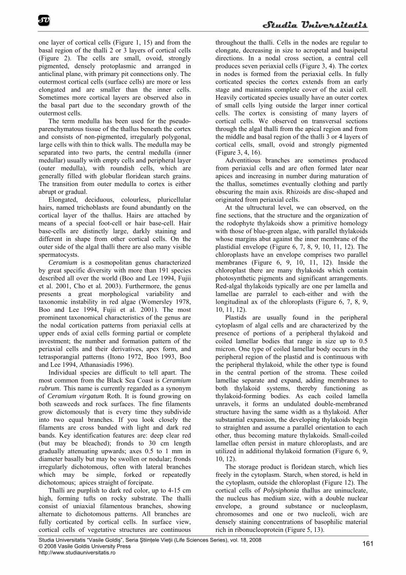
one layer of cortical cells (Figure 1, 15) and from the basal region of the thalli 2 or 3 layers of cortical cells (Figure 2). The cells are small, ovoid, strongly pigmented, densely protoplasmic and arranged in anticlinal plane, with primary pit connections only. The outermost cortical cells (surface cells) are more or less elongated and are smaller than the inner cells. Sometimes more cortical layers are observed also in the basal part due to the secondary growth of the outermost cells.
The term medulla has been used for the pseudo-parenchymatous tissue of the thallus beneath the cortex and consists of non-pigmented, irregularly polygonal, large cells with thin to thick walls. The medulla may be separated into two parts, the central medulla (inner medullar) usually with empty cells and peripheral layer (outer medulla), with roundish cells, which are generally filled with globular floridean starch grains. The transition from outer medulla to cortex is either abrupt or gradual.
Elongated, deciduous, colourless, pluricellular hairs, named trichoblasts are found abundantly on the cortical layer of the thallus. Hairs are attached by means of a special foot-cell or hair base-cell. Hair base-cells are distinctly large, darkly staining and different in shape from other cortical cells. On the outer side of the algal thalli there are also many visible spermatocysts.
Ceramium is a cosmopolitan genus characterized by great specific diversity with more than 191 species described all over the world (Boo and Lee 1994, Fujii et al. 2001, Cho et al. 2003). Furthermore, the genus presents a great morphological variability and taxonomic instability in red algae (Womersley 1978, Boo and Lee 1994, Fujii et al. 2001). The most prominent taxonomical characteristics of the genus are the nodal cortication patterns from periaxial cells at upper ends of axial cells forming partial or complete investment; the number and formation pattern of the periaxial cells and their derivatives, apex form, and tetrasporangial patterns (Itono 1972, Boo 1993, Boo and Lee 1994, Athanasiadis 1996).
Individual species are difficult to tell apart. The most common from the Black Sea Coast is Ceramium rubrum. This name is currently regarded as a synonym of Ceramium virgatum Roth. It is found growing on both seaweeds and rock surfaces. The fine filaments grow dictomously that is every time they subdivide into two equal branches. If you look closely the filaments are cross banded with light and dark red bands. Key identification features are: deep clear red (but may be bleached); fronds to 30 cm length gradually attenuating upwards; axes 0.5 to 1 mm in diameter basally but may be swollen or nodular; fronds irregularly dichotomous, often with lateral branches which may be simple, forked or repeatedly dichotomous; apices straight of forcipate.
Thalli are purplish to dark red color, up to 4-15 cm high, forming tufts on rocky substrate. The thalli consist of uniaxial filamentous branches, showing alternate to dichotomous patterns. All branches are fully corticated by cortical cells. In surface view, cortical cells of vegetative structures are continuous
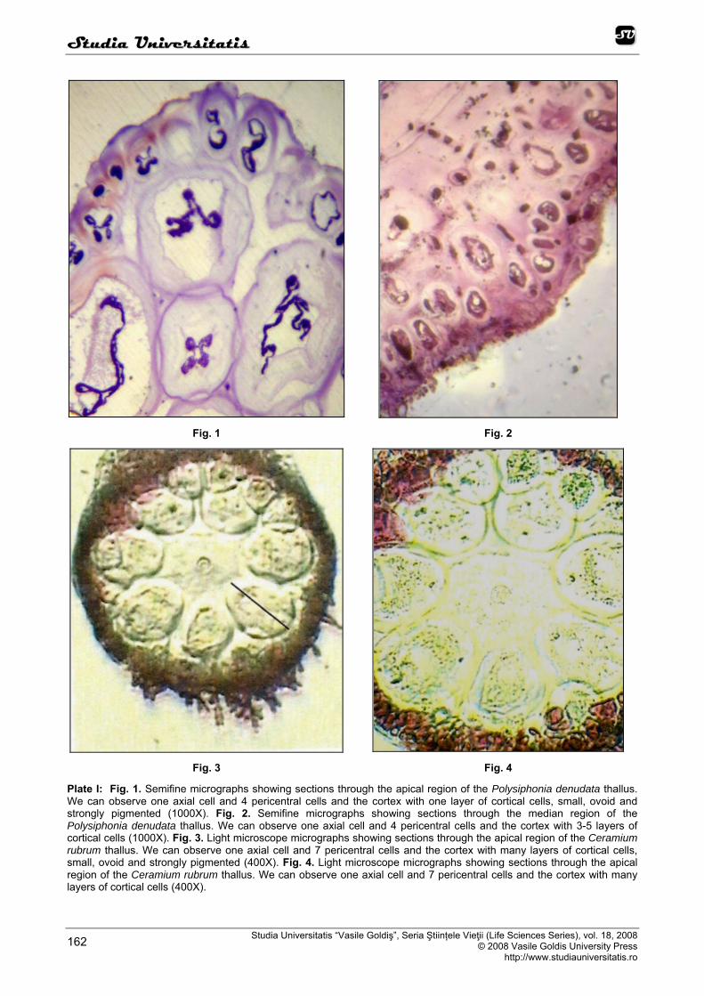
throughout the thalli. Cells in the nodes are regular to elongate, decreasing in size to acropetal and basipetal directions. In a nodal cross section, a central cell produces seven periaxial cells (Figure 3, 4). The cortex in nodes is formed from the periaxial cells. In fully corticated species the cortex extends from an early stage and maintains complete cover of the axial cell. Heavily corticated species usually have an outer cortex of small cells lying outside the larger inner cortical cells. The cortex is consisting of many layers of cortical cells. We observed on transversal sections through the algal thalli from the apical region and from the middle and basal region of the thalli 3 or 4 layers of cortical cells, small, ovoid and strongly pigmented (Figure 3, 4, 16).
Adventitious branches are sometimes produced from periaxial cells and are often formed later near apices and increasing in number during maturation of the thallus, sometimes eventually clothing and partly obscuring the main axis. Rhizoids are disc-shaped and originated from periaxial cells.
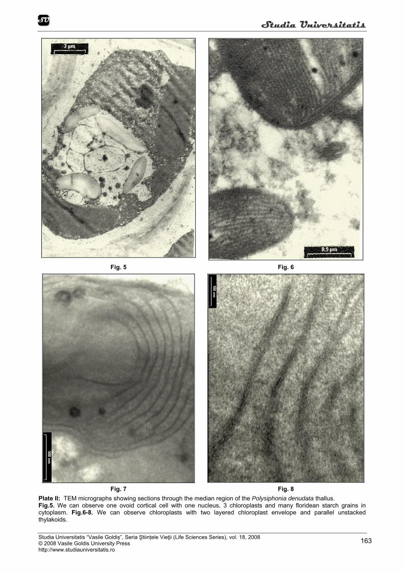
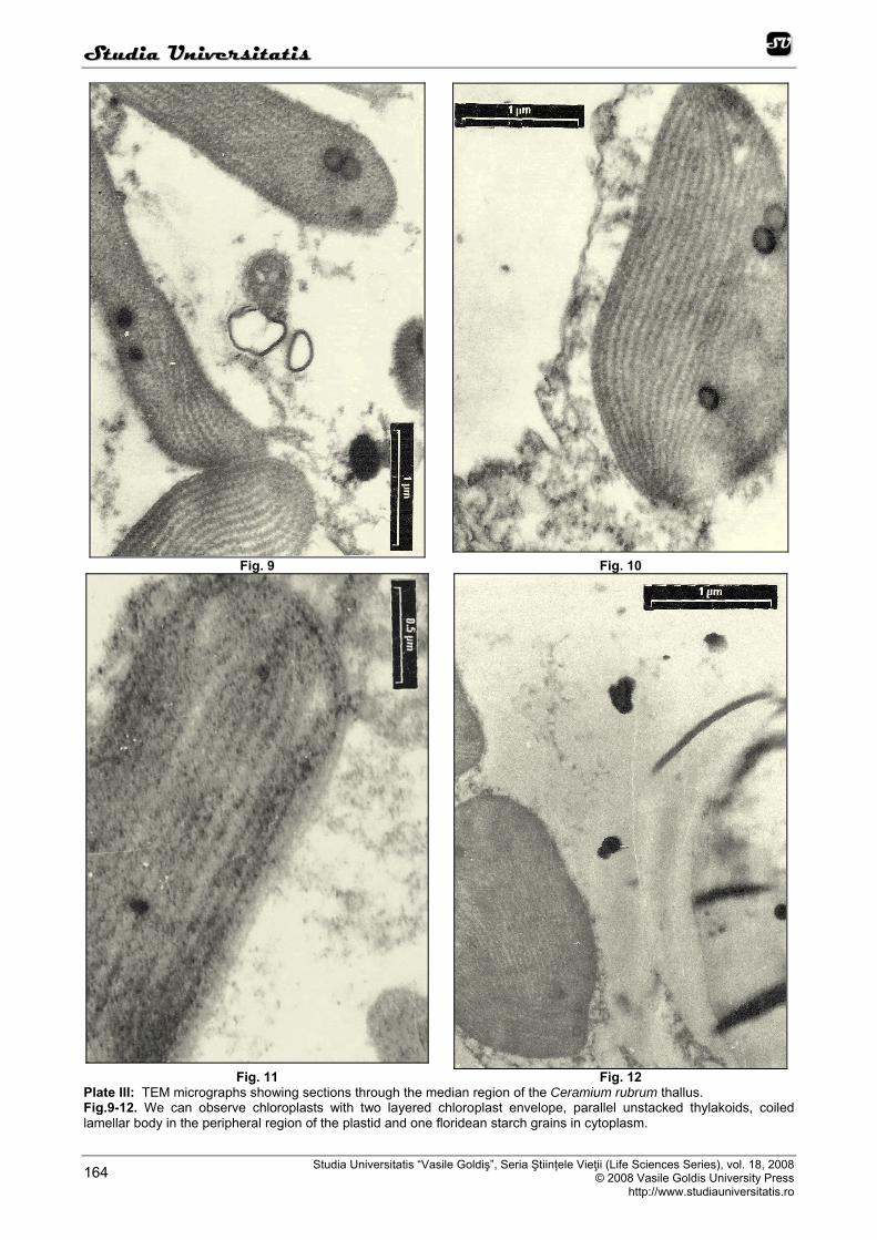
At the ultructural level, we can observed, on the fine sections, that the structure and the organization of the rodophyte thylakoids show a primitive homology with those of blue-green algae, with parallel thylakoids whose margins abut against the inner membrane of the plastidial envelope (Figure 6, 7, 8, 9, 10, 11, 12). The chloroplasts have an envelope comprises two parallel membranes (Figure 6, 9, 10, 11, 12). Inside the chloroplast there are many thylakoids which contain photosynthetic pigments and significant arrangements. Red-algal thylakoids typically are one per lamella and lamellae are parralel to each-either and with the longitudinal ax of the chloroplasts (Figure 6, 7, 8, 9, 10, 11, 12).
Plastids are usually found in the peripheral cytoplasm of algal cells and are characterized by the presence of portions of a peripheral thylakoid and coiled lamellar bodies that range in size up to 0.5 micron. One type of coiled lamellar body occurs in the peripheral region of the plastid and is continuous with the peripheral thylakoid, while the other type is found in the central portion of the stroma. These coiled lamellae separate and expand, adding membranes to both thylakoid systems, thereby functioning as thylakoid-forming bodies. As each coiled lamella unravels, it forms an undulated double-membraned structure having the same width as a thylakoid. After substantial expansion, the developing thylakoids begin to straighten and assume a parallel orientation to each other, thus becoming mature thylakoids. Small-coiled lamellae often persist in mature chloroplasts, and are utilized in additional thylakoid formation (Figure 6, 9, 10, 12).
The storage product is floridean starch, which lies freely in the cytoplasm. Starch, when stored, is held in the cytoplasm, outside the chloroplast (Figure 12). The cortical cells of Polysiphonia thallus are uninucleate, the nucleus has medium size, with a double nuclear envelope, a ground substance or nucleoplasm, chromosomes and one or two nucleoli, wich are densely staining concentrations of basophilic material rich in ribonucleoprotein (Figure 5, 13).
SSttuuddiiaa UUnniivveerrssiittaattiiss
Studia Universitatis “Vasile Goldiş”, Seria Ştiințele Vieţii (Life Sciences Series), vol. 18, 2008 © 2008 Vasile Goldis University Press
http://www.studiauniversitatis.ro 162
Fig. 1 Fig. 2
Fig. 3 Fig. 4
Plate I: Fig. 1. Semifine micrographs showing sections through the apical region of the Polysiphonia denudata thallus. We can observe one axial cell and 4 pericentral cells and the cortex with one layer of cortical cells, small, ovoid and strongly pigmented (1000X). Fig. 2. Semifine micrographs showing sections through the median region of the Polysiphonia denudata thallus. We can observe one axial cell and 4 pericentral cells and the cortex with 3-5 layers of cortical cells (1000X). Fig. 3. Light microscope micrographs showing sections through the apical region of the Ceramium rubrum thallus. We can observe one axial cell and 7 pericentral cells and the cortex with many layers of cortical cells, small, ovoid and strongly pigmented (400X). Fig. 4. Light microscope micrographs showing sections through the apical region of the Ceramium rubrum thallus. We can observe one axial cell and 7 pericentral cells and the cortex with many layers of cortical cells (400X).
SSttuuddiiaa UUnniivveerrssiittaattiiss
Studia Universitatis “Vasile Goldiș”, Seria Ştiințele Vieţii (Life Sciences Series), vol. 18, 2008 © 2008 Vasile Goldis University Press http://www.studiauniversitatis.ro
163
Fig. 5 Fig. 6
Fig. 7 Fig. 8 Plate II: TEM micrographs showing sections through the median region of the Polysiphonia denudata thallus. Fig.5. We can observe one ovoid cortical cell with one nucleus, 3 chloroplasts and many floridean starch grains in cytoplasm. Fig.6-8. We can observe chloroplasts with two layered chloroplast envelope and parallel unstacked thylakoids.
SSttuuddiiaa UUnniivveerrssiittaattiiss
Studia Universitatis “Vasile Goldiş”, Seria Ştiințele Vieţii (Life Sciences Series), vol. 18, 2008 © 2008 Vasile Goldis University Press
http://www.studiauniversitatis.ro 164
Fig. 9 Fig. 10
Fig. 11 Fig. 12
Plate III: TEM micrographs showing sections through the median region of the Ceramium rubrum thallus. Fig.9-12. We can observe chloroplasts with two layered chloroplast envelope, parallel unstacked thylakoids, coiled lamellar body in the peripheral region of the plastid and one floridean starch grains in cytoplasm.
SSttuuddiiaa UUnniivveerrssiittaattiiss
Studia Universitatis “Vasile Goldiș”, Seria Ştiințele Vieţii (Life Sciences Series), vol. 18, 2008 © 2008 Vasile Goldis University Press http://www.studiauniversitatis.ro
165
Fig. 13 Fig. 14
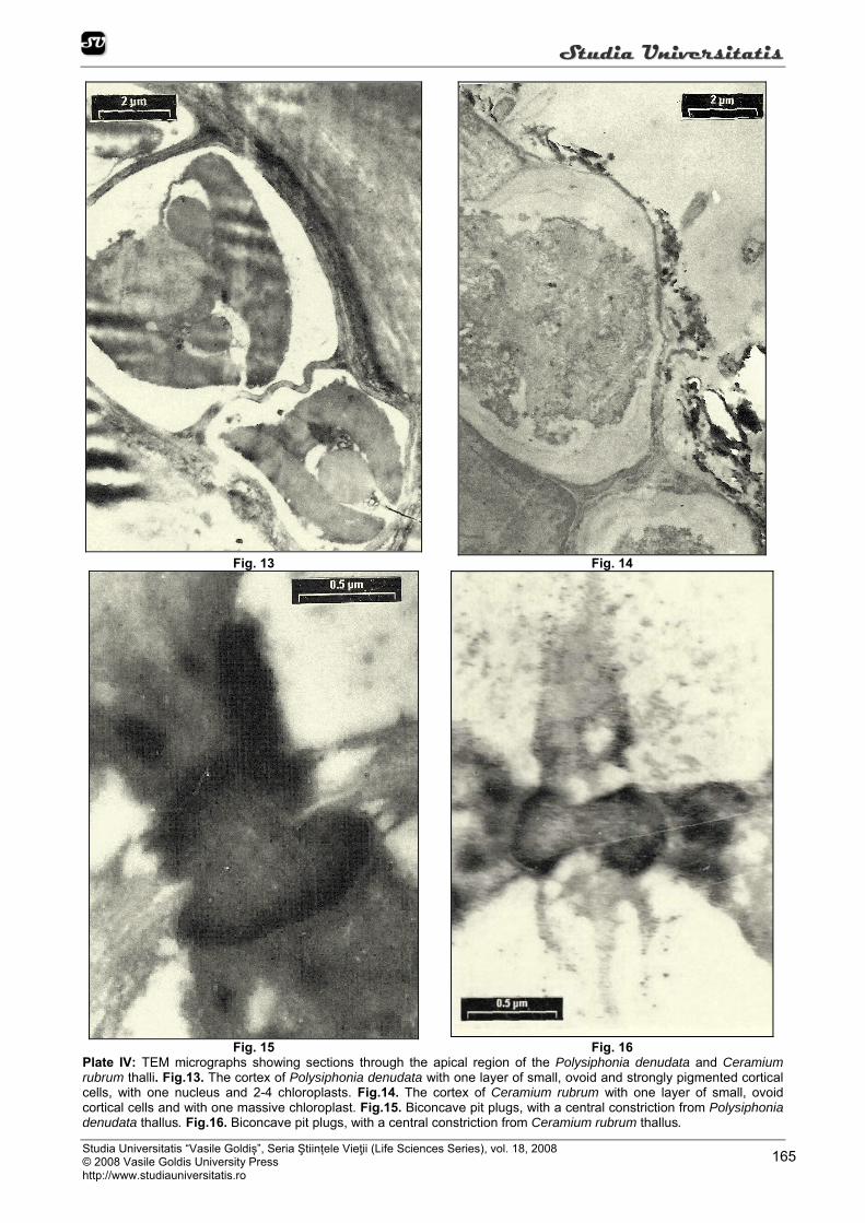
Fig. 15 Fig. 16 Plate IV: TEM micrographs showing sections through the apical region of the Polysiphonia denudata and Ceramium rubrum thalli. Fig.13. The cortex of Polysiphonia denudata with one layer of small, ovoid and strongly pigmented cortical cells, with one nucleus and 2-4 chloroplasts. Fig.14. The cortex of Ceramium rubrum with one layer of small, ovoid cortical cells and with one massive chloroplast. Fig.15. Biconcave pit plugs, with a central constriction from Polysiphonia denudata thallus. Fig.16. Biconcave pit plugs, with a central constriction from Ceramium rubrum thallus.

SSttuuddiiaa UUnniivveerrssiittaattiiss
Studia Universitatis “Vasile Goldiş”, Seria Ştiințele Vieţii (Life Sciences Series), vol. 18, 2008 © 2008 Vasile Goldis University Press
http://www.studiauniversitatis.ro 166
There are numerous mitochondria in apical region and a small number in the basal region of the thallus (Figure 5, 9), while the endoplasmic reticulum seems to be well developed in middle and especially in apical region of the thalli. There are also microbodies (ca 0.2 µm in diameter).
The cell wall surrounds the cells and is comprised of electron-dense reticulated cellulose microfibrils embedded in an amorphous matrix, with a randomly arrangement. Cell wall thickness decreased from very thick in discoid base and basal region of the thallus to thin in apical region of the thallus (Figure 13, 14).
A specialized feature is the pit connection or “pit plug” which has been linked with intercellular transport, structural strengthening in filamentous and pseudoparenchymatous thalli (Figure 15, 16). In the rhodophytes primary pit plugs are formed between two cells during division and secondary pit plugs develop between laterally adjacent cells. The plugs are usually biconcave, with a central constriction (Figure 15, 16). CONCLUSIONS
Our studies show the importance of cytological features in algae systematic, particularly for the Rhodophyta marine pluricellular algae studied. So, at the ultrastructural level the most distinctive features for Rhodophyta are: possession of simple plastids with unstacked thylakoids; in the chloroplasts number of thilakoids per lamella is one, the girdle lamella is not present and grana not formed; chloroplast envelope two layered; lack of chloroplasts ER; coiled lamellar body in the peripheral region of the plastid; the storage product is floridean starch, which lies freely in the cytoplasm; the thallus heaving unicleate cells, the nucleus being small and with one or two nucleoli; cell wall composed of cellulose with the fibrils randomly arranged; primary and secondary pit plugs usually biconcave, with a central constriction.
REFERENCES Alley CD, Scott JL, Unusual dictyosome morphology
and vesicle formation in tetrasporangia of the marine red alga Polysiphonia denudata. J. Ultrastr. Res. 58, 289-298, 1977.
Athanasiadis A, Morphology and classification of the Ceramioidae (Rhodophyta) based on phylogenetic principles. Opera Botanica, 128, 1, 216, 1996.
Bavaru A, Bologa AS, Skolka VH, A checklist of the benthic marine algae (except diatoms) along the Romanian Shore of the Black Sea – Oebalia. Int. Jour. Mar. Biol. Ocean., 17, 2 (suppl.), Toronto, Italia, 535-551, 1991.
Bavaru A, Brezeanu A, Doroftei E, Jianu L, Ultrastructure of algal cells at Ceramium rubrum (Huds) C. Ag. In: Current problems in cellular and molecular biology, IV, 562-566, 1999. Edited by Ardelean A. şi Crăciun C., Ed. Risoprint, Cluj-Napoca.
Bavaru A, Brezeanu A, Doroftei E, Jianu L, Ultrastructural aspects at multicellular green seaweeds Ulva rigida Ag., Enteromorpha intestinalis (L.) Link, Cladophora sericea
(Huds.) Kutz. In: Current problems in cellular and molecular biology, V, 567-575, 2000. Edited by A. Ardelean şi C. Crăciun. Ed. Risoprint, Cluj-Napoca.
Bold HC, Wynne MJ, Introduction to the Algae. Structure and reproduction, 706, 1978. Ed. University Press, Cambridge.
Boo SM, Intermediately corticated species, Ceramium puberubem (Ceramiaceae, Rhodophyta). Jpn. J. Phycol (sorui) 41, 143-149, 1993.
Boo SM, Lee AK, Ceramium and Campylaephora (Ceramiaceae, Rhodophyta). In: Biology of Economic Algae, 1-33, 1994. Edited by I. Akatsuka. SPB Academic Publishing, The Hague.
Cho TO, Riosmena-Rodríguez R, Boo SM, First record of Ceramium giacconei (Ceramiaceae, Rhodophyta) in the North Pacific: developmental morphology of vegetative and reproductive structures. Botanica Marina 46, 548-554, 2003.
Doroftei E, Bavaru A, Brezeanu A, Sava D, Comparative ultrastructure of some Chlorophyta and Rhodophyta from the Romanian coast of the Black Sea. In: Current problems in cellular and molecular biology, V, 462 – 470, 2000. Edited by Crăciun C. şi Ardelean A. Ed. Risoprint, Cluj-Napoca
Doroftei E, Sava D, Brezeanu A, Moldoveanu D, Particularităţi citofiziologice la Ulva rigida şi Cladophora vagabunda în condiţiile cultivării “in vitro”. In: Probleme curente de biologie celulară si moleculară, VII, 476-483, 2002. Edited by C. Crăciun şi A. Ardelean. Ed. Risoprint, Cluj-Napoca.
Doroftei E, Sava D, Brezeanu A, Arcuş M, Modificãri ultrastructurale la algele marine verzi pluricelulare în condiţiile cultivãrii „in vitro”, VIII, 202-212, 2003. In: Analele Societãţii Naţionale de Biologie Celularã. Edited by C. Crăciun şi A. Ardelean. Ed. Risoprint, Cluj-Napoca.
Doroftei E, Sava D, Mihăilă F, Aspecte ultrastructurale ale celulei algale la Ceramium elegans Ducl. In: Analele Societãţii Naţionale de Biologie Celularã, IX, 1, 357-370, 2004. Edited by C. Crăciun şi A. Ardelean. Ed. Risoprint, Cluj-Napoca.
Doroftei E, Sava D, Bavaru A, Modificãri ultrastructurale la algele marine verzi pluricelulare, sub acţiunea unor metale grele. In: Analele Societãţii Naţionale de Biologie Celularã, X, 467-473, 2005. Edited by C. Crăciun şi A. Ardelean. Ed. Risoprint, Cluj-Napoca.
Doroftei E, Sava D, Bavaru A, Zamfirescu S, Ultrastructura celulei algale la Bryopsis plumosa (Huds) Ag. In: Analele Societãţii Naţionale de Biologie Celularã, XI, 554-562, 2006a. Edited by C. Crăciun şi A. Ardelean. Ed. Risoprint, Cluj-Napoca.
Doroftei E, Jianu L, Brezeanu A, Bavaru A, Particularitati citologice la Cystoseira barbata

SSttuuddiiaa UUnniivveerrssiittaattiiss
Studia Universitatis “Vasile Goldiș”, Seria Ştiințele Vieţii (Life Sciences Series), vol. 18, 2008 © 2008 Vasile Goldis University Press http://www.studiauniversitatis.ro
167
(Good et. Wood Ag.). In: Analele Societãţii Naţionale de Biologie Celularã, XI, 563-571, 2006b. Edited by C. Crăciun şi A. Ardelean. Ed. Risoprint, Cluj-Napoca.
Edwards P, The life history of Polysiphonia denudata (Dillwyn) Kützing in culture. Journal of Phycology 4, 35-37, 1968.
Edwards P, Field and cultural observations on the growth and reproduction of Polysiphonia denudata from Texas. British Phycological Journal 5, 145-153, 1970.
Fujii MT, Concentino ALM, Pereira, SMB, Ceramium nitens (Ceramiaceae, Rhodophyta), an uncommon species from Brazil. Rev. Bras. Bot., 24, 3, 359-363, 2001.
Hayat MA, Basic Electron Microscopy Techniques, 2, 1972. Ed. Van Nostrand Reinhold Company.
Itono H, The genus Ceramium (Ceramiaceae, Rhodophyta) in southern Japan. Botanica Marina, 15, 74-86, 1972.
Reynolds E S, The use of lead citrate at high pH as an electron-opaque stain in electron-microscopy. J. Cell Biol., 17, 208, 1963.
Van den Hoeck, Algae: an introduction to phycology, 300-342; 419-436, 1997. Ed. Cambridge University Press, Cambridge.
Womersley HBS, Southern Australian species of Ceramium Roth Greville (Rhodophyta). Australian Journal of Marine and Freshwater Research, 29, 205-257, 1978.
SSttuuddiiaa UUnniivveerrssiittaattiiss
Studia Universitatis “Vasile Goldiş”, Seria Ştiințele Vieţii (Life Sciences Series), vol. 18, 2008 © 2008 Vasile Goldis University Press
http://www.studiauniversitatis.ro 168