ultrasonography of the painful hip in … simon godefridus... · simon godefridus franciscus robben...
TRANSCRIPT

ULTRASONOGRAPHY OF THE PAINFUL HIP IN CHILDHOOD
Simon G.F. Robben

ISBN 90-9012721-6
Cover designed by A.W. Zwamborn Type-setting: A.W. Zwamborn Illustrations: A.w. Zwamborn Photographs: T. Rijsdijk
Printed by ~ Ridderprint B.V. Ridderkerk
©.1999, S.G.F. Robben
Dermatologicall1y tested. Not recommended for children under 3 years, small parts may be swallowed.
All rights reserved. No part of this dissertation may be reproduced, stored in a retrieval system or transmitted in any other form or by any means, without the prior written permission of the author, or, when appropriate, of the publishers of the publications.

ULTRASONOGRAPHY OF THE PAINFUL HIP IN CHILDHOOD
ECHOGRAFIE VAN DE PIJNLlJKE HEUP BIJ KINDER EN
Proefschrift
Ter verkrijging van de graad van doctor aan de Erasmus Universiteit Rotterdam
op gezag van de Rector Magnificus Prof. Dr. P.W.C. Akkermans M.A.
en volgens het besluit van het College voor Promoties
De openbare verdediging zal plaatsvinden op woensdag 16 juni 1999 om 15.45 uur
door
Simon Godefridus Franciscus Robben geboren te Tilburg

Promotiecommissie
Promotor Overige leden
Prof. dr. M. Meradji
Prof. dr. J.A.N. Verhaar Prof. dr. J.8. Lameris Prof. dr. F.W.J. Hazebroek

Vaar mijn vader.


Chapter 1
Chapter 2
Chapter 3
Chapter 4
Chapter 5
Chapter 6
Chapter 7
Chapter 8
Contents
INTRODUCTION Pathology of the hip joint in childhood .......................................................................... .
Imaging techniques .................... .................................... . ............................... . Conventional radiography and tomography ................................................ . Computer tomography ............................................................................................ . Magnetic resonance imaging " .. ""'''''''" .. "."'''''" ............ '''''''" ................. ,,'',,,, .. Radionuclide studies .,"""'''''',." ............. " ........... ,,''', .......... ,,''''''''''"""""""",,,,,,, Ultrasonography .. ...................................... ...................... . ..................... .
9 11 13 13 13 14 14 15
Basic principles ........................... ....................................................... 15 Doppler ultrasonography ......... .......................................................... 16 Biomedical effects and safety....... .................... ............................. 17
Purpose and outline of this thesis ......................................................... 17
THE ANTERIOR JOINT CAPSULE OF THE HIP an anatomic and histologic study with ultrasound correlation
THE ANTERIOR JOINT CAPSULE OF THE HIP an ultrasound study in 105 patients with transient
synovitis .................................... n ....................................................... " .................................... .
ATROPHY OF THE QUADRICEPS MUSCLE IN CHILDREN WITH A PAINFUL HIP an ultrasound study, physiological aspects ....................................................... .
THE IMPORTANCE OF ATROPHY OF THE QUADRICEPS MUSCLE IN THE DIFFERENTIAL DIAGNOSIS OF CHILDREN WITH A PAINFUL HIP
21
37
53
an ultrasound study, clinical aspects " .. ,""'''', ... ,'''''''" ........ ",."''" .. "" ............. "'',, .. ,,,',. 73
ULTRASONOGRAPHY OFTHE PAINFUL HIP IN CHILDHOOD the diagnostic value of cartilage thickening and muscle atrophy in the detection of Perthes' disease .......................................................... ..
DOPPLER ULTRASONOGRAPHY OF THE HIP JOINT IN CHILDREN WITH A PAINFUL HIP
SUMMARY AND CONCLUSION SAMENVATIING EN CONCLUSIE
91
111
129 135
Acknowledgments ... 139
Curriculum vitae 143
7

Contents
8

Chapter 1
INTRODUCTION
9

Chapter 1
10

Introduction
INTRODUCTION
Pathology of the hip joint in childhood There are many diseases in childhood that affect the hip joint. Some diseases are systemic in origin and initially may present themselves as hip disorders, such as rheumatoid arthritis. other diseases are localized specifically in the hip joint, such as transient synovitis and Perthes' disease, mostly unilateral. Neoplastic or infectious diseases around the hip joint may also manifest themselves as a painful hip. If both hips are affected the differential diagnosis should include skeletal dysplasias (multiple epiphyseal dysplasia) and metabolic diseases (hypothyroidism, Gaucher's disease, mucopolysaccharidoses and mucolipidoses).
From a diagnostic point of view, the most frequently occurring hip diseases can be categorized in age groups
Age (in years)
0-3 Developmental dysplasia of the hlp
Osteomyelitis
3-10 Transient synovitis
Perthes' disease
Osteomyelitis/septic arthritis
Juvenile chronic arthritis
10-17 Slipped capital femoral epiphysis
The most frequently observed diseases that cause a unilateral painful hip will be discussed briefly in the next paragraphs:
Transient synovitis Transient synovitis is the most frequent hip disease in children of 3-10 years of age, boys being affected more often than girls (1, 2). Symptoms are unilateral, usually have a sudden onset (often at night) and consist of pain in the groin (extending to thigh and knee), refusal to bear weight and limping. The hip is fixed in flexion and external rotation. Physical examination reveals decreased rotation (especially internal rotation) and abduction. Body temperature is normal, although some patients have a subfebrile temperature (3).
11

Chapter 1
Mild leukocytosis may be present (3), but some authors report normal leucocyte counts (1,4). The etiology of transient synovitis remains unclear. Trauma, infection, antibody response to bacterial and viral antigen, or allergic predisposition have all been cited as possible factors, although the few controlled studies have failed to sUbstantiate any of these etiologies (1, 5). The treatment consists of rest and the course of the disease is benign. The pain and limitation of motion usually subside within several days (2) and the mean duration of complaints is 13 days (6).
Perthes' disease (7) Perthes' disease is an avascular necrosis of the epiphysis of the femoral head. The mean age of the patients is 5 (range 3-11) years. Boys are affected 4 times as much as girls; in 15% of all patients the disease is bilateral. The symptoms and age of the patients can be identical to transient synovitis, although the onset is usually more insidious and the symptoms persist longer. Physical examination shows an abnormal gait, a positive Trendelenburg's sign, decreased range of motion of the affected hip, especially rotation and abduction. Adduction contracture can simulate leg length discrepancy. Although it is generally accepted that Perthes' disease is an avascular necrosis of the epiphysis of the femoral head, the exact cause is unknown. Several etiological factors have been proposed, such as recurrent trauma, relative hypovascularity at the age of 4-8 years (8, 9) and coagulation disturbances (10). The prognosis depends on age of onset and on extent of necrosis. The disease follows distinct stages (initial, fragmentation, reparative and remodelling stage) and the femoral head eventually heals with more or less deformity. This process takes approximately 2 years. Treatment consists of rest, traction, bracing, or femoral varus derotation osteotomy.
Slipped capital femoral epiphysis (SCFE) (11) SCFE is a displacement of the proximal femoral epiphysis from its central orientation on the metaphysis, usually in posterior direction. Two types can be recognized: acute [10%) and chronic [90%) slip. There is a moderate male predilection [2:1), the mean age in boys is 11.5 years and in girls 13.5 years. A high incidence of obesity is found in patients with SCFE. Bilateral slipping occurs with significant frequency, reported to range from 16% to 49%. Symptoms and signs depend on the type of slip. Acute slip occurs suddenly or is of short duration. Patients complain of severe pain in the groin, buttocks or thigh and are unable to bear weight. The leg is in external rotation and is shortened. Prodromal symptoms of dull pain in the affected hip or knee may be present prior to the acute period. Gradual or
12

Introduction
chronic slip is generally slowly progressive over a long period and the complaints consist of intermittent pain in hip or knee. intermittent limping and progressive limb shortening. The specific cause of SCFE is unknown. Possible causes include epiphyseal plate weake· ning due to endocrine factors. mechanical factors related to the angle of the growth plate and trauma. Treatment consists of fixation in situ by inserting multiple pins or a single cannulated hip screw. The prognosis depends on the severity of the slip and is good in cases of minimal or moderate slip.
IMAGING TECHNIQUES
Patients with diseases of the hip present with a variety of non specific symptoms and signs (pain in the groin. knee or leg; inability to bear weight and limping; decreased rotation. abduction and extension). Moreover. other diseases. such as spondylodiscitis and retrocecal appendicitis can present as a painful hip. Therefore. these patients remain a diagnostic challenge and their diagnosis depends heavily on imaging techniques. This is especially true for very young patients in whom the history is often unreliable. Several imaging techniques can be used to visualize the hip joint: conventional radio· graphy. conventional tomography. ultrasound. computed tomography. magnetic resonance imaging and radionuclide studies.
Conventional radiography and tomography Conventional radiography has proven its value in screening for changes of cortical and cancellous bone. and follow·up of known osseous abnormalities. It is neither expensive nor time·consuming. readily available. and has a good reproducibility. However. one should realize that early disease may not be recognized because: a) a large volume of bone must be destroyed before it will become visible. and b) periosteal apposition of new bone (another early sign of disease) takes 1·2 weeks to develop. Moreover. soft tissue abnormalities are difficult to depict with conventional radiography; displacement of fat planes was considered as evidence of hip joint effusion. but this proved to be unreliable (12·16). Another important drawback is ionizing radiation. which should be kept to a minimum in a pediatric popUlation: the mean effective dose of a pelvic examination (2 radiographs) in the Netherlands is 54 JASv. resulting in a chance of fatal tumor induction of 1:150.000 (17). Many of the disadvantages of conventional radiography cannot be overcome by conven· tional tomography. the radiation dose being even higher. Nowadays conventional tomo· graphy is replaced by computed tomography in almost every hospital in the Netherlands.
Computer tomography Computer tomography (CT) had a tremendous impact on diagnostic imaging. CT of the hip has proven its value in traumatology and diseases of the musculoskeletal system. Effu·
13

Chapter 1
sion can be detected indirectly by thickening of the capsule (18, 19). However, CT is limited to the transverse plane, the child has to lie still for at least 30 seconds, and the radiation dose of a pelvic examination exceeds the dose of conventional radiographs 180 times (10000 ilSV) (20, 21). Another disadvantage is the relatively high cost compared to conventional radiography and US (Table 1). Although the soft tissues can be seen to a better advantage with CT than with conventional radiography, its role in discriminating different tissues is limited compared with magnetic resonance imaging.
Table 1. Outline of the costs of imaging studies of the lower extremity (22).
Conventional radiography f 35.95
Ultrasonography ! 81.80
Conventional tomography ! 100.00
Radionuclide study ! 150.00
Computer tomography (CIl ! 360.00
Magnetic resonance Imaging(MRI) ! 871.00
Magnetic resonance imaging (23-28) MRI had an even greater impact on diagnostic imaging than CT had previously. The hip can be visualized in multiple planes and contrast between different soft tissues is very high. This results in a high sensitivity for effusion (25). However, because calcium does not have paramagnetic properties, cortical and cancellous bone do not generate a signal; also, the fibrous capsule and ligaments have a very low signal intensity. Moreover, MRI is expensive, time consuming and not readily available. Because data acquisition takes 3-4 minutes for each image sequence, children have to be able to lie motionless for that period of time. Therefore, most of the children under 6 years of age have to be imaged under general anaesthesia or sedation. Many of these disadvantages will be overcome in the future due to technical improvement and MRI will definitely play an important role in the diagnostic work-up of the painful pediatric hip.
14

Introduction
Radionuclide studies The examination offers little anatomical details, but gives a good estimation of vascularization and bone metabolism. The radiation dose in radionuclide studies is relatively low compared to CT. The radiopharmacon is administered intravenously, which is a relative disadvantage in childhood.
Ultrasonography Ultrasonography (US) can visualize the soft tissues and bony contours of the hip joint, but it can not visualize inside the bony structures. In this way, US is complementary to conventional radiography. US lacks ionizing radiation, it is relatively inexpensive, readily available, offers a high spatial resolution, and can be performed as a bedside examination. Because of the physical contact, US can be considered a modified physical examination, offering the sonographer the opportunity to correlate US findings with physical signs immediately. However, US has several disadvantages. It is operator-dependent as it relies on the skills of the sonographer to display the anatomy properly and to recognize pathology during the examination, because it is not possible to recognize these later on the films. Moreover, most US eqUipment is unable to provide an overview of the hip joint because high-frequency transducers have a small field of view of a few centimeters only. The following sections will briefly discuss the basic US principles, the biomedical effects and safety.
Basic principles of US In daily routine, pediatric ultrasonography uses soundwaves with frequencies of 5-10 MHz for visualizing structures in daily routine. The soundwaves are generated, emitted, and received by the transducer containing a piezoelectric plate. In the work presented here, the US examinations were done with a linear array transducer with a longitu-dinal row of elements which are electronically activated in groups one at a time. While traversing tissues, the waves are absorbed, refracted and reflected. The reflected waves are involved in the imaging. Reflection occurs when there is a difference in acoustic impedance at the interface between two types of tissue. The reflected signals are received by the same piezoelectric element which emits the signal and are converted into electric signal that can be processed by the electronics of the ultrasound system. The received signals are amplified to compensate for the attenuation; the amount of amplification generally depends on the time the signal takes to arrive at the transducer. This depends on the depth at which the tissue reflection occurred. Early arriving signals which arise from interfaces nearby the transducer are displayed superficially and signals that arrive late are displayed more deeply. Interfaces that cause strong echoes are displayed as bright spots, weak echoes are displayed as dim spots. The appearance of
15

Chapter 1
the displayed image is further affected by the processing curves and log compression which distribute the gray shades over a certain range of echoes. Wavelength and frequency The relation between wavelength and frequency follows from the equation: v = f x A, where v is average velocity in solid tissue (1580 m/s), f is the frequency (5-10 MHz in this thesis), and A is the wavelength (minimal 0.15 mm in this thesis). The wavelength affects the spatial resolution which can be expressed as axial and lateral resolution. The axial resolution is the minimum distance between two points along the axis of the sound beam that can be separated. For a 10 MHz transducer with a 0.15 mm wavelength the axial resolution would be, at best, 0.22 mm. The lateral resolution is the minimum distance beween two points perpendicular to the sound beam; it depends on the beam width and on focusing. Generally the lateral resolution is 2-3 times the axial resolution. For a 10 MHz transducer, the lateral resolution will be approximately 0.5 mm (29).
Doppler ultrasonography (30, 31) The previous paragraph described how the analysis of the amplitude of the reflected echo generates a gray-scale image. However, analysis of the frequency of the returning echo also gives important information. When a wave is reflected from a moving target, the frequency of the wave received differs from that which was transmitted. This difference in frequency is Imown as the Doppler shift and depends on the speed at which the target is moving and whether the motion is toward or away from the receiver. The greater the relative velocity, the larger the Doppler shift. In diagnostic US, two applications of the Doppler principle are frequently used: pulsed Doppler and color Doppler examinations. Pulsed Doppler: In pulsed Doppler US the transducer transmits a short pulse of sound and then listens for the returning echo. Because the speed of sound is constant, the delay in time between the transmitted signal and the returning echo is proportional to the distance. By varying the delay between transmission and reception, it is possible to select a defined pOint along the Doppler beam from which the signal arises. With this capability, one can use standard gray-scale US as a road map to visualize the vessels of interest, and to position the Doppler sample volume at various pOints within the vessel. This combination of gray-scale image with pulsed Doppler is also known as duplex Doppler. Color Doppler: With duplex Doppler only a small area of flow can be analyzed at any time as the Doppler sample volume is moved across the image. Color Doppler US offers a major advantage over duplex Doppler because it is sensitive to Doppler signals throughout the field of view. Color Doppler US operates by analyzing the returning echoes for amplitude, frequency and phase information. Moving targets produce a phase shift and will be assigned a color based on the direction of phase shift. The shade of the color will depend on the mean Doppler frequency shift from the pixel. Stationary objects produce no phase shift in the direction of the US beam and are assigned a gray-scale value based on amplitude, as in conventional gray-scale imaging.
16

Introduction
Biomedical effects and safety (32) Although diagnostic ultrasound has been used for more than 30 years and no harmful biological effects have been demonstrated, energy is nevertheless absorbed by the tissues during US examination. Biological effects of this energy transmission can be heating and cavitation, both of which can cause tissue damage. - Heating is not a major concern in daily US practice. Several indicators of thermal effects are used, such as the Thermal Index (TI), and Spatial Peak, Temporal Average Intensity (I,pt,). No adverse biological effects have been reported when I"" < 100 mW/cm' was used under conditions that are comparable with the conditions of the US examinations in this thesis. - Cavitation has occurred at diagnostic US exposure levels in plants and lower organisms that contain small air spaces. In addition, lung hemorrhage in mice has been demonstrated at intensities used in diagnostic US. Therefore, an indicator of the likelihood for cavitation was introduced; the Mechanical Index (derated peak rarefaction pressure, divided by the square root of tile transducer frequency in MHz). The upper limit of the mechanical index for tissues with well·defined gas bodies is 0.3. For other tissues no adverse effects have been reported; nevertheless, the Food and Drugs Administration (FDA) has set the upper limit of MI at 1.9 (33). This is well above the MI used in this thesis. Although the risk of biological effects is negligible in diagnostic US, it is advisable to adhere to the ALARA principle (As Low As Reasonably Achievable). The intensity levels used in this thesis are well below the safety limits.
PURPOSE AND OUTLINE OF THIS THESIS
Ultrasound of the hip was first described by Kramps and Lenschow in 1979 (34). One year later, Seltzer et al. showed that hip joint effusion could be demonstrated with US (35). However, the diagnostic role of US in patients with a painful hip has been limited to the detection of joint effusion although state·of-the·art US machines with high frequency transducers can visualize an abundancy of anatomical details in the hip joint, especially the capsule and cartilage. Proper knowledge of the sonographical anatomy will prevent erroneous interpretation and inaccurate measurements. Moreover, this detailed anatomical knowledge could help improve the diagnostic value of ultrasonography in children with a painful hip. The purpose of this thesis was to identify US criteria to improve the diagnostic value of US.
Chapter 1 summarizes the pathology of the hip joint in childhood and discusses briefly the different imaging techniques. In chapter 2 the anatomy of the hip joint is studied in a cadaver study with sonographic and microscopic correlation. In addition, the sonographic anatomy of 58 normal children is described.
17

Chapter 1
Chapter 3 describes the sonographic findings in 105 patients with transient synovitis, utilizing the anatomic knowledge derived from chapter 2. Chapters 4 and 5 describe a method to measure muscle thickness, and describe the effects of disease on the thickness of the muscle in 348 children with a painful hip. The physiological aspects of muscle wasting and the clinical applications are discussed. Chapter 6 describes the value of ultrasound in the detection of Perthes' disease by measuring cartilage thickness and muscle thickness. Chapter 7 introduces color Doppler sonography and duplex Doppler sonography to the arsenal of potentially useful tools in evaluating the painful hip. Chapter 8 summarizes the results of the studies in this thesis.
18

Introduction
REFERENCES
1. Hardinge K The etiology of transient synovitis of the hip in childhood, J Bone Joint Surg 1970;528:100-107.
2. Haueisen DC, Weiner OS, Weiner SO. The characterization of "transient synovitis of the hlpR in children, J Pediatr Orthop 1986;6:11-17.
3. Hermel MB, Albert SM. Transient synovitis of the hip. Clin Orthop 1962;22:21-26. 4. Adams JA. Transient synovitis of the hip joint in children, J Bone Joint Surg 1963;45B:4 71-4 76. 5. Blockey NJ, Porter BB. Transient synovitis of the hip: a virological investigation. Br Med J
1968:557-558. 6, Rosenberg NJ, Smith EE, Transient synovitis of the hip. J Pediatrics 1956;48:776-781. 7. Edgren W. Coxa plana. Acta Orthop Scand 1965;84 (suppl):1-129. 8. Chung SMK, The arterial supply of the developing proximal end of the human femur, J Bone Joint
Surg 1976;58-A:961-970. 9, Trueta J. The normal vascular anatomy of the human femoral head during growth. J Bone Joint Surg
1957;398:358-394. 10. Glueck CJ, Crawford A, Roy 0, Freiberg R, Glueck H, Stroop D. Association of antithrombotic factor
deficIencies and hypofibrinoJysis with Legg-Perthes disease. J Bone Joint Surg 1996;78-A:3-12, 11. I<atz JF, Siffert RS. Management of hip disorders in children. (first ed.) Philadelphia, Toronto: J.8.
Lippincott company, 1983: 105-120. 12. Wilson OJ, Green OJ, MacLarnon JC. Arthrosonography of the painful hip. Clin Radial 1984;35:17-19. 13. Meradji M, Diepstraten AFM. Coxitis Fugax, sonografisches und radlologisches Blld in 65 Fallen.
Radlologe 1988;28:473-478. 14. Adam R, Hendry GM, Moss J, Wild SR, G1tIespie I. Arthrosonography of the irritable hip In childhood:
a review of 1 year's experience. Br J RadloI1986;59:205-208. 15. Marchal GJ, Holsbeeck MT, Raes M, et al. Transient synovitis of the hip in children: role of US.
Radiology 1987;162:825-828. 16. Rosenborg M, Mortensson W. The validity of radiographic assessment of childhood transient
synovitis of the hlp. Acta Radiol.Diagnosis 1986;27:85-89. 17. Geleijns K, Vliet van M. Stralenbelasting bij klnderradiologie. IRS rapport 98-0iA 18. Dihlman W, Nebel G. Computed tomography of the hlp joint capsule. J Comp Assist Tomogr
1990;7:278-285. 19. Egund N, Wingstrand H, Forsberg L, Petterson H, Sunden G. Computed tomography and
ultrasonography for diagnosis of hip joint effusion in children. Acta Orthop Scan 1986;57:211-215. 20. Fearon T, Vucich J. Pediatric patient exposure from CT examinations: GE CTfT 9800 scanner. AJR
1985;144:805-809. 21. l<amelIR, Hernandez RJ, Martin JE, Schlesinger AE, Niklason LT, Guire I<E. Radiation dose reduction
in CT of the pediatric pelvis. Radiology 1994;190:683-687. 22. Ziekenfondsraad. Diagnostisch I<ompas. 1997. 23. Erickson SJ. High-Resolution Imaging of the Musculoskettal System. Radiology 1997;205:593-618. 24. Jaramillo D, Villegas-Medina OL, Doty 01<, et al. GadOlinium enhanced MR imaging demonstrates
abduction-caused hip ischemia and its reversal in children. Pediatr RadioI1995;25:578-87. 25. Moss SG, SchweItzer ME, Jacobson JA, et al. Hip Joint fluid: Detection and Distribution at MR
Imaging and US with Cadaveric Correlation. Radiology 1998;208:43-48. 26. Sebag G, Ducou Le Pointe H, Klein I, et al. Dynamic gadolinium-enhanced subtraction MR imaging. A
simple technique for the early diagnosis of Legg-Calve-Perthes disease: preliminary findings. Pedlatr Radiol 1997;27:216-220.
19
Chapter 1
27. Tsukamoto H, Kang YS, Jones lC, et al. Evaluation of marrow perfusion in the femoral head by dynamic magnetic resonance imaging. Effect of venous occlusion in a dog model. Invest Radiol 1992;27:275·281.
28. Rush SH, Bramson RT, Ogden lA. legg-Calve-Perthes disease: detection of cartilagenous and synovial changes with MR imaging. Radiology 1988;167:473-476.
29. Walter JP. Physics of high-resolution ultrasound-practical aspects. ClIn Radial N Am 1985;23:3-11. 30. Burns PN. The physical principles of Doppler and spectral analysis. 1 Clin Ultrasound
1987;15:567·590. 31. Taylor !UW, Holland S. Doppler US. Part I: basic principles, Instrumentation, and pitfalls. Radiology
1990;174:297·307. 32. Beek FlA, Thijssen 1M, Sandstrom 1<, Mali WPTM. Twee velllgheidsmaten voor diagnostisch
ultrageluid: de mechanlsche en de thermische Index. Ned Tijdschr Geneesk 1996;140:706-708. 33. Tenner MS. Letter from the President. AlUM Reporter 1994;10:1-3. 34. Kramps HA, lenschow E. Einsatzmogllchkeiten der Urtraschalldlagnostlk am Bewegungsapparat.
Zeitschrift fUr Orthopedie und Ihre Grenzgebiete 1979;118:355-364. 35. Seltzer S, Finberg H, Weissma SA. Arthrosonography; technique, sonographic anatomy and pathology.
Invest RadloI1980;15:19·28.
20

Chapter 2
THE ANTERIOR JOINT CAPSULE OF THE HIP An anatomic and histologic study
with ultrasound correlation
Simon G.F. Robben, MOl, Jan C. den Hollander, MD2, Cees A.C. Entius3,
Morteza Meradji, MOl
Department of lpediatric Radiology, Sophia Children's Hospital, University Hospital Rotterdam, The Netherlands
Departments of 2Pathology and 3Anatomy,
Erasmus University Rotterdam, The Netherlands
The contents of this chapter are published in: Radiology 1999;210:499-507
21

Chapter 2
22

US, histology and anatomy of the hip joint
ABSTRACT Purpose: To identify the anatomic components of the anterior joint capsule of the hip. Material and methods: Five cadaveric specimens (3 adult, 2 fetal) were imaged with ultrasound (US) with special attention to the anterior jOint capsule. Subsequently the specimens were analyzed by cryosectioning in a parasagittal plane parallel to the femoral neck, followed by histologic examination. These anatomic findings were correlated with the US findings in 58 healthy children. Results: The anterior joint capsule comprises an anterior and posterior layer, mainly composed of fibrous tissue, lined by only a minute synovial membrane. In healthy children, both fibrous layers were identified separately at US in 98 of 116 (84%) hips. The synovial membrane was too thin to be visualized separately with US examination. A small slit-like indentation is present near the insertion of the anterior layer of the anterior joint capsule into the acetabular labrum representing the superior articular recess. Conclusion: Various components of the anterior joint capsule of the hip can be visualized with high resolution US equipment. Correct identification of both layers, especially the posterior layer, will prevent erroneous measurements and interpretation, such as mistaking the posterior layer for debris, blood, or pathological thickening of the synovial membrane.
Keywords Children, skeletal system Hip, anatomy Hip, US Joints, US
23

Chapter 2
INTRODUCTION The anatomy of the hip joint is known from conventional dissection studies. The advent of computed tomography (GT) created the need for detailed axial crosssectional anatomic studies. Magnetic resonance imaging (MRI) and ultrasonography (US) are multiplanar imaging techniques, and the need for cross-sectional anatomic studies extended to other than axial planes, especially coronal and sagittal plane. US of the hip joint is performed to detect effusion in the anterior recess of the joint capsule in patients with a painful hip by measuring the difference in thickness of the anterior recess between symptomatic and asymptomatic hip. The hip is examined in an unconventional plane (i.e., parasagittal) that parallels the femoral neck by an anterior , pproach. To our knowledge, however, no detailed cross-sectional anatomic study has I.een performed in this plane. Therefore, standardized measurements are not possible Gecause detailed description of the local anatomy is not available. Moreover, the need for anatomic detail is increased because US techniques have improved and can depict the anatomy in greater detail. The aim of this study was to examine the anatomy of the joint capsule of the hip in a plane that is relevant to high resolution US imaging.
MATERIAL AND METHODS This study included (a) study of five cadaveric specimens, and (b) US study of the hip in 58 healthy children. The study was approved by the institutional review board. In the study of the normal children, informed consent was obtained from parents and children before the examination.
Cadaver study Five cadavers (3 adult, 2 fetal) were available for imaging, anatomic, and histologic examinations. The fetal specimens had a gestational age of 18 and 24 weeks and were fixated with 10% formaldehyde solution. One adult cadaver was embalmed by vascular infusion of a preserving fluid (phenol and formaldehyde). The other 2 adult cadavers were not fixated during imaging studies. All cadavers were examined with US by using a high-resolution 7-10 MHz linear array transducer (Advanced Technical Laboratories, Ultra mark 9 HDI, Bothell, Washington, USA). The adult cadavers were examined in supine position with hips in neutral position. Both hips were examined by an anterior approach along the long axis of the femoral neck to visualize the anterior joint capsule (1, 2). After imaging, the hips were deep frozen and sawed, by using a bandsaw (5 teeth per inch), into 5-mm-thicl< slices parallel to the femoral neck, identical to the US plane, with use of a technique described by Entius (3) modified for the hip joint. This was followed by
24

us, histology and anatomy of the hip joint
histologic examination of the slices; the tissues were fixed in formalin and embedded in paraffin. Sections 10 ~m thick were cut and routinely stained with hematoxylin-eosin.
US in normal children Fifty-eight asymptomatic children without a history of hip disease were examined with US. Both hips were examined (n=116). There were 37 boys and 21 girls; age range 1.7 to 12.5 (mean 6.7) years. The anterior joint capsule was identified, and the thickness of the capsule was assessed by measuring the maximal distance between the anterior surface of the femoral neck and posterior surface of the iliopsoas muscle (4).
Statistical analysis The difference in thickness of the anterior joint capsule between both sides was tested by the paired t-test. The correlation between the thickness of the anterior joint capsule and age was examined by calculating Pearson correlation coefficients. A p value less than 0.05 was considered statistically significant.
RESULTS Cadaveric study The anatomic sections show that the space between the iliopsoas muscle and the femoral neck is occupied by a fold of the joint capsule. This fold is composed of two layers (anterior and posterior), each of considerable thickness (2-4 mm), that are separated by the anterior recess of the joint space (Fig. 1). The anterior layer is slightly thicker than the posterior layer.
Figure la. Figure lb. Figure 1. Adult cadaver. (a) A 5-mm-thick, parasagittat, gross anatomic section parallel to the femoral neck. (b) Detail of a. Note the anterior joint capsule (arrows in al, anterior and posterior layer (arrowheads in b), and femoral cartHage (arrows in b). Heterogeneous appearance of bone marrow is caused by metastatic disease. AR = anterior recess of joint space, F = femoral head, I = Iliopsoas muscle, l = labrum.
25

Figure 2a. Adult cadaver. A 5-mm-thick, parasagittal, gross anatomic section parallel to the femoral neck. Joint space is spread open to view structures to a better advantage.
Figure 2c. Adult cadaver. Low-power photomicrograph of the superior articular recess, between labrum (L) and anterior layer of the capsule (el.
Chapter 2
Figure 2b. Normal hip of a 5-year-old child. US image shows the superior articular recess outlined by a small amount of physiologic joint fluid between the anterior
layer of the capsule and labrum.
Figure 2d . Adult cadaver. High-power photomicrograph of c. (original magnification, x60.) The superior articular recess is lined by synovial membrane (small arrows)
Figure 2. Superior articular recess. Superior articular recess (curved arrow), L = labrum, F = femoral head. Arrowheads mark the anterior layer of the joint capsule.
The joint capsule inserts at the outer labrum, runs caudally to form the anterior layer, and inserts on the intertrochanteric line; here it blends with the periostium. However, many fibers are reflected upwards, covering the femoral neck, to form the posterior layer of the joint capsule. The posterior layer runs upward and ends at the caudal edge of the articular cartilage of the femoral head (Fig. 1b).
26

US, histology and anatomy of the hip joint
At the junction of the anterior layer of the joint capsule with the labrum, a slit-like indentation is identified (Fig. 2a). On macroscopic inspection this simulates an artificial tear. Microscopically, however, this indentation is lined by synovial cells and therefore represents an anatomic structure (Fig. 2d). US examination sometimes shows this structure as a hypoechoic triangular area (caused by a small amount of physiological joint fluid, Fig. 2b). This slit-like indentation is known from arthographic studies as a small extension of contrast material adjacent to the labrum and represents the superior articular recess. Histologic examination shows that both layers of the anterior joint capsule consist of collagen fibers, with the inner surfaces lined by a synovial membrane .. The synovial membrane is a one-three cell thick synovial intima (Figs. 2d and 3) that lies directly on the fibrous capsule without interposition of fatty or areolar tissue.
"$
'~.<
Figure 3. Adult cadaver. High-power photomicrograph of synovial membrane (arrows). (original magnification, x100)
-The collagen fibers in the posterior layer run strictly longitudinal; the fibers in the anterior layer show a mixed pattern because the longitudinal fibers of the capsule are traversed by circular fibers in the zona orbicularis (Fig. 4). All the anatomic structures described above could also be visualized in the fetal specimens, the fibrous capsule being relatively thicker and more cellular (Fig. 5).
The fetal specimens were too small to visualize the anterior recess by US. In the fixated adult cadaver the joint capsule could not be visualized with US because of the changed tissue echogenicity caused by the fixation process. In the noh-fixated adult cadavers the anterior joint capsule was seen at US as a band of tissue between the anterior femoral neck and the fascia of the iliopsoas muscle that is composed of two layers, and is isoechoic to the iliopsoas muscle . The interface between both layers is seen as a linear reflection of increased echogenicity ("stripe-sign") (Fig. 6).
27

Chapter 2
~~~~> ....... . ....... ...::' ·'!2"~:11!:wc!!!glJ!
A
Figure 4a. Figure 4b.
Figure 4. Adult cadaver. (a) PhotomIcrograph of anterior recess of the joint space (AR) shows different orientation of fibers In posterior (P, predominantly longitudInal) and anterior (A, mixed) layers. Scale bar indicates 0.8 mm. (original magnification, x3.5.) F = femur. (b) Photomicrograph of the anterior Joint capsule shows no separate layer at the border (arrows) between iliopsoas muscle (IL) and anterior layer (A) of the joint capsule.
Figure 5 Fetal specimen, 24 gestational weeks. Low-power photomicrograph. Anterior and posterior layers (arrowheads) of anterior joint capsule. L = labrum, F '" femoral head, AR = anterior recess of the joint space.
28

US, histology and anatomy of the hip joint
Normal children The anterior joint capsule was easily identified in all hips (n~116) as a layer of tissue between the femoral neck and overlying muscles (Figs. 7a-c). The mean thickness was 4.7 mm. There was no significant difference in thickness between both sides; the maximal difference was 1 mm. There was no significant correlation with age (p ~ 0.1) In 98 hips (84%) the anterior layer could be differentiated from the posterior layer: • In 82 hips (70%), a linear reflection was visible centrally in the anterior joint capsule
paralleling the femoral neck, representing the interface between the anterior and posterior layer of the joint capsule (stripe sign) (Fig. 7a).
• In seven hips (6%), a small amount of synovial fluid was present in the anterior recess, separating both layers. (Fig 7b).
• In nine hips (8%), both layers could be distinguished by a difference in echogenicity (Fig. 7c).
Figure 6. Adult cadaver. US image of anterior joint capsule (arrows). Interface between anterior and posterior layers Is seen as a linear reflection (arrowhead), representing an empty anterior recess. F = femoral neCk, I = Iliopsoas muscle.
In the majority of the children (81%) the anterior layer of the joint capsule had the same echogenicity as the posterior layer, being isoechoic to psoas muscle in 48%, hyperechoic in 31% and hypoechoic in 2%. In 19% of the children both layers had a different
29
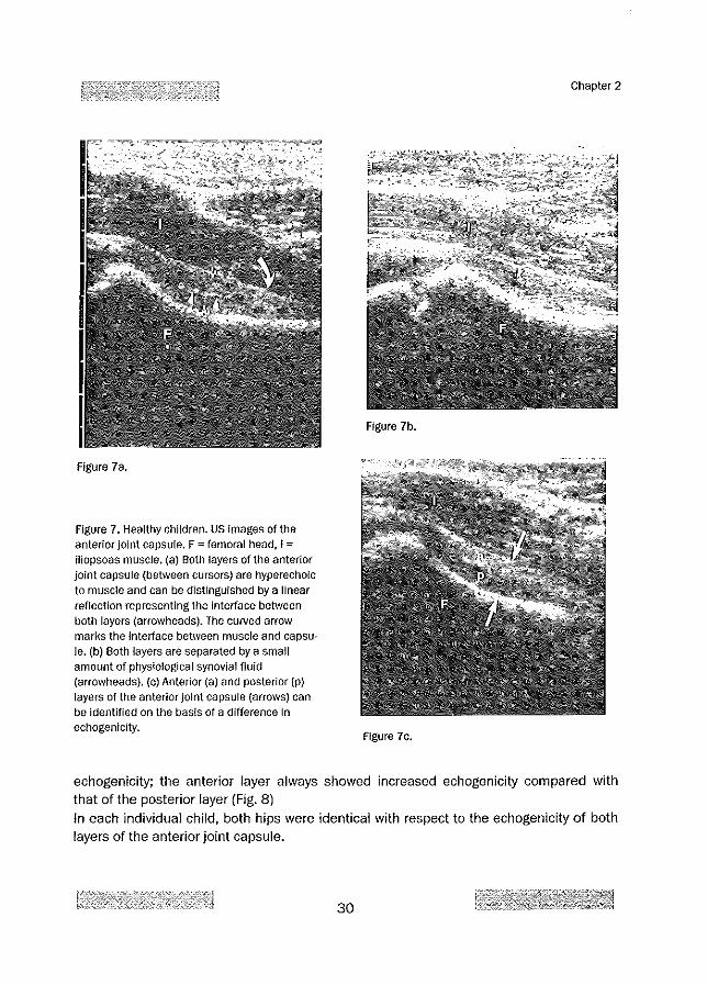
Figure 7a.
Figure 7. Healthy children. US images of the anterior joint capsule. F "" femoral head, I =
iliopsoas muscle. (a) Both layers of the anterior joint capsule (beuveen cursors) are hyperechoic to muscle and can be distinguIshed by a linear reflection representing the interface between both layers (arrowheads). The curved arrow marks the interface between muscle and capsule. (b) Both layers are separated by a small amount of physiological synovial fluid (arrowheads). (c) Anterior (a) and posterior (p) layers of the anterior joint capsule (arrows) can be identified on the basis of a difference in echogenicity.
Chapter 2
Figure 7b.
Figure 7e.
echogenicity; the anterior layer always showed increased echogenicity compared with that of the posterior layer (Fig. 8) In each individual child, both hips were identical with respect to the echogenicity of both layers of the anterior joint capsule.
30

US, histology and anatomy of the hip joint
n = 45 (39%) n = 11 (9%)
n = 22 (19%) n = 14 (12%)
n = 2 (2%)
n=13(11%) n = 9 (8%)
Figure 8. Healthy children. Schematics show the echogenicity of the anterior (a) and posterior (p) layers of anterior joint capsule in 116 hips. The echogenlcity of each layer is expressed as shades of gray compared with that of the iliopsoas muscle (I). f = femur. In the images on the left, the white tine between the layers represents the interface between the layers, or stripe sign (55), whIch was seen at US as a linear reflection. In the images on the right, there was no stripe sign.
DISCUSSION The US technique for investigation of the hip joint to evaluate a painful hip was first described by Seltzer et al. (5), to our knowledge, and has not changed thereafter. An anterior approach is used in the parasagittal plane parallel to femoral neck with the leg in slight external rotation. Some investigators included additional planes and rotation of the femur but this did not improve reproducibility (4, 6·12). In numerous reports, the thiclmess of the anterior joint capsule is addressed without a discussion of its separate layers (2, 11, 13-16). Findings in the present cadaveric study show that the anterior joint capsule is composed of two layers. Both layers have a thickness, substantial enough to enable US measurements
31

Chapter 2
with state-of-the-art equipment. This was confirmed at sonography in the adult cadavers that demonstrated both layers of the anterior joint capsule (Fig. 6). Knowledge of this anatomy is essential for accurate measurements and good interpretation. The border between the iliopsoas muscle and the joint capsule is visible as a hyperechoic line (Fig. 7a), but does not represent an anatomic structure (Fig. 4b). This line should not be interpreted as the fibrous capsule (2,4,7,8,17,18) but merely represents the interface between muscle and joint capsule. The thickness of this line depends on, among other factors, the transducer frequency (19-21). In previous studies, a wide range of values is reported for thiclmess of the anterior joint capsule. These values can be divided into two main groups: those with a mean of 5 mm, comparable to that in the present study (4, 14, 19, 22, 23), and those with a mean of 2-3 mm (1, 13, 24-26). This discrepancy was already observed by Terjesen and Osthus (11), who found it "difficult to explain, as similar US techniques and anatomic landmarks seem to have been employed." Apparently, this discrepancy is largely caused by unfamiliarity with the anatomy. In the latter studies, the distance was measured between the femoral neck and the interface between both layers of the joint capsule (stripe sign); therefore only the posterior layer of the anterior joint capsule was measured instead of the total capsule. Even the cartilage of the femoral head was sometimes mistaken for anterior joint capsule (26). According to Rohrschneider et al. (4) there was no statistically significant correlation between age and thickness of the anterior joint capsule in normal hips (in children aged more than 3 years). This finding was confirmed in the present study. In the present study the difference between both sides did not exceed 1 mm. This difference is in agreement with other studies which report a difference of 2 mm as pathologic limit (1, 4, 8, 11, 24, 26).
Histologic examination shows that both layers of the joint capsule are composed mainly of collagen fibers lined by only a thin layer of synovial membrane. The thickness of the synovial membrane is approximately 0.025 mm (27), which exceeds the spatial resolution of modern radiologic techniques such as US. Moreover, the synovial membrane of the anterior recess is of the fibrous type. In contrast to the areolar and adipose types of synovial membrane-in which the synovial intima is separated from the fibrous capsule by loose connective tissue or fatty tissue-in the fibrous type, the synovia rests directly on the fibrous layer (28). This also contributes to the inability of US to visualize the synovial membrane as a distinct layer.
In the present study, the anterior layer showed an increased echogenicity compared with that in the posterior layer in 19% of the hips of healthy subjects. This difference in echogenicity may, in part, be attributed to the difference in fiber texture of both layers, as demonstrated in the histologic examination (Fig 4a).
32

us, histology and anatomy of the hlp joint
The superior articular recess can be seen at US as a small band of decreased echogenicity adjacent to the labrum at the insertion of the joint capsule at the labrum. This should not be mistaken for a pathological condition, such as a rupture.
CONCLUSION Findings in this study show that the anterior capsule of the hip joint is composed of anterior and posterior layers that were visualized separately at US in 84% of healthy children. Both layers are mainly composed of fibrous tissue (representing the fibrous capsule) and lined by only a minute layer of synovial membrane, which is too thin to be visualized separately at US examination. Knowledge of the US anatomy of the capsule of the hip joint is essential for future studies, especially since state-of-the-art US equipment with high frequence transducers allows visualization of the hip joint in greater detail. Correct identification of both layers, especially the posterior layer, will prevent erroneous measurements and interpretation, such as mistaking the posterior layer for debris, blood, or pathological thickening of the synovial membrane.
ACKNOWLEDGMENTS The authors wish to thank T. Rijsdijk and A. Zwamborn for preparation of the photographs and drawings.
33

Chapter 2
REFERENCES 1. Marchal GJ, Hoisbeeck MT, Raes M, et al. Transient synovitis of the hip in children: role of US.
Radiology 1987;162:825·828. 2. Zieger MM, Dorr V, Schulz RD. Ultrasonography of hip joint effusions. Skeletal Radiol
1987;16:607·611. 3. Entius CAC, Kuiper JW. Koops W, de Gast A, A new positioning technique for comparing sectional
anatomy of the shoulder with sectional diagnostic modalities: Magnetic Resonance Imaging, Computed Tomography and Ultrasound. J Int Soc Plastination 1993;7:23-26.
4. Rohrschneider WK, Fuchs G, Trager J. Uftrasonographic evaluation of the anterior recess in the normal hip: a prospective study on 166 asymptomatic children. Pedlatr RadioI1996;26:629-634.
5. Seltzer S, Finberg H, Weissma SA. Arthrosonography; technique, sonographic anatomy and patho
logy. Invest RadloI1980;15:19·28. 6. Dorr U, Zieger M, Hauke H. The painful hip. Diagnostic possibilities of sonography. ROFO Fortschr
Geb Rontgenstr Nuklearmed 1988;148:487-491. 7. Dorr 0, Zieger M, Hauke H. Ultrasonography of the painful hlp. Prospective study in 204 patients.
Pediatr RadioI1988;19:36-40. 8. Kallio p, Ryoppy S, Jappinen S, Siponmaa AK, Jaaskelalnen J, Kunnamo r. Ultrasonography in hip
disease in children. Acta Orthop Scand 1985;56:367-371. 9. Konermann W, Pelligrin de M. The differential diagnosis of juvenile hip pain In the ultrasonographic
picture. Transient synovitis, Legg-Calve-Perthes disease, epiphysiolysis capitis femoris. Orthopede 1993;22:280·287.
10. McGoldrick F, Bourke T, Blake N, Fogarty E, Dowling F, Regan 8. Accuracy of ultrasonography In
transient synovitis. J Pediatr Orthop 1990;10:501-503. 11. Terjesen T, Osthus P. Ultrasound in the diagnosis and follow-up of transient synovitis of the hip.
J Pediatr Orthop 1991;11:608·613. 12. Wilson DJ, Green DJ, Maclarnon Je. Arthrosonography of the painful hlp. Clln RadioI1984;35:17-19. 13. Bickerstaff DR, Neal lM, Booth Al, Brennan PO, Bell MJ. Ultrasound examination of the Irritable hlp.
J Bone Joint Surg Br 1990;72:549·553. 14. Meradji M, Diepstraten AFM. Coxitis Fugax, sonografisches und radiologisches BlId In 65 Fallen.
Radiologe 1988;28:473-478. 15. Miralles M, Gonzales G, Pulpeiro JR, et al. Sonography of the painful hip in children: 500 consecu
tive cases. AlR 1989;152:579-582. 16. Peck RJ. Ultrasound of the painful hip In children. Br J Radiol 1986;59:293-294. 17. Shiv VI~, Jain AK, Taneja K, Bhargava SK. Sonography of hip joint In infective arthritis. J Can Assoc
RadioI1990;41:76·78. 18. Gaucher H, Hoeffel J. Anterior synovial recess of the hip: how to assess a pathological condition?
Pediatr RadioI1997;27:835·836. 19. Egund N, Wingstrand H, Forsberg l, Petterson H, Sunden G. Computed tomography and
ultrasonography for diagnosis of hip joint effusion in children. Acta Orthop Scan 1986;57:211-215. 20. Walter JP. Physics of high·resolution ultrasound·practical aspects. Clin Radial N Am 1985;23:3-11. 21. Egund N, Wingstrand H. Pitfalls in ultrasonography of hip joint synovitis in the child. Acta Radiologica
1989;30:375·378. 22. Castriota-Scandebeg A, De Micheli V, Orsi E. Letter to the editor: Ultrasound and hip joint effusion.
Eur J Radiol 1994;18:74-75. 23. Rosenborg M, Mortensson W. The validity of radiographic assessment of childhood transient
synovitis of the hip. Acta Radial DiagnOSiS 1986;27:85·89. 24. Adam R, Hendry GM, Moss J, Wild SR, Gillespie r. Arthrosonography of the irritable hip in chifdhood:
a review of 1 year's experience. Br J Radial 1986;59:205-208. 25. Alexander JE, Seibert JJ, Glasier CM, et aJ. High·resolution hlp ultrasound in the limping child. J Clin
Ultrasound 1989;17:19·24.
34

US, hIstology and anatomy of the hip joint
26. Gopakumar TS, Vaishya R, Klenerman l, Carty H. The role of ultrasound and isotope scanning in the management of irritable hips. Eur J RadioI1992;15:113-117.
27. Fawcett OW. A TextbooK of Histology. (12th ed.) New YorK London: Chapman & Hall, 1994: 229-233. 28. Ham WA, Cormack DH_ Histology. (8th ed.): J.B. lippincott Company, 1979: 473-477.
35

Chapter 2
36

Chapter 3
THE ANTERIOR JOINT CAPSULE OF THE HIP: An ultrasound study in 105 patients
with transient synovitis
Simon G.F. Robben, MD\ Maarten H. Lequln, MD\ Ad F.M. Diepstraten, MD2, Morteza MeradJi, MDt,
Departments of Pediatric Radiology' and Pediatric Orthopedic Surgery2, Sophia Children's Hospital, University Hospital Rotterdam,
The Netherlands
The contents of this chapter are published in: Radiology 1999;210:499-507
37

Chapter 3
38

US of the Hip Joint in Transient Synovitis
ABSTRACT Purpose: To evaluate the sonographic appearance of the anterior joint capsule of the hip joint in children with transient synovitis. Patients and Methods: Patients (n=105) with unilateral transient synovitis were examined with ultrasonography (US) in a prospective study. Special attention was paid to the anatomy of the joint capsule of the hip, measuring the anterior and posterior layer of the anterior joint capsule. The symptomatic hip joint was always compared with the contralateral asymptomatic hip. Results: Transient synovitis was right·sided in 57 patients and left-sided in 48 patients. Mean thickness of the symptomatic joint capsule was 9.9 mm, of the asymptomatic joint capsule 4.9 mm. In all patients the effusion itself was easily visualized, being anechoic in 96 patients and showing reflections in 9 patients. The anterior and posterior layers were identified separately at US in all hips with transient synovitis and in 83 of 105 (79%) contralateral normal hips. Overall, the anterior layer was thicker than the posterior layer. In transient synovitis compared to normal hips, no significant thickening of both layers was present (p = 0.24 and 0.57 for the anterior and posterior layers, respectively). Normal variants include plicae, local thickening of the capsule and pseudodiverticula. Conclusion: Increased thickness of the anterior joint capsule of the hip joint in transient synOVitis is caused entirely by effusion. There is no US evidence for additional capsule swelling or synovial hypertrophy. Many normal anatomic landmarks can be visualized by state·of-the-art sonography equipment and should not be mistaken for pathology.
Keywords Children, skeletal system Hip, diseases Hip, US Joints, fluid Normal variant
39

Chapter 3
INTRODUCTION The role of ultrasonography (US) in children with a painful hip has been limited to detection of thickening of the anterior joint capsule. Both thickening of the anterior joint capsule and bulging of the capsule are considered evidence of joint effusion (1-10). However, the different anatomic components of the anterior joint capsule that constitute the thickening have not been described in detail although nowadays they can be better visualized using modern US equipment with high frequency transducers. Also, the US aspect of the effusion itself has received little attention (11-13). The purpose of this study was to investigate the US anatomy of the anterior joint capsule of the hip joint in patients with transient synovitis.
PATIENTS AND METHODS This study included (a) prospective US study of 105 children with transient synovitis, and (b) US study of four cadaveric specimens.
Patient study Between January 1994 and May 1997, 105 consecutive patients with transient synovitis were examined with US in a prospective study. Patients with bilateral involvement or fever were excluded. There were 74 boys and 31 girls; aged from 2 to 12.8 years (mean, 6.0 years). The right hip was involved in 57 patients, the left hip in 48 patients. Patients with an irritable hip were considered to have transient synovitis if US depicted a thickening of the joint capsule of more than 2 mm compared with that in the asymptomatic hip (6,7,10,14) and the complaints subsided completely within 4 weeks without specific therapy and the patient remained symptom free for at least 6 months thereafter.
Cadaver study Four adult human cadavers were used for this study. The cadavers were examined prior to the embalming process. With US guidance, all 8 hip joints were injected with 10 mL of saline solution (0.9% NaCI). The needle entered the joint cavity at the level of the labrum to preserve the anatomy of both layers of the anterior joint capsule. Anatomic landmarks, such as the layers of the anterior joint capsule, were identified before and after administration of saline solution.
Method The US examinations were performed by one investigator (SR) with use of US equipment with high·frequency 7-10 MHz (Ultramark 9 HDI; Advanced Technology Laboratories, Bothell, Washington, USA) and 7 MHz (model 128 XP10; Acuson, Mountain View, California, USA) linear array transducers. The children were examined in the supine position with the hips in neutral position (extension and slight external rotation). An anterior approach along the long axis of the femoral neck was used to visualize the anterior capsule of the hip joint to the best
40

US of the Hip Joint in Transient Synovitis
advantage (12, 15). A similar approach was used in the cadaver study. In children with transient synovitis, the contralateral asymptomatic hip was used as the normal reference. The anterior jOint capsule occupies the space between femoral neck and iliopsoas muscle. It is composed of two layers of jOint capsule, separated by an anechoic space in case of effusion. The anterior joint capsule was identified, and the thickness of the capsule was assessed by measuring the maximal distance between the anterior surface of the femoral neck and posterior surface of the iliopsoas muscle (16). Moreover, the following parameters were examined: (a) identification and measurement of both layers of the anterior joint capsule in both symptomatic and asymptomatic hips, (b) identification and characterization (clear or turbid) of the effusion in the anterior recess of the joint capsule, and (c) evaluation of the anterior contour of the joint capsule.
Statistical analysis The difference in thickness of the anterior joint capsule between both sides, as well as the difference in thickness of the various layers of the anterior joint capsule of both hips, were tested by means of the paired t-test. The correlation between the thickness of anterior joint capsule and age was examined by calculating Pearson correlation coefficients. A p value less than 0.05 was considered statistically significant.
RESULTS PATIENT STUDY Anterior joint capsule of the hip joint The anterior joint capsule could be visualized in all hips, both symptomatic and asymptomatic. The anterior joint capsule is composed of two layers: the anterior and posterior layer. In all symptomatic hips, both layers could easily be distinguished from surrounding bone, muscle and anechoic effusion and could be measured (Table 1 and Fig. la). In the asymptomatic hip, the posterior layer could be differentiated from the anterior layer in 83 patients (79%). This was facilitated by: (a) a linear reflection representing the interface between both layers in 65 patients (62%). This phenomenon will be further referred to as the stripe sign (Fig. lb), (b) a small amount of physiological joint fluid in 12 patients (11%), and (c) a difference in echogenicity between both layers in 6 patients (6%). In these asymptomatic hips both layers could be measured separately (Table 1). The anterior layer was thicker than the posterior layer in both symptomatic (p = 0.01) and asymptomatic hips (p < 0.001). However, there was no difference between symptomatic and asymptomatic hips with regard to the thickness of the anterior layer (p = 0.24) or posterior layer (p = 0.57) The thickness of the anterior joint capsule of the asymptomatic hip showed no correlation with age (p = 0.1). However, the amount of effusion did show a positive correlation with age (p = 0.001).
41

Chapter 3
Table 1. Measurements in the anterior joint capsule of both hip joints In 105 patients with unilateral transient synovitis.
No. of mean
patients thickness (mm) SO
Symptomatic hip
Joint capsule 105 9.91 1.71
Anterior layer 105 2.38 0.66
Posterior layer 105 2.14 0.44
Asymptomatic hip
Joint capsule 105 4.90 1.02
Anterior layer 83 2.51 0.63
Posterior layer 83 2.10 0.58
In 50 of the symptomatic hips (48%), a local thickening of the posterior layer of the anterior joint capsule, referred to as the "hump", was visible. The thickness of this hump varied considerably but it was invariably localized at the insertion of the posterior layer near the cartilage of the femoral head (Fig. 2a and 2b). This phenomenon was almost exclusively observed in hips with effusion. Occasionally an identical structure was seen on the anterior layer (Fig. 2c).
Effusion In all symptomatic hips, the effusion in the anterior recess could be clearly differentiated from the surrounding layers of the anterior joint capsule (Figs. 1 and 2). The effusion was anechoic in 95 patients (90%) and showed some reflections in 9 patients (9%) (Fig. 3). In one patient (1%), an obese boy, the visualization of the anterior recess was insufficient for reliable evaluation of the clarity of the effusion. A small amount of joint fluid was also present in the anterior recess of 12 (11%) asymptomatic hips. The fluid in the anterior recess of the asymptomatic hips was always clear. The mean thickness of this layer of synovial fluid was 1.0 mm (range 0.2-1.6 mm).
42

US of the Hip Joint in Transient Synovitis
Figure la.
Figure lb.
43
Figurel. Anterior joint capsule, sagittal US image.
Figure la. TransIent synovitis. The anterior (A) and posterior layers (P) of the joInt capsule are separated by anechoic effusion and can be easily identified and measured (between cursors, 2,5 and 1,9 mm, respectively),
Figurelb. Normal hip. The anterior joint capsule is measured between cursors. Anterior and posterior layers are separated by their interface (arrowheads) and can be measured separately (3.1 and 2.9 mm, respectively).
F = femoral neck, I = Iliopsoas muscle

Figure 2a.
Figure 20.
Figure 2d.
Chapter 3
Figure 2b.
Figure 2. Hump in hips with transient synovitis and in normal hips. Sagittal US Images. F "" femoral neck, I "" itiopsoas muscle, C = cartilage of femoral head.
Figure 2a. Patient with transient synovitis. Local thickening (arrows) of the posterior (P) layer of the joint capsule at its insertion near the articular cartilage of the femoral head.SAR = superior articular recess, L = labrum.
FIgure 2b. Another patient with transient synovitis. Large hump (arrows) originating from the posterior (P) layer of the anterior joint capsule.
Figure 20. Local thickening (arrows) at the anterior layer (A) of the jOint capsule in a patient with transient synovitis.
Figure 2d. Cadaver study after injection of saline solution In hlp joint. The hump is clearly visualized (arrows).
FIgure 2e). Asymptomatic hip without effusion. The hump Is delineated by the stripe sign (small arrows). Anterior jOint capsule is marked by cursors.
Figure 2e.
44

US of the Hip Joint in Transient Synovitis
Anterior bulging of the capsule The results are shown in Table 2. Convex bulging anterior joint capsule has a sensitivity of 94%, specificity of 91%, positive predictive value of 92% and negative predictive value of 94% for effusion. None of the patients with a concave border had an effusion: therefore, the presence of a concave border virtually rules out effusion.
Table 2. Shape of the anterior border of the anterior joint capsule of the hip joint in 105 patients with transient synovitis. Data are number of patients.
Shape
convex
straight
concave
total
Symptomatic Hip
99
6
o
105
(94%)
(6%)
(0%)
(100%)
45
Asymptomatic Hlp
9
31
65
105
(9%)
(29%)
(62%)
(100%)
Figure 3. Sagittal US image depicts the anterior joint capsule (cursors) in a patient with transient synovitis of 7 days duration. Small particles (arrows) are floating In the effusion.

Figure 4a.
Additional findings
Figure 4. Plicalike structure (arrowheads), traversing the effusion in a patient with transient synovitis (a) and in a cadaver (b) after intra-articular administration of saline solution.
Chapter 3
Figure 4b.
In four patients (4%), a linear structure traversing the fluid-filled anterior recess (Fig. 4a), demonstrated the same echogenicity and texture as did both layers of the anterior joint capsule. In 2 patients (2%), a thin-walled cystic protrusion of the joint effusion was seen. This arose from the synovial surface of the anterior layer of the capsule and protruded through the anterior layer into the space between the iliopsoas muscle and anterior border of the joint capsule with a collar-button configuration (Fig. 5). All patients with additional findings had an uneventful recovery.
CADAVER STUDY US could visualize the anterior joint capsule in all cadaveric hips, except for one hip that was replaced by a prosthesis. The anterior joint capsule was seen at US as a band of tissue between the anterior femoral neck and the fascia of the iliopsoas muscle. Centrally, a linear reflection was seen in 5 hips, thought to represent the interface between both layers of the anterior joint capsule (collapsed anterior recess). This was subsequently proved by means of intra-articular injection of saline solution, after which the linear reflection was replaced by hypoechoic fluid in the anterior recess (Figs. 6a and b). Obviously, the presence of this linear reflection (stripe sign) indicates absence of effusion. After the injection of saline solution, one cadaveric hip showed a plical ike structure within the fluid-filled joint space (Fig. 4b), identical to the structure that was seen in 4% of the hips with transient synovitis (Fig. 4a), and 2 hips showed a local thickening of the posterior layer (Fig. 2d), corresponding with the hump that was seen in approximately 50% of the hips with transient synovitis.
46

US of the Hlp Joint in Transient Synovitis
Figure 5a. Figure 5b.
Figure 5. Sagittal (a) and transverse (b) US image of the anterior joint capsule in a patient with transient synovitis depict a cystic protrusion of effusion through the anterior layer (A) of the joint capsule is seen, with the aspect of a pseudo·diverticulum (arrows).
DISCUSSION
The anterior joint capsule in transient synovitis Until recently, thickening of the anterior joint capsule of the hip joint was considered as evidence of effusion. However, the continuous improvement of US equipment enables the sonologist to visualize anatomic structures that could not be seen in the past. Cadaveric studies showed that the anterior joint capsule of the hip joint is composed of two layers of joint capsule (17). Both layers consist of fibrous tissue and are thick enough to be visualised by US. The posterior layer is a continuation of the periosteal membrane, the anterior layer blends with the iliofemoral ligament. The synovial membrane is lined by a one to three celis thick (20AO ,umeter) layer of synoviocytes (18). This layer of synoviocytes is too thin to be seen on US.
The results of the present study are compatible with those in the above-mentioned cadaveric study: Both layers of the joint capsule can be readily visualized and measured separately with US in ali hips with transient synovitis, facilitated by the effusion. In all these hips the effusion itself could be readily discerned from the joint capsule as a separate hypo echoic or anechoic layer. In the contralateral normal hip, the thickness of the anterior and posterior layers of the anterior joint capsule could be measured in 79% of the patients and compared with measurements in the symptomatic hip. In both symptomatic and asymptomatic hips the anterior layer of the joint capsule was significantly thicker than the posterior layer (Table i), which is consistent with the
47

Chapter 3
findings in a previous cadaveric study (17). The anterior layer is thicker probably because it is reinforced by the iliofemoral ligament and the orbicular zone.
Figure 6a. Figure 6b.
Figure 60. Figure 6d.
Figure 6. The stripe sign in a cadaver and a patient. The stripe sign is marked by small arrows in a cadaver (a). It disappears after introduction of saline solution (arrowheads) into the anterior reoess of the joint capsule (large arrows) (b). The reverse process is seen in a normal hlp. The stripe sign (small arrows) appears when a physiological effusion, marked behveen arrowheads (e), is squeezed out of the anterior joint capsule by compression of the transducer (d).
48

US of the Hlp Joint in Transient Synovitis
Moreover, the anterior and posterior layers of the anterior joint capsule showed no statistically significant difference between the symptomatic and asymptomatic sides (Table 1). Synovitis apparently does not cause a measurable thickening of the anatomic components of the anterior joint capsule. Theoretically, the synovial membrane is thickened in synovitis, but because it forms only a minute part of both layers of the anterior joint capsule, its thickening cannot be appreciated at US. In many articles dealing with transient synovitis, both layers of the anterior joint capsule are either not identified or not mentioned, probably due to use of inadequate eqUipment or unfamiliarity with the anatomy of the anterior joint capsule. However, those articles that do describe both layers erroneously identify them as synovium or synovial hypertrophy or thickening (3, 9, 12, 13, 19). According to the results in the present study, neither of the layers of the anterior joint capsule are diffusely thickened in transient synovitis. Moreover, "synovium or synovial membrane" is a misnomer because the anterior joint capsule is composed of a fibrous capsule with only a minute layer of synovial membrane. Because (a) both layers of the anterior joint capsule are not thickened, and (b) the effusion was always discernible, it seems more rational to detect effusion by visualizing the effusion itself rather than by relying on indirect signs such as differences between hips in measurements of the joint capsule (4-10, 19). In cases of poor US visualization of the joint capsule, this indirect method can be a good alternative, although it should be realized that it fails when bilateral effusions are present.
Hump of the posterior layer This phenomenon was almost exclusively observed in hips with effusion, probably because the effusion facilitates demarcation of the synovial surface. This hump could be interpreted as debris or flocculation, but several facts argue against this. (a) In some patients, we were able to demonstrate vessels in this hump at Doppler US; and (b) in the supine position, the hump does not migrate to the most dependent section of the recess. The stripe sign phenomenon does not offer enough anatomic detail to confirm the presence of such a local thicl(ening in hips without an effusion. However, evidence of a hump was present in one asymptomatic hip (Fig. 2e), and moreover we observed this structure in two cadaver hips (Fig. 2d). Apparently this local hump is not restricted to synovitis but should be regarded as a normal anatomic landmark representing the insertion of the capsule in the femoral neck. Marchal et al. (15) observed an identical local thickening in a patient with septic arthritis, but the findings in the present study demonstrate that this sign certainly is not pathognomonic for septic arthritis.
Effusion Because both layers of the anterior jOint capsule are not thickened in transient synovitis, the widening of the anterior joint capsule can be attributed solely to effusion. In all symptomatic hips, the effusion could be readily discriminated from the layers of the anterior joint capsule and measured separately.
49

Chapter 3
In 9 patients, the effusion was not completely clear. None of these patients had clinical signs of septic arthritis, and all had an uneventful recovery. This phenomenon was also observed by D6rr et al. (13) and Marchal et al. (15) in 21% and 10% of their patients, respectively, and Zawin et al. (3) demonstrated that turbid effusion is not diagnostic for infection. We found that the mean duration of complaints in patients with turbid effusion was considerably longer than in patients with a clear effusion (7.2 versus 4 days, p < 0.001). This may be the result of progressive accumulation of cellular debris in effusion of longer duration. Obviously, the presence of turbid effusion is associated with the duration of the disease, but it has no further diagnostic value. Suprisingly, the amount of fluid in transient synovitis shows a positive correlation with age (p < 0.001). Assuming that the pathophysiological mechanism of transient synovitis is age-independent, this correlation must be attributed to geometrical factors. Because the anterior recess is larger in older children, the same pressure will induce more expansion, according to the physical law of Laplace. A small layer of joint fluid in the anterior recess of the asymptomatic hip was present in 12 patients (11%), with a mean thickness of 1.0 mm and a maximum of 1.6 mm (Figs. 6c and d). This finding is similar to that of Rohrschneider et al. (16), who found this in 12% of healthy children, with a maximum thickness of 1.5 mm. Therefore, 2 mm seems to be a sound threshold to differentiate a pathologic from a physiological effusion. This is in keeping with a difference of more than 2 mm between both symptomatic and asympto· matic anterior joint capsules that is considered pathological in several reports (7, 8, 12, 14-16, 19). The stripe sign, representing the collapsed anterior recess, can be of additional value in excluding small amounts of effusion in the joint. This sign is visible only when both layers lie close together, that is, in the absence of effusion (Figs. 6c and d).
Bulging of the capsule Some authors consider anterior bulging of the joint capsule the best criterion for effusion (1-3). However, the contour of the anterior joint capsule depends on rotation, being concave in external rotation and convex in internal rotation (16). Moreover, joint capsules of normal hips can have a straight or convex contour (16). This is also true in the present study, in which 9% of the asymptomatic hips showed anterior bulging in the absence of effusion, making this sign less reliable. Six percent of the symptomatic hips had no convex contours, despite the fact that a substantial amount of effusion was present. It seems more rational to detect effusion by visualizing the effusion itself than by relying on indirect signs such as contours of the capsule. On the other hand, a concave border of the anterior joint capsule seems to be a reliable indicator for the absence of effusion because in the present study, none of these hips showed joint fluid on US.
50

US of the Hlp Joint in Transient Synovitis
Plica In 6 patients (6%) a platelike structure traversed the effusion, mostly running from the hump of the posterior layer to the anterior layer. The structure and echogenicity were identical for both layers of the anterior joint capsule. This structure was also demonstrable in one of the cadaver hips after intraarticular injection of saline solution. It probably represents a plica, comparable with the well-known plicae in the knee joint. None of the patients with such a plica had previous complaints, and all had an uneventful recovery.
Diverticulum The cystic protrusion of joint effusion through the anterior layer of the anterior joint capsule probably represents a herniation of the synovial membrane through a defect in the fibrous capsule, which creates a pseudodiverticulum of the synovial membrane. The thin wall also suggests it is a pseudodiverticulum of the synovial membrane rather than a real diverticulum of the joint capsule.
CONCLUSION Findings in this study show that no measurable thickening of both layers of the anterior joint capsule is present in transient synovitis. Therefore, enlargement of the anterior joint capsule in transient synovitis is caused solely by the presence of effusion. It is more rational to detect effusion by visualizing the effusion itself than by relying on indirect signs such as contours of the capsule or differences in joint capsule measurements between hips. These indirect methods can be good alternatives only in cases of poor US visualization of the joint capsule, although the latter method fails when bilateral effusions are present. Moreover, findings in this study describe normal variants that can be visualized by stateof-the-art US equipment, such as local humps of the posterior layer of the joint capsule, the stripe sign, pseudodiverticula, and plicae. Sonologists should be aware of these findings to prevent misinterpretation of normal variants mistaken for pathologic conditions.
ACKNOWLEDGMENTS The authors thank Teun Rijsdijk and Andries Zwamborn for preparation of the photographs and drawings for this article.
51

Chapter 3
REFERENCES 1. Alexander JE, Seibert JJ, Aronson J, et al. A protocol of plain radiographs, hip ultrasonography,
and triple phase bone scintigraphy in the evaluation of the painful pediatric hip. Clin Pediatr (Phila) 1988;27:175-181.
2. Alexander JE, Seibert JJ, Glasier CM, et a!. High-resolution hip ultrasound in the limping child. J Clin Ultrasound 1989;17:19·24.
3. Zawin JK, Hoffer FA, Rand FF, Teele RL. Joint effusion in children with an irritable hip: US diagno· sis and aspiration, Radiology 1993;187:459-463.
4. Bickerstaff DR, Neal lM, Booth AJ, Brennan PO, Bell MJ. Ultrasound examination of the irritable hip, J Bone Joint Surg Br 1990;72:549-553,
5, Wilson OJ, Green OJ, MacLarnon JC. Arthrosonography of the painful hip. Clin Radial 1984;35:17-19.
6. Meradji M, Dlepstraten AFM. Coxitis Fugax, sonograflsches und radiologisches BUd in 65 Fallen. Radiologe 1988;28:473-478.
7. Terjesen T, Osthus p, Ultrasound In the diagnosis and follow-up of transient synovitis of the hip. J Pediatr Orthop 1991;11:608-613.
8. I~onermann W, Pelligrin de M. The differential diagnosis of juvenile hlp paIn In the ultrasonographic picture. Transient synOVitis, legg-Calve-Perthes disease, epiphysiolysis capitis femoris. Orthopede 1993;22:280-287.
9. Mlralles M, Gonzales G, pulpelro JR, et al. Sonography of the painful hip In children: 500 consecutive cases. AJR 1989;152:579-582.
10. McGoldrick F, Bourke T, Blake N, Fogarty E, Dowling F, Regan B. Accuracy of ultrasonography in transient synovitis. J Pediatr Orthop 1990;10:501-503,
11. Schlesinger AE, R.J. H. Diseases of the Musculoskeletal System in Children: Imaging with CT, Sonography, and MR. AJR 1992;158:729-741.
12. ZIeger MM, Dorr U, Schulz RD. Ultrasonography of hlp JoInt effusions. Skeletal Radiol 1987;16:607-611.
13. Dart U, Zieger M, Hauke H. Ultrasonography of the painful hip, Prospective study In 204 patients. Pedlatr Radiol 1988;19:36-40.
14. Gopakumar TS, Vaishya R, Klenerman l, Carty H. The role of ultrasound and Isotope scanning in the management of irritable hips, Eur J RadioI1992;15:113-117.
15. Marchal GJ, Holsbeeck MT, Raes M, et al. Transient synovitis of the hip tn children: role of US. Radiology 1987;162:825-828.
16. Rohrschneider WK, Fuchs G, Trager J. Ultrasonographic evaluation of the anterior recess in the normal hip: a prospective study on 166 asymptomatic children. Pediatr RadioI1996;26:629-634_
17. Robben SGF, lequin MH, Diepstraten AFM, Hollander JC, EnUus CAC, Meradji M. Anterior joint capsule of the normal hip and in children with transient synovitis: US study with anatomic and histologic correlation. Radiology 1999;210:499-507.
18. Gray H. Anatomy of the Human Body. (30th ed.) Philadephla: Lea & Febiger, 1985: 390-395. (Clemente CD, ed. ).
19. Adam IR, Hendry GM, Moss J, Wild SR, Gillespie I. Arthrosonography of the irritable hip in childhood: a review of 1 year's experience. Br J Radlol 1986;59:205-208.
52

Chapter 4
ATROPHY OF THE QUADRICEPS MUSCLE IN CHILDREN WITH A PAINFUL HIP:
An ultrasound study, physiological aspects
Simon G.F. Robben, MD', Maarten H. Lequin, MD" Morteza Meradji, MD', Ad F.M. Diepstraten, MD2, Wim C.J. Hop, PhD3
Departments of Pediatric Radiology' and Pediatric Orthopedic Surgery2, Sophia Children's Hospital, University Hospital Rotterdam,
Department of Epidemiology and Biostatistics3,
Erasmus University, Rotterdam, The Netherlands
Accepted for Publication in Clinical Physiology
53

Chapter 4
54

Atrophy of the quadriceps muscle in children
ABSTRACT
Objective: To study the degree of muscle wasting of various components of the quadriceps muscle in children with a painful hip. Methods: Between January 1994 and September 1997, 327 consecutive patients with a unilateral painful hip and/or limping were evaluated prospectively with ultrasonography (US). Quadriceps thickness was measured on both sides. Moreover, muscle thickness was measured in 59 control subjects. Results: The patients were divided into 8 groups; transient synovitis (n=134), Perthes disease (n=35), slipped capital femoral epiphysis (n=5), osteomyelitis (n=4), non-specific synovitis (n=5), rheumatoid arthritis (n=3), miscellaneous (n=16). In 125 patients no sonographic and radiological abnormalities were found and during follow-up the complaints disappeared ("no pathology" group). Ipsilateral muscle wasting was present in all patient groups, whereas the control subjects showed no significant difference between both legs. The degree of muscle wasting was compared between transient synovitis, "no pathology" group, Perthes' disease, and control subjects. For both quadriceps and vastus intermedius muscles there was a significant difference between these groups, except between control subjects and "no pathology" group. For the rectus femoris muscle there was a significant difference between these groups, except between transient synovitis and "no pathology". Muscle wasting showed a positive correlation with duration of complaints and preexisting muscle mass. Conclusion: Different diseases show different degrees of muscle wasting and different patterns of muscle wasting of various components of the quadriceps femoris muscle.
Keywords Atrophy Muscle Ultrasonography Childhood
55

Chapter 4
INTRODUCTION
The development of muscle atrophy as a result of inactivity is a well·known phenomenon (1-4). Especially the effects of decreased activity on the muscles of the leg have been the subject of many investigations. Most of these investigations have focused on: a) animal models (5·9), b) experimental studies in adults (10·13), or c) immobilization studies in adults (14·16). The effect of (subclinical) inactivity on muscle wasting in children with various orthopedic diseases has not been the subject of investigation, except for one study that describes muscle atrophy in Perthes' disease (17). Ultrasonography (US) is a noninvasive technique that can visualize muscles to a good advantage (18). Because it is not time·consuming and it is readily accepted by the patients, US is an ideal modality for screening large patient groups. Moreover, US mea· sures the muscle itself, which is an advantage over tape measurements, because the latter is an indirect method that also includes the skin and subcutaneous fat. The purpose of this study was threefold: 1) to study the presence and degree of muscle wasting in various pediatric diseases of the musculoskeletal system that cause a painful hip or limping; 2) to investigate the influence of several factors on the degree of muscle wasting; and 3) to study the recovery from atrophy.
PATIENTS AND METHODS Patients Between January 1994 and September 1997, 348 consecutive children who had clinical symptoms of hip disease were evaluated prospectively. There were 221 boys and 127 girls; age range 1.1 to 15.9 (mean 6.3) years. Patients with bilateral complaints were excluded. A control group was composed of 59 children without complaints or previous orthopedic disease. There were 37 boys and 22 girls; age range 1.7 to 18.1 (mean 7.4) years. The study was approved by the institutional Medical Ethical Review Board. Informed consent was obtained before the examination.
US measurements All patients and control subjects were examined sonographically by the same investigator (SR). High·resolution 7·10 MHz (Advanced Technical Laboratories, Ultramark 9 HDI, Bothell, Washington, USA) and 7 MHz (Acuson 128 XP10, Mountain View, California, USA) linear array transducers were used. Patients were examined in supine position with hips in neutral position. Both hips were examined, in which the contralateral asymptomatic hip was used as normal reference. Because US is sensitive to bias (the investigator is aware of the symptoms), the use of a standardized US measurement technique is mandatory: the transducer is placed on the anterior thigh in the transverse plane, at the mid·point between the superior border of the patella and anterior superior iliac spine. Any pressure of the transducer on the skin is
56

Atrophy of the quadriceps muscle in children
eliminated by using an excess amount of echogel to avoid compression of the muscle (Figs. 1 and 2). Moreover, the patient's attention is diverted to optimize muscle relaxation. Three parameters were measured: 1) the thickness of the rectus femoris muscle as the distance between posterior and anterior fascia, 2) at the same level, the vastus intermedius muscle as the distance from the femoral cortex to the anterior fascia of the vastus intermedius muscle, 3) total thickness of the quadriceps muscle (both rectus femoris and vastus intermedius) as the distance from the femoral cortex to the anterior fascia of the rectus femoris muscle (Fig. 1).
Echogel
Subcutan&ous fal Skin
Femur
Figure la, Figure lb.
Figure 1, a) Schematic drawing of transverse ultrasound image at the mid-thigh position. b) Transverse ultrasound image. Quadriceps muscle is measured between electronic calipers, Notice generous use of contact gel to avoid any compression of muscle.
All measurements were performed on both sides and the differences between the normal and symptomatic side were calculated. In the control group the difference between left and right side was calculated. These differences will be denominated [; quadriceps, [;rectus femoris and [; vastus intermedius (asymptomatic side minus symptomatic side). The final clinical diagnosis was established 6 months after presentation by reviewing the medical and radiological records.
Reproducibility study In 21 legs of normal children (age range 4 to 14 years), two independent measurements with an interval of 5-10 minutes were performed of all muscles to assess the degree of reproducibility. First, the measurement site was determined and marked on the skin by a pen on both legs, followed by the US measurements. Prior to the second set of measurements, the skin markings were erased and the measurement sites were defined again. The skin marks could be easily rubbed off by wads of cotton wool drenched in 70% ethanol.
57

Chapter 4
Follow-up study In 38 patients with transient synovitis follow-up US examination was performed to verify the resorption of the effusion. At the same time, the muscles were measured again as described above. There were 24 boys and 14 girls, mean age was 5.6 (range 2.0-9.2) . years. Mean duration between initial US examination and follow-up examination was 39 (range 8-86) days.
Figure 2a. Figure 2b.
Figure 2. Five-year-old boy with Perthes' disease, duration of symptoms is 40 days. Transverse ultrasound images show severe muscle wasting. a) Asymptomatic right side. Quadriceps thickness is 22.3 mm. b) Symptomatic left side. Quadriceps thickness is 13.4 mm
Statistical analysis For statistical analysis the Statistical Package for the Social Sciences (SPSS version 7.5, Chicago, USA) was used. The paired t-test was used to evaluate left/right differences within groups. Analysis of variance, with multiple comparison procedure of Bonferroni, was used to analyse the differences in muscle wasting between the various groups. Univariate correlation studies were performed by calculating Spearman's coefficients of correlation. Multiple regression analysis was performed to simultaneously evaluate various factors such as age, duration and preexisting muscle mass. In this analysis the duration of complaints was logarithmically transformed to eliminate the influence of outlying data. A p-value of ~0.05 was considered statistically significant. Reproducibility of the measurements for each muscle was evaluated by calculating the mean coefficient of variation.
58

Atrophy of the quadriceps muscle in children
RESULTS Reproducibility study In this group, the mean muscle thickness for quadriceps, rectus femoris, and vastus intermedius muscle was 32.6, 18.6, and 13.9 mm, respectively. The reproducibility of US measurements was excellent: The mean difference between both quadriceps, rectus femoris and vastus intermedius muscle measurements was 0.4 mm (sd 0.6), 0.3 mm (sd 0.3) and 0.4 mm (sd 0.3), respectively. This resulted in a mean coefficient of variation for the quadriceps muscle measurements of 1%, for the rectus femoris muscle 1%, and for the vastus intermedius muscle 2%.
Table 1. Descriptive statfstfcal data on the patient population (n = 327) and control subjects
number of Diagnosis patients mean age median duratfon of
(years) symptoms in days (range)
Transient synovitis 134 6.0 3 (1-120)
~No pathology" 125 6.5 21 (1-1100)
Perthes disease 35 5.4 42 (2 - 360)
SCFE 5 12.7 35 (21-100)
Osteomyelitis 4 7.3 21 (14 - 21)
Non-specific synovitis 5 10.3 21 (5-120)
Rheumatoid arthritis 3 7.7 21 (5 - 60)
Miscellaneous 16 6.7 14 (2 - 210)
Control subjects 59 7.4
Patients and control subjects In 21 patients muscle measurements were not possible or considered to be unreliable, because of anxiety and agitation. These patients (mean age 4.5 years) were generally younger than the other patients (mean age 6.4 years). The remaining patients (n=327) could be divided into 8 groups; the final clinical diagnoses are given in Table 1.
59

Chapter 4
• The "no pathology" group consists of 125 patients without sonographic and radiological abnormalities. No definite diagnosis could be made and in all patients the complaints disappeared without specific treatment and remained symptom-free thereafter. This group includes 8 patients with the tentative diagnosis of postinfectious or parafebrile arthralgia.
• The non-specific synovitis group includes patients with reactive (postinfectious) synovitis.
• The "miscellaneous" group consists of patients with neoplasms (n=4), occult fractures (n=2), synovitis of the knee (n=2), septic arthritis, and Baker's cyst, retrocecal appendix, ureteric calculus, juvenile osteoporosis, leukemia, lymphadenitis, bone marrow edema eci, and (all n=1).
The muscle measurements are given in Table 2, including the calculated values of I1vastus intermedius, I1rectus femoris and I1quadriceps. Figure 3 shows the mean value of I1quadriceps for each diagnosis group. No significant difference in thickness between left and right quadriceps muscles (l1quadriceps) was present in control subjects. Maximal difference between both legs in control subjects was 3 mm. The number of patients with SCFE (n=5), osteomyelitis (n=4), aspecific synovitis (n=5), and rheumatoid arthritis (n=3) was considered too low to perform a reliable statistical analysis. Also, the "miscellaneous" group did not undergo statistical analysis because of its heterogeneity. Therefore, the difference in muscle wasting was tested between transient synovitis, "no pathology" group, Perthes' disease, and control subjects: for both I1quadriceps and I1vastus intermedius there was a significant difference between all groups, except between control subjects and "no pathology". For I1rectus femoris there was a significant difference between all groups, except between transient synovitis and "no pathology" (Table 3). In Perthes' disease a significant positive correlation (r=0.67, p<0.001) was found between I1vastus intermedius and I1rectus femoris. Multiple regression analysis demonstrated that this relation was independent of the duration of the complaints. In transient synovitis and "no pathology" group, the degree of wasting of both rectus femoris muscle and vastus intermedius muscle did not appear to be related.
Muscle wasting versus age and gender There was no correlation between age and muscle wasting for quadriceps muscle, rectus femoris muscle and vastus intermedius muscle in separate disease groups (Table 4). However, multivariate analysis demonstrated a significant weak correlation (p=0.02) between age and atrophy for the vastus intermedius muscle when the groups were combined (i.e. transient synovitis, "no pathology" and Perthes' disease): per year the average increase of wasting was 0.06 mm. None of the groups showed a significant correlation between the degree of muscle wasting and gender.
60

Atrophy of the quadriceps muscle in children
Table 2. Absolute muscle thickness (in mm), and 8 quadrIceps, 8 rectus femoris and 8 vastus intermedius, in patient groups and control subjects.
Diagnosis
it Z 'U en 0 G; ;u " ()
'" ()
* ~ iii 0 ~
0 ;+ '" '0 '" ~
"' 'U ~ '" 0 " c 0
~ iii' " " 3 0 3 '>.
rl "' ::ti " " " ~ a. '< 0 5 ~ "' !!!
0 U;. '>. /!J. " c a
" a ~ 0: 0 !'r
~ ~ " "' 0 " c " 0 "' < ;+ "' Ii! < " 2: ~ ~
"' a: "'
Quadr. sympt 24.7 25.8 22.3 28.6 20.1 26.1 25.9 25.3 28.2
Quadr. asympt 25.5 26.2 25.2 34.0 23.6 27.9 29.6 27.1 28.4
Rectus sympt 13.6 13.9 12.3 13.8 12.4 14.3 15.3 12.9 16.1
Rectus asympt 14.0 14.1 14.0 16.3 13.7 15.2 15.8 13.5 16.2
VI sympt 11.1 11.9 10.1 14.8 7.7 12.1 10.7 13.2 12.4
VI asympt 11.6 12.0 11.4 17.7 9.9 12.9 13.8 14.3 12.4
tJ. Quadriceps 0.8 0.3 2.9 5.5 3.6 1.8 3.6 1.8 0.2 SE 0.1** 0.1* 0.4** O.gNi 2.1NT 1.1'IT 1.61iT 0.51fT 0.2NS
8 Rectus fem. 0.3 0.2 1.7 2.5 1.3 0.9 0.5 0.6 0.1 SE 0.1** 0.1* 0.2** 0.7NT 1.2NT O.gNT 1.0m 0.31fT O.1NS
II VI 0.5 0.1 1.3 3.0 2.2 0.8 3.1 1.1 0.0 SE 0.1** O.1NS 0.2** 1.3NT 1.2!lT O.sm 0.6* O.4NT 0.21<5
8 quadriceps '" quadriceps asymptomatic side minus symptomatic side tJ. rectus femoris = rectus asymptomatic side minus symptomatic side
tJ. Vastus intermedius (VI) vastus asymptomatic side minus symptomatic side (In control subjects= left minus right)
NS= not statistically sIgnIficant left/right difference NT= not tested because number of patient was too small SE= standard error
*= 0.05> P >0.01
**= P <0.01
61

Table 3. Differences in muscle wasting between various disease groups
8. quadriceps and Perthes fJ. vastus Intermedius
Control subjects * Transient synovitis * uNo pathology" *
fJ. rectus femoris Perthes
Control subjects
Transient synovitis
"No pathology"
* = significant (p< 0.05) NS = not significant
*
*
*
"No pathologyu
NS
*
"No pathologyu
*
NS
Muscle wasting and duration of complaints (Table 4)
Transient synovitls
*
Transient synOVitis
*
Chapter 4
In Perthes' disease a positive correlation was found between duration and wasting for quadriceps muscle, rectus femoris muscle and vastus intermedius muscle, also when the effect of age was taken into account. For each doubling of the duration, lIquadriceps, lIrectus femoris and lIvastus intermedius increased with 0.7 mm (p=0.007), 0.3 mm (p=0.05) and 0.4 mm (p=0.002), respectively. For transient synovitis similar findings were found for lIquadriceps and lIrectus femoris: 0.3 mm (p=0.003), and 0.2 mm (p=0.02), respectively.
Muscle wasting and preexisting muscle mass (Table 4) The muscles of the asymptomatic leg were considered to represent the preexisting muscle mass. A statistically significant positive correlation between preexisting muscle mass and degree of muscle wasting was found in a) transient synovitis for both quadriceps muscle and vastus intermedius muscle, b) "no pathology" group for the vastus intermedius muscle, and c) Perthes' disease for the rectus femoris muscle. All these correlations remained unchanged after adjustment for age and duration by multivariate analysis.
62

Atrophy of the quadriceps muscle in children
10
8 a. :::J e
6 Ol
:g U) c a. 4 0 !'l () ·c
" 2 '" :::J 0
0 <1
-2
-4
-6
1 2 3 4 5 6 7 8 Disease groups
Figure 3. Boxplot of mean muscle atrophy In various diseases. The lower and upper boundaries of the box represent 25th and 75th percentlle, respectively. Whiskers represent smallest and largest values. l=translent synovitis, 2="no pathology", 3=Perthes' disease, 4=SCFE, 5=osteomyelitis, 6=aspecific synovitis, 7=rheumatold arthritis, 8=miscellaneous.
Follow-up study Thirty-eight patients with transient synovitis had a follow-up US examination after a mean interval of 39 (range 8-86) days. Mean /lquadriceps at the initial US examination was 0.7 (range -1.8 to 5.6) mm. At the follow-up US examination it was 0.0 (range -2.6 to 2.4) mm. This difference was tested by the paired t-test and was significant (p=0.003). All patients were symptom-free at the time of the follow-up examination.
DISCUSSION Inactivity induces many changes in muscles; functional (strength, electro myographically, contraction characteristics), structural (muscle weight loss, fiber size reduction, histologic changes), and biochemical changes. Most data are derived from experimental and/or animal studies. Modern imaging techniques can visualize the structural changes that occur in muscle atrophy both quantitatively (13, 19, 20) as well as qualitatively (18, 21, 22). US is an ideal modality to examine a large pediatric patient population. First, US is readily available, easy to perform, relatively inexpensive, not time-consuming, painless, lacks ionizing radiation, and is reproducible (coefficient of variance for various components of the quadriceps muscle is 1-2%, which is even slightly better than reported in previous
63

Chapter 4
Table 4. Spearman correlation coefficients between atrophy and age, atrophy and duration of complaints, and atrophy and preexisting muscle mass for various muscles. Numbers between parentheses denote p-values
age and wasting
tl quad tl rect tl vast
TransIent synovitis 0.08 (0.34) -0.05 (0.56) 0.16 (0.07)
UNo pathology" 0.11 (0.20) 0.03 (0.76) 0.12 (0.18)
Perthes disease 0.03 (0.86) 0.10 (0.54) -0.08 (0.65)
duration and wasting
tlquad tl reet !J.. vast
Transient synovitis 0.21 (0.01) 0.17 (0.05) 0.12 (0.16)
"No pathology" 0.03 (0.71) 0.15 (0.10) 0.00 (0.97)
Perthes disease 0.49 (0.003) 0.45 (0.008) 0.54 (0.001)
preexisting muscle mass and wasting
tl quad tl rect tl vast
Transient synovitis 0.20 (0.02) 0.11 (0.21) 0.30 «0.001)
UNo pathology" 0.14 (0.12) 0.09 (0.30) 0.30 (0.001)
Perthes disease 0.33 (0.05) 0.37 (0.03) 0.20 (0.24)
studies (23-26)). This method is accepted very well by the patients. as is reflected in the low number of patients that could not be investigated in the present study (6%). Secondly. in many institutions US is part of the evaluation of children with a painful hip and/or limping. so measuring the muscles is only a small extension of the investigation. We measured muscle thickness (11. 19. 23. 27-29) instead of cross-sectional area (CSA). because the latter method is elaborate. is more sensitive to sources of error. and requires a compound ultrasound scanner that is no longer used in daily routine (13. 26. 30).
64

Atrophy of the Quadriceps muscle in children
US of normal skeletal muscle has been described in normal children and adults (23, 26, 27,31). Ultrasonographic assessment of muscle architecture and/or wasting is reported in children with neuromuscular disease (27,28,31-34) and leukemia (19, 29). One study describes muscle atrophy in Perthes' disease (17), but no other sonographic literature is available on muscle atrophy in patients with a painful hip. In the present study we evaluated muscle thickness in patients with various diseases and in control subjects.
Presence of muscle wasting In each disease group a difference in thickness between both quadriceps muscles (Ilquadriceps) was present. Invariably, the muscle thickness on the symptomatic side was less than on the contralateral asymptomatic side. This difference was most pronounced in SCFE (5.5 mm) and smallest in transient synovitis (0.8 mm) and "no pathology" group (0.3 mm). The difference is small in the latter groups, but highly significant, due to the large number of patients in these groups. Although a substantial difference was present in several groups (SCFE, osteomyelitis, aspecific synovitis and rheumatoid arthritis) the number of patients in these groups was considered too low to perform statistical analysis. In the control subjects no significant difference between both quadriceps muscles was present. Obviously, muscle wasting is a subclinical phenomenon that occurs in many diseases, even "benign" diseases of short duration, such as transient synovitis, and in self-limiting diseases in which no diagnosis can be made ("no pathology" group).
Difference in muscle wasting between various diseases Several patient groups (transient synovitis, "no pathology", and Perthes' disease) were large enough to test the difference in muscle wasting (Table 3). Patients with Perthes' disease and transient synovitis demonstrated a statistically significant wasting of all muscles compared to normal children. Moreover, patients with Perthes' disease show significant more wasting than patients with transient synovitis and patients of the "no pathology" group. However, there was a difference in behaviour between the rectus femoris muscle and the vastus intermedius muscle as shown in Table 3. Moreover, in Perthes' disease a significant positive correlation was found between Ilvastus intermedius and Ilrectus femoris. In the transient synovitis and "no pathology" groups the degree of wasting of both rectus femoris muscle and vastus intermedius muscle was independent. Obviously, there is not only a different response of muscles in different diseases, but also a different response between muscle groups. The possible causes of these differences will be discussed in the following paragraph.
65

Chapter 4
Factors that influence muscle wasting Muscle atrophy in patients with a painful hip probably results from decreased ipsilateral postural and locomotor activity. There are several factors that influence the degree of muscle atrophy (2,35); • duration and degree of inactivity • preexisting muscle mass • fiber type and extensor/flexor type of muscle • age and gender The influence of these factors on muscle wasting in the patients of the present study will be discussed briefly:
Duration The influence of duration of symptoms on the degree of muscle wasting could be confirmed by the present study for Perthes' disease and transient synovitis (Table 4). The "no pathology" group showed no significant correlation between duration of complaints and muscle wasting for all muscle groups. The positive correlation between duration and wasting of the quadriceps muscle in transient synovitis (median duration 3 days) implies that the process of muscle wasting has a rapid onset.
Degree of inactivity This is an important contributor to muscle wasting, however, no reliable parameter was available to quantify the degree of inactivity in childhood. It is therefore difficult to draw conclusions regarding the effect of degree of inactivity on muscle wasting. For instance, the muscle wasting in Perthes' disease is more pronounced than in "no pathology" group and transient synovitis (Table 2), but this can be attributed to the longer duration of complaints (42 versus 21 versus 3 days, respectively). However, the difference between Perthes' disease and "no pathology" group, and the difference between "no pathology" group and transient synovitis cannot be explained by a difference in duration. Obviously, in patients within the "no pathology" group the degree of inactivity is less pronounced, probably because they experience less discomfort.
Preexisting muscle mass The present study confirms the correlation between preexisting muscle mass (in which the contralateral asymptomatic muscle is considered to represent the preeXisting muscle mass) and the degree of muscle wasting for several muscles in Perthes' disease, transient synOVitis and "no pathology" group (Table 4), even after correction for differences in age and duration. In other words, thicller muscles show more pronounced wasting.
66

Atrophy of the quadriceps muscle in children
Fiber type and flexor/extensor function Inactivity induces more atrophy in type I fibers ("slow" muscles) than in type II fibers ("fast" muscles) (2, 6, 7, 36). Additionally, extensor muscles atrophy to a greater extent than their antagonists (2, 7,36). In the present study the rectus femoris shows a different pattern of wasting than the vastus intermedius: • in the "no pathology" group the rectus femoris muscle shows wasting, whereas the
vastus intermedius does not (Table 2). • the vastus intermedius muscle shows a significant difference in wasting between
transient synovitis and "no pathology", whereas the rectus femoris muscle does not (Table 3).
• the degree of wasting of the vastus intermedius muscle showed a positive correlation with age, whereas the rectus femoris muscle did not.
• also, both muscles show a different response to the duration of the complaints and preexisting muscle mass (Table 4).
50
40
30
20
' . • •
~ ....
.. "
/~-~-r'-~~~~~~~-• • •....... e •••••• .".. ... I·' .. .
.. ,/ e I .. , ••• It". «It .. • •
• ...... ••• !,e. • •••• ............ .. ,.:Ii·· ., .. , .. , : ..... . . .. ......... ''''''''i
•
• • • • • . \~::~.~ ~:?~~>;-'?/-• .... ........ .
.. ! ....... ,,-
~ ............ . ... " .. ,
.....
• •
.. .. ,
•
10~~~--~----~--~--~----~--~--~ o 2 4 6 8 10
Age (years) 12 14 16
Figure 4. Muscle thickness versus age of asymptomatic legs of the patient groups (n·327). Drawn curves represent the mean thickness ± 2 SD. Mean values are obtained by quadratic regression.
67

Chapter 4
This different behavior between both muscles is probably caused by different fiber type composition and different function; The rectus femoris is composed of more type II fibers (37) and acts both as a hip flexor and knee extensor, the other three heads of the quadriceps muscle act solely as knee extensors. In the present study, the separate contribution of fiber type and muscle function to muscle wasting could not be determined.
Age and gender According to Heckmatt et al. (23), there is a good correlation between age and muscle thickness in normal legs and this could be confirmed in the present study as shown in Figure 4 (r~ 0.7, p<0.001). Moreover, the response of muscle to inactivity is age'dependent (35). In addition, Steffen and colleagues demonstrated that atrophy occurs at a faster rate in young animals that were immobilized compared to adult animals (6). No correlation between age and muscle wasting could be demonstrated for the diagnostic groups separately. However, mUltivariate analysis showed a weak, though significant, positive correlation between age and degree of muscle wasting of vastus intermedius muscle when the groups of patients that were large enough to be tested (Perthes' disease, trahsient synovitis, and "no pathology" group) were combined. There was no such correlation for rectus femoris and quadriceps muscle. Obviously, in general there is no influence of age on the degree of muscle wasting in childhood, except for a weak correlation for the vastus intermedius muscle, found by multivariate analysis. Therefore, in evaluating muscle wasting in individual patients, it is not necessary to consider the age of the patient. There was no significant difference in degree of wasting between boys and girls, neither in the whole study group, nor in separate diagnostic groups. Obviously, in childhood, gender has no influence on the development of muscle wasting, probably because most of the children in this study were prepubertal and no influence of gonadotrophic hormones could be expressed. Therefore it is not justified to extrapolate the findings of this study to pubertal and postpubertal age group.
Recovery from atrophy The muscle wasting that was induced by transient synovitis had completely disappeared within a mean period of 39 days. Apparently, minor degrees of muscle wasting, acquired in a short time, recover fast. This is consistent with the results of Rosemeyer, who demonstrated a positive correlation between duration of immobilisation and time to recover from atrophy (38). Moreover, the fact that young individuals respond more rapidly to changes in locomotor and postural activity might also contribute to this fast recovery (6). Other studies that examined recovery from atrophy, demonstrated prolonged atrophy after immobilization; almost complete recovery from atrophy is possible although the recovery phase is often much longer than the immobilisation period (2, 4, 7, 14, 38). However, these latter studies refer to recovery from severe atrophy, which explains the discrepancy with the findings in the present study.
68

Atrophy of the quadriceps musc!e in children
CONCLUSION This study shows that the quadriceps muscle mass in childhood is very sensitive to changes in activity patterns: many diseases in childhood that cause a painful hip demonstrate ipsilateral muscle wasting. Different diseases show different degrees of muscle wasting and different patterns of muscle wasting of various components of the quadriceps femoris muscle. Minor degrees of wasting, acquired in a short period, recover within several weeks. To a certain extent this clinical study confirms the findings from experimental and animal studies that several parameters can influence the degree of atrophy, such as the duration of inactivity, muscle type, preexisting muscle mass, and disease-specific parameters, such as degree of inactivity. The clinical relevance of these findings is speculative: Muscle wasting is an objective clinical sign and the severity of the atrophy might reflect the severity of the disease. This can be a helpful diagnostic tool, especially in very young children, in which the history and physical examination is often unreliable. Therefore the recognition of muscle wasting could change the clinical attitude.
ACKNOWLEDGMENTS The authors wish to thank T eun Rijsdijk and Andries Zwamborn for preparing the illustrations and photographs.
69

Chapter 4
References 1. Appell HJ. Skeletal muscle atrophy during immobilization. Int J Sports Med 1986;7:1-5. 2. Appell HJ. Muscular atrophy following immobilization: A review. Sports Med 1990;10(1):42-58. 3. Booth FW. Effect of limb immobilization on skeletal muscle. J Appl PhysioI1982;52:1113-1118. 4. Booth FW. PhysIologic and biochemical effects of Immobilization on muscle. CUn Orthop Rei Res
1987;219:15-20. 5. Fahim FA. Rapid neuromuscular remodeling following limb Immobilization. The Anatomical
Record 1989;224:102-109. 6. Steffen JM, Fell RD, Geoghegan TE, Ringel lC. Musacchia XJ. Age effects on rat hindlimb muscle
atrophy during suspension unloading. J Appl Physio11990;68:927-931. 7. Ueber Rl, McKee-Woodburn T, Gershuni DH. Recovery of the dog quadrIceps after 10 weeks of
immobilization followed by 4 weeks of remobilization. J Orthop Res 1989;7:408-412. 8. Booth FW, Time course of muscular atrophy during immobilization of hindlimbs in rats. J Appl
Physiol 1977;43:656·661. 9. Ueber Rl. Frieden JO, Hargens AR, Danzig LA, Gershuni DH. Differential response of the dog
Quadriceps muscle to external skeletal fixation of the knee. Muscle Nerve 1988;11:193-201. 10, Sipila S, Suominen H, Quantitative Ultrasonography of muscle: detection of adaptlons to training
in elderly women. Arch Phys Med RehabIl1996;77:1173-1178. 11. Ellis S. Kirby lC, Greenleaf JE. lower extremity muscle thIckness during 30·day 6 degree head
down bed rest with isotonic and isoklnetic excercise training. Aviat Space Environ Med 1993;64:1011-1015.
12. Berry p, Berry I, Manelfe C. Magnetic resonance Imaging evaluation of lower limb muscles during bed rest. A microgravity simulation model. Aviat Space Environ Med 1993;64:212-218.
13. Sipila S, Suominen H. Muscle ultrasonography and computed tomography in elderly traIned and untrained women. Muscle Nerve 1993;16:294-300.
14. Rutherford OM, Jones OA, Round JM. long-lasting unilateral muscle wasting and weakness fOllowing Injury and immobilisation. Scan J Rehab Med 1990;22:33-37.
15. Sargeant Al, Davies CTM, Edwards RHT, Maunder C, Young A. Functional and structural changes after disuse of human muscle. Clin Sci Mol Med 1977;52:337-342.
16. Veldhuizen JW, Verstappen FTJ, Vroemen JPAM, Kuipers H, Greep JM. Functional and morphological adaptions following four weeks of knee immobilization, Int J Sports Med 1993;14:283-287,
17. Robben SGF, Meradji M, Oiepstraten AFM, Hop WCJ. Ultrasonography of the painful hip In childhood: the value of cartilage thickening and muscle atrophy in the detection of Perthes disease. Radiology 1998;208:35-42.
18. Erickson SJ. High·Resolution Imaging of the Musculoskeltal System. Radiology 1997;205:593-618. 19. Taskinen M, Saarinen UM. Skeletal muscle protein reserve after bone marrow transplantation In
children. Bone Marrow Transplant 1996;18:937-941. 20. JUbrias SA, Odderson IR, Esselman PC, Conley KE, Decline in isokinetic force with age: muscle
cross-sectional area and specific force, Pflugers Arch 1997;434:246-253. 21. Dock W, Happak W, Grabenw6ger F, ToUf K, Bittner R, Gruber H. Neuromuscular diseases:
Evaluation with high-frequency sonography. Radiology 1990;177:825-828. 22. SOdergard JMB. JaaskeJainen JJ, Ryoppy SA. Muscle ultrasonography in arthrogryposis. Acta
Orthop Scand 1993;64:357-361. 23. Heckmatt JZ, Pier N, Dubowitz V, Measurement of Quadriceps muscle thickness and subcuta
neous tissue thickness in normal children by real·time ultrasound imaging, J Clin Ultrasound 1988;16:171-176.
24. Young A, Hughes I, Russell p, Parker MJ, Nichols P JR. Measurement of Quadriceps muscle wasting by ultrasonography. Rheumatol Rehabil1980;19:141-148.
70

Atrophy of the quadriceps muscle in children
25. Hides JA, Richardson CA, Jul1 GA. Magnetic resonance imaging and ultrasonography of the lumbar multifidus muscle. Spine 1995;20:54·58.
26. Weiss LW, Clark FC. Ultrasonic protocols for separately measuring subcutaneous fat and skeletal muscle thickness In the calf area. Physical Therapy 1985;65:477-481.
27. Schmidt R, Voit T. Ultrasound measurement of quadriceps muscle In the first year of life. Normal values and application to spinal muscular atrophy. Neuropediatrics 1992;24:36-42.
28. Heckmatt JZ, Pier N, Dubowitz V. Assessment of quadriceps femoris muscle atrophy and hypertrophy in neuromuscular dIsease in children. J Clin Ultrasound 1988;16:177-181.
29. Koskela EK, Saarinen UM, Siimes MA. Skeletal muscle wasting and protein-energy malnutrition in children with newly diagnosed acute leukemia. Cancer 1990;66:373-376.
30. Sipil§. S, Suominen H. Ultrasound imaging of the quadriceps muscle in elderly athletes and untrained men. Muscle Nerve 1991;14:527-533.
31. Dock von W, Grabenw6ger F, Happak W, et a1. Sonographie der Skelettmuskulatur mit hochfrequenten Ultraschallkopfen. Fortschr Rontgenstr 1990;152:47-50.
32. I~amala 0, Suresh S, Githa K. Real-time ultrasonography in neuromuscular problems of children, J Clin Ultrasound 1985;13:465-468.
33. Heckmatt JZ, Pier N, Oubowitz V. Real-time ultrasound imaging of muscle. Muscle & Nerve 1988;11;56·65.
34. Cady EB, R.H.T. G. Ultrasonic tissue characterization of skeletal muscle. Eur J CUn Invest 1983;13;469-473.
35. Wills CA, Caiazzo VJ, Yasakuwa 01, Prietto CA, McMaster WC. Effect of ImmoblllsaUon of human skeletal muscle. Orthop Rev 1982;11:57-64.
36. Booth FW, Gollnick PD. Effects of disuse on the structure and function of skeletal muscle. Med Sci Sports Exerc 1983;15:415-420.
37. Johnson MA, Polgar J, Weightman D, Appleton D. Data on the distribution of fibre types in thIrty-six
human muscles. J Neural Sci 1973;18:111-129. 38. Rosemeyer B, Storz H. Musculus quadriceps femoris bel 1m mobilisation und remobilisation.
Z Orthop 1977;115:182·188.
71

Chapter 4
72

Chapter 5
THE IMPORTANCE OF ATROPHY OF THE QUADRICEPS MUSCLE IN THE DIFFERENTIAL DIAGNOSIS OF
CHILDREN WITH A PAINFUL HIP: An ultrasound study, clinical aspects
Simon G.F. Robben, MDt, Morteza Meradji, MDt, Maarten H. Lequin, MD', Ad F.M. Diepstraten, MD2, Wim C.J. Hop, PhD3
Departments of Pediatric Radiology' and Pediatric Orthopedic Surgery2,
Sophia Children's Hospital, University Hospital Rotterdam, Department of Epidemiology and Biostatistics3
,
Erasmus University, Rotterdam,
The Netherlands
Submitted for publication
73

Chapter 5
74

Differential Diagnosis of Children with a Painful Hip
ABSTRACT Objective: Muscular components respond rapidly to changes in activity patterns. In a prospective ultrasound study we evaluated quadriceps muscle thickness in children with a painful hip to determine the clinical value of muscle wasting. Methods: Between January 1994 and September 1997, 348 consecutive patients with a painful hip and/or limping were evaluated prospectively. Quadriceps thickness was measured at mid-thigh. A control group of 69 children with no symptoms was also measured. Results: In the control group (n=69) the mean difference between both quadrjceps muscles was 0.1 ± 2.7 mm (2 standard deviations). Therefore, pathological wasting was defined as a difference of more than 2.7 mm between both quadriceps muscles. In 327 patients the muscles could be measured; these patients were divided into 2 groups: Group A (n=270) had self-limiting diseases (e.g. transient synovitis) or no sonographic and radiological diagnosis with spontaneous disappearance of symptoms during followup. Group B (n=57) had severe pathology requiring specialist treatment or hospitalization (e.g. Perthes' disease, slipped capital femoral epiphysis, low grade osteomyelitis, osteoid osteoma). Increasing degrees of ipsilateral muscle wasting result in an increasing probability of severe disease. A threshold of 2.7 mm muscle wasting results in a positive predictive value of 64%, negative predictive value of 92%, sensitivity of 61% and specificity of 93% for severe pathology. Conclusion: The degree of muscle wasting is an indicator of the severity of the disease in children presenting with a painful hip or limp, and may be used as guideline for further radiological work-up.
75

Chapter 5
INTRODUCTION Clinical diagnosis of a painful hip in childhood can be difficult because many underlying diseases produce the same clinical picture, such as Perthes' disease, transient synovitis, slipped capital femoral epiphysis, juvenile chronic arthritis, neoplasms, trauma and nonaccidental injury, and even low-grade osteomyelitis. The symptoms are non-specific: localized or referred pain, limping, refusal to bear weight, and limitation of movement of the hip. Because the medical history is often less reliable in childhood, especially in the very young, the diagnosis in children depends heavily on imaging studies. The role of conventional radiographs is limited and recently ultrasonography (US) has been introduced as an additional diagnostic modality, but its use in general has mainly been restricted to the detection of effusion. Muscle volume responds rapidly to changes in activity patterns, as can occur in disease. Therefore, further studies on muscle behavior are justified. US is an excellent modality to visualize the musculature and has been employed in many studies to measure the muscles (1-5) and to analyze muscle fiber pattern (6, 7). Almost all these studies were experimental or post-immobilization studies. It has not been investigated whether muscle thickness might contribute to the diagnosis in children presenting with a painful hip, except for one study that shows muscle wasting to be helpful in identifying Perthes' disease (8). In a prospective study the quadriceps muscle was measured to: a) define the physiological left/right difference in asymptomatic children and to evaluate muscle wasting in disease; and b) to determine the clinical value of muscle wasting in children with a painful hip and/or limp.
PATIENTS AND METHODS Between January 1994 and September 1997, 348 consecutive children who had clinical symptoms of unilateral hip disease were evaluated prospectively. There were 221 boys and 127 girls, aged 1.1 to 15.9 (mean 6.4) years. Patients with bilateral symptoms were excluded. A control group was composed of 69 children without symptoms or previous orthopedic disease: there were 46 boys and 23 girls, aged 1.7 to 18.2 (mean 7.1) years. The study was approved by the Institutional Review Board. In the study of the normal children, informed consent was obtained from parents and children before the examination. All patients and control subjects were examined with US by the same investigator (SR). High-resolution 7-10 MHz (Ultra mark 9 HDI, Advanced Technology Laboratories, Bothell, Washington, USA) and 7 MHz (Model 128 XP10, Acuson, Mountain View, California, USA) linear array transducers were used. Patients were examined supine with hips in neutral position (extension and slight external rotation). Both hips were examined, the contralateral asymptomatic hip was used as normal reference.
76

Differential Diagnosis of Children with a Painful Hip
Echogel
Subcutaneous fat
Femur
Figure 1a. Figure 1b.
FIgure 1c. Figure 1d.
Figure 1. Normal anatomy and the effect of compression of the transducer on quadriceps muscle thickness in a 5·year-old boy_ F = femur. A) Schematfc shows the transverse anatomy at mid-thigh position. 8) Transverse US image. Quadriceps muscle is measured between cursors (22 mm). Notice
generous use of ultrasound gel to avoid any compression of the muscle. C) The effect of the compression caused by the weight of the transducer. Muscle thickness is
18.7 mm. D) The effect of manual compression. Muscle thickness is 10.5 mOl.
Firstly, the anterior joint capsule was examined for the presence of effusion, according to the US protocol for children with a painful hip (9) Secondly, the transducer was placed on the anterior thigh in the transverse plane, at the mid-point between the superior border of the patella and anterior superior iliac spine. At this level the thickness of the quadriceps muscle (both rectus femoris and vastus intermedius) was measured as the distance from the femoral cortex to the anterior fascia
77
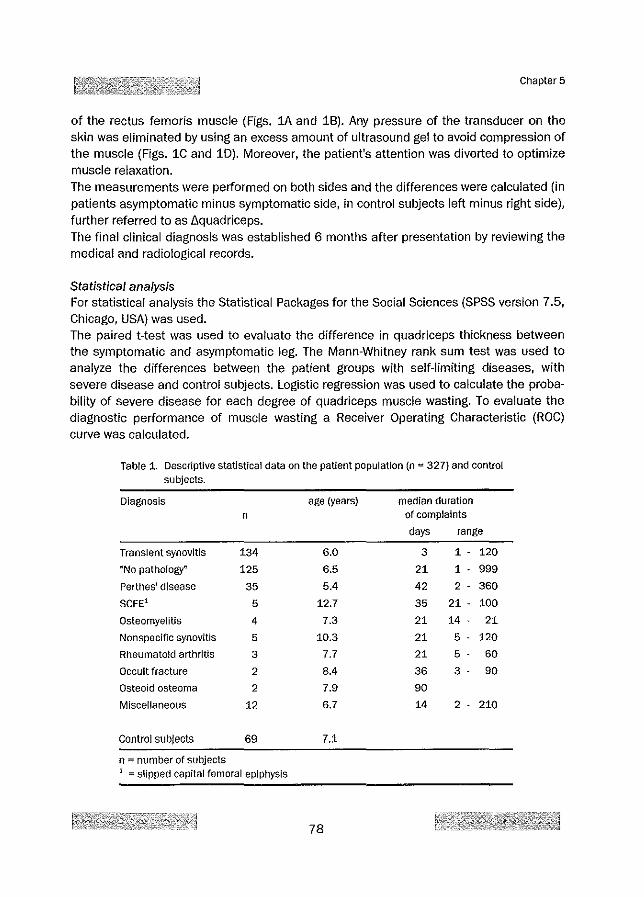
Chapter 5
of the rectus femoris muscle (Figs. 1A and 18). Any pressure of the transducer on the skin was eliminated by using an excess amount of ultrasound gel to avoid compression of the muscle (Figs. 1C and 10). Moreover, the patient's attention was diverted to optimize muscle relaxation. The measurements were performed on both sides and the differences were calculated (in patients asymptomatic minus symptomatic side, in control subjects left minus right side), further referred to as L\quadriceps. The final clinical diagnosis was established 6 months after presentation by reviewing the medical and radiological records.
Statistical analysis For statistical analysis the Statistical Packages for the Social Sciences (SPSS version 7.5, Chicago, USA) was used. The paired t-test was used to evaluate the difference in quadriceps thickness between the symptomatic and asymptomatic leg. The Mann-Whitney rank sum test was used to analyze the differences between the patient groups with self-limiting diseases, with severe disease and control subjects. Logistic regression was used to calculate the probability of severe disease for each degree of quadriceps muscle wasting. To evaluate the diagnostic performance of muscle wasting a Receiver Operating Characteristic (ROC) curve was calculated.
Table 1. Descriptive statistical data on the patient population (n '" 327) and control subjects.
Diagnosis age (years) median duration n of complaints
days range
Transient synovitis 134 6.0 3 1 - 120
"No pathorogy~ 125 6.5 21 1 - 999
Perthes' disease 35 5.4 42 2 - 360
SCFEi 5 12.7 35 21 - 100
Osteomyelitis 4 7.3 21 14 - 21
Nonspecific synovitis 5 10.3 21 5 - 120
Rheumatoid arthritis 3 7.7 21 5 - 60
Occult fracture 2 8.4 36 3 - 90
Osteoid osteoma 2 7.9 90
Miscellaneous 12 6.7 14 2 - 210
Control subjects 69 7.1
n"" number of subjects 1 '" slipped capital femoral epiphysis
78

Differential Diagnosis of Children with a Painful Hip
RESULTS Patients Muscle measurements were not possible, or considered to be unreliable, in 21 (6%) patients because of anxiety and movement. These patients (mean age 4.5 years) were generally younger than the other patients (mean age 6.4 years). Therefore, 327 (94%) patients could be evaluated. The final clinical diagnoses are given in Table 1. In 125 patients no definite diagnosis could be made and in all these patients the symptoms disappeared without specific treatment and remained symptom free thereafter. This group is denominated as "no pathology" group, and includes 8 patients with the tentative diagnosis of postinfectious or parafebrile arthralgia.
Table 2. Mean quadriceps muscle thickness and {). quadriceps in patient groups and control subjects
Diagnosis Quadriceps Quadriceps I!. Quadriceps (SO) symptomatic asymptomatic
(mm) (mm)
TranSient synovitis 24.7 25.5 0.8 (1.6)"
"No pathology" 25.8 26.2 0.3 (1.5 )'
Perthes' disease 22.3 25.2 2.9 (2.5)"
SCFE1 28.6 34.0 5.5 (2.0)'"
Osteomyelitis 20.1 23.6 3.6 (4.2)"'
Nonspecific synovitis 26.1 27.9 1.8 (2.5)"'
Rheumatoid arthritts 25.9 29.6 3.6 (2.8)"'
Occult fracture 29.2 31.8 2.6 (0.6)"'
Osteoid osteoma 26.2 29.2 2.9 (1.8)'"
Miscellaneous 24.5 25.9 1.4 (2.1)'
Quadriceps Quadriceps {). Quadriceps (SO) right (mm) left (mm)
Control subjects 27.7 27.6 -0.1 (1.3}'"
!J. quadriceps = quadriceps asymptomatic side minus symptomatic side (in control subjects left minus right)
NS ~ P > 0.05 NT = not tested due to insufficient number of patients * = p < 0.05 ** = P < 0.01
1 "" slipped capital femoral epiphysis
79

Chapter 5
Nonspecific synovitis was diagnosed when effusion was present in the hip joint (n~5) without the classical presentation of transient synovitis (10) and without clinical and serological signs of rheumatoid arthritis. Discharge diagnosis in this group was often reactive or postinfectious synovitis. The miscellaneous group (n~12) consisted of patients with malignant osseous tumors (n~2), synovitis of the knee (n~2), Baker's cyst, lymphadenitis, edema of bone marrow e causa ignota, retrocecal appendix, ureteric calculus, juvenile osteoporosis, leukemia, and septic arthritis (all n~1).
Control subjects Control subjects showed no significant difference in thickness between left and right quadriceps muscles (l\quadriceps). Mean difference was ·0.1 mm ± 2.7 (two standard deviations). Therefore 2.7 mm (2SD) was considered as the upper limit of normal left/right difference. The muscle measurements of patients and control subjects are given in Table 2 and Figure 2.
3 4 5 6 7 8 9 10 Disease groups
Figure 2. Boxplot of mean muscle atrophy in various diseases. The lower and upper boundaries of the box represent the 25th and 75th percentiles, respectively. Whiskers represent smallest and largest values. 1 == transient synOVitis, 2 "" Dno pathology", 3 '" Perthes' disease, 4 '" SCFE, 5 "" osteomyelitis. 6 "" aspecific synovitis, 7 = rheumatoId arthritis, 8 = occult fracture 9 = osteoid osteoma, 10 = miscellaneous. IJ. quadriceps "" difference in quadriceps thickness (asymptomatic side minus symptomatic side, in control subjects left minus right side).
80

Differential Diagnosis of Chifdren with a Painful Hip
To determine the clinical value of US detection of muscle wasting, the patients were divided into 2 groups: Group A: Consisting of 270 patients with self-limiting diseases: transient synovitis
(n=134); "no pathology" group (n=125); nonspecific synovitis (n=5); miscellaneous self-limiting diseases (n=6).
Group B: Consisting of 57 patients with severe diseases requiring hospitalization or a consultation with an orthopedic surgeon: Perthes' disease (n=35); SCFE (n=5); osteomyelitis (n=4); rheumatoid arthritis (n=3); occult fracture (n=2); osteoid osteoma (n=2); and miscellaneous severe diseases (n=6).
Table 3 and Figure 3 give data on the measurements of muscle thickness in group A and B, compared with control subjects. All groups differed significantly (p<0.001) from each other. Figure 4 presents the ROC curve of quadriceps muscle wasting in differentiation between self-limiting and severe diseases. This curve confirms the significant difference in muscle wasting between self-limiting diseases and severe pathology and shows that a quadriceps wasting of >2.7 mm (upper limit of physiologicalleftjright difference) can serve as a threshold between severe and self-limiting diseases. Using this threshold for severe disease, the positive predictive value is 64%, the negative predictive value 92%, the sensitivity 61%, and the specificity 93% (Table 4). Figure 5 shows the probability of severe disease as a function of the degree of wasting, and the number of patients for each degree of wasting.
Table 3. Absolute muscle thickness, and !J. quadriceps, in patients groups and control subjects
DIagnosis
Self-limiting disease (n =270)
Severe dIsease (n =57)
Control subjects (n =69)
Quadriceps symptomatic
side (mm)
25.3
23.3
Quadriceps left side (mm)
27.7
Quadriceps asymptomatic
side (mm)
25.9
26.5
Quadriceps right side (mm)
27.6
Data are mean values, with standard deviation In parentheses
!J. Quadriceps
0.6 (1.6)"
3.2 (2.5)"
-0_1 (1.3)"'
!J. quadriceps = quadriceps asymptomatic side minus symptomatic side (In control subjects left minus right)
NS = P > 0_05 ** :: p < 0_01 n :=: number of subjects
81

10
8
6 I '" c. 4
~ 1l ."
~ ." 2 '" => a
0 <1
·2 I
-4
-6 Controls Self·limitlng disease Severe disease
Figure 3. Boxplot of mean muscle atrophy In control subjects, In patients with self· limiting diseases, and in patients with severe diseases. The boundaries of the box represent 25th and 75th percentile, respectively. Whiskers represent smallest and largest values. !J. quadriceps = difference in quadriceps thickness (asymptomatic side minus symptomatic side, in control subjects left minus right side).
1.00
0.75
~ ~050 c .
Jl
0.25
0,00
Area under ROC curve. 0.7917 A Quad;:::" 0.0
0.00
.6 Quad ~ 1.0
""::;;---h. Quad ~ 0,5 r----....r"~ ~
~-~~
o Quadriceps wasllng (mm)
0.25 0.50 0.75 1.00
1 - Specificity
Figure 4. Receiver operating characteristic curve shows sensitivities and specificities of muscle wasting In differentiation between serf-limiting and non self-limiting disease. Four cut-off revers are indicated. The area under the ROC curve equa!s 0.79. The diagonal line represents an imaginary test without discriminative power.
82
Chapter 5

Differential Diagnosis of Children with a Painful Hlp
Table 4: Value of quadriceps wasting for the diagnosis of severe orthopedic disease, using 2_7 mm as a threshold value
Severe Self-limi- Total dIsease
8 quadriceps?: 2_7 mm 35
8 quadriceps < 2_7 mOl 22
Total number of patients 57
100
80
ling
20 55
250 272
270 327
[J Severe dlsaase
o Self-fmlllng disease
~ ~ ~ ~ ~ ~ ~ ~ ~ ~ ~ ~ ~ 'V)' '~)' ''?)' '':')' 'q$l '($I '~$1 '''$I ''1"0 '1'0 'ero '-:'0 '<?$1
Muscle wasting (mm)
100
80
20
o
Figure 5. Curve showing the probability of severe disease (right axis) versus the degree of wasting_ The absolute number of patients (feft axIs) for each degree of wasting is given as bars_ Muscle wasting is defined as the difference in quadriceps thickness (asymptomatic side minus symptomatic side).
DISCUSSION Although us is an excellent modality to visualize the musculature, its clinical application in childhood has been limited to study the atrophy and aspect of muscles in neuromuscular diseases (11·16). Another clinical application was the assessment of the skeletal muscle protein reserve in children after bone marrow transplantation and in newly diagnosed acute leukemia (17, 18). One study describes muscle atrophy in Perthes' disease (8), but no other sonographic literature is available on muscle atrophy in patients with a painful hip. In many institutions, US is part of the evaluation of children with a painful hip. Because (a) muscles respond rapidly to changes in activity pattern, (b) early diagnosis of severe orthopedic disease is important for the prognosis, especially in childhood, and (c) as US
83

Figure 6a. Signiffcant wasting of the right quadriceps muscle (27.0 mm, between cursors) is present versus the asymptomatic left side (31.2 mm).
Figure 6b. Radiograph of the hips was normal.
Chapter 5
Figure (;ie. An additional radiograph of the right femur was made because of ipsilateral muscle wasting, revealing cortical sclerosis (arrows) that was caused by an osteoid osteoma,
Figure 6. A 12-year-old girl with a painful right hip for 3 weeks, US showed no effusion, Definite diagnosis was osteoid osteoma of the right femur,
muscle measurement is not time-consuming and easy to perform, it is justified to extend the US examination with muscle measurements. This is supported by the fact that all patients in the present study showed more or less muscle wasting compared with the control subjects. SCFE showed the most severe wasting, the "no pathology" grou p the least wasting. However, because the differences between various diseases are small and there is much overlap (Fig. 2), it is not justified to even attempt making a diagnosis based
84
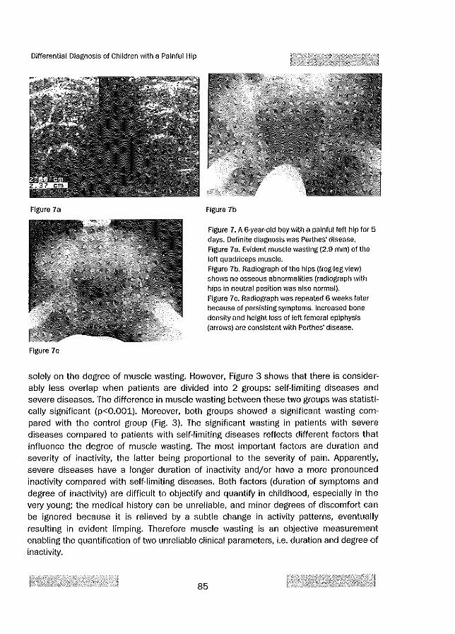
Differential Diagnosis of Children with a Painful Hip
Figure 7a
Figure 7c
Figure 7b
Figure 7. A 6-year-old boy with a painful left hip for 5 days. Definite diagnosis was Perthes' disease. Figure 7a. Evident muscle wasting (2.9 mm) of the left quadriceps muscle. Figure 7b. Radiograph of the hips (frog-leg view) shows no osseous abnormalities (radiograph with hips in neutral position was also normal). Figure 7e. Radiograph was repeated 6 weeks later because of persisting symptoms. Increased bone density and height loss of left femoral epiphysis (arrows) are consistent with Perthes' disease.
solely on the degree of muscle wasting. However, Figure 3 shows that there is considerably less overlap when patients are divided into 2 groups: self-limiting diseases and severe diseases. The difference in muscle wasting between these two groups was statistically significant (p<O.OOl). Moreover, both groups showed a significant wasting compared with the control group (Fig. 3). The significant wasting in patients with severe diseases compared to patients with self-limiting diseases reflects different factors that influence the degree of muscle wasting. The most important factors are duration and severity of inactivity, the latter being proportional to the severity of pain. Apparently, severe diseases have a longer duration of inactivity and/or have a more pronounced inactivity compared with self-limiting diseases. Both factors (duration of symptoms and degree of inactivity) are difficult to objectify and quantify in childhood, especially in the very young; the medical history can be unreliable, and minor degrees of discomfort can be ignored because it is relieved by a subtle change in activity patterns, eventually resulting in evident limping. Therefore muscle wasting is an objective measurement enabling the quantification of two unreliable clinical parameters, i.e. duration and degree of inactivity.
85

Chapter 5
Figure 8a. Sagittal US image of the left hip joint shows large effusion (E). F = femur.
Figure 8. A 7-year-old boy with a painful right hip for 2 days. Definite diagnosis was transient synovitis.
Figure 8b. Transverse US images of both quadriceps muscles show symmetrical muscle thickness (23 mm versus 24 mm, measured with calipers).
As demonstrated in Figure 3, muscle wasting can be a potential US criterium to discriminate between severe pathology and self-limiting diseases. In order to extrapolate these findings to individual patients we tested several threshold values for L\quadriceps for differentiating severe pathology from self-limiting diseases by establishing a ROC curve, and by calculating a probability curve for severe disease (Figs. 4 and 5). These curves confirm the difference in muscle wasting among patients with severe and self-limiting disease. Assuming the consequences of a false-positive result and a false-negative result to be equal, the optimal threshold value for muscle wasting to
86

Differential Diagnosis of Children with a Painful Hip
differentiate between both patient groups is 2.7 mm, which is also the upper limit of the 95% confidence interval of physiological left-right difference (Figs. 6-8). This threshold value results in a sensitivity of 61%, positive predictive value (PPV) of 64%, specificity of 93%, and negative predictive value (NPV) of 92%, suggesting that muscle wasting is more effective in ruling out severe disease than in predicting it. However, it should be noted that the capability of muscle measurements to rule out severe disease should not be overestimated based on these data: there is a discrepancy between the number of patients with self-limiting disease and severe disease (5:1). Therefore, a NPV of 93% means that 22 patients (39%) with severe disease will still go unrecognized. Moreover, we assumed that the consequence of a false-negative result equals the oonsequence of a false-positive result. This assumption may not be correct because a false negative result is clinically more detrimental than a false positive result. From this point of view, a lower threshold value would be more acceptable. For instance, a threshold value of 0 mm would result in only 7 patients with severe disease (12%) not being detected (Fig. 5). Therefore, although 2.7 mm seems an optimal threshold value to discriminate between both disease groups, its clinical use is limited. However, in terms of radiation protection in a pediatric population, this threshold value can be useful: some authors recommend US as the examination of choice for painful hips, omitting radiography in uncomplicated cases (8, 19-24). Assuming that additional radiographs of the hips were to be obtained if the US indicates wasting of more than 2.7 mm, then only 19 of 270 patients with self-limiting diseases (7%) in our study population would have received needless radiation. It is even justified to extend the radiological examination to the whole limb if the radiograph of the hip is normal (Fig. 6). On the other hand, a threshold value of less than 0 mm muscle wasting will protect 88 patients with self-limiting disease (33%) from needless radiation. In conclusion, patients submitted for US because of a painful hip can benefit from additional muscle measurements, because this technique can determine those individuals at high risk for severe disease; in patients with pathological quadriceps muscle wasting (more than 2.7 mm on the ipsilateral side), additional examinations or control examinations are mandatory (Fig_ 7). Patients without muscle wasting (less than 0 mm on the ipsilateral side) are at low risk for severe disease and additional radiographic examinations should be omitted (Fig. 8). However, it should be emphasized that the results of the present study should always be held in perspective with regard to the clinical degree of suspicion.
ACKNOWLEDGMENTS The authors thank Teun Rijsdijk and Andries Zwamborn for preparing the illustrations and photographs_
87

Chapter 5
REFERENCES 1. Young A, Hughes I, Russell p, Parker MJ, Nichols PJR. Measurement of quadriceps muscle wasting
by ultrasonography. Rheumato[ RehabII1980;19:141-148. 2. Sipih] S, Suominen H. Quantitative Ultrasonography of muscle: detection of adaptions to training in
elderly women. Arch Phys Med RehabIl1996;77;1173·1178. 3. Ellis S, Kirby Le, Greenleaf JE. Lower extremity muscle thickness during 30-day 6 degree head-down
bed rest with isotonic and Isokinetic excercise training. Aviat Space Environ- Med 1993;64:1011·1015.
4. Dons B, Bollerup K, Bonde-Peterson F. The effect of weight-lifting excerclse related to muscle fiber composition and muscle cross-sectionat area in humans. Eur J Applied PhysloI1979;40:95·106.
5. Sipila S, Suominen H. Muscle ultrasonography and computed tomography In elderly trained and untrained women. Muscle Nerve 1993;16:294-300.
E. Cady ES, R.H.T. G. Ultrasonic tissue characterization of skeletal muscle. Eur J Ciln Invest 1983;13:469-473.
7. Heckmatt J, Rodil/o E, Doherty M, Willson K. Quantitative sonography of muscle. J Child Neurology 1989;4 (suppl):101·106.
8. Robben SGF, Meradji M, Diepstraten AFM, Hop WCJ. Ultrasonography of the paInfUl hlp In childhood: the value of cartilage thickening and muscle atrophy In the detection of Perthes disease. Radiology 1998;208:35-42.
9. Rohrschneider WK, Fuchs G, Trager J. Ultrasonographic evaluation of the anterior recess in the normal hip: a prospective study on 166 asymptomatic children. Ped/atf RadioI1996;26:629-634.
10. Haueisen DC, Weiner OS, Weiner SO. The characterization of ~transient synovitis of the hip" In children. J Pediatf Orthop 1986;6:11-17.
11. HeckmaU JZ, Pier N, Dubowitz V. Assessment of Quadriceps femoris muscle atrophy and hypertrophy In neuromuscular dIsease in children. J Clln Ultrasound 1988:16:177-181.
12. SchmIdt R, Voit T. Ultrasound measurement of Quadriceps muscle In the first year of life. Normal values and application to spinal muscular atrophy. Neuropediatrics 1992;24:36-42.
13. Dock W, Happak W, Grabenwager F, Toilf 1<, Bittner R, Gruber H. Neuromuscular diseases: Evaluation with high-frequency sonography. Radiology 1990;177:825-828.
14. Kamala 0, Suresh S, Githa K. Real-time ultrasonography in neuromuscular problems of children. J Clin Ultrasound 1985;13:465-468.
15. Heckmatt JZ, Pier N, Dubowitz V. Real-time ultrasound imaging of muscle. Muscle & Nerve 1988;11:56·65.
16. Sodergard JMB, Jaaskelainen JJ, Ryappy SA. Muscle ultrasonography in arthrogryposls. Acta Orthop Scand 1993:64:357·361.
17. Koskelo EK, Saarinen UM, Slimes MA. Skeletal muscle wasting and protein-energy malnutrition in children with newly diagnosed acute leukemia. Cancer 1990;66:373-376.
18. Taskinen M, Saarinen UM. Skeletal muscle protein reserve after bone marrow transplantation in children. Bone Marrow Transplant 1996;18:937-941.
19. Castriota-Scanderbeg A, Orsi E, De Micheli V, Pedrazzi G, Letico M, Coppl M. Ultrasound in the diagnosis and follow-up of hlp disorders in children. Radiol Med (Torino) 1993;86:808-814.
20. Bickerstaff DR, NeallM, Booth Al, Brennan PO, Bell MJ. Ultrasound examInation of the irritable hip. J Bone Joint Surg Br 1990;72:549-553.
21. Dorr U, Zieger M, Hauke H. The painful hip. Diagnostic possibilities of sonography. ROFO Fortschr Geb Rontgenstr Nuklearmed 1988;148:487-491.
22. Terjesen T, Osthus P. Ultrasound In the diagnosis and follow-up of transient synovitis of the hip. J Pediatr Orthop 1991;11:608·613.
23. Futami T, I~asahara Y, Suzuki S, Ushikubo S, Tsuchiya T. Ultrasonography in transient synovitis and early Perthes' disease. J Bone Joint Surg Bf 1991;73:635-639.
88

Differential Diagnosis of Children with a Painful Hip
24. Meradji M, DJepstraten AFM. Coxitis Fugax, sonografisches und radiologisches Blld In 65 Fallen.
Radiologe 1988:28:473-478. 25. Wills CA, Caiazzo V), Yasakuwa DI, Prietto CA, McMaster WC. Effect of immabifisatian of human
skeletal muscle. Orthop Rev 1982;11:57-64.
89

Chapter 5
90
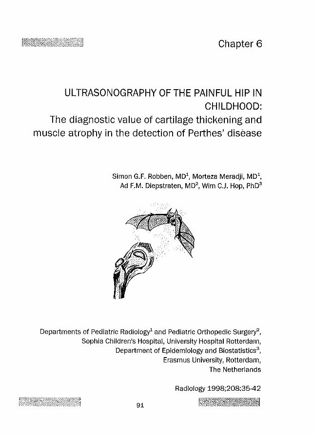
Chapter 6
ULTRASONOGRAPHY OF THE PAINFUL HIP IN CHILDHOOD:
The diagnostic value of cartilage thickening and muscle atrophy in the detection of Perthes' disease
Simon G.F. Robben, MDl, Morteza Meradji, MDl, Ad F.M. Diepstraten, MD2, Wim C.J. Hop, PhD3
Departments of Pediatric Radiology1 and Pediatric Orthopedic Surgery2, Sophia Children's Hospital, University Hospital Rotterdam,
Department of Epidemiology and Biostatistics3,
Erasmus University, Rotterdam, The Netherlands
Radiology 1998;208:35-42
91

Chapter 6
92

Cartilage Thickening and Muscle Atrophy in Perthes Disease
ABSTRACT
Purpose: To evaluate the combined use of several ultrasonographic (US) criteria in the detection of Perthes' disease. Material and Methods: In a prospective study, 144 consecutive patients with a painful hip underwent US. The thickness of the (a) joint capsule, (b) cartilage of femoral head, and (c) quadriceps muscle were assessed.
Sixty-eight children with no symptoms, the control group, were also examined. Results: The final diagnosis was transient synovitis(n = 58), Perthes' disease(n = 21), slipped capital femoral epiphysis (SCFE; n = 5), or miscellaneous (n = 6). Fifty-four patients had no US or radiological abnormalities, and symptoms disappeared during follow-up. The anterior joint capsule in patients with transient synovitis was significantly wider than that in the other patients and control subjects (P < 0.001). Patients with Perthes' disease showed significant cartilage thickening in the symptomatic hip compared with the other patients and control subjects (p<O.OOl). Patients with Perthes' disease and patients with SCFE showed significant atrophy of the ipsilateral quadriceps muscle compared with all other groups (p<O.OOl). The combined use of these US criteria for the diagnosis of Perthes' disease resulted in a positive predictive value of 94%, a negative predictive value of 95%, a sensitivity 71% and a specificity 99% Conclusion: The combination of several US criteria increases the diagnostic value of US of the painful hip in patients with Perthes' disease.
93

Chapter 6
INTRODUCTION The pediatric hip is susceptible to a large variety of diseases that may manifest as localized or referred pain, limping, refusal to bear weight, and limitation of movement of the hip. These symptoms, however, are nonspecific, and many diseases produce the same clinical picture (eg, Perthes' disease, septic arthritis, slipped capital femoral epiphysis [SCFE], osteomyelitis, juvenile chronic arthritis, neoplasm, trauma, and non-accidental injury). Some authors recommend ultrasonography (US) as the examination of choice for painful hips, omitting radiography in uncomplicated cases (1-6). Unfortunately, the role of US has been limited to the detection of effusion; moreover, because several diseases are accompanied by effusion, the discriminatory value of US is limited. other US criteria, however, such as cartilage thickening and muscle atrophy, have the potential to be useful. Thickening of articular cartilage is recognized as an early US sign of Perthes' disease (5, 7). Muscle atrophy is important because muscular components respond rapidly to changes in activity patterns (8), and clinical studies have reported considerable muscle atrophy in patients with Perthes' disease (9, 10). In this prospective study, the thickness of musculature, joint cartilage and anterior joint capsule were assessed in all patients presenting with a painful hip. We performed this study to determine whether the use of a combination of these criteria increased the US capacity to help differentiate between various diseases and to help detect Perthes' disease in a population of children with painful hips.
MATERIAL AND METHODS Between January 1994 and August 1995, 144 consecutive children with clinical symptoms of hip disease were examined in a prospective study. There were 88 boys and 56 girls aged 1.1 to 15.6 years (mean, 6.4 years). Patients with bilateral symptoms were excluded. We also examined 68 children without symptoms or previous orthopedic disease. This control group comprised 45 boys and 23 girls aged 1.6 to 14.3 years (mean, 6.7 years). The study was approved by the institutional medical ethical review board. Informed consent was obtained from parents and children before the examination. All patients and control subjects were examined with US by the same investigator (S.G.F.R); 7-10 MHz (Ultramark 9 HDI, Advanced Technology Laboratories, Bothell, Washington, USA) and 7 MHz (model 128 XPlO, Acuson, Mountain View, California, USA) linear array transducers were used. Patients were examined in supine position with hips in neutral position. Both hips were examined; the contralateral, asymptomatic hip was used as normal reference. The thickness of the (a) joint capsule, (b) articular cartilage of the femoral head, and (c) quadriceps muscle were assessed. To evaluate the anterior joint capsule, an anterior approach along the long axis of the femoral necll was used. The anterior joint capsule is seen as a stripe of tissue between
94

Cartilage Thickening and Muscle Atrophy in Perthes Disease
M.lliopsoas
Figure 1a.
Figure 1b.
M. Rectus Femoris
Figure 1. (a) Schematic shows the anterior recess of a normal right hlp. M. = musculus. (b, 0) Parasagittal US scans of (b) the normal right hlp and (c) the left hlp with effusion (anechoic space) that caused widening of the anterior recess of the joint capsule in a 4-year-old boy with transient synovitis. The wIdth of the anterior joint capsule was measured with the calipers.
Figure 1e.
the anterior femoral neck and the fascia of the iliopsoas muscle. It consists of two fibrous layers that in the case of an effusion are separated by an anechoic space (Fig. 1). The maximal width of the joint capsule perpendicular to the femoral neck was measured (11). To evaluate the articular cartilage of the femoral head, the transducer was placed on the anterior femoral head and aligned parallel to the femoral neck. Subsequently it was moved laterally and simultaneously angulated slightly in the anterior oblique plane to depict the cartilage of the femoral head at the level of the labrum to the best extent possible (Fig. 2). The cartilage was seen as a hypoechoic rim around the femoral head, and the thickness of it was measured perpendicular to the femoral head at the level of the labrum. To evaluate the quadriceps muscle, the transducer was placed on the anterior thigh in the transverse plane, at the midpoint between the superior border of the patella and anterior superior iliac spine. Any pressure of the transducer on the skin was eliminated by using an excess amount of ultrasound gel to prevent compression of the muscle. Moreover, the patient's attention was diverted to optimize muscle relaxation.
95

Fibrous c~~sule M.Hiopsoas '>. __
------------------ -------Aceta,bular
carU!age
Figure 2a.
Figure 2b.
Labrum
'-T::
Chapter 6
Figure 2. (a) Schematic shows a normal right hip. M. = musculus. (b, c) Parasagittal US scans of (b) the normal right hlp and (c) the affected left hlp In a 5·year-old boy with Perthes' dIsease. The cartilage thickness (arrows) was 1.8 mm in band 3.5 mm in c. Note the mild flattening of osseous nucleus (arrowheads) In c.
Figure 2C.
The distance from the femoral cortex to the anterior fascia of the rectus femoris muscle was measured (Fig. 3). All measurements were performed in both hips and the differences between the asymptomatic and symptomatic hips were calculated (for cartilage thickness, symptomatic minus asymptomatic hip; for joint capsule thickness, symptomatic minus asymptomatic; for muscle thickness, asymptomatic minus symptomatic).
Additional Imaging Studies Additional radiographs of both hips (frontal and lateral frog-leg views [Lauenstein views; anteroposterior radiographs with the thighs in abduction and external rotation, projecting the hips in a lateral fashion)) were obtained in all patients immediately after US examination. In selected cases, radiographs of the upper and lower parts of the leg were also obtained. If no abnormalites were found and the symptoms persisted after a period of rest, additional imaging studies were performed (eg, control radiography, bone scintigraphy, magnetic resonance [MR] imaging, computed tomography [eT)); the choice of imaging study depended on the individual symptoms. In patients with classic signs of transient synovitis (eg, acute onset of symptoms, irritable hip, effusion at US, absence of fever, age of 4-10 years) (12), however, radiography was
96

Cartilage Thic[{ening and Muscle Atrophy in Perthes Disease
not performed at the initial examination. Radiographs were obtained only if the symptoms persisted for more than 4 weeks.
FIgure 3a. Figure 3b.
Figure 3. Transverse US scans of quadriceps muscles (arrowheads) In a 9-year-old boy with a left hlp affected by Perthes' disease. (a) US scan of the normal right quadriceps muscle (muscle thIckness, 31.9 mm). (b) US scan of the left quadriceps muscle demonstrates muscle atrophy (muscle thIckness, 23.7 mOl).
Diagnostic Criteria Patients with an irritable hip were considered to have had transient synovitis if US depicted effusion of any size (or a difference in thickness of the anterior recess between both hips of more than 2 mm), the symptoms subsided completely within 4 weeks without specific therapy, and the patient remained symptom-free for at least 6 months thereafter. If the symptoms persisted, radiographs of the hips (frontal and lateral views) were obtained. Perthes' disease was diagnosed with radiography. A patient was considered to have Perthes' disease if the typical changes of avascular necrosis appeared on serial radiographs (ie, condensation, subchondral fracture, collapse, and, eventually fragmentation (Table 1). SCFE was diagnosed if the epiphysis slipped at radiography of the hip.
97

Chapter 6
In patients with fever, septic arthritis was diagnosed with bacteriologic analysis of aspirated fluid. Neoplasm (eg, osteoid osteoma) was diagnosed with histologic analysis. The final clinical diagnosis was established 6 months after presentation by reviewing the medical and radiological records. Six patients with the tentative diagnosis of transient synovitis did not show up for regular checkups. To confirm the assumption that the symptoms had disappeared, the parents were interviewed by telephone. All patients appeared to have had a fast and complete recovery and stayed symptom free thereafter.
Table 1. Summary of data for 21 patients with Perthes' disease
Difference Difference Patient Duration of in cartilage in Quadriceps
No/SeX; Complaints Thickness Muscle Age(y) (d) Stage * Remarks (mm)t Thickness
(mm)t 1/M/7.0 10 Initial small subchondral fissure 0.4 0.0 2/M/6.6 14 Initial no osseous abnormalities 0.2 -0.9 3/M/4.4 14 Initial no osseous abnormalities 0.9 1.0 4/M/3.9 14 Initial no osseous abnormalities 0.7 4.3 5/M/6.6 36 Initial small subchondral fissure 0.3 2.6 6/M/7.1 51 IniUal small subchondral fissure 0.4 3.7 7/M/4.9 60 Initial no osseous abnormalities 0.0 1.5 8/F/6.4 70 Initial Height loss 0.6 4.5 9/F/5.1 90 Initial Subchondral fissure 0.6 5.1 10/M/5.9 90 Initial Subchondral fissure 1.7 4.3 1i/F/11.7 90 Initial Height loss 1.5 -2.0 12/M/4.7 90 Initial Height loss, sclerosis -0.1 3.4 13/M/3.2 120 Initial Height loss, sclerosis 0.8 1.2 14/F/5.7 150 Initial Subchondral fissure 1.5 3.5 15/F/5.2 150 Initial Incipient sclerosis, height loss 0.4 5.0 16/M/4.9 150 Initial sclerosis, Height loss 1.8 0.7 17/F/3.9 180 Initial Inclpient sclerosis, anterior collapse 1.1 4.4 18/M/9.3 180 Initial Incipient sclerosis, height Joss 0.1 6.8 19/M/4.7 180 Initial Height loss, sclerosis 0.6 3.1 20/M/8.0 180 Fragmentation Fragmentation, sclerosis, collapse 0.3 1.9 21/M/6.5 360 Initial no osseous abnormalities 1.4 3.4
* according to Edgren (13).
t difference between the two hips.
Statistical Analysis For statistical evaluation of measurements among different patient groups the KruskalWallis one-way analysis of variance and the Mann-Whitney rank sum test were used. The correlation between measurements and age was analyzed by using Pearson correlation coefficients. A p value of less than .05 was considered statistically significant. Multivariate analysis (logistic regression) was used to differentiate between groups. This analysis proceeded in two stages. At the first stage, patients with Perthes' disease were differenti-
98

Cartilage Thickening and Muscle Atrophy in Perthes Disease
ated from both those with no visible abnormality and those with transient synovitis. At the second stage, the latter two groups were differentiated from each other.
RESULTS The final clinical diagnoses are given in Table 2. Fifty-four patients had no joint effusion at US, normal radiographs, and spontaneous resolution of symptoms without specific therapy.
Table 2. Final diagnoses and descriptive data
Duration of Symptoms before US (d)
Final DIagnosIs Mean Age(y) Mean Range
Transient synovitis (n=58) 6.1 7.2 1·69 No visible abnormality (n=54) 6.6 67.4 0-720
Perthes' disease (n=21) 5.9 10S.5 10-360
SCFE (n=5) 12.6 49.2 21·100 Occult fracture (n=2) S.5 2.5 Septic arthritis (n=2) 3.7 3.7 Articular hemorrhage (n=l) 11.5 1.0
Osteoid osteoma (n=l) 3.2 90.0
Table 3. Mean Thickness of the Anterior Recess according to Diagnosis
Mean Thickness of the Anterior Recess (mm)
Painful Normal Difference between Hip Hip Hips (mm)
Transient synovitis (n=58) S.S 4.S 4.0 1.9 " No pathology (n=54) 4.5 4.5 0.0 0.6 " Perthes' disease (n""21) 6.S 4.9 1.9 1.5 " SCFE (n=5) 5.7 4.5 1.3 0.6 " Occult fracture (n=2) 4.0 4.0 0.0 0.3 " SeptiC arthritis (n""2) 8.0 4.9 3.1 0.05 " Articular hemorrhage (n""l) 15.7 9.3 6.4
Osteoid osteoma (n=l) 3.5 2.S 0.7
Note. -In the 68 control subjects, the mean thickness of the anterior recess was 4.6 mm for the left hlp and 4.7 mm for the right hlp (difference, -0.1 mm iOA (standard deviatfon]).
" is the standard deviation
Five patients with Perthes' disease showed no osseous abnormalities on initial radio· graphs (Table 1). These patients had symptoms that persisted and changes typical of avascular necrosis on serial radiographs.
99

Chapter 6
Two patients had an occult fracture: One patient, a 15-year-old boy, had an avulsion fracture of the minor trochanter, and the other, a 2-year-old boy, had a fissure of the fibula. These fractures were diagnosed with radiography. One patient with hemophilia had a large hyperechoic effusion after a minor trauma. The tentative diagnosis of articular hemorrhage was made and the patient recovered completely after intravenous administration of coagulation factor VIII. The numbers of patients with occult fracture, articular hemorrhage, septic arthritis, or osteoid osteoma were too small for statistical analysis.
Difference between Hips in Anterior Joint Capsule Patients with transient synovitis and patients with Perthes' disease showed statistically significant thickening of the anterior joint capsule as compared with the control subjects and the patients with no visible abnormality (p < 0.001) (Table 3). Moreover, the difference between the symptomatic and asymptomatic hips in the mean thiclmess of the anterior joint capsule in patients with transient synovitis was significantly different from that in patients with Perthes' disease (p < 0.001). No significant difference was present among other groups.
Table 4. Mean artIcular cartilage thickness according to diagnosis.
Diagnosis
Transient synovitis (n=58)
No Visible abnormality (n="54) Perthes' disease (n=21)
SCFE (n=5)
Occult fracture (n=2)
SeptiC arthritis (n=2)
Articular hemorrhage (n=l)
Osteoid osteoma (n=1)
Mean Articular Cartilage Thickness (mm)
Painful Normal Hip Hip
2.4 2.4
2.3 2.3
3.2 2.5
1.5 1.4
2.0 2.0
3.0 3.0
1.5 1.5
2.4 2.5
Difference between Hips (mm)
0.0 0.2 * 0.0 0.2·
0.7 0.6 .-
0.1 0.1·
0.0 0.0 .-
0.0 0.0 t
0.0 -0.1
Note. -In the 68 control subjects, the mean articular cartilage thickness was 2.3 mm for both the left and right hIps (standard deviation, 0.2 mm) .
.. standard deviation
100

Cartilage Thickening and Muscle Atrophy in Perthes DIsease
8 0 •
E 6
• B. s 4 • • ,., •• .c c. g 2 « •• • 11. 0 1l ." -g ~2 • => 0 a
-4 0 0
.jl
-0.4 --0.2 0 0.2 0.4 0.6 0.8 1 1.2 1.4 1.6 1.8
Cartilage Thickening (mm)
Figure 4. Graph shows the difference in quadriceps muscle thickness versus the difference In articular cartilage thickness ber.veen the painful hip and the normal hip for all patients (n = 144) . • = patients with Perthes' disease; 0 '" other patients. The dotted line Indicates the threshold of a O.5-mm difference In articular cartilage thickness.
Difference between Hips in Femoral Articular Cartilage Thickness The femoral articular cartilage of the symptomatic hip was significantly thicker in patients with Perthes' disease than it was in the other patient groups and control subjects (p<0.001)(Fig.4, Table 4). The other patient groups did not show statistically significant cartilage thickening. Figure 4 shows that all but one patient with cartilage thickening greater than 0.5 mm had Perthes' disease. The patient with thickening and no Perthes' disease was a 1.7 -year-old boy who had unexplained cartilage thickening and an uneventful recovery. Use of the threshold of 0.5-mm cartilage thickening to diagnose Perthes' disease resulted in a specificity of 99% (122 of 123). The sensitivity was 57% (12 of 21), the negative predictive value was 93% (122 of 131), and the positive predictive value (PPV) is 92% (12 of 13). No statistically significant correlation was found between the difference in cartilage thickness between hips and age for all groups, including control subjects.
Difference between Hips in Quadriceps Muscle Thickness Statistially significant ipsilateral muscle wasting was present in the patients with Perthes' disease and in the patients with SCFE as compared with muscle wasting in the other patients and control subjects (p < 0.001)(Fig. 5, Table 5). The difference in muscle wasting between patients with Perthes' disease and those with SCFE showed a trend but was not statistically significant (p = 0.079). The patients with no visible abnormality did not differ significantly from those with transient synovitis (p = 0.93),
101

Chapter 6
or control subjects (p = 0.33) in terms of muscle wasting. The difference between the transient synovitis group and the control group, however, was statistically significant (p =
0.02). No statistically significant correlation was found between the changes in quadriceps muscle thickness and age for all groups, including control subjects.
Table 5. Mean quadriceps muscle thickness according to diagnosis.
Diagnosis
Transient synovitis (n-58) No Visible abnormality (n=54)
Perthes' disease (n=21) SCFE (n=5) Occult fracture (n=2)
SeptiC arthritis (n=2) Articular hemorrhage (n=1) Osteoid osteoma (n=1)
Mean Quadriceps Muscle Thickness (mm)
Painful Hip Normal Hip
23.8 24.4 24.0 24.2 22.4 25.1
28.4 33.3 30.6 32.6
23.1 22.5
30.5 31.0 20.0 21.6
Difference between Hips (mm)
0.6 1.4' 0.2 1.7·
2.7 2.1*
4.9 1.5·
2.0 1.0· -0.6 2.5·
0.5 1.6
Note. -In the 68 control subjects, the mean quadriceps muscle thickness was 28.1 mm for the left hip and 28.2 mm for the right hip (difference, -0.1 mm ± 1.4 [standard deviation)).
t standard deviation
When we compared the patients with Perthes' disease with the patients with no visible abnormality and the patients with transient synovitis combined, we found that the optimal combination of the difference between hips in cartilage thickness and the difference between hips in quadriceps muscle thickness that enabled differentiation of both groups could be written as follows: 6 x (II cartilage) + 0.75 x (II quadriceps), where II cartilage is the difference beween hips in the cartilage thickness (symptomatic hip minus asymptomatic hip) and IIquadriceps is the difference between hips in the quadriceps muscle thickness (asymptomatic side minus symptomatic side). When we compared patients with transient synovitis with those with no visible abnormalities, we found that only the difference in anterior joint capsule thickness had a discriminatory value. Figure 6 shows the total (in millimeters) derived with the formula (vertical axis) versus the difference between hips in in the anterior joint thickness (horizontal axis) for all patient groups. />s shown in Figure 6, all but one patient with a total of at least 5 mm had Perthes' disease. The patient with a total of more than 5mm and no Perthes' disease was a patient with SCFE who had considerable muscle atrophy.
102

Cartilage Thickening and Muscle Atrophy in Perthes Disease
8
E 6 , ' g
4 ' , ~ • .~ . c- ol, i .: .:. ,
~ 2 .I, n • 'I' '"
,', 001, c- O 2l ~~ ',' III .~ "it 'I' u ·2 ro "
,: a
·4
·6 Control No TS PO SCFE Mise
Pathol Figure 5. Graph shows the difference in quadriceps muscle thickness bet\veen the painful hip and the normal hlp in all groups. Control = control subjects, No Pathol = patients with no visible abnormality, TS = patients with transient synovitis, PO "" patients with Perthes' disease, SCFE "" patients with Slipped Capital Femoral Epiphysis, Mise"" patients with miscellaneous abnormalities.
Therefore, the positive predictive value for Perthes' disease was 94% when the total of 6 x (/1 cartilage) + 0.75 x (/1 quadriceps) was at least 5 mm. The sensitivity was 71%, the specificity was 99%, and the negative predictive was 95% (Table 6).
Table 6. Value of the Formula 6 x (8. Cartilage) + 0.75 x (8. Quadriceps) In the Diagnosis of Perthes' Disease, with a Total of 5 mm as a Threshold.
No. of No. Of Patients
Patients without with Perthes' Perthes'
Total (mm) Disease Disease Total
>5 15 1 16
<5 6 122 128
Total 21 123 144
103

15
" -c. Il -. • 'c i lO .. " 0' .. So X 5 ..iii ,., <0 • ..., 0 0 0
+ 0
0;- 0 g> 0 Q) 'e II -5 So X <D
-10 -1 0 2 3 4 5 6 7 8
Difference in Recess Thickness (mm) Figure 6. Graph shows the total of 6 x (ll cartHage) + 0.75 x (8 quadriceps), where !J.. cartilage is the difference In cartilage thickness (sympto·matic hip minus asymptomatic hlp) and fJ. quadriceps Is the difference in quadriceps thlcness (asymptomatic hlp minus symptomatic hip) between the painful hlp and the normal hip, versus the difference in the anterior joint capsule thickness between the painful hip and the normal hip. All but one patient with a total of at least 5 mm had Perthes' disease. CD = patients with Perthes' disellse; 0 =
patients with transient synovitis; CD = other patients.
DISCUSSION
Chapter 6
US of the joint was first described by Kramps and Lenschow in 1979 (12). One year later, Seltzer et al.(14) showed that fluid collections as small as 10 mL could be demonstrated in the hip with US (14). Subsequently, the results of several studies confirmed that US is more sensitive for detecting hip effusion than is conventional radiography (6, 15-20). Although there is excellent correlation between US and CT for the detection of intraarticular effusion (21, 22), US is preferable because it is less expensive, requires no sedation, and is not associated with ionizing radiation. Although US is operator-dependent, the anatomical landmarks in the present study are easy to depict. The present study depends heavily on the comparison of the abnormal hip joint with the contralateral normal hip joint; therefore, patients with bilateral disease were excluded. This is a limitation of the study; however, the number of patients with bilateral hip pain was negligible. Moreover, bilateral synchronous involvement is very rare in Perthes' disease and transient synovitis, with reported frequencies of 0.4% and 1%, respectively (23-24).
104

Cartilage Thickening and Muscle Atrophy in Perthes Disease
Hip Joint Effusion Until recently, the role of US in patients with a painful hip has been limited to the detection of joint capsule thickening as evidence of joint effusion. Unfortunately, many diseases cause an increase in the capsular joint space (eg, transient synovitis, Perthes' disease, SCFE, rheumatoid or septic arthritis (3, 5, 18, 25-31). Although the mean capsular joint widths in Perthes' disease, transient synovitis, and septic arthritis differ significantly, there is considerable overlap in individual patients (5, 28, 32). In the present study, all patients with Perthes' disease had capsular joints widths within the range seen in patients with transient synovitis. The duration of the effusion can be of help in differentiating transient synovitis from other diseases. An effusion lasting 3-6 weeks is suggestive of Perthes' disease (2-4, 33-35); however, this observation can not be applied at presentation. Moreover, other diseases, such as rheumatoid arthritis, show effusions that persiSt. Therefore, the US detection of effusion is useful in identifying the hip joint as the source of the symptoms, but the discriminatory role of US is limited when only the presence of effusion, the amount of effusion, or both are evaluated.
Cartilage Thickness The cartilage of the femoral head also can be depicted with US. There is a good correlation between bone age and articular cartilage thickness (36). Thickening of the articular cartilage in Perthes' disease is a well-known phenomenon in both experimental and clinical studies (37-40). This cartilage thickening is attributed to swelling (41-42), hyperplaSia (39, 43-44), or ceased enchondral ossification in combination with continued cartilaginous growth (40, 45-46). To our knowledge, few reports have emphasized the importance of cartilage thickening in the differential diagnosis of the painful hip (5, 7). Unfortunately, in all of those studies, Perthes' disease was compared with transient synovitis. Other entities that cause a hip to be painful were not included. To overcome that drawback, we included all patients with a painful hip in our study and correlated the US criteria with the final diagnosis. Patients with Perthes' disease showed a statistically significant increase in the cartilage thickness of the symptomatic hip compared with the other patient groups; cartilage thickening in other patients was virtually absent. Among the patients with cartilage thickening of more than 0.5 mm, all but one - a 1.7 -year-old boy with cartilage thickening of 0.7 mm - had Perthes' disease (Fig.4). At this age, cartilage thickening can be caused by irregular mineralisation of the osseous nucleus of the femoral head. A threshold value of more than 0.5 mm seems, therefore, to be a reliable discriminator for Perthes' disease (positive predictive value, 92%; negative predictive value, 93%; specificity, 99%). Unfortunately, some patients with Perthes' disease had no evident cartilage thickening, resulting in a sensitivity of 57% (Table 1).
105

Chapter 6
Muscle Atrophy US of normal skeletal muscle has been described in healthy children and adults (47,48). US assessment of muscle wasting has been performed in children with neuromuscular disease (49-52) and in children with acute leukemia (53). To our knowledge, however, there are no reports in the US literature about muscle atrophy in patients with a painful hip. Muscle atrophy in patients with a painful hip probably results from decreased ipsilateral postural and locomotor activity. In our study, the maximal difference between the left and right quadriceps muscles in the control group was 3 mm (Fig. 5). Therefore a decrease of more than 3 mm on the 'side with the symptomatic hip can be considered as atrophy. Patients with Perthes' disease and patients with SCFE showed a statistically significant decrease in ipsilateral quadriceps muscle thickness compared with patients with transient synovitis, patients with no visible abnormality, and control subjects. In addition, patients with transient synovitis showed slight, although statistically significant, muscle wasting compared with the control subjects. According to Wills et al. (54), several factors can influence the degree of muscle atrophy, including duration and degree of inactivity. These factors probably contribute to the differences in muscle atrophy among patients with transient synovitis, Perthes' disease, and SCFE. Apparently, muscle atrophy can be a potential US criterion for detecting abnormalities. There was considerable overlap, however, among the groups (Fig. 5), so it is hazardous to extrapolate these findings to individual patients. Twenty-four patients had muscle atrophy (quadriceps muscle wasting at the ipsilateral side of >3 mm), 17 of whom had a severe abnormality (12 had Perthes' disease, four had SCFE, and one had an occult fracture) and seven of whom had self-limiting diseases (three had transient synovitis and four had no visible abnormality). Therefore, a threshold of at least 3 mm for muscle wasting results in a positive predictive value of 50% (12 of 24) for Perthes' disease, a negative predictive value of 92% (111 of 120), and a specificity of 90% (111 of 123). The relatively low sensitivity of 57% (12 of 21), however, indicates that many cases of Perthes' disease will go undetected. The results of the present study show that each US criterion (thickness of anterior joint capsule, thickness of the cartilage, and muscle atrophy) can be useful for differentiating among various diseases. Combining these criteria and applying thresholds for muscle atrophy (>3 mm) and cartilage thickening (>0.5 mm) can improve the diagnostic performance of US. Multivariate analysis optimizes this multipara meter approach with the use fa the formula 6 x (/'; cartilage) + 0.75 x (/';quadriceps). All patients with a total of at least 5 mm had severe hip abnormality (15 had Perthes' disease, and one had SCFE). This results in a positive predictive value for Perthes' disease of 94%, a negative predictive value of 95%, a sensitivity of 71%, and a specificity of 99% (Table 6). US of the hip joint is very sensitive in the detection of even small amounts of synovial effusion. Several frequently encountered diseases (eg, transient synovitis, Perthes'
106

Cartilage Thickening and Muscle Atrophy In Perthes Disease
disease and SCFE), however, can manifest as hip joint effusion. The results of this study show that the role of US should not be limited to the detection of effusion. Patients with a painful hip who have ipsilateral cartilage thickening, muscle atrophy, or both at US are highly suspected of having Perthes' disease, regardless of the duration of symptoms. Conventional radiography should be performed in these patients to confirm Perthes' disease. If the conventional radiographs are negative, additional investigations, such as radionuclide studies or MR imaging, are justified to confirm or rule out early Perthes' disease.
ACKNOWLEDGMENTS The authors thank Teun Rijsdijk and Andries Zwamborn for preparing the illustrations and photographs.
107

Chapter 6
REFERENCES 1. Castriota-Scanderbeg A, Drsi E, De Micheli V, Pedrazzj G, letico M, Coppl M. Ultrasound in the
diagnosis and follow-up of hip disorders in children. Radiol Med (Torino) 1993;86:808-814. 2. Bickerstaff DR. Neal lM. Booth AJ, Brennan PO, BeH MJ. Ultrasound examination of the irritable hip_
J Bone JoInt Surg Bf 1990;72:549-553. 3. Dorf U, Zieger M, Hauke H. The painful hip. Diagnostic possibilities of sonography. ROFO Fortschr
Geb Rontgenstr Nuklearmed 1988;148:487-491, 4. Terjesen T, Osthus P. Ultrasound in the diagnosis and follow-up of transient synovitis of the hip_
J Pediatr Orthop 1991;11:608-613. 5. Futami T. I{asahara Y, Suzuki S, Ushikubo S, Tsuchiya T. Ultrasonography In transient synovitis and
early Perthes' disease. J Bone Joint Surg Br 1991;73:635-639. 6. Meradjl M, Dlepstraten AFM. Coxitis Fugax, sonografisches und radlologisches Bild in 65 Fallen.
Radiologe 1988;28:473-478. 7. Wirth T, LeQuesne GW, Paterson DC. Ultrasonography in Legg-Calve-Perthes' disease. Pediatr Radlol
1992;22:498-504. 8. Booth FW, Gollnick PD. Effects of disuse on the structure and function of skeletal muscle. Med Sci
Sports Exerc 1983:15:415-420. 9. Ferguson AB jr. Synovitis of the hlp and legg-Perthes' disease. Clin Orthop 1954;4:180-188. 10. Schiller MG, Axer A. Legg-Calve-Perthes' syndrome (loC.P.S.). A critical analysis of roentgenographic
measurements. Clin Orthop 1972;86:34-42. 11. Rohrschnelder WK, Fuchs G, Trager J. Ultrasonographic evaluation of the anterior recess In the
normal hlp: a prospective study on 166 asymptomatic children. Pediatr RadioI1996;26:629-634. 12. Kramps HA, Lenschow E. Einsatzmoglichkeiten der Urtraschalldiagnostik am Bewegungsapparat.
Zeitschrift fur Orthopedie und Ihre Grenzgebiete 1979;118:355-364. 13. Edgren W. Coxa plana. Acta Orthop Scand 1965; 84(suppl): 1-129. 14. Seltzer S, Finberg H, Weissma BA. Arthrosonography; technique, sonographic anatomy and patho
logy. Invest Radiol 1980;15:19-28. 15. Wilson OJ, Green OJ, Maclarnon JC. Arthrosonography of the painful hlp. Clin RadloI1984;35:17-19. 16. Adam R, Hendry GM, Moss J, Wild SR, Gillespie I. Arthrosonography of the Irritable hlp in childhood:
a review of 1 year's experience. Br J RadfoI1986;59:205-208. 17. Marchal GJ, Holsbeeck MT, Raes M, et al. Transient synovitis of the hip in children: role of US.
Radiology 1987:162:825-828. 18. Mlralles M, Gonzales G, Pulpeiro JR, et a1. Sonography of the painful hip in children: 500 consecu
tive cases. AJR 1989;152:579-582. 19. Peck RJ. Ultrasound of the painful hip In children. Br J RadloI1986;59:293-294. 20. Rosenborg M, Mortensson W. The validity of radiographic assessment of childhood transient
synovitis of the hip. Acta Radiol Diagnosis 1986;27:85-89. 21. Wingstrand H, Egund N. Ultrasonography in hip joint effusion. Acta Orthop Scand 1984;55:469-471. 22. Egund N, Wingstrand H, Forsberg L, Petterson H, Sunden G. Computed tomography and
ultrasonography for diagnosis of hlp joInt effusion in children. Acta Orthop Scan 1986;57:211-215. 23. Nevelos AB. Bilateral Perthes' disease. Acta Orthop Scand 1980;51:649-654. 24. Haueisen DC, Weiner OS, Weiner SO. The characterization of ~transient synovitis of the hlp· In
children. J Pedlatr Orthop 1986:6:11-17. 25. Shiv Vf~, Jain AI~, Taneja K, Bhargava SK. Sonography of hip joint in infective arthritis. J Can Assoc
RadioI1990;41:76-78. 26. Sell S, Zacher J, !\onjg J, Goethe S. Ultrasound in inflammatory rheumatic joint disease. Ultraschall
Med 1993:14:63-67. 27. Koski JM, Antilla P, Hamalainen M, Isomaki H. Hlp joint ultrasonography: correlation with
intra-articular effusion and synovitis, Br J RheumatoI1990;29:189-192.
108

Cartilage Thickening and Muscle Atrophy in Perthes Disease
28. Kallio P, Ryoppy S, Jappinen S, Siponmaa AK, Jaaskelainen J, I<unnamo I. Ultrasonography in hip disease in children. Acta Orthop Scand 1985;56:367-371.
29. I<onermann W, Pelligrin de M. The differential diagnosis of juvenile hip pain in the ultrasonographic picture. Transient synovitis, legg-Calve-Perthes' disease, epiphysiolysIs capitis femoris. Orthopede 1993;22:280-287.
30. Castriota-Scanderbeg A, Orsl E. Slipped capital femoral epiphysis: ultrasonographic findings. Skeletal RadioI1993;22:191-193.
31. I<allio PE, leQuesne GW, Paterson DC, Foster BK, Jones JR. Ultrasonography in slipped capital femoral epiphysis. J Bone Joint Surg 1991;73B:884-889.
32. Alexander JE, Seibert JJ, Glasier CM, et al. High-resolution hip ultrasound in the limping child. J Clin Ultrasound 1989;17:19-24.
33. Terjesen T. Ultrasonography in the primary evaluation of patients with Perthes' disease. J Pediatr Orthop 1993;13:437-443.
34_ Kallio P, Ryoppy S. Hyperpressure in juvenile hlp disease. Acta Orthop Scand 1985;56:211-214. 35. linnenbaum FJ, Woltering H, Karbowski A, Harle A. Ultrasonography of the hlp for Perthes's disease.
Arch Orthop Trauma Surg 1989;108:166-172. 36. Castriota-Scanderbeg A, Micheli de V. Ultrasound of femoral head cartilage: a new method of
assessing bone age. Skeletal Radiol 1995;24:197-200. 37. Rush BH, Bramson RT, Ogden JA. legg-Calve-Perthes' disease: detection of cartilagenous and
synovial changes with MR imaging. Radiology 1988;167:473-476. 38. Katz JF. Arthrography in legg-Calve-Perthes' disease. J Bone Joint Surg(Am) 1968;60:567-472. 39. Gershuni DH, kl.er A, Hendel D. Arthrographic findings in legg-Calve-Perthes' disease and transient
synovitis of the hip. J Bone Joint Surg(Am) 1978;60:457-464. 40. Zahlr A, Freeman MAR. Cartilage changes following a single episode of infarction of the capital
femoral epiphysis in the dog. J Bone Joint Surg(Am) 1972;54:125-136. 41. Gershuni DH, Amiel 0, Gonsalves M. The biochemical response of rabbit articular cartilage to an
induced talcum synovitis. Acta Orthop Scand 1981;52:599-603. 42. Ueo T, Tsutsumi S, Yamamuro T, Okumura H. Biomechanical analysis of Perthes' disease using the
finite element method: the role of swelling of articular cartilage. Arch Orthop Trauma Surg 1987;106:202-208.
43. Axer A, Schiller MG. The pathogenesis of the early deformity of the capital femoral epiphysis in legg-Calve-Perthes' syndrome (l.C.P.S.). An arthrographic study. Clin Orthop 1972;84:106-115.
44. larsen E, Reimann J. Calve Perthes' disease with special reference to histological observations. Acta Orthop Scand 1973;44:426-438.
45. Singleton WB, Jones El. The experimental induction of subclinical Perthes' disease in the puppy following arthrotomy and intracapsular tamponade. J Comp Path 1979;89:57-71.
46. Kemp HBS, Bolero Jl. Radiological changes in Perthes' disease. Br J RadioI1966;39:744-760. 47. Heckmatt JZ, Pier N, Dubowitz V. Measurement of quadriceps muscle thickness and subcutaneous
tissue thickness in normal children by real-time ultrasound imaging. J Clin Ultrasound 1988;16:171-176.
48. Weiss lW, Clark FC. Ultrasonic protocols for separately measuring subcutaneous fat and skeletal muscle thickness in the calf area. Physical Therapy 1985;65:477-481.
49. Heckmatt JZ, Pier N, Dubowitz V. Real·time ultrasound imaging of muscle. Muscle & Nerve 1988;11:56-65.
50. Dock W, Happak W, Grabenwoger F, Toilf 1<, Bittner R, Gruber H. Neuromuscular diseases: Evaluation with high-frequency sonography. Radiology 1990;177:825-828.
51. Kamala D, Suresh S, Githa K. Real-time ultrasonography in neuromuscular problems of children. J Clln Ultrasound 1985;13:465-468.
109

Chapter 6
52. Heckmatt JZ, Pier N, Dubowitz V. Assessment of quadriceps femorIs muscle atrophy and hypertrophy in neuromuscular disease in chIldren. J Clin Ultrasound 1988;16:177-181.
53. Koskelo EK, Saarinen UM, Siimes MA. Skeletal muscle wasting and protein-energy malnutrition in children with newly diagnosed acute leukemia. Cancer 1990;66:373-376.
54. Wills CA, Caiozzo VJ, Yasakuwa 01, Prietto CA, McMaster we. Effect of immobilisation of human skeletal muscle. Orthop Rev 1982;11:57-64.
110

Chapter 7
DOPPLER ULTRASONOGRAPHY OF THE HIP JOINT IN CHILDREN WITH A PAINFUL HIP
Simon G.F. Robben, MD\ Maarten H. Lequin, MD', Ad F.M. Diepstraten, MD2, Wim C.J. Hop, PhD3, Morteza Meradji, MD'
Departments of Pediatric Radiology' and Pediatric Orthopedic Surgery2, Sophia Children's Hospital, University Hospital Rotterdam,
Department of Epidemiology and Biostatistics3,
Erasmus University, Rotterdam, The Netherlands
Submitted for publication
111

Chapter 7
112

Doppler US of the Hip Joint in Children with a Painful Hlp
ABSTRACT Objective: To examine Doppler ultrasonography (US) for the feasibility of in vivo demonstration of the small feeding arteries to the femoral head in children, and to examine waveform characteristics in normal and diseased hips. Subjects and Methods: In a prospective study 224 hips in 112 patients (mean age 5_9 years) with a painful hip were examined with US. The anterior ascending cervical arteries of the hip were identified with color Doppler US, and the resistive index (RI) was measured with pulsed Doppler US. Eleven hips with transient synovitis were re-examined after 4-6 weeks. Results: In 137 of 224 hips (61%) a Doppler signal could be obtained. In asymptomatic hips (n = 64) the mean RI was 0.58. In symptomatic hips the definitive diagnoses and mean Rl's were as follows: transient synovitis (n = 31) 0.92; Perthes' disease (n = 9) 0.67; miscellaneous (n = 5) 0.68. In 28 symptomatic hips no definite diagnosis could be made and the complaints disappeared spontaneously during follow-up, the mean RI was 0.57. There was no statistically significant difference in RI of symptomatic versus asymptomatic hips except in children with transient synovitis (p < .001) In 11 hips with transient synovitis that were re-examined after 4-6 weeks the RI returned to normal (0.57). The RI in the symptomatic hips showed a positive correlation with the amount of effusion (r = 0.73, P < 0.001). In both symptomatic and asymptomatic hips there was no correlation with age (p = 0.9 and 0.1, respectively). Conclusion: 1) The deep capsular vessels of the hip joint can be evaluated with Doppler US in over 60% of the hips. 2) The RI is age independent and correlates with the amount of effusion.
113

Chapter 7
INTRODUCTION The blood supply of the femoral head is unique and vulnerable because the epiphysis and most of the femoral neck is intracapsular. Moreover, after infancy the growth plate constitutes an absolute barrier to blood flow between the epiphysis and bone metaphysis (1, 2). The contribution of the artery from the ligamentum teres in childhood is negligible (1, 3, 4). Therefore, the major source of blood supply to the femoral head is from the ascending cervical arteries. These vessels arise from the medial and lateral circumflex arteries and traverse the capsule along its femoral attachment, pass beneath the synovium, and then branch to supply the metaphysis and epiphysis (Figs. 1 A ami B). Therefore, they are located intracapsularly and thus, theoretically, can be compromised by high intracapsular pressure. These vessels have been visualized in vitro by various injection/fixation techniques (1-3) and in vivo by selective angiography (5). The arterial blood supply to the femoral head can also be studied indirectly by isotope scanning (6-9), or magnetic resonance imaging (10-12). However, no non-invasive techniques were available for in vivo real-time evaluation of the arterial flow to the femoral head until the introduction of Doppler US equipment, which offers a non-invasive means of real-time evaluation of the blood flow with color Doppler, duplex Doppler and, more recently, power Doppler US. Using Doppler US, Graif et al. (13) examined the medial and lateral circumflex arteries with Doppler US in healthy adults, but these vessels have many muscular branches and do not exclusively supply the femoral head. However, the application of these techniques is promising because continuing technical improvements enable the sonographer to visualize even the smallest vessels (14-16). Because of their small size, children may particularly benefit from these techniques (17): Bearcroft and colleagues were even able to visualize the vessels within the cartilaginous femoral head in neonates (18). The purpose of this study was threefold: a) to examine Doppler sonography for the feasibility of in vivo demonstration of the small feeding arteries to the femoral head in children; b) to determine the characteristics of the waveform in normal hips; and c) to examine waveform characteristics in diseases that affect the hip joint.
114

Doppler US of the Hlp Joint in Children with a Painful Hip
Femoral artery
Medial circumflex artery
Jliopsoas muscle
/ ~ /
Posterior ascending cervical artel)'
Posterior ascending cervical branches
Reclus femoris muscle
Femur
Trochanter
_lslerslcircumflex artery
_ Anterior sswn<ling cervical artery
"< Medi.,.1 drcumflex - artal)'
Flgure1a. Crosssection of the femoral neck at the level of the insertion of the Joint capsule
Figure 1b. Parasagittal section along the femoral neck, correspondIng to the ultrasonographic plane. Anterior recess of the jOint space is widened (as in effusion) to better understand anatomical relationships.
A::: anterior layer of the capsule P ::: posterior layer of the capsule E "" osseous nucleus of epiphysis of femoral head C "" articular cartilage of acetabulum and femoral head.
Figure 1. Schematic drawing of arterial blood supply to the femoral head. Slice orientation is demonstrated in the inserts.
115

SUBJECTS AND METHODS Patients
Chapter 7
Between July 1996 and September 1997 all consecutive patients with a painful hip or limping (n ~ 112) who were referred to the radiology department were examined with ultrasonography (US) in a prospective study. The patients were referred from outpatients' clinics (orthopedic, pediatric and emergency department), as well as from general practitioners. The mean age was 5.9 (range 0.8-15.9) years. There were 81 boys and 31 girls. The complaints were unilateral in 108 and bilateral in 4 patients. In 10 patients with transient synovitis (unilateral in 9 and bilateral in 1) the examination was repeated after 4-6 weeks.
Method The US examinations were performed by the same investigator (SR) using US equipment with high frequency 7-10 MHz (Advanced Technical Laboratories, Ultramark 9 HDI, Bothell, Washington, USA) and 7 MHz (Acuson 128 XP10, Mountain View, California, USA) linear array transducers. The patients were examined supine, with their legs in extension and slight external rotation. Both hips were examined with US in a plane parallel to the femoral neck. In this plane the anterior capsule of the hip can be seen to the best advantage. In patients with unilateral symptoms a pathological amount of joint effusion was diagnosed if US depicted a thickening of the anterior jOint capsule of more than 2 mm compared with that in the asymptomatic hip (8, 19-21). In patients with bilateral symptoms, the thickness of the effusion itself was measured (22), and considered pathological if it exceeded 2 mm. The dorsal layer of the anterior joint capsule was subsequently examined with Doppler US to identify the anterior ascending cervical vessels. Low pulse-repetition frequency (1.25 kHz) and wall filter of 50 kHz was used. The color gain was set at a level just below the disappearance of color noise. When no vessels were found with color Doppler sonography, another attempt to identify vessels was made with power Doppler. Next, a pulsed Doppler examination was performed, placing the Doppler sample over the vessel. The pulse· repetition frequency was adapted to the amplitude of the waveform. The resistive index (RI), defined as the difference between the peak systolic and end-diastolic flow velocities divided by the peak systolic velocity, was measured at least twice in each vessel. The Doppler examination was limited to 5 minutes for each hip. In an attempt to estimate the overall vascularization of the femoral head, two parameters were recorded: a) the number of vessels that could be identified within the limited time frame; and b) the effort to find these vessels, expressed as the interval of time (in minutes) between the beginning of the Doppler examination and the actual detection of the first vessel. If no vessel could be detected within 5 minutes, the time assigned was 5 minutes. None of the hips with an effusion were aspirated.
116

Doppler US of the Hip Joint in Children with a Painful Hip
Diagnostic criteria Afebrile patients with an irritable hip were considered to have had transient synovitis if US depicted a pathological effusion and the symptoms subsided completely within 4 weeks without specific therapy and remained symptom free for at least 6 months thereafter. Perthes' disease was diagnosed with radiography. A patient was considered to have Perthes' disease if the typical changes of avascular necrosis appeared on serial radiographs (ie, condensation, subchondral fracture, collapse, and, eventually fragmentation). Aspecific synovitis was diagnosed when effusion was present in the hip joint without the classical presentation of transient synovitis (23) and without clinical and serological signs of rheumatoid arthritis. Discharge diagnosis in this group was reactive or postinfectious synovitis. The final clinical diagnosis was established 6 months after presentation by reviewing the medical and radiological records.
Statistical analysis For statistical analysis the Statistical Package for the Social Sciences (SPSS version 7.5, Chicago, USA) was used. Kruskal-Wallis analysis of variance was used to analyse the differences between patient groups. As the differences between the symptomatic and asymptomatic hips in each patient group were roughly normally distributed, a comparison of both hips was done using paired t-test. For each patient group, it was verified that outcomes did not differ significantly, whether or not the measurement on the contralateral side was available. The 97.5 percentile was calculated to determine the upper limit of RI in asymptomatic hips. For quantification of hyperemia, the paired Wilcoxon test was used for comparison between both hips in each disease group, the Kruskal-Wallis analysis of variance was used to analyse the differences between patient groups. The correlation between RI and age and between RI and effusion was tested by calculating Spearman's coefficients of correlation.
RESULTS In 137 of 224 hips (61%) a Doppler signal could be obtained (Figs. 2 and 3). The diagnoses, resistive indices amount of effusion and duration of symptoms are given in Table 1. All hips with Perthes' disease (n = 9) were in the initial stage according to Edgren (24). The miscellaneous group (n = 5) consisted of hips with aspecific synovitis (n = 3), occult fracture of the tibia in a 1.9-year-old girl that was diagnosed with conventional radiography, and low-grade osteomyelitis of the distal tibia in a 1.1-year-old boy that was diagnosed with both conventional radiography and clinical findings.
117

Figure 2. Doppler findings in asymptomatic hips. F = femoral head.
Chapter 7
FIgure 2a. Color Doppler and duplex Doppler examInation of an asymptomatic hip of a 5-year-old boy_ An ascending cervical artery, situated In the posterior layer of the capsule, runs towards the femoral head parallel to the femoral neck, The waveform resembles a lowresistance vasu[ar bed.
Figure 2b. Duplex Doppler examination of an asymptomatic hip of a 3-year-old boy. The artery in the anterior layer of the joint capsule demonstrates a highresistance vascular bed. This vessel does not supply blood to the femoral head,
In 47 hips no abnormalities were found at US and conventional radiography, and the symptoms disappeared without specific therapy and the patients remained symptom free at six months. This group will be referred to as "no pathology" group.
In asymptomatic hips the mean RI was 0.58 (97.5 percentile = 0.70, 2.5 percentile = 0.45) (Table 1 and Fig. 2). There was no correlation with age (p = 0.1). Also in symptomatic hips there was no correlation with age (p=0.9) In transient synovitis the RI of the symptomatic hip is significantly higher compared to the contralateral normal hip (p < 0.001) (Fig. 3). In the other disease groups the differences between symptomatic and asymptomatic hips in Rlwere not statistically significant. The RI of the symptomatic hip in transient synovitis was significantly higher than in other disease groups, which showed no mutual differences.
118

Doppler US of the Hlp JOint in Children with a Painful Hlp
Table 1. Diagnoses and Doppler results of 224 hips.
Diagnoses Number Mean Resistive Size of Effusion In Median of hips Index1 mm1,2 duration of without symptoms Doppler n (d) signal
Asymptomatic (n=108) 44 0.58 (0.11) 64 0.0 (0.2) N.A.
Transient synovitis (n=44) 13 0.92 (0.23) 31 4.5 (1.7) 2
Perthes'disease (n=11) 2 0.67 (0.21) 9 2.6 (2.1) 28
"No pathology" (n"47) 19 0.57 (0.09) 28 0.15 (0.6) 21
Miscellaneous (n=14) 9 0.68 (0.22) 5 3.5 (3.3) 14
n = number of hips. 1 Number in parentheses is the standard deviation 2 The amount of effusion is expressed as the thickness of the layer of effusion in the anterior recess or
as the difference in thickness of the anterior joint capsule between both hips, NA = not applicable
Table 2. Number and diagnoses of hips with a pathological effusion1
n Transient Perthes' Oise- Non-specific Mean Age (y) Synovitls ase Synovitis
RI > 0.70 26 24 1 1 5.2
RI < 0.70 11 6 4 1 6.1
1 effusion with a thickness of more than 2 mm or a thickening of the anterior joint capsule of more than 2 mm compared with that in the asymptomatic hip.
The relation between diagnosis, RI, and amount of effusion is shown in Figure 4 and Table 2. There is a linear correlation between RI and the amount of effusion (r = 0.73, P < .001).
119

Figure 3a. A 4-year-old boy shows decreased diastolic velocity resulting in an Rlo10.81
Figure 30_ A 3-year-old girl shows reversal of diastolic flow resulting in an RI of 1_33
Chapter 7
Figure 3b. A 5-year-old boy with absent diastolic flow
Figure 3d. The same patient as in Figure 3C, one month later; the effusion has disappeared, the waveform has normalized (Rl = 0.58).
Figure 3: Duplex Doppler sonography in patients with transient synovitis with variable amounts of effusion_ F = femoral neck E'"' effusion
In 11 hips with transient synovitis the examination was repeated after 4 to 6 weeks. The patients were symptom free, the effusion had disappeared and the RI returned to normal (0.57, standard deviation 0.07) (Figs. 3 C and D). The number of vessels that could be identified and the time it took to identify these vessels are given in Table 3. No significant differences could be found, neither between symptomatic and asymptomatic hips in all groups, nor between groups mutually. In 87 hips no vessels could be identified, neither with color- (or power), nor with duplex Doppler US; in 20 hips accurate Doppler measurements were impossible because of agitation (mean age 3.2 years), the other 67 hips in which no signal could be obtained had a mean age of 7.8 years (standard deviation 3.6, range 1.6-15.9 years). This is a significantly older age than the hips in which a Doppler signal could be detected (mean age 5.4 years, p < 0.001).
120

Doppler US of the Hip Joint in Children with a Painful Hlp
1.4 0 0
0 0 Patient groups
1.2 • o Transient synovitis a. 0 ... No pathotogy • :c 0 • Perthes disease :ai 1.0 .. ""= 0 cf)
E ¢ Miscellaneous
% 0° ¢ ~ 0.8 0 0
UI • 0:: 0
0.6
~ .0 eo·., 0
¢ 0. 0.4
·2 0 2 4 6 8 10
Effusion (mm) Figure 4. Scatterplot of the resistive index as a function of the amount of effusion (expressed as distension of the capsule compared to the contralateral normal hip or thickness of the effusion itself). Thin line represents the regression line.
Table 3. Quantification of hyperemia.
Number of Time In sympt vessels mInutes
(SD) sympt (SD)
Transient synovitis 1.0(0.6) 1.9(1.6)
Healed TS 1.0(0.0) 1.8 (1.0)
Perthes' disease 1.2 (004) 1.6 (0.9)
"No pathology~ 1.0 (004) 1.9 (1.6)
Miscellaneous 1.0 (0.0) 1.0(0.0)
Asymptomattc hips 1.0(0.5) 2.1 (1.6)
Number of vessels that could be identtfled during the examination was assessed in symptomatIc and asymptomatic hips. Also the time it took to identtfy the first vessel from the beginning of the examination. Standard deviation is given between parentheses.
121

DISCUSSION Feasibility
Chapter 7
In the present study only the anterior ascending cervical arteries were examined because
the joint capsule can be visualized to the best advantage with US using the anterior
approach. The lateral, medial and posterior ascending vessels could not be visualized reliably because of unfavorable geometrical factors (large distance and over projection of
the greater trochanter) (Figs. 1A and 1B). However, it seems justified to extrapolate the
findings in the anterior ascending cervical arteries to the latter vessels because all
ascending cervical arteries a) lie in the same anatomic (intraarticular) compartment, b)
share the same origin (femoral artery), and c) form a subsynovial anastomotic ring (1). We were able to identify the ascending cervical vessels with color Doppler US in 61% of
all examined hips. Doppler evaluation failed in 20 hips due to agitation, usually in very young children (mean age 3.2 years). In older children it was more difficult to obtain
Doppler signals. This can be attributed to geometrical factors, such as increasing dis
tance between transducer and femoral neck in older children, but also to the progressive
reduction of the number of vessels with age (1). In adults, it would be even more difficult to study the ascending cervical arteries with Doppler US; therefore the use of US contrast
media may be a rewarding subject for further studies in adults.
Asymptomatic hips
In asymptomatic hips the duplex Doppler waveform resembles a low resistance vascular bed with a mean RI of 0.58, which was fairly constant. The 97.5 percentile was 0.70 and
considered to be the upper limit of normal. In this context it must be emphasized that it is
important to identify the posterior layer of the anterior joint capsule, because vessels in the anterior layer characteristically show a high resistive type waveform (Figs. 1B and
2B), and do not contribute to the blood supply of the femoral head. Especially in hips
without effusion special attention must be paid to correct identification of the posterior
layer.
Symptomatic hips
In the present study the RI in transient synovitis was significantly higher compared to other disease groups. The RI in these other disease groups did not show mutually signifi
cant differences. This difference between transient synovitis and other diseases can be
explained for the most part by a relatively large effusion in patients with transient synovi
tis (Table 1). In fact, there is a linear correlation between RI and amount of effusion
(r = 0.73, P < .001)(Fig. 4); increasing amounts of effusion cause progressive decrease of
diastolic velocity resulting in arrest of diastolic flow (RI = 1) and, eventually, even in reversal of diastolic flow (RI > 1) (Figs. 3 A-D).
122

Doppler US of the Hip Joint in Children with a Painful Hlp
In the present study, distension of the capsule is considered to represent the volume of effusion because there is a linear correlation between capsular thickening and volume of effusion (25, 26). A capsular distension of more than 2 mm seems to be a threshold value beyond which the RI changes: the RI is constant (mean = 0.58) when the amount of effusion is less than 2 mm, but if the capsule distension exceeds 2 mm, there is a positive correlation between the width of the effusion and the Rio This supports the assumption of many studies that a distension of the anterior joint capsule of more than 2 mm is the threshold between physiological and pathological amount of effusion (8, 19, 22, 27-31). Although there is a linear correlation between the amount of effusion and RI, no increase in RI was identified, despite a large effusion (Fig. 4 and Table 2). To evaluate this finding, we divided hips with a pathological effusion into two groups: group A (n = 26, mean age 5.2 years) with an increased RI (>0.70), and group B (n = 11, mean age 6.1 years) with a normal RI «0.70)(Table 2). The composition of diseases within both groups differs significantly: group A consists of only 1 hip with Perthes' disease (4%), 24 hips with transient synovitis and 1 hip with aspecific synovitis, whereas group B consists of 4 hips with Perthes' disease (36%), 6 hips with transient synovitis and 1 hip with aspecific synovitis. Apparently, in many hips with Perthes' disease a certain amount of effusion does not affect the vascularization to the same degree as in transient synovitis. An explanation is that the compliance of the capsule increases due to long standing effusion (median duration of complaints was 28 days in Perthes' disease, versus 2 days in transient synovitis) resulting in less pressure per ml effusion (32). This hypothesis is supported by animal studies that found a decay of intra-articular pressure due to non-elastic capsular stretching (33). According to Wingstrand et al. (26), a positive correlation between age and capsular compliance exists, but this does not contribute to the differences between group A and B because there is no significant difference in age (p = 0.31). It is hazardous to translate the RI changes to absolute flow changes (mljs) to the femoral head because Doppler sonography measures blood velocity (m/s). However, the finding of diminished, absent or reversed diastolic blood velocity, in the presence of normal flow characteristics in the contralateral normal hip, indicates increased vascular impedance, and therefore decreased absolute flow. Theoretically, this increased vascular impedance can be caused by venous obstruction (thrombosis or compression), increased intraosseous pressure or extra-osseous compression. Hip joint effusion that causes increased intra-articular pressure and vascular compromise can be a common denominator. Indeed, scintigraphic studies (6, 26) showed evidence of compression of blood supply by a tense joint effusion on bone scintigraphy; also Kallio et al. (32) concluded that the intraarticular pressure in transient synovitis is capable of causing a reduction of blood flow in the femoral head with the hip in neutral position.
123

Chapter 7
Although no intra-articular pressure was measured in this study, there are several reasons to hypothesize that the RI correlates positively with intra-articular pressure:
• The RI show a positive correlation with the amount of effusion. This is analogous to the positive correlation between amount of effusion and intra-articular pressure, as found by Kallio et al. (r = 0.52) (25, 32).
• Both RI (this study) and intra-articular pressure (26, 34) return to normal when the effusion has disappeared (Fig. 3C and 3D)
• The findings in the present study show no correlation between RI and amount of effusion in hips with Perthes' disease. This is analogous to the lack of correlation between intracapsular pressure and aspirated volume of fluid in Perthes' disease (35). This also indicates a correlation between RI and intra-articular pressure.
In the present study the hip joints with transient synovitis were not aspirated because, to our knowledge, there is no evidence for an etiologic relation between transient synovitis and subsequent development of Perthes' disease (36). Nevertheless, both our study and others (6, 26, 32) demonstrate that effusion can cause vascular compromise and many institutions still puncture the hip joint, not only for exclusion of septic arthritis, but also to avoid complications of long-standing increased intra-articular pressure (37, 38). In these institutions, Doppler US can be used to identify hips "potentially at risk" (i.e. hips with a RI > 0.70). In this respect, it would also be interesting to perform a Doppler US study to establish the relation of the position of the hip to arterial blood flow, because it is known that the intraarticular pressure depends on the position of the hip. The technique used in the present study can also be used to assess the femoral head blood supply in infants and older children during abduction treatment for hip dysplasia, to identify hips at risk of avascular necrosis (18). It is likely that hip joint effusion is the sole cause of increase of RI. This is supported by the fact that: a) RI drops to normal values when the effusion disappears, and b) symptomatic hips without effusion show a normal RI, regardless of the underlying disease. In the present study we did not find evidence of decreased perfusion in Perthes' disease. On the contrary, many patients showed a normal RI despite considerable effusion. The finding that the RI in Perthes' disease is lower than in transient synovitis does not rule out the importance of intracapsular tamponade in the development of avascular necrosis: the pressure may have been higher during the preceding stages of the disease. Table 3 demonstrates that the mean number of ascending cervical arteries that could be evaluated was one, and the mean time necessary to depict the vessel was approximately 2 minutes. There were neither statistically significant difference between diseases, nor between symptomatic and asymptomatic hips. Therefore, we did not find evidence for hyperemia of the joint capsule in patients with synovitis, which is in accordance with the study of Strouse et al. (39). These authors made a qualitative evaluation of perfusion by
124

Doppler US of the Hip Joint in Children with a Painful Hip
visual comparison of both hips using power Doppler sonography, and found no difference in flow between both hips in children with unilateral transient synovitis. We even found evidence for decreased flow with a mean RI of 0.92. Apparently, power Doppler sonography cannot depict these differences in flow characteristics. In conclusion, the present study shows the feasibility of in vivo demonstration of the vascularization of the femoral head in children and this technique provides a tool for further studies on hemodynamic changes in diseased and normal hips. In children with hip joint effusion a statistically significant positive correlation with RI exists, supporting the view that hip joint effusion is potentially harmful to the vascularization of the femoral head.
Acknowledgments The authors wish to thank Teun Rijsdijk and Andries Zwamborn for preparing the photo· graphs and illustrations.
125

Chapter 7
REFERENCES 1. Chung SMft The arterial supply of the developing proximal end of the human femur. J Bone Joint
Surg 1976;58-A:961-970. 2. Crock HV. An atlas of the arterial supply of the head and neck of the femur In man. Clin Orthop
1980;152:17-27. 3. Trueta J. The normal vascular anatomy of the human femoral head during growth. J Bone Joint Surg
1957;398:358-394. 4. Lauritzen J. The arterial supply to the femoral head In children, Acta Orthop Scand 1974;45:724-736. 5. Theron J. Angiography in Legg-Calve-Perthes disease. Radiology 1980;135:81-92. 6. Kloiber RR, Pavlovsky M, Portner 0, Gartke K. Bone scintigraphy of hlp Joint effusions In children.
AJR 1983;140:995-999. 7. Alexander JE, Seibert JJ, Aronson 1, et a1. A protocol of plain radiographs, hip ultrasonography, and
triple phase bone scintigraphy in the evaluation of the painful pediatric hip. Clin Pediatr (Phila) 1988;27:175-181.
8. Gopakumar TS, Vaishya R, Klenerman l, Carty H. The role of ultrasound and Isotope scanning in the management of irritable hips. Eur J Radial 1992;15:113-117 .
9. KanikHdes C, Sahlstedt B, l6nnerholm T, Moberg A. Conventional radiography and bone scintigraphy in the prognostic evaluation of legg-Calve-Perthes disease. Acta RadioI1996;37:561-566.
10. Sebag G, Ducou Le Pointe H, Klein I, et al. Dynamic gadolinIum-enhanced subtraction MR imaging. A sImple technique for the early diagnosis of legg-Calve-Perthes disease: preliminary findings. Pediatr RadioI1997;27:216-220.
11. Jaramillo D, Villegas-Medina Ol, Doty OK, et a!. Gadolinium enhanced MR imaging demonstrates abduction-caused hip ischemia and its reversal In children. Pediatr RadioI1995;25:578-87.
12. Tsukamoto HI Kang YS, Jones LC, et a!. Evaluatton of marrow perfusion In the femoral head by dynamic magnetic resonance imaging. Effect of venous occlusion in a dog model. Invest Radlol 1992;27:275-281.
13. Graif M, Schweitzer ME, Nazarian l, Matteucci TM, GOldberg BB. Color Doppler hemodynamic evaluatlon of flow to normal hlp. J Ultrasound Med 1998;17:275-280.
14. Newman JS, Adler RS, Bude RO, Rubin JM. Detection of soft-tissue hyperemia: value of power DOPPler sonography. AlR 1994;163:385-389.
15. Newman JS, Laing TJ, McCarthy C1, Adler RS. Power Doppler sonography of synovitis: assessment of therapeutic response. Preliminary observations. Radiology 1996;198:582-584.
16. Newman JS, Adler RS, Rubin 1M. Power Doppler sonography: use in measuring alterations in muscle blood volume after exercise. AIR 1997;168:1525-1530.
17. Keller MS. Sonographic detection of femoral head vascularity In neonates. Radiology 1996;200:28-29.
18. Bearcroft PW, Berman LH, Robinson AHN. Butler G1. Vascularity of the neonatal femoral head: in vivo demonstration with power Doppler US. Radiology 1996;200:209-211.
19. Terjesen T, Osthus P. Ultrasound in the diagnosis and follow-up of transient synovitis of the hip_ J Pediatr Orthop 1991;11:608-613.
20. Meradji M, Diepstraten AFM. Coxitis Fugax, sonografisches und radlologisches Bl1d In 65 Fallen. Radiologe 1988;28:473-478.
21. McGoldrick F, Bourke T, Blake N, Fogarty E, Dowling F, Regan B. Accuracy of ultrasonography in transient synovitis. J Pediatr OrUlOp 1990;10:501-503.
22. Robben SGF, LeQuin MH, Diepstraten AFM, Hol1ander 1C, Entius CAC, MeradJi M. Anterior joint capsule of the normal hip and In children with transient synovitis: US study with anatomic and histologic correlation. Radiology 1999;210:499-507.
23. Haueisen DC, Weiner OS. Weiner SO. The characterization of Vtransient synovitfs of the hlpw in children. J Pedlatr Orthop 1986;6:11-17.
126

Doppler US of the Hip Joint In Children with a Painful Hip
24. Edgren W. Coxa plana. Acta Orthop Scan 1965;84 (suppl):1-129. 25. Kallio P, Ryoppy S, Japplnen S, Siponmaa AK, Jaaskelalnen J, f<unnamo r. Ultrasonography In hip
dIsease In children. Acta Orthop Scand 1985;56:367-371. 26. Wingstrand H, Egund N, Carlin NO, Forsberg L, Gustafson T, Sunden G. lntracapsular pressure in
transIent synovitis of the hip. Acta Orthop Scan 1985;56:204-210. 27. Rohrschneider WK, Fuchs G, Troger J. Ultrasonographic evaluation of the anterior recess in the
normal hip: a prospective study on 166 asymptomatic children. Pediatr Radio/1996;26:629-634. 28. Konermann W, PeJligrin de M. The differential diagnosis of juvenile hip pain in the ultrasonographlc
picture. Transient synovitis, Legg-Calve-Perthes disease, epiphysiolysis capitis femoris. Orthopede 1993;22:280-287.
29. Zieger MM, Dorf U, Schulz RD. Ultrasonography of hlp joint effusions. Skeletal Radiol 1987;16:607-611.
30. Adam R, Hendry GM, Moss J, Wild SR, Gillespie I. Arthrosonography of the irritable hip in childhood: a review of 1 year's experience. Sr J Radiol1986;59:205-208.
31. Marchal GJ, Holsbeeck MT, Raes M, et al. Transient synovitis of the hip in children: role of US. Radiology 1987;162:825-828.
32. Kallio P, Ryoppy S. Hyperpressure in juvenile hip disease. Acta Orthop Scand 1985;56:211-214. 33. Gershuni DH, Hargens AR, Lee YF, Greenberg EN, Zapf R, Akeson WHo The questionable significance
of hip joint tamponade in producing osteonecrosis in Legg-Calve-Perthes syndrome. J Pediatr Orthop 1983;3:280-286.
34. Vegter J. The influence of joint posture on intra-articular pressure. A study of transient synovitis and Perthes disease. J Bone Joint Surg 1987;69B:71-74.
35. Eckerwall G, Hochbergs P, Wingstrand H, Egund E. Sonography and intracapsular pressure In Perthes' disease. Acta Orthop Scand 1994;65:575-580.
36. Kallio P, Ryoppy S. Transient synovitis and Perthes' disease: Is there an aetiological connectlon? J Bone Joint Surg 1986;68B:808.
37. Wingstrand H, Egund N. Ultrasonography in hip joint effusion. Acta Orthop Scand 1984;55:469·471. 38. Rauch G, Schuler P, Wirth, Griss p, Dorner P. Diagnosis and treatment of transient synovitis of the
hip in children. Z Orthop 1993;131:105-110. 39. Strouse PJ, DiPietro MA, Adler RS. PediatriC hip effusions: evaluation with power Doppler sonography.
Radiology 1998;206:731-735.
127
Chapter 7
128

Chapter 8
SUMMARY AND CONCLUSION
SAMENVATIING EN CONCLUSIE
129

Chapter 8
130

Summary and Conclusion
PURPOSE OF THIS THESIS Although (a) a painful hip is a frequently encountered in childhood, and (b) clinical ultrasound is commercially available for more than 30 years, the use of ultrasound in children with a painful hip has been limited to the detection of hip joint effusion. Moreover, the sonographic anatomy of the hip joint is poorly documented. The purpose of this thesis is to optimize the use of ultrasonography in children with a painful hip.
Chapter 1 This chapter is an introduction to (a) diseases in childhood that can cause a painful hip, and to (b) imaging modalities that can be used to visualize the hip joint. Moreover, this chapter briefly summarizes the studies in this thesis and puts them into perspective to define the clinical role of diagnostic ultrasound (US) in children with a painful hip and/or limp.
Chapter 2 In this chapter the US anatomy of the anterior joint capsule was studied to identify its anatomical components. For this purpose 5 cadaveric specimens (3 adult, 2 fetal) were imaged by ultrasound (US) with special attention to the anterior joint capsule. Subsequently the specimens were analyzed by cryosectioning in a parasagittal plane parallel to the femoral necil, followed by histological examination. Moreover, the anterior joint capsule of 58 normal children was examined with ultrasound. This study showed that the anterior joint capsule is made up of an anterior and posterior layer. Both layers are mainly composed of fibrous tissue (representing the fibrous capsule), lined by only a minute layer of synovial membrane. Both anterior and posterior layer have a considerable thickness and can be visualized separately by US examination in 84% of normal children. Correct identification of both layers, especially the posterior layer, will: a) prevent erroneous measurements, and b) prevent erroneous interpretation, such as mistaking the posterior layer for debris, blood, or pathological thickening of the synovial membrane.
Chapter 3 This chapter describes a prospective study that applies the anatomical knowledge from chapter 2 to a population of 105 patients with transient synovitis. Special attention was paid to the anatomy of the joint capsule of the hip, measuring the anterior and posterior layer of the anterior recess. The symptomatic hip joint was always compared with the contralateral asymptomatic hip. The mean thickness of the symptomatic joint capsule was 9.9 mm, of the asymptomatic joint capsule 4.9 mm. In all patients the effusion itself could be easily visualized, being anechoic in 96 patients and showing reflections in 9. Mean thickness of the effusion was 5.2 mm at the symptomatic side, 0.1 mm at the normal side.
131
Chapter 8
The ventral layer of the anterior recess was thicker than the dorsal layer, both in the symptomatic hip and in the contralateral asymptomatic hip. However, there was no difference in thickness of both layers of the anterior recess between both sides. Normal variants include plicae, local thickening of the capsule and pseudodiverticula. This study showed that the increased thickness of the anterior recess of the hip joint in transient synovitis is entirely caused by effusion, there is no evidence for additional capsule swelling and/or synovial hypertrophy. Many normal anatomical landmarks can be visualized by state-of-the·art sonography equipment and should not be mistaken for pathology.
Chapters 4 and 5 These chapters describe a prospective study of 348 patients with a unilateral painful hip and/or limp. Quadriceps muscle thickness was measured by US in a standardized manner. Sixty-nine children with no symptoms, the control subjects, were also measured. In chapter 4 the patients were categorized into 8 disease groups to study physiological aspects of muscle wasting. Ipsilateral muscle wasting was present in all patient groups, whereas the control subjects showed no significant difference between both legs. Obviously, muscles respond to even small changes in activity patterns by wasting. Muscle wasting showed a positive correlation with duration of complaints and preexisting muscle mass, but no correlation with age and gender was demonstrable. Moreover, it was demonstrated that muscle wasting, acquired in a short period of time, recovers fast. In chapter 5 the patients were divided into 2 groups to establish the diagnostic value of US detection of muscle wasting: group A (n=270) with self-limiting diseases (e.g. transient synovitis), and group B (n=57) with severe pathology requiring specialistic treatment or hospitalization (e.g. Perthes' disease, slipped capital femoral epiphysis, low grade osteomyelitis, osteoid osteoma). Increasing degrees of ipsilateral muscle wasting result in an increasing probability for severe disease. A threshold of 2.7 mm wasting results in a positive predictive value of 64%, negative predictive value of 92%, sensitivity of 61% and specificity of 93% for severe pathology. Therefore muscle measurements provide the sonographist valuable additonal information besides the detection of effusion. substantial muscle wasting proved to be a predictor of severity of the disease, changing the diagnostic work-up. In conclusion, patients that are submitted for sonography because of a painful hip can benefit from additional muscle measurements, because this technique can depict those individuals with high risk for severe disease; in patients with pathological quadriceps muscle wasting ( >2.7 mm on the ipsilateral side), additional examinations or control examinations are mandatory. Patients without muscle wasting ( <0 mm on the ipsilateral side) are at low risk for severe disease and additional radiographic examinations should be omitted.
132
Summary and Conclusion
Chapter 6 In this prospective study we evaluated the combined use of several US criteria in the detection of Perthes' disease in 144 consecutive patients with a painful hip. The thickness of the a) anterior jOint capsule of the hip, b) cartilage of femoral head, and c) quadriceps muscle were assessed. Sixty-eight children with no symptoms, the control group, were also examined. The final diagnosis was transient synovitis(n = 58), Perthes' disease(n = 21), slipped capital femoral epiphysis (SCFE; n = 5), miscellaneous (n = 6). Fifty-four patients had no US or radiographical abnormalities, and symptoms disappeared during follow-up. The anterior joint capsule in patiens with transient synovitis was significantly wider than in the other patients and in control subjects (p<0.001). Patients with Perthes' disease showed significant cartilage thickening in the symptomatic hip compared with the other patients and control subjects (p<0.001). Patients with Perthes' disease and patients with SCFE showed significant atrophy of the ipsilateral quadriceps muscle compared with all other groups (p<0.001). The combined use of these sonographic criteria for the diagnosis of Perthes' disease resulted in a positive predictive value of 95%, negative predictive value 95%, sensitivity 71% and specificity 99% The combination of several sonographic parameters increases the diagnostic value of ultrasound examination of the painful hip in patients with Perthes' disease.
Chapter 7 In a prospective study 112 patients with a painful hip were examined by Doppler US. The anterior ascending cervical arteries of both hips were identified by color Dopper US, and the resistive index was measured by pulsed Doppler US. In 61% of the hips a Doppler signal could be obtained. In asymptomatic hips (n = 64) the mean resistive index was 0.58. In symptomatic hips the definitive diagnoses and mean resistive indices were as follows: transient synovitis (n = 31) 0.92; Perthes' disease (n =
9) 0.67; miscellaneous (n = 5) 0.68. In 28 hips no definitive diagnosis could be made and the complaints disappeared spontaneously during follow-up, the mean resistive index was 0.57. In 11 hips with transient synovitis that were re-examined after 4-6 weeks the resistive index returned to normal (0.57). The resistive index in the symptomatic hips showed a positive correlation with the amount of effusion (r = 0.73, P < 0.001). In both symptomatic and asymptomatic hips there was no correlation with age (p = 0.9 and 0.1, respectively). This study shows the feasibility of in vivo demonstration of the feeding arteries to the femoral head. This will facilitate future studies of the vascularization of the femoral head. In this study the only factor that seemed to influence the perfusion was the amount of effusion that showed a positive correlation with the resistive index.
133

Chapter 8
CONCLUSION In the past, US of the painful hip in childhood has proved to be a valuable tool in diagnostic work-up. However, its use was limited to detect hip joint effusion, and the US anatomy of the anterior joint capsule was not studied properly and was, for the most part, based on assumptions. This study describes the US anatomy of the joint capsule of the hip in a plane that is relevant to high resolution US imaging. Moreover, the diagnostic value of other US criteria (besides effusion) was examined, such as cartilage thickening, muscle wasting, and resistive index of the femoral ascending cervical arteries. The use of these US criteria expands the physiological knowledge of muscle wasting and perfusion, improves the diagnosis of Perthes' disease, and helps differentiating self-limiting diseases from severe diseases. As in all imaging studies, it should be emphasized that the results of this study should be put in the right perspective with regard to the clinical degree of suspicion.
134

Samenvatting en Conc!usie
DOEL VAN DIT PROEFSCHRIFT
Ondanks het feit dat diagnostische echografie reeds 30 jaar beschikbaar is, is de toepassing van echografie bij kinderen met een pijnlijke heup steeds beperkt gebleven tot het aantonen of uitsluiten van vocht in het heupgewricht. Ook de echografische anatomie van het heupgewricht is slecht gedocumenteerd. Het doe I van dit proefschrift is de diagnostische waarde van echografie bij kinderen met een pijnlijke heup te verbeteren.
Hoofdstuk 1. Oit hoofdstuk beschrijft de pathologie van het heupgewricht op de kinderleeftijd en de beeldvormende technieken die gebruikt kunnen worden am het heupgewricht af te beelden. Tevens wordt een overzicht gegeven van de hoofdstukken in dit proefschrift.
Hoofdstuk 2. Oit hoofdstuk beschrijft de echografische anatomie van het kapsel van de heup. Vijf kadavers (3 volwassenen en 2 foetussen) werden eerst echografisch onderzocht, daarna in coupes gezaagd evenwijdig aan de echografische vlakken, en vervolgens histologisch onderzocht. Tevens werden de heupkapsels van 58 gezonde kinderen echografisch onderzocht. Uit deze studie bleek dat het ventrale heupkapsel bestaat uit een anteriore en posteriore laag. Beide lagen bestaan uit bindweefsel, bekleed door slechts een zeer dunne synoviafe membraan. Zowel de anteriore als de posteriore laag zijn dik genoeg om afzonderlijk echografisch gemeten te worden. In 84% van de gezonde kinderen konden beide lagen afzonderlijk gei'dentificeerd worden. Juiste interpretatie van de anatomie voorkomt verkeerde metingen en verkeerde interpretatie. Vooral de posteriore laag wordt regelmatig in de literatuur verward met debris, bloed of pathologische synoviale verdikking.
Hoofdstuk 3. In dit prospectieve onderzoek werden 105 patienten met unilaterale coxitis fugax onderzocht, waarbij gebruik werd gemaakt van de anatomische kennis uit hoofdstuk 2. Beide lagen van het ventrale heupkapsel werden gemeten, de symptomatische heup werd steeds vergeleken met de asymptomatische heup. De gemiddelde dikte van het heupkapsel aan de symptomatische kant was 9.9 mm, aan de asymptomatische kant 4.9 mm. Het gewrichtsvocht zelf kon in aile patienten duidelijk onderscheiden worden van het kapsel. In 96 patienten was de gewrichtseffusie echoloos, in 9 patienten waren kleine reflecties te zien. De ventrale laag van het heupkapsel was dikker dan de dorsale laag, zowel in de symptomatische als in de asymptomatische he up. Er was geen verschil in dikte van beide lagen tussen symptomatische en asymptomatische zijde.
135

Hoofdstuk 8
Normvarianten zijn plicae, locale kapselverdikkingen en pseudodiverticula. Dit onderzoek toont aan dat kapselverbreding bij coxitis fugax volledig wordt veroorzaakt door gewrichtseffusie. Kapseloedeem en/of synoviale verdikking kon niet worden aangetoond. Bovendien werden in dit onderzoek diverse echografische normvarianten gei"dentificeerd die niet ge"interpreteerd moeten worden als pathologie.
Hoofdstukken 4 en 5. Deze hoofdstukken beschrijven een prospectieve studie van 348 patienten met een unilaterale pijnlijke heup. De spierdikte van de musculus quadriceps werd echografisch gemeten volgens een gestandaardiseerde methode. Tevens werden de spieren van 69 gezonde kinderen gemeten: de controlegroep. In hoofdstuk 4 worden de fysiologische aspecten van spieratrofie onderzocht. Hierbij werden de patienten verdeeld in 8 ziektegroepen. De controlegroep toonde geen verschillen in spierdil<te tussen be ide ben en, echter in aile ziektegroepen was een ipsilaterale spieratrofie aanwezig. Blijkbaar reageren spieren van het bovenbeen snel met atrofie op geringe variaties van het bewegingspatroon. Er was een positieve correlatie tussen de mate van spieratrofie en lOwel de duur van de klachten als de preexistente spiermassa. Er was echter geen correlatie tussen spieratrofie en lOwel leeftijd als geslacht. Bovendien werd aangetoond dat spieratrofie, ontstaan in korte tijd, snel reversibel is. Hoofdstuk 5 gaat over de klinische betekenis van echografische spiermeting. Hierbij werden de patienten verdeeld in 2 groepen: groep A (n=270) met self limiting afwijkingen (bijvoorbeeld coxitis fugax) en groep B (n=57) met ernstige pathologie waarvoor een specialistisch consult noodzakelijk was (ziekte van Perthes, epifysiolysis, low grade osteomyelitis en ostedid osteoma). Naarmate de atrofie toeneemt stijgt ook de kans op een ernstige afwijking. Indien een drempelwaarde van 2.7 mm spieratrofie wordt aangehouden is de positief voorspellende waarde voor ernstige pathologie 64%, de negatief voorspellende waarde 92%, de sensitiviteit 61% en de specificiteit 93%. Hieruit blijkt dat echografie niet aileen gebruikt kan worden voor het aantonen van gewrichtseffusie, maar door het meten van spierdikte ook een inclicatie geeft voor de ernst van de ziekte. Derhalve is het aan te bevelen om van elke patient met een pijnlijke heup oak echografisch de spierdikte te meten; patienten met een spieratrofie van meer dan 2.7 mm aan de ipsilaterale zijde hebben een grote kans op ernstige afwijkingen en aanvullende beeldvormende onderzoekingen of controle onderzoekingen zijn noodzakelijk. Patienten lOnder spieratrofie daarentegen hebben slechts een geringe kans op ernstige pathologie en een afwachtend beleid is gerechtvaardigd.
136

Samenvatting en Conclusfe
Hoofdstuk 6. In deze prospectieve studie van 144 patienten met een unilaterale pijnlijk he up werd onderzocht welke echografische criteria gebruikt kunnen worden om de ziekte van Perthes aan te tonen. De volgende criteria werden geevalueerd: kapselverdikking, dikte van het gewrichtskraallbeen van de heupkop en spierdikte (musculus quadriceps). Tevens werden 68 gezonde kinderen geevalueerd als controlegroep. Achtenvijftig patienten hadden coxitis fugax, 21 patienten de ziekte van Perthes, 5 patienten epifysiolysis en 6 patienten toonden overige ziektebeelden. Bij 54 patienten kon geen definitieve diagnose gesteld worden aangezien deze patienten geen echografische gewrichtseffusie vertoonden en evenmin riintgenologische afwijkingen. Bovendien verdwenen de symptomen van deze patienten tijdens follow up. Het heupkapsel van patienten met coxitis fugax was duidelijk wijder dan van de andere patiiinten en de controlegroep (P<0.001). Patienten met de ziekte van Perthes toonden een statistisch significante kraakbeenverdikking van de heupkop in symptomatische heupen ten opzichte van de andere patienten en de controlegroep (P<0.001). Patienten met de ziekte van Perthes en pati8nten met epifysiolysis toonde statistisch significante atrofie van de ipsilaterale musculus quadriceps vergeleken met aile andere groepen (P<0.001). Indien rekening gehouden wordt met kraakbeenverdikking en spieratrofie is de positief voorspellende waarde van echografie voor de ziekte van Perthes 94%, negatief voorspellende waarde 95%, sensitiviteit 71% en specificiteit 99%. Door gebruik te maken van verschillende echografische parameters verbetert de diagnostische waarde van echografie voor de diagnostiek van de ziekte van Perthes.
Hoofdstuk 7. In dit prospectieve onderzoek werden 112 patienten met een pijnlijke he up onderzocht met Doppler echografie. De resistive index werd gemeten in de kapselvaten van de posteriore laag van het ventra Ie heupkapsel. In 61% van de heupen kon een Dopplersignaal gemeten worden. In asymptomatische heupen (n=64) was de gemiddelde resistive index 0.58. In heupen met coxitis fugax was de resistive index 0.92 (n=31); ziekte van Perthes 0.67 (n=9); overige 0.68 (n=5). In 28 symptomatische heupen kon geen definitieve diagnose gesteld worden waarbij echter de klachten spontaan verdwenen tijdens follow up. De gemiddelde resistive index in deze groep was 0.57. Tevens werden 11 heupen met coxitis fugax na 4 tot 6 weken gecontroleerd waarbij de resistive index normaal geworden was (0.57). De resistive index van symptomatische heupen toont een positieve correia tie met de hoeveelheid gewrichtseffusie (r=0.69, p<0.001). Er is geen correlatie tussen resistive index en leeftijd, noch in symptomatische, noch in asymptomatische heupen. Dit onderzoek toont aan dat de voedende bloedvaten van de heupkop echografisch geevalueerd Ilunnen worden.
137

Hoofdstuk 8
De mate van gewrichtseffusie was de enige factor in dit onderzoek die invloed had op de resistive index en derhalve op de mate van doorbloeding. Echografisch onderzoek biedt de mogelijkheid om fysiologische processen, zoals doorbloeding van de heupkop, te onderzoeken. De klinische relevantie hiervan moet nog geevalueerd worden in toekomstige studies.
CONCLUSIE De waarde van een echografisch onderzoek bij kinderen met een pijnlijke heup is in het veri eden beperkt gebleven tot het aantonen of uitsluiten van gewrichtseffusie. In' dit proefschrift wordt allereerst de echografische anatomie van het ventrale heupkapsel gedefinieerd. Vervolgens wordt aangetoond dat andere diagnostische criteria waardevolle informatie kunnen geven lOals kraakbeendikte, spieratrofie en Dopplerevaluatie van de kapselvaten. Deze echografische parameters vergroten de fysiologische kennis van spieratrofie en heupperfusie, verbeteren de diagnose van de ziekte van Perthes en geven een indicatie voor de ernst van de ziekte.
138

Acknowledge
ACKNOWLEDGEMENTS
139

Acknowledge
140

Dankwoord
DAN KWOORD
Op de eerste plaats wil ik diegenen bedanken die me niet aileen het meest gesteund hebben, maar oak zelf het meest geleden hebben. Leonie en Stijn, ik hoop tach dat jullie genoten hebben van aile "vrije zondagen", omdat ik moest werken.
Het leven op de afdeling kinderradiologie van het Sophia kinderziekenhuis heeft vele gezichten, maar niet op de eerste plaats een academisch. Dat dit proefschrift tach tot stand is gekomen zou niet mogelijk geweest zijn zonder de hulp van anderen. Morteza Meradji: beste Morteza, als ik dit schrijf moet ik weer denken aan al de\ afscheidsspeeches die ik voor je gehouden heb. Daarin heb ik je nooit bedankt voor datgene waardoor dit proefschrift mogelijk werd. Ten eerste je stellige overtuiging dat dit onderwerp de moeite van het promoveren waard was. Uiteraard geloofde ikje aanvankelijk niet wantje bent eigenlijk altijd van alles stellig overtuigd, maar je had weer gelijk, en vele editors waren het met je eens. Ten tweede je onvoorwaardelijke steun zodat ik me regelmatig uit ons pandemonium kon terugtrekken in mijn kamer op de medische faculteit. Op experimentele rontgen, tussen patten en zallken kadaver femora achter een 286 PC, bijna onbereikbaar voor de clinici. Toch heerste daar een klimaat dat ik wetenschappelijk zou willen noemen, en dat zonder meer aan de basis he eft gestaan van dit proefschrift. Andries Zwamborn: beste Andries, jouw accuratesse en digitale grafische kennis waren onmisbaar voor de lay-out van dit proefschrift. Je hebt me altijd doen herinneren aan de vele sergeant-majoors die ik in militaire dienst heb leren kennen. In mijn tijd (en misschien nag steeds) waren die de spil van de landmacht die, als een arachnoide laag, de scheiding vormde tussen hoger en lager kader. In de medische faculteit bestaat ook zo'n la~g. Van vele mensen uit deze laag die jij bij mij hebt ge·'ntroduceerd zijn vele onmisbaar geweest voor mijn proefschrift: Cees Entius, de sergeant-majoor van de afdeling anatomie. Beste Cees, jouw hulp heeft de basis gelegd voor twee artikelen, en het was de moeite van het afborstelen van de bandzaag (2 uur, met pauze) zeker waard. Je was een graag geziene gast op onze rongenafdeling en ik heb het gevoel dat dat wederzijds is. Laraine Visser-Isles, je hebt wat rode pennen versleten, maar ik ben ervan overtuigd dat jouw engelse correcties een aantal reviewers over de streep getrokken heeft. En dan die snelheid I Teun Rijsdijk, het valt me moeilijk am iets aardigs over je te zeggen, het is onze gewoonte niet om verbaal aardig tegen elkaar te zijn. Maar je maakt prachtige foto's voor iemand die niet weet wat een "vanitas stilleven" is. Dat je beweert dat het werkwoord opstruffen niet bestaat neem ik je desondanks nag steeds kwalijk. Wim Hop, als ik koffievlekken op mijn manuscripten zie moet ik weer aan jou denken. Een bezoek aan jou blijft een narcistische krenking. Ik had altijd het idee dat ik even
141

Acknowledge
langs zou komen voor enl<ele kleine aanvullingen. Ik bleef altijd veel langer en werd geconfronteerd met statistische testen waarvan ik het bestaan liever niet wist. Maar het was nooit voor niets ! Met name jouw vermogen om een statistisch probleem nooit los te zien van het klinische probleem heeft bij mij overtuigd van het bestaansrecht van "biomedische statistiek". Ik hoop in de toekomst nog veel metje te mogen samenwerken. Ad Diepstraten, van de ruim 400 patienten uit deze studie kwamen de meesten van jouw poli. Ais ik extra onderzoeken wilde doen, of patienten wilde terugzien, was dat nooit een probleem. Zonder jouw gigantische patientenbestand was dit onderzoek nooit gestart. Jan den Hollander, dankzij jouw bemiddeling stonden medewerkers van de afdeling pathologische anatomie altijd voor me klaar en zonder jouw hulp waren de hoofstukken 2 en 3 niet van de grond gekomen.
Verder vergt een proefschrift een aantal slachtoffers, want het werk gaat gewoon door. Maarten Lequin, jouw tijd komt nog wei en je wraak zal dan zoet zijn. Jonathan Verbeke, door jouw onverzettelijke werklust heeft de afdeling gewoon door kunnen draaien, ook toen Morteza met emiritaat ging.
Aile laboranten en administratieve medewerkers die archieven hebben doorgeploegd (met spoed), statussen hebben opgevraagd (met spoed), artikelen hebben gekopieerd (met spoed), me hebben afgeschermd als ik aan het schrijven was, kadavers hebben gescand, etc., hartelijk dank en ik beschouw het als een persoonlijke bijdrage aan dit proefschrift.
142

Curriculum
CURRICULUM VITAE
De schrijver van dit proefschrift werd geboren op 19 mei 1957 te Tilburg. In 1975 behaalde hij het Atheneum-B diploma aan het St. Paulus lyceum in Ti/burg. In 1982 behaalde hij het artsdiploma aan de Erasmus Unversiteit te Rotterdam. Na vervulling van de militaire dienstplicht werd in 1983 aangevangen met de opleiding tot radioloog in het Academisch Ziekenhuis Dijkzigt (Opleider Prof. K. H. Hoornstra).lnschrijving als specialist geschiedde in 1987. Sindsdien is hij als kinderradioloog werkzaam in het Academisch Ziekenhuis Rotterdam, localie Sophia Kinderziekenhuis.
143

CUrriculum
144