trauma pelvic fracture ortho prespective
TRANSCRIPT
Management of Sever Pelvic InjuriesOrthopedic Perspective
Dr. Yasir JameelOrthopedic Trauma Fellow
Supervised by: Dr. Ghalib AhmedOrthopedic Consultant
Orthopedic Department HMC
Acknowledgment of OTA ,Robert M. Harris for the use of his content
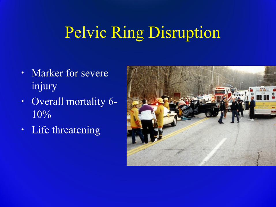
• Marker for severe injury
• Overall mortality 6-10%
• Life threatening
Pelvic Ring Disruption
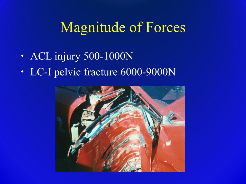
Magnitude of Forces
• ACL injury 500-1000N• LC-I pelvic fracture 6000-9000N
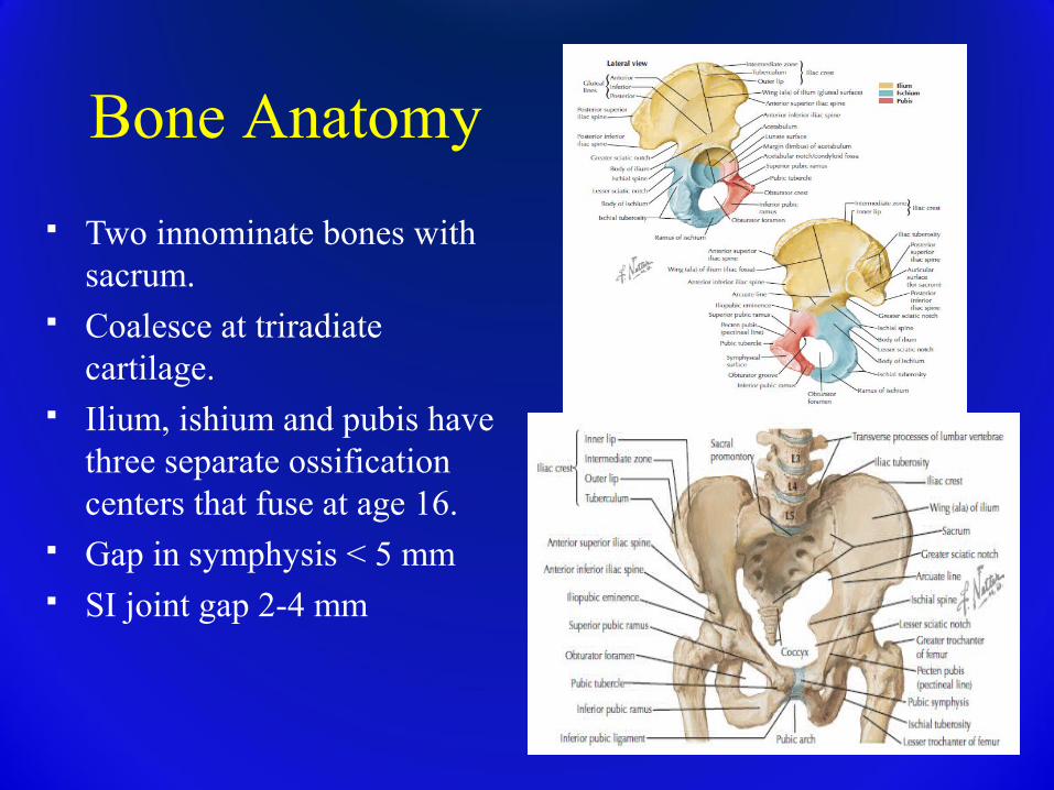
Bone Anatomy
Two innominate bones with sacrum.
Coalesce at triradiate cartilage.
Ilium, ishium and pubis have three separate ossification centers that fuse at age 16.
Gap in symphysis < 5 mm SI joint gap 2-4 mm
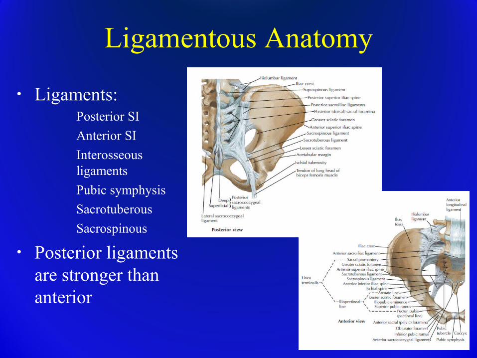
Ligamentous Anatomy
• Ligaments:� Posterior SI� Anterior SI � Interosseous
ligaments� Pubic symphysis � Sacrotuberous� Sacrospinous
• Posterior ligaments are stronger than anterior
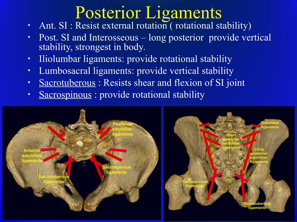
Posterior Ligaments• Ant. SI : Resist external rotation ( rotational stability)• Post. SI and Interosseous – long posterior provide vertical
stability, strongest in body.• Iliolumbar ligaments: provide rotational stability • Lumbosacral ligaments: provide vertical stability• Sacrotuberous : Resists shear and flexion of SI joint • Sacrospinous : provide rotational stability
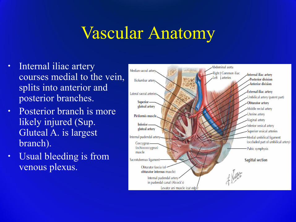
Vascular Anatomy
• Internal iliac artery courses medial to the vein, splits into anterior and posterior branches.
• Posterior branch is more likely injured (Sup. Gluteal A. is largest branch).
• Usual bleeding is from venous plexus.
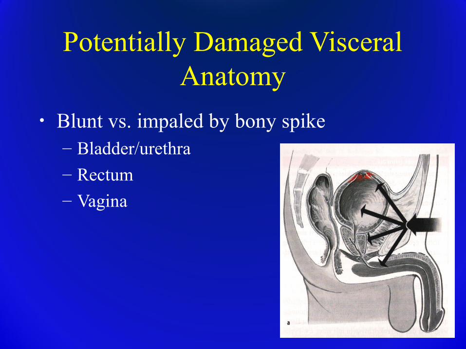
Potentially Damaged Visceral Anatomy
• Blunt vs. impaled by bony spike– Bladder/urethra
– Rectum – Vagina
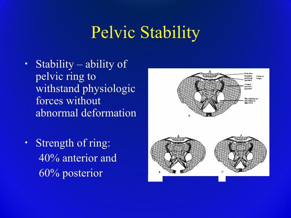
Pelvic Stability
• Stability – ability of pelvic ring to withstand physiologic forces without abnormal deformation
• Strength of ring: 40% anterior and 60% posterior
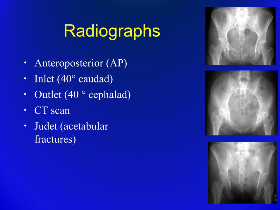
Radiographs
• Anteroposterior (AP)• Inlet (40° caudad)• Outlet (40 ° cephalad)• CT scan• Judet (acetabular
fractures)
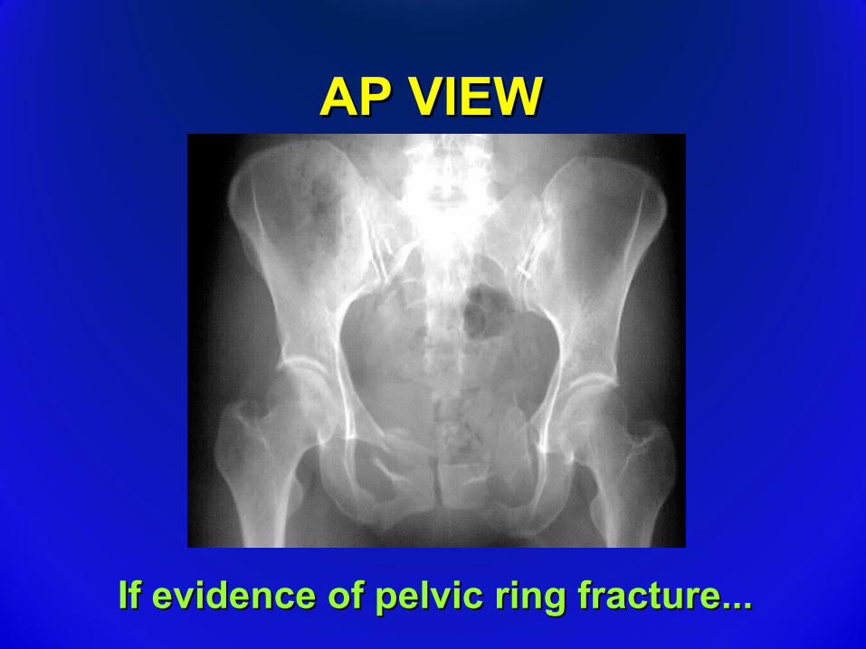
AP VIEWAP VIEW
If evidence of pelvic ring fracture...If evidence of pelvic ring fracture...
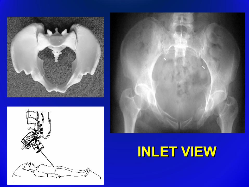
INLET VIEWINLET VIEW
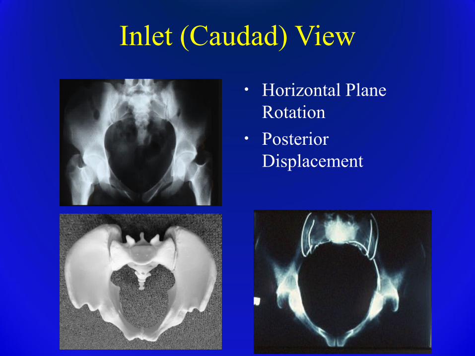
Inlet (Caudad) View
• Horizontal Plane Rotation
• Posterior Displacement
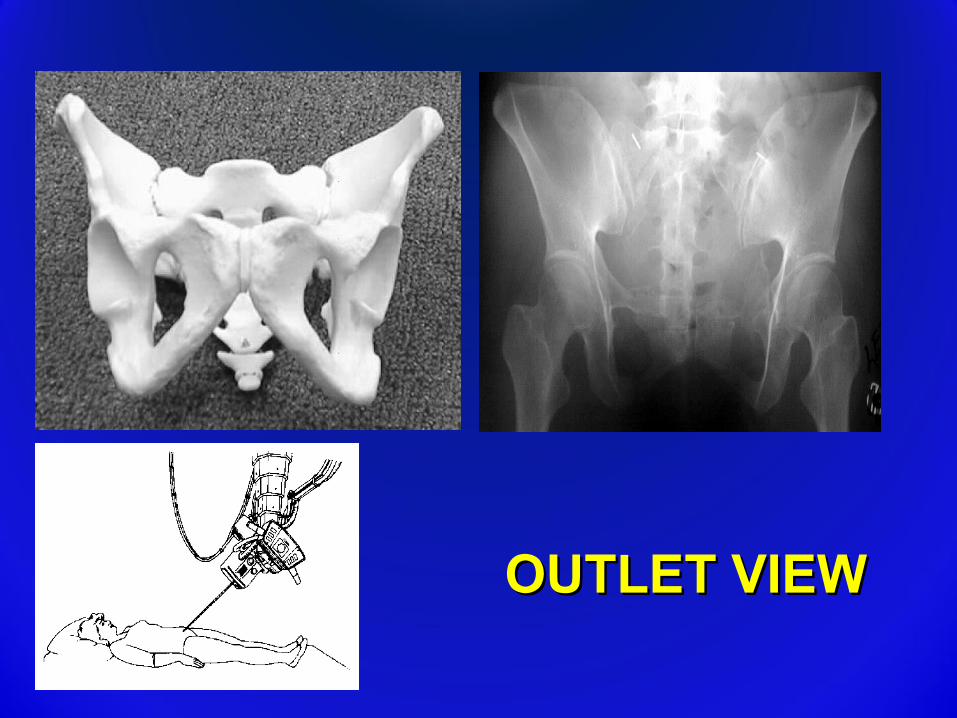
OUTLET VIEWOUTLET VIEW
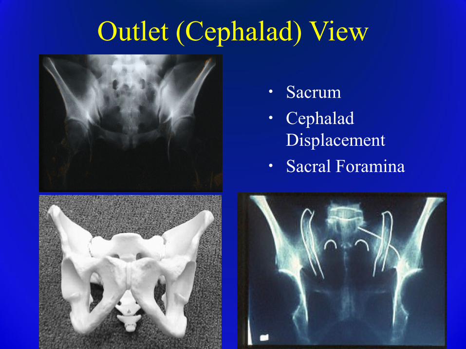
Outlet (Cephalad) View
• Sacrum• Cephalad
Displacement• Sacral Foramina
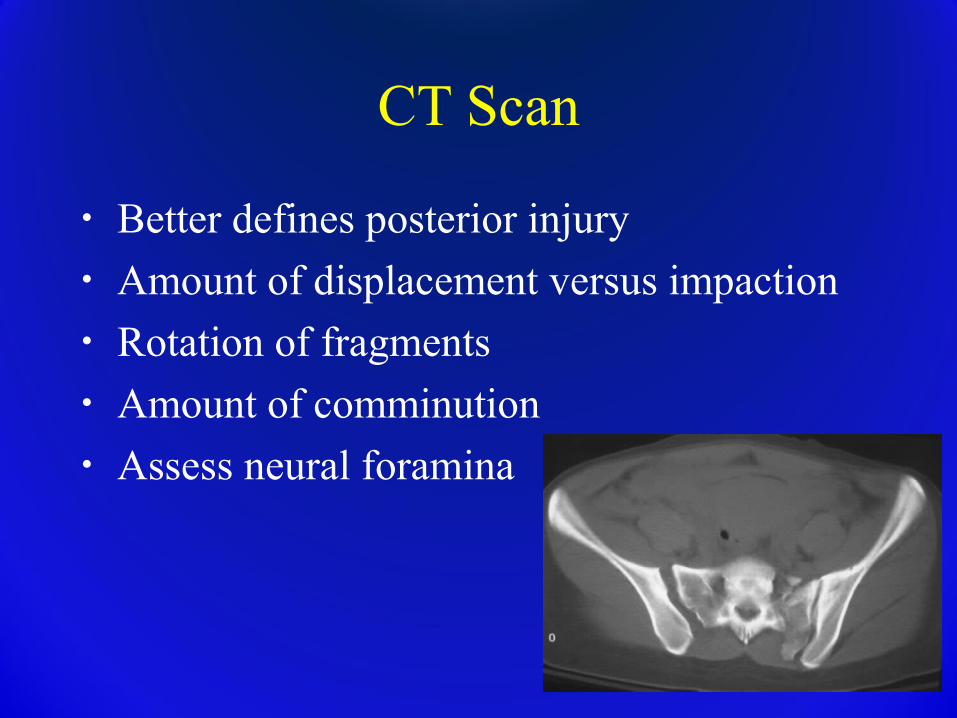
CT Scan
• Better defines posterior injury• Amount of displacement versus impaction• Rotation of fragments• Amount of comminution• Assess neural foramina
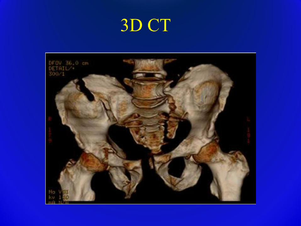
3D CT
Radiographic Signs of Instability
• Sacroiliac displacement of 5 mm in any plane
• Posterior fracture gap (rather than impaction)
• Avulsion of � fifth lumbar transverse process� lateral border of sacrum (sacrotuberous
ligament), � ischial spine (sacrospinous ligament)

Classification
Aids in predicting:– Hemodynamic instability
– Visceral and g.u. injuries– Pelvic instability– Mechanism of injury, force vector of injury.
• Planning surgical tactic for reduction & fixation
Classification Systems
• Anatomical (Letournel)• Stability & Deformity (Pennal, Bucholz,
Tile)• Vector force and associated injuries (Young
& Burgess)• OTA-research
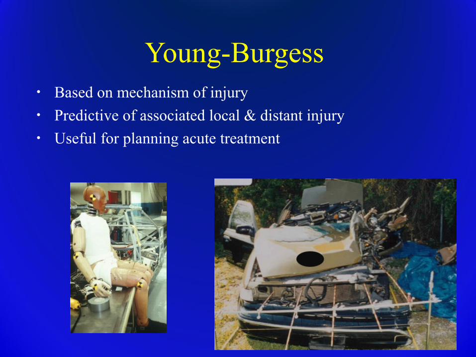
Young-Burgess • Based on mechanism of injury• Predictive of associated local & distant injury• Useful for planning acute treatment
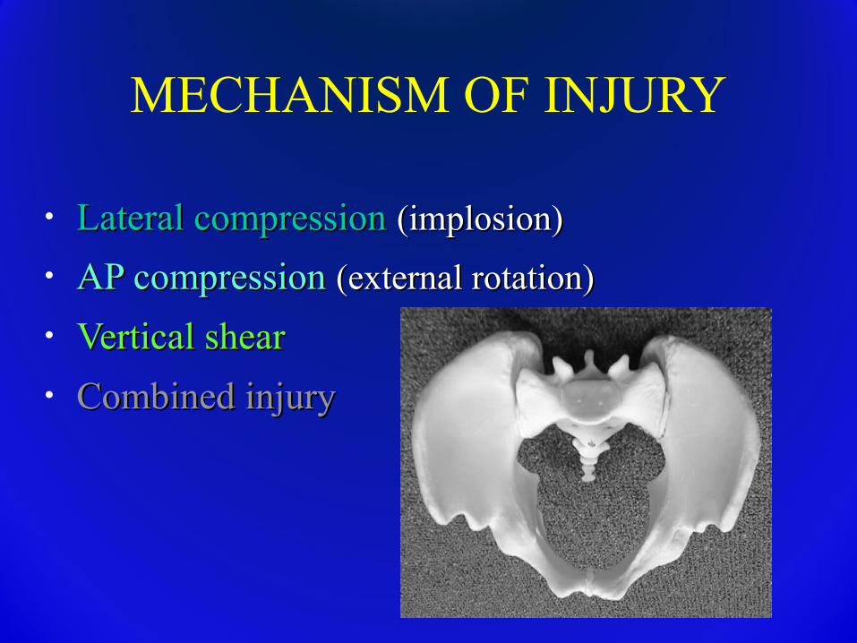
MECHANISM OF INJURY
• Lateral compressionLateral compression (implosion)(implosion)
• AP compressionAP compression (external rotation)(external rotation)
• Vertical shearVertical shear
• Combined injuryCombined injury
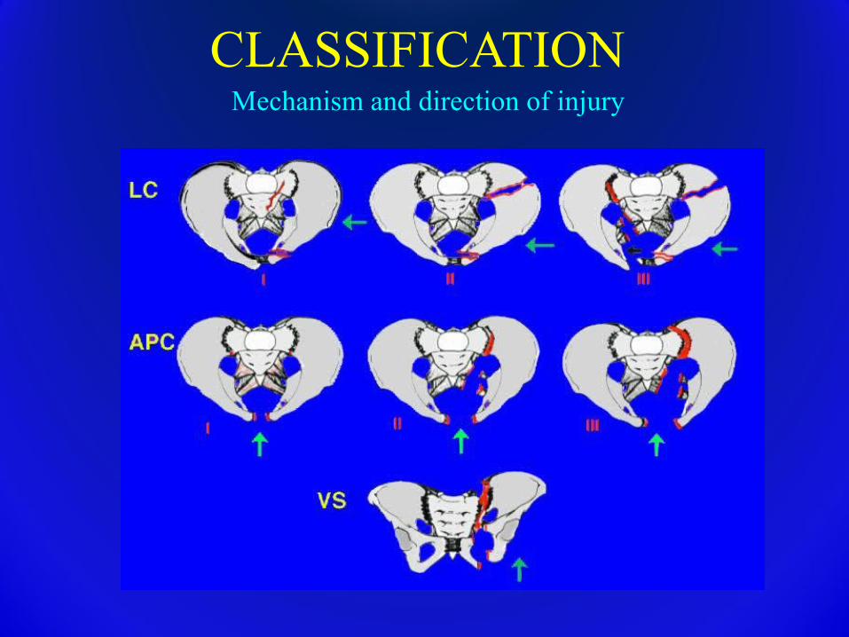
CLASSIFICATIONMechanism and direction of injury
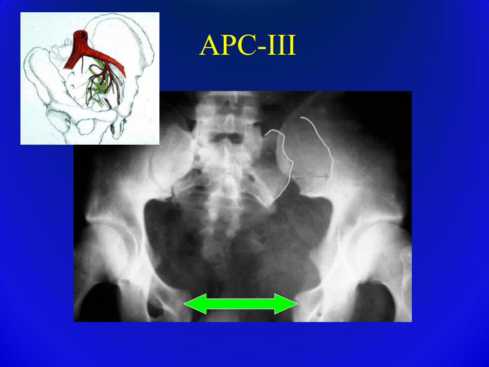
APC-III
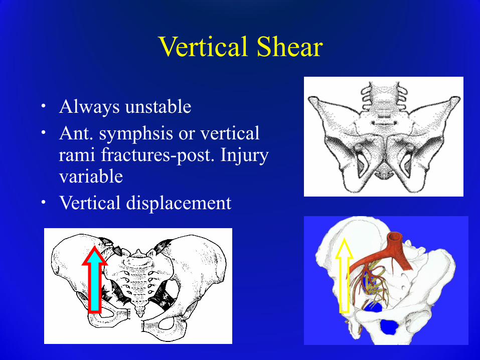
Vertical Shear
• Always unstable• Ant. symphsis or vertical
rami fractures-post. Injury variable
• Vertical displacement
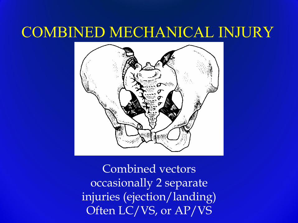
COMBINED MECHANICAL INJURY
Combined vectors occasionally 2 separate
injuries (ejection/landing)Often LC/VS, or AP/VS
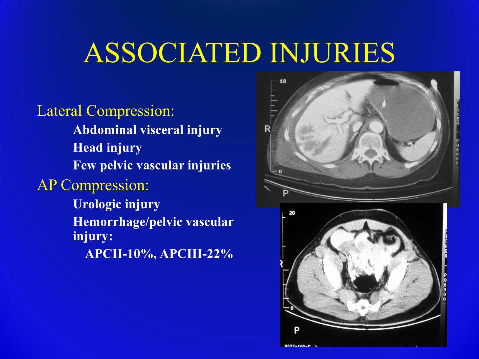
ASSOCIATED INJURIES
Lateral Compression:� Abdominal visceral injury� Head injury� Few pelvic vascular injuries
AP Compression:� Urologic injury� Hemorrhage/pelvic vascular
injury:APCII-10%, APCIII-22%
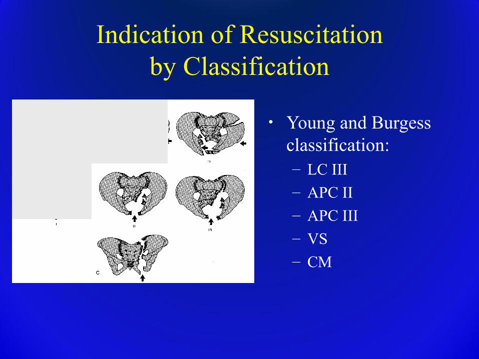
Indication of Resuscitationby Classification
• Young and Burgess classification:– LC III– APC II
– APC III– VS– CM
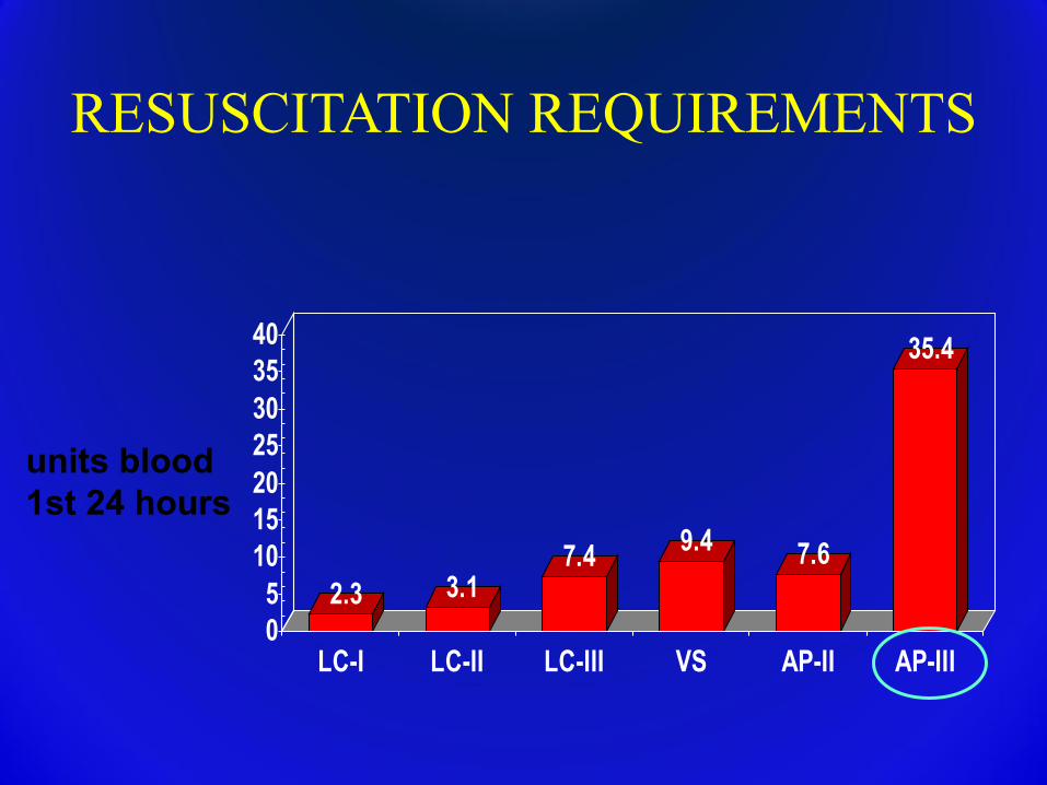
2.3 3.17.4 9.4 7.6
35.4
05
10152025303540
LC-I LC-II LC-III VS AP-II AP-III
units blood 1st 24 hours
RESUSCITATION REQUIREMENTS
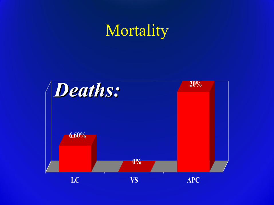
6.60%
0%
20%
LC VS APC
Deaths:Deaths:
Mortality
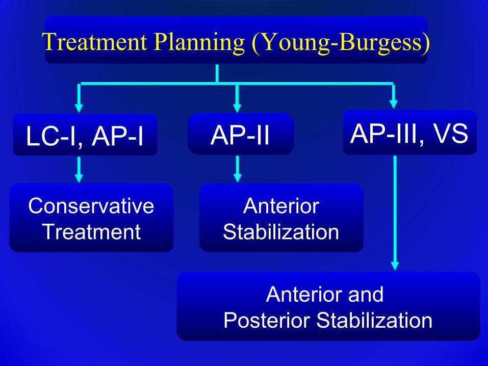
Treatment Planning (Young-Burgess)Treatment Planning (Young-Burgess)
LC-I, AP-ILC-I, AP-I AP-IIAP-II AP-III, VSAP-III, VS
ConservativeTreatment
ConservativeTreatment
AnteriorStabilization
AnteriorStabilization
Anterior and Posterior Stabilization
Anterior and Posterior Stabilization
Management of Pelvic Injuries
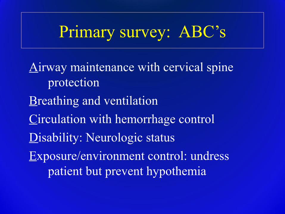
Primary survey: ABC’s
Airway maintenance with cervical spine protection
Breathing and ventilation
Circulation with hemorrhage control
Disability: Neurologic status
Exposure/environment control: undress patient but prevent hypothemia
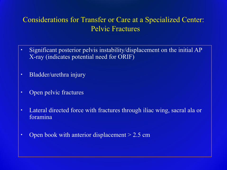
Considerations for Transfer or Care at a Specialized Center: Pelvic Fractures
• Significant posterior pelvis instability/displacement on the initial AP X-ray (indicates potential need for ORIF)
• Bladder/urethra injury
• Open pelvic fractures
• Lateral directed force with fractures through iliac wing, sacral ala or foramina
• Open book with anterior displacement > 2.5 cm
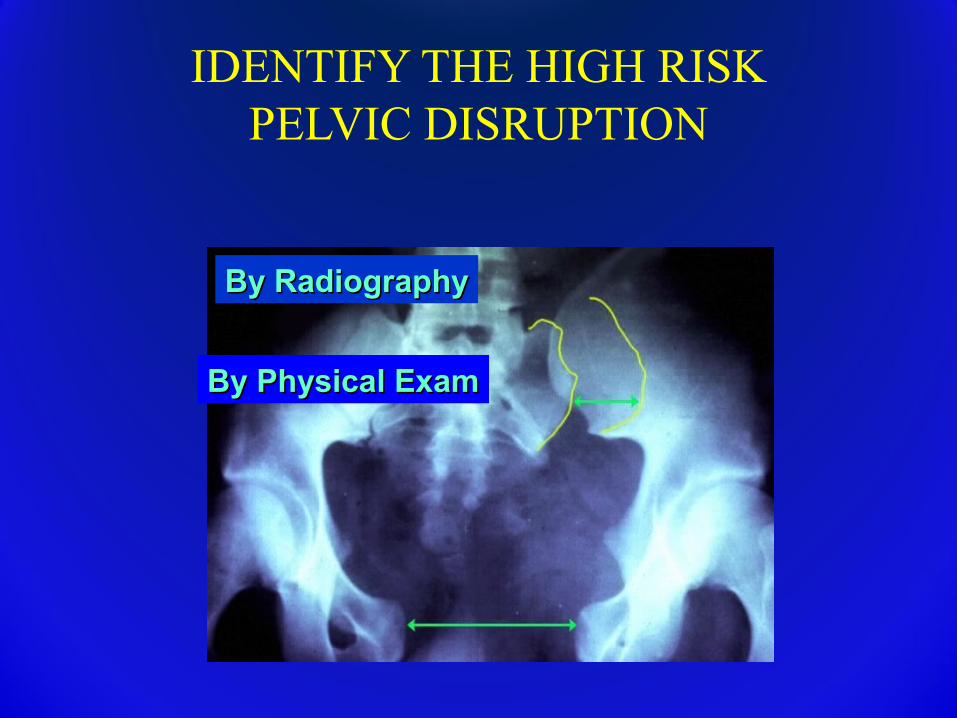
IDENTIFY THE HIGH RISK PELVIC DISRUPTION
By Physical ExamBy Physical Exam
By RadiographyBy Radiography
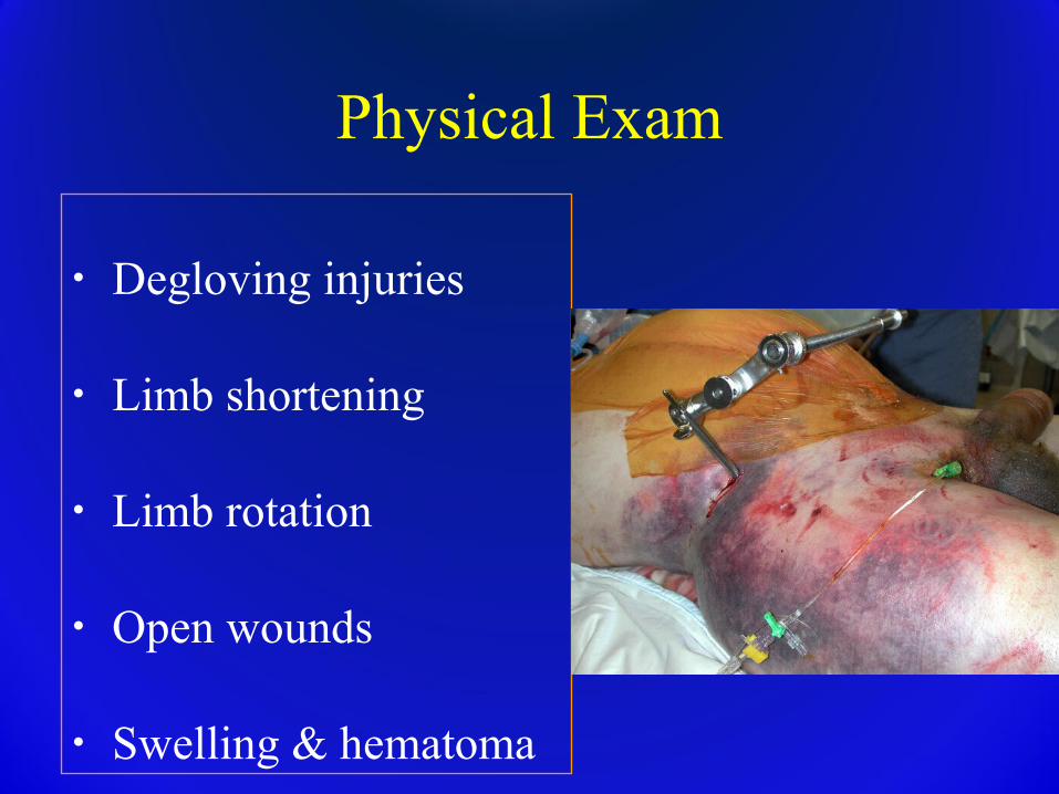
Physical Exam
• Degloving injuries
• Limb shortening
• Limb rotation
• Open wounds
• Swelling & hematoma
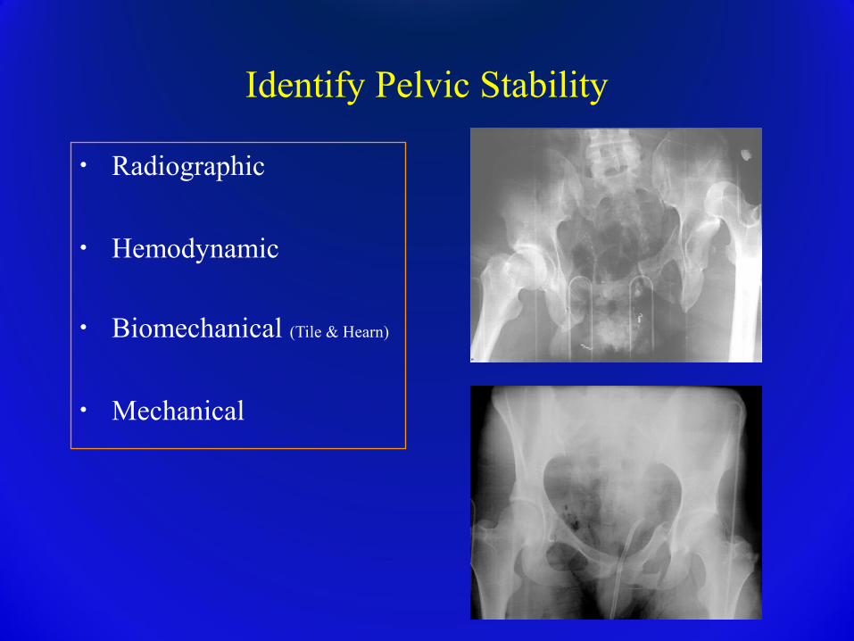
Identify Pelvic Stability
• Radiographic
• Hemodynamic
• Biomechanical (Tile & Hearn)
• Mechanical
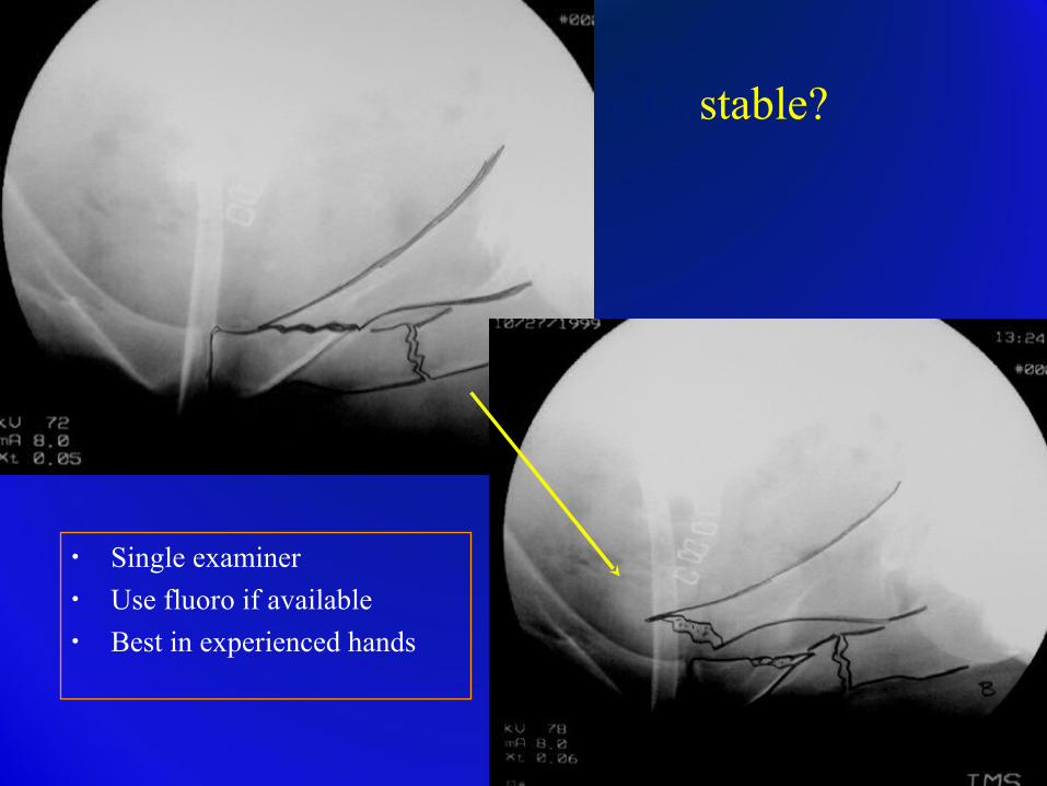
Stable or Unstable?
• Single examiner• Use fluoro if available• Best in experienced hands
Open Pelvic Injuries
• Open wounds extending to the colon, rectum, or perineum: strongly consider early diverting colostomy
• Soft-tissue wounds should be aggressively debrided
• Early repair of vaginal lacerations to minimize subsequent pelvic abscess
Urologic Injuries• 15% incidence• Blood at meatus or high riding prostate• Eventual swelling of scrotum and labia
(occasional arterial bleeder requiring surgery)• Retrograde urethrogram indicated in pelvic
injured patients• A foley catheter is preferred • If a supra-pubic catheter it used, it should be
tunneled to prevent anterior wound contamination
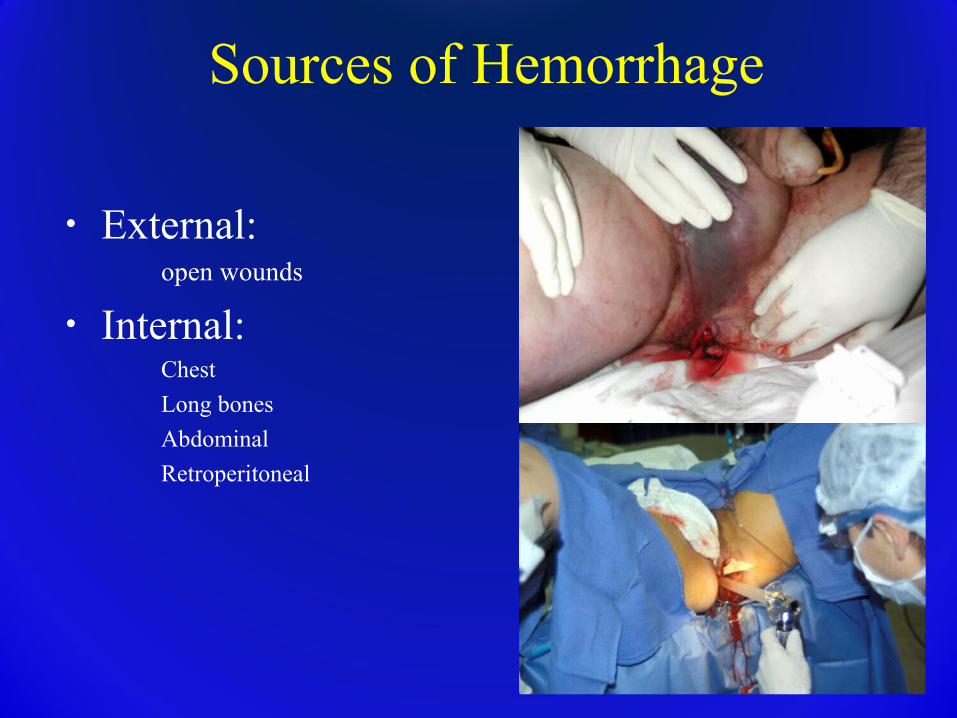
Sources of Hemorrhage
• External: open wounds
• Internal:Chest
Long bones
Abdominal
Retroperitoneal
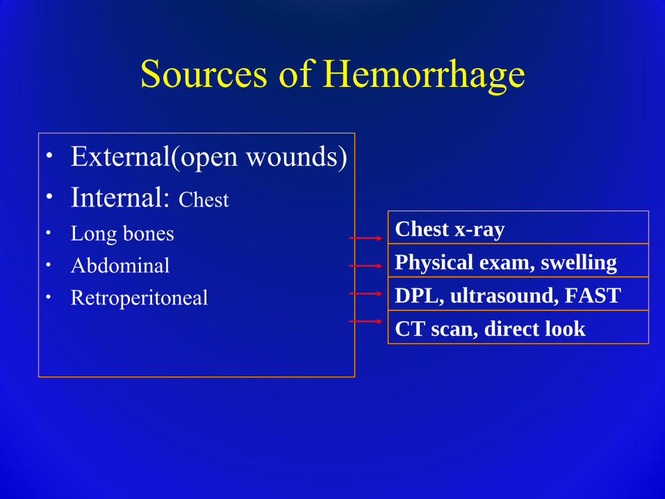
Sources of Hemorrhage
• External(open wounds)• Internal: Chest
• Long bones• Abdominal• Retroperitoneal
Chest x-ray
Physical exam, swelling
DPL, ultrasound, FAST
CT scan, direct look

Hemorrhage Control: Methods• Pelvic Containment
– Sheet– Pelvic Binder– External Fixation
• Angiography• Laparotomy• Pelvic Packing
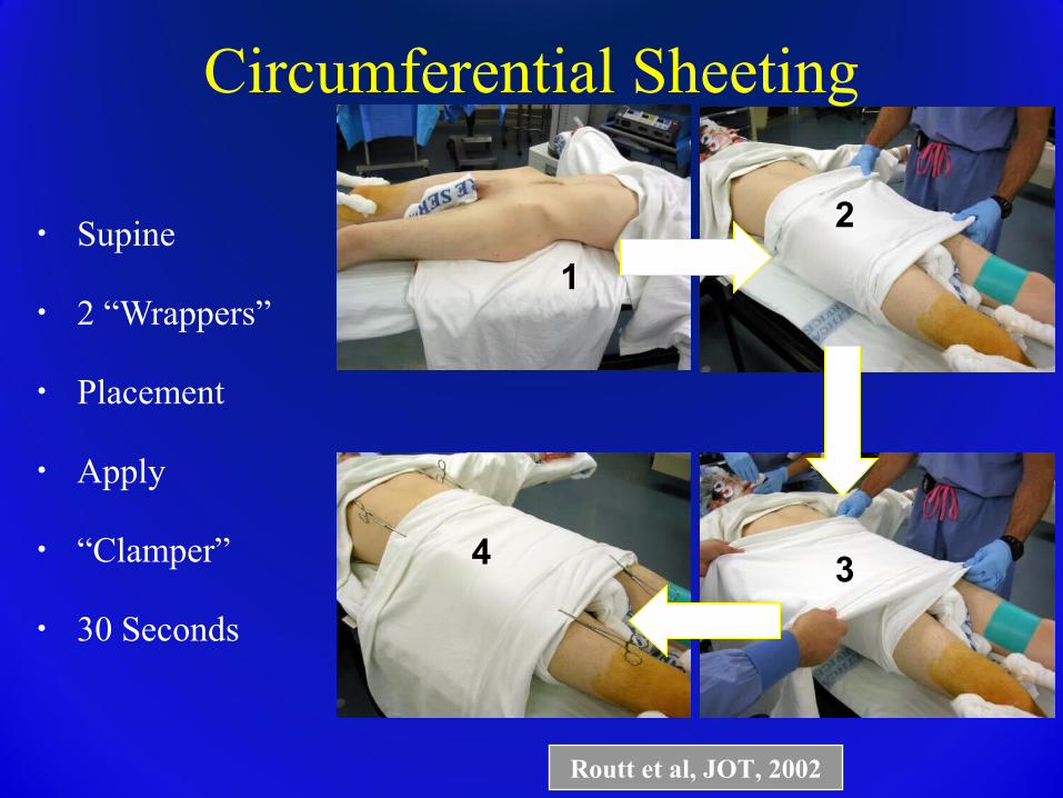
Circumferential Sheeting
• Supine
• 2 “Wrappers”
• Placement
• Apply
• “Clamper”
• 30 Seconds
1
2
34
Routt et al, JOT, 2002
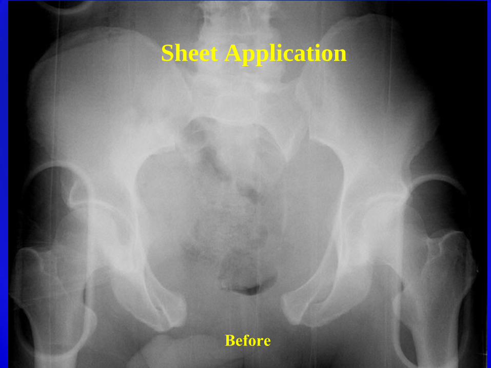
Sheet Application
Before
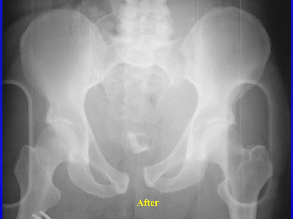
After
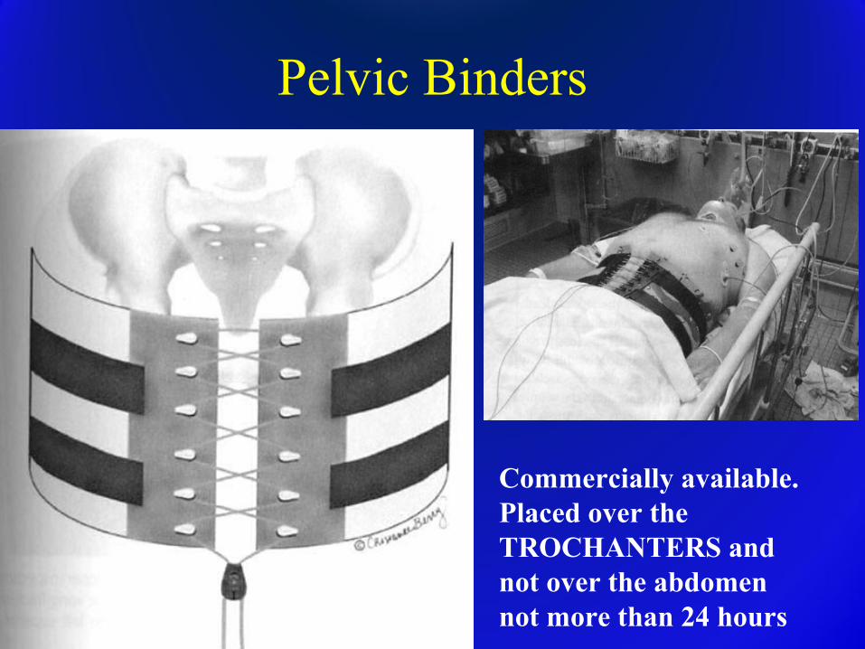
Pelvic Binders
Commercially available.Placed over the TROCHANTERS and not over the abdomen not more than 24 hours
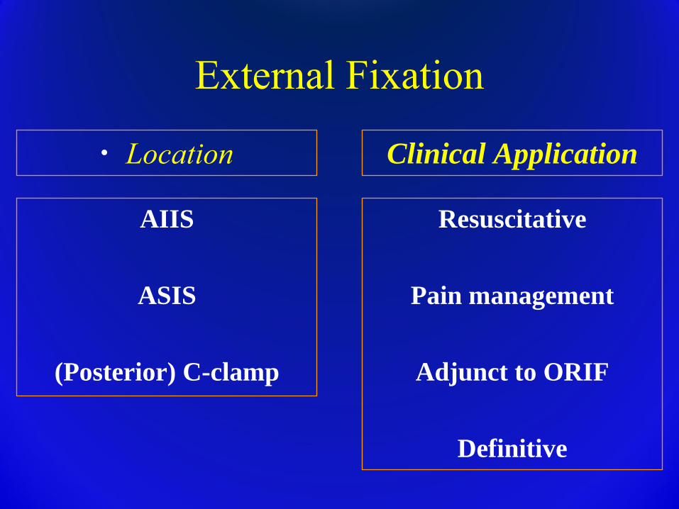
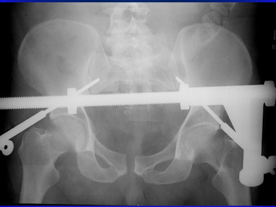
External Fixation
• Location
AIIS
ASIS
(Posterior) C-clamp
Clinical Application
Resuscitative
Pain management
Adjunct to ORIF
Definitive
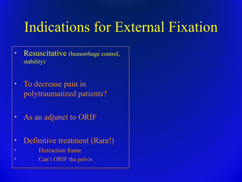
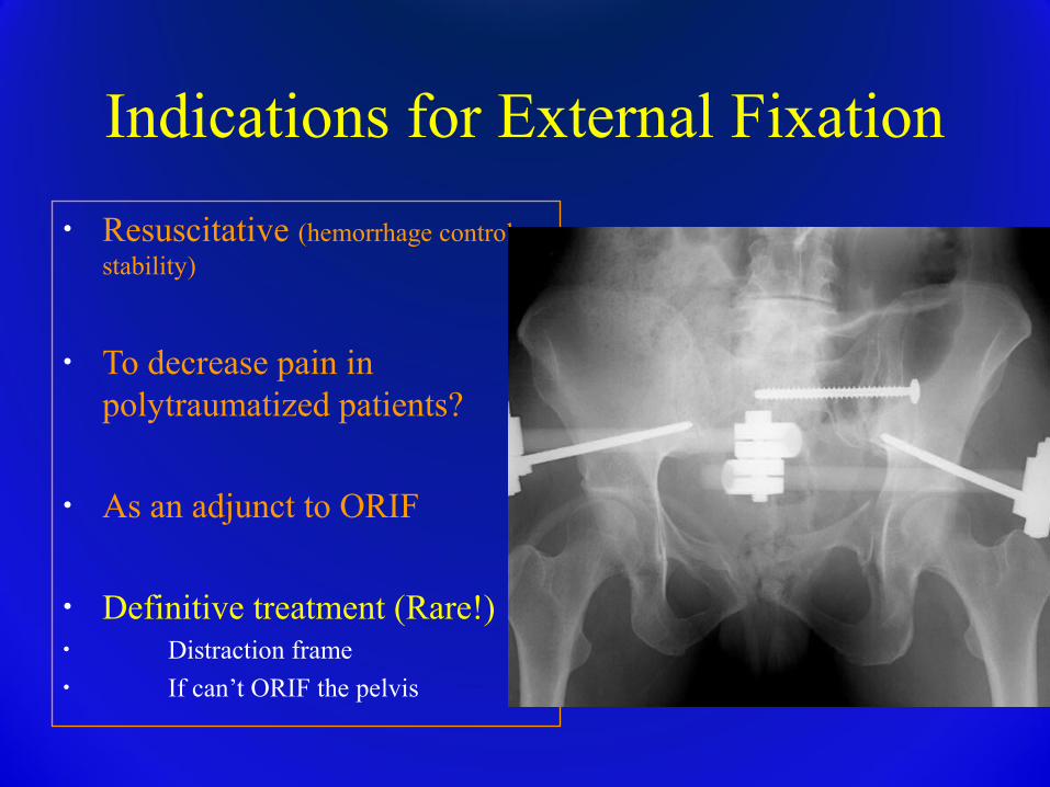
Indications for External Fixation
• Resuscitative (hemorrhage control, stability)
• To decrease pain in polytraumatized patients?
• As an adjunct to ORIF
• Definitive treatment (Rare!)• Distraction frame• Can’t ORIF the pelvis
Indications for External Fixation
• Resuscitative (hemorrhage control, stability)
• To decrease pain in polytraumatized patients?
• As an adjunct to ORIF
• Definitive treatment (Rare!)• Distraction frame• Can’t ORIF the pelvis
Theoretical and a marginal indication, but there is literature support
Barei, D. P.; Shafer, B. L.; Beingessner, D. M.; Gardner, M. J.; Nork, S. E.; and Routt, M. L.: The impact of open reduction internal fixation on acute pain management in unstable pelvic ring injuries. J Trauma, 68(4): 949-53, 2010.
Indications for External Fixation
• Resuscitative (hemorrhage control, stability)
• To decrease pain in polytraumatized patients?
• As an adjunct to ORIF
• Definitive treatment (Rare!)• Distraction frame• Can’t ORIF the pelvis
Indications for External Fixation
• Resuscitative (hemorrhage control, stability)
• To decrease pain in polytraumatized patients?
• As an adjunct to ORIF
• Definitive treatment (Rare!)• Distraction frame• If can’t ORIF the pelvis
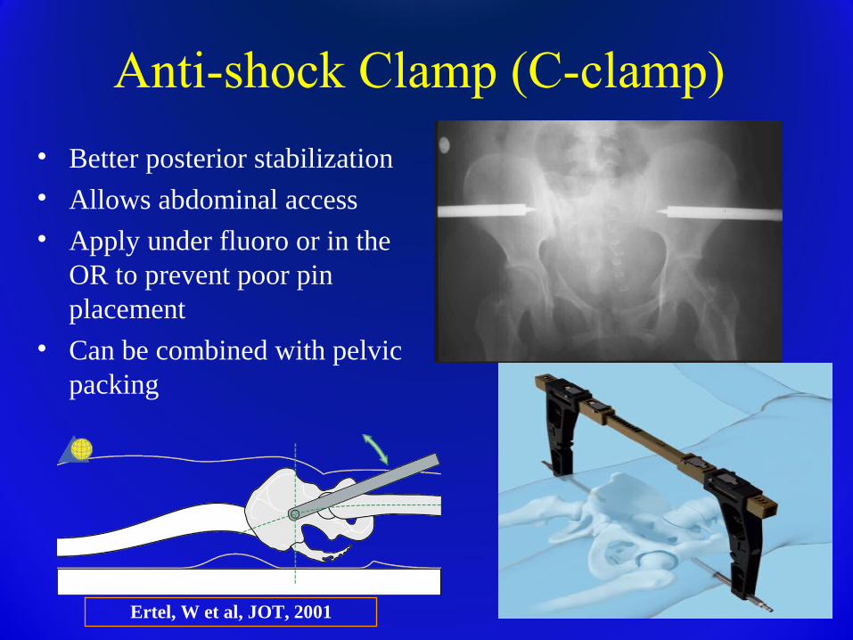
Anti-shock Clamp (C-clamp)
• Better posterior stabilization• Allows abdominal access• Apply under fluoro or in the
OR to prevent poor pin placement
• Can be combined with pelvic packing
Ertel, W et al, JOT, 2001
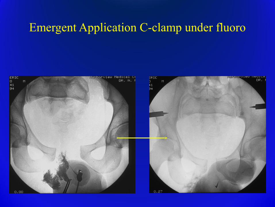
Emergent Application C-clamp under fluoro
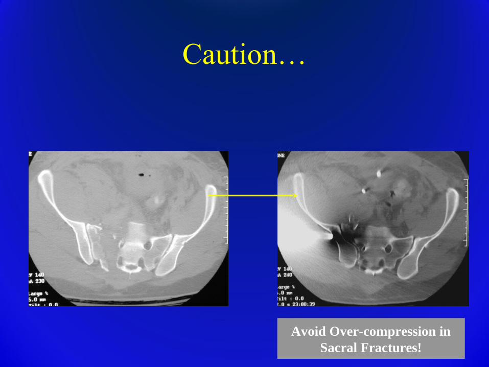
Avoid Over-compression in Sacral Fractures!
Caution…
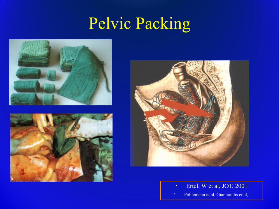
Pelvic Packing
• Ertel, W et al, JOT, 2001• Pohlemann et al, Giannoudis et al,
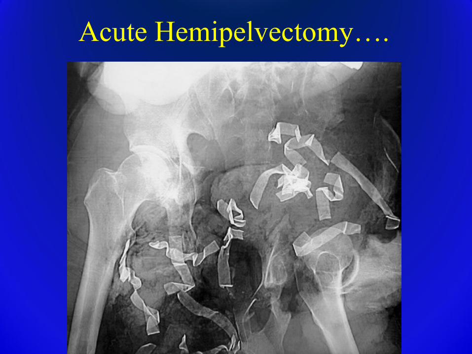
Acute Hemipelvectomy….
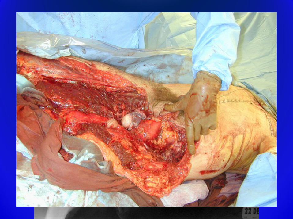
Acute Hemipelvectomy….
Pelvic FractureDefinitive Management
Non-Operative Management
• Lateral impaction type injuries with minimal (< 1.5 cm) displacement
• Pubic rami fractures with no posterior displacement
• Minimal gapping of pubic symphysis– If NO associated SI joint injury
Non-Operative Management
• X-rays are static picture of dynamic situation– Stress radiographs may help– Post-mobilization radiographs are essential
• Regular follow-up during conservative management.– In case of displacement reassess stability.
Operative Indications
• Resuscitation• Assist in mobilization
– Just as stabilizing long bones helps in mobilization of polytrauma patients
• Prevent long term functional impairment– Deformity of pelvic ring can impact function
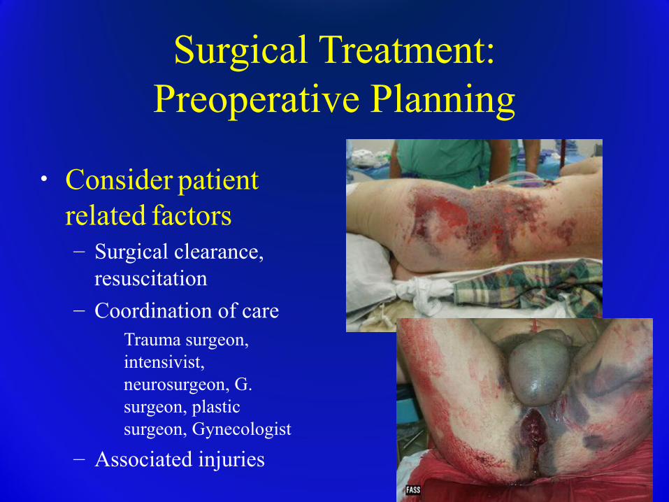
Surgical Treatment:Preoperative Planning
• Consider patient
related factors– Surgical clearance,
resuscitation
– Coordination of care� Trauma surgeon,
intensivist, neurosurgeon, G. surgeon, plastic surgeon, Gynecologist
– Associated injuries
Preoperative Planning
• Timing of surgery– Reduction may be easiest in first 24-48 hours– Patients often not adequately resuscitated in
first 24 hours– Potential for surgical “secondary hit” on post-
injury days 2-5� May be a significant issue in open procedures
Conclusions Multidisciplinary approach (general surgery,
urology, interventional radiology, neurosurgery) Understand the fracture pattern Do something (sheet, binder, ex fix, c-clamp) Combine knowledge of the fracture, the patients
condition, and the physical exam to decide on the next step
Treatment is based on:Pelvic instability, Displacement, Associated injuries
Surgical techniques for reduction and stabilization continue to evolve
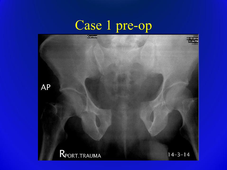
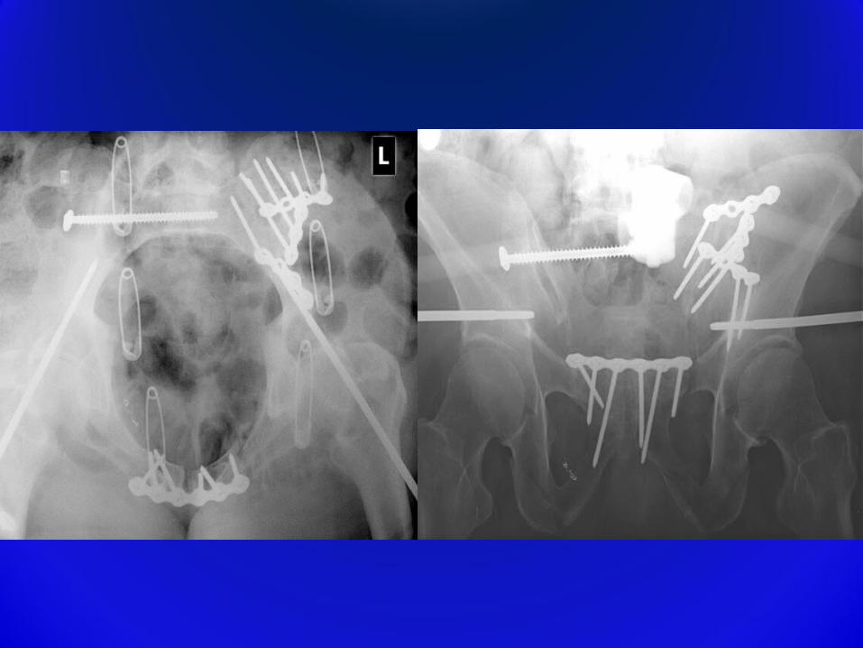
Case 1 pre-op
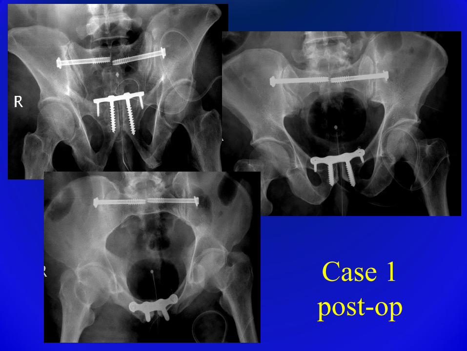
Case 1post-op
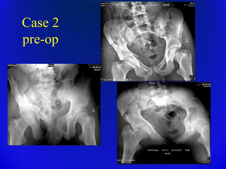
Case 2pre-op
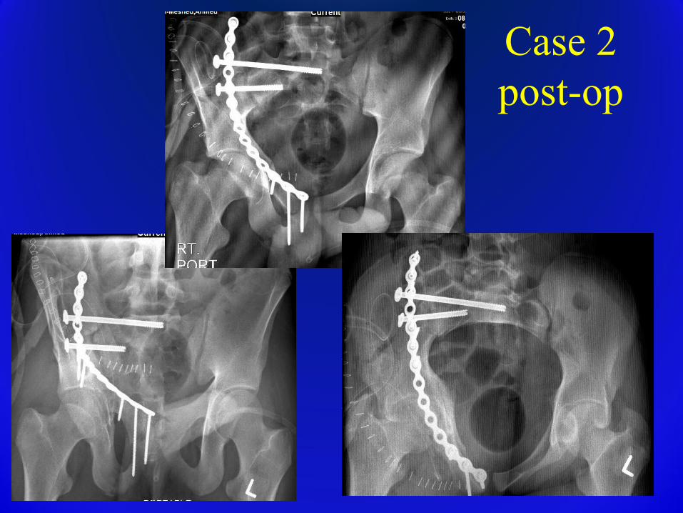
Case 2 post-op

Thank you