thermodynamics of amyloid fibril formation from chemical
TRANSCRIPT

26184 | Phys. Chem. Chem. Phys., 2019, 21, 26184--26194 This journal is© the Owner Societies 2019
Cite this:Phys.Chem.Chem.Phys.,
2019, 21, 26184
Thermodynamics of amyloid fibril formationfrom chemical depolymerization†
Nicola Vettore a and Alexander K. Buell *ab
Amyloid fibrils are homo-molecular protein polymers that play an important role in disease and
biological function. While much is known about their kinetics and mechanisms of formation, the origin
and magnitude of their thermodynamic stability has received significantly less attention. This is despite
the fact that the thermodynamic stability of amyloid fibrils is an important determinant of their lifetimes
and processing in vivo. Here we use depolymerization by chemical denaturants of amyloid fibrils of two
different proteins (PI3K-SH3 and glucagon) at different concentrations and show that the previously
applied isodesmic linear polymerization model is an oversimplification that does not capture the
concentration dependence of chemical depolymerization of amyloid fibrils. We show that cooperative
polymerization, which is compatible with the picture of amyloid formation as a nucleated polymerization
process, is able to quantitatively describe the thermodynamic data. We use this combined experimental
and conceptual framework in order to probe the ionic strength dependence of amyloid fibril stability.
In combination with previously published data on the ionic strength dependence of amyloid fibril growth
kinetics, our results provide strong evidence for the product-like nature of the transition state of amyloid
fibril growth.
1 Introduction
Filamentous protein structures are ubiquitous in biology. Theycan fulfill functional roles, as in the case of the cytoskeletalproteins actin1 and tubulin,2 or be associated with diseases, asin the case of sickle hemoglobin polymers3 or amyloid fibrils.4
For the cytoskeletal filaments, both the mechanical and thermo-dynamic stability, as well as the molecular origin of theirreversibility have been the subject of extensive studies in thelast decades.5 Indeed, actin filaments were the first proteinpolymers the thermodynamic stability of which has beencharacterized in detail in seminal work by Oosawa.6 Aktinfilaments can be described as helical polymers, whereby eachmonomeric building block interacts not only with its nearestneighbors in the polymer, but also with building blocks furtheraway. Actin monomers undergo relatively minor structuralchanges upon polymerization,7 and the same is also true fortubulin and sickle hemoglobin.8 Amyloid fibrils, on the otherhand, are protein polymers in which the protein buildingblocks usually adopt a very different structure inside the fibrilcompared to the isolated protein molecule.9 Amyloid fibrils are
mostly known as being the hallmark of a wide range ofdiseases, but mounting evidence demonstrates that amyloidfibrils can also play functional roles in biology.10 Many differentproteins entirely unrelated in structure (IDPs or folded proteins)and function (peptide hormones,11 lipid-binding proteins,12 milkproteins13) have been found to form amyloid fibrils, either in vivoor in the test tube. While much insight has been generated in thelast two decades on the kinetics and mechanisms of amyloid fibrilformation,14–17 much less is known about the thermodynamicstability of these structures,18 and to what extent common drivingforces govern the amyloid formation of different proteins. It isvery important to be able to quantify and rationalize the thermo-dynamic stability of amyloid fibrils because the thermodynamicstability is likely to be a decisive factor in determining whetheramyloid fibrils of a certain protein can be cleared in a biologicalcontext. Such clearance could happen either through spontaneousbinding of molecules, such as antibodies, to soluble or aggregatedprotein19 or else through active, energy consuming processes,such as the action of chaperone complexes on amyloid fibrils.20
Experimental data of amyloid fibril stability21,22 has so farbeen analysed in the framework of the linear polymerizationmodel.6 The simplest, so-called isodesmic form of this modelcontains only a single equilibrium constant, that for the addi-tion of monomer to all possible species, including to anothermonomer. Under this assumption, the stability of the fibrilscan be directly determined from a measurement of the freemonomer concentration at equilibrium, for sufficiently high
a Institut for Physical Biology, Heinrich-Heine-Universitaet Duesseldorf,
Universitaetstrasse 1, Duesseldorf, Germanyb Technical University of Denmark, Department of Biotechnology and Biomedicine,
Lyngby, Denmark. E-mail: [email protected]
† Electronic supplementary information (ESI) available. See DOI: 10.1039/c9cp04524d
Received 15th August 2019,Accepted 16th November 2019
DOI: 10.1039/c9cp04524d
rsc.li/pccp
PCCP
PAPER
Ope
n A
cces
s A
rtic
le. P
ublis
hed
on 1
9 N
ovem
ber
2019
. Dow
nloa
ded
on 2
/21/
2022
10:
55:2
0 PM
. T
his
artic
le is
lice
nsed
und
er a
Cre
ativ
e C
omm
ons
Attr
ibut
ion-
Non
Com
mer
cial
3.0
Unp
orte
d L
icen
ce.
View Article OnlineView Journal | View Issue

This journal is© the Owner Societies 2019 Phys. Chem. Chem. Phys., 2019, 21, 26184--26194 | 26185
total concentrations. In many practical cases, the free monomerconcentration is very low (nM range23) and difficult to determineaccurately. In order to overcome this practical problem, amyloidfibrils can be destabilized by chaotropes, such as GndSCN orGndHCl.21,22 By assuming a linear relationship between theconcentration of denaturant and the free energy difference DGbetween the monomeric and polymeric state, equivalent to theassumption in protein unfolding experiments,24 sigmoidal fibrildepolymerization data can be fitted and the value of DG extra-polated to the absence of denaturant.21,22 While this method ofdetermination of amyloid fibril stability is commonly used, itsvalidity has not yet been thoroughly tested. Here we use intrinsicprotein fluorescence to monitor the fraction of fibrillar vs. solubleprotein. We investigate and rationalise the influence of the typeof chemical denaturant, the ionic strength and the proteinconcentration on the depolymerization curves and show that acooperative model provides a better description of the thermo-dynamics of amyloid fibril formation than the simple isodesmicpolymerization model. The cooperative model, adapted from thefield of supramolecular chemistry,25,26 describes the thermo-dynamics of polymerization by two different equilibrium constants:a nucleation constant and an elongation constant. We find thatexperiments in which the peptide concentration is varied allow abetter discrimination between the isodesmic and coopera-tive models than the standard experiments in which only thedenaturant concentration is varied.
We apply our insight and methodology to the analysis ofthe ionic strength dependence of amyloid fibril stability andcompare it with the ionic strength dependence of the fibrilgrowth kinetics.27 Our combined analysis of this data providesstrong evidence for the fact that the transition state of thefibril growth reaction is highly product-like with respect to thedistance of the newly adding peptide to the fibril end.
2 Experimental section2.1 Proteins
The human glucagon employed in the study was a kind giftfrom Novo Nordisk. The bovine PI3K-SH3 domain was purifiedaccording to the protocol in ref. 28. The construct contains a6xHis-tag linked to the protein by a thrombin cleavage site. Thesequence of the WT protein after cleavage is the following, withthe dipeptide Gly-Ser remaining as overhang from the cleavage:
GS MSAEGYQYRA LYDYKKEREE DIDLHLGDIL TVNKGSLVALGFSDGQEAKP EEIGWLNGYN ETTGERGDFP GTYVEYIGRK KISP
The protein was expressed in a BL21 E. coli strain with TBmedium for auto induction containing 0.012% Glucose and0.048% Lactose. The cells were grown for over 24 h and thenharvested by centrifugation. After resuspension in sodiumphosphate buffer (50 mM sodium phosphate pH 8, 5 mMImidazole and 100 mM NaCl), the cells were disrupted bysonication, in the presence of protease inhibitors (cOmpleteMini EDTA-free, Roche) and DNAse (Sigma-Aldrich). The lysatewas centrifuged, and the supernatant loaded on a Ni-NTASuperflow Cartridge (Qiagen, Venlo, Netherlands) equilibrated
in 50 mM sodium phosphate pH 8, 5 mM Imidazole and100 mM NaCl. The protein was eluted with a linear gradientfrom 5 to 300 mM imidazole in 50 mM sodium phosphate pH 8,100 mM NaCl in 25 ml elution volume. Fractions containing theprotein were collected and cleaved overnight at 7 1C with 1 unitof thrombin (from bovine plasma, Sigma-Aldrich Saint Louis,Missouri, USA) per 1 mg of protein. The cleaved solution wasthen concentrated and loaded on a SEC HiLoad 26/60 Superdex75 column (GE Healthcare, Chicago, Illinois, USA) equili-brated with 5 mM ammonium acetate pH 7. Fractions containingthe PI3K-SH3 domain were collected and lyophilised forfurther use.
2.2 Fibril preparation
Glucagon fibrils were formed from protein solutions preparedafter resuspension of the lyophilized peptide in 10 mM glycinehydrochloride pH 2 at 1 mM final concentration. This solutionwas incubated under shaking at 37 1C for 1–2 hours. The fibrilsobtained through this procedure were used as seeds for furthersolutions of monomeric protein, which were prepared at aconcentration range between 1 and 2 mM and seeded atca. 10% (monomer equivalents). The solution was left overnightat room temperature without shaking or stirring.
PI3K-SH3 fibrils were formed from protein solutionsprepared from the lyophilized protein (produced as describedabove), resuspended in 10 mM glycine hydrochloride pH 2 at200 mM final concentration. This solution was incubated undershaking at 42 1C overnight. The fibrils obtained through thisprocedure were then used as seeds for further solutions ofmonomeric protein, which were prepared at a concentrationrange between 200 and 300 mM and then seeded at ca. 10%(equivalent monomer mass). The solution was left overnight atroom temperature without shaking or stirring.
Such high concentrations of fibril stock solutions areneeded, as the fibrils are strongly diluted upon addition ofthe denaturant, which is necessary to achieve denaturant concen-trations high enough for depolymerization. Before preparingthe samples, the fibril preparations were sonicated with aVialTweeter-sonotrode (Hielscher, Teltow, Germany). Glucagonfibril solutions were sonicated in a volume of at least 700 ml,twice for 3 seconds, 100% amplitude, with a pause of ca. 30 s.PI3K-SH3 fibrils were sonicated for 10 s at 100% amplitude in avolume of at least 700 ml. The fibril preparations were imagedby AFM, both before and after the sonication protocol,to evaluate the effect of the sonication on the lengths of thefibrils.
2.3 Atomic force microscopy imaging
Atomic force microscopy (AFM) was performed on dried fibrilsamples. The fibril samples were diluted to ca. 3 mM in thesame buffer, then 20 ml were placed on freshly cleaved mica andincubated at room temperature for 10 min. After 10 min themica was washed with MilliQ water. Imaging was performedwith a Bruker Multimode 8 (Billerica, Massachusetts, USA)using OMCL-AC160TS cantilevers (Shinjuku, Tokyo, Japan).
Paper PCCP
Ope
n A
cces
s A
rtic
le. P
ublis
hed
on 1
9 N
ovem
ber
2019
. Dow
nloa
ded
on 2
/21/
2022
10:
55:2
0 PM
. T
his
artic
le is
lice
nsed
und
er a
Cre
ativ
e C
omm
ons
Attr
ibut
ion-
Non
Com
mer
cial
3.0
Unp
orte
d L
icen
ce.
View Article Online

26186 | Phys. Chem. Chem. Phys., 2019, 21, 26184--26194 This journal is© the Owner Societies 2019
2.4 Depolymerization experiments
The fibril samples were mixed with different volumes of urea orGndHCl stock solutions and buffer. In order to maintain asolution pH of 2 constant throughout the whole denaturationseries, an 8 M urea stock solution was prepared by dissolving12 g of urea (SigmaAldrich) in 16 ml of concentrated buffer, toyield a final concentration of 10 mM glycine hydrochloride and16 mM HCl. The HCl is necessary, as the urea has a weakbuffering capacity. In order to keep the ionic strength constantin all samples, NaCl was added to the 10 mM glycine HCl bufferto a final concentration of 16 mM. In order to ensure that thesamples have reached equilibrium when they are analyzed, weassessed the time of equilibration (see Fig. S5, ESI†) and basedon these assessments left the samples to equilibrate for oneweek in the case of glucagon fibrils and two weeks in the case ofPI3K-SH3 fibrils.
2.5 Fluorescence measurements
Fluorescence spectra were recorded on a Tecan M1000proinstrument using Greiner UV-transparent 96 well plates. Thewells were filled with 140 ml of solution and the temperaturewas maintained constant at 27 1C. The fluorescence spectrawere measured by top reading, exciting at 280 nm for glucagonsamples and 290 nm for PI3K-SH3 samples (5 nm bandwidth),while the emission was recorded between 300 nm and 420 nm(5 nm bandwidth). A blank spectrum (buffer) resulting from theaverage of 10 different spectra is subtracted before analysis.In order to determine the relative populations of soluble vs.fibrillar protein, we computed the ratios of the fluorescenceintensities at 340 and 320/310 nm (glucagon/PI3K-SH3) foreach spectrum. This choice of wavelengths was based on thedifference between the fibrillar and monomeric spectra in eachcase (Fig. S2, ESI†).
2.6 Absorbance measurements
In order to measure the degree of aggregation of the fibrillarsamples, they were centrifuged at 16.100 g for 1 hour. Thesupernatant was removed and its absorbance was measuredbetween 220 and 350 nm with a Nanodrop 2000 (ThermoFisher)spectrophotometer. The extinction coefficients at 280 nm are8480 M�1 for glucagon and 15 930 M�1 for PI3K-SH3.
3 Results
In order to establish a reliable methodology for the determinationof the thermodynamic stability of amyloid fibrils, we chose twopolypeptide systems that have been shown to form amyloid fibrilsunder acidic conditions, the peptide hormone glucagon29,30 andthe protein PI3K-SH3.28,31 Both of these polypeptides containthe amino acid tryptophan and therefore the use of intrinsicprotein fluorescence can be explored for the distinctionbetween monomeric and aggregated protein32 (Fig. S2, ESI†).In several previous studies of amyloid fibril stability based onchemical depolymerization, the equilibrated samples weresubjected to centrifugation and the protein concentration in
the supernatant was determined.22,33 Experimental data con-firming the equivalence between the data from concentrationmeasurements and the intrinsic fluorescence data can be foundin Fig. S6 and S7 (ESI†).
For both PI3K-SH3 and glucagon, homogeneous fibril pre-parations without appreciable amounts of non-fibrillar material(as judged by AFM imaging, see Fig. S1, ESI†) can be produced byadjusting the solution conditions appropriately.32,34 In our experi-ments of fibril depolymerization we started from fully equilibratedfibrillar samples that we prepared by seeding, i.e. the addition ofpreformed fibrils to monomeric samples. We then homogenizedthe samples and shortened the average fibril length by subjectingthe samples to ultrasonication (Fig. S1, ESI†). Details about thesample preparation can be found in the methods section.
3.1 Choice of denaturant
Having chosen the polypeptide systems to investigate, we nextproceeded to the choice of the chemical denaturant to be usedto destabilise the fibrils. Previous reports mostly used strongdenaturants, such as GndHCl or GndSCN21,22,33 rather thanmilder ones, such as urea. It is well established that the ionicdenaturants are more powerful than urea in the unfolding ofproteins.35 However, we found that in our case the fibrils couldbe dissociated by urea, and even that in the case of PI3K-SH3amyloid fibrils, urea was a more powerful denaturant thanGndHCl (see Fig. S4, ESI†). This is in contrast to the unfoldingof monomeric PI3K-SH3 that was unfolded at lower concentra-tions of GndHCl compared to urea, at neutral pH (see Fig. S4,ESI†). Based on these initial results, we decided to chose ureaas the denaturant of choice for our amyloid fibril depolymeri-zation experiments, as this choice of a neutral denaturantallows us to explore the role of electrostatic interactions inamyloid fibril stability in more detail. The use of GndHCl wouldlead to an almost complete screening of the electrostaticinteractions at the high denaturant concentrations used in theseexperiments. It has to be noted that prolonged incubation ofproteins in high urea concentration can lead to carbamylationreactions.36 The acidic pH conditions employed in the presentstudy strongly disfavour this reaction. We showed by massspectrometric analysis (Fig. S9, ESI†) that even after 9 daysincubation in 6 M urea, no sign of carbamylation was observedfor PI3K-SH3, which required the longest equilibration times.
3.2 Analysis of chemical depolymerization with an isodesmicmodel
Equilibrium denaturation curves of folded proteins are oftenanalysed with a two state model, whereby it is assumed that thedenaturant linearly shifts (with proportionality constant m) thefree energy difference between the folded and the unfoldedpolypeptide.37 Due to the large number of aggregate species ofdifferent sizes, a two state model is not appropriate in the caseof protein polymerization. It has first been proposed by Gotoand coworkers21 to apply the isodesmic form of the linearpolymerization model6 to fit equilibrium depolymerizationcurves of amyloid fibrils, and this method of analysis has beenexclusively used to-date. The mathematical formulation of this
PCCP Paper
Ope
n A
cces
s A
rtic
le. P
ublis
hed
on 1
9 N
ovem
ber
2019
. Dow
nloa
ded
on 2
/21/
2022
10:
55:2
0 PM
. T
his
artic
le is
lice
nsed
und
er a
Cre
ativ
e C
omm
ons
Attr
ibut
ion-
Non
Com
mer
cial
3.0
Unp
orte
d L
icen
ce.
View Article Online

This journal is© the Owner Societies 2019 Phys. Chem. Chem. Phys., 2019, 21, 26184--26194 | 26187
model can be found in Section S2 (ESI†). Individual sigmoidaldepolymerization curves can be fitted and free energy differ-ences between the fibrillar and the soluble states, as well asm-values, can be determined. The absolute concentration ofpeptide is an important parameter in the model that deter-mines the shape of the depolymerization curve (see Section S2,ESI†). If depolymerization curves are acquired at differentpeptide concentrations, the extracted free energies andm-values are expected to be identical, within error. We havefitted data from experiments at three (glucagon) and two(PI3K-SH3) different peptide concentrations. It is convenientto display such data in a normalised way, i.e. as a fraction ofdepolymerised protein, rather than as an absolute concen-tration (Fig. 1). In Fig. S8 (ESI†), we show the same datawithout normalisation. We find that while all the individualcurves can be well-fitted (Fig. 1, left panel), in the case ofglucagon the fits at the three different concentrations yieldsignificantly different values for the free energy difference DG0
(�36.7 vs. �38.8 vs. �42.2 kJ mol�1 for the lowest, intermediateand highest concentrations, respectively). In the case of PI3K-SH3, the difference is similar (�64.7 vs. �71.1 kJ mol�1),despite the fact that here the difference in concentrations isless significant. The reason for the difference between theindividual fits is that both DG0 and m are free parametersand can both vary between fits to the data sets at differentprotein concentrations. If, however, the data at the differentconcentrations are fitted globally (Fig. 1, right panel), we findthat in the case of PI3K-SH3, a satisfactory fit result is achievedand the value for the free energy of fibril stability is intermediatebetween the two values of the individual fits (�66.5 kJ mol�1).On the other hand, a global fit to the data for glucagon is not asgood and yields a value for the free energy comparable to thatfrom a local fit of the lowest concentration (�36.7 kJ mol�1).Tables S1 and S2 in the ESI† show all the free energy and m-values,when neither, either of these or both parameters are globallyfitted. Therefore, already a global fit to denaturation curves at onlytwo or three different protein concentrations reveals potential
inadequacies of the isodesmic model for the description ofchemical depolymerization experiments of amyloid fibrils.
3.3 Extension of the isodesmic model
The isodesmic version of the linear polymerization modelassumes that all equilibria in a solution of polymers have thesame equilibrium constant. However, this assumption is clearlynot in agreement with the known mechanistic features ofamyloid fibril formation. Amyloid fibrils form through anucleated polymerization process,14,16 whereby the formationof the initial oligomeric nucleus is energetically less favourablethan the addition of a monomer to a fully grown fibril.38
We hypothesised that this simplification could be at the originof the inability of the isodesmic model to quantitativelydescribe the concentration dependence of chemical depolymeri-zation curves of amyloid fibrils (Fig. 1). We therefore increasedthe complexity of the model by allowing a different equilibriumconstant for monomer association to any aggregate below acertain aggregation number n, i.e. a distinct equilibrium con-stant of nucleation. The nucleation process is thus defined as aseries of less favourable interactions between a monomer andany species up to an n-mer, where n defines the size of thenucleus. By allowing for two distinct equilibrium constants with
a ratio s ¼ kn
ke, it is possible to account for the fact that the
formation of a pre-fibrillar structure is thermodynamically lessfavourable than its growth.
This cooperative model, which is slightly more general thanthe helical polymerization model of Oosawa5 (see Section S2 fordetails on possible extensions of the isodesmic polymerisationmodel, ESI†) has been successfully used to describe the aggre-gation process of supramolecular non-covalent polymers.25
It has for example allowed to explain the differential effectsof a gradual change in solvent conditions on the stability ofseveral supramolecular polymer systems.39 The building blocksof supramolecular polymers are usually simpler molecules thanpolypeptides, with much fewer degrees of freedom and less
Fig. 1 Equilibrium depolymerization profiles of glucagon and PI3K-SH3 fibrils at different peptide concentrations. (a) The data for each peptideconcentration have been fitted individually (continuous lines). The insets are AFM images showing the sample at low denaturant and at high denaturantconcentrations. The image size is 5 � 5 mm. (b) Global fits to all concentrations simultaneously for each peptide (dotted lines).
Paper PCCP
Ope
n A
cces
s A
rtic
le. P
ublis
hed
on 1
9 N
ovem
ber
2019
. Dow
nloa
ded
on 2
/21/
2022
10:
55:2
0 PM
. T
his
artic
le is
lice
nsed
und
er a
Cre
ativ
e C
omm
ons
Attr
ibut
ion-
Non
Com
mer
cial
3.0
Unp
orte
d L
icen
ce.
View Article Online

26188 | Phys. Chem. Chem. Phys., 2019, 21, 26184--26194 This journal is© the Owner Societies 2019
potential for polymorphism, allowing the systems in somecases to be characterised very accurately and in great detail.40
Our concentration-dependent measurements of amyloid fibrildepolymerization allow us for the first time to test the applic-ability of this type of model also in the case of amyloid fibrils.Fig. 2 shows a comparison of fits of the same data sets as inFig. 1 to both the isodesmic and cooperative models. Comparedto the isodesmic model, the cooperative model has two addi-tional free parameters, s and n. We performed fits for differentfixed values of n, ranging from 1 to 50. The resulting fitparameters are recorded in Tables S3 (PI3K-SH3) and S4(glucagon) in the ESI.† s and n are not independent of eachother, the higher the fixed value of n, the closer s is to unity.We also compared the model, whereby all species other thanthe monomer have the same spectroscopic signature (oligomersame as aggregate, ‘osaa’), to the model whereby all species upto size n have the same spectroscopic signature as the monomer(oligomer same as monomer, ‘osam’). Given the intrinsicallydisordered nature of both PI3K-SH328 and glucagon41 under thesesolution conditions, we think it more likely that the oligomers willdisplay a signature of intrinsic fluorescence closer to that of thefibrillar aggregate than to that of the monomer. Furthermore, theosaa model yields more consistent values for both DG0 and s.The best fits are achieved for small to intermediate values of n(2–5),whereby the difference between the fit to the isodesmic model andthe best fit to a cooperative model is more significant in the case ofglucagon compared to PI3K-SH3 (Fig. 2). The physical significanceof n and s are discussed in more detail in the ESI.†
3.4 Exploring the concentration dimension in chemicaldepolymerization
The finding that an increase in the concentration of urea allowsto gradually depolymerize amyloid fibrils reflects that thedenaturant shifts the equilibrium in a concentration depen-dent manner towards the soluble state. Therefore, while thecritical concentration in the absence of denaturants can be verylow and difficult to measure, at higher denaturant concentrations,
it will eventually approach the total concentration of the sample.In order to explore this behaviour, we investigated the depen-dence of both glucagon and PI3K-SH3 amyloid fibril dissocia-tion on the protein concentration at fixed concentrations ofdenaturant. A suitable denaturant concentration for each proteinwas chosen based on the data in Fig. 1 and 2; we chose 3 M ureafor glucagon and 4 M urea for PI3K-SH3. In Fig. 3, we plot theconcentration of soluble protein as a function of the total proteinconcentration. We performed these measurements by using themore conventional method of sample centrifugation, followed bymeasurement of the supernatant concentration. In Fig. S7 (ESI†),we show that for both proteins, the results from fluorescenceand absorbance measurements are very similar, in particular athigher concentrations. We fit these data to both the isodesmicand the cooperative model and find that for both proteins, thecooperative model provides a significantly better fit than theisodesmic model. This is in particular also true for PI3K-SH3, forwhich both models gave very similar fits when two denaturant-dependent depolymerization curves were globally fitted at twodifferent monomer concentrations (Fig. 2, left panel).
Therefore, the exploration of the protein concentration inaddition to the denaturant concentration in amyloid fibril depoly-merization experiments represents a powerful combination,allowing a more rigorous test and comparison of differentmodels compared to an exploration of the denaturant concen-tration dimension alone.
3.5 Influence of ionic strength on amyloid fibril stability
Having established an experimental and conceptual frameworkin which to analyze the thermodynamic stability of amyloidfibrils quantitatively, we then proceeded to apply this metho-dology in order to probe the dependence of amyloid fibrilstability on the ionic strength of the solution, by addingdifferent concentrations of NaCl. We found that an increasein the concentration of NaCl stabilizes the amyloid fibrils ofboth PI3K-SH3 and glucagon (Fig. 4a and b), manifest througha shift of the depolymerization midpoint towards higher
Fig. 2 Cooperative polymerisation describes the concentration dependence of chemical depolymerization profiles of PI3K-SH3 (a) and glucagon (b)amyloid fibrils. The black lines show the best global fits of the two different linear polymerization models, corresponding to n = 4 in the case of thecooperative model for both proteins (see ESI† for a more detailed discussion of the effect of changes in n).
PCCP Paper
Ope
n A
cces
s A
rtic
le. P
ublis
hed
on 1
9 N
ovem
ber
2019
. Dow
nloa
ded
on 2
/21/
2022
10:
55:2
0 PM
. T
his
artic
le is
lice
nsed
und
er a
Cre
ativ
e C
omm
ons
Attr
ibut
ion-
Non
Com
mer
cial
3.0
Unp
orte
d L
icen
ce.
View Article Online
This journal is© the Owner Societies 2019 Phys. Chem. Chem. Phys., 2019, 21, 26184--26194 | 26189
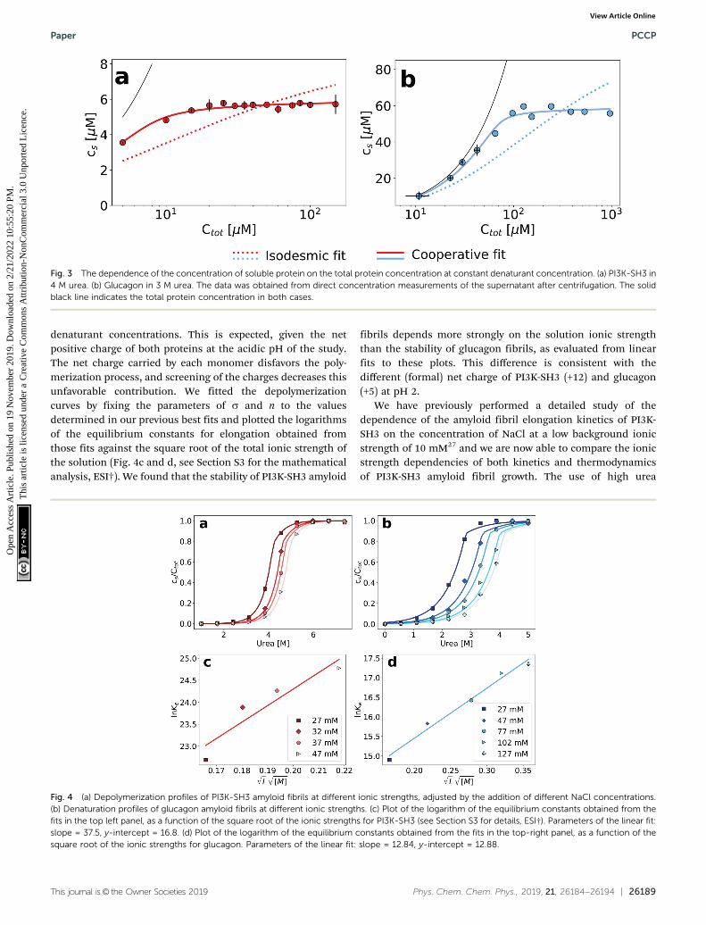
denaturant concentrations. This is expected, given the netpositive charge of both proteins at the acidic pH of the study.The net charge carried by each monomer disfavors the poly-merization process, and screening of the charges decreases thisunfavorable contribution. We fitted the depolymerizationcurves by fixing the parameters of s and n to the valuesdetermined in our previous best fits and plotted the logarithmsof the equilibrium constants for elongation obtained fromthose fits against the square root of the total ionic strength ofthe solution (Fig. 4c and d, see Section S3 for the mathematicalanalysis, ESI†). We found that the stability of PI3K-SH3 amyloid
fibrils depends more strongly on the solution ionic strengththan the stability of glucagon fibrils, as evaluated from linearfits to these plots. This difference is consistent with thedifferent (formal) net charge of PI3K-SH3 (+12) and glucagon(+5) at pH 2.
We have previously performed a detailed study of thedependence of the amyloid fibril elongation kinetics of PI3K-SH3 on the concentration of NaCl at a low background ionicstrength of 10 mM27 and we are now able to compare the ionicstrength dependencies of both kinetics and thermodynamicsof PI3K-SH3 amyloid fibril growth. The use of high urea
Fig. 3 The dependence of the concentration of soluble protein on the total protein concentration at constant denaturant concentration. (a) PI3K-SH3 in4 M urea. (b) Glucagon in 3 M urea. The data was obtained from direct concentration measurements of the supernatant after centrifugation. The solidblack line indicates the total protein concentration in both cases.
Fig. 4 (a) Depolymerization profiles of PI3K-SH3 amyloid fibrils at different ionic strengths, adjusted by the addition of different NaCl concentrations.(b) Denaturation profiles of glucagon amyloid fibrils at different ionic strengths. (c) Plot of the logarithm of the equilibrium constants obtained from thefits in the top left panel, as a function of the square root of the ionic strengths for PI3K-SH3 (see Section S3 for details, ESI†). Parameters of the linear fit:slope = 37.5, y-intercept = 16.8. (d) Plot of the logarithm of the equilibrium constants obtained from the fits in the top-right panel, as a function of thesquare root of the ionic strengths for glucagon. Parameters of the linear fit: slope = 12.84, y-intercept = 12.88.
Paper PCCP
Ope
n A
cces
s A
rtic
le. P
ublis
hed
on 1
9 N
ovem
ber
2019
. Dow
nloa
ded
on 2
/21/
2022
10:
55:2
0 PM
. T
his
artic
le is
lice
nsed
und
er a
Cre
ativ
e C
omm
ons
Attr
ibut
ion-
Non
Com
mer
cial
3.0
Unp
orte
d L
icen
ce.
View Article Online
26190 | Phys. Chem. Chem. Phys., 2019, 21, 26184--26194 This journal is© the Owner Societies 2019
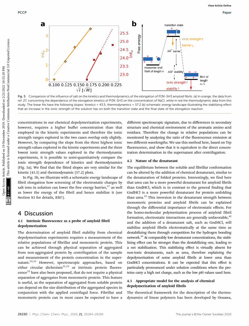
concentrations in our chemical depolymerization experiments,however, requires a higher buffer concentration than thatemployed in the kinetic experiments and therefore the ionicstrength ranges explored in the two cases overlap only slightly.However, by comparing the slope from the three highest ionicstrength values explored in the kinetic experiments and the threelowest ionic strength values explored in the thermodynamicexperiments, it is possible to semi-quantitatively compare theionic strength dependence of kinetics and thermodynamics(Fig. 5a). We find that the fitted slopes are very similar for thekinetic (43.5) and thermodynamic (57.2) plots.
In Fig. 5b, we illustrate with a schematic energy landscape offibril elongation how screening of the electrostatic charges bysalt ions in solution can lower the free energy barrier,27 as wellas lower the energy of the fibril and hence stabilize it (seeSection S3 for details, ESI†).
4 Discussion4.1 Intrinsic fluorescence as a probe of amyloid fibrildepolymerization
The determination of amyloid fibril stability from chemicaldepolymerization experiments requires a measurement of therelative populations of fibrillar and monomeric protein. Thiscan be achieved through physical separation of aggregatedfrom non-aggregated protein by centrifugation of the sampleand measurement of the protein concentration in the super-natant.22,33 However, spectroscopic approaches, based oneither circular dichroism21,32 or intrinsic protein fluores-cence32 have also been proposed, that do not require a physicalseparation of aggregates from monomeric protein. This featureis useful, as the separation of aggregated from soluble proteincan depend on the size distribution of the aggregated species inconjunction with the applied centrifugal force. Fibrillar andmonomeric protein can in most cases be expected to have a
different spectroscopic signature, due to differences in secondarystructure and chemical environment of the aromatic amino acidresidues. Therefore the change in relative populations can bemonitored by analyzing the ratio of the fluorescence emission attwo different wavelengths. We use this method here, based on Trpfluorescence, and show that it is equivalent to the direct concen-tration determination in the supernatant after centrifugation.
4.2 Nature of the denaturant
The equilibrium between the soluble and fibrillar conformationcan be altered by the addition of chemical denaturant, similar tothe denaturation of folded proteins. Interestingly, we find herethat urea can be a more powerful denaturant for amyloid fibrilsthan GndHCl, which is in contrast to the general finding thatGndHCl is a more powerful denaturant for protein unfoldingthan urea.35 This inversion in the denaturant strength betweenmonomeric proteins and amyloid fibrils can be explainedthrough the differential importance of electrostatic effects. Forthe homo-molecular polymerization process of amyloid fibrilformation, electrostatic interactions are generally unfavorable,42
and the addition of a denaturant salt, such as GndHCl, willstabilise amyloid fibrils electrostatically at the same time asdestabilizing them through competition for the hydrogen bondingnetwork.43 At comparably low denaturant concentrations, the stabi-lizing effect can be stronger than the destabilizing one, leading toa net stabilization. This stabilizing effect is virtually absent fornon-ionic denaturants, such as urea, therefore explaining thedepolymerization of some amyloid fibrils at lower urea thanGndHCl concentrations. It can be expected that this effect isparticularly pronounced under solution conditions where the pro-teins carry a high net charge, such as the low pH values used here.
4.3 Appropriate model for the analysis of chemicaldepolymerization of amyloid fibrils
The theoretical framework for the description of the thermo-dynamics of linear polymers has been developed by Oosawa,
Fig. 5 Comparison of the influence of salt on the kinetics and thermodynamics of the elongation of PI3K-SH3 amyloid fibrils. (a) In orange, the data fromref. 27, concerning the dependence of the elongation kinetics of PI3K-SH3 on the concentration of NaCl, while in red the thermodynamic data from thisstudy. The linear fits have the following slopes: kinetics = 43.5; thermodynamics = 57.2 (b) schematic energy landscape illustrating the stabilizing effectthat an increase in the ionic strength of the solution has on both the transition state and the final state of the elongation reaction.
PCCP Paper
Ope
n A
cces
s A
rtic
le. P
ublis
hed
on 1
9 N
ovem
ber
2019
. Dow
nloa
ded
on 2
/21/
2022
10:
55:2
0 PM
. T
his
artic
le is
lice
nsed
und
er a
Cre
ativ
e C
omm
ons
Attr
ibut
ion-
Non
Com
mer
cial
3.0
Unp
orte
d L
icen
ce.
View Article Online
This journal is© the Owner Societies 2019 Phys. Chem. Chem. Phys., 2019, 21, 26184--26194 | 26191
initially for actin polymerization. There it was found thatthe system was best described by so-called helical polymers,whereby each monomer interacts with several other monomersin the fibril, not only with the two next neighbors, as expectedfor a perfectly one-dimensional polymer. This structural featureleads to the fact that the initial nucleus is difficult to form, as amonomer will not be able to form the same number of favor-able interactions with the nucleus compared to when it addsonto a complete fibril. Amyloid fibril thermodynamics has sofar exclusively been analyzed with the simplest form of thelinear polymerization model,21,22,33 whereby a single equili-brium constant for monomer addition is postulated. As weshow in this work, individual chemical depolymerization curvescan be fitted very well with this simple model, but as soon asthe total concentration of the protein is varied, it becomesobvious in some cases that this model cannot accuratelydescribe the equilibrium behavior of amyloid fibrils. Inspiredby the field of supramolecular polymerization, we find that amodel that allows for a less favorable equilibrium constantof monomer attachment to species below a threshold sizen provides a consistent theoretical description that allows toquantitatively account for the data. Interestingly, we find thatthe best fits are achieved for small values of n (approximatelybetween 2 and 5) and for values of s that are one to two ordersof magnitude smaller than unity. We would like to stress herethat this model is likely to still represent an oversimplification,given the real complexity of amyloid fibril formation, wherebyin most cases different steps of monomer addition and struc-tural rearrangement44–46 lead to the formation of a minimalfibril. Nevertheless, it is probably the simplest extension to thebasic linear polymerization model and the fact that our datacan be well-fitted suggests that at least for the thermodynamicbehaviour of amyloid fibrils, a description in terms of aneffective equilibrium constant for the addition of monomersto small, pre-fibrillar species captures the essence of theprocess. It is interesting to note that the numerical values ofthe free energy of monomer addition and s together suggestthat also the addition of monomers to the smaller structures isfavorable, albeit less so than the addition of monomer to a fullyformed fibril. These results can be compared to the kinetics ofamyloid fibril nucleation, which is generally found to be veryslow and therefore the formation of the nucleus is viewed asbeing highly unfavorable. Indeed, the finding that in manyamyloid systems, fibrils are found that consist of thousands ofindividual monomers suggests that the rate of nucleation of afibril is at least 3–4 orders of magnitude slower than the rate ofits growth. These observations can be reconciled with ourpresent analysis by considering that for the kinetic behavior,the height of the free energy barriers, as well as the reactionorder of nucleation and growth processes, have to be taken intoaccount. A detailed analysis of these parameters in the case ofthe amyloid b peptide has recently revealed that the free energybarrier for primary nucleation is indeed several times higherthan that for fibril elongation.47 In our experiments, however,we probe the thermodynamic behavior of fibril formation.While the energy barrier for the formation of a dimer, trimer
or tetramer might be much higher than for monomer additionto a fibril,47 our results suggest that once such a small oligomeris formed, it can be similarly stable, per monomer, as a fullygrown fibril.
It is also interesting to note that the increasing number ofavailable high resolution structures of amyloid fibrils34,48,49
paints the consistent picture of a minimal fibril unit, consistingof between 2 and 8 monomers, which displays the full range ofinteractions of a fully grown fibril. It is plausible that structuressmaller than this minimal unit are thermodynamically some-what less stable than bigger structures, in contrast to classicalnucleation theory, where structures smaller than the criticalnucleus are considered unstable with respect to individualmonomers.
4.4 Electrostatic effects to probe the nature of the transitionstate for amyloid fibril elongation
Our experimental and conceptual framework allows us to probethe effect of changes in solution ionic strength on the thermo-dynamic stability of amyloid fibrils. Here we study two proteinsthat form fibrils at acidic pH conditions, where the individualprotein molecules carry a positive net charge that opposeshomomolecular polymerization.27,42,52,53 The electrostaticrepulsion of the individual monomers within the fibril can beexpected to be screened by salt ions in solution, similar to whathas been reported for the formation of surfactant micelles54 orvirus capsids.55 The use of urea as a denaturant allowed us toprobe this effect and we represent the resulting data by plottingthe logarithm of the equilibrium constant against the squareroot of the total ionic strength (Fig. 4a). The slope of such a plotdepends on the effective charge of the molecular interaction inquestion (see Section S3, ESI† for a detailed discussion of theunderlying model). In agreement with the higher formal netcharge of PI3K-SH3 compared to glucagon at acidic pH, we findthat the stability of PI3K-SH3 amyloid fibrils is more stronglyaffected by solution ionic strength than that of glucagon fibrils.It is insightful to compare the dependence of both PI3K-SH3amyloid formation thermodynamics and kinetics27 on NaClconcentration. For technical reasons (see above), the range ofionic strengths exploited in both studies overlaps only slightly,but if we extrapolate the respective slopes we are neverthelessable to compare kinetic and thermodynamic ionic strengthdependencies. We find that the slopes of the kinetic andthermodynamic ionic strength dependency plots are very similar(Fig. 5a), with the thermodynamic slope being slightly higher.This result suggests that the monomer adopting the transitionstate for fibril elongation experiences very similar, albeit slightlyless, electrostatic repulsion compared to the monomer fullyincorporated into the fibril, thereby implying a high degree ofsimilarity between the transition state and the final state of thefibril elongation reaction. Under conditions of acidic pH, whereeach monomer carries only positive charges and therefore asubstantial net charge, electrostatic repulsion is probably mainlydefined by the principal reaction coordinate,50 the center of massdistance between the monomer and the fibril end. Therefore, ourresults suggest that the transition state of PI3K-SH3 amyloid fibril
Paper PCCP
Ope
n A
cces
s A
rtic
le. P
ublis
hed
on 1
9 N
ovem
ber
2019
. Dow
nloa
ded
on 2
/21/
2022
10:
55:2
0 PM
. T
his
artic
le is
lice
nsed
und
er a
Cre
ativ
e C
omm
ons
Attr
ibut
ion-
Non
Com
mer
cial
3.0
Unp
orte
d L
icen
ce.
View Article Online

26192 | Phys. Chem. Chem. Phys., 2019, 21, 26184--26194 This journal is© the Owner Societies 2019
elongation corresponds to a monomer in very close proximity ofthe fibril end, only marginally removed from its final positionwhen incorporated into the fibril. This conclusion is in closeagreement with the finding that the hydrophobic effect plays amajor role in stabilizing the transition state of fibril elongation,as revealed through a strongly favorable entropy of activation.47,56
The picture that therefore emerges from this analysis is that therate-limiting step of amyloid fibril elongation consists of astructural rearrangement of the monomer while being in closecontact with the fibril end. Therefore, the defining energy barrierfor fibril elongation is of an inter-molecular, rather than intra-molecular nature (see Fig. 6 for a comparison of these twodistinct scenarios). This result is an important contribution tothe ongoing discussion about the intra- vs. intermolecular natureof the protein misfolding events that lead to amyloid fibrilformation. It has been proposed for several amyloid systems,such as poly-glutamine57 and tau58 that the crucial and rate-limiting event along the pathway of amyloid fibril formation is apurely intra-molecular misfolding event to form an ’aggregationcompetent state’ that can add onto a fibril without any significantbarrier crossing. Our results for PI3K-SH3, on the other hand,are more compatible with the picture whereby the misfoldingtransition is a highly cooperative event between monomer andfibril end.
5 Conclusions
In summary, we have been able to show for the first time thatthe thermodynamics of amyloid fibril formation is bestdescribed by cooperative polymerization rather than by simpleisodesmic linear polymerization. This result is in excellentagreement with mechanistic insight into amyloid fibril for-mation, as well as with the emerging high resolution structuralinformation on amyloid fibrils. Furthermore, we have been ableto accurately probe the role of electrostatic effects in amyloid
fibril stability. In combination with previously available data onthe influence of solution ionic strength on the kinetics ofamyloid fibril growth, we are able to reveal the product-likenature of the transition state-ensemble for amyloid fibril elon-gation by PI3K-SH3 monomers.
Conflicts of interest
There are no conflicts to declare.
Acknowledgements
AKB and NV thank the Deutsche Forschungsgemeinschaft(DFG) for financial support. AKB thanks the Novo NordiskFoundation for support through a Novo Nordisk Foundationprofessorship. The authors thank Sabine Metzger for help withthe mass spectrometry, Florian Platten for helpful discussionsand Novo Nordisk for a kind gift of glucagon.
References
1 F. Oosawa, S. Asakura, K. Hotta, N. Imai and T. Ooi, J. Polym.Sci., 1959, 37, 323–336.
2 G. G. Borisy and J. B. Olmsted, Science, 1972, 177,1196–1197.
3 J. Hofrichter, P. D. Ross and W. A. Eaton, Proc. Natl. Acad.Sci. U. S. A., 1974, 71, 4864–4868.
4 C. M. Dobson, Nature, 2003, 426, 884–890.5 F. Oosawa, Thermodynamics of the Polymerization of Protein,
Academic Press Inc, 1975.6 F. Oosawa and M. Kasai, J. Mol. Biol., 1962, 4, 10–21.7 T. Oda, M. Iwasa, T. Aihara, Y. Maeda and A. Narita, Nature,
2009, 457, 441–445.8 B. Magdoff-Fairchild and C. C. Chiu, Proc. Natl. Acad. Sci.
U. S. A., 1979, 76, 223–226.
Fig. 6 Comparison of two distinct scenarios of the transition state of amyloid fibril growth. The free energy barriers that define the kinetics of fibrilgrowth are marked in red in both cases; however, we plot here only the electrostatic component of the free energy as a function of the center of massseparation between the approaching monomer and the fibril end. (a) The transition state of fibril elongation corresponds to an isolated misfolding eventof the free monomer (1), which is electrostatically unfavorable followed by a diffusive search of the fibril end (2). The electrostatic signature of the lattercannot be elucidated from kinetic experiments, because only the highest free energy barrier is probed.50,51 The final state (monomer incorporated) iselectrostatically less favorable than the initial state and the transition state, but has the lowest free energy. (b) The transition state of fibril elongationcorresponds to a situation after a diffusional search (1), where the monomer is in close contact with the fibril end. The misfolding reaction is aided by thepresence of the fibril end (2, templating effect). The transition state and the final state are similarly electrostatically unfavorable, because these two stateshave a very similar center-of-mass distance that dominates the electrostatic interactions. Therefore, we argue that our data on the ionic strengthdependence of both kinetics and thermodynamics of fibril elongation is best compatible with a late, product-like transition state (scenario b).
PCCP Paper
Ope
n A
cces
s A
rtic
le. P
ublis
hed
on 1
9 N
ovem
ber
2019
. Dow
nloa
ded
on 2
/21/
2022
10:
55:2
0 PM
. T
his
artic
le is
lice
nsed
und
er a
Cre
ativ
e C
omm
ons
Attr
ibut
ion-
Non
Com
mer
cial
3.0
Unp
orte
d L
icen
ce.
View Article Online

This journal is© the Owner Societies 2019 Phys. Chem. Chem. Phys., 2019, 21, 26184--26194 | 26193
9 M. Sunde, L. C. Serpell, M. Bartlam, P. E. Fraser, M. B. Pepysand C. C. Blake, J. Mol. Biol., 1997, 273, 729–739.
10 J. Greenwald and R. Riek, Structure, 2010, 18, 1244–1260.11 S. K. Maji, M. H. Perrin, M. R. Sawaya, S. Jessberger,
K. Vadodaria, R. A. Rissman, P. S. Singru, K. P. R. Nilsson,R. Simon, D. Schubert, D. Eisenberg, J. Rivier, P. Sawchenko,W. Vale and R. Riek, Science, 2009, 325, 328–332.
12 K. A. Conway, J. D. Harper and P. T. Lansbury, Biochemistry,2000, 39, 2552–2563.
13 W. S. Gosal, A. H. Clark and S. B. Ross-Murphy, Biomacro-molecules, 2004, 5, 2408–2419.
14 A. Lomakin, D. B. Teplow, D. A. Kirschner and G. B.Benedek, Proc. Natl. Acad. Sci. U. S. A., 1997, 94, 7942–7947.
15 T. P. J. Knowles, C. A. Waudby, G. L. Devlin, S. I. A. Cohen,A. Aguzzi, M. Vendruscolo, E. M. Terentjev, M. E. Wellandand C. M. Dobson, Science, 2009, 326, 1533–1537.
16 S. I. A. Cohen, M. Vendruscolo, M. E. Welland, C. M.Dobson, E. M. Terentjev and T. P. J. Knowles, J. Chem. Phys.,2011, 135, 065105.
17 A. K. Buell, C. Galvagnion, R. Gaspar, E. Sparr, M. Vendruscolo,T. P. J. Knowles, S. Linse and C. M. Dobson, Proc. Natl. Acad.Sci. U. S. A., 2014, 111(21), 7671–7676.
18 E. P. O’Brien, Y. Okamoto, J. E. Straub, B. R. Brooks andD. Thirumalai, J. Phys. Chem. B, 2009, 113, 14421–14430.
19 F. Bard, C. Cannon, R. Barbour, R. Burke, D. Games,H. Grajeda, T. Guido, K. Hu, J. Huang, K. Johnson-Wood,K. Khan, D. Kholodenko, M. Lee, I. Lieberburg, R. Motter,M. Nguyen, F. Soriano, N. Vasquez, K. Weiss, B. Welch,P. Seubert, D. Schenk and T. Yednock, Nat. Med., 2000, 6,916–919.
20 X. Gao, M. Carroni, C. Nussbaum-Krammer, A. Mogk,N. B. Nillegoda, A. Szlachcic, D. L. Guilbride, H. R. Saibil,M. P. Mayer and B. Bukau, Mol. Cell, 2015, 59, 781–793.
21 T. Narimoto, K. Sakurai, A. Okamoto, E. Chatani,M. Hoshino, K. Hasegawa, H. Naiki and Y. Goto, FEBS Lett.,2004, 576, 313–319.
22 A. J. Baldwin, T. P. J. Knowles, G. G. Tartaglia, A. W.Fitzpatrick, G. L. Devlin, S. L. Shammas, C. A. Waudby,M. F. Mossuto, S. Meehan, S. L. Gras, J. Christodoulou,S. J. Anthony-Cahill, P. D. Barker, M. Vendruscolo andC. M. Dobson, J. Am. Chem. Soc., 2011, 133, 14160–14163.
23 E. Hellstrand, B. Boland, D. M. Walsh and S. Linse, ACSChem. Neurosc., 2010, 1, 13–18.
24 C. N. P. J. K. Myers and J. M. Scholtz, Protein Sci., 1995, 4,2138–2148.
25 D. Zhao and J. S. Moore, Org. Biomol. Chem., 2003, 1,3471–3491.
26 M. M. J. Smulders, M. M. L. Nieuwenhuizen, T. F. A. deGreef, P. van der Schoot, A. P. H. J. Schenning andE. W. Meijer, Chem. – Eur. J., 2010, 16, 362–367.
27 A. K. Buell, P. Hung, X. Salvatella, M. E. Welland, C. M.Dobson and T. P. J. Knowles, Biophys. J., 2013, 104, 1116–1126.
28 J. Zurdo, J. I. Guijarro, J. L. Jimenez, H. R. Saibil andC. M. Dobson, J. Mol. Biol., 2001, 311, 325–340.
29 G. H. Beaven, W. B. Gratzer and H. G. Davies, Eur.J. Biochem., 1969, 11, 37–42.
30 J. S. Pedersen, J. Diabetes Sci. Technol., 2010, 4, 1357–1367.31 J. I. n. Guijarro, M. Sunde, J. A. Jones, I. D. Campbell and
C. M. Dobson, Proc. Natl. Acad. Sci. U. S. A., 1998, 95,4224–4228.
32 J. S. Pedersen, D. Dikov, J. L. Flink, H. A. Hjuler,G. Christiansen and D. E. Otzen, J. Mol. Biol., 2006, 355,501–523.
33 R. Porcari, C. Proukakis, C. A. Waudby, B. Bolognesi,P. P. Mangione, J. F. S. Paton, S. Mullin, L. D. Cabrita,A. Penco, A. Relini, G. Verona, M. Vendruscolo, M. Stoppini,G. G. Tartaglia, C. Camilloni, J. Christodoulou, A. H. V.Schapira and V. Bellotti, J. Biol. Chem., 2015, 290, 2395–2404.
34 C. Roeder, N. Vettore, L. N. Mangels, L. Gremer, R. B.Ravelli, D. Willbold, W. Hoyer, A. K. Buell and G. F.Schroeder, Nat. Commun., 2019, 10, 3754.
35 J. S. Martin, J. Parker and A. R. Clarke, J. Mol. Biol., 1995,253, 771–786.
36 L. Kollipara and R. P. Zahedi, Proteomics, 2013, 13, 941–944.37 A. Fersht, Structure and Mechanism in Protein Science, W.H.
Freeman, New York, 1999.38 A. K. Buell, Int. Rev. Cell Mol. Biol., 2017, 329, 187–226.39 P. A. Korevaar, C. Schaefer, T. F. A. de Greef and
E. W. Meijer, J. Am. Chem. Soc., 2012, 134, 13482–13491.40 P. A. Korevaar, S. J. George, A. J. Markvoort, M. M. Smulders,
P. A. Hilbers, A. P. Schenning, T. F. De Greef and E. Meijer,Nature, 2012, 481, 492.
41 C. Boesch, A. Bundi, M. Oppliger and K. Wuthrich, Eur.J. Biochem., 1978, 91, 209–214.
42 S. L. Shammas, T. P. J. Knowles, A. J. Baldwin, C. E.Macphee, M. E. Welland, C. M. Dobson and G. L. Devlin,Biophys. J., 2011, 100, 2783–2791.
43 E. P. O’Brien, R. I. Dima, B. Brooks and D. Thirumalai, J. Am.Chem. Soc., 2007, 129, 7346–7353.
44 T. R. Serio, A. G. Cashikar, A. S. Kowal, G. J. Sawicki, J. J.Moslehi, L. Serpell, M. F. Arnsdorf and S. L. Lindquist,Science, 2000, 289, 1317–1321.
45 N. Cremades, S. I. A. Cohen, E. Deas, A. Y. Abramov, A. Y.Chen, A. Orte, M. Sandal, R. W. Clarke, P. Dunne, F. A.Aprile, C. W. Bertoncini, N. W. Wood, T. P. J. Knowles,C. M. Dobson and D. Klenerman, Cell, 2012, 149, 1048–1059.
46 G. A. Garcia, S. I. A. Cohen, C. M. Dobson and T. P. J.Knowles, Phys. Rev. E: Stat., Nonlinear, Soft Matter Phys.,2014, 89, 032712.
47 S. I. Cohen, R. Cukalevski, T. C. Michaels, A. Saric,M. Tornquist, M. Vendruscolo, C. M. Dobson, A. K. Buell,T. P. Knowles and S. Linse, Nat. Chem., 2018, 10, 523.
48 L. Gremer, D. Scholzel, C. Schenk, E. Reinartz, J. Labahn,R. B. G. Ravelli, M. Tusche, C. Lopez-Iglesias, W. Hoyer,H. Heise, D. Willbold and G. F. Schroder, Science, 2017, 358,116–119.
49 A. W. Fitzpatrick, B. Falcon, S. He, A. G. Murzin,G. Murshudov, H. J. Garringer, R. A. Crowther, B. Ghetti,M. Goedert and S. H. Scheres, Nature, 2017, 547, 185.
50 A. K. Buell, J. R. Blundell, C. M. Dobson, M. E. Welland,E. M. Terentjev and T. P. J. Knowles, Phys. Rev. Lett., 2010,104, 228101.
Paper PCCP
Ope
n A
cces
s A
rtic
le. P
ublis
hed
on 1
9 N
ovem
ber
2019
. Dow
nloa
ded
on 2
/21/
2022
10:
55:2
0 PM
. T
his
artic
le is
lice
nsed
und
er a
Cre
ativ
e C
omm
ons
Attr
ibut
ion-
Non
Com
mer
cial
3.0
Unp
orte
d L
icen
ce.
View Article Online

26194 | Phys. Chem. Chem. Phys., 2019, 21, 26184--26194 This journal is© the Owner Societies 2019
51 T. C. Michaels, L. X. Liu, S. Curk, P. G. Bolhuis, A. Saric andT. P. Knowles, Mol. Phys., 2018, 116, 3055–3065.
52 F. Massi, D. Klimov, D. Thirumalai and J. E. Straub, ProteinSci., 2002, 11, 1639–1647.
53 D. K. Klimov and D. Thirumalai, Structure, 2003, 11, 295–307.54 G. Gunnarsson, B. Joensson and H. Wennerstroem, J. Phys.
Chem., 1980, 84, 3114–3121.55 W. K. Kegel and P. van der Schoot, Biophys. J., 2004, 86,
3905–3913.
56 A. K. Buell, A. Dhulesia, D. A. White, T. P. J. Knowles,C. M. Dobson and M. E. Welland, Angew. Chem., Int. Ed.,2012, 51, 5247–5251.
57 K. Kar, M. Jayaraman, B. Sahoo, R. Kodali and R. Wetzel,Nat. Struct. Mol. Biol., 2011, 18, 328–336.
58 H. Mirbaha, D. Chen, O. A. Morazova, K. M. Ruff,A. M. Sharma, X. Liu, M. Goodarzi, R. V. Pappu, D. W.Colby, H. Mirzaei, L. A. Joachimiak and M. I. Diamond,eLife, 2018, e36584.
PCCP Paper
Ope
n A
cces
s A
rtic
le. P
ublis
hed
on 1
9 N
ovem
ber
2019
. Dow
nloa
ded
on 2
/21/
2022
10:
55:2
0 PM
. T
his
artic
le is
lice
nsed
und
er a
Cre
ativ
e C
omm
ons
Attr
ibut
ion-
Non
Com
mer
cial
3.0
Unp
orte
d L
icen
ce.
View Article Online