aggregate geometry in amyloid fibril nucleation. irbäck ... · aggregate geometry in amyloid...
TRANSCRIPT
LUND UNIVERSITY
PO Box 117221 00 Lund+46 46-222 00 00
Aggregate geometry in amyloid fibril nucleation.
Irbäck, Anders; Jonsson, Sigurdur; Linnemann, Niels; Linse, Björn; Wallin, Stefan
Published in:Physical Review Letters
DOI:10.1103/PhysRevLett.110.058101
Published: 2013-01-01
Link to publication
Citation for published version (APA):Irbäck, A., Jonsson, S., Linnemann, N., Linse, B., & Wallin, S. (2013). Aggregate geometry in amyloid fibrilnucleation. Physical Review Letters, 110(5), [058101]. DOI: 10.1103/PhysRevLett.110.058101
General rightsCopyright and moral rights for the publications made accessible in the public portal are retained by the authorsand/or other copyright owners and it is a condition of accessing publications that users recognise and abide by thelegal requirements associated with these rights.
• Users may download and print one copy of any publication from the public portal for the purpose of privatestudy or research. • You may not further distribute the material or use it for any profit-making activity or commercial gain • You may freely distribute the URL identifying the publication in the public portalTake down policyIf you believe that this document breaches copyright please contact us providing details, and we will removeaccess to the work immediately and investigate your claim.
Download date: 19. Jun. 2018
Aggregate Geometry in Amyloid Fibril Nucleation
Anders Irback, Sigurður Æ. Jonsson, Niels Linnemann, Bjorn Linse, and Stefan Wallin
Computational Biology and Biological Physics, Department of Astronomy and Theoretical Physics, Lund University,Solvegatan 14A, SE-223 62 Lund, Sweden
(Received 1 September 2012; published 28 January 2013)
We present and study a minimal structure-based model for the self-assembly of peptides into ordered
�-sheet-rich fibrils. The peptides are represented by unit-length sticks on a cubic lattice and interact by
hydrogen bonding and hydrophobicity forces. Using Monte Carlo simulations with >105 peptides, we
show that fibril formation occurs with sigmoidal kinetics in the model. To determine the mechanism of
fibril nucleation, we compute the joint distribution in length and width of the aggregates at equilibrium,
using an efficient cluster move and flat-histogram techniques. This analysis, based on simulations with
256 peptides in which aggregates form and dissolve reversibly, shows that the main free-energy barriers
that a nascent fibril has to overcome are associated with changes in width.
DOI: 10.1103/PhysRevLett.110.058101 PACS numbers: 87.14.em, 87.15.A�
Many proteins and peptides share the ability to self-assemble into amyloid fibrils, aggregates with a cross-�structure and remarkable mechanical properties, that areassociated with a range of disorders as well as with func-tional roles [1,2]. The formation of amyloid fibrils, usuallymonitored by thioflavin T (ThT) fluorescence, is known tooccur with reproducible sigmoidal kinetics [3], indicatinga nucleation-dependent process. A powerful method forinterpreting the experimental kinetic profiles is by meansof rate equations [4]. This approach can reveal some gen-eral properties of intermediate species participating in thegrowth process. It has proven useful for some related self-assembly phenomena as well, such as hemoglobin S aggre-gation [5] and microtubule assembly [6]. Another methodto elucidate the mechanisms of amyloid formation is byphase equilibria analysis [7].
By coarse-grained structure-based approaches [8],additional insights have been gained into the nucleationof amyloid fibrils [9–14]. Fibrillation pathways involve,however, a host of different aggregated species of widelyvarying size, and studying the competition among thesespecies without restrictive assumptions represents a chal-lenge even in coarse-grained models.
In this Letter, we introduce a minimal structure-basedmodel that describes amyloid fibril formation in terms ofphysically inspired peptide-peptide interactions and yetallows for representative sampling of the model statespace for relatively large systems. Using flat-histogrammethods [15,16] and an efficient cluster move resem-bling the Swendsen-Wang algorithm for spin systems[17], we determine equilibrium distributions in sizeand shape of the aggregated structures, in order to elu-cidate the free-energy landscape that a nascent fibril hasto navigate.
We consider N identical peptides, represented by unit-length sticks on a periodic cubic lattice with dimensionsL3. We assume that the internal dynamics of a peptide are
fast compared to the time scales for fibril formation, andtherefore can be averaged out.Each peptide i is centered at a lattice site ri, and two
peptides cannot simultaneously occupy the same site.
Associated with each peptide are two unit vectors bi
and pi that can point in any of the six lattice directions
[Fig. 1(a)]; bi represents the N-to-C backbone orientation,whereas �pi are the directions in which hydrogen bonds
can form. The vectors bi and pi are perpendicular, leavinga total of 24 possible orientations of a peptide. The vectors
si ¼ bi � pi and �si represent side-chain directions. Theþsi and �si sides of a peptide are assumed to have differ-ent interaction properties and are referred to as hydro-phobic and polar, respectively.The energy function describing the interactions between
the peptides is assumed pairwise additive, E ¼ Pi<j�ij,
where �ij � 0. The pair potential �ij is nonzero only if
(i) peptides i and j are nearest neighbors on the lattice, and
(ii) bi and bj are perpendicular to rij ¼ rj � ri and aligned
either parallel or antiparallel to each other. When theseconditions are met, we set �ij ¼ �1 except in the three
cases illustrated in Fig. 1. The first two cases correspond toparallel [Fig. 1(b)] and antiparallel [Fig. 1(c)] � structure,respectively, and the third [Fig. 1(d)] to hydrophobic side-chain attraction. The corresponding interaction energiesare given by
�ij ¼
8>><
>>:
�ð1þ apÞ parallel� structure
�ð1þ aapÞ antiparallel� structure
�ð1þ bÞ hydrophobic attraction:
(1)
The hydrophobic attraction is included because of evi-dence suggesting that a pairwise (steric zipper) �-sheetorganization is a common architecture for the core ofamyloid fibrils [18]. The b parameter must not be too large,in order for extended � sheets to form. Because the �sheets often are parallel in amyloid fibrils, we take
PRL 110, 058101 (2013) P HY S I CA L R EV I EW LE T T E R Sweek ending
1 FEBRUARY 2013
0031-9007=13=110(5)=058101(4) 058101-1 � 2013 American Physical Society
ap > aap, but the model can also be studied for aap > ap.
In what follows, for simplicity, we stick to a singleparameter set, namely ap ¼ 5, aap ¼ 3, and b ¼ 1. With
this choice, the parallel �-strand organization dominates,but the suppression of antiparallel strand pairs is not pro-hibitively strong.
We simulate the thermodynamics of this model usingsingle-peptide as well as cluster moves. A cluster updatemakes it possible for aggregates to move without havingto be first dissolved and then reassembled. To be able toalso split and merge aggregates, we follow a stochasticSwendsen-Wang–type cluster construction procedure [17].The construction is recursive and begins by picking a randomfirst cluster member, i. Then, all peptides j interacting withpeptide i (�ij < 0) are identified and added to the cluster with
probability pij ¼ 1� e��ij , where � ¼ 1=kBT is inverse
temperature. This step is iterated until no cluster memberhas any unchecked interaction partner. Finally, the resultingcluster is subject to a trial rigid-body translation or rotation,drawn from a symmetric distribution, which is acceptedwhenever it does not cause any steric clashes. It can beverified that this algorithm fulfills detailed balance withrespect to the canonical ensemble p� / e��E� .
To further enhance the sampling, we employgeneralized-ensemble methods [15,16], along withreweighting techniques [19]. After estimating the densityof states gðEÞ by the Wang-Landau method [16], we simu-late the ensemble p� / 1=gðE�Þ [15], where the distribu-tion of E is flat. This approach was recently used foratomic-level aggregation simulations [20] and is usefulfor the present system as well, which displays phase coex-istence at the fibrillation temperature, Tm (see below). Oursimulations sample a limited energy range, Emin < E � 0.The cutoff Emin is needed to prevent the formation ofartificial cyclic aggregates, which otherwise may occurdue to the periodic boundary conditions, but is sufficientlylow to permit studies of temperatures in the fibrillar phase.
The above cluster update can be adapted for thegeneralized-ensemble simulations by adding an accept orreject step, with acceptance probability paccð� ! �0Þ ¼min½1; gðE�Þe��E�=gðE�0 Þe��E�0 �. Here, � changes itsmeaning to become a tunable algorithm parameter. Wedid not fine-tune �, but expect the optimal � to be in theneighborhood of �m ¼ 1=kBTm, as supported by prelimi-nary runs.Using these methods, we studied the thermodynamics of
the model for several different system sizes. Here, we focuson the results obtained for N ¼ 256 and L ¼ 64, corre-sponding to a peptide concentration of � � 10�3 per unitvolume. This system size would have been very time-consuming to study with standard Monte Carlo methods.In our simulations, two distinct major phases occur: a
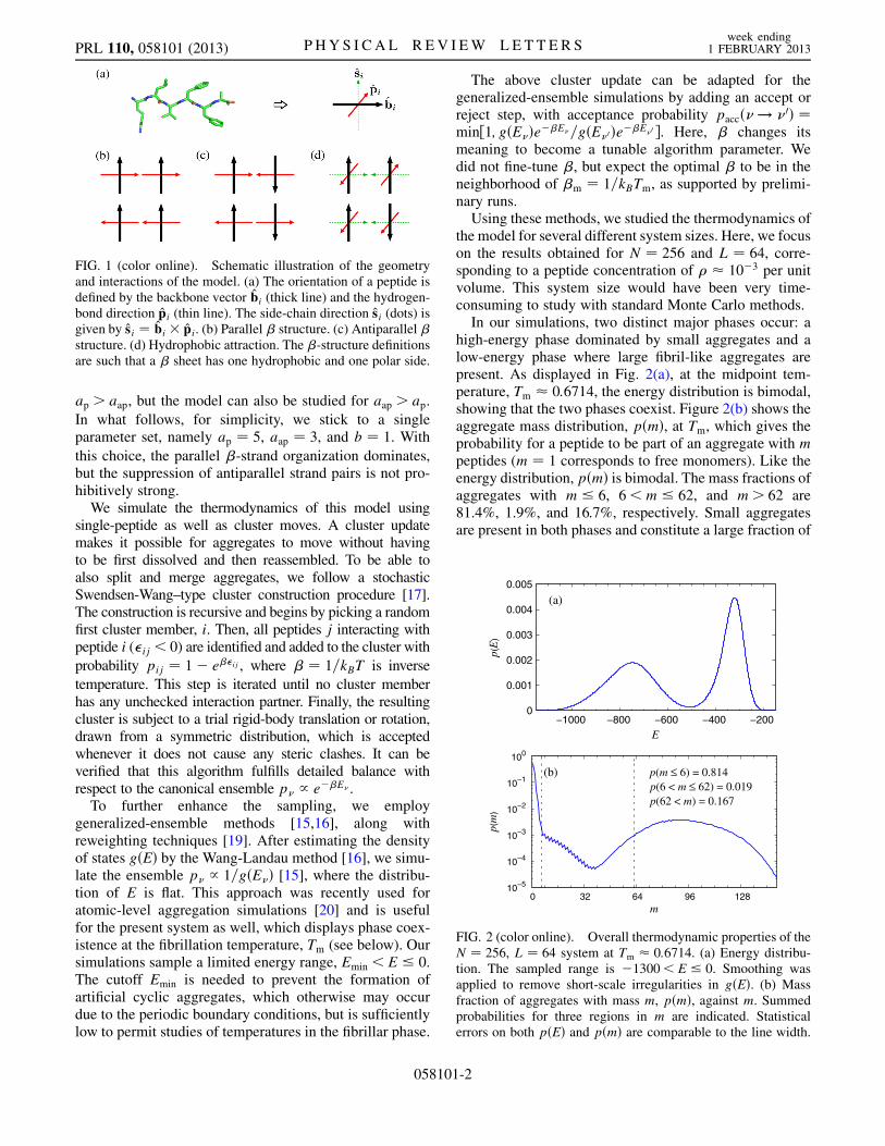
high-energy phase dominated by small aggregates and alow-energy phase where large fibril-like aggregates arepresent. As displayed in Fig. 2(a), at the midpoint tem-perature, Tm � 0:6714, the energy distribution is bimodal,showing that the two phases coexist. Figure 2(b) shows theaggregate mass distribution, pðmÞ, at Tm, which gives theprobability for a peptide to be part of an aggregate with mpeptides (m ¼ 1 corresponds to free monomers). Like theenergy distribution, pðmÞ is bimodal. The mass fractions ofaggregates with m � 6, 6<m � 62, and m> 62 are81.4%, 1.9%, and 16.7%, respectively. Small aggregatesare present in both phases and constitute a large fraction of
FIG. 1 (color online). Schematic illustration of the geometryand interactions of the model. (a) The orientation of a peptide isdefined by the backbone vector bi (thick line) and the hydrogen-bond direction pi (thin line). The side-chain direction si (dots) isgiven by si ¼ bi � pi. (b) Parallel � structure. (c) Antiparallel �structure. (d) Hydrophobic attraction. The �-structure definitionsare such that a � sheet has one hydrophobic and one polar side.
0
0.001
0.002
0.003
0.004
0.005
−1000 −800 −600 −400 −200
p(E
)
E
(a)
10−5
10−4
10−3
10−2
10−1
100
0 32 64 96 128
p(m
)
m
(b) p(m ≤ 6) = 0.814p(6 < m ≤ 62) = 0.019p(62 < m) = 0.167
FIG. 2 (color online). Overall thermodynamic properties of theN ¼ 256, L ¼ 64 system at Tm � 0:6714. (a) Energy distribu-tion. The sampled range is �1300< E � 0. Smoothing wasapplied to remove short-scale irregularities in gðEÞ. (b) Massfraction of aggregates with mass m, pðmÞ, against m. Summedprobabilities for three regions in m are indicated. Statisticalerrors on both pðEÞ and pðmÞ are comparable to the line width.
PRL 110, 058101 (2013) P HY S I CA L R EV I EW LE T T E R Sweek ending
1 FEBRUARY 2013
058101-2
the total mass at Tm (see also Fig. S1 in the SupplementalMaterial [21]).
At first glance, the bimodality of pðmÞ may seem toindicate that fibril nucleation occurs when a critical mass isreached. However, this picture is geometrically incom-plete, because the species involved are neither strictlyone dimensional nor sharing one common shape, such asspherical. A simple but useful way to extend the analysisis via the inertia tensor. As measures of the length andwidth of an aggregate, we define l ¼ ffiffiffiffiffiffiffiffiffiffiffiffiffiffiffiffiffiffiffiffi
12�1 þ 1p
and w ¼ffiffiffiffiffiffiffiffiffiffiffiffiffiffiffiffiffiffiffiffi12�2 þ 1
p, where �1 � �2 are eigenvalues of the inertia
tensor. In our model, there is no interaction between lon-gitudinally aligned peptides to support growth in a thirddimension. With these definitions, for a rectangular aggre-gate, l and w are the numbers of peptide layers in the twodirections.
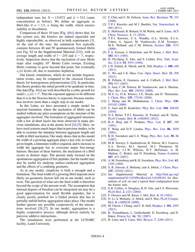
Figure 3 shows the probability pðl; wÞ for a peptide to bepart of an aggregate with length l and width w, at Tm.Consistent with Fig. 2(b), pðl; wÞ is highest for aggregateswith l small and w � 1. Among larger aggregates, a clearpreference can be seen for even over odd values of w,reflecting a pairwise �-sheet organization, although aggre-gates with six or more layers are severely constrained byfinite-size effects. A second trend is that single-layeraggregates are shorter than two-layer ones, which in turnare shorter than those with four layers. We expect thistrend to persist beyond the four-layer level if the systemis sufficiently large. These overall features of pðl; wÞ arelikely to be quite robust, although the locations of thedifferent maxima depend on both T and �.
The shape of pðl; wÞ has implications for how fibrilsnucleate and grow in the model. It suggests that the mainfree-energy barriers faced by a growing aggregate areassociated with changes in width, and it must increase inwidth to be able to grow.
Having examined the thermodynamics of the model,we now turn to the aggregation kinetics, studied usingconstant-temperature Monte Carlo dynamics. Because ofevidence that amyloid growth occurs by monomer addition[22], here we use single-peptide moves only. The simula-tions start from random initial conditions and the tempera-ture is T ¼ 0:66.
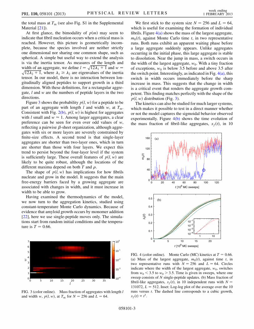
We first stick to the system size N ¼ 256 and L ¼ 64,which is useful for examining the formation of individualfibrils. Figure 4(a) shows the mass of the largest aggregate,m0ðtÞ, against Monte Carlo time t, in two representativeruns. Both runs exhibit an apparent waiting phase beforea large aggregate suddenly appears. Unlike aggregatesoccurring in the initial phase, this large aggregate is stableto dissolution. Near the jump in mass, a switch occurs inthe width of the largest aggregate, w0. With a tiny fractionof exceptions, w0 is below 3.5 before and above 3.5 afterthe switch point. Interestingly, as indicated in Fig. 4(a), thisswitch in width occurs immediately before the sharpincrease in mass. This suggests that the change in widthis a critical event that renders the aggregate growth com-petent. This finding matches perfectly with the shape of thepðl; wÞ distribution (Fig. 3).The kinetics can also be studied for much larger systems,
which makes it possible to test in a direct manner whetheror not the model captures the sigmoidal behavior observedexperimentally. Figure 4(b) shows the time evolution ofthe mass fraction of fibril-like aggregates, xfðtÞ, in 10
FIG. 3 (color online). Mass fraction of aggregates with length land width w, pðl; wÞ, at Tm for N ¼ 256 and L ¼ 64.
0
40
80
120
160
0 20 40 60 80 100 120
m0(
t )
t [106 MC sweeps]
(a)
0
0.1
0.2
0.3
0.4
0.5
0 5 10 15 20
x f(t)
t [106 MC sweeps]
(b)
10−410−310−210−1
1 10
FIG. 4 (color online). Monte Carlo (MC) kinetics at T ¼ 0:66.(a) Mass of the largest aggregate, m0ðtÞ, against time t, intwo representative runs with N ¼ 256 and L ¼ 64. Circlesindicate where the width of the largest aggregate, w0, switchesfrom w0 < 3:5 to w0 > 3:5. Time is given in sweeps, where onesweep consists of N single-peptide updates. (b) Mass fraction offibril-like aggregates, xfðtÞ, in 10 independent runs with N ¼131072, L ¼ 512. Inset: Log-log plot of the average over the 10runs versus t. The dashed line corresponds to a cubic growth,xfðtÞ / t3.
PRL 110, 058101 (2013) P HY S I CA L R EV I EW LE T T E R Sweek ending
1 FEBRUARY 2013
058101-3
independent runs for N ¼ 131072 and L ¼ 512 (sameconcentration as before). We define an aggregate asfibril-like if w> 3:5, w being the width, which ensuresstability to dissolution.
Comparison of these 10 runs [Fig. 4(b)] shows that, forthis system size, the kinetics are indeed sigmoidal andhighly reproducible, as observed in bulk experiments [3].At the end of the runs, the simulation box typicallycontains between 40 and 50 spontaneously formed fibrils(see Fig. S2 in the Supplemental Material [21]), with anaverage length and width of l� 210 and w� 7, respec-tively. Inspection shows that the nucleation of new fibrilsstops after roughly 107 Monte Carlo sweeps. Existingfibrils continue to grow beyond that point, but eventuallyxfðtÞ levels off, due to monomer depletion.
Our kinetic simulations, which do not include fragmen-tation events, may be compared to the classical Oosawatheory for homogeneous polymerization [23]. In particular,this theory predicts the initial growth to be quadratic in time.Our data [Fig. 4(b)] are well described by a cubic growth forsmall t, xfðtÞ / t3. That the exponent appears to be different
than it is in the Oosowa theory is expected, because nuclea-tion involves more than a single step in our model.
In this Letter, we have presented a simple model foramyloid formation, where the nucleation of fibrils can bestudied without any prior assumptions on the structure of theaggregates involved. The formation of aggregated structureswith a few �-sheet layers has been observed in many pre-vious simulations, also at the atomic level [24,25]. Here, wehave used systemsmuch larger than in previous studies, to beable to examine the interplay between aggregate length andwidth in fibril nucleation. Our study shows that in this modelthe width of a growing aggregate plays a key role; to reach agiven length, a minimumwidth is required, and to increase inwidth the aggregate has to overcome major free-energybarriers. Because of these barriers, the nucleation of a fibriloccurs in distinct steps. The present study focused on thespontaneous aggregation of free peptides, but the model mayalso be useful for studying surface-catalyzed aggregationand the effects of a confining geometry.
As in any model, simplicity is both a strength and alimitation. The final width of a growing fibril depends mostlikely on geometric factors left out in our model, such astwist. The question of what sets the final width is thereforebeyond the scope of the present work. The assumption thatinternal degrees of freedom can be integrated out may be agood approximation for small flexible peptides, but isclearly poorly justified for a folded protein that has topartially unfold before aggregation takes place. Our modelfurther ignores any possible cooperativity of the interac-tions involved [26,27]. In our model, aggregation is ahighly cooperative process, although driven entirely bypairwise additive interactions.
The simulations were performed at the LUNARCfacility, Lund University.
[1] F. Chiti and C.M. Dobson, Annu. Rev. Biochem. 75, 333(2006).
[2] T. P. J. Knowles and M. J. Buehler, Nat. Nanotechnol. 6,469 (2011).
[3] E. Hellstrand, B. Boland, D.M.Walsh, and S. Linse, ACSChem. Neurosci. 1, 13 (2010).
[4] T. P. J. Knowles, C. A. Waudby, G. L. Devlin, S. I. A.Cohen, A. Aguzzi, M. Vendruscolo, E.M. Terentjev,M. E. Welland, and C.M. Dobson, Science 326, 1533(2009).
[5] F. A. Ferrone, J. Hofrichter, and W. Eaton, J. Mol. Biol.183, 611 (1985).
[6] H. Flyvbjerg, E. Jobs, and S. Leibler, Proc. Natl. Acad.Sci. U.S.A. 93, 5975 (1996).
[7] J. D. Schmit, K. Ghosh, and K. Dill, Biophys. J. 100, 450(2011).
[8] C. Wu and J.-E. Shea, Curr. Opin. Struct. Biol. 21, 209(2011).
[9] R. Pellarin, E. Guarnera, and A. Caflisch, J. Mol. Biol.374, 917 (2007).
[10] S. Auer, C.M. Dobson, M. Vendruscolo, and A. Maritan,Phys. Rev. Lett. 101, 258101 (2008).
[11] M. S. Li, D.K. Klimov, J. E. Straub, and D. Thirumalai,J. Chem. Phys. 129, 175101 (2008).
[12] J. Zhang and M. Muthukumar, J. Chem. Phys. 130,035102 (2009).
[13] S. Auer and D. Kashchiev, Phys. Rev. Lett. 104, 168105(2010).
[14] N. S. Bieler, T. P. J. Knowles, D. Frenkel, and R. Vacha,PLoS Comput. Biol. 8, e1002692 (2012).
[15] B. A. Berg and T. Neuhaus, Phys. Lett. B 267, 249(1991).
[16] F. Wang and D. P. Landau, Phys. Rev. Lett. 86, 2050(2001).
[17] R. H. Swendsen and J.-S. Wang, Phys. Rev. Lett. 58, 86(1987).
[18] M. R. Sawaya, S. Sambashivan, R. Nelson, M. I. Ivanova,S. A. Sievers, M. I. Apostol, M. J. Thompson, M.Balbirnie, J. J.W. Wiltzius, H. T. McFarlane, A. Ø.Madsen, C. Riekel, and D. Eisenberg, Nature (London)447, 453 (2007).
[19] A.M. Ferrenberg and R.H. Swendsen, Phys. Rev. Lett. 63,1195 (1989).
[20] S.Æ. Jonsson, S. Mohanty, and A. Irback, J. Chem. Phys.135, 125102 (2011).
[21] See Supplemental Material at http://link.aps.org/supplemental/10.1103/PhysRevLett.110.058101 for illus-trations of pðE;mÞ and a typical final configuration fromthe kinetic runs.
[22] S. R. Collins, A. Douglass, R. D. Vale, and J. S. Weissman,PLoS Biol. 2, e321 (2004).
[23] F. Oosawa and M. Kasai, J. Mol. Biol. 4, 10 (1962).[24] D. Li, S. Mohanty, A. Irback, and S. Huo, PLoS Comput.
Biol. 4, e1000238 (2008).[25] M. Cheon, I. Chang, and C.K. Hall, Biophys. J. 101, 2493
(2011).[26] K. Tsemekhman, L. Goldschmidt, D. Eisenberg, and D.
Baker, Protein Sci. 16, 761 (2007).[27] B. Linse and S. Linse, Mol. Biosyst. 7, 2296 (2011).
PRL 110, 058101 (2013) P HY S I CA L R EV I EW LE T T E R Sweek ending
1 FEBRUARY 2013
058101-4