therapy using adenovlral vectors tumou. alpha · 2005-02-10 · mrna splicing. as well, adenohes...
TRANSCRIPT

TUMOUR GEM THERAPY USING ADENOVlRAL VECTORS EXPRESSING TUMOU. NECROSIS FACTOR ALPHA
BY
ROBERT ANTHONY MARR, BSc.
A Thesis
Submitted to the School of Graduate Studies
in Partial FuUUnent of the Rquirements
for the Degree
Doctor of Philosophy
McMaster University
Q Copyright by Robert A. Marr, September 1999

National Library IJll ,,-da Bibliothèque nationale du Canada
Acquisitions and Acquisitions et Bibliographie Services services bibliographiques 395 Wellington Street 395, rue Wellington Ottawa ON K i A ON4 OtIawaON K 1 A W Canada Canada
The author has granted a non- L'auteur a accordé une licence non exclusive licence allowing the exclusive permettant à la National Library of Canada to Bibliothèque nationale du Canada de reproduce, loan, distriiute or sell reproduire, prêter, distribuer ou copies of this thesis in microform, vendre des copies de cette thèse sous paper or electronic formats. la forme de microfiche/film, de
reproduction sur papier ou sur foxmat électronique.
The author retains ownership of the L'auteur conserve la propriété du copyright in this thesis. Neither the droit d'auteur qui protège cette thése. thesis nor substantial extracts from it Ni la thèse ni des extraits substantiels may be printed or othenvise de celle-ci ne doivent être imprimés reproduced without the author's ou autrement reproduits sans son permission. autorisation.

TUMOUR GENE THERAPY USING ADENOVIRAL VECTORS EXPRESSING TUMOUR NECROSIS FACTOR ALPHA
ROBERT A. MARR, BSc.

DOCTOR OF PHILOSOPHY (1999) McMaster University
(Medical Sciences, Molecular Biology, Genetics, and cancer) Hamilton, Ontario
Title: Tumour Gene Therapy Using Adenoviral Vectors Expressing Turnour Necrosis Factor alpha.
Author Robert Anthony M a . , BSc. (University of Guelph).
Supervisoc Dr. Frank L. Graham
Number of pages: 176

ACKNOWLEDGMENTS
1 would like to thank my supervisor Dr. Frank Graham and rny supervisory
cornmittee for their help and guidance over the years. 1 would also like to extend m y
th& to Mary Hitt, Christina Addison, and Jonathan Bramson, for their technical
assistance and constructive cnticism which contributed to the generation of my
manuscripts. Thanks are also extended to Stevie Baker for her support and assistance
over the last year of my degree (snifne). FinalIy 1 would like to thank m y parents, Don
and Carmela Marr (&e d e ) , for their moral and finsncial support over the years.
iii

TABLE OF CONTENTS
.............................................. CHAPTER I: Abstract I
AAdenoviruses .............................................. 2 .................................. 1.neadenovirusvirion 3 ................................... 2 . Adenovirus infection 6
3.Adeno+genome ................................... 10 ................................... 4 . Early region I 10 .................................. 5 . Earlyredon 11 14 ................................. 6 . Early region III 15 ................................. 7 . Early region IV 16
................................... 8 . Ad late genes 17
.......................................... B.Adenovectorology 19 .............................. . 1 First generation Ad vectors 20 ............................. 2 . Disadvantages of Ad vectors 24
......................... . 3 2d and 3" generation ~d vectors 25 4.Geneth-y ......................................... 27
C.TumourBiology ........................................... 30 1.Oncogenesis ......................................... 30
......................................... 2.Angiogenesis 31 . 3 Metastasis .......................................... -32
......................... . 4 Polyoma-middle-T tumour model 33
...................................... D . Tumour Immunology -34 ............................... . 1 Immunotherqy of cancer 37
............................ 2 . Turnour necrosis factor alpha 39 .................................. 3 . TNF receptors 40
.................... 4 . Immunological activity of TNFa 41 ...................... 5 . Antitumour activity of TNFa 42
................................. CHAPTER II: Materials and Methods 44
A. Recombinant DNA Techniques ............................. - 4 4 1 . Bacterial cell culture .................................. - 44 2 . Bacterial tsansfomiatiofls ............................... 44 3 . Small scale plasmid DNA preparations .................... 45 4.EnzymaticmanipdationofDNA ......................... 46 5 . Preparations of large quantities of plasmid DNA ............. 47 6 . Polymerase chah reactions @CR) ........................ 48
B . Mammalian ce11 culture ..................................... 48
C . Adenovirus Construction and Propagation ..................... 49 1 . Rescue of recombinant adenovirus vectors ................. -49 2 . Plaque pudication of recombinant adenovinis vectors ........ 50
....................... 3 . Production of high titer virai stocks 51 ................................. 4 . Inclusion body staining 52
................. 5 . Cesium chloride gradient banding of virus -53 6 . Plaque titration of virai stocks ........................... 54
................... 7 . Lnfection of rnammaiian cells in culture -54
D . Molecalar Biologicai Techniques ............................ -54 1 . Tmmruio-blots ........................................ 54
............................... 2 . Coimmuno-precipitatiom 56 ...................................... 3 . Flow cytometry -56
E . Detection of TNFa Expression I Activity ...................... -57 1.TNFaELISA ......................................... 57 2 . TNFa Bioassay ....................................... 57
............................... 3 . TNFa profifefafion assay -58
........................................... F.TumonrStndies 5 9 1 . Preparation of primary middle-T tumour cells .............. -59
.......... 2 . Generating polyoma-middle-T hunour bearing mice 59 ............................... 3 . Tumour immunotherapy -60
4 . Production of haematoxylon and -sin stained tumour sections . . 60 5 . Preparation of tumour homogenate for TNFa ELISA ......... 61
.................... 6 . Preparation of semm for TNFa ELISA 61
.............................. G . Cytotoxic T Lymphocyte Assay 61

CHAPTER III: Tumour Immunotherapy using an Adenoviral Vector Expressing a Membrane-Bound Mutant of Murine TNFa ................ -63
AlIntroduction .............................................. 63 ....................... B . Manascript: Gene Therapy 4.1181-1188 66 C.Summary ................................................. 74
1 . Expression / activity of membrane bound murine TNFa mutant . . 74 2 . Reduced toxicity of membrane bound mutant expressed h m Advector .............................................. 74
.............. 3 . Antitumour activity of membrane bound TNFa 74 ........................ 4 . Immune memory and CTL activity 75
CHAPTER IV: Tumour Therapy in Mice using Adenoviril Vectors ........................................... Expressing Human TNFu 76
A,IEtroducfion .............................................. 76 ...... . B Manuscript: International Journal of Oncology 12: 509.515 78 ................................................. c . s u mmary 85
....... 1 . M W promoter more efficient than HCMV promoter 85 ...................... 2 . Human TNFa is higbly toxic to mice 85
CHAPTER V: A p75 TNF Receptor SpeciCic Mutant of Murine TNFa Erpressed from an Adenovins Vector Induces an Antitamoar Response .............................................. with Reduced Toxicity 86
Ah&oducfion .............................................. 86 ................... B . Manuscript: Cancer Gene Therapy (in press) 88 ................................................. C.Summary 126
.............. 1 . Mutant D142N-A144R is p75 receptor specific 126 .............. 2 . Mutant murine TNFa showed reduced toxicity 126
................ 3 . Antihimour activity of mutant murine TNFa 126
.................................... APPENDIX 1: Plasmid Constructs 163

....................... APPENDIX II: Recombinant Adenovira! Vectors 165
....................... APPENDM III: Combinational Immunotherapy -166
APPENDDCW:CeNLines ........................................... 173
.................................. APPENDIX V: List of Abbreviations 174
vii

LIST OF FIGURES
Figure 1-1: The Adenovinis Virion .. .. ............................... -5
Figure 1-2: Adenoviras DNA Replication ....... .. .. ......... .. ......... 9
Figure 1-3: Transcriptionai Map of Adenovhs Type 5 .................. -12 Figure 1-4: Construction of First Generation Ad Vectors via the Two PIasmidSystem .................................................... 23
Figure 3-1: Generation of Murine TNFa Mutant Al.9KllE. and Organization of Ad-murine Vectors ..... .. ....................... -67
Figure 3-2: Expression of Ad-mTNFu Vectors h viho and in vivo .......... 68
Figure 3-3: Tumour Pathology Post Ad-mTNF Injection ................. -69
Figure 34: Middle-T Specific CTL Activity ............................ 70
Figure 4-1: Organization of Ad-human TNFa Vectors ................... -80
Figure 4-2: In VIbo Expression of Ad-hTNF Vectors ..................... 81
Figure 4-3: In Expression of Ad-hTNF Vectors ..................... 82
Figure 4-4: Tumour Pathology Post Ad-hTNF Injection .................. 82
Figure 5-1: Organization of Ad Vectors Expressing TNFa ................ 98
Figure 5-2: Immunobolt Showing Expression of Mutant (D142N-A144R) .............................................. formofMurheTNFa 101
Figure 5-3: Cytotoncity and Cellular ProMeration Induced by Mutant ...................................... and Wiid Type Forms of TNFa -105
Rigure 5 4 : In Vitro Binding of Mutant Murine TNFa to the TNF Receptors -108

............. Figure 5-5: Tumour Pathology Post Ad-TNF Vector Injection 111
............. Figure 5-6: Turnour Growth Kinetics Post Ad-TNF Injection -115
Figure Al-1: Shuttle PIasmIds Used to Constract Ad Vectors Expressing TNFU.*.. ..................*..................................... 163
Figure A2-1: Left End Genomic OrgPnizPtion of Ad-TNF Veetors Used for ........................................................ aisThsis 165

LIST OF TABLES
Table 3-1: Performance of Ad-mTNF Vectors in Tumour Gene Therapy of Polyoma MiddleT Tumour Bearing Mice. . . . . . . . . . . . . . . . . . . . . . . . . . . . . -69
Table 4-1: Performance of Ad-hTNF Vectors in Tnmonr Gene Therapy of Polyoma Middle-T Tumour Bearing Mice. . . . . . . . . . . . . . . . . . . . . . . . . . . . . -82
Table 5-1: In Vitro Expression of Ad-hTNF and Ad-mTNF Vecton . . . . . . . . 102
Table 5-2: Performance of Ad-TNF Vectors in Tumour Gene Therapy of Polyoma Middle-T Tumour Bearing Mice.. . . . . . . . . . . . . . . . . . . . . . . . . . . . -113
Table A3-1: Combinationai Immunotherapy Using Ad-MW-TNF and Ad-CA-2 .......................................................166
Table A3-2: Combinational Immunotherapy Using Ad-mTNF'-MEM and AdXA--2 .......................................................167
Table A3-3: Combinational Xmmunotherapy Using Ad-mTNF-75 and AdXA--2 .......................................................168
Table A 3 4 Combinational Immunotherapy Using Ad-mTNF-MEM and Ad--12 ........................................................169
Table A3-5: Combinational Immunotherapy Using Ad-mTNF-MEM and Ad-mM-L12R .................................................. 170
Table A3-6: Combinationaï Immunotherapy Using Ad-mm-75 and Ad-MEM-L12R .................................................. 171
Table A3-7: Combinational Immunotherapy Using Ad-mTNF'-75 and A d - m m - m M . . . . . . . . . . . . . . . . . . . . . . . . . . . . . . . . . . . . . . . . . . . . . . . . . . .172
Table A4-1: Cells and CeU lines Used for This Thesis. . . . . . . . . . . . . . . . . . . . 173

The general focus of my project was the production of recombinant adenovirus
vectors for use in immunotherapy of cancer. The basic strategy involved the inféction of
hunour ccils with Ad-vectors expressing cytokines, inducing a local anti-tumour
response. The cytokine of interest for my project was hmiour necrosis factor alpha
m a ) , which 1 used for treatment of a murine transgenic mode1 of breast cancer. TNFa
is a pluripotent cytokine with a wide variety of physiological hct ions including
antitumour activity. TNFa was orighlly discovered through the anticancer activity of
sera of mice treated with endotoxin (Carswell et al., 1975). There are two hown ceii
surface receptors for TNFa termed pSS and p75. Both receptors signal a variety of -
fhctions and some redmdancy exists between than. However the p55 TNF receptor is
the major activator of cytotoxicity and cytokine secretion, whde the p75 TNF receptor is
primarily responsible for proinflammatory and lymphoproliferatve signals.
Perhaps the major limiting factor flécting the use of TNFa in tumou thefapy is
its systemic toxicity, thmugh the induction of septic shock and cachexia (Tra~ey, 1995;
Tracey et al., 1986). We have been investigating techniques which would reduce the
systemic side effects of TNFa, while retahhg its mtitumour activity. We found that
local expression of TNFa h m within a tumour (transduced cells) alone is not enough to
eliminate its lethal side effects, therefore two other approaches are being investigated in
an attempt to deal with systemic toxicity induced by TNF'a The first was the

2
construction of a . Ad vector expressing a membrane bound mutant of murine TNFa (see
chapter III). The second approach involved specific targeting of the two cell surface
receptors of TNFa. To accomplish this, we used Ad vectors expressing human TNFa, and
a novel p75 TNF receptor specific mutant of murine TNFa for use in tumeur
immunotherapy (See chapters IV and V). It was found that restricting TNFa to the
membrane produced a marked reduction in lethaIity while re-g near normal
antitumour activity. Targeting the p55 TNF receptor proved to be ineffective, while
targeting the p75 TNF receptor drastically reduced the lethatity of the cytokine while
retaining some antitumour activity. However the overall efficacy of this therapeutic
technique was poor, as only a low percentage of mice were cured with our Ad-TNF
vectors.
Literature Review
Adenoviruses
Adenovinises were first identified through the observation of spontaneous
degeneration of primary human ceii cultures derived h m adenoid tissue (Reviewed in
(Shenk, 1996)). A large number of adenovirus serotypes have been isolated,
characterized and identified as the etiologic agents causing a variety of diseases including
acute respiratory infections, conjunctivitis, and gastrointestinal infections. AdenoVintSes
are classified as part of the Adenovinndae family belonghg to the Mmtudenovirur genus.
There are at least 47 diffcrent human adenoviral serotypes based on the neu-g

characteristics of antisera prepared against adenovinises of other serotypes. These
serotypes can be arrangeci into six subgroups (A to F) according to their ability to
agglutinate red blood cells (among other properties). Adenovins types 2 and 5 (subgroup
C) are probably the best characterized of the serotypes, and complete gemme sequences
have been detennined for both (Chroboczek et al., 1992; Roberts et al., 1984). The
extensive research conducted on adenovinises has provided unique insight into a variety
of cellular mechanimu including celi cycle regulation, transcriptional regulation, and
mRNA splicing. As well, a d e n o h e s have also proven very useful as vehicles for gene
deI ivq into mammalian cells both for the purposes of research and gene therapy.
The Adenovhs V i o n
The adenovinis virion consists of a linear double-stranded DNA molecule of
approximately 36 kb containhg short inverteci terminal repeats (ITRs) at each end,
packageci within the core of the virion. The protein shell or capsid of the virion is an
icosahedon composed of 240 hexon and 12 penton units forming 20 triangular surfaces
and 12 vertices. Each penton unit at the vertices is attached to a projecting fiber. The
entire capsid is approximately l4Onrn in diameter (Shenk, 1996) (see fig 1 - 1).
The core of the adenovins virion contains four proteins in association with the
Wal genome. Proteins V, W, and mu are al1 found within the core associated with the
viral DNA. Protein VI. is thought to act similarly to nuclear histones providing a
substrate for the Wal DNA to wrap around. The fourth protein, t m e d the terminai

Fimre 1 - 1 : The adenovims virion.
A cross sectionai view of the adenovirus virion. The virion is composed of the
outer capsid components (shown below on the left) and the inner core components
(shown below on the right) including the viral DNA. This diagram was modified from
She& "Adenoviridae": The Vimses and Their Replication", In Fields ViroIogy, 3d
Edition (1996).

Fiber (N) 8 TP
Penton (III) 4 x Hexon (II) 0 v
Protein IX - DNA + VI1
Il la
Vlll
VI

6
protein (TP), is covdently attached to the 5' end of the viral chromosome (Rekosh et al.,
1 977). The TP is attached via a phosphdiester bond between a key serine residue within
TP and the 5' hydroxyl of each terminal cytosine residue of the Ad DNA. The function of
this protein in Ad DNA replication wiU be discussed below. The adenovinis capsid is
composed of seven proteins, the most abundant being polypeptide II which f o m the
trimenc hexon unit of the capsid (Horwitz et al., 1970). The penton base, found at the
vertices of the capsid, is wmposed of a pentoma of polypeptide III. The fiber is
cornposed of a trimer of polypeptide IV and projmts outward h m the penton base at
each vertex fiuictioning as the ligand for the primary Ad receptor (van Oostnim and
Buniett, 1985), (Bergelson et al., 1997). The heron cupsomere is the primary unit
fomYng the capsid while the penton fills the gaps produced at the vertices. Other proteins
associated with the capsid (VI, W, IX, IIIa) are thought to act by bridging various parts
of the capsid and core as well as serving to stabilize the virion.
Adenovirus Infection
As mentioned above, the adenovirus fbst recognizes and binds to its receptor, the
Coxsackie virus Adenovinis receptor (CAR), on the ceil sudace as the first step of
infection. CAR is a 46kD transmembrane protein thought to possess two extraceliulat
immunoglobulin-like domains (Bergelson et al., 1997). Intemaiization of the virus is
mediated via an association between the av integrins (avp3, av$S) on the target cell's
surface and the pmton base (Wickham et al., 1993). This association triggers

7
internalization of the virus via receptor-mediated-endocytosis. Viral escape fiom the
endosorne is mediated by the penton base and is triggered by the acidic environment
inside (Pasten et al., 1986; Svensson, 1985). Once inside the cytosol, the virus is directe-
toward the nucleus, likely through association with microtubules mediated by hexon
(Dales and Chardonnet, 1973). The viral DNA enters the nucleus by passing Uirough the
nuclear pores, leaving the capsid behind to be proteolytically degraded (Greber et al.,
1993). The first gene products to be expressed nom the viral genome are h m the El
region, which hct ions in transactivation of other early gene transcription (E2, E3, E4) as
well as inducing cell cycle progression. Six to eight hours after early gene expression late
gene synthesis is upregulated ('1-L5) in conjunction with the onset of viral DNA
synthesis (reviewed in (Shenk, 1996)).
Adenoviral DNA replication is a two-step process involving the viral ongin of
replication containeci within the ITR. The k t round of replication produces one double
stranded copy of the genome and a single stranded intemediate. Secondly, replication of
the displaced single strand is primed by self-association mediated by the ITRs (see figure
1-2). Both cellular and viral encoded proteins contribute to replication of the genome.
The viral encodexi pre terminal protein @TP) primes the initiation of DNA synthesis by
providùig a fiee hydroxyl group for the addition of dCTP by the viral encoded
polymerase (Rekosh et al., 1977). Cellular nuclear factors 1 and ï l I (NF1 and NFm) also
contribute to the initiation of virai DNA replication, while NFII and the viral encoded
(E2) 72kD DNA binding protein are required for chah elongation

Fimire 1 -2: Adenovims DNA replication.
Graphical representation of the two phases of adenovirai DNA replication. The
fust round involves replication of one strand primed by the TP at the ongin of replication
within the ER. This produces a double stranded and a single stranded intennediate. The
second round of replication is facilitateci by cornplementary association of the ï ïR
sequenceç to f o m a pan-handle structure which resembles the ends of the adenoviral
genome. Replication pnmed from this structure produces the second wmplete double
stranded copy of the adenovirai genome.

First Round
Second Round

10
(Chen et al., 1990; Lindenbaurn et al., 1986; Nagata et al., 1983; Vemjzer et al., 1991).
Incorporation of the replicated viral genome into the capsid requires only one cis acting
element termed the packaging signal. This short sequence is situated at the left end of
the genome near the ITR, and wiil only fiuiction when positioned near the ends of the
chromosome (Grable and Hearing, 1992). Proteolytic degradation of the cytoskeleton
and the induction of apoptosis faciltate escape of the new virus particles fiom the
infectecl celis, resulting in release of aproximately 10000 progeny virus= per ce11 (Green
and Daesch, 1961).
Adenovirus Genome.
In terms of genome organization the Ad genome comprises five early regions
(E 1 A, E lB, E2, E3, E4) and one late region which utilizes different polyadenylation sites
to give five distinct mRNA f h l i e s (LI to L5) (see figure 1-3). Extensive splicing of the
viral m R N h results in synthesis of a variety of mRNA species cncoding over 40
polypeptides (reviewed in (Shenk, 1996)) involved in a l l aspects of viral function. Each
early region serves a variety of fûnctions ranging fiom transactivation of viral and cellular
gents, inhibition of apoptosis, host transcriptional and translational shut off, and immune
evasion, while the late gene products are primarily virion structurai components.
EarIy Region 1
There are two groups of transcripts which are produced h m El, which utilize

Figure - 1-3: Transcrbtional mar, of adenovirus twe 5.
Al1 the major Ad transcript regions are indicated by brackets. AI1 early transcripts
are designated by an E, and the late transcripts by an L. Arrows represent the particular
mRNA species. Those rnRNAs derived from rightward transcription of the genome are
positioned above the genome, and those mRNAs derived from lehard transcription are
positioned below the genome. This diagrarn was modifiecl from Shenk, "Adenoviridae":
The Viruses and Their Replication", In Fields Virology, 3" Edition (1996).


different promoters and polyadenylation signals (E 1 A and E 1 B). The E 1 A genes,
(designated 12s and 13s) expresseci fïrst, are involved in activation of transcription and
ce11 cycle progression. The two products differ in a 46 amino acid portion that is present
in the 13s form and absent h m the 12s (Downey et al., 1984). EIA expression is
required for tramactivation of all other viral promoters and deletion or mutation of E1A
renders the virus replication deficient. The E1A proteins are thought to activate
transcription and ce11 cycle progression through association with several cellular factors.
One of the retinoblastoma @Rb) tumou. suppressors, binds to the cellular E2F
transcriptional activator and represses its hction. Both E1A 12s and 13s can bind to
pRb and prevent its repression of E2F (Bandara and La Thangue, 1991; Chellappan et al.,
199 1). The E1A proteins also modulate transcription thmugh association with many other
cellular proteins including the TATA binding protein (TBP), Drl, ATF-2, and p300. The
TBP binds to the TATA box which is a Thymine and Adenine rich region located in many
promoter regions (Pugh and Tjian, 1992). TBP associates with the auxiliary transcription
factor ID (TFIID), which madiates f o d o n of the pre-initiation complex, facilitating
transcription. Dr1 can bind to TBP and repress its activity (Inostroza et al., 1992). The
association of El A 12s with Dr1 is thought to relieve this repression and promote gene
expression. ATF-2 is a member of the ATF family of transcription factors (Papavassiliou,
1994). and many cellular and Ad viral promoters contain ATF binding sites (Liu and
Green, 1990). The p300 transcriptional coactivator is thought to promote cellular
differentiation (Webster et al., l988), and El A blocks the ability of p3OO to regulate gene

expression, thus facilitating entry into S phase (Arany et al., 1994).
The ElB gene products are involved in the prevention of apoptosis, as weU as
inducing host cell shut O& There are two major products produced f?om EIB: EIB-55kD
and E1B-19kD (Green et al., 1982). ElB-55kD inhibits apoptosis through binding and
repressing the activity of p53 (Ibo et al., 1990) involved in ceil cycle arrest and apoptosis.
E1B-19K is a homolog of the Bcl-2 family of proteins that are involved in regdation of
apoptosis. Bcl-2 act to inhibit apop tosis through dimerization with other pro-apoptotic
family members (Bax, Bad) thus blocking apoptosis (reviewed in (Kroemer, 1997)). The
relative abundance of the various pro and anti apoptotic factors determines the cells
susceptibility to apoptotic stimuli. The E1B-19kD fhctions similarly to Bcl-2, thus
protecting the celi from apoptosis. ElB-SSkD is also thought to work in cooperation with
E4 products to facilitate the block of host ceil mRNA accumulation and to promote viral
mRNA accumulation (Pilder et al., 1986; Samow et al., 1984). Both E1A and E1B
regions are required for transformation of target ceils as one is responsible for induction of
cell cycle progression while the other blocks apoptosis induced by the foxmer.
Early Region IT
The products of E2 are aü involved primarily in viral DNA replication. The DNA
polymerase is a 140kD polypeptide, encoded by the viral E2b region, which also exhibits a
3'4' exonuclease activity believed to be involved in proof reading (Field et al., 1984). The
E2b encoded pTP is an 80kD polypeptide that complexes with the polymerase to initiate

15
DNA synthesis (Ternperley and Hay, 1992). As discussed above pTP primes DNA
synthesis by providing a fiee $-hydroxyl group fiom serine residue 562 for the formation
of a phosphodiester bond with the 5' hydroxyl group of dCTP (the fïrst base pair of the
genome) (Smart and Stillman, 1982). The E2-72kD single stranded DNA binding protein
is involved in binding to and protecting single stranded DNA replication intermediates
produced during replication (van der Vliet and Levine, 1973). This protein is rquited for
chain elongation and its presence greatly enhances the processivity of the viral DNA
polymerase (Lindenbaum et al., 1 98 6).
Early Region III
The early region 3 (E3) gene products are not required for viral replication and
elimination of this region does not affect viral infection and virus production in ceil
culture. The products of E3 are involved in evasion of the hoçt's immune system and
protect the vins h m multiple antiviral mechanisms. The E3-19kD protein is localized
to the endoplasmic reticulum and fiinctions through association with the antigen
presenting domain of MHC class-1 preventing antigen presentation on the ce11 surface
(Burgert and Kvist, 1987). This is thought to protect Ad infecteci cells fiom recognition
and iysis by specific cytotoxic T lymphocytes (Cm). Another mechanism by which the
host can control viral infection is through the production of TNFa from monocytes,
macrophages, and lymphocytes. TNFa can induce apoptosis in virus infecteci c e k (Koff
and Fann, 1986) and adenoviruses lacking E3 have been found to be susceptible to TNFa

16
cytotoxicity (Gooding et al., 1 98 8). The activation of phospholipase-A2 following
activation of the TNFR-I (produchg arachidonic acid) is an essential step in the cytotoxic
response (Sues et al., 199 1). The E3- l4.7kD and 10.4kD proteins are thought to
interfere with arachidonic acid production (Zilli et al., 1992). thus protecting the ceil fkom
apoptosis. The transcription fmtor NF-- and its regdators can influence a cell's
susceptibility to qoptotic stimuli. Activation of NF-* is believed to protect ceils h m
apoptosis, through its transactivation of various genes including W C , which in tum
pmmote ce11 cycle progression through the upregulation of cyclins A and D3 (reviewed in
(Foo and Nolan, 1999)). E3-14.7kD has been shown to interact with the cellular factor
FIP-3, and this association is beLieved to interfere with FP-3's ability to repress NF-
kappaB activity thus favoring cell Survival (Li et al., 1999). In addition to the TNFR the
Fas receptor can also trigger apoptosis, and can be used by cytotoxic lymphocytes (CTLs)
to kill target cells (Stalder et al., 1994). TntereSfingIy E3-14.7kD has been shown to block
Fas induced apoptosis in Ad infecteci ce& (Shisler et al., 1997). The E3-11.6kD protein
is localized to the nuclear membrane and is thought to play a role in the induction of
apoptosis near the end of the virai replicative cycle (Tollefkon et al., 1 9 9 6 ~ Tollefkon et
al., 1996b).
Early Region ïV
Products of the adenovins early region 4 (E4) have a seemingly less focused
spectnim of activities compared to the other viral transcriptional unitS. Similar to El A the

17
E4 encoded M 6 / 7 polypeptide is able to transactivate transcription h m the E2 promoter.
This is also thought to occur through an association with E2F. It has been shown that
M617 dimerizes to join two E2F factors and stabilize their binding to the E2 promoter,
which contains two E2F binding sites (Obert et al., 1994). in addition to transactivation of
viral transcription os617 is thought to upreguiate expression h m cellular promoters
which carry paired E2F binding motifk similar to the E2 promoter (Johnson et al., 1994).
Both the Orf3 and Orf6 proteins are believed to facilitate accumulation of late viral
mRNAs by enhancing mRNA transport and stability. Orf6 operates in conjunction with
EIB-55kD to upregulate late virai mRNA export and inhibit host cellular mRNA export
h m the nucleus (Bridge and Ketner, 1990). Orf6 has also been reported to bind to p53
(independent of E1B-5SkD) and repress its activity in a similar fashion to E1B-55kD
(Dobner et al., 1996). The Orf4 polypeptide has been shown to downregulate both E1A
and E4 promoter activity through its interaction with the cellular phosphatase (PP)2A
(Bondesson et al., 1996; Kleinberga and Shenk, 1993). The E4Orf4-(PP)2A complex is
thought to dephosphorylate / inactivate key cellular transcription factors (AP-1) involved
in E1A mdated transcriptional upregulation. Recently 0x54 has also been implicated as a
key factor in the final induction of host cell apoptosis during the viral life cycle in a
pathway independent of p53 (Marcellus et al., 1998).
The Adenovirus Late Genes
Expression of Ad late gents coincides with the omet of virai DNA replication and

18
is regulated by the major late promoter (MLP). Several theones have been put forth to
explain this delay in expression. One possibility is simply that the viral core needs a set
amount of time to fhe itself fiom associated viral proteins before active transcription can
begin h m regions closer to the middle of the genome. This is supporteci by the
observation that El and E4 gene products are the first to be expressed during infection.
However, this can not M y accomt for the regulated / delayed expression of the Ad genes.
Activation of the MLP is also controlled by virai encoded transcription fxtors. The viral
"delayed early" NaS protein has been demonstrateci to bind to and enhance MLP
expression (Tnbouley et al., 1994). Early gene products are believed to release the N a 2
prornoter fkom its repressor(s) allowing it to transactivate the MLP. The late gens are
expressed as one long transcnpt which utilizes alternative splicing and polyadenylation to
produce the mRNAs encoding structural capsid components. AU the late gene regions are
divided into five families h m LI to L5 based on the usage of these different
polyadenylation signals. The LI-52-/55kD polypeptide is not acaially incorporated into
the capsid but is thought to act as a scaffold protein facilibthg capsid assembly (Hasson et
al., 1989). L2 mRNh encode penton and core proteins while L3 encodes hexon. The L3
region also encodes a protease which fûnctions in facilitating capsid assembly by cleaving
viral proteins VI, W. VIII, and pTP into their mature f o m (Tihanyi et al., 1993). IA
encodes polypeptide VIII (see figuix 1-l), and L5 mRNA encodes the fiber (reviewcd in
(Shenk, 1 996)).
The adenoviral V h Associated RNAs (VA RNAs) are expressed carly d e r

adenovirai infection but are dramatidy upregulated during the late phases of the
infection. Both Ad 2 and Ad 5 express two VA RNAs each transcribed by the cellular
RNA polymerase III (Mathews, 1990). These small RNAs are not translated and fiinction
to counter the translational block induced by a and $ interferon, and the presence of large
amounts of double-stranded RNA(dsRNA) present in infected ceils. They are thought to
act in maintaùiing efficient translation of viral transcnpts during host shut off in the late
phases of infection (Thimmappaya et al., 1982). It has been shown that the VA RNAs
inhïbit the activity of protein kinase R (activated by interferon and dsRNA) which can
phosphorlyate and inactivate eIF-2, the cellular translational initiation factor (Kitajewski et
al., 1986). The VA RNAs are thought to be localized to areas of viral mRNA transcription
thus selectively protecting viral translation while host translation is inhibiteci (Mathews,
1980).
AdenovectoroIogy
Adenovinses have become one of the most intensely studied and popular vehicles
for the delivery of foreign genes to target cells. Of the adenoviral serotypes, Ad 2 and Ad
5 are the best characterized. These serotypes also belong to subgroup C whose members
cause generally mild upper respiratory infections, and have not been shown to be
oncogenic in rodents. Tbus these serotypes have been developed and wideIy used for gene
delivey and gene therapy. The adenovins also exhibits a number of advantages which
make it a popular gene vector system. Both Ad2 and Ad5 replicate to very high titers, and

20
infect a wide variety of mamrnalian ce11 types (both quiescent and replicating). Ad vectors
also produce high levels of expression in transduced cells, and can be manipuiated easily
using recombinant DNA technology due to the fact that its genome is double stranded
DNA, and purifiecl virai DNA can generate infectious ~ ~ I U S in transfected ceiis.
First Generation Ad vectors
The adenoviral type 5 capsid can package (or acco~nmodate) DNA up to 105% of
its normal genome size (36kb) (Bett et al., 1993). This allows for the addition of 1.8 kb of
foreign DNA to the viral genome without preventing packaging of the recombinant virus,
but laves little room for the insertion of a heterologous promoter and cDNA. The El
region has been shown to exhibit transfoxming properties, even though Ad2 and Ad5 have
not shown oncogenic properties in vivo. Thus, the removal of El fkom the vector is
preferable for several reasons. It reduces the risk associated with the Ad vector, increases
the capacity for transgme insertion, and renders the vector replication incompetent. The
rernoval of El sequences (keeping cis acting elements) results in the addition of 3.2 kb to
the cloning capacity of the Ad5 vector (Bett et al., 1994). These El- vectors can be
constructeci and propagated in an El expressing 293 cell line (Graham et al., 1977).
As described above, the E3 region is not required for adenoviral replicaticc in virro
and is primarily involved in host immune evasion. Thus the E3 region has also been
deleted nom most modem Ad vectors allowing for furttier increased packaging capacity.
Ad5 vectors with both El and E3 deletions can theoretically incorporate up to 8-8.5 kbp of

21
foreign DNA (Bett et al., 1994). A cornmon method used to construct recombinant Ad
vectors utilizes homologous recombination between adenoviral sequences. One of the
most recent systems developed utihes a "two plasmid system" in which the majority of
Ad sequences are carrieci on a bacterial plasmid (with deletions in El and E3, and the
packaging signal). This plasnid is wtransfected with a second "shuttle" plasmid ca-g
a packaging signal and a portion of the ieft end adenoviral sequences. Usually, the shuttle
plasmid aiso contains a convenient multicloning site for the insertion of the desired cDNA
and regulatory elernents. This technique is viable because circular Ad genomes have the
capacity to repiicate within transfected permissive ceils (Graham, 1984). Only a relatively
small region of homology (1000bp) between the shuttle plasmid and the genome sized
plasmid is required to facilitate efficient homologous recombination WcGrory et al.,
1988). Once homologous recornbination has occurred, a vector is produced which
possesses the packaging signal, tnrnsgene, and ail other elements required for viral
propagation in 293 cells (see fig 1-4).
Transgenes can be inserted in El in either the leftward or rightward orientation,
however, it has been reported that higher expression is attainable in the rightward
orientation (Kitt et al., 1995). A potential problem with transgenes onented rightwards is
the possible interference with viral replication by extended transcription past the foreign
cDNA. This is remedied by the use of a heterologous polyadenylation sequence
immediately downstream of the transgene cassette, which also serves to increase transgene
expression. A variety of promoters have been usad to drive expression of transgenes in Ad

Figure 1-4: Construction of first generation Ad vectors via the two vlasrnid svstem.
Both the shuttie plasrnid with transgene insert and packaging signal (q) (top
right), and the Ad genome containing plasmid, without packaging signal (center left),
were cotransfected into 293 cells. Homologous recombination between the two plasmids
within a 2kb region common to both, generates an adenoviral genome containing the
transgene insert, capable of replication in 293 cells. This figure was adapted fiom
Christina Addison's PhD. thesis (Fig. 2 4 , Construction and chatacterization of
adenoviral vectors expressing cytokines for cancer irnmunotherapy (1997).


24
vectors, and several have been direcly compareci including the f%-actin promoter, human
cytomegalovinis h e d i a t e early promoter (HCMV). the major late promoter (ML,P), and
the SV40 early and late promoters m u et al., 1995). Often it has been shown that the
HCMV promoter is the strongest. Cornparisons of the HCMV promoter with the murine
cytomegalovinis immediate early promoter (MCMV) have suggested that the MCMV
promoter is more efficient, particularly in murine celi lines (Addison et al., 1997b) (&O
see chapter IV). One may wish to express multiple transgenes in a single vector.
Aithough this can be accomplished by simply placing one expression cassette down-
stream of the other, transcription h m the upstream cassette can reduce expression fkom
the downstream cassette. Anottier option for the expression of two cassettes in a f b t
generation Ad vector is to place one transgene in El and the other in W. Howeveq if one
prefas to express multiple transgenes inserted in the same deletion area, an internai
ribosomal entry site (IRES) can be used. This aUows for efficient expression of multiple
c D W using a single promoter and poly adenylation signal (Gurtu et al., 1996).
Disadvantages of Ad vectors
Perhaps the most comrnon cxiticisrn of Ad vectors is that the adenovirai genome
persists as an episome. Thus in replicating cells the vector is diluted out. The transient
expression of Ad vectors is M e r exacerbated by the host immune respome (reviewed in
(Ett et al., 1997)). Despite the deletion of the El sequences in fïrst generation vectors,
low levels of Ad gene expression do occur. Immune responses directed against existing

25
Ad vector sequences have been detected and resuit in reduced transgene expression and
blockage of vector re-administration m g et al., 1994). Another problem- with first
generation Ad vectors is the potential production of replication competent adenoviruses
@CA). This can occur through homologous recombination between El sequences in the
genome of 293 ceils and raidual El sequences in the vector backbone, producing an El'
vector. This can wmplicate the use of these vectors for gene therapy, as a low level of
replicating virus would enhance the antiviral immune response, and facilitate replication
of the vector thus increasing expression of the transgene and the acîual vector dose. In
addition, replicating v h e s would have a selective growth advantage in culture rapidly
overtaking the gent vector titers. The production of RCA can be prevented through the
use of other complementing ceh which lack overlapping homologous sequences. These
problems can also be addressed by h r h e r disrupting other viral sequences to construct 2d
and 3" generation adenoviral vectors.
2" and 3d Generation Adenoviral Vectors
Second generation Ad vectors are those in which regions of the Ad genome, other
than El and E3, have been disrupted and/or deleteci. One of the most highly expressed
genes in first generation Ad vectors is the E2a-72kD DNA binding protein (DBP). To
address this background expression problem, vectors expressing temperature sensitive
mutants of DBP have been developed which are not active at 37OC (Engelhardt et al.,
1994). These vectors show an increased duration of expression in vivo and are also less
inflammatory. Ad vectors deficient in El, E2a, and E3 were shown to produce
substantially lower levels of residual late gene expression thus l o w e ~ g the potential for
immune activation (Amalfitano et al., 1998). Celi lines which would complement
deletions in El, E4, and pIX have been produced, which would allow for insertions of up
to 1 1 kb (Krougliak and Graham, 1 995). Both E4 and pIX were placed under inducible
- wntroIs to avoid cytotoxicity. 0 t h sequences including Elb-SSkD, pTP, and the
140kD-polymerase, have also been deleted h m Ad vectors (reviewed in (Hitt et al.,
1 997)).
Thùd generation Ad vectors are those in which all or most of the Ad genome is
deleted thus requiring the presence of a "helper virus" to provide the necessary tram acting
factors. In theory, all that is required for replication and encapsidation of DNA is the ITRs
and a packaging signal. Thus all other requirements could be provided in trans by the host
cell and a helper virus. SeveraI groups have developed strategies for achieving this goal.
A capacity of up to 28kb has been achieved by (Kochanek et al., 1996). with the use of a
helper virus with El and E3 deletions. This helper vector also contained a partial deletion
in the packaging signal resulting in a 100 fold decrease in efficiency of encapsidation of
the helper. Serial passase of this vector mixture resulted in as little as 1% helper
contamination in vector preparations. More recently an improved helper dependent
system has been developed in which Cre mediatecl recombination is used to remove a
floxed packaging signal fiom an El/ E3 deleted helper vector in Cre expressing 293 ceils
(Parks et al., 1996). This allows for up to 36 kb of foreign DNA to be inserted into a

27
recombinant helper-dependent vector. Serial passage of this vector mixture into 293&
cells and CsCl gradient purification, resulted in approximately 0.1% helper virus
contamination. The use of a helper-dependent vector has been shown to greatly enhance
the duration of expression of the transgene and reducx inflamation in Mvo (Morral et ai.,
1998; Morsy et al., 1998; O'MaUey et al., 1999).
Gene Therapy
Adenoviral vectors are being extensively investigated as gene therapy vectors for
the treatment of a wide variety of diseases. One of the potential diseases which is actively
being investigated as a . target for Ad gene therapy is cystic fibrosis (CF). CF is a genetic
disorder characterized by decreased chloride conductance and increased sodium uptake,
resulting in respiratory failure. The treatment of CF with Ad vectors expressing the CFTR
gene has shown some promise (Johnson et al., 1995). Some have reported transient
correction of chlonde transport with no vector associated complications (Zabna et al.,
1993). However, reports have bcen variable as to the effectiveness and immunogenicity of
this treatment (Hay et ai., 1995; Knowles et al., 1995). Athemsclerosis is another disease
to which Ad gene therapy has been applied. Ad vectors expressing the LDL receptor have
been used in an attempt to increase the uptake and metabolism of low density lipids (LDL)
with some success in animal models, showing transient reductions in LDL levels
(Ishibashi et al., 1993; Kozarsky et al., 1994).
One of the most widely studied applications of Ad vectors in the treatment of

28
disease is for cancer gene therapy. Some of the disadvantages of Ad vectors may actually
prove to be advantageous when applied to the gene therapy of cancer. The transient
expression induced by adenoviraI vectors is favorable for tumour therapy as one would not
wish continued expression of a potentially inflammatory or toxic gene after the tumour has
regressed. The host immune response against Ad vectors (lu generation in particular)
could also act to enhance or promote an antitumour immune response. Direct
intratumoural injection of Ad veztors might aUow for the expression of the antitumour
agent localIy ( h m transduced tumow celis) thus enhancing the antitumour response and
reducing any systemic side effects. Cancer gene therapy typically involves one of three
approaches (or combinations of hm). The first approach utilizes tumow suppressor
genes. Tumour suppressor genes are commonly used to arrest or kill infecteci tumour
celis: One of the most weU-characterkd-tumour suppressor genes is p53. Approximately
half of aII tumours have some form of mutation in p53. P53 bemmes stabiliztd in
response to certain stimuli, including DNA damage, virus infection, and oncogenesis (de
Stanchina et al., 1998; Kuerbitz et al., 1992; Zindy et al., 1998). It is a transcription factor
which can activate genes involved in ceIi cycles amst at the Gl/S @21d) or the G2/S
(CE 1, SDII) checkpoints. It can also induce apoptosis by influencing Bax expression and
the expression of gens involved in regulatiag the cellular redox state (reviewed in
(Kaelin, 1999)). Introducing wild type p53 via gene therapy, has proven to be somewhat
useW for tumour therapy, through the induction of tumour growth arrest and regression
(Liu et al., 1994), (Pu- et al., 1998)). Also cyclin-dependent kinase inhibitors such as

29
p21d and ~ 1 6 ~ have been used to treat tumours in animal models. Both induce ce11
cycle amest through binding to and inactivating cych-dependent kinases, and Ad vectors
expressing these cell cycle inhibitors have been shown to delay tumour growth in vivo
(Easthm et al., 1995; Jin et al., 1995; Schreiber et ai., 1999; Yang et al., 1995). The
second approach involves the use of suicide gens. The herpes virus thymidine kinase
(HSV-TIC) gene and the cytosine deaminase (CD) gene confer sensitivity to the cytotoxic
effects of ganciclovir and 5-fluorocytosine respectively. Both have shown promise for use
in tumour gene therapy. An Ad vector expressing CD was shown to facilitate inhibition of
tumour growth in nude mice (Hirschowitz et al., 1995). Similarly an Ad vector expressing
HSV-TK facilitated complete tumour regressions in 20% of treated mice (Chen et al.,
1994). Both the use of tumour suppressor genes and suicide genes in tumour therapy
involve the induction of a bystander effect which kills tumour cells surromding the
transduced ceil (reviewed in Wtt et al., 1997)). fmmunotherapy is the third cornmonly
used method of cancer gene therapy. This strategy ofien involves the Ad-directed
expression of immune activating cytokines which will break the tolerance to, or induce an
immune response against, tumour associated antigem. This technique has the advantage
of potentiaily being able to induce a systemic antitumour response which would seek out
and destmy any tumour metastases. Tumour imrnunotherapy will be discussed in more
detail below.

Tumour BioIogy
Oncogenesis
The process of oncogenesis generally starts with a breakdown in the proliferative
controls of the celi cycle. CeUular factors whose expression or overexpression promote
uncontrolled prolifkration are calleci oncogenes. Many of these onmgenes fit into one of
three categories : Protein kinases, G pmteins, -and transcrip tional activators (reviewed in
mishop, 1991)). Receptor tyrosine kinases (RTK) play a pivotal d e in oncogenesis.
These cell s u r f " receptors typically exhibit an extracellular ligand binding domah, a
trammembrane domain and a tyrosine kinase domain (reviewed in (Porter and
Vaillancourt, 1 998)). Receptor dimerkation upon ligand binding facilitates
autophosphorlyation of key tyrosine residues and activation of signal transduction
pathways leading to transformation (Biswas et al., 1 985; Heldin et al., 1 989). The
se~dthreonine kinases are part of a phosphorylation cascade hown as the mitogen-
activated protein kinase cascade (MAPK) leading to the activation of transcription factors
involved in cellular transformation (Seger et al., 1995). A key player in this pathway is
Raf which is activated via RTK phosphorylation through the signaling intermediate Ras
(Wood et al., 1992). Ras is a member of the GTP binding protein family (G-proteh) and
has been implicated in oncogenesis (Sukumar, 1989). Ras is activated by being recruited
to the RTKs and activated through the association with adaptor proteins (e.g. Grb2)
culminating in the exchange of bound GDP for GTP (Lowenstein et al., 1992).
Phosphoinositide 3'-kinase (PU-K), which phosphorylates lipids, is also an important

signal transduction protein (reviewed in (Porter and Vaillancourt, 1998)). PI3'-K is
recruited to the membrane by activated RTKs and catylizes the phosphorylation of
membrane lipids which act as second messenger (Truïtt et al., 1994). Transcription factors
bind to DNA in the promoter/enhancer regions of genes and upregdate expression.
Transcription factors which induce genes involved in ceii cycle progression are the
downstream targets of many proliferative signal transduction pathways involved in
oncogenesis. The process of oncogenesis also involves inactivation of turnour suppressor
genes. Tumour suppressors such as p53, pRb, and NF1 act to inhibit proliferation and
counter the eEects of oncogenes by inducing apoptosis (reviewed in (Bishop, 1991)).
Angiogenesis
Tumourigenesis is a multistep step process beginning with the uncontrolled
proliferation of the celi. A m e r step of tumourigenesis is the induction of new blood
vessel formation to feed the expanding turnour mas. This development of new blood
vessels nom pre-existing capillary beds is called angiogenesis. Without the formation of
new blwd vessels the volume of a tumour is limited to a diameter of ody a few
millimeters (reviewed in (Bouck, 1990)). Tumours can induce angiogenesis by two
approaches: i) the production / secretion of angiogenesis promoting factors, and ii)
recruitment of cells which will secrete angiogenic factors (reviewed in (Folkman, 1992)).
Many of the angiogenic factors secreted by nimour cells are growth factors which induce
phenotypic changes in endothelia1 ce& facilitating vessel formation. Sorne pro-

32
augiogenic polypeptides include basic fibroblast growth fctor @FGF), epidermal growth
factor (EGF), tumour necrosis factor alpha m a ) , and vascular endothelial growth factor
(VEGF). These proteins interact with endothelial cells to alter their proiiferative capacity,
motility, and interactions (degradation, attachment, detachment) with the extracellular
rnatrix. Infiammatory cells attracted to the turnour site can dso contribute to the
angiogeneic potential of a tumeur. These celis include macrophages (Moore and Sholley,
1985) and mast cells (Dethlefsen et al., 1990). Many of these factors and the process of
angiogenesis itself are also believed to enhance the metastatic potential of tumours.
Metas tasis
The most critical aspect of cancer is the production of tumour metastases. For
metastasis to take place the tumour cells must migrate and traverse the basement
membrane of the capillaries and then re-attach and traverse at another location. This
complex process requires changes in ce11 adhesion, motility, and proteolytic degradation of
the extracellular matrix @.CM) (reviewed in (Aznavoorian et al., 1993)). Integrins and
adhesion molecules mediate cellular interactions with the ECM and o t k cells. Integrins
are composed of a variety of ae subunit heterodimers which bind to specific types of
ECM. The cadherin family of cellular adhesion molecules (CAM) mediate cell-celi
interactions. The cadherins have been found to inhibit metastasis presumably by binding
tumour cells together vakeichi, 1991), while the integrins have generally been found to
promote metastasis (Humphries et al., 1986; Saiki et al., 1989). This complex requirement

33
for attachent underscores the wmplicated interactions of the metastatic tumour cell with
its surroundings.
For movement of a cell through the ECM repeated atîachment to the ECM and
release is required in a highly regulated and directional mariner. Proteolysis of the ECM is
required in this proccss. MatrU< metalloproteinase expression has been linked to the
metastaticpotential of a tumour (Liotta and Stetler-Stevenson, 199 1). These enzymes
degrade various components of the ECM including collagens, gelatin, fibronectin, and
proteoglycans. The degradation of these matrix components is required for the passage of
cells through the ECM and basement membranes of capillaries.
Cellular motility has also been correlated with metastatic potential (Mohler et al.,
1987). A wide variety of secreted factors including growth factors and ECM components
have been &O- to facilitate motility. Induction of ceUular motility can be divided into
two categories: chernotaxis and chemokinesis. Chernotaxis is the induction of directional
movement of a cell towards the stimulus, while chemofanesis is the general induction of
motility of a cell (in any direction). Thus the combined action of altered cellular adhesion,
proteolytic degradation, and chemotaxis/chemokinesis facilitate the "escape" of tumour
cells h m the primary site to new locations in the body.
The Polyoma-middle-T Mammary Tumour Mode1
The use of transgenic murine systems is a powerful tool for Uzvestigating the
role(s) of particular signal transduction pathways in oncogenesis. The polyoma virus

34
middle-T antigen is well known for its transforming ability, through association with a
variety of cellular factors involved in signal transduction. Middle-T can act by triggering
the tyrosine kinase activity of members of the c-src family of kinases (Cheng et al., 1988;
Courtneidge and Smith, 1983; Kombluth et al., 1986). It can also associate with and
activate other cellular factors including the regulator subunit of PU'K (Courtneidge and
Heber, 1987). A polyoma middle-T (PyMidT) breast cancer model has been made
through the construction of a transgenic mouse which expresses the middle-T antigen
under the control of the mouse mamrnaxy tumour vins promoter enhancer (MMTV) (Guy
et al., 1992). These transgenic mice developed multifocal mammary adenocarcinornas
rapidly, with m u e n t metastasis to the h g . It was later shown that explanted twnour
celis nom PyMidT mice could be cultured in vitro and injected subcutaneously into
syngeneic m.&, resulting in the establishment of a subcutaneous himour (Addison et al.,
1995a). These tumours were also shown to be treatable with Ad vectors expressing IL-2,
resulting in the elimination of cancer in a percentage of treated mice. This tumour model
is the primary tool used for the tumour gene therapy experiments presented in this thesis.
Tnmour Immunology
The immunological state within the region of a tumour has been the subject of
considerable research for quite some tirne. It has long been known that some tumours can
be recognized by the immune system, and therapies based on provoking the immune
system to attack tumours have been attempted since the fïrst observations that acute

35
bacterial infections couId mediate tumour regression (Bast et al., 1975a; Bast et al.,
1975b). Tumours invariably employ a variety of strategies by which they avoid detection /
elimination by the immune system, and even manage to exploit an inflammatory response
to promote growth and metastasis. Researchers are currently e x p l o ~ g a variety of
strategies with which to counter this immuue evasion and facilitate eradication of the
tumour.
The major immune cells involved in an effective antitumour response are T
lymphocytes, natural killer (NK) cells, macrophages, and B lymphocytes (reviewed in
(Verbik and Shtaram, 1995)). There are two major subgroups of T lymphocytes: the
CD4' (T helper) and the CD8+ (cytotoxic) populations. Helper T cells facilitate a
productive h u n e response through the recognition of foreign antigens in the wntext of
MHC-class II on professional antigen presenting ce&, through the T ceil receptor (TCR)
and coreceptor (CD4). Once a particular antigen specifïc helper T dl has been stimulated
it begins to proliferate and produces various factors (cytokines) involved in stimulating
and guiding the immune response (Th1 vs. TM). A T helper 1 ml) response is
characterized by a primarily cellular mediated effector fhction and the production of
cytokines such as FNy, and IL-12. A T helper 2 (Th2) type response is characterized by a
primarily humoral or antibody mediated responçe with the production of cytokines such as
L10 . The cytotoxic T ce11 population (CIZ) fiuictions by specificaiiy inducing the lysis
of target cells presenting "foreign" antigen in the context of MHC-class 1. The recognition
of antigen is mediated by the TCR and CD8 coreceptor (Julius et al., 1993). In addition to

36
recognition by the TCR, T cells also require a costirnulatory signal provided typically by
B7 which binds and signals through its receptor (CD28) on the T ce11 (Freeman et al.,
1989; Linsley et ai., 1990). Failure to costirnulate in the presence of specific antigen
results in induction of anergy and T ce11 death (Harding et al., 1992).
T lymphocytes are the major effector celis which can mediate an effective
antitumour response, however, other ceil types can also play an important d e . Naturai
killer celis are a subpopulation of lymphocytes that can kiil hunour cells in a mamer
similar to that of CIZs. NK ceiis typically lack a TCR and CD4 / CD8 markers but are
able to recognize and lyse target cells by s e v d altemate mechanisrns, including antibody
dependent ceil mediated cytotoxicity (ADCC) (Lanier et al., 1988). The other specific
activating receptors on NK cells are poorly understood, however the role of inhibitory
receptorswhich bind MHC-class-1 is better understood NK makers Ly49 and SW5E6
have been demoflsfrated to bind to MHC alleles ~ - 2 * and H-2b (respectively) and inhibit
lysis of ceh expressing those aileles (Karlhofn et ai., 1992; Sentman et al., 1989). It is
said that NK cells are able to recognize the absence of self(ME3C deficient) as opposed to
the presence of non-self (antigen with MHC). NK cells are also responsive to the
cytokines produced by lymphocytes and other immune cells (IL-2, IL-12). which can
induce them into a highly active foxm callexl a lymphokine activated killer (LAK)eell
(Gately et al., 1992; Ritz et al., 1988). Macrophages fûuction in the phagocytosis and
destruction of foreign materials. Macrophages can have antitumour activity in a variety of
ways. They can act as professional antigen presenting ceUs theoretically facilitating a

37
specific immune response agauist the tumour. Also, they can act directly against hunour
cells through the production of cytotoxic factors such as tumour necrosis factor alpha
m a ) or kill by direct contact with tumour ce11 resulting in apoptosis (Bucana et al.,
1 983). An antitumour role for B ceb, mast ctlls, and eosinophils has also been suggested
b y various investigators (Sogn, 1998; Verbik and Shantaram, 1995).
Cytokines play a key role in any effective immune response. Perhaps the most
fundamental cytokine reqWred for immune activity is intdeukin 2 (IL-2). IL-2 (&O
called T ce11 gmwth factor) is primarily produced by T celis, and is requll-ed for T cell
proliferation. IL-2 also activates NK cells and enhances antibody production by B ceils
(Kohler and Sondel, 1989). IL-12 is a key mediator of the Th1 response and is produced
primarily by monocyteslmacrophages and B ceils @'Andrea et al., 1992) IL-12 has been
found to promote LAK cell activity, activate CïL activity, promote the release of other
cytokines such a s IFNy and TNFa, plus upregulate IL-2 and TNFa receptors on T ceils
(reviewed in (Verbik and Shantaram, 1995)). Tumour n m s i s factor alpha m a ) is a
key mediator of inflammation and has many antitumour properties. TNFa will be
discussed in more detaiI below.
Immunotherapy of cancer
Tumours by their very nature are not significantly inhibited by the immune system.
However, tumour cells and derived products have commonly been found to be antigenic
but non-imrnunogenic. There are a variety of ways in which a tumour can escape

38
detection by the immune system (reviewed in (Sogn, 1998)). Tumour cells can modi@ the
resident endothelium resulting in decreased infiltration of immune cells. The surrounding
stroma cm also be affectai in a similar rnanner. Tumour ceiis themselves may be
deficient in antigen presentation or the tumour may inhibit professional antigen
presentation. Furthemore, T ceil anergy c m be induced through the absence of a
costimulatory signal kom the himour cd, and T cell apoptosis m u g h expression of the
Fas ligand. Cytokines produced by tumour cell (II,-10, TGF-p) may also inhibit immune
activation and effector hc t i on (Hisatsune et al., 1994; Stmsmann et ai., 1991).
Thus, the goal of immunotherapy is to stimulate the immune system and induce an
effective antitumour immune response to eliminate the disease. A major attraction of this
type of approach is that the induction of tumour specific immunity would potentidy result
in elimination of metastases, which are usuaiiy the main cause of fatality. It has been
clearly demonstrated in murine models, that effective immune responses can be generated
against even non immunogenic tumours (Liu et al., 1996). Investigators have used a
variety of strategies to induce an immune response against tumours, including specific
antibody administration, administration of stimulated T cells, and cytokine administration.
Cytokine therapy is attractive as it is relatively simple to implement. It is based on the
hope that the presence of high levels of cytokine will overcorne any inhibitoiy effects
mediated by the himour. A major problem associated with this approach is the induction
systemic of toxicity. Cytokines such as Tt2 and IL-12 have been shown to be toxic at
doses required for himour therapy (Atkins et al., 1997; Sondel et al., 1987). In this respect

39
adenovins vectors may be well suited for himou. immunotherapy. Ad vectors, if injected
into solid turuours, might allow for the possibility of locally expressed high levels of
cytokine in the area of infection. First generation Ad vectors express transiently, which is
desirable in tumour therapy as one does not typicaliy want continued expression afier
tumour eradication. Also, the high level of immunogenicity of the first generation Ad
vectors could advantageously act as an adjuvant.
Tumour Necrosis Factor a
TNFa is a multipotent cytokine with a variety of physiological activities. It was
originaily discovered through the anticancer activity of sera of mice treated with endotoxin
(Carswell et al., 1975). It is associated with a wide variety of responses including the
activation of immune ceus, changes in the extracellular matrix, apoptosis, cachexia, and
septic shock. TNFa is first produced as a 26 kD membrane bound precursor protein that is
subsequently cleaved h m the membrane by a metaioproteinase caiied TNFa converting
enzyme (TACE) (Black et al., 1997; Tracey and Cerami, 1994). The secreted (mature)
17kD product acts as a homotrimer in the circulation. TNFa is sezreted by a wîde variety
of cell types including macrophages, lymphocytes, and polymoxphonuclear granulocytes
(reviewed in @im, 1991)). Its expression c m be induced by bacterial endotoxin, virus
infection, parasites, senun complement, antibody-antigen complexes, and cytokines.

TNF receptors
There are two ce11 surface receptors for TNFa referred to as p55 and p75. The
extracellular domains of these receptors are homologous and contain four highly
conserved cysteine rich regions. However, the intracellular domains are very different,
and the receptors are believed to act through distinct signal transduction pathways
(reviewed in (Fiers, 1991)). In many cases the fidl effect of TNFa requires activation of
both receptors. It is thought the p75 TNFR can potentiate signahg through the p55
TNFR through a ligand passing mechanism, involving the association of TNFa with the
lower affinity p75 TNF receptor and subsequent "passing" of TNFa to the higher affinity
p55 TNF receptor @ri et al., 1999; Tartaglia et al., 1993). Cooperation is also thought to
occur through intracelIular signahg, as both TNF receptor 2 associated factors 1 and 2
(TRAF1, -TRAF;) binding domains have been shown to be required for potentiation of
cytotoxicity (Declercq et al., 1998). The p55 TNFR has been shown to induce
cytotoxicity, cytokine secretion, as well as mediating changes in the extracellula. matrix,
whiie the p75 TNFR is primarily involved in proidammatory and p ro l i f dve signais
(reviewed in (Sarraf, 1994)). The p75 TNFR is believed to be responsible for the TNF
mediated activation of T cells (Tartaglia et al., 1991; Vandenabeele et al., 1992). It is the
dominant receptor in human lymphoid tissue @*el and Mihatsch., 1993). and human
tumour infiltrathg lymphocytes (TlL) were found to express the p75 TNFR exclusively
(Trentin et al., 1995). Human TNFa has been found to be species specific, it does not bind
to the murine p75 TNFR but does bind the murine p55 TNFR (Lewis et al., 1991; m g e s

et al., 1989).
As previously mentioned, TNFa displays many toxic side effects. The spectmm of
toxic effects cm be organized into acute and chronic. Large quantities in the serum can
rspidly induce septic shock, vascular leak syndrome, and disseminated intravascular
thrombosis (Tracey et al., 1986). Extended exposure to lower levels of TNFu can cause
cachexia, marked by weight loss, and dehydration It was once assumai that the p75 TNFR
was primarily responsible for the induction of amte systemic toxicity. It has been
demonstrated that human TNFa is much less toxic to mice compared to murine TNFa
(Brouckaert et al., 1989). Since human TNFa spacifïcally recognizes the murine p55
TNFR alune, it was thought the p75 receptor must mediate systemic toxicity.
Subsequently, studies with gene knockout mice, demonstrated that both receptors are
involved in the induction of septic shock, with the p55 TNFR being the primary mediator
of septic shock and the p75 receptor providing a potentiating hc t ion (Bluethmann et al.,
1994; Enckson et al., 1994). This has subsequently been supportesi by more recent data
(Sheehan et al., 1995) (&O see chapter V), including an experiment demonstrating the
induction of toxicity in baboons by p55 TNFR agonist and not p75 TNFR agonists
(Welborn et al., 1996).
Immunological activity of TNFa
TNFa is a key mediator of inflammation (reviewed in ( S e 1994)). It has been
shown to rapidly activate neutrophils, macrophages, NK cells, and T cells, and to induce

42
the production of other cytokines. TNFa is a potent inducer of IL-6 which in tum induces
acute phase protein synthesis, fever, and lymphocyte proliferation. TNFa cm also induce
IL-1. GM-CSF, IL-8, and IFNy production. Lymphocyte expression of the IL2 receptor
has also been shown to be induced by TNFa (Pimentel-Muinos et al., 1994). In a papa by
Collins et al., TNFa was shown to upregulate MHC class 1 expression on vascuiar
endothelid cells and fibrobIasts, demollstrating a potential effect on antigen presentation
(Collins et al., 1 986). In addition to cytokine production, TNFa mediates inûsunmation
through its effects on the extracellular matrix. It can induce the expression of adhesion
molecules (ELAM-1, I-CAM- 1, VCAM-1) f%om vascular endo thelial ceils facilitating the
infiltration of leukocytes into the affectecl area (PaleoIog et al., 1994). TNF has also been
demonstrated to act as a costirnulatory molecuie. The presence of membrane associated
TNFa on the suface of CD4' T cells has been shown to enhance antibody production, and
I-CAM4 and B7-1 presentation h m B cells, and this activity seems to be linked to the p75
TNF receptor (Aversa et al., 1993; Ranheim and Kipps, 1995).
Antitumour activity of TNF'a
There are primarily three ways in which TNFa can kill tumour ceils. The first is by
direct cytotoxicity through the induction of apoptosis. This most likely plays a s m d role
in tumour regression, as most tumour cells are resistant to the cytotoxic/cytostatic effects of
TNFa in the absence of protein synthesis inhibitors (reviewed in (Balkwiil et al., 1990)).
For example meth-A-sarcoma cells are not sensitive to TNFa in vitro, but meth-A-sarcoma
C
43
deriveci tumours are highiy sensitive in vivo (Nawroth et al., 1988). The second manner in
which TNFa can kill tumours is through its effects on the tumour vasculature. TNFa
induces a prothrombotic environment within the tumour vasculature, inducing hypoxia
TNFa inhibits thrombomodulin, and induces plasrninogen activator inhibitor, and tissue
factor procoagulant activity (Clauss et al., 1992; Prober, 1987). The tumour vascuiature is
uniquely sensitive to TNFa, as the quality of tumour vasculature is typically poor and
disorganized mntaining long vesse1 loops high in catabolites and low in nutrients
@enelcamp, 1993). Thus the disruption of tumou. vessels has a greater chance of
producing necrosis. Furthennore, factors secreted by tumour cells may cooperate with the
pro-thrombotic effects of TNFa, as it has been demonstrated that factors isolated fkom
meth-A-fibrosarcoma ceiis can sensitize endothelid ceus to the procoagulant effects of
TNFa (Clauss et al., 1990; Nawroth et al., 1988). Fibrin deposition occurs rapidly (30min)
in meth-A-sarcoma tumours treated with TNFa, and treatment with anticoagulants reverses
the necrotic effects (Shimomura et ai., 1988). The third mechanisrn by which TNFa can
induce tumour regession is t h u g h immune activation TNFu has bcen shown to activate
various immune cells (Palladino et al., 1987). In murine systems the antitumour activity of
TNFa has been shown to be dependent on both CD4+ and CD8+ T cell populations, and to
promote antitumour CTL activity (Asher et al., 199 1; Blankenstein et al., 199 1; Marincola
et al., 1994). The multiple routes by which TNFa produces 8~.titumour activity makes it an
attractive candidate for tumour therapy.

CHAPTER II - MATEIUALS AND METHODS
Recombinant DNA Techniques
Bacteriai ceU culture
Eschenchia coli (E. colz] strain DHSa ( s~pE44~ hsaRl7, rew41, gyrA96, thi-1,
relAl) was.usedto cary recombinant DNA plasmids. These bacteria were grown on solid
phase Luria-Bertani broth agar &BA) or in liquid culture in Luria-Bertani broth (LB) with
the appropriate antibiotic (IOpg/mL ampicillin). For the production of large sale
quantities of plasmid, bacteria were grown in Super Broth (SB) supplemented again with
the appropriate antibiotic.
Bacterial transformations
Recombinant plasrnid DNA was introduced into bacterial cells by calcium chioride
transformation. Calcium chlonde competent cells were prepared by inoculation of LB with
1/10 volumes of an overnight culture of E. coli, and incubated at 37OC with shacking until
an OD,, of 0.375 was reached. The cells were then centrifugai for 10 min at 2000 xg at
4OC in prechilled polypropylene tubes. After decanting the supematant, the ceil pellets
were resuspended in lOmL of icecold CaCl, solution (60mM CaCl,, lOmM PIPES pH 7,
15% glycerol) and centrifugai again for 5 min at 1400 xg at 4OC. The ceil pellets were
again resuspended in lOmL of icecold CaCl, solution and let stand for 30 min on ice. The
cells were recentrifiiged and the pellet rauspended in 2mL of the ice-cold CaCl, solution.

45
The ce11 suspension was then aliquoted into microfige tubes (200pL) and stored at -80°C
until used. For transformation of cells with recombinant plasmids, 200pL of the competent
E coli. were mixed with varying quantities of plasmid DNA (5 to 50pL), and incubated on
ice for 10 min.. Mer this, the mixture was heat shocked at 42OC for 2 min, and ailowed to
"recovei' in 1 mL of LB for 30 to 60 min at 37OC- FoUowing the transformation, ceh
were plated onto LBA containhg the appropnate antibiotic, in various quantities. Potential
bacterial colonies were then picked from the plates and screened for the proper plasmid (see
below).
Smaiï scale DNA preparations (mini preps)
Antibiotic resistant colonies were removed for agar plates with a sterile wooden
pick or a metal lwp, and inoculated into 2mC of LB plus antibiotic. Mer incubation
~vemight with shalong at 37OC, 1.5mL of each culture was aliquoted into niicrofiige tubes
and centrifiiged at 16000 xg for 3 min. The celi pellet was resuspended in 100pL of lysis
buffer (50mM glucose, 25mM Tris-HCl pH 8, lOmM EDTA, Rnase 10pg/rnL), and let sit
for 5 min at room temperature (Rn. Two hundrtd pL of alkaline-SDS buffer (0.2N
NaOH, 1% SDS) was added, and the sample was quickly mixed and incubated on ice for 5
min. One hundred and fifty pL of 3M sodium acetate (pH 4.8) was then added and m i x a
and incubated on ice for 20 min. The ceil debris was then pelieted by centrifugation (16000
xg, 5 min) and the supematant transferred to a fksh tube containhg ImL of isopropanol.
M e r vortexing the mixture was centrifuged for 10 min at 16000 xg, and the supematant

46
was aspirated off. The remaining pellet (may not be visible) was resuspended in 100pL of
TE buffer (IûmM Tris-HC1, pH 8, ImM EDTA) and mixed with 200pL of 96% ethanol
and centrifùged for 10 min at 16000 xg. The supematant was aspûated off and resuspended
with 150pL 70% ethanol and again centrfiged for 3 min at 16000 xg. The supematant
was thcn aspirated off and the pellet dried at 60°C for 5 min and resuspended in 50pL of
TE buffer. This solution containhg the plasmid DNA was then incubated at 60°C for 20
min before being stored at 4OC.
Enqmatic manipulation of DNA
Plasmid DNA was "cut" with restriction enzymes according to the manufacturer's
instnictions. Restriction digested DNA was visualiztd by electrophoresis on a 1% agarose
gel (Maniatis et al., 1989). DNA hgments were isolated h m low melting temperature
and regular agarose by excision of the desired hgment h m the gel with a scalpel, and the
DNA purificd h m the gel using a Wizard PCR DNA isolation kit (Promega), accordhg to
the manufacturer's instructions. Sequence analysis of DNA was pdorrned at the MOBM
central facility, McMaster University, via an automated sequencer. The Ligation of DNA
bgments for cloning was perfonned using T4 DNA ligase, according to the manufacturers
instructions. Ligation reactions were incubated ovcmight at 14°C. or at room temperature
for 2 hrs.

Preparation of Iarge quantities of plasmid DNA (large scales)
For the production of large quantities of plasmid DNA, 1L of SB (plus antibiotic)
was inoculateci with 3mL of an 8 hrs bactenal culture, and incubated ovemight with
shaking at 37OC. Cells were collected by centrifugation at 6000 xg (10 min, 4OC). The ce11
pellet was then resuspended in 40mL lysoymt b a e r (lysis buffer (see mini preps) plus
S m g / d lysoyme), and incubated at room temperature for 20 min. Eighty mL of alkaline-
SDS buEer was mixed in, and incubated for 5 min on ice. After the allcaline lysis 40mL of
5M potassium acetate solution (made by the addition of 147.2 g of KOAc to 300 mL of
distilled (d) water, plus 57.5mL glacial acetic acid and adjusting k a i volume to SOOmL +\
with dH20), rnixed and incubated on ice for 20 min. Ten mL of dH20 were added and the
mixture was centrifugeci at 6000 xg (10 min, 4OC). The supernatant was then filtered
through 2 to 3 layers of cheese cloth into 100 mL of isopropanol and let stand at room
temperature (Rn for 30 min. The precipitated DNA was peLleted by centrifugation (6000
xg, 10 min, RT), and the resulting pellet resuspended in 7mL of TE. Cesium chloride was
then added to the solution (1 .lg per mL), mixed thoroughly to dissolve the CsCl, and left
on ice for 30 min. After centrifugation (6000 xg, 20 min, 4OC) the supernatant was
transfmed to a Bechman 13mL quickseal ultracentrifuge tube, 200pL of 10mg/mL
ethidium bromide was added, and the solution overlayeû with light paraflnn oil. Mer this,
the tube was heat-sealed, mixeci, and centrifugecl to eqcilibrium in a Bechman Vt65.rotor
(55000 rpm, lS°C, 18-20 hrs). Bands of plasmid DNA were visualized by the r d colour
produced by ethidium bromide. The plasmid DNA was removed fhn the tube with a

48
syringe (18 gauge needle), and the ethidium bromide was extracted 4X wïth 6 volumes of
CsCl saturated isopropanol. Three volumes of TE were then added and the DNA was
precipitated by the addition of 8 volumes of 96% ethanol. The DNA was pelleted by
centrifugation (6000 xg, 4OC, 15 min), resuspended in 70% ethanol (pelleteci again, 5 min),
dried, and dissolved in 0.5-lmL of TE. Any possible contamination with nucleases was
inactivated by incubation at 60°C for 20 min, before storage at -20°C. The final
concentration of DNA was detenriined by fluorometric assay in a Hoeffer TKO fluororneter
by cornparison to a h o w n DNA standard.
Polyrnerase Chain Reactions @CR)
Amplification of srnail quafltities of DNA for diagnostic purposes or cloning was
done using the polymerase chain reaction @CR). PCR reactions were conducteci as
described in Maniatis et al. (Maniatis et al., 1989) using a Perkin Elmer PCR thexmocycler.
PCR prirners were synthesized at the MOBM Central Facility, McMaster University.
Mammalian Cell Culture
Al1 ce11 lines and tissue culture were grown in a 37OC, 5% CO2 humidified
incubator. Al1 media reagents were obtained h m Gibco, unless otherwise stated. Ail
culture media was supplemented with 100U/mL peniciliin, 100pghL streptomycin, 2mM
L-glutamine, and 2.5pg/mL fungizone (Squibb Canada). Cells were routinely seeded into
new dishes following two washes with phosphate buffered saline (137mM NaCl, 3mM

49
KCI, 4mM NqHPO,, 1 SmM KH,PO,) (PBS) and incubated for 2-5 min with -sin-
EDTA (Gibco), with the exception of 293 ceUs which were passaged by washing twice
with citric saline (1 3OmM KC1, 15 mM sodium citrate). Al1 cells were grown in 10% fetai
bovine serum (FBS) unless otherwise stated (see appendix table A4-1).
Adenovirus Construction and Propagation
Rescue of recombinant adenovirus vectors
Recombinant adenovirus vectors expressing TNFa were generated by homologous
recombination in 293 ceiis using the two plasmid system (see literature review) (Bett et al.,
1994). The shuttle plasmids containing the gene cassette of interest (in El) were
wtransfected into 293 celk with the Ad-genomic plasmid pBHGl0 ushg the calcium
phosphate method (Graham and Van der Eb, 1973). Briefly, lOpg of salmon spem DNA
(sheared by vortexing), 5 or lOpg of shuttle plasmid DNA, 5 to 10pg of pBHG10,25pL of
2.5M CaCl, (dropwise) was added to 0.5d HEPES buffered saline (HeBS) (21mM N-2-
hyhxyethyl-piperazine-N'-2-ethandonic acid (HEPES), 137mM NaCl, SmM KC1,
0.5m.M Na, HP0,-H20, 5.6mM glucose, pH 7.1) (Hitt et al., 1995). This solution was
mixed and let stand at RT for 15-30min, before it was added to 60mm dishes of 293 cells
(70% to 80% coduent) containhg 5mL of media Dishes were then incubated ovemight
(37"C, 5% CO&, d e r which the medium was removed and cells were overlayed with
1 ûmL of agarose medium (0.5% agarose, 293 medium plus 0.2% yeast extract). After
solidification of medium the dishes were stored in an 37°C hcubator (5% CO3 and
50
monitored for viral plaque formation (7 to 15 days). See appendix for diagrams of ail the
shuttle plasrnids used to rescue recombinant Ad vectors.
Plaque purification of recombinant Ad vectors
To isolate newly rescued Ad vectors, an agar plug containhg the Wal plaque was
taken from the dish using a Pasteur pipette, and stored in 1mL of PBS supplemented with
0.68rnM CaCI, and OSmM MgCl, (BPS'C)@lus 10% glycerol), at -80°C. Serial dilutions
of the agar plug were made in PBS*, and 0.2mL of each dilution were used to infect
confluent 60mm dishes of 293 celis. This was done by removing the medium h m the
cells. adding the virus mixture to the dish, and incubating the cells for 30-45 min at 37OC
(5% CO& Following this the dishes were overlayed with agarose-medium as described
above, and incubated until visible plaques wcre seen. At that time several well isolateci
plaques were picked (as before), and 0 . 2 d of the agar plug mixture was used to infect a
60mm dish of 293 cells (as above). After the infection, 5mL of 293 cell medium was added
to the dishes, and lefi to incubate at 37°C (5% CO& Once complete cytopathic effect
(CPE) was observed on the infectai dishes (ie. 95%+ cells rounded up and or lifteci off of
the surface of the dish), the cells were allowed to settle for 15 min at RT, and the
supernatant was removed, glycerol added to IO%, and stored at -80°C. The residual cells
left on the dish were digestecl in 0.5mL of pronase-SDS (500 pg/mL pronase, lOmM Tris
pH 7.4, lûmM EDTA, 0.5% SDS) at 37OC ovemight. The cell lysate was then transferred
to a microfuge tube, the DNA extracted with 1X volume of bufTer-saturated phenol, and

5 1
precipitated with 1/10 volume 3M NaOAc and 2X volumes of 96% ethanol. The DNA was
pelleted by centrifugation (16000 xg, 15 min). After washing the DNA pellet first in 70%
ethanol and second in 96% ethanol, the pellet was dned and resuspended in 50pL TE. The
structure of the recombinant Ad vector DNA was analyzed by restriction enzyme digest
(HindIII), and purified isolates, with the correct digest band pattern on an agarose gel, were
fbther amplified and used in subsequent experirnents.
Production of high titer viral stocks
Medium was removed fiom dishes of 293 cells and virus inoculum fiom high titer
stock or plaque purification (20pL or 1 5OpL respectively, in 1xnL of PBS? per l6Omm
dish) was added. M e r incubation at 37OC @%CO3 for 30-45 min medium was added and
the ceus incubated until complete CPE was obse~ed. At this point the ceus were scraped
into the medium, collecteci in a 50mL coniing tube, and centrifüged 6000 xg for 15 min.
For the storage of high titer crude lysate, the ceii peilet was resuspended in PBS* + 10%
glycerol(1mL per 1 6Omm dish) and stored at -80°C until needed. If the viral stock
solution was to be M e r purified (concentrated) by CsCl banding the ce11 pellet was
resuspended in 0.1M Tris-HC1 (pH 8) (15mL Tns-HCI per 30 160mm dishes). For the
infection of 293N3S cells (in suspension) three litters of 293N3S "spinnef' ce11 culture
(5x10' cells/ml in Joklik's medium supplemented with 5% fetal bovine s e m ) was
cenûifûged at 2300 xg for 30 min. Half of the conditioned medium was saved and the
remaining ce11 pellet was resuspended in 300mL of fiesh Joklik's medium. High titer crude

52
lysate was added to the mixture (4mL per 3L spinner culture or a multiplicity of infection
of 1 to 5). Cells were incubated at 37OC with stimng for 1-1 -5 hrs, after which the infecteci
cells were transferred to the original spinner flask containing 3L of culture medium (50%
&ah, 50% conditioned). The infected spinner cells were then incubated at 37OC with
stirring until 85-95% of cells were observed with inclusion bodies. After this, the ceiis
were hanrested by centrifugation at 2300 xg for 30 min, and resuspended in the residual
conditioned medium. The cells were then centrifiged in 50mL polypropylene tubes at 600
xg, and finally resuspended in 30mL 0.1M Tris-HCL (pH 8) and stored at -80°C until
needed.
Inclusion body staining
To determine if the spinner cell infection is near completion, inclusion bodies
produced by the virus are determined. At various time-points post infection, a 1.5mL
sample of celIs is taken and centrifbged at 600 xg for 5 min. The ce11 pellet was
resuspended in 0.5mL of 1% sodium citrate, and let stand at RT for 10 min. One half mL
of Carnoy's fixative (3:l methanol : glacial acetic acid) was added, and the cells were
incubated further for 10 min. An additional 2mL of Carnoy's was added, and the cells
were centrifuged for 5 min at 600 xg. The ce11 pellet was resuspended in 5-10 drops of
Carnoy's, and a drop of this mixture was placed onto a microscope slide and allowed to dry
(1 hr). The inclusion bodies were made visible by staining with 1-2 drops of orcin stain
(2% orcin in 50% glacial acetic acid). Inclusion bodies appeared as refiactile densely-

staining ( rd ) nuclear localized particles.
Cesium chloride gradient banding of vims
High titer stocks of virus were treated with 1/10 volumes of 5% sodium
deoxycholate (30 min, RT) and mixed regularly. Aftenivards 1/100 volumes of 2M MgCl,
and 11200 volumes of a DNase 1 solution (100mg/mL DNase I in lOmL of 20mM Tris-HC1
pH 7.4,SûmM NaCl, 1mM dithiothreitol, O . l r n g / d bovine serum ab* 50% glycerol).
The solution was mixed by inversion and incubated at 37°C for 1 hr (with occasional
mixing). The solution was then centrifûged 6000 xg for 15 min at SOC. A step gradient
was prepared with 0.5 mL of 1.5d CsCl solution gently overlayed with 3 mL of 1.35d CsCl
solution, and again with 3 mL of 1.256 CsCl solution, in a SW41 ultracentrifuge tube. Five
mL of the celi lysate supematant were gently overlayed onto this gradient. The sample was
centrifiged at 35,000 rprn in a SW41 rotor at 10°C for 1 hr. Adenoviral bands appeared as
a greyhlue zones located at the interface between the 1.25d and 1.35d soîutions. These
virus bands were wllected and pooled into SWSO.l ultracentrifbge tubes and the tubes
were topped up with the 1.35d CsCl solution, mixed welI, and centrifiged in a SWSO.l
rotor at 35000 rpm, 4OC, for 18-22 hrs. Virus bands were coliected in 0.5-lmL volumes,
and dialyzed at 4OC in 500m.L 0.01M Tris-HC1 @H 8) for 4 hrs, and again in fksh Tris-
HCl ovemight. Dialysis was perfomed in a Slide-A-Lyzer dîalysis cassette. Merward,
glycerol was added to the virus preparation (to 1 O%), and stored at -80°C until needed.

Plaque titration of viral stocks
To detexmine the concentration of adenovirus vectors, plaque titrations were
perfiormed. Virus stocks were serially diluted in PBS*, and 0.2mL was used to infkct
60mm dishes of confluent 293 celis (as above). CeUs were overlayed with agarose medium
(as above) and incubated until visible Wal plaques appeared (7 to 15 days). The average
number of plaque fomiing units @.Lu.) were detemllned nom plaque counts.
Infection of Mammalian Cens in Culture
The nwnber of cells per dish was determined by trypsinization and counting of cells
on a hemocytometer. After removal of the medium, virus was added to the monolayer
(1mL for 15- dish and 0.2mL for a 60mm dish, in PBS"), as to produce a multiplicity
of infection (moi) of 50 p h per ce11 (typically). The moi may ciiffer between expaiments.
Afler incubation for 30-45 min (37OC, 5%C03, the celi culture medium was replaced
(25mL for 150mm dishes, and 5mL for 6ûmm dishes) and cells were placed in an incubator
(37OC, 5%CO& Ce11 culture supernatants were sampled at various time points post
infection and analyzed for expression of the viral transgene by various methods.
Molecular BioIogical Techiques
Imrnuno-Mots (Western Blots)
Ce11 culture supematants fkom ceUs transduced with Ad vectors, were added to 1X
volume of 2X polyacrylarnide gel electrophoresis (PAGE) loading buffer (125mM Tris-

. -- 55
HCl pH 6.8'4% SDS, 20% glycerol, 10% j3-mercaptoethanol, 0.2% bromophenol blue) and
boiled in a water bath for 2-3 min. The samples were then separateci on a 12.5%
acrylamide gel (Maniatis et al., 1989) by electrophoresis at 5-10 m h p s ovemight. The
acrylamide gel was then electrotransfered ont0 a nitrocelidose membrane ~ o b i l o n - P ) .
This was accomplished by placing the gel between a Scotch bnte pad, and 2 pieces of 3M
Whatmann paper (on either side) with the nylon membrane against the gel facing the anode.
Pnor to this the nylon membrane was soaked in methanol, then water, and finally tramfer
buffer. The entire gel "sandwich" was immersed in transfer b a e r (20m.M Tris-HCL
150m.M glycine pH 8,20% v/v methanol), and electrotransferred at 1 amp for 3 hrs (at
4°C). The membrane was then placed in blocking solution (5% powdered milk in PBS) at
RT for 2 hrs. Once blocking was complete, the membrane was incubated in 20-50mL of
primary antibody solution (1 : 100 to 1 : 1000 dilution of antibody in blocking solution), with
gentle shacking for 1 to 4 hrs at RT. The membrane was washed v e y well(4X) in 100-
200mL of PBS. The membrane was then incubated in 154OmL of the secondary
antibody solution (1 : 1000 to 1 5000 dilution of HRP-conjugated sccondary antibody in
blocking solution), with shalong for 1 to 4hrs at RT. After washing vigorously again, the
membrane was subjected to cherniluminescent detection using the ECL system
(Arnersham) according to the manufacturer's protocol. After treatment with ECL the
membrane was wrapped in plastic wrap and exposed to Kodak XAR-5 film for 30 sec to 30
min until a satisfactory signal was observed. For western blot analysis of human TNFa the
primary antibody was polyclonal rabbit anti human TNFa (Chiron) at a dilution of 1 : 1000.

56
The secondary antibody was HRP conjugated goat anti-rabbit antibody (Santa Cruz
Biotechnology) at a 1 :2000 dilution. Murine TNFa was detected using a polyclonai goat
anti-mouse TNFa antibody (R&D Systems) at a dilution of 1500, and a secondary HRP
conjugated donkey anti-goat antibody (Santa Cruz Biotechnology) at a dilution of 1 : 1000.
Co-immunoprecipitations
One hmdred pL of pre-swollen (PBS) pr0tein-G sepharose (Pharmacia) was added
to 0.5 mC of ceIl culture supernatant. Two pg of anti-murine p55 or p75 TNFR antibody
(SR-286, and TR75-89 respectively), and 2 pg of soluble murine p55 or p75 TNFR (R&D
Systems) was added respectively. The mixture was rotated for 4 hrs at 4OC, then
centrifuged for 2 min (16000 xg). The pellet was resuspended in fiesh PBS and centrikged
again. This was repeated for a total of six washes. The pellet was then resuspended in
50pL of protein loading buffer (lx), and boiled for 3 min. The sepharose beads were then
centrifhged down (aç before) and 15-25pL of the supernatant was loaded onto a
polyacrylarnide gel (1 2.5%) for SDS-PAGE and immuno-blot analysis.
Flow cytometry
MT1 A2 cells were infected with Ad vectors at a MOI of 50 pfb per cell. One ciay
post infection the cells were trypsinized (1X trypsin-EDTA), diluted with 1: 10 volumes of
FBS, centrifugai (2300 xg), washed once in MEM (20mM Hepes), and finally resuspended
in MEM (2- Hepes) at a concentration of 1x10~ cells per mL. Flow cytometry was

57
perfomed on these cells by Dr. Denis Snider's laboratory (Immunology Facility, McMaster
University). Cells were stained with primary rat anti-murine TNFa antibody (Irnmunocorp
Science), and secondary fluoresceh(F1TC)-conjugated goat ad-rat IgG antibody (Southern
Biotechnology Associates). Viable cells were seltcted for analysis by excluding propidium
iodide (PI) positive (dead) celis.
Detection of TNFa Expression / Activity
T M a ELISA
Biotrak Kits, designed for the detection of human and murine TNFa, were
purchased fiom Amersham, and used according to the manufacturer's protocol. The
concentration of TNFa was caiculated by extrapolation fiom a standard curve produced by
standard dilutions of recombinant TNFa. The kits used horseradish peroxidase (HRP)
conjugated streptavidin to detect biotinylated anti-TNFa antibodies.
TNFa bioassay
The cytotoxicity bioassay measured direct killing of TNFa sensitive cell lines
(A673/6 or L929). Cells were dispensecl into 96 well plates (2.5 or 5x10~ cells respectfully,
in 50pL), and incubated overnight at 37OC (5-20 hrs) in growth medium. The next day the
medium was aspirated and replaced with 40pL of medium plus soyabean ûypsin inhibitor
(SBTI) (1 00pg/mL) and cycloheximide (Sigma) (2Opg/mL). Ten pL of samples (ce11
culture supematants fiom infected cells) or standards were added to the microtiter wells and

58
incubated at 37 OC oveniight (= 1 8 hrs). Afterwards 1 OpL of 5mgImL 3-[4,5-
dimethylthiazol-2-yl]-2,5-diphenyl telrazolium bromide @UT) was added to each well and
incubated for 4 hrs, after which 50pL of 50% dimethylformamide (20%SDS p H 4.7) was
added. Following an additional ovemight incubation, the OD,, was measured. For the
quantitation of TNFa, the measured OD, fiom diluted samples was compared to the OD,
obtained fkom serial dilutions of a standard TNFa recombinant stock solution.
Cycloheximide, SBTI, MT', and recombinant murine TNFa used as the standard were
purchased î?om Sigma, St. Louis, MO, USA. Recombinant human TNFa was purchased
fiom R&D Systems, Minneapolis.
TNFa proliferation assay
CT6 cells (donated by Mike Rothe, Tularilc, Inc.) were depnved of IL-2 for 24 hrs
before being aliquoteci into 96 well microtiter plates (5x1 0' cells per well in 40pL growth
medium (without IL-2)) with lOpL of samples (transfected ce11 culture supernatant) or
standards. Three days later 10pL of Smg/mL M ï T was added to each well and incubated
for 4hrs, after which 50pL of 50% dimethylformamide (2O%SDS pH 4.7) was added.
Following an additional ovexnight incubation, the OD, was measureà. For receptor
blocking, soluble p55 mTNFR (R&D Systems, Minneapolis, MN, USA) was added to the
appropnate wells to a concentration of 33pg/ml.

Preparation of polyoma-middIe-T tumour cells
Tumours were surgicdly removed fiom mammary glands of M T femde mice, and
placed in a Petri dish, containing approximately 1 OmL of collagenase-dispase (Boehringer)
solution (0.25mg/mL collagenase-A, 2.5mg/mL dispase-II in PBS). Forceps and razor
blades were used to slice tumours into smder chunks, M e r this, the tumours were fiirther
divided with forceps and scissors. The "processed" tumour tissue was transferred to a
lOOmL bottle, and filled to a total volume of 2SmL with collagenase-dispase. Next the
mixture was stirred vigorously for 1.5-2 hrs at 37OC. The botüe was left to stand for 2 min,
then the upper Iayers of the mixture were carefbliy transfmed into a SOmL Corning tube
(carefiilly not to suck up too many of the larger floaty things). This was centrifige. (2300
xg), washed in MEM, and resuspended in approximately 22mL of medium. The celis were
then pIated onto 1 1 150 mm dishes (2mL of cell mixture plus 23mL of b h cdture
medium and incubated ovemight, 37OC, S%COJ. The next day the oId media was
removed and replaced with fksh media Cells grew to confluence within 2-4 days.
Generating polyoma-middle-T tumour bearhg mice
Polyoma-middle-T (PyMidT) cell monolayers were trypsinized vigorously using 2X
trypsin-EDTA for 15 min, and harvested into a 50 mL Corning tube with about 5mL of
fetal bovine senim. The cells were centrifuged (23 00 xg, 5 min) and washed several times
in sterile PBS. The final concentration of PyMidT tumour cells was adjusted to 2 .5~10~

60
cells/ml. Syngeneic FVB mice were injected with these cells subcutaneously on the nght
hind flank using a 1 mL syringe and 25g needle (200pL per mouse to deliver 5x10~ cells).
Sizable tumours (approximately 60mm)) developed within 18-21 days post injection.
Tumour immunotherapy
Established PyMdT tumours were h t l y injected with various concentrations of
Ad vectors expressing cytokines in SOpL of PBS, with an insulin syringe (Becton
Dickinson). Ad vector preparations used for tumour immunotherapy were purified and
concentrated using the CsCl banding procedure (above). Tumours were measured pnor to
injection with catipers, and every 2-7 days post-injection. Tumour volumes were
calculated assuming a prolate spheroid using the equation: d24 (length -(width + de~th)~) .
Mice which underwent complete tumour regression were challenged 2-4 months later with
PyMidT celis (as before) on the left hind flank.
Production of hematoxylin and eosin stained tumour sections
Tumours were removed f?om treated mice, cut in two with a razor and fked in 10%
Neutra1 BufEered Foxmalin (NBF) for 18-24 hrs. After fixation the tissue was immersed in
50% ethanol, then 60% ethanol, for 30 minute penods until finally being stored in 70%
ethanol. Turnours were then para* embedded, sectioned and stained with hematoxylin
and eosin (H&E) at the pathology central facility, McMaster University.

Preparation of tumour homogenate for TNFa ELISA
Tumours were removed fiom mice and snap fiozen in liquid nitrogen and stored at -
80°C. Thawed tumour tissue was then homogenized in 0.5mL of PBS @lus 1 O O p M
phenylmethylsuIfonylfloride and 10pg/mL aprotinin. using a Tissueimizer (Janke &
Kunkel, Ultra-Turrax). Aftewards the sample was sonicated (Branson Sonifer 450) twice
for 10 seconds. The homogenate was cleared by centrifbgation (16000 xg) and stored at -
80°C until assayed by ELISA.
Preparation of semm for TNFa ELISA
BIood fiom sacn'ificed mice was collected by extraction fiom the thoracic cavity
(after cervical dislocation) with a plastic disposable pipet. The collected blood was left at
RT for one hou and then centrifbged gently (2000 xg). The serum was collected h m the
clotted blood and stored at -80°C until assayed by ELISA.
Cytotoxic Lymphocyte Assay
For the detection of cytotoxic T lymphocytes (CTL), splcens nom immunisr.ed mice
were removed and minced into single ce11 suspensions by cnishing with a fine metal mesh
in PBS plus 1% FBS. Cells were suspended in 0.83% N a C l for 5 min on ice (to remove
erythrocytes), after which they were washed and resuspended in PBS (1 % FBS). Polyoma-
middle-T expressing MT3 cells were exposed to 5000 rads of y-radiation then cocultured
with splenocytes (1.2~1 O' splenocytes: 1.2~ 10' stimulators) in RPMI-1640 medium @lus

62
20mM Hepes, and 1 00 p M p-mercaptoethanol), for 5-6 days. Stimulatecl spleen cells were
incubateci for 6 hrs with "~r-labeled MT3 target cells or labeled nonspecific targets (PT,,,,
cells), at ratios of 90: 1, 30: 1, 10: 1, and 3.3: 1 (effector to target) in 300pL total volume.
Target cells were prepared by incubation for 1 hr in 100pL (1 pCUpL) of fiesh "Cr (sodium
chromate, Dupont), after which they were washed 5 times by repeated centrifugation (2300
xg) and resuspension in CTL medium. The lysis step was perfomed in V-bottomed
microtiter plates at 37OC @%Cod. As an additional negative wntml, 10pL of anti-CD3
antibody (hamster anti-mouse CD3 (145-2C11) ascites at a dilution of 1:8) was added to
control weUs. The radioactivity released by the lysis of target cells was detennined by
adding 80pL of cell medium to a glass tube and measuring y-radiation using a y-radiation
counter. The percentage of specinc " ~ r release was then deterrnined by the equation:
fixperimental release - spontaneous releasel x 100 (Maximum release - spontaneous release)
Spontaneous and maximum release were determineci by incubating target celis with
medium alone (no effector splenocytes) or with 1N HC1 respectively.

63
CHAPTER III: Tumour Lmmunotherapy using an adenoviral vector expressing a
membrane-bound mutant of murine TN?a
Introduction
Tumour necrosis factor alpha is a multipotent cytokine with a plethora of
physiological functions. It can attack tumours in three ways: i. direct cytotoxicity; ii.
disniption of tumour vasculature; and iii. immune activation. Unfortunately TNFa is also
involved in a variety of disease processes including septic shock (Tracey, 1995; Tracey et
al., 1986). The systemic administration of TNFa for tumour therapy has been associated
with acute systemic toxicity. Physicians have atternpted to deal with this problem through
the use of isolated limb perfusion (ILP) techniques. This approach has met with some
success in reducing the systemic toxicity associated with TNFa while retaining the
antihimour activity of the cytokine (reviewed in (Lejeune et ai., 1998)). However, ILP is
limited to the treatment of affected h b s and requires surgery. A more effective and less
cornplicated means of delivering TNFa to the tumour site would be highly desirable. Gene
therapy is ideally suited for this task, as the transduction of tumour cells with a TNFa
expression cassette would allow for the local and continuous production of the cytokine
from the tumour ce1ls themselves. First generation adenovins vectors are well suited for
this task, as they can infect a wide range of mammalian cells, and can express transient but
high levels of the transgene. Therefore we constructed an Ad vector expressing wild type
murine TNFa for testing in hunour gene therapy of a murine tmmgenic polyoma-middle-T

64
breast cancer model developed by Dr. William Muller's laboratory (Addison et al., 1995a;
Guy et al., 1992). However, it was anticipated that sufficient quantities of murine TNFa
wouId d i f h e fiom the turnour site, to cause lethality at doses required for efficacy. For
this reason a second Ad vector was constmcted which expressed a membrane bound mutant
of murine TNFa. The mutant, used in our studies, was previously s h o w to be active and
uncleavable fiom the membrane (Decoster et al., 1995). This was done in the hope that the
presentation of TNFa on the cell surface of transduced celis would activate target cells via
cell to ce11 contact, but restrkt cytokine exposure to the tumour site. Thus, by preventing
escape of TNFa into the circulation, the systemic toxicity would be reduced while retaining
local antitumour activity. Both these vectors were characterized and used in hunour gene
therapy experiments in syngeneic mice bearing transgenic polyoma-middle-T (PyMidT)
tumours, through direct injection of the vectors intratumouraly.
The second author of this paper Chnstha Addison contributed by assisting the k t
author with the cytotoxic T lymphocyte (CTL) assay by preparing al1 the ce11 lines involved
and assisting with al1 other steps in the assay (see fig. 3-4). Christina Addison also
onginally generated the MTIA2, PT,,,,, and MT3 ceil luies (see appendix table A4-1). Dr.
Denis Snider provided the flow cytomeûic analysis of the expression of membrane bound
TNFa on the surface of transduced MTlA2 cells (see figure 3-2). Dr. William Muller
provided our laboratory with the PyMidT mouse mammaiy tumour model. Dr. Jack
Gauldie assisted through providing access to the pathology lab run by Brian Hewlett. Dr.
Frank Graham supervised and provided direction (as well as lab space, reagents, and a pay

65
check) for al1 of the work done for this manuscript. The f h t author Robert M m performed
al1 the remaining work which lead to the completion of this manuscript including the design
and performance of the experiments. Dr. Graham also contributecl by proof readuig the
manuscript before its submission.

Tumour immunotherapy using an adenoviral vector expressing a membrane-bound mutant of murine TNFa
The most Iimiting factor regarding the use of MFa in tumour Eheqy is syslemk tomdtymdty The atpress&m of membrane-bound (nonseaeted) TNFa Win a tumur may senfe to r e d m systemk toxkiry whirïe relaining anü- tumour adiviy. T w ademvims (Ad) m o t s wre um- s t d e d : (1) Ad-mTNFwt expressing wild-type mutine TIF&; and (2) Ad-mTNF-MEM expressing a muiant mm- secreted (membrane-bound) fom. Only the Ad-mtNFW vedor ind& high levels of TNFa secretkm in f r a n s d d ce/& (approximarely 400 ngflO œlls), howver. both veo lors WuOed effment ceIl Mace expressrior, as deteded by FACS. These vectors were used in tumour immunolher- apy iriak m a muniie transgenk breast cancer modeII High
Keywords: tumour Iherapy; adenovirws vedor; tumour ne-
Tumour necrosis factor alpha CR\IFa) was original1 y dis- covered through the anticancer activity of sera of rnice treated with endotoxin.' It is associatecl with a wide var- iety of responsg involving the activation of immune cells, altera rions in the extracellular matrix, antiviral activity, cachexia, septic shock and more." TNFQ is first produced a s a cell surface 26 kDa pmtein. most of which is then cleaved from the membrane by mctallopmteascrs, to produce the mature smc ted 17 kDa fo rml TNF is secreted by many cell types including macrop1i;iges. [ym- phocytes, polymorphonuclcar cells. astrocytes and Kupffer c e l l ~ . ~ ' lts expression can bc induced by bacterial endotoxins, viruscs, parasites, coniplcrncnt, antibody/ antigcn complexes and cytokines.'
TNFa can act against tumours in t1irc.e basic ways. The first is thraugh dircct cytotoxicity against tumour ceils. Induction of apoptmis is the priniiiry mcvhanism of dircvt cytotoxicity by TNFa. mcdiated tlirougli engagc- ment of thc p55 TNF rcucptor prcwnt cm nimi ccll t y p ~ . ~ - ' I-~owcvc'~, ninst iiorniril aiid tuniour cclls arc wsistaiit to dircct killing by TNflr.'." Ttic .second mccli- atiism is tlirougli cffcct?; on tIw tunicitir v;i?;cul;iturc. II hns hvii sliciwn [liai TNFir cati tirtitiioics iiitravasaihr llironi-
semm mncenlralEons of m M F a (appmximately 1 ngt'ml) wiere deteded on& in AdmTNF-wt-Lreated mke. white both V6dots tnd(K#d subslantial distuplion of tunrcwrr path- dogy. The wt TNF vedor was highiy toxic* kr7fing 12 of 16 mice at a &se of 5 x 1 P p.Lu., whereas dhe AdniTNF- MEM vedor d m w d &w toxicity kiIIing three of 27 at the same dcrse. 80th vedors id& pbuüal, and üt some cases, permanent turnour regcessEons, with w d mice dis- pkylng protedive rinmuniîy and speciri Cm adiMty againsl the tumour. These msults hdrcate îhat the use of a nonsecreted fonn of TNFa can resuiî in a reI;e(NeIy &qe redudrudron h systemk toxicity wilh M e or no reduction in antiiumour aclivity.
si$ factor a; gene iherapy; membrane-bound TNF; immuno-
bosis wi thin tumours, inducing a hypoxic/necrotic envir~nment.~'" The third antitumour mechanism is through immune activation. TNFa is a key mediator of inflammation, and can rapidly adivate neutrophils. macrophages and Nk cells, as well as induce the pro- duction of other cytokines (IL-6, IL-1, G M G F and IL- 8) and adhesion molecules.'-'" The antitumour activity o f TNFa has been shown to be due. in part, to T ceil (CD4/CDS) activation and to induction of tumour- sp&fic irnrn~nity.~ '- '~
Perhaps the major limiting factor preventing the clini- c i i l use of TNFa in tumour therapy is its induction of systemic toxicity due to septic shock and cachexia.'-"-" The use of adenoviruses for delivery of I I N k miglit serve ta circumvent tliis problem. There are many advan- tages to using adenovirus vectors to express potentially toxic cytokines for tumour therapy. Tlic vcctor can pro- duce high levcls of continuous/loc;rl expression whcn dirtxtly administered to the tumour site. which could serve to rcducc the systcniic side-effccts a-ssociatcd with the cytokine. wliilc enl~nncing thc I c m l antituniciiir activity.'" Furthcrmorc, the scicallcd first gcncraticii~ Ad vwtors o d y tmnsientl y cxprsq t Iic transpnc. thils avoiding long-tcrm stiniuIati(iti of tlic iii~niiiiic- ?;ystcn~.'~.'*
Two - nrTNFtr cxprcxsiiig Ad v~ut<irs wcrc u s d in inimuiiot hcrapy cif a niuriiic [r;in.;gcvi ic hrcasc caiicCr nit Jcl." l ï ic f i rs t vcrtor cxprcs.qd I I W «,niplr.tr* wild- i y p nrTNhi cl3Nh. Iku;iusc* s r ~ r c - t ~ d 'ytokiiir..;

Il8' Jiffusc iiito thc circulatory syrteni (rom tlw site of pm- Juction wc constructcd a second vcctor which cxprcsscd a gcnctically cnginecrd murinc TNFa (Al-9KllE). firsl d c x r i k d by Dccostcr ci 0 1 , ~ ' that liad k n altcrrd to a nonmclc*l (mcrnbraiw-bund) forrn, to prevcnt i ts dis- wmiii;iticiii. Our ritsults show tliat botli vcctors Iiad sifi- iiificaiii aniituniour activity, but that tlic mutant v~urt(ir w.is far ICS ioxic.
Results
Expression of T W . from Ad vector-transduced cells Two vcctors have been constructeci which exprc-s murinc TNFu (Figure Ibl, the first expressing the mm- plctc wild-typc n i M F a cDNA (Ad-rnTNF-wt) and thc second expressing a mutant form of rnTNFa which prc- vents its cnzymatic cleavage from the membrane (Ad- mTNFMEM) while retaining b ioac t iv i ty ïhe mutant cDNA generated by PCR mutagenesis of the wild-type cDNA, has amino acids one to nine deleted and lysine 11 mutated to glutamic atid.
To measure TNF expression, cells were infecteci a t a multiplicity of infection (MOD of 50 p.f.u. per ceIl and culture supematants were harvested at variow times, frozen ( - 7 0 , and later assayed for m'MFa activity using a TNFa-sensitive ceIl line (A673/6), as described in Materials and rnethodr. Ad-mTNFwt direct& higli levels of expression (up to 400 n g / W cells) in human MRCS and murinc M n A 2 cells (Figure 24. Howevcr, negligible levels of secreted TNFa were detected from Ad-rnlMFMEM-transduced célls. Expression of secreted TNFa by Ad-mRuFwt-infected celis was aIso confirrned by EUSA (data not shown).
To detect membrane-associateci TNF, FACS analysis was done on hATlA2 celk etansduceci with either Ad- mTNFwt or Ad-rnTNF-MEM F~gure 2b). Similat levels of expression were detected on the surface of celk tram- duced with both m T N k vtxtors (79 and i7% positive, respectively), while only background levek of staining were observeci on mock-infecteci and control virus (Ad- dl70-3)-infected celk.
ln vivo expression of TNFa liom Ad vectors The turnour mode1 used for our in vNo studies was gener- ated through suixutaneous injection of normal syngeneic mice with mamrnary tumour cefls derived from trans- genic micc carrying polyoma middle-T antigen ( under th? control of the M W promoter. I W ~ "
subcutaneous tumours were injected with 5 x IV p.f.u. of Ad-mlNFwt, Ad-rnTNF-MEM o r control vector (Ad- d170-3). Blmd was taken from mice killed on days 1 and 3 and sera werc assaycâ for mTNFa by ELISA. Sorne Ad- rnTNF-wt-treatcd niice became sick (appeared wasted with ruffted fur and rapid sliallow breathing) on day 2 and were killed. In addition to hl&, tumours were also removcd fmm ilic aiiim;rk and snap-frozen or fixcd in 10% ncutrnl-buffcrcd fornialin (NBn for Iatcr liistological analysis. Higli lcvcls of niTNFa werc dctcvtcd in tlic scruni a f micc irijcutcrl witli tlic Ad-mTNF-wt vrVttrr (Ftgurc -3.1). prticularly cm &y 2 whcn visible sigw of toxicity werc nitwt appirr*iit. 111 contrasi. only back- g n ~ ~ i i d Icvcls cil niT NFtt wrrc dctcrtcd in tlic s m n i of niicc inkx1cd witli Ad-tii7'NF-MEM cir Ad-d170-3. wlirw.is si~iiific.iiii Icvc.1~ of tiiTNF(r wcrc dctcvtcd hy
EUSA in I iomogenid tumour tissue from mice trcated with both mMFa vectors (Figure 3b). Turnour pa thol~gy from treated mice was examin4 to detcnnine if bimctive . TNFa was prcscnt in Ad-mTNF-MEM-trcatd tumours. Hacmatoxylin and e a i n staincd tuniour sections show& ccmsidcrablc dismption of tuniciur n i o r p l i o l ~ y m r k c d by vast arcas of cells undcrgoin): pycnosis within tumtrurs injcctcd witli citlicr niTNFu vcutors. wliilc nor- mal pitfitilogy was ol=wcqi iii Ad 4170-3 inwed tuniours (Figure 4). Tiic-w data dcmcrnqtnte t h 1 the mcnrhr;inc-bound niutniit is iiot rc.;idily r c l c i id into the circulaiory sysicati. but is prvwiii mit1 hioactivc in vcvtor- i tifvct~d k w v s

F I E fluorescence
Anlilumour acthdy of murine TNFa vectors Miec bcaring subcutanmus PyMidT tuniours wcrc u - 4 in immunothcrapy cxpcrimcnts to dcterniiiic i l ic toxicity a - m a t c d with a singlc injcution o f A d vc-cttirs as wcll as the cffccts on tuniour progression. Micr- wcrr- injrutcd intratumorally wit l i various J t ~ s o f Ad-inTNI:-wt. Ad-mTNF-MEM or AJdl70-3, aftcr wii icl i i l ic tuniour volumc was nionitcircd wrvkly. Tahlc 1 ?;tinininriï.c"; tl ic toxicity and aiititunitiiir r rspu iw iiiidii~-rxi by tlic v.iric~us Ad vrutors.
The Ad-mTNF-wt vector proved Io be quite toxic showing a dosedependent rate of rnortality. Approxi- mateIy 50, 75 and 100% of thc mice died or luid to be killed wi th in 2-3 days aftcr injection at doses of 1 x 10". 5 x lb and 1 x 10Y p.f.u. respctivcly. Howevcr, mice sur- viving doses of 1 and 5 x lû" showed partial or cornpletc tumour regressions. wi th tunmurs slirinking to at Icast lialf of thcir original volume Ixfcwr! rclapsing (12 and 25% at 1 and 5 x lû" p.f.u., resputivcly). o r cor.~plctcly disap- p r i n g (25 and 50% at doms tif 1 and 5 x 10" p-f.~.. rcsF>cctivcl y). C t i n v c ~ l y ttii* Ad-iiiTNFMEM vcyior sliciwcd a suhstantial rcdiictitai i n systcrnic tcixicity: only aproxinvitcly t 1-15% Ic.tIiality w.is tilr?ic*rv~rl .II do%.. of S x IO" and 1 x 10- p.f.u.. r rspx~ ivc ly . Most inipirtanlly. t I ~c tuniaors of niicc trmtcul wi l l i tlirsc* dt>sc's of Ad- iirTNF-MEM uiidrrwcti i partial or cxiniplcic rqrrssions .it frcupc.iicic.s siniilar to i l i t~sc ~ l i * ~ r v r d in iiiicr- wrviv i i ig

treatment with Ad-mTNF-wt. TypicaUy in c u d mice tumours disappead within >5 weeks after injection with either vector. Addl7(b3-injected mice showed no tumour response as weU as no systeaiic toxkity. These results show that both wüd-type and membrane-bowid mutant mTNFa wctors can induce a potent antihimour Ce5ponse, with the muGant form being far less toxic.
Anlitumocn imunity Immune memoty against the transgenic hunour ceüs could provide a mechanisai for protection against poten- rial mehs%&s In similar stud'k using Ad vectors expressing IL-2, IL4 and iL-12 in this himour modd we have achieved long-krm protection and specific cytotoxic
T lymphocyte (Cn) a c t i ~ i t y . ~ ' ~ To kestigate whether Ad mTNF vfftor treatment h i the same effect, curecf mice were tested for their ability to rqect a semndary challenge with the transgenic tumour celis Approxi- mately 2 mon& after the tumour was deareci. two Ad- mTNFMEM-treated and three Ad-m'TNFwt-treated mice were chaiienged with the bansgenic middle-T tumour dis (se Materiais and methods). All chaiienged mice were immune to the second injection. while naiw mice dwcloped tumou~s within a normal time frame (approxirnateiy 18 days). The detection of antihunour immune memory sug-
gested that tumourspecific precursor CïL wete ptesent in cured mKe. Five rnonths alter the second chatlenge,

splccns wcrc rcnwvcd frorn Ad-n1TNF-MEM-trcatc.ri micc and tcstcd for I'yMidT5pccific CTL activity. In both micc. strong specific CTL activity was dctcctcd against M T 3 cclls wllicll cxprcss PyMidT (Figurc 5). Ncgativc controls includcd tlic use of m,,, targct cclls (PyMidT ncgativc) and anti-CD.7-trcatcd splcnocytw. k t 1 1 of which gcncratcd background lcvcls of "Cr r c l c a ~ . Also. splcncqtc?; from n.aivc micc s h o w d kxkground Icvcls of activity (not sllcwn).
Discussion Using adcnovirus vccton to cxpress TNFu within a tumour produces a dramatic antitumour rcsponsc. Unfortunatcly. direct administration of the vector to the tumour docs not adequately prevent thc taxicity observcd by othcrs (reviewed in Refs 2 and 3). tcvcls of thc cytokine sufficient to cause a high incidence of mor- tality arc still releiwd into the circulation. To prevent high levels of circulating TNF we constructed an Ad vcc- tor which directs the expression of a nonsecreted mem- brane-bound form of murine TNk, and have demon- strated the efficacy of using this vectar to deliver and express this mutant within the turnour tissue, resulting in reduced systemic toxicity while retaining antitumour activity. The wild-type MFa vector did seem to have somewhat more antitumour activity than the mutant membranebound form at equal doses. This could be duc to a reduced interaction with immune and endotheliai cells when using the membrane-restricted form. How- cvcr. we believe that in this case the benefits of restricted exposure to TNFa outweigh the disadvantages. As well, an increase in the dose of the Ad-mlMFMEM vector seems to compensate to a large extent for this slightly reduced antitumour activity. More importantly, it is clear that large doses of TNFu did not enter the circulatory system following Ad-mTNFMEM administration, while a strong response was still initiated within the tumour. This reduction in circulating TNF correlated with a reduction in mortality associated with the cytokine. The
fact that high d m c>f the Ad-mTNF-MEM vcctor wvrc 1185 still toxic to a rclativdy small proportion of the micc indi- catcs that this system is not perfect. There was no d m of the Ad-mTNF-MEM vcctor that was able to cure mice while not causing systcmic toxicity; tlius the probleni o f systemic toxicity Ilns not been climinatcd. The low lcvcls of toxicity seen in Ad-mMF-MEM-treated micc could bc a rcsult of tcakagc of tntraccllular mTNFm into thc system frorn dcad or dying cclls. Altcmativcly, if sornc of thc vcutor disscminatd fmrn t l ~ c tumour. toxicity could havc b c ~ n induccd by the cxprcssion of membrane-bound mTNFa, in vital organs such as thc livcr, It has been prc- viously shown in this laboratory that the injection of sub- cutaneous tumours with Ad vector can lead tosubstantial expression of the transgene in nlapr organs such as the liver.24 Attempts have been made to assay for TNFa in the livers o f treated mice by ELSA, but they were unsuc- clrsful d u e to a high background signal. It is also possible that the induction of other potentially toxic cytokincs. such as IMy, could be responsible at least in part for the low levels of toxicity induced by Ad-mTNFMEM.
We have not yet fully elucidated the nature of the anti- tumour response induced by the mTNF expressing Ad vectors. PyMidT tumour bearing mice successfully treated with Ad-rnTNFMEM or Ad-mTNFwt demon- strated long-term immunity by rejecting a challenge with PyMidT tumour cells. This long-term immunity was likely to be mediated by memory T cells, and indeed, a high level of PyMidT-specific Cn. activity was detected in mice tested. Our results are in agreement with pre- vious findings in this field. In murine systems the admin- istration of recombinant T N k has k e n shown to pro- mote antitumour Cll irnrn~nity. '~ In addition, Marincola cf all3 showed that implanted methylcholanthrene induced rnurine sarcoma cells, engineered to secrete hTNFa, showed significant growth inhiition when injected into animals which possessed pulmonary metast- ases of the parental (wild-type) cell type. This inhibition was shown to be CD4 and CD8 cekiependent by immu- nodepletion. indicating the involvement of T cells in the antitumour activity of T N k .
Conh-ary to our results, Karp et UP 1992 found that only secreted and not membrane-bound human TNk- t r a n s d u d tumour cells prevented tumour growth, albeit thcm are many differences between our two experimental systems. They used a model involving the subcutaneous injection of retrovirally t r a n s d u d turnour cell lines (205F and 203 E4) into mice, in contrast to our model of directly injecting pre-cstablished PyMidT tumours with Ad vectors. However, the most notable difference between the two systems is their use of human rather than murine TNFa. Most TNFa studies in the mouse have been done using human TNFar. Much of this was done before the species specificity of the cytokine was fully understood. Human TNFa &cs not bind thc murinc p7S TNF receptor, but docs bind the p55 rccep- tormhm One advantage to the use of hunwn TNFa in the murinc system is the lack 01 systemic toxicity. I t has c ~ t i m a t d that in micc lrTNFu is ,50 times less toxic than mTNF4t. T h i s implies that tlrr. pZ TNF rcwptor is prim- arily rcspcmsiblc for tl~c indudicm of systcrnic toxicity. I-lowwcr. i t has rcwtitl y kvn suggc.stcd that the rcd ucc! toxicity o i hTNFu in thc nrurinc system is a m u l t o f the lack of ct~opcration clicikd wl~ctr h<itlr r ~ ~ p t c w s arc- Ixtund. In fad i t .tpl~-.ars t11,it ~ I I C r5.S ratl1t.r ttian tlw p75

TNFRismorertronglyinvolvcJititIieinductionof~ys- tcmic toxi~ity.~--" Thc p75 TNF rcucptor CMFR) is involved in proliferativc and proinfiammatory signak, as well as TNF-mcdiatcd activation of T c ~ l l s . - ~ As onc would prcdict. hTNFa docs nat activate murinc T ccll lincs irr ~ i l r o . ~ " lt lias bccn dcinonstratcd tliat. tlic p7S rcceptor is the dominant M F R in Iiuman lymphoid tissue. and in particular, Iiuman tumour infiltratins lym- phacytcs CIL) wcrc found to cxpr- cxclusivcly tlic p7S TNFR-sl-U niercfore. WC prrdict thaï thc usc of murinc TNFa in a murine system would bc a bcttcr mode1 for the bcliaviour of human TNFa in tlic human systcm, since the full range of physiological effcxts are induccd.'
The immunological activity of transmembane TNFa rnay also be of particular interest. It has becn suggcstd that the membrane-hund form of TNFa is the major acti- vator of the p75 TNF receptor on T cetk, and evcn pro- vides a costirnulatory signal to B ce11 when e x p r e s d on T helper ~ e l l s - ~ ~ ~ A membrane-bound mutant of human T N h has been constructed by Perez d aLm This hurnan mutant varies from the murine mutant, used in this study, in that amino adds one to 12 were deleted and there are no point mutations. It was found to be non- . . secreted and retained the ability to signal cell death thmugh ceII to ce11 contact with sensitive cells. It is con- ceivable that such a mutant could lx useful in the treat- ment of human cancers. However, a s with other poten- tially toxic cytokine therapies, combinations with other cytokines (IL-2, IL-12. IFNy etc1 may improve the effec- tiveness of the treatment. Work in this area is presently ongoing in our labontory as well as in others.
Materials and methods
Cell culture AU ce11 culture media and reagents were purchased h m GIBCO (Miissauga, Ontario. Canada) except for Fungi- zone which was purchas& from Squibb (Montreal, Que- bec, Canada). MRCS ce& (ATCC CCL-171; Rockville, MD, USA) wem cultured in minimal essential medium (a-MEM) supplemented with 2 mM L-glutamine, 100 U/ml penicillin, 0.1 mg/ml streptomydn, 25 ~ g / m l Fungizone, and 10% fetat bovine serum (FBS). 293 Ceils and MTlAî cells (denved [rom prirnary transgenic PyMidT expressing tumour cells) were cultured in s u p plemented MEM Fll (as ab~ve).'~**< PT,,, ceIls (a non- transformd murinc kidney cell line. PyMidT negative) and h4T3 ~ l l s (a murine kidney cell Iine, derived by transduction of PT,,, cells with the PyMidT cDNA) were cultured in Dulbecco's MEM supplemented as above. In addition, MT3 cells wcre culturcd in G418 (400 ~ g / m l ) until the time of use. Tlic TNFaeMitive A673/6 human ceIl line (obtained from Dr Jean MarsliaIl, McMaster Uni- versity, Hamilton, Ontario, Canada) was cultured in u- MEM supplemented witli 100 U/ml pcnicillin, 0.1 mg/nd streptornycin and 5% Fi3S.
Vector conslfuction The niurinc TNFu cDNA (pUCniTNhx) was kindly donatcd by Dr Gracmc* Douglwrty (Tcrry Fox t i lxmtory Vancouvw, British Ccilumbia. C.~iwJa). Tlic cDNA was itiscrtcd inta pMH4 placing TNF traiiscription undcr tlic cruitrol of tlic MCMV lirotii~itc-r .iiici tlir SV40 prly A sig- ii.11 (pniTNF-wt)." A dc-lc*iioir/piiii1 iriiiintioii tif tlw
niTNIk cDNA(A1-9/KI 1 E) was criiistnictc~ using l'CH mutagciicsis (Figurc la). Tlic gcnc was amplificd from tlic circular plasmid pUCrnTNFct using thc primcrs AB7330 (STCTGAGGGTCKGGCCATAGA) and AB7329 (SCACC AGCCTmACCCCACCTCmACCA) wliicli prinicd PCR replication lcftward and riglitward ciutsidc the dclction rcspcctivcly (AB7329 containcd tlic point mutatioii KIIE), and Vcnt polynicras (Ncw Engbn J Uicilabs, MLssissauga, Ontario, Canada). Tlic niutaicd liiicar prcduct was wlf lifiitrd tci gcncntc tlic plasniid pA l -9KI 1 E, tlicri cut witli Xlrtl ([kwliringcr Mannlrcim. Laval, Qucbcu, Canada) to isolale tlir altcrcd cDNA, wliich was insertd into pMH4 tas above) to yicld p m M FMEM. Scquencc anal ysis con firmed the prcxncc of the dciction/mutation and thc absence of any unde- sircd PCR related mutations. These plasmids wcre latcr cotransfccted with pBHGlO using the calcium phosphate mcthod to abtain the desired Ad viral vectors, Ad-mTNF- wt and Ad-mTNFMEM, containing the wild-type and mutant mTNFa cDNAs, re~pect ive ly .~ Adcnoviral vec- tors were isolated and propagated in 293 cells as prc- viouly daaibed.'" Ad-dt7û-3 which has a deletion in Et and a dcletion/substitution in W. was used as a negative control vector." Al1 v i m used were purified by band- ing in CsCl gradients."
TNFa bioassay Thc bioassay used to quantify secreted TNF activity mea- sured direct killing of a sensitive ce11 line (A673/6). Celis wcre dispensed into 96-well plates (25 x 10' cells in 50 4 medium per welI) and incubated overnight a t 37°C (approximately 20 hl. ïhe ncxt day the medium was replaced with 40 p.l of ce11 culture medium plus çoybean trypsin inhibitor CçBïï ) (100 &ml) and cycloheximide ('20 &ml). Ten microtitres of the standards or sarnples were added to the microtitre weils and incubated a t 37°C ovemight (approximatdy 18 hl. Afterwards, 10 d of 5 mg/ml3-(4,54imethylthiaz01-2-yl~~iphenyl tetrazol- ium bromide WiTl was added to each well. and the plates were incubated for 4 h, after which 50 4 of 50% dimethyl formamide (20% SOS, pH 4.7) mas added. Fol- lowing a n additioml ovemight incubation, the OD, was rneasured, and the ïNk concentration calculated wi th reference to the OD, obtained h m serial dilutions of a standard IMFa recombinant stock solution. Cydohexim- ide. SBTI, hiTTT and the recombinant murine ïNFa d as the standard werc p u r c l d from Sigma (St Louis, MO, USA).
FACS analysis MTlA2 celis were transduced at an MOI of 50 p.f-u. per ceIl with Ad-mm?-wt, Ad-mMF-MEM. Ad-d17û-3 (Et/W-deleted control virus) or uninfected (mock). Twenty-four hours after infection the cells were har- vested and resuspcnded in MEM supplemented with 20 m u Hcpcs for FACS analysis. Cells werc stained with rat anti-TNFa rncinoclonal antibody purchascd from Immu- nomrp Scicncc (Montrml, Qucbru, Canada). fotlawcd by RTC-anti-rat-16 (!%utlic.rii UicitculinoIogy Asx~iatcs. Binningliam. Al,. USA). Viable cclls wcrc gatcd for analy- sis by cxclusicin of propidiuni icdidc (IW-psitivc (dcad) cclls.

(MMTV) were gcnerated by Guy ri ni." Brcast tuniours from transgcnic fcmalcs wcre explanted. and mcclian- ical ty homogcnized in collngcnasc (0.025% w/v)/dispasc (0.25% w/v) (Boehtinger Mannheim, Laval. Qucbec, Canada). Tumour cells wcre then culturcci in MEM FI 1 plus 10% FBS. 100 U/ml pcnicillin, 0.1 mg/ml strcpto- mycin. 2 mM L-glutaminc. and 2 5 pg/ml Fungizonc for 2 4 4 8 h at 37°C (untii confluent). Tfic cultures wcrc tryp- s i n i r d (0.1% trypsine 1.06 m u EDTA; Sigma), waslied and rcsuspcnded in phospliate-buffcrcd salinc (I'OS). tlien injected subcutaneously (1 x IO" cells in 200 pl PBS pcr animal) into the right hind Rank of syngcneic N O micc (Charles River, Canada). Reasonably Large tumours (approximately % mm3 developed by 18-21 days after injection- These tumours were then directly injected witli various doses of Ad vectors in 50 pI PBS and tumour progression was then monitored thmugh routine measurementç of tumour size with callipers. Tumour vol- ume was calculateci h m the longest diameter and aver- age width assuming a pmlate spheroid. For secondary challenges transgenic tumour cells werc prepared and injected (as described above) in the Ieft hind flanks of mice 2 months after primary tumour regression.
mTNFu €USA B l d from kiiied mice was collecteci on days 1 and 3 after Ad vector injection ( s r n e Ad-mmf-wt mice were killed on day 2 due to sickness), and the senim was s n a p frozen in tiquid nitrogen, then stored at - T C until ana- lysed by ELiSA. Tumours were removed from mice that had k n killed, snapfrozen in liquid nitrogen, homo- genizeâ in 05 ml PBS (100 FM phenylmethylsulfonyl fiuoride and 10 &ml aprotinin), and sonicated twice for 10 S. The homogenate was d e a d by centrifugation (5 min in microhge at m m temperature) and stored at -70°C for later assay by ELSA. An EUÇA kit specific for murine TNFa (Biotrak; Amersham. Budcs, UK) was used to quanti@ TNFat levels. î h e assay was camed out according to the manufacturer's directions using strepta- vidin-conjugated HRP to detect specifically mTNFa coupled with biotinyiated rnti-TNFâ antibodies.
Tumour motpho@y Tumours were removed from treated mice. cut in two with a razor and fixed in 10% NBF for 18-24 h. After fixation, the tissue was immersed once in 50% ethanol, then 60% ethanol for 30 min periods each, then stored in 70% ethanol. NBF fixed hlmours were paraffin-embed- ded, sxtioned and stained witli haematoxylin and eosin.
PyMidT-specrspecrfic CTL assays Spleens were rcmoved from irnmunizcd mice and minced into single ce11 suspensions by passage through a metal mesh in PBS plus 1% FBS. Splenocytes were resuspended in 0.83% NH,CI for 5 min on ice, after wliich thcy wcrc wwhcd and again rcsuspcndcd in PBS plus serum. PyMidT expmsing MT3 cells werc ex@ to 50 C y y-radiation then coculturcul witli splcnocytcs (1 2 K IO7 splcnocytcs: 1.2 x stiniulators) in RPMI-1640 (10% FOS. 100 U/ml pnicilliii. 0.1 nig/ml streptomycin, 2 mM L-glutarninc. 20 m M , .licpcs, aiid 100 p~ 0- nwrcaptoctlianol) for 5-6 days. Stiniulatcd splccn cells wcrc incubatcd for 6 II witli "Cr-IalwllcJ M7T1 targct cclIs tir nc~nspcuific contrd IT , , , , . hrgct cclls (l(K1 +Ci/I(r' dis) at ratios cd W:I, Xkl, 10: 1 aiid 3.3: 1 (cffcctor:tarp,~t)
in V-bottomed microtiter plates nt 37'C. As a n additional 1187
ncgativc control, anti-CD3 antikdy-trcatcd wclls (hamster anti-rnorisc CD3 (145-2C11) ascites at a 1:s dilution, 10 FI pcr wcll) were also uxd. nie pcrcentagc spccific cliromium rclcasc was tlicn determincd:
(Expcrimental releasc - spcintanmus . - rclcasr) x 100 ( ~ a ; i m u & rclca&? &mtaiicwus rcleasc)
Spontanmus and ninximum rclcasc wcrc detcrmincd by incubating targct cells with niediuni alonc or witli 1 N
HCI, rcspcctively.
Acknowledgements We would like to thank Dr Craemc Douglicrty from tlic Terry Fox Laboratory, Vancouver. British Columbia for his donation of the murine TNFa: cDNA. We would al& like to thank Jonatlun Brarnson and Mary Hitt for their help and suggestions. This work was supported by grants from the National Cancer imtitute of Canada (NCIC), the Medicat Researcli Council of Canada (MRCI, the Canadian Breast Cancer Initiative, and b n d o n Life insurance. F U 3 is a Terry Fox Research Scientist of the NCIC and WJM is an MRCScientist. Al1 PCR primer syn- thesis and DNA sequencing was done a t the Mobix Cen- tral Facility, McMaster University, Hamilton, Ontario.
References 1 Canwcll EA d al. An erdotoxin induced xrum factor that
c a s necrosis of tumours. P m Nail A d Sc; USA 1975; 72: s s ~ 6 r n .
2 Fias W. Tumour ne& factor. ~mctcrization rt the molecu- iar, d lu l r r and in oirio lewl. FEES Lat 1991; 285: 199-212
3 BalkwiU FR Tumour necrosis factor and cancer. Prog Cnnuik FIcl Rb 1992- 4: 121-137.
4 Trray KJ. Carimi A. Tumour n d factor. a plaotmpic cyt0- fine and thempeutic trrgcl Ann Rm Md 1994; 6: 491-503.
5 Smf CE Tumour ncamis factor and ceü d a t h in htmours (mim). Int 1 Oncol 1994; S: 1333-1339.
6 Balkwiii FR Nayior MS. M a l i S. Tumour - factor as an ant ianar agent Eur I 0- 1990; 26: 641444.
7 Prokr fi. Effcck of tumour ncciwir factor and &td cflo- IUnes on mir endothelid celis In: &xk G, Murh J kds). Tumaut Naru& Facior and Rrlalcd C~ocoxim. Wilcy: Chichester.

Ilsa 1s Trdccy KI. TNF and Mac West or death [rom toa much of a
good thing. b i t u l 1995; US: 75-76. 16 Bramson J L d al. Direct iniratumoural inpction of an adcnovims
ex pressing inlerlcukin-12 induces rcgression and long lasting immunily thai is d t d with highty localizcd cxprcszion of inlerlcukin-12. Hum Corr TIvr 1996; 7: 1995-2002.
17 Engclhardi J F d al. Direct transferof a human CFïR into human bronchul epithelia of xemografts with El-dcldcd adcnoviruscs. Nat Giirct 1993: 4: 27-34.
18 Guzman RI et al. Effiient gcnc transfcr into mycmrdium by direct injceiion of admoviw vectors. Circ M 1993; 73: 1202- 1207.
19 Guy CT.Cardiff RD. Muller WJ. Induction of mammary tirmors by exprmion of polyoma vins middle T oncogene: a rransgcnic mouse madel for metastatic dise-. Md C d B i d 1992: 12: 954-96 1.
2û Decoster E d al. Ceneration and biologial charaderiution of &&und. unduvabk murine tumor necrosis factor. 1 Bid Chrm 1995; 270: 18G318178.
21 Addison CL rl al. Intra-tumouml injection of an adcmivirus cxprpsing intaieukin-2 uduca -ion and immunity in a munne birasî a n m modeL Proc Nat1 M Sci USA 1995; 92: 852î-8526.
22 ~~~n J et al- ConsÉNetion of a double reCOmbinint aden- ovinir vcclor -g a hetawdimaic cytokine: in uifro and in Ditra producbon of biologiciIIy active interkukin-12 Hurri Cmc Thcr 1996; 7: 33X342
U Addison CL GuMii J, Mdls Wj, Grrham FL An adenovin1 wctor exprrrsing interkukin4 modubtes tumorigmicity and induces rcgmsion in a murine breast cancer model. h l / Oncd 1995; 7: 12!5%1260.
24 Bramon Jl Hitt M. Chuldie J, Graham FL Pruxisting immun- ity d o a nut v e n t rrgraJion following intrahimouni admin- istration of a vtctor expressing IL12 but inhibib virus dissemi- rution Gme 7 k q y 1997; (: 1069-1076.
25 k r p SE d i l . In a i w activity of hunour necrosis éactor mi=) mutanls. Sccrecory Sut Mt manbnncbound TNF mediates the rrgrrPion d rctroWIIly &ansduccd murine hirnouxs lmmund 19% 149: zolba#Il.
26 Lewis M d i l . Cbning and expression of cDN& for two distinct murine tumor cmmxk factor rcapton demonstrate one rrcep tot L rpàa spKific Pmc Notl AcPd SQ USA 1991; 88: Zû3& 2834.
27 Ranges CE d d. TUW ncawis éactor-cl as a prolifera tive signal for an K-2 dcpadent T di tiril: rtnct specia spccificity of action / lmmund 1989; 1(2: 12û3-1208.
28 Tartaglia LA d al. Thc IWO dilfcrent receptors for tumor nccrmis factor mcdiatc distinct ccflular resporim- Pmc Natl Arad Si USA 1991 : 88: 92924%
29 Bluethmann H c l al. Establishment of the raie of I L 4 and TNF rcccpior 1 using gme knackout mice. 1 Lnrkqte Binl 1994; 56: 565-570.
M Erickson SLcI al. Dccreased scnsiliviiy Io lumour-necrosis factor but n o m l T-cell deveIopmcni in M F mptor-2-deficicnt mim. Natur; 1994; 372: SM)-563.
31 Shmhan KCF et al. Mnnoclonal aniibodia spccilic for murinr. p55 and p75 tumor m i s factor rcccptors: identification of a novcl itr viuo d e for p75. 1 f i l> M d 1995; 181: 607-617.
32 Vandenabcele P et al. Funclionai duracterizalion of the human tumor necmsis factor receptor p z in a tnnsfccted rat/moux T cc11 hybridom. 1 G p Md 19% 176: 101!X024. Ryffcl B. Miharsbi MJ. M F mxptor distribution in human tissues. Int & &p Pathd 1993; 348: 149-156.
M T m t i n L ri al. Tumourinfiltnting lymphocytes b r the 75 kDa tumour necmsis factor rrctp<or- Br 1 Cânar 1995; 71: 24&245.
35 Crd i M et i l . ï h e tr;insmcmbrane fonn of himor mcmsis factor is the p r i e activrting ligand of the 80 kûa tumour CKIC~OS~~
factor rccrptor. Cd1 1995; 83: 7'93482 36 Awrs i G. h ~ 4 n m J. de %CS JE fhe 26-kD transmcmbrane
form of hirnour neausis factor a on activrted CM' T ceil d o n a provida a cudnuhtory s i p l for human B ceIl activation. / tp Md 1993; 17ï: 157SlS85.
37 Perez C et al. A nonscrrct.ble dl surface mutant of tumor mcrosis fador CMR kilk by ceil-to-~ell contact. G l l 1990; 63: 251 -258.
38 Graham FL SMley J. R-1 W C Nairn R chanderiution of r human dl Iine hansformed by DNA fmm human adenovinas type 5.1 Cm V Ï d 1977; 36: 54-72
39 Addison Ct. Hitt M. Kumkn D. Graham FL Comprison of the human venus rnurine eytomegalovirus i m m c d i a t ~ r t y gmc promolas for tnnsgme expression by idenoviral vectois. 1 Gm Vird 19m; 78: 1653-1661.
(O Hitt M et d. Tcchnigues for humui a d e n o h vector construc- tion and duractaiution. in: Adolph KW (ed). Viml Ccric Tech- nqua. MrLhods in Udazi lar Careticr. A a d d c Prar San Diego, 1995. VOL 7, pp 13-30.
41 8ett AJ, Haddara W. Pmec L. Graharn FL An efficient and flexible systcm for construction of adenovirus vectors with uisertions or deletions in u r i y regions 1 and 3. Pmc Natl Arad Sci USA 1991: 91: lMû2-8806.

74
Summary
Expression / activity of membrane bound murine TNFa mutant
Expression of murine TNFa was detected in the ce11 culture supernatant of cells
transduced with Ad-mTNF-wt (Ad vector wnstmcted to express wild type murine TNFa).
Levels up to OApgImL were detected after two days post infection. No secreted TNFa
activity was detected fiom celis transduced with Ad-mTNF-MEM (Ad vector expressing
the membrane bound mutant of murine m a ) . However the expression of murine TNFa
was detected on the surface of ceils transduced with both Ad vectors by FACS analysis.
Murine TNFa was detected in the s e m of mice intratumourally injected with Ad-mTNF-
wt only, while murine TNFa was detected in tumours injected with either vectors.
Reduced toricity of membrane bound mutant expressed fkom Ad vector
The Ad-mTNF-wt vector proved to be highly toxic to treated mice, killing 8 of 8
mice at a dose of 1x10~ pfb, and 12 of 16 mice at a dose of 5x10~ ph. Even at a dose as
low as 5x10' p h this vcçtor killed 2 of 7 treated animals. However, the Ad-mTNF-MEM
vector killed only 2 of 13 mice at a dose of lxlo9 pfy and 3 of 27 mice at 5x10' pfu. No
toxicity was observed at 1x10~ p h . This demonstrated a marked reduction in lethality by
the use of a membrane bound mutant.
Antitumour activity of membrane bound TNFa
Within 24 hours post vector injection a high degree of intratumoural necrosis was

'nJd 801xS
le a3p.1 p ~ j o 1 prre n ~ d , o l x ~ JO asop s JE a 3 p I I JO pm3 JOWA pqrn-mu~-pv aw
q q ~ 'qd 8 ~ ~ x l JO asop aq) P a o p 8 JO pm 'qd ,OIXSJO asop a q m aqtu pjo pams
~ 0 3 % ~ m - a m - p v aqLL = S I O J . ~ ~ A a v u x q o q qp~ p a 3 ~ 4 smoum~ urog pa~asqo
SL

C U T E R IV: Tumour Immunotherapy in Mice using Adenovins Vectors
Expressing Human TNFa
Introduction
Human tumour necrosis factor alpha is species specific. In the murine system
human TNFa dots not recognize the murine p75 TNF receptor, while it binds normaily to
the p55 TNF receptor (L,ewis et al., 1991; Ranges et al., 1989). It has also been shown that
human TNFQ is much less toxic to mice compared to the murine form (Brouckaert et ai.,
1989). This led us to the hypothesis that targeting of the TNF receptors could result in
reduced systemic toxicity while retaining antitumour activity. Since human TNFa was p55
specific and showed reduced toxicity, two Ad vectors were constructed expressing human
TNFa. Both vectors expressed the mature form of human TNFa secreted under the
interferon gamma signal peptide. The Ad-HCMV-TNF vector utilized the human
cytomegalovinis virus immediate early promoter (HCMV) to drive expression of the
transgene, and the Ad-MCW-TNIF vector utilized the murine cytomegalovirus immediate
early promoter (MCMV) to drive expression. Both these vectors were tested in tumour
irnmunotherapy experiments (as in Chapter III), to detennine the lethality and antitumour
properties. Also, direct cornparisons of the efficiencies of the two promoters were made.
The second author of this manuscnpt, Dr. Mary Hitt, generated the Ad-HCMV-TNF
vector (see fig. A2-l), and also provided usefil insight into the work which generated this
manuscnpt. Dr. William Muller provided the PyMidT mouse mammary tumour model.

77
Dr. Jack Gauldie provided access to the pathology laboratory nui by Brian Hewlett. Dr.
Frank Graham s u p e ~ s e d and provided direction (as well as lab space, reagents, and a pay
check) for al1 of the work done for this manuscript. The k t author Robert Marr perfomed
all the remaining work which lead to the completion of this manuscript including the design
and performance of the experiments. Both Dr. Graham and Dr. Hitt also contributed by
proof reading the manuscript before its submission.

- Tumour therapy in mice using adenovirus vectors expressing human TNFa
ROBERT A. MARRI. MARY f-fn-r2. WILLIAM J. MULLER~. JACK GAULDIE' and FRANK L. GR AH AM'.^
Dcpannicnl~ of ' ~ a l l i o ~ o ~ ~ . 2~io[ogy , McMastcr University. 1280 Main Strcct Wcst. Hanidton. Oiitario L8S 4K 1 . Canada
Rcccivcd Dcccmbcr 17. 1997; Acccptcd lanuary 9. 1998
Abstract. Induciion of systemic toxicity is a major factor lirniting the use of TNFa in tumour therapy. T h e local expression of TNFa from Ad vector infccicd tumour cells. might result in reduccd systemic toxicity and induce an enhanceci local antiiumour ritsponse. Two adcnovinl voctors expressing human TNFa were consinicted for use in tumour immunothcrapy. one utilizing the HCMV promoter (Ad- HCMV-TNF) and the second utiliring the MCMV promotcr (Ad-MCMV-TNF). Both vtctors induced thc sccreiion of hTNFa from transduced cells in vitro. howcvcr chc MCMV promoter direcicd strongcr expression in murinc cells. Expression from the rwo vectors was a l s o kinetically d i f f e r e n l wi th t h e MCMV promoter i n d u c i n g e a r l i e r expression. from murine tumour celts in vitro and in vivo. Both vecton induccd inwaiurnouraf necrosis. h o w e v a only the Ad-MCMV-TNF vcctor induced systemic toxiciiy and significant antitumour activity when dirccily injected into tumours. killing 8 of 20 rnice and inducing partial (2 of 12) and permanent (1 o f 12) tumour regras ions a t a dose of Sx 10. pfulmousc. These data indicate chat hTNFa exp& from an Ad vecror is considerably toxic to m i a h i l e inducing a moderate antiturnour tespome.
sccrction (7.8). whilc ilic p75 TNF rcccptor is primarily rcspnsible for lymphoprolifcniivc signals (6).
Pcrhaps the greatest probIcm limiting thc u.sc of TNFu in tumour (herPpy is i<s induction of systmic toxicity: TNFu is 8 key medi- of septic s h ~ k Prid cacticxia (8-10). Human TNFor binds rhe munne pSS TNF rcceptor but docs not bind the murine p75 TNF rcccptor (4.1 1.12). It has also becn demonsrratcd that hTNFa is 50 Cimes less toxic to micc when compared to the murine form which binds borh rcccptors (1 ). Thus use of the less ioxic hTNFa cytokine to ireat murinc iumoun could bc a uscful model in which to cvaluatc thc pssibility of using p5S spccific agonise to rcducc toxiciiy in the human systern. Furihermore. Ihe exprcssion of cytokines h m adcnoviml vectors could allow furthcr rcduccd systemic toxiciiy and enhanced antitumour activiiy. by inducing continuous TNFa expression from infected iumour cclls. resulfing in high TNFa levels IocaIly wirhin Lhe turnour. H u c we describe îhe consrniction of two Ad v a o r s expressing human TNFu using the human cyiomegalov;nts (HCMV) or the murine cytomegalovirus (MCMV) promoten io drive expression and assess the cfficacy of using these vectors for turnour thcnpy in a polyoma middle-T iransgenic breast cancer mode!.
Materials and methods Tumour nccrosis facior a (ïNk) is a muItipotent cytokine with a variety of physiological effecrs including antkumour activity (1.2). Mature (secrctcd) TNFa! is a 17 kD polypeptide which functions as a homotrimer h a i binds to two cc11 surface nceptors @5S and p75). Boh these -tors have homologous cxuacelluar domains, but share little homology inuacellulariy, and signal h m u g h distinct signal transduction parhways (3-5). The pS5 TNF rcceptor is primarïly rcsponsible for signalling a variety of rcsponses including cytoioxiciiy (1.6). and cyiokinc
Correspondencc fa: Prolusor Frank L. Gnhatn. Dcpartmuit ol' niofogy. McMastcr Univcnity. 1280 Main S i r a t Wcst. Hamilton. Ontario LW 4K f . C d a
Key w0rd.r: adcnovirus. gcnc thcnpy. immunothcrtpy. iumour illcnpy. human cumour nerosis rac1or U. IiCMV proiwtcr. MCMV prornoccr
Gfl culrure. Ce11 culture media and rcagents wcrt purchascd from Gibco (Missisauga, Ontario. Canada) with the exception of Fungizoneœ purchascd from Squibb (Monlrtal. Qucbec. Canada). Human fibrobtast MRCS (ATCC CCL- 17 1 ) cells wert cultured in minimat essential medium (a-MEM). murinc melanoma B I6BL6 cells (1 3) were culturtd in F- 15 medium. El complcmcnting human cmbryonic kidney 293 cclls (14). MTIA2 (15) cclls (ccll Iinc dcrivcd from polyoma middlc-T expressing iransgenic mammary tumour cclls). and primary tumour cclls from thc same line.of iransgcnic polyoma rniddle-T (PyMidT) micc (16) wcrc cultured in MEM-FI 1. The TNT- sensitive Iiurnan ceIl Iinc A67316 was culrurcd in u-MEM supplernentcd with 100 U/ml pcnicillin. 0.1 inglrnl streptomycin and 5% fctal bovine semm (FBS). Al1 othcr d l culturc mcdia wcrc supplemcntcd with 2 mM L-glulaniinc. 1 0 0 Ulml pcnicillin. 0.1 ing/tnt sircptomycin. 2.5 pg/ml Fungimnc.. and 10% FûS.

iccliiiiquc with ilic Ad-gcnociiic pliisiiitd pIJHG I O ( 17) plus (lit apprripriaic E l shuiilc plasniid c<iiii;iiiiinp cxprcssioti c;ts.sciics (illusiraicd in Fig. 1 A). Ad-HCMV-'IWF (Fig. 1 B 1 was consirucicd hy coiranslccticrn wiili ilic sliuiilc plasinid pCMV-TNF coninininp ilic niaturc 1iuiii;in TNFm c D N A lusscd io n scqucncc cnctding tlic INFy signal pcpiidc undcr tlic crinirol of ihc Iiuman cyioi:icp;ilovirus inimcdi;itc c;irly (HCMV) promoicr ( 15). Ad-MCMV-TNF w;is ccinsrrucicd hy cotransïcction wiili pBM4-R\(F (gcncr;iicd hy ixiscriion 01. tlic IiTNF cxprcssion c;issclic Iioiii pCMV-'MF iiiiti pMH4) coniaining ihc rnurinc cyiorncgalovirus iinincdiriic carly (MCMV) prornotcr (18). Thc IiTNFu cDNA (fuscd with INFy signal pcpiidc) W.% obtaincd from Dr Patrick Hwu (NCI. Bcthcsdrt. MD. USA). Adcnoviral vcciors wcrc isolatcd. propagatcd in 293 cells. and purificd using CsCl gndicnis as prcviously dcscribed ( 19).
T~tnronr stuclies. Transgcn ic micc carrying ihc polyoma middlc-T oncogene expresscd from ihc mousc mammary rumour virus promciicr/enbncer wcrc gcnenicd by Guy et a l (16). B m i tumours from Lransgenic fernales were cxplantcd. and homogcnized in collagenase (0.025% w/v) and dispasc (0.25% w/v) (Boehringer Mannheim. Laval. Qucbcc, Canada). Tumour cells w e r e then culturcd in MEM-FI 1 plus 10% fetal bovine serum. 1% penfsirep. 1 % L-gluiminc. and 1% Fungizonem for 24-48 h at 37'C (uniil conflucni). ï h c cclls w c n ihen irypsinizcd (O. 1 % irypsin in 1 -06 mM EDTA. Signa, Si. Louis. MO. USA). washcd and rcsuspcndcd in phospharc buffercd saline (PBS), afier which ihcy wcrc injccicd into syngcncic FVB mice subcumncousiy (1x10" cclls in 200 pl o f PBS) on the righi hind flrink. Siwblc tunioun (96 mm') dcvclopcd in 18-2 1 days p s t injcction. ïhcsc tumours wcrc ihen dirccily injecteci with various doscs of Ad vcctors in 50 pl of PBS. Tumour progression was moniiorcd ihrough routine mcasurcmenls with callipcrs.
Preparariort of sant pl es for exprcssiorr ussays. 111 vitro expression o f T N F u was analysed following infcciion o f 6 0 mm dishes o f MRCS. B16BL6. MTlA2. o r primary PyMidT cclls at a muliipticity o f infection (moi) o f SOplaque fonning units (pfu) per ccll. sampling ccll supcrnaianis a i various iime points post infcciion. and sioring s;implcs ai -70'C uniil ihc lime o f assay. III vivo exprcssion was dcierrnincd following direct injeciion of PyMidT tumours wiih 5x 10" pfu o f Ad vcqor. af ier which hlood from sacrificed micc was collcctcd on days 1 . 3 and 7 post injcction. and ihc scrum was snap frozcn in liquid niirogen ihcn storcd ai -70'C uniil rinalyzcd hy ELISA.
TNFu bicrassuy. T h c bioassay uscd io quaniiiütc sccrcicd TNF aciiviiy m c m u r d direct kirling of J. T N F scnsiiivc cc11 linc (A673/6). Cel ls wcrc dispcnscd inio 96-wcll plaies (25x10' cclls in 50 pl pcr wcll). and incubaicd ovcrnighi ai 37'C (-20 h). T h c ncxi day ihc nicdiü was aspiraicd and rcplaccd with 4 0 FI of tncdia plus soyahan irypsin inhibiior (SBTI) (100 (rglml) and cyçlolicxiinidc (Sigma) (20 (rdml). Tcn pl o f thc siandards or samplcs wcrc Adcd to ilx: iiiicroliicr wclls and incuhnicd ai 37'C rivcrnighi (-18 I i) . alicr whicli IO pl o r 5 inghiil 3-[4.S-diiiictIiyliliiazcr1-2-yl~-2.5-diph~nyl tctrn~.oliuiii hroiiiidc (MTT) wcrc iddcd io c d i wcll and
IiTNFr~ ELISA. An ELISA kir spcci l ïc Icir Iiuiiiiiii 'I-NFtt (Bioirak; Aiiicrshatii. Bucks. UK) wüs uscd i o qu;iniiI'y T N F u Icvcls. The assay wns carricd oui acccirding i c i iIie tiiünufnciurcr's dircciions using sircpiadvidiii-coiijug:itctl HRP IO spccifiwlly dctcci hTNFcc couplcd wiili hioiinylaicd ünii-TNFu aniihodics.
Tunrour rriorphology. Tumours wcrc rcmovcd froin ircaicd mioc. cut in iwo with a =or and f i x d in ICI% neutml b u f f c d formalin for 18-24 h. Afiw fixation the tissuc was immcrscd in 50% elhanol, ihcn 609b eihanol. for 30 min pends until finally k i n g storcd in 70% cthanol. Tumours wcrc ihcn paraffin crnbedded. scciioncd and siaincd with haemaioxylin and cosin.
Resutts
111 vitro atid irr vivo exprcssi~~r /ru111 Ad-TNF vcctors. Two adcnovirus iypc 5 vcciors were construcied wiih hTNFct cxpression casscties innscr ibing rightwards within thc E l region o f a n EUE3 dclcied vector (Fig. 1)- Bo!h vcctors c x p r d ihc maiurc 17 kD forrn of human TNFu ruscd wiili ihc iNfi signal pcptide. Ad-HCMV-TNF utilizcd ihc HCMV promoicr to drive expression o f the iransgcnc. Rcccni data from our laboniory has rhown thai the MCMV promoicr direcls higher levcls of expression [han the HCMV promoier particularly in munnc cclls (15). ihcrefore ihc sccond vccior (Ad-MCMV-TNF) was consirucicd uiilizing thc MCMV promoier.
Bo ih vcciors d i rec ted h i g h l eve l s o f cxprcss ion in innsduced human MRC5 c d l s and d a t i v c l y low lcvcls of cxprcssion from murinc B 16BL6 cells as detecicd by ELISA and bioasïay (Fig. 2). T h c Ad-HCMV-TNF vcctor induccd cxprcssion lcvcls up io aboui 3 p g per IV cells. whilc tlic Ad-MCMV-TNF vccior direcicd exprcssion lcvcls up to . aboui 4 pg pcr 10'' cclls (assayed by ELISA) in transduccd MRCS cclls (Fig. 2A). Howcvcr. ihc diffcrence in cxprcssion wz tnuch grcatcr in iransduced murinc B16BL6 çclls. willi ihc HCMV vccior producing lcvcls of TNFtc up IO only 4 iig p ~ t I W. whilc ihc MCMV vccior dircctcd l c v c l ~ O I ' C X ~ ~ C S S I O ~ ~
up t a 25 ng pcr 1 0 " (wsaycd hy ELISA) [Ftg. 2B). Siiiiil;ir trends of cxprcssion wcrc nlso dctccicd by hioassiy hüscd on çyioioxiciiy on a TNFcr scnsiiivc cc11 Iinc (A67U6) (Fig. 2C . and D). Expression IeveIs in injcctcd pr imary PyMidT iransgenic mammary turnour cclls and ihc MT1 A2 inrimmary iuinour ccll linc. dcrivcd from ihc samc transgcnic mousc inodcl. wcrc also msaycd hy hio.i';sûy (Fig. 2E). Tiic HCMV vccior induccd cxprcssion lcvcls aï up 10 0:s and 4.25 pg pcr IW cçlls Troiii tr;iiisduc~d MT1 A2 riiid priiii;iry l'y MidT cclls rcspcctivcly. wl icrc:~~ ilic MCMV vçcior tlircctcrl crprcsstoii

E l Reglon
figurr: 1 - h a i o n of Ad *mots cncoding hTNFu. A. MW of shuiik plasniids. Thc rliuiik pl;iztnids çmuin Ad wq- +22 IO W i +XQ4 5790 OnJiaicd by d d bu). CoUanskction of 293 cclls with p0HGlQ and cithcr pCMV-MF or p B M 4 - M F gencmtcd ihc vecron AJ-HCMV-MF Ad- MCMV-TNF rcspcaivdy. B. Ad e a o m upmssing human T N L . Bath vcccors exp- il= tiuturr (rccrrted) TNFu CONA fuscd wiih (ln: l N f i signa1 (r~<i& a d h k e d by the SV40 plyIdcnyluion signal. Exprrruion OC ihc ir;rnz- ir Jri- by ihc hunun cytomcplovicus IE promoisr (HCMV) in Ad-HCMV-MF v~ccor whik ihc murinc cy<ompîovirus IE pmotn<MCMV) is d in ihc A d - M C M V - M F v a o r .
lcvcls up to about 1.2 and 1.5 pg pcr l W cclls rcspxtivcly. Funhcrmorc. tl ic kinctics of cxprcssion i n murinc turnour ccI ls dirfcrcd bctwccn i f ic two vcctars. w i t h cxprcssion proprcssivcly incrcasing to mûxiniuin lcvcts ovcr tlircc days in Ad-HCMV-TNF ir~nsduccd cclls, niid pcak lcvcls k i n g cxprcsscd a.. soon as 24 h i n Ad-MCMV-TNT= 1r;insduccd sclts (Fig. 2E). No TW(i was dctcctcd C r m i t i i o ~ k infcctcd çclls (na1 sliown).
Scruiii lcvcls oC Iiumcin wcrc wsaycd frorn inicc intratuiti<iurriIl y injcctcd wi t l i Sx l Ou +que ktrining units (pfu) of' A ~ - ' I N F vcctors. Sici1il;ir scniiii tcvsls wcrc dc~cctctl in tii icc dirccrly tnjccicd wi t l i ci i l icr vcccor (I'ig. 3) (ilircc d;iys p s t iiijcctioii) ;is tlslcctctl I i y a Ii'[WI:tc ~ lwci l i r : I%ISA.

Figure 2- In vilru upnsrion o f h u m TNFa. H u m MRCS eclls (A. C). inurine B l 6 8 U ecllz (8 16) (B. D). a d inunne M T I A 2 ( IA2) and p n m ~ r mutine WMim (NB) celk (El. were trYisductd nt n moi ot 50 pfu pcr di. with cithu Ad-HCMV-TNF (open symbolr) or A ~ - M C M V - T W (cf& xymbols) Jnd -plcd Ji m r k s timc points. Samplcd cell nillurc sugcnuunu - - p i by hRJ& spccific ELISA (A. B) or by a b i ~ 3 . y uiilizing a MFu scnsiiiw d l h c (A67OI (C-m. Emr byr reprocnt standard &vimian (ELSA dam noc done in dupliutc).
Aftcrwards iumours were rcmoved from sacnficcd mice on days 1.3. and 7 posi injeciion. and fixcd in 10% NEF for hisio- IogicaI analysis (Fig. 4). No disruplion o f iumour morphology was obscrved in control virus ireaicd turnoun (Fig. 4A and B) cornparcd îo PBS ircatcd iumours (no1 shown). Ad-HCMV- TNF trcatcd tumours did not display any significant d is~pt ion o f tumour paihology on day onc (Fig. 4C). but nccrosis wûs visible ihrce days p s i injection. markcd hy largc disruptd aras o f ihc iumour con taining cclls undcrgoing pycnosis (Fig. 4D). I n conirast Ad-MCMV-TNF ircatcd iumours showcd high amounis of disrupiion on hotli days (Fig. 4E and F). No significani disruptian o f turnour pathology was
visible in tumours by 7 days after tnatrnent with any vector (noi shown). This ircnd in the induciion o f nccrosis was also consisieni with rhe expression data described above, and indicaied a marked diffcrcncc in the kinciics of expression betwecn the HCMV and ihc MCMV promoiers hoih in ccll culture and irr vivo. In addition i t is dcmonsiraicd ihat hTNFcr cxprcsscd from Ad vccion can inducc subsrantiaI intr;riumoural nccrosis. cvcn nt doscï, üs low as 2% IO7 pfu pcr inousc (no[ shown).

INTERNATIONAL JOURNAL OF ONCOLûGY 11: S09-SIS. 1998 513
Fyrrr 3. In * uprrrrioa OC hunnn TNFL. M i (rp3) wnr inrYwnnrnlly i a m i*irb SLIP p r ~ or A~I-HCMV-TNF (HCMV). a A~I-MCMV-TNF (MCMV). tkn rxrificcd ai 1 anci 3 days pmt injmioa Sen r?l colhcd h m bf#d m'cc 4 a y e d T o r h7NFo by ELISA (rn Matda& ;rnJ mahodr). Errer hur q m s c m nvldvd cmw.
wirh vviws doses of Ad-HCMV-MF, Ad-MCMV-TNF. or Ad-d170-3 (control) (Table !). The HCMV vcctor did not show any ioxicity at ri dose o f 5x tW pfu. but dso did noi induce any significant antiiumour rcsponse manifcstcd as c i h r putid (iurnour rtgrcsses io half iu original sizc or les) or cornplcte iumour rcgrtssionr. I n contmst thc MCMV vcctor proved io bc quite ioxic killing 8 o f 20 micc within 1-3 days posc injection. ai a dose o f Sx 10" plu. Doms of 1
Table 1. Antilumour rcspunsc and toxiciiy induccd by h m cxprcssing Ad vcctors.
Tumour rcsponsc
Vcciar D o r Mortÿliiy No M a l Complctc (pfu) rcsp. rcsp." rcgmssiorF
Ad-HCMV- SEM8 TNF 2E&8
Ad-MCMV- 2E+W TNF I €49
SE48 I €+O8 2E+07
Ad-df7O-3 SE48
(Y13 OIS
1/12
OIg
a'3
NT4
*Maridiry rrpercnts ihc proportion of micc which d i d as a wli o f systcmic toxicity. iypically wiihin 1-3 days posr injection: 'Pnnial msponw? rcfcn ta the pmporiion of survivon disphying turnours which rcgrcucd 10 less ihon haif ihcir origiruil si= kfore staning IO gmw ignin: cCornpIac regmion dm to che proponion of surviw duplaying cornplde and pam~icnr disappamc of the tumour.
and 5x10' pfu of Ad-MCMV-TNF produccd smdl numbcrs of pmiol rcgrcssiom (4/8 and 2/12 rrspectivcly) or complctc tumour regrrssions ( Ill 2 ai 5x [(Y plu).

I'rcviously ilrc HCMV proiiioicr w;i> sli<iwii r i , tiitliicc Ii igli iy crlicicni cxprcssim liotii Ad vccior ir;iiistluccil cclls (20). Our data supports il i is conclusiriii. Iiowcvcr our rcsirlis oht;iiiicd l i t i i i i coiiip;iriscin o r vcctors ccintnining I i C M V iiiid M C M V prociioicrs wcrc dso consistent wit l i tliosc of Addisriii cl r d ( 1 5 ) . dciiionsirating tliai tllc MCMV pri~ri ioicr i s iiicirc poisnr Iw i l i NI vilru and Ni vivo fliiin tlic H C M V vccior. piirricu1;irly i n inurinc sysicriis. III vivrr h o ~ l i Ad-TNF vcctilrs Jcscribcd show tlic ahi l i iy i o inducc iniriiiuiiiourril nccrosis. ttowcvcr ihc vcctor.posscssing thc MCMV (Ad-MCMV-TNF) proiiioicr proved i o bc mucti inorc ioxic than ~ h c H C M V vcctclr (AJ- HCMV-TNF). I n addition only thc M C M V vccior sliowcd significant antitumour activity whcn dircctty ii i jccicd into PyMidT iurnours. This incrcascd aciivity coulcl bc a rcsuli of' the n p i d onsct o f cxprcssion obscrvcd by ihc MCMV vccior cornparcd to thc HCMV vector. Conrinuous adniinistmtion ol' l ow dwcs of hTNFa h u been shown i o inducc iolcrrincc to ihe effecls of the cytokine (1). also ii has bccn cstablishcd in vivo ihai TNFa can induce thc sccni ion o f solubtc TNF binding proreins (soluble TNF receptor shcclding) in io thc circulation (2.21). i n addition. ihe prc-trcaimeni o f cclls with TNFa can inducc the exprcssion of protccrivc protcins. Perhaps the slower kinetics elicited by ihc Ad-HCMV-TNF vecior allowcd for cime for such a down ngulatory raponsc to occur ihus rcducing the systernic toxiciiy and aniiiumour rcsponsc induccd by that vccior.
The aniiiumour activity o f TNFa u n manifest i isc l f i n three dif fcrtnt ways. Thc first is ihrough direct cytotoxiciry i o tumour cclls. by inducing apopiosis rhrough cngagcmcnt o f ihc p55 TNF rcceptor (1.6). Howcvcr most normal and tms fonn td cells art mistant to the dirccr cyioioxic cffccis al' TNFa (2.22). The second mechanism by which TNF induccs tumour regrcssion is rhrough irnmunc riclivaiion. TNFu is a kcy'rncdiator o f inflammation. and can açtivaic ncutrophils. macrophages and NK cells, as well as inducc the production o f 0th- immune facioa such as IL&. IL- 1. GM-CSF, IL-8 and adhcsion molcculcs (8.n). The antitumour aciivity o f TNFu has becn reportcd to be due i n parc IO T c t l l activation and has thc ab i l i i y to inducc iumour spccific immuni iy (24-26). Considcring the n p i d nature o f ihc induction o f nccrosis (24 h) i n our cxpcrirnenis, ii is unlikcly thai ihc aciivalion o f T cclls is invoivcd, alihough thc aciivation o f oihcr immunc cclls (ncuirophils, Ma, NK cclls) tnay bc irtvolvcd. Thc ihird mcchanism by which TNF rncdiatcs iumour rcgrcssion is ihrough its'ef'fects on iumour vriscuiaiurc. Ir l ias hccn shown iha i TNFu can promotc iniravascuirtr ihronibosis wi th in iumours. inducing iscliemic necrosis ( 1 J7.28). Tuinour vascula iurc i s uniqucly susccpi ib lc i c > TNF<r i nduccd coagularion. as ihc quality o r iumour vcsscls is iypically vcry poor. k i n g l ow in nuiricnts and high i n catabolitcs (2.6). duc i o thc disorganizcdlabnormal dcvclopmcni o r iuniour vcsscls. This is ihc mosi l ikcly mcchanisiii hy which rapid intntumoural nccrosis is induccd.
Mutants o f IiTNFu havc bccn gcncratcd ~ii l i spcçificity for on l y Ihc p55 TNF rcccpior (29.30). A p55 rcccpior spcc i k fortii of' human RJFct wiis li)unJ i c i inducc Iîhrinolysis. coügulaiion, and ncuiroplril dcgr;iiiul;iticni III irc;iicd hrihooiis iii lcvcls swi~p;ir;ililc i o 111s w ~ l d - t ~ ~ l ~ III-oiciti ( 3 I 1. 11 13
WC would likc io tI~:iiik Brinn Hcwlctt and Uoiiii;~ Cii~siliw;iric fi~r ilicir I ~ c l p witl i tlic iuiiiour tiiorpliology. 'Iliis wcirk w;is supporisd hy grants I ior i i ilrc N;itionril C i i n c ~ r Ii isitiutc o l
Ctin;ida (NCIC). i l ic Mcdic;il Rcscarcli Council ol' C;inrid;i (MRC). ilic Cünüdian Brcasi Crinccr 1niti;itivc. iiiid L i d o i i Lik Insumncc. F.L.G. is a Tcrry Fox Rcsctirçh Scirniist c i l '
thc NCIC and WJ-M. is an M R C Scicntisi.
Rcfcrences
1. Ficrs W: Tumour nccrosis factor. Characicrizaiion ai ihc molaular. ccllular ruid Ni v i w Icvci. FEBS hi[ 285: 1'99-212. 1991.
2 Balkwill FR: Tumour narosis factor and cancer. Prog Growili hctor Rcs4: 121-137. t992.
3. Grcll M. Zimmermann G. Hulscr D. Plizcnmaicr K and Schcurich P: TNF rccepiors TR60 and TR80 can mcdiatc apoptosis via induction of disiinct signal pathwriys. I lmmunoi 153: 1963- 1972. 1994.
4. Tantglia LA. Weber RF. Figan 1s. Reynolds C. Palladino MA Ir and GocddJ DV: The I ~ O diffcrcni rcccpton for tumor nccrusis factor mediate distinct ccllular rcsponscs. Proc Nad Acad Sci USA 88: 9292-9299. 199 1.
S. Vilcck S and Lcc TH: Tumor necrosis factor. New insighis inio ihc molcculsr mcchztnism of irr multiplc actions. J Biol Clicin 206: 7313-7316. 1991.
6. Sarnr CE: Tumour nccrosis factor and ccll d o ~ l i in iuiniiurs (rcvicw). Ini J Oncol S: 1333-1339. 1994.
7. Bcnigni F. Faggiorii R. Sironi M. Fanluzzi G. Vanckiwbcclc P. Takrhashi N. Sacco S. Fiers W. Buuman WA ancl Ghc~z i P: TNF rcccpior p55 plays a major rolc in ccntrolly mcdiatcd incrtases of scrum 1- and conicostcronc aftcr inimccrchro- ventricular injection of TNF. I lmmunol 157: 5563-5568. f YO6.
8. Tncey KJ and k m i A: Tumour necrosis factor: a plcintropic cyiolrinc and thaaputic targct. Annu Rev M d 45: 49 1-503. 1W4.
9. Traccy KI. Bcutfer B. towry SF. Mcrrywcathcr J. Wolpc S. Milark IW. Hariri RI. Fahey TI III. Zcntclla A. Albcri JD. Shirts T and Ccrami A: Shock and tissue injury induccd hy recombinant human c;rduain. Science 234: 470474. 1986.
10. Tr;lccy KJ: TNF and Mac West or dcaih from tw much of' il g o 4 thing, bncct 345: 75-76. 1995.
II. Lcwis M. Tartaglia LA. Lcc A. Benncit CL, Ricc GC. Wang GHW. Chcn €Y and Goeddcl DV: Cloi i ing and cxprcssion of a cDNA. for IWO disiinci murinc iuinor nccrusis factor rcccptors demonsinie one rcceptor'is specics spc'cil?~. Proc Neil Acad Sci USA 88: 2830-2834. 199 1.
12. Ranges GE. Bomban MP. Aiycr RA. Ricc GG an? Palladirio MA Jc Tumor nccrosis facioru as a prolifcniivc sipniil liir iin IL-2 dcpcndcni T ccll linc: sirici spccics spccificiiy ol acrioir. J Iinmunol 142: 1203- 1208. 1989.
13. Fidlcr IJ: Biological~bchaviour of malignanl ntçlarioiii~t cclls coml;iicd to ihcir survivjl in Mvo. Cmccr Rcs 35: 2 18-2.14. Ii)7S.
14. Graham fL. Smiley I. Russel WC an3 Naini R: Cliüri~tcri~~riic,i i or a human çcll linc tnnsrormcd by DNA ïroin Iitirnati ;ideno- virus iypc S. J Gcn Virol 36: 59-72. 1977.
15. Addison CL. Hiti M. Kunskcn D ancl Grahriin FL: Coriip:ir~xoii of ihc human venus murinc ~y iomeg i i lov i~s irnir\cdi;iic c;irIy promoicrs ïor iransgene cxprcssion by nclcnoviral vccrors. J Gcii.. Virol 78: 1653- 166 1. !997.
16. Guy C-T. Cardifl' RD a d Mullcr WJ: Indiiçiiciii (11' iiiiirrrtilary iiintcirr hy crpr~'ssion ol. pilyorn;r viriis iiiiddlc 'l' oiiciigciiC: a irittisgcticc ti lo~sc in<xicl Ior itict;isi;titc <~i .~; isc. M d Ccll 11101 12: 'J54.1)(11. IV91

17 13cii Al. Iiaddara W. Prcvcc L ;itid Gra1i;iiii FI,. hii cflicrcni ;riid l lcxiblc sysiciii o r çoiisiruciiuii t i f . ;idciioviriis vccion w t l i i!iscniom or dclcirons in carly rcgion I aiid 3. P r c ~ Nil11 Ac;id Sei USA YI: 8802-88CKi. 119Y4.
I H Donch-14aslcr K. Kcil CM. Wcbcr F. J;isi~i M. Sc1i;ilïCr W ;ind Koszinowski UH: A long and coinplcx ciiliaticcr nciivafcs ini is- cripiion o r i l ic gcnc codtng lbr tlic lirplily ahuiidaiii iinincdiaic crirly mRNA in iiiurinc cyt~i i i icgalovir~i~. Prcir Nnil Aç;iJ Sei USA 82: 8325-8329. 1985.
IO. I i i t t M. flctt A. Addi.wn C. Prcvcc L ;aiid Gr;ili;iiii R: 'T~~linrquc* for Iiuinan adcnovinis vccior consiniciion ;ilid ch;ir~cicriwtin~i. 111: Viral Gcnc Tccliniqucs. Mcihods in Molccu1;ir CCII~I~C~. Adolpli K W (cd). Acadciiiis Press. San Dicgu. pp13-30. 1995.
20. Guo ZS. Wang L-H. Eiscns~iiiili RC and Wlio SLC; E*alu;iiion o l pronmicr sirengih Cor Iicpaiic gcnc cxprcssioii ici v iw Idlowing lidcnovims-mcdiatcd gcnc rnnslcr. Gcnc Thcr 3: 802-8 10. 1907.
21. Pinckard JK. Slicchan KC. Ari l iur CD and Sclircihcr RD: Consiirutivc shcdding ol' ho i l i p55 and p75 i i iurinc T N F racpors ~ J J vivu. J Imniunol 158: 3860-M73. l W 7 .
22. Balkwill 1%. Naylor U S and Malik S: Tuinour ncçrosis I i d o r as an aniianccr agcni. Eur J O n c c r 26: G4 1-644. 1990.
23. Palailog EU. Dclvallc SAJ. Buunnm WA and fi ldmann M: Functioml activitics of rcccpiors for iuinour nccrosis facior-cc on human vascular cnâothclial cdts. Blood 84: 2578-2590. 1994.
24. Marincola FM. Eitinghausen S. Cohcn PA. Cheshire LB. Rcnifo NP. Mulc JJ and Roseabccg SA: Trwtmcnt o f c r t ~ b l i s h d lung meiasiasu wiih mmar-infi l imiing lympl~ocytcs dcrivcd h m a p r l y immunogcnic iumor cnginccrcd io sccrcic humm TNFY J lmmunol 152: 350 1-35 13. 1994.
25 1~i1ll;dinci M A Jr. 1Zc~;i;ii SM. Kraii icr SM. l=c i i . i i t~ la~ l i t - . l$;ii1g111~1~1n RA. i k l c i i AlS. C8.u~ D- M;ir;iIiii~~ 15. Agg.~i\\.il 1311. fig;iri iS- CI d. CI~;ir; icicr~~;~~ti i i~ 111 IIIC ~II~IIIIIIIIII~I~ .KII\ rtic, III ~111111;111 lUIlI<lUI I I C E ~ < i h l ~ l i l ~ l l 1 i .111i11.1 ;i11d I~IC l.'<1111~i.i11\~111 \\'il11
ciificr cyitikiriçs. tiiducrtoii t r i t i i i i io i i r -ywci l i ~ i i i i r i i ~ i i i t p I I~ii inunol 13X: 4023-4032. l 9 S 7
20 Trcl>ct R I ;ind Miilc JJ: Expcriiiicrii;il ;incl çl~i i iç; i l ~ i i i t l t c ~ o i cy idmc ~~IIC-III~JII~C~ 1~1111itir CCIIS. l f i m C h e Thcr 5 1 5.1- 104. 1 994.
27. Dciick;iiiip J. I<cvtrw ;irriclc. ~II~III~CIICSIS. ~CII \ . I~CXI~;I I
~ ~ r ~ i I i l c r ; ~ i i ~ x i ; i i i c I v;isciil;ir p ; i i l ~ c i ~ l i y s ~ c ~ l i ~ ~ y ;th tiirgcts 1111 c.iiicci iIicr;il~y 13r J R;idiol (16. 1 XI - 1 90. ltJ93
2X. Priilicr JS. Ellir-1s ol iiiiiiour iicr-msis I;icior ;iiiJ rcl.iicd ~yiiikrtics oti v;t.wul;ir ctid~iil~ciiai cclls. III: Tutiiour Nccrow F;ictoi .iiiil Rc1;iicd Cyioicixins. I i w k G ;inJ M;trali J (culs). Wilcy. Cliiclicsici (Ciba i;Ound;iiicin Syiiipszuiii I Z I 1. pli 170- 18.3. 19X4
2'). k i w l i c r H. Siuchcr D. Raiiiicr D. M;ickay F and Lc4.iiicr W Iiuniaii iurnor iiccrosis lacitir II (TNFII) rnur;ini~ wii l i c ~ ~ l i i s i v ~ spcçil'iciiy l i r r ilic 55-kDa ctr 75-kD;i T N F rcccpiors J I3iol Clicrn 268: 20350-26357. 1 l.).i.
3 0 . Van Osiadc X. Vnndcnahcclc P. Tnvcrnicr J i i i id Fiers W: Ilunian tunior ntirosis lacior niuianis wii l i p rc fcr~ i l i i i~ l hiiidiiig i o and activiiy on citlicr ilic RSS or R75 rcccptor. Eur J Riwlici i i 220: n I -779.1994.
31. Van clcr Poll T. Janscn PM. van Zce KJ. Wclburn Mn III. Dc long 1. Hÿck CE. Loctxhcr H. Lcsslnucr W. Lowry SF and Moldawcr U: Tumor nccrosis fac~or-aIpha incluc~s .mi valiein of coagulation and fibrinolysis i n briboanc iliruugh aii cxclusivc clCeci on tlic pS5 rcccptor. Blmxi 88: 922-927. 1996.

Summary
MCMV promoter is more efficient than HCMV promoter
In vitro both vectors produced sirnilar levels of expression in human cells, while the
MCMV vector seemed to be more effective in murine cells. Expression fiom transduced
primary PyMidT cells, and MTlAî ceiis showed that the MCMV promoter directed high
levels of expression sooner than the HCMV promoter (24 hrs and 72 hrs for maximal
expression respectively). This trend was also reflected in the senun levels of human TNFa
detected fiom treated mice, and in the induction of iniratumourai necrosis by these vectors.
The Ad-MCMV-TNF vector cured 1 of 12 mice and produced partial regressions in 2 of 12
mice at a dose of 5xld pfq while the Ad-HCMV-TNF vector produced no partial or
permanent tumour regressions.
Human TNFQ is highly toxic to mice
The Ad-MCMV-TNF vector was also found to be highly toxic to treated mice,
killing 4 of 4 mice at a dose of 1x10' p h and 8 of 20 mice at 5x10' p h . The Ad-HCMV-
TNF vector did not show any lethality to treated mice. This demonstrates that the MCMV
promoter may be better suited to gene therapy in mice. Furthemore, it is clear that the
targeting of the p55 TNF receptor results in high levels of systemic toxicity and little
antitumour efficacy .

86
CKAPTER V: A p75 TNF Receptor Specifk Mutant of Murine TNFa Expressed from
an Adenovirus Vector Induces an Antitumour Response with Reduced Toxicity
Introduction
The TNF receptors are highiy homologous in their extracel1uIa.r domains, but differ
wnsiderably intracellularly. The receptors are believed to signal via distinct signal
transduction pathways and have separate fiinctions. The p55 TNF receptor is a major
mediator of cytotoxicity, and cytokine secretion. The p75 TNF receptor is primarily
responsible for prolifedve and proinflamrnatory signals (reviewed in (Sarraf, 1994)). As
mentioned above, human TNFa is much less toxic to mice compared to murine TNFa.
Considering the species specificity of human TNFa, many had thought the p75 TNF
receptor was primarily responsible for inducing systemic toxicity. However, it has now
becorne clear that it is the p55 TNF receptor that is responsible for induction of systemic
toxicity, while the p75 TNF receptor plays a potentiating role (Bluethmam et al., 1994;
Erickson et al., 1994). Thus, targeting the p7S TNF receptor could result in m e r reduced
toxicity while retaining the ability to activate an antiturnour immune response. Many
mutants of human TNFa have been characterized with specificity for either the p55 or p75
TNF receptors. However, none had been identified for the murine form. Human and
murine TNFa are higldy homologous, particularly in the region which recognizes the p55
TNF receptor. Therefore, we constmcted an Ad vector expressing a double point mutant of
murine TNFa (D142N-A144R) which was homologous to the most p75 TNF receptor

87
specific form of human TNFa @143N-A145R) (Loetscher et al., 1993). The product of
this vector (Ad-mm-75) was tested for its specificity for the p75 TNF receptor, and its
performance in tumour gene therapy experiments was also tested.
The second author of this manuscript, Dr. Mary Hitt, onginally generated the NDL
ce11 line (see appendix table Aél) , and also provided useful insight into the work which
generated this manuscript. Dr. William Muller provided the PyMidT mouse mamrnay
tumour model. Dr. Jack Gauldie provided access to the pathology laboratory run by Bnan
Hewlett. Dr. Frank Graham s ~ p e ~ s e d and provided direction (as well as lab space,
reagents, and a pay cheque) for al1 of the work done for this manuscnpt. The first author
Robert Marr performed all the rernaining work which lead to the completion of this
maouscript including the design and performance of the experiments. Both Dr. Graham
and Dr. Hitt also contributecl by proof reading the manuscnpt before its submission.

88
Revised
A p75 TNF Receptor Specific Mutant of Murine TNFa Expressed from
an Adenovirus Vector Induces an Antitumour Response with Reduced
Toxicity
Running Title:
"Tumour therapy using a mutant p75TNFR specific mTNFa"
*Robert A. Marr, BSc; Mary Hitt, PhD; *Jack Gauldie, PhD; 'william J. Muller, PhD; and
*'~rank L. Graham, PhD.
*Department of Pathology, and '~epartment of Biology, McMaster University, 1 280 Main
Street West, Hamilton, Ontario, Canada, L8S 4K1.
Correspondence to Dr. Frank L. Graham: Phone: (905)-525-9140 Ext. 23545; Fax: (905)-
521-2955.
Key Words: Adenovirus, Gene Therapy, Immunotherapy, Tumour Therapy, TNFa, TNF
receptor.

89
Abstract
The toxic effécts of TNFa have greatly limited its use in tumour therapy. Recently,
clear evidence has been obtained linking the p55 TNF receptor to the induction of systemic
toxicity. We have generated a p75 murine TNF receptor (mTNFR) specific mutant of
murine TNFa @ 142N-A l44R), cloned this gene into a recombinant adenovirus vector
(Ad-79, and studied its efficacy for tumour immmotherapy of a murine transgenic breast
cancer model.
Ce11 culture supernatants £kom Ad-75 transduced cells showed no cytotoxic activity
on L929 cells, but retained the ability to induce proiiferation of a murine T ce11 line (CT6),
and this activity was not blocked by soluble p55 mTNFR. Furthemore it was shown that
the mutant nom of mTNFa was able to CO-immunoprecipitate only with the p75 rnTNFR,
and not with the p55 mTNE;R Tumours injected with Ad-75 becarne necrotic and mice
injected with up to 1x10' pfu showed no mortality, while both wild type murine and human
TNF vectors induced lethality at dosa of 1 and 5x10' pfu. Al1 Ad-TNF vectors induced
partial or permanent tumow regressions, with cured mice showing immune memory against
the ttunour. These results demonstrate that a p75 mTNFR receptor agonist expresseci from
a recombinant adenovinis vector does not induce mortality at doses which cause tumour
regression.
Introduction
Tumour necrosis factor alpha (TNFa) was first identified through the detection of

90
antiturnour activity in the sera of mice treated with endotoxin (1). Mature TNFa is a 17k.D
polypeptide which recognizes two ce11 surface receptors (pS5 and p75 TNF receptors) (2).
These receptors are highly homologous in their extracellular domains but show little
homology between their intracellular domains, and are believed to act via distinct signal
transduction pathways (3,4). The p55 TNF receptor (TNFR) is primarily responsible for
signalhg a variety of responses including cytotoxicity (2, 5), and cytokine secretion (6,7),
while the p75 TNF receptor is primarily responsible for lymphoproliferative signals (5),
and activation of T cells. TNF receptors are ubiquitously expressed on nearly all cell types,
however the p75 TNFR is preferentially expressed by lymphoid cells.
Human TNFa does not recognize the murine p75 TNFR, but binds to the p55 TNFR
(8,9). ALso it is estimated that hTNFa is 50 times less toxic in mice compared to the
murine form (2). This observation suggested that the p75 TNFR is primarily responsible
for the induction of systemic toxicity, however it has become clear in recent years that the
reduced toxicity exhibited by hTWa in mice is a result of the lack of cooperation between
signalhg through both receptors and not to lack of activation of the p 7 5 ~ T N F ~
Furthemore, it is now believed that the p55 TNFR is prirnarily responsible for the
induction of systemic toxicity (1 0,11,12).
The treatment of cancer with a p75 TNFR specific agonist should result in greatly
reduced systemic toxicity compared to that induced by wt TNFa, as well as being able to
activate an antitumour immune response. Point mutants of human TNFa have been
generated which show specificity for either the p55 hTNFR or the p75 hTNFR (1 3,14).

91
Since human and murine TNFa are higlily homologous, particularly in the domains which
recognize the pSS TNFK we constructeci a point mutant of murine TNFa @ 142N-A144R)
analogous to the human TNF mutant @143N-A145R). Following rescue of this mutant
gene into an Ad vector we were able to demonstrate that this murine mutant is p75 rnTNFR
specific. In addition we have investigated the efficacy of an adenovirus expressing this
mutant for tumour immunotherapy.
Materiais and Methods
Cell Culture
Celi culture media and reagents were purchased fkom GIBCO (Mïssasauga, Ontario,
Canada) with the exception of ~ut lgizone~ purchased fÎom Squibb (Montreal, Quebec,
Canada). MRCS (human lung fibroblasts) cells (ATCC CCL-17 1) were cultured in
minimal essential medium (a-MEM). Ad4 El-complementing human embryonic kidney
celis (293), MTlA2 cells (a line derived nom polyoma middle-T transgeaic primary
mamrnary tumour cells), and transgenic polyoma middle-T (PyMidT) primary tumour cells
were cultured in MEM-F1 1 (15,16,17,18). The NDL ce11 h e (generated fiom mutant
transgenic neu marnmary tumour cells ) was cultured in Dulbecco's medium supplernented
with 30 nglmL epidermal growth factor (EGF) (Boehringer Mannheim, Laval, Quebec)
(1 9). The TNFa sensitive murine ce11 line L929 was cultured in RPMI-1640 supplemented
with 100 UlrnL penicillin, 0.1 mg/mL streptornycin, 2 rnM L-glutamine, 20 mM HEPES,

92
and 10% fetal bovine serum (FBS). CT6 cells (murine T cell) were also cultured in RPMI-
1640 supplemented with 100 U/mL penicillin, 0.1 mg/mL streptomycin, 2 mM L-
glutamine, 10 p M B-rnercaptoethanol, 20 UlmL IL-2 (Gibco BRL), and 10% FBS. Al1
other ce11 culture medium was supplemented with 2 mM L-glutamine, 100 U/mL penicillin,
0.1 m&nL streptomycin, 2.5 pg/rnL ~ungizone~, and 10% FBS.
Vector Construction
A cDNA encoding the mature (ie secreted fom) m d e TNFa cDNA fused with
the IEWy signal peptide cDNA was mutated using the four primer PCR technique with the
primes 5 ' ~ ~ ~ ~ ~ m G ~ ~ ~ ~ ~ ~ ~ ~ ~ ~ ~ 3 ' (Outside Rightward),
'ACAATGCTTCCATCAAACGA~ (Outside Leftwards),
*AAGTACTTAAACTTTCGGGAGTCCGGG~ m i d e Rightward), and
S ~ ~ ~ ~ ~ ~ ~ ~ ~ ~ ~ ~ ~ ~ ~ ~ ~ ~ ~ ~ ~ ~ ~ 3 ' (Inside Leftwards), ventQ polymerase
(New England Biolabs, Mississagua, ONT), and the plasmid pV1L-TNF (containing the
constmct) as the template (20). The double point mutant @ l42N-Al44R) was cloned into
pMH4 creating the plasmid pmTNF-75 (2 1).
The Ad-mTNF-75 (Ad-75) (Fig. 1) vector was generated by in vivo recombination
between the Ad-genomic plasmid pBHGlO and the shuttle plasmid pmTNF-75, which
express the mutant secreted mTNFa cDNA under the control of the murine
cytomegaIovirus (MCMV) promoter and the SV40 poly adenylation signal (22).
Adenoviral vectors were isolated, propagated in 293 cells, and purifieci (in CsCl gradients)

as previously described (23).
Expression assay preparation
In vitro expression assays were done by infecthg celis at a multiplicity of infection
(moi) of 1, IO, and 50 plaque forming units @fu) per ceil, sampling cell culture
supernatants at various time points post infection, and storing samples at -70°C until the
time of assay.
TNFa bioassay / Cytotoxicity Assay
The cyto toxicity bioassay measured direct kiIIing of a TNF sensitive ceil line
(L929). Ceiis were dispensed into 96 weil plates (5x10' celis in 50pL). and incubated
ovemight at 37OC (=20hrs) in growth medium. The next &y the medium was aspirate.
and replaced with 40 pL of medium plus soyabean trypsin inhibitor (SBTI) (100 pg/mL)
and cyclohexirnide (Sigma) (20 pg/mL). Ten pL of samples or standards were added to the
microtiter wells and incubated at 37 OC overnight (= 18 hrs). Afterwards 10 pL of 5 mg/mL
3 -[4,5-dimethylthiam1-2-yl]-2,5-diphenyl tetrazolium bromide (M'IT) was added to each
well and incubated for 4 hrs, after which 50 pL of 50% dimethyIfomamide (ZO%SDS pH
4.7) was added. Following an additional ovemight incubation, the OD,, was measured.
For the quantitation of TNFq the measured OD, fkom diluted sarnples was cumpared to
the OD, obtained kom serial dilutions of a standard TNFa recombinant stock solution.
Cycloheximide, SBTI, MTT, and recombinant murine TNFa used as the standard were

94
purchased fiom Sigma, St. Louis, MO, USA. Recombinant human TNFa was purchased
kom R&D Systerns, Minneapolis, MN, USA.
CT6 proliferation assay
CT6 cells (donated by Mike Rothe, Tularik, Inc.) were deprived of IL-2 for 24 hrs
before being aliquoted into 96 well microtiter plates (5x10~ celis per well in 40 pL growth
medium (without IL-2)) with 10 pL of samples (transfected ce11 culture supernatant) or
standards. Three days later 10pL of Smg/mL MTT was added to each well and incubated
for 4hrs, after which 50pL of 50% dimethylfoxmamide (20%SDS p H 4.7) was added.
Following an additional overnight incubation, the OD, was measured. For receptor
blocking, soluble p55 mTNFR (R&D Systems, Minneapolis, MN, USA) was added to the
appropnate wells at a concentration of 33 pg/mL.
Receptor Binding by TNP I n vitro
For CO-immunoprecipitations 500 pL of NDL cell culture supernatants fkom Ad
vector transduced cells (moi 50 pfû per cell) were cm-immunoprecipitated with 2pg of
hamster anti-p55 mTWR antibody (55R-286) or hamster anti-p75 mTNFR antibody
(TR75-89) (both acquired fiom Dr. Robert D. Schreiber's laboratory (Washington
University, St. Louis, MO)) (12), 2 pg of soluble p55 mTNFR or p75 mTNFR respectively
(R&D S ystems, Minneapolis, MN) and protein -G sepharose@ (Pharmacia Biotech,
Quebec, Ontario, Canada) (100 pL pre-swollen slurry) in PBS for four hours. Neither

95
mTNFR antibody blocked TNFa binding. Sepharose beads were washed 6 tirnes in PBS.
Samples were analyzed by SDS PAGE on a 12.5% polyacrylamide gel, and
trans ferred to a nitrocellulose membrane ( h o bilon-pTM, Mil li pore, Bedford, MA, USA).
Western blotting was done by blocking the membrane with 5% powdered m i k in PBS,
then incubating with primary polyclonal goat anti mouse TNFa antibody (R&D Systems)
for 4hrs at roorn temperature or overnight at 4OC. Mer extensive washing the membrane
was incubated with secondary horseradish peroxidase (HRP) conjugated donkey anti goat
antibody (Santa Cruz Biotechnology Inc., Santa CIUZ, CA, USA) for 4hrs at room
temperature. For detection of human TNFa, polyclonal rabbit anti mouse hTNFa antibody
was used as the primary antibody (Chiron) (2hrs at room temp), and HRP conjugated goat
anti-rabbit antibody was used as the secondary sntibody (Santa Cruz Biotechnology, Inc.)
(lhr at room temp.), this was done afler blotting for the murine fom.
Tumour studies
Transgenic mice carrying the polyoma middle-T oncogene under the Mouse
Mammary Tumour Virus promoter/enhancer (MMTV) were generated by Guy et al., 1992
(18). Breast hunours £tom transgenic females were explanted, and homogenized in
collagenase (0.025% w/v) and dispase (0.25% w/v) (Boehringer, Mannheim, Laval, Quebec
Canada). Tumour cells were then cultured for 24 to 48 hrs at 37'C (until confluent), then
trypsinized (0.1% trypsin, 1.06m.M EDTA in PBS, Sigma, St. Louis, Missouri, USA),
washed, and resuspended in phosphate bufferd saline (PBS), after which they were

96
injected into syngeneic FVB mice subcutaneously (5x10~ cells in 200pL of PBS) on the
nght hind flank. Visible palpable tumours (96mm3) developed in 1 8-2 1 days post injection.
These tumours were then directly injected with various doses of Ad-vectors in 50 pL of
PBS. Tumour progression was rnonitored through routine measurements with callipers.
For re-challenge, cured mice were injected with 5x10' tumour cells subcutaneously on the
lefi hind flank.
Tumour morphology
Tumours were removed fiom treated mice, cut in two with a razor and fïxed in 10%
Neutra1 Buffered Formalin for 18-24 hrs. After kation the tissue was immersed in 50%
Ethanol, then 60% Ethanol, for 30 minute periods until fÏnally being stored in 70% Ethanol.
Tumours were then paraffin embedded, sectioned and stained with haematoxylin and eosin.
RESULTS
In Vitro Expression of Ad-mTW-75
A double point mutant of murine TNFa (D142N-A144R) was constructeci similar to
that previously reported for human TNFa, for optimal p75 TNFR specificity (13) (see
matenals and methods). This mutant murine cDNA fûsed with IFNy signal peptide coding
sequences, expressed under the control of the muRne cytomegalovirus immediate early
promoter (MCMV) and a downstream SV40 polyadenylation signal, was rescued into an
EUE3 deleted adenovirus type 5 vector (Ad-mTNF-75) (Fig 1). Other vectors used are also

Fig. 1. Ad vectors containing TNFa expression cassettes: Both Ad-75 (Ad-mTNF-75)
and Ad-hTNF (Ad-MCMV-TNF) (23) vectors express the mature murine and human
TNFa cDNAs respectively, fused with the IMy signal peptide. Ad-mTNF (Ad-mTNF-
wt) (22) contains the entire murine TNFa cDNA. Al1 TNFa expressing vectors utilize
the MCMV promoter and a SV40 poly adenylation signal. Al1 vectors are E1E3 deleted
Ad-5 based vectors.

€3 deletion (77-86mu)
n l ~ m "
Ad5 - Mat ~TNFCDNA
g--FN
1- MCMV Ad-hTNF Ad5 Mat hTNF cDNA
hll
MCMV Ad-mTNF Ad5 W t mTNF cONA

99
illustrated, which express the wild type murine and human forms of TNFa also under the
control of the MCMV promoter (24,25).
To determine if Ad-mTNF-75 induced expression of the mutant TNFa cDNA,
human fibroblast MRCS and mouse adenocarchoma denved NDL and MTlA2 cells were
transduced with Ad-mTNF-75 at multiplicities of infection of 1, 10 and 50 p h per cell. For
comparison, cells were also transduced with Ad vectors expressing wild type murine T W a
(Ad-mm-wt or Ad-mTNF) (24), wild type human TNFa (Ad-MCMV-TNF or Ad-hTNF)
(25) (Fig. 1) or an ElE3 deleted control vector (Ad-BHGAE1.3). Expression of the mutant
protein was detected by western blot analysis with a polyclonal anti mouse TNFa antibody
(Fig 2). Comparable expression levels of mutant and wild type mTNFa were detected from
transduced MTlA2 cells (Lanes 4 & 7). Furthemore, expression of mutant mTNFa was
dso detected in transduced NDL and MRCS cells (Lanes 5 & 6). Interestingly, it appears
that the electrophoretic mobility of the mutant mTNFa band is slightly p a t e r than that of
wild type TNFa. Expression levels of wild type human and murine TNFa were quantitateci
by a bioactivity assay using a TNFa sensitive ce11 line (L929). Levels of TNFa up to
approximately 500 @ml were detected in culture supernatant fkom transduced murine
cells and about 5 to 10 fold lower levels fiom infected human MRCS cells (Table I).
Cytotoxicity and Proliferation Assays
The induction of cytotoxicity by TNFa is prirnarily signalled through the p55 TNFR
(2,s). Therefore we tested the cytotoxic activity of ce11 culture supematants fiom

Fig. 2. TNF expression in Ad-vector transduced ce11 supematants: Detection of mutant
(D142N-A144R) mTNFcr expression by Western Blot with a polyclonal goat anti mouse
TNFa antibody. Expression was detected from supernatants of Ad-mTNF or Ad-75
transduced MRCS, NDL, and MTlA2 (1A2) ce11 lines as described in the materials and
methods. Samples (12.5~1 of supernatant per well) were mixed 5050 with toading
buffer. Negative control: ~d-BHGAE 1,3 transduced MT 1 A2 cells.

rmTNF (5QOng/mi)
Ad-mTNF Ad-75 A E I , ~ Ad-75 moi 50 moi 50 mol 50 1 A2

Table 1: Concentration of TNF expressed €rom transduced cells as detected by bioassay on L929 cells.
Cet! Line Vector [TNF]ng/ml ' Std. Error
MRC5 Ad-hTNF 9 1 1
NDL Ad-hTN F 262 7
MT1A2 Ad-hTNF 531 I I 1 Concentration of TNF alpha in culture supematants from cells transduced with'various Ad-vectors ar a moi of 50pfu/celI (48 hrs post infection). 2 Standard error (n = 4)

1 O3
cells transduced with the mutant and wild type TNFa vectors, on the welI characterized
L929 ce11 line. Both wild type murine ('55 and p75 TNFR specific) and human TNFa (p55
TNFR specific) expressed fiom MRCS, NDL, and MTlA2 cells were cytotoxic to L929
cells, as was demonstrated by greatly reduced absorbance (570nm) levels (Fig. 3A),
indicating the lack of chromogenic processing of MTT by viable cells (see materials and
methods). However no cytotoxic activity was detected f?om cells transduced with the Ad-
75 vector when compared to a control Ad-BHGAE1,3 vector. Cytotoxicity generated by
standard concentrations of recombinant murine TNFa showed îhat near background
absorbance (ie. maximal cytotoxicity) was induced by a concentration of only 2000pg/mL.
The approximate concentration of mutant TNFa in ce11 culture supernatant was similar to
that of Ad-mTNF transduced cells (-40Ung/mL,) as estimated by western blot analysis (Fig.
2). Thus, even at a mutant mTNFa concentration roughly 100 to 200 times higher than
that needed to induce maximal celi death by recombinant wild type protein, no cytotoxicity
was observed. These data strongly suggest that the mutant form of m'INFa no longer binds
to the p55 mTNFR.
Prolifedve signals are commonly induced through activation of the p75 TNFR
(5.12). Therefore we tested the ability of ce11 culture supematants fiom cells transduced
with the mutant and wild type vectors to induce the proliferation of a murine T ce11 line
(CT6) (proliferation was measured by MTT as well) (see Materials and Methods). Al1
three forms of TNFa induced the proliferation of CT6 cells (Fig. 3B), with the wild type
and mutant murine TNFa vectors inducing the highest levels. Furthemore, the addition of

Fig. 3. In vitro bioactivity of mutant mTNFa: A: Cytotoxicity 0 assay on TNFa
sensitive L929 cells with lOpL supernatants from Ad-75 (75), Ad-mTM: (mTNF'), Ad-
hTNF (hm, and ~ d - B H G A E ~ ,3 (El ,3) transduced MRCS. NDL, and MTIA2 ceils
(moi = 50pfidcell). Cytotoxicity of standard dilutions of recombinant munne TNFa is
indicated by the line graph on the right. B: Proliferation assay on the TNFa responsive .
CT6 ce11 line (detected by Mm), treated with 1 0 ~ 1 supematants from Ad vector
transduce MTlA2 cells (moi = SOpfu/cell). Solid bars represent wells treated with -
soluble murine p55 TNF receptor (33pghL). Proliferation of CT6 cells in response to
recombinant munne TNFa is indicated by cross hatched bars dn the right.


106
soluble murine p55 TNFR blocked activity by both murine and human TNFa, but had no
effect on the mutant form. The ability of the mutant protein to induce proliferaüon of CT6
cells in both the presence and absence of soluble p55 mTNFR suggests that the mutant
specifically activates only the p75 mTNFR.
In viiro binding of mutant and wiId type TNFQ to TNF receptors
The physical associations of the various forms of TNFa with the murine p55 and
p75 TNF reçrptors were demoasfrated by CO-immunoprecipitating ligands secreted by
veztor infecteci cells, with soluble forms of the receptors. Hamster anti-mouse p55 and p75
mTNFR antibodies were used to immunoprecipitate the murine receptors. Both these
antibodies were chosen for their ability to bind the respective receptor without blocking
TNF binding (12). Co-immunoprecipitates were analyzed by western blot analysis with
primary anti murine and primary anti human TNFa antibodies. Wild type murine TNFa
was CO-precipitated with both p55 and p75 rnTNFR (Fig. 4; Lanes 2 & 3). However only
the p75 and p55 mTNFR CO-precipitated the mutant murine and wild type huma. forms of
TNFa respectively (Lanes 7 & 10). Unknown cross reacting bands appeared at
approximately 30kD in al1 sample and control lanes. In al1 cases TNFa was not
precipitated in the absence of either mTNFR. These results clearly demonstrate that the
double point mutant @142N-A144R) specifically binds to the murine p75 TNFR

Fig. 4. In vitro binding of mutant and wt TNFa to TNFR: Co-imrnunoprecipitation /
Western Blot of wild type human and murine TNFa, and mutant (D I42N-A144R)
murine TWa in 500pL culture supernatant from NDL cells transduced with the
respective Ad vectors (moi = 50pfu/cell) using soluble murine p55 and p75 TNF
receptors (2pg per precipitation), as described in the materials and methods. Receptors
were imrnunoprecipitated by specific antibodies which did not interfere with ligand
binding (2pg per precipitation). Co-immunoprecipitates were anaiyzed by Western Blot,
first for murine TNFa, and second for human TNFa (see Materials and Methods).
sTNFR refers to soIuble TNF receptor.
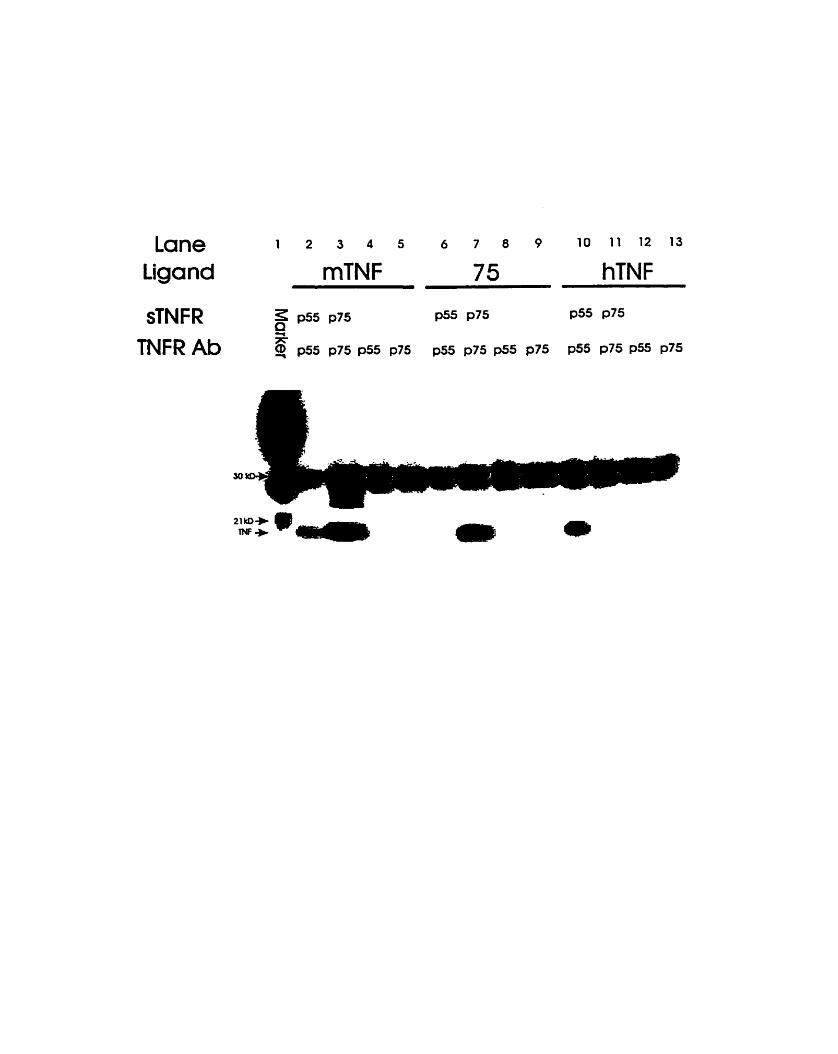
Lane Ligand
sTNFR TNFR Ab

109
Effect of the mutant form of mTNFa on tumour pathology
Mice bearing transplanted polyoma middle-T tumours (PyMidT) were injected
intratumourally with 5x 1 o8 p h of Ad vectors. Two days post injection the heours were
removed fkom sacrificed mice, and processed for paraffin sectioning. Haematoxylin and
eosin stains of tumour sections showed thaî, by cornparison to n o d tumour morphology
as exhibited by hunours injected with control vector (Fig 5A). the mutant form of mTNFa
was able to induce intratumoural necrosis (Fig SB), as did the wild type vectors (Fig.
5C,&D), however the extent of the necrosis induced by Ad-75 was sigdicantly lower. It
should also be noted that the Ad-75 vector did not induce sigdïcant levels of necrosis at a
lower dose of 1x10~ p h (not shown), while it has been previously detennïned that both
wild type vectors readily induced intratumoural necrosis at that dose (24,25).
Systemic and antitumour effects of the mutant form of mTNFa
Previous studies have suggested that the triggering of the p55 and not the p75
TNFR is prirnarily responsible for the induction of systemic toxicity (1 0,11,1 Z), and that
triggering of the p75 TNFR is responsible for signalling the activatiordproliferation of
lymphoid cells. Therefore, we decided to test the systemidantitumour effects of the Ad-75
vector directly injected into PyMidT himours. Both the wild type TNFa expressing vectors
(Ad-mRJF & Ad-hTNF) have previously been tested in this tumour mode1 (24,25). These
vectors proved to be quite toxic to treated rnice killing 12 of 16 and 8 of 20 injected
anirnals in two experiments, while curing 2 of 4 and 1 of 12 respectively at a dose of 5x10'

Fig. 5. Tumour rnorphology: Haematoxylin and eosin staining of p M 1 n sections of
tumours from mice treated with control ( A ~ - B H G L \ E ~ , ~ ) vector (A), Ad-75 (B) (*
necrotic region), Ad-mTNF (C) and Ad-hTNF (D), two days post intratumoural injection
with 5x10' pfu per mouse (200X).


112
pfu per mouse. We chose to use these vectors dong with the mutant vector for a direct
cornparison in vivo.
Table II shows the results of the immunotherapy treatments with the various Ad
vectors. The Ad-75 vector showed no mortality even at a dose of 1x10' pfu, which in
previous experirnents with both wild type vectors resulted in 100% mortality within 1-3
days (24,25). Both wild type vectors showed lethality at doses of 5 and 1x10~ pfu, kiliing
2 of 4 and 3 of 8 (Ad-mTNF) and 3 of 5 and 1 of 5 (Ad-hTNF) mice respectively. No p s s
toxicity was observed in control vector injected mice. Even though the Ad-75 vector
showed no mortality, it did induce an antitumour response in treated mice, causing partial
regessions in 5 of 15 mice and permanent elimination of the tumour in 2 of 15 mice at a
dose of 5x10~ pfu. Mice surviving treatment with the wild type vectors also showed an
antitumour response, with one in two being cured at a dose of 5x10~ pfu (Ad-mTNF) and
one in four behg cured at a dose of 1x10' pfû (Ad-hTNF), as well as induchg partial
regressions (see TabIe II). Figure 6 displays the growth kinetics typicdly exhibited by
tumours treated with Ad-75 (5x 1 0bfÛ). Al1 mice cured in these experirnents were resistant
to subsequent challenge (two months later) with PyMidT tumour cells, which implied the
induction of immune memory against the tumour. These data clearly show that a TNFa
mutant that fllnctions through activation of the p75 mTNFR alone does not induce gross
mortality while retaining the ability to induce an antiturnour response.

Tablc 2: Antitumour rcsponse and toxicity induced by intratuniourai injection witli TNF cxpressing Ad vectors.
1 Mortality represents the proportion of rnice which died as a result of vector administration, typically within 14 days post injection.
2 Partial response refers Co the proportion of survivors displaying tumours which regressed to less than half iheir origional ske before starting to grow again.
3 Complete regression refers to the proportion of sunrivors displaying cornplete and pemanent
I
Vector Dose(Pfu) Mortality
disappearance of th& tumour (within 2 4 weeks post vector injection).
Turnour Response 7 3
No Parîial Complc fe Response Response Regression
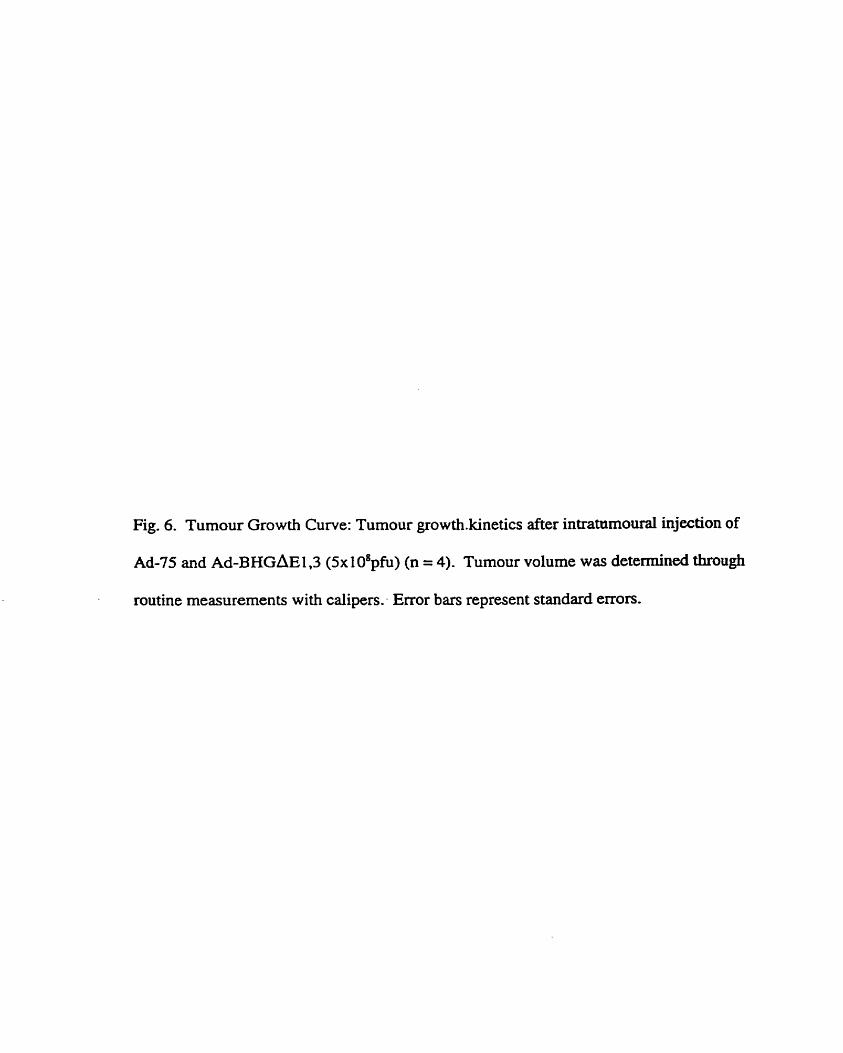
Fig. 6. Tumour Growth Curve: Tumour growthkinetics after intr~moural injection of
Ad-75 and ~d-BHGAEI ,3 (5x 1 08~fu) (n = 4). Tumour volume was detennined through
routine measurements with caiipers: Error bars represent standard errors.


Discussion
The p55 TNF receptor has been implicated as the primary receptor for the induction
of systemic toxicity by TNFa (1 0,11,12). Our in vivo experiment data support this
conclusion by showing that, compared to the wild type f o m of murine and human TNFa,
a p75 TNFR specific form showed no lethality, however less obvious side effects may have
been present. Furthemore, the fact that p75 TNFR activation does have immune
stimulatory properties (3,5,7), allows one to specifically bypass W u related toxicity
while retaining the ability to activate an antitumour response. Here we have shown such an
application of a p75 TNFR specinc ligand and compared it with Iigands which bind both
receptors (mTNFa) or o d y the p55 receptor (hTNFa).
We have not yet f U y characterized the nature of the antitumour response induced
by this receptor specific mutant, however the detection of immune mernory against the
tumour suggests that an immune response is involved. As well, it has been demonstrated
thaî mice cured by cytokine expressing Ad vectors, including Ad-mTNF and Ad-75 (24,
26), showed PyMidT specific CTL activity (data not shown). The one mouse cured by the
human TNFa vector has yet to be tested for PyMidT specific CTL activity. The role of the
immune system in the antitumour activity of T'N'Fa is well established (7,27,28,29,30). By
comparing the effects of human and murine TNFa on murine T ce11 lines transfected with
the human p75 TNFR cDNA, Vandenabeele et al. (7) showed that activation of the p75
TNFR was able to specifically induce cytokine secretion by T cells. Recently the effects of
administration of a p75 TNFR specific mutant of human TNFa were tested in baboons (3 l),

and the results of this study indicated that this form of TNFa did not induce hypotension,
IL-6 or IL-8 production, or skin necrosis as did wild type and p55 TNFR specific ligands.
However a modest infiltration of lymphocytes and macrophages was detected in the dermis.
A cornpaison of the effectiveness of the Ad-75 vector at doses of 5x10' and
1xl0~pfu indicates a potential inhibitory effect at higher doses, however greater numbers of
mice are needed to confirm this (see Table II). TNFa has been irnplicated as part of the
immune regdatory mechanism modulating T ceil responses through elimination of reactive
T ceUs (32,33). TNFa was shown to be an important mediator of T ce11 anergy in HIV
infection and TNFa alone was shown to inhibit CD4+ T ce11 fiinction (34). Other examples
of the suppressive properties of TNFa have been described (35,36). Perhaps higher levels
of mutant mTNFa produccd at the dose of 1xl0~pfu induced an immunosuppressive
response.
The data presented in Table II show that exclusive activation of the p55 TNFR by
human TNFa can produce an antitumour response in up to haif of treated animals. The p55
TNFR is a powerfirl inducer of a variety of irnrnunologic/antitumo~~ activities, including
the induction of cytokine secretion, neutrophil activation, endothelial ce11 activation
(6,27,37,3 8), and cytotoxicity (2,s). The induction of intratumoural thmmbosis is a major
mechanism by which TNFa can induce rapid tumour necrosis (2,39,40,41). As well, Van
der Pol1 et al., 1996 (38) demonstrateci that only wild type and p55 specific agonists
induced coagulation and fibrinolysis in baboons. Considering the dominant role of the p55
TNFR in endothelial ce11 activation and cytotoxicity one would not predict that a p75

118
TNFR specific agonist would induce intratumoural necrosis in contrast to our obsewations
(Fig. 5). We did note however, that the degree to which the necrosis was induced was
somewhat less in Ad-75 treated mice. It is possible that the overproduction of TNFa
generated by the Ad vector codd account for this observation. It is also possible that
production of the mutant protein induced the secretion of endogenous mTNFa or other
infiammatory proteins, at levels sufficient to cause tissue necrosis but not systemic toxicity.
The obsewation that hTNFa induced proliferation of the CT6 ce11 line was also unexpected,
considering that lymphoproliferative signals are primarily induced via the p75 TNFR. It is
however possible that the longer incubation tirne with TNFa used in ou. assay (three days
compareci to one) (3), is at least partly responsible for the discrepancy between our data
and results observed by others.
The results of our studies in our murine tumour mode1 suggest that a p75 TNFR
specific agonist may be very useful for the treatment of human cancers if the non toxic and
antitwnour properties of Ad-75 could be extrapolated to huma. patients. The expression of
such a mutant fiom an Ad vector would be advantageous, considering adenoviral vectors
allow for the possibility of local and transient high level expression of the transgene over a
period of several days. However work m u t still be done to improve the effectiveness of
this approach, possibly through the combination of TNF with other cytokines (IL-2, IL-12)
and costimulatory molecules (B7). Work on this and other approaches is presently
ongoing.

119
Acknowledgements
We wouId Iike to thank Dr. David Goeddel and Dr. Mike Rothe (Tularik, Inc., San
Francisco, CA) for providing us with the CT6 ce11 line, and Dr. Robert D. Schreiber and Dr.
Cora Arthur (Washington University School of Medicine, St. Louis, MO) for providing us
with the murine TNF receptor specific antibodies (55R-286 and TR75-89). Ail PCR primer
synthesis and sequencing was done at the Mobix Central Facility, McMaster University,
Hamilton, Ontario, Canada This work was supported by grânts h m the National Cancer
hstitute of Canada (NCIC), the Medical Research Council of Canada (ME) , the Canadian
Breast Cancer Initiative, and London Life Insurance. F.L.G. is a Terry Fox Research
Scientist of the NCIC and W.J.M. is a MRC Scientist.
References
1. Carswell, E.A.,Old, L.J.,Kassel, RL.,Green, S., Fiore, N., and Williamson B. An
endotoxin-induced serum factor that causes necrosis of tumours. Proc. Natl. Acad. Sci.
USA., 72(9):3666-3670. 1975.
2. Fiers, W. Tumour Necrosis Factor. Characterization at the molecular, cellular and in vivo
level. FEBS Letters., 285(2): 199-212. 1991.
3. Tartaglia, L.A., Weber, RF. Figari, I.S., Reynolds, C., Palladino, Jr., M.A., and Goeddel,
D.V. The two different receptors for tumor necrosis factor mediate distinct cellular
responses. Proc. Natl. Acad. Sci. USA., 88:9292-9296. 1991.

120
4. Vilcek, J., and Lee, T.H., Turnor necrosis factor: New insights into the molecular
mechanisms of its multiple actions. J. Biol. Chem, 266(12):73 13-73 16. 1991.
5 . Sarraf, C.E. Turnour necrosis factor and ce11 death in tumours (review). International
Journal of Oncology., 5: 1333-1339. 1994.
6 . Benigni, F., Faggioni, R., Sironi, M., Fantuzzi, G., Vandenabeele, P., TakRhashi, N., Sacco,
S., Fiers, W., Buunnan, W.A., and Ghezzi, P. TNE; receptor p55 plays a major role in
centrally rnediated increases of semm IL-6 and corticosterone after intracerebroventricular
injection of TNF. J. Immunol., 157(12):5563-5568. 1996.
7. Tracey, KI., and Ce- A. Tumour necrosis factor: a pleiotmpic cytokine and
therapeutic target. Ann. Rev. Md., 45:49 1-503. 1994.
8. Lewis, M., Tartaglia, L.A., Bennett, L.A., Rice, G.C., Wong, G.H.W., Chen, E.Y., and
Goeddel, D.V. Cloning and expression of a cDNA for two distinct murine tumor necrosis
factor receptors demonstrate one receptor is species specific. Proc. Natl. Acad. Sci. USA.,
88:2830-2834. 199 1.
9. Ranges, G.E., Bombara, M.P., Aiyer, R.A., Rice, G.G., and Palladino, Jr., M.A. Tumor
necrosis factor-a as a proliferative signal for an IL-2 dependent T ce11 line: strict species
specificity of action. J. Immunol., 142: 1203-1208. 1989.
10. Bluethmann, H., Rothe, J., Schultze, N., Tkachuk, M.,and Koebel, P. Establishment of the
role of IL-6 and TNF receptor 1 using gene knockout mice. J. Leukocyte Biol., 56:565-570.
1994.

121
1 1. Enckson, S.L., de Sauvage, F. J., Kikly, K., Carver-Moore, K., Pitts-Meek, S ., Gillet, N.,
Sheehan, K.C.F., Schreiber, R.D., Goeddel, DY., and Moore, M. W. Decreased sensitivity
to tumour-necrosis factor but normal T-ce11 development in TNF receptor-2-deficient mice.
Nature, 372560463. 1994.
12. Sheehan, K.C.F., Pinkard, LEC, Arthur, C.D ., Dehner, L.P., GoeddeI, D.V., and Schreiber,
RD. Monoclonal antibodies specific for murine p55 and p75 tumor necrosis factor
receptors: identif ication of a novel in vivo role for p75. J. Exp. Med., 1 8 1 :607-6 1 7. 1 995.
13. Loetscher, H., Stueber, D., Banner, D., Mackay, F., and Lessauer, W. Human tumor
necrosis factor a (TNFa) mutants with exclusive specincity for the 55-kDa or 75-kDa TNF
receptors. J. Biol. Ch=, 268(35):26350-26357. 1993.
14. Van Ostade, X., Vandenabeele, P.,Tavernier, J., and Fiers, W. Human tumor necrosis factor
mutants with preferential binding to and activity on either the R55 or R75 receptor. Eur. J.
Biochem., 22O:77 1-779. 1994.
15. Fidler, I.J. Biological behaviour of malignant melanoma cells correlateci to their suMval in
vivo. Cancer Res., 35:218-234. 1975.
16. Graham, F.L., Smiley, J., Russel, W.C., and Naini, R. Characterization of a human ce11 line
transfomed by DNA fiom human adenovims type 5. J. Gen. Virol., 3659-72- 1977.
17. Brarnson, J., Hitt, M., Gallichan, W.S., Rosenthal, K.L., Gauldie, J., and Graham, F.L.
Construction of a double recombinant adenovixus vector expressing a heterodimenc
cytokine: in vitro and in vivo production of biologically active interleukin- 12. Hum. Gene
Ther., 7:333-342. 1996.

122
18. Guy, C.T., Cardiff, R.D., and Muller, W.J. Induction of mammary tumours by expression
of polyoma virus middle T oncogene: a transgenic mouse mode1 for metastatic disease.
Mol. Ce11 Biol., 12:954-961. 1992.
19. Putzer, B.M., Hitt, M., Muller, W.J., Emtage, P., Gauldie, J., and Graham, F.L. Interle-
12 and B7- 1 costirnulatory molecule expressed by an adenovinis vector act synergistically
to facilitate tumor regression. Proc. Natl. Acad. Sci. USA., 94: 10889- 10894. 1997.
20. Higuchi, R Recombinant PCR. In PCR protocols: a guide to methods and applications.
Innis, MA, Gelfand, D.H., Sninski, J.J., and White, T.J. editors. Academic Press, Inc., San
Diego, CA. 177-1 80. 1990.
21. Addison, CL., Hitt, M., Kunsken, D., and Oraham, F.L. Cornparison of the human and
murine cytomegalovinis immediate early gene promoters for transgene expression by
adenoviral vectors. J. Gen. Virol., 78:1653-166 1. 1997.
22. Bett, A.J., Haddara, W., Prevec, L., and Graham, F.L. An efficient and flexible system for
construction of adenovirus vectors with insertions or deletions in early regions 1 and 3.
Proc. Natl. Acad. Sci. USA., 9 1 :8802-8806. 1994.
23. Hitt, M., Bett, A., Addison, C., Prevec, L., and Graham, F.L. Techniques for human
adenovirus vector construction and characterization. In Viral gene techniques. Meîhods in
molecular genetics. Adolph, K.W. editors. Academic Press, Inc., San Diego, CA. 13-30.
1995.
24. Marr, R.A., Addison, C.L., Snider, D., Muller, W.J., Gauldie, J., and Graham, F.L. Tumour
immunotherapy using an adenoviral vector expressing a membrane-bound mutant of

123
murine TNFa. Gene Ther., 4:1181-1188. f 997.
25 Marr, R.A., Hitt, M., Muller, W.J., Gauldie, J., and Graham, F.L. Tumour therapy in mice
using adenovirus vectors expressing human TNFa. Int. J. Oncol., 12: 509-5 15. 1998.
26. Bramson, J.L., Hitî, M., Gauldie, J., and Graham, F.L. Pre-existing immunity to adenovins
does not prevent tumor regression foUowing intraunioral administration of a vector
expressing IL- 12 but inhibits virus dissemination. Gene Ther., 4: 1069- 1076. 1997.
27. Paleolog, E.M., Delasalle, A-A.J., Buumian, W.A., and Feldman, M. Functional activities
of receptors for tumor necrosis factor-a on human vascular endothelial cells. Blood,
84(8):2578-2590. 1994.
28. Palladino, M.A. Jr., Ref* S.M., Kramer, S.M., Femaiolo, B.L., Baughman, RA., Deleo,
AB. Crase, D., Madino, B., Aggarwal, B.B., Figari, I.S., Liggit, D., and Patton, J.S.
Characterization of the antitumor activities of human tumor necrosis factor* and the
cornparison with other cytokines: induction of tumor-specific immunity. J. ImmunoL,
l38(l l):4023-4032. 1987.
29. Trepper, RI., Mule, J.J. Experimental and clhical studies of cytokine gene-modifieci
tumour cells. Hum. Gene Ther., 5: 153-164. 1994.
30. Marincola, F.M., Ettinghausen, S., Cohen, P.A., Cheshire, L.B., Reftifo, N.P., Mule, J.J.,
and Rosenberg, S.A. Treatment of established Iung metastases with tumor-infiltrathg
lymphocytes derived fiom a poorly inimunogenic tumor engineered to secrete human TNF-
a. J. Immunol., l52:3SOl-35 13. 1994.

124
3 1. Welborn, M.B. III, Van Zee, K., Edwards, P.D., Pruitt, J.H., Kaibara, A., Vauthey, J.N.,
Rogy, M., Castleman, W.L., Lowry, S.F., Kenney, J.S., Stuber, D., Ettlin, U., Wipf, B.,
Loetscher, H., Copeland III, E.M., Lesslauer, W., and Moldawer, L.L. A human tumor
necrosis factor p75 receptor agonist stimulates in vitro T cell proliferation but does not
produce inflamation or shock in the baboon. J. Exp. Md., 184: 165-1 7 1. 1996.
32. Glickstein, L.J., and Huber, B.T. Karoushi-Death by overwork in the immune system. J.
I ~ ~ f l ~ l . , lS5(2):522-524. 1995.
33. Zheng, L., Fisher, G., Miller, RE., Peschon, J., Lynch, D.H., and Lenardo, M.J. Induction
of apoptosis in mature T cells by tumour necrosis factor. Nature, 3 77:348-3 5 1. 1 995.
34. Kaneko,H.,Hishikawa,T.,Sekigawa,I.,Hashimoto,H.,Okumura,K,andKaneko,Y.
Role of tumour necrosis factor-alpha (TNF-alpha) in the induction of HIV- 1 gp 120-
mediated CD4+ T cell anergy. C h . Exp. ImmunoL, 109(1):41-46. 1997.
35. Alleva, D.G., Burger, C.J., and Elgert, K.D. Tumor-induced regdation of supressor-
macrophage nitric oxide and TNFu production: Role of -or-derivd IL-10, TGF-p, and
prostaglandin &. J. Immunol., 153: 1674-1686. 1994.
36. Cope, A.P., Londei, M., Chu, N.R, Cohen, S.B.A., Eiliott, M.J., Brennan, F.M., Maini,
RN., and Feldmann, M. Chronic exposure to tumor necrosis factor (TNF) in vitro impairs
the activation of T cells through the T ce11 receptodCD3 complex; reversal in vivo by anti-
TNF antibodies in patients with rheumatoid arthritis. J. Clin. Invest., 94:749-760. 1994.

125
37. Barbara, J.A.J., Smith, W.B., Garnble, J.R, Van Ostade, X., Vandenabeele, P., Tavernier,
J., Fiers, W., Vadas, M.A., and Lopez, A.F. Dissociation of TNF-a cytotoxicity and
proinflamatory activities by p5S recep tor- and p75 receptor-selective TNF-a mutants.
EMBO J., 13(4):843-850. 1994.
38. Van der Poll, T., Jansen, P.M., Van Zee, K.J., Welbom ID, M.B., de Jong, I., Hack, C.E.,
Loetscher, H., Lesslauer, W., Lowry, S.F., and Moldawer, L.L. Tumor necrosis factor-alpha
induces activation of coagulation and fibrinolysis in baboons through an exciusive effect on
the p55 receptor. Blood, 88(3):922-927. 1996.
39. Prober, J.S . Effects of tumour necrosis factor and related cytokines on vascular endothelial
cells. In Tumour necrosis factor and related cytotoxins. Bock, G. and Marsh , J. editors.
Wiley, Chichester, (Ciba Fomdation Symposium). 170-1 85.1984.
40. Claus, M. Ryan, J., and Stem, D. Modulation of endothelial ceIl haemostatic properties by
TNF: insights into the role of the endothelium in the host response to inflammatory stimuli.
I n Tumour necrosis factors. The molecules and their emerging role in medicine. Beutler,
D., editor. Raven Press, NY. 49-64.1992.
41. Denekamp, J. Review article: angiogenesis, neovascular proliferation and vascular
pathophysiology as targets for cancer therapy. Brit. J. Radiol., 66: 18 1- 196. 1993.

Summary
Mutant D142N-A144R is p75 receptor specific
The homologous mutant of human TNFQ @143N-A145R), was previously shown
to be specific for the human p75 TNF receptor (#36 Loetscher et tz& 1993). Here it is
shown that TNFa with a mutation D142N-A144R is specific for the murine p75 TNF
receptor. This was demonstrated by showing a lack of cytotoxic activity on L929 ceils
@55 specific function), while retaining the ability to Uiduct proliferation of CT6 tells.
Furthermore the proiiferaton was not blocked by the addition of soluble p55 TNF receptor
to the assay. Also, a physical association of the mutant TNF with the p75 TNF receptor
alone was demonstrated by CO-immunoprecipitating the mutant ligand with either the p55
or p75 TNF receptors.
Mutant murine TNFa showed reduced toxicty
Direct injection of Ad-mTNF-75 (expressing the double point mutant of mTNFa)
failed to induce lethality at doses as high as 1x10~ p h . While both the Ad-mTNF-wt (wild
type murine) and Ad-hTNF-wt (wild type human, also called Ad-MCMV-TNF) both
produced lethality at lower doses.
Antitumour activity of mutant murine TNFa
Mice treated with the A d - m m - 7 5 vector showed some partial (5 of 15) and

127
permanent (2 of 15) tumour regressions at a dose of 5x10' pfu. Cured rnice were resistant
to subsequent challenge with PyMidT tumour ceils on the lefi hind flank. Thus, a p75 TNF
receptor agonist showed drastically reduced systemic toxicity while retaining some ability
to regress tumours.

CHAPTER VI: DISCUSSION
Tumour necrosis factor alpha has been the subject of a great deal of research ever
since its discovery as an anticancer agent in the sera of mice treated with endotoxin
(Canwell et al., 1975). Its apparent anticancer properties led to its testing in patients as a
possible cancer treatment. The direct injection of recombinant TNFa into the circulation or
intratumourally has been shown to induce toxicity at doses required for therapeutic e@ects
(Kimura et al., 1987; Sherman et al., 1988). Patients treated systemically showed a
relatively low minimum tolerated dose and responded with organ failure and hypotension,
with little antitumour effect. More recently clinicians have used isolated Limb perfusion
(ILP) or isolated organ perfusion (IOP) to deal with the toxicity induced by TNFa. ILP
entails surgical isolation of the vascular root and connection to a heart and lung machine,
which enable the constant perfusion of therapeutic agents. This procedure ailows for the
addition of approximately 10 times the normal minimal tolerated dose, and in combination
with other therapeutic agents (melphaian), resulted in the complete regression of 70% of
patients with in transit melanoma metastasis (reviewed in (Lejeune et al., 1998)). Even
though this procedure has met with some success, it is still limited to regional therapy of
advanced cancer, with ultimately Little effêct on patient survival. Also, the requirement for
surgery is labour intensive and unpleasant. Thus improved methods for the delivery of
TNFa to the tumour site are needed.
In recent years the use of Wal vectors to deliver therapeutic genes for the treatment
of cancer has become the subject of intense investigation. The potential for local

129
expression of anticancer agents within the region of the tumour is an obvious advantage of
gene therapy, and Ad vectors are ideally suited for this task (discussed above). However,
our observations have indicated that the local expression of TNFa fkom the tumour site is
insufficient for the prevention of systemic toxicity. We have shown that an Ad vector
expressing wild type murine TNFa induced high levels of lethality at doses required for
antitumour activity (see chapter III). However, the restriction of TNFa to the ceil surface
resulted in a drastic reduction in lethality. It is also apparent that this form of TNFa retains
the ability to cause partial and permanent tumour regressions. A membrane bound mutant
of human TNFa has been characterized (Perez et al., 1990), allowing for the possibiiity of
testing this type of therapy in human cancer patients.
Targeting either of the TNF receptors has ais0 met with some success in our tumour
model. Using human TNFa to specifically activate the p55 TNF receptor was shown to
provide little protection kom the lethal side effects of TNFa. Human TNFa failed to
produce a powerfùl antitumour response as weii. Targeting the p75 TNF receptor with the
double point mutant showed a drastic reduction in lethality, however this mutant also failed
to produce a powerful antihunour response. It should be noted that in both cases
(membrane bound mutant, and p75 specific mutant) the antitumour ac t ive of the treatment
was reduced in cornparison to wild type TNFa. The reduction in activity of the membrane
bond mutant could be a result of reduced availability of TNFa for target cells. The
antihunour activity of Ad-mTNF-MEM appeared to follow a positive dose response, and a
higher dose of the vector partialiy compensated for the reduced activity. The reduction in

130
efficacy was more pronounceci for the p75 receptor specific mutant. As stated above,
activation of both TNF receptors has commonly been found to produce the greatest
induction of TNF activity (see Literature review). Also, the lack of p5 5 activation would
result in the loss of any antiturnour contribution provided by that receptor. The elimination
of lethality, should have enabled us to increase the dosage to levels in which a higher
degree of antitumour activity might be observed. This, however, was not the case. The
higher dose used did not show any indication of increased antitumour activity. In addition
to the abiiity of TNF to activate the immune system, TNF has also been implicated as a
down-regulator of the immune system. It was shown to be hvolved in the induction of
CD4' T ce11 anergy by HIV gpl2O (Kaneko et ai., 1997). Similar to Fas, TNFa was aiso
shown to induce apoptosis in T ceils (CD89 through activation the p75 TNF receptor
(Zheng et al., 1995). This could explain the lack of increased efficacy at greater doses, as
the higher levels of TNFa produced may have lead to immunosuppression rather than
immune stimulation. In al1 mes, mice cured by the Ad TNF vectors were resistant to
subsequent challenge with PyMidT tumour cells, demonstrating the induction of protective
immune memory, critically important to the elimination of metastesis. The specific
involvement of the immune system in regression of the prirnary himour was not directly
detennined, however, considering the lympho-specific effects of the p75 TNF receptor, it is
likely that immune activation was involved in regressions induced by the Ad-mTNF-75
vector. It is also likely that an immune response was also involved in regressions caused
by the other Ad-TNF vectors, as it has been shown that the antitumour properties of TNFa

131
involve an immune response (see Literature Review) (Asher et al., 1 99 1 ; B lankenstein et
al., 1991 ; Marincola et al., 1994).
Although the techniques used were effective for reducing the lethality associated
with TNFa, the antitumour activity is low relative to similar treatments with other
cytokines such as IL,-2 and IL12 (30-40% complete regressions) (Addison et al., 199%;
Addison et al., 199%; Bramson et al., 1996a). The wmbination of TNF treatment with
other cytokine and or cytotoxic treatments could prove fkidùl. However, the various
mutants used in the above tumou. therapy expcrircents have also been used in combination
with Ad vectors expressing IL2 and L 1 2 (see Appendix). No wmbination of Ad-TNF
vector with Ad-IL2 or Ad-IL12 vector produceci any signifïcant improvanent in
antitumour activity, with the exception of the combination of Ad-mm-MEM with an IL-
12 expressing Ad vector (Ad-mlG12) (Bramson et ai., 1996b), in which case a dramatic
increase in lethaiity was also observed.
The constructecl Ad-TNF vectors wuld prove to be usefûl tools for the investigation
of the role(s) of TNFa in other physiological processes. Lei et al. found that ovalbumin
(OVA) induced airway eosinophilia in CD40L knockout mice, was dependent on the
combination of IL4 and TNFa Gei et al., 1998). This was done through the co-
administration of Ad vectors expressing IL4 and murine TNFa (Ad-mTNF-wt). Ad-
mTNF-wt was also used in a mode1 of pulmonary inflammation, in which it was suggested
that TNFa induced fibrosis through the induction of TGF-P (Sime et al., 1998). Mutant
forms of human TNFa specific for the p75 TNF receptor (recombinant protein) have been

132
introduced into baboons, showing greatly reduced systemic toxicity similar to our fïndings
(Welbom et al., 1996). The receptor specific TNFa expressing Ad vectors could prove to
be usefùl for the investigation of the functions of either TNF receptor within the murine
system. The Ad-mTNF-MEM vector wuld prove useful for the investigation of the
specific role of tht membrane bound f o m of TNFa, or Iocally expresseci TNFa, in a variety
of physiological processes. Furthemore, the partial success of the Ad-mTNF-MEM vector
in tumour therapy, demonstrated the principal that a nom- secreted cytokine can be
restricted of to the cell surface and remain effective. This strategy could be applied to other
toxic cytokines such as IFNy, L 2 , and IL12 using membrane anchored h i o n proteins.
This type of therapy would require a gene delivery technique, and adenoviral vectors would
be ideal for such a task. With respects to tumour therapy, the techniques described here
show that the lethality associated with TNFa can be reduceà while retaining the ability to
regress tumours, however the overall efficacy is poor. Fewer than 9% of the mice were
cured with the Ad-mTNF-MEM vector (at doses of 1x10~ and 5x10~ pfq with none cured
at doses of 1x10' and 2x10~ p h ) and approximately 5% were cured with the Ad-mTNF-75
vector (at doses of 1x1 09, 5x1 0' and 1x1 0' p h , with none cured at a dose of 2x1 o7 pfû).
Thus we were not able to increase the oved l therapeutic index of our TNFa gene therapy
techniques through mutation of TNFa.

133
CHAPTER VII: REFERENCES
Addison, C.L. (1997a) Construction and characterization of adenoviral vectors expressing
cytokines for cancer immunotherapy, McMaster.
Addison, CL., Braciak, T., W o n , R, Muller, W.J., Gauldie, J. and Graham, F.L. (1 995a)
Intratumoral injection of an adenovirus expressing interle* 2 induces regression
and immunity in a murine breast cancer model. Ptoc Nat1 Acad Sci U S A 92(18),
8522-6.
Addison, C.L., Braciak, T., Ralston, R, Muller, W.J., Gauldie, J. and Graham, F.L. (1995b)
An adenoviral vector expressing interleukin-4 modulates tumourigenicity and
induces regression in a murine breast cancer model. ïnt J Oncol 7, 12534260.
Addison, C.L., Hitt, M., Kunsken, D. and Graham, F.L. (199%) Cornparison of the human
versus murine cytomegalovinis immediate early gene promoters for transgene
expression by adenoviral vectors. J Gen Virol78(Pt 7), 1 653-6 1 .
Amalfitano, A., Hauser, M.A., Hu, H., Serra, D., Begy, C.R. and Chamberlain, J.S. (1 998)
Production and characterization of improved adenovirus vectors with the El, E2b7
and E3 genes deleted. J Virol72(2), 926-33.
Arany, Z., Sellers, W.R., Livingston, D.M. and Eckner, R. (1994) ELA-associated p300 and
CREB-associated CBP belong to a conserved family of coactivators [letter]. Ce11
77(6), 799-800.
Asher, A.L., Mule, J.J., Kasid, A., Restifo, N.P., Salo, J.C., Reichert, CM., Jaffe, G.,

134
Fendly, B., Kriegler, M. and Rosenberg, S.A. (1991) Murine tumor cells transduced
with the gene for tumor necrosis factor- alpha. Evidence for paracrine immune
effects of tumor necrosis factor against tumors. J Immmol l46(9), 3227-34.
A t m , M.B., Robertson, M.J., Gordon, M., Lotze, M.T., DeCoste, M., DuBois, J.S., Ritz,
J., Sander, A.B., Edington, H.D., -ne, P.D., Mier, J. W., Canning, C.M.,
Battiato, L., Tahara, H. and Sherman, M.L. (1997) Phase 1 evaluation of intravenous
recombinant human interleukin 12 in patients with advanced malignancies. Clin
Cancer Res 3(3), 409- 17.
Aversa, G., Punnonen, J. and de Vries, J.E. (1993) The 26-kD transmembrane form of
-or necrosis factor alpha on activated CD4+ T cell clones provides a
costhnulatory signal for human B cell activation. J Exp Med 177(6), 1575-85.
APiavoorian, S., Murphy, AN., Stetler-Stevenson, W.G. and Liotta, L.A. (1993) Molecular
aspects of hunor ce11 invasion and metastasis. Cancer 71(4), 1368-83.
Balkwill, F.R, Naylor, M.S. and Malik, S. (1990) Tumour necrosis factor as an anticancer
agent. Eur J Cancer 26(5), 641-4.
Bandara, L.R. and La Thangue, N.B. (1991) Adenovins Ela prevents the retinoblastoma
gene product fkom complexing with a cellular transcription factor. Nature
35 1 (6326), 494-7.
Bast, RC., Jr., Zbar, B., Mackaness, G.B. and Rapp, H.I. (1975a) Antiturnor activity of
bactenal infection. 1. Effwt of Listeria monocytogenes on growth of a murine
fibrosarcoma. J Nat1 Cancer Inst 54(3), 749-56.

135
Bast, RC., Jr., Zbar, B., Miller, T.E., Mackaness, G.B. and Rapp, H.J. (1975b) Antitumor
activity of bacterial infection. II. effect of Listeria monocytogenes on growth of a
guinea pig hepatoma J Natl Cancer Inst 54(3), 757-61.
Bergelson, J.M., Cunningham, J.A., Droguete, G., Kurt-Jones, E.A., Krithivas, A., Hong,
J.S., Horwitz, M.S., Crowell, IU. and Finberg, RW. (1997) Isolation of a cornmon
receptor for Coxsackie B vinises and adenovinises 2 and S. Science 275(5304),
1320-3.
Bett, A.J., Haddara, W., Prevec, L. and Graham, F.L. (1994) An efficient and flexible
system for construction of adenovins vectors with insertions or deletions in early
regions 1 and 3. Proc Natl Acad Sci U S A 9 1(19), 8802-6.
Bett, AL, Prevec, L. and Graham, F.L. (1993) Packaging capacity and stability of human
adenovinis type 5 vectors. J Virol67(1 O), 59 1 1-2 1.
Bishop, J.M. (1991) Molecular themes in onwgenesis. Cell64(2), 235-48.
Biswas, R, Basu, M., Sen-Majumdar, A and Das, M. (1985) Intrapeptide
autophosphorylation of the epidermd growth factor receptor: regulation of kinase
catalytic function by receptor dimerization. Biochemistry 24(14), 3795-802.
Blaclc, R.A., Rauch, C.T., Kozlosky, C.T., Peschon, J.J., Slack, J.L., Wolfson, M.F.,
Castner, B.J., Stocking, ICL., Reddy, P., Srinivasan, S., Nelson, N., Boiani, N.,
Schooley, KA., Gerhart, M., Davis, R., Fitzner, J.N., Johnson, R.S., Paxton, R. J.,
March, C. J. and Cerretti, D.P. (1 997) A metallopro teinase disintegrin that releases
himour-necrosis factor- alpha fkom cells. Nature 385(6618), 729-33.

Blankenstein, T., Qin, Z.H., Uberla, K., Muller, W., Rosen, H., Volk, H.D. and
Diarnantstein, T. (199 1) Tumor suppression after tumor cell-targeted tumor necrosis
factor alpha gene transfer. J Exp Med 173(5), 1047-52.
BIuethmann, H., Rothe, J., Schultze, N., Tkachuk, M. and Koebel, P. (1994) Establishment
of the role of IL6 and TNF receptor 1 using gene knockout mice. J Leukoc Bi01
56(5), 565-70.
Bondesson, M., Ohman, K., Manervik, M., Fan, S. and Akuçjarvi, G. (1996) Adenovirus
E4 open reading k e 4 protein autoregdates E4 transcription by inhibiting E1A
tramactivation of the E4 promoter. J Virol70(6), 3844-5 1.
BOU&, N. (1990) Tumor angiogenesis: the d e of onmgenes and tumor suppressor genes.
Cancer Cells 2(6), 179-85.
Bramson, J., Hitt, M., Gallichan, W.S., Rosenthal, KL., Gauldie, J. and Graham, F.L.
(1996b) Construction of a double recombinant adenovinis vector expressing a
heterodimeric cytokine: in vitro and in vivo production of biologically active
interleukin- 1 2- Hum Gene Ther 7(3), 3 33-42.
Brarnson, J.L., Hitt, M., Addison, C.L., Muller, W.J., Gauldie, J. and Graham, F.L. (1996a)
Direct intratumoral injection of an adenovinis expressing inter1euki.u- 12 induces
regression and long-lasting immunity that is associated with highly localized
expression of interleukin- 12. Hum Gene Ther î(l6), 1995-2002.
Bridge, E. and Ketner, G. (1 990) Interaction of adenoviral E4 and E 1 b products in late gene
expression. Virology l74(2), 345-53.

137
Brouckaert, P.G., Everaerdt, B., Libert, C., Talcahashi, N. and Fiers, W. (1989) Species
specificity and involvement of other cytokines in endotoxic shock action of
recombinant tumour necrosis factor in mice. Agents Actions 26(1-2), 196-8.
Bucana, CD., Hoyer, L.C., Schroit, A.J., Kleinerman, E. and Fidler, IJ. (1983)
Ultrastructural studies of the interaction between liposome-activated human blood
monocytes and aliogeneic hunor cells in vitm. Am J Pathol 1 12(1), 101 -1 1.
Bwgert, H.G. and Kvist, S. (1987) The E3/19K protein of adenovinis type 2 binds to the
domains of histocompatibility antigens required for CTL recognition. Embo J 6(7),
20 19-26.
Carswell, E.A., Old, L.J., Kassel, RL., Green, S., Fiore, N. and Williamson, B. (1975) An
endotoxin-induced semm factor that causes necrosis of tumors. Proc Nat1 Acad Sci
U S A 72(9), 3666-70.
Chellappan, S.P., Hiebert, S., Mudsj, M., Horowitz, J.M. and Nevins, J.R.. (1991) The E2F
transcription factor is a cellular target for the RB protein. Ce11 65(6), 1053-61.
Chen, M., Memod, N. and Honvitz, M.S. (1 990) Protein-protein interactions between
adenovinis DNA polymerase and nuclear factor 1 mediate formation of the DNA
replication preinitiation cornplex. J Bi01 Chem 265(30), 18634-42.
Chen, S.K., Shine, H.D., Goodman, J.C., Grossman, R.G. and Woo, S.L. (1994) Gene
therapy for brain tumors: regression of experimental gliomas by adenovinis-
mediatecl gene transfer in vivo. Proc Nat1 Acad Sci U S A 9 l(8), 3054-7.
Cheng, S.H., Piwnica-Worms, H., Harvey, RW., Roberts, T.M. and Smith, A.E. (1988)

138
The carboxy terminus of pp6Oc-src is a regdatory domain and is involved in
complex formation with the middle-T antigen of polyomavinis. Mol Ce11 Bi01 8(4),
1736-47.
Chroboczek, J., Bieber, F. and Jacrot, B. (1992) The sequence of the genome of adenovints
type 5 and its cornparison with the genome of adenovins type 2. Virology l86(l),
280-5.
Clauss, M., Murray, J.C., Vianna, M., de Waal, R., Thurston, G., Nawroth, P., Gerlach, H.,
Bach, R, Familletti, P.C. and Stem, D. (1990) A polypeptide factor produced by .
fibrosarcoma ce& that induces endothelid tissue factor and enhances the
procoagulant response to tumor necrosis factor/cachectin. J Bi01 Chem 26$(12),
7078-83.
Clauss, M., Ryan, J. and Stem, D. (1992) Modulation of endotheliai cell haernostatic
properties by TNF: insights into the role of endothelium in the host response to
infiammatory stimuli. In: D. Beutler (Ed), Tumour necrosis factors. The molecules
and their emerging role in medicine, Raven Press, New Yourk, pp. 49-64.
Collins, T., Lapierre, L.A., Fiers, W., Strominger, J.L. and Pober, J.S. (1 986) Recombinant
human tumor necrosis factor increases mRNA levels and surface expression of
HLA-A,B antigens in vascular endothelid cells and dermal fibroblasts in vitro. Proc
Nat1 Acad Sci U S A 83(2), 446-50.
Courtneidge, S.A. and Heber, A. (1987) An 8 1 kd protein complexeci with middle T
antigen and pp6Oc-WC: a possible phosphatidylinositol kinase. Cell 50(7), 103 1-7.

139
Courtneidge, S.A. and Smith, A.E. (1 983) Polyoma Wus transfomllng protein associates
with the product of the c- src cellular gene. Nature 303(5916), 435-9.
Dales, S. and Chardonnet, Y. (1 973) Early events in the interaction of adenoviruses with
HeLa cells. W . Association with microtubules and the nuclear pore cornplex during
vectorial movement of the inoculum. Virology 56(2), 465-83.
D'Andrea, A., Rengaraju, M., Valiante, NM., Chehimi, J., Kubin, M., Aste, M., Chan,
S.H., Kobayashi, M., Young, D., Nickbarg, E. and et al. (1 992) Production of
nahiral killer ce11 stimulatory fâctor (interleukin 12) by penpheral blood
mononuclear cells. J Exp Med 176(5), 1387-98.
de Stanchina, E., McCurrach, M.E., Zindy, F., Shieh, S.Y., Fexbeyre, G., Samuelson, AV.,
Prives, C., Roussel, M.F., Shm, C.J. and Lowe, S.W. (1998) E1A signahg to p53
involves the p19(ARF) -or suppressor. Gens Dev 12(15), 2434-42.
Declercq, W., Denecker, G., Fiers, W. and Vandenabeele, P. (1998) Cooperation of both
TNF receptors in induchg apoptosis: involvement of the TNF receptor-associated
factor binding domain of the TNF receptor 75. J Immun01 161(1), 390-9.
Decoster, E., Vanhaesebroeck, B., Vandenabeele, P., Grooten, J. and Fiers, W. (1995)
Generation and biological charactezization of membrane-bound, uncleavable murine
tumor necrosis factor. J Bi01 Chem 270(3 l), 18473-8.
Denekamp, J. (1 993) Review article: angiogenesis, neovascular proli feration and vascular
pathophysiology as targets for cancer therapy. Br J Radio1 66(783), 18 1-96.
Dethlefsen, S.M., Matsuura, N. and Zetter, B.R. (1990) Tumour growth and angiogenesis

140
in wild type and mast ce11 deficient mice. FASEB J 4, A623.
Dobner, T., Horikoshi, N., Rubenwolf, S. and Shenk, T. (1 996) Blockage by adenovirus
E4orf6 of transcriptional activation by the p53 tumor suppressor. Science
272(5267), 1470-3.
Downey, J.F., Evelegh, CM., Branton, P.E. and Bayley, S.T. (1984) Peptide maps and N-
t a a l sequences of polypeptides fkom early region 1A of human adenovirus 5. J
VirolSO(l), 30-7.
Dri, P., Haas, E., Cramer, R., Menegazzi, R, Gasparini, C., Martinelli, R, Scheurich, P.
and Patriarca, P. (1999) Role of the 75-kDa TNF receptor in TNF-induced
activation of neutmphïi respiratory burst. J Immun01 162(1), 460-6.
Eastham, J.A., Hall, S.J., Sehgal, I., Wang, J., Timme, TL., Yang, G., Connell-Crowley,
L., Elledge, S.J., Zhang, W.W., Harper, J.W. and et al. (1995) In vivo gene therapy
with p53 or p21 adenovirus for prostate cancer. Cancer Res 55(22), 5 15 1-5.
Engelhardt, J.F., Li-, L. and Wilson, J.M. (1994) Pmlonged transgene expression in
cotton rat Iung with recombinant adenoviruses defective in E2a. Hum Gene Ther
5(1 O), 121 7-29.
Erickson, S.L., de Sauvage, F.J., KikIy, K., Carver-Moore, K., Pitts-Meek, S., Gillett, N.,
Sheehan, KC., Schreiber, RD., Goeddel, D.V. and Moore, M. W. (1 994) Decreased
sensitivity to tumour-necrosis factor but n o d T-ce11 development in TNF
receptor-2-deficient rnice. Nature 372(6506), 560-3.
Fidler, 1. J. (1 975) Biological behavior of malignant melanoma cells correlatecl to their

141
survival in vivo. Cancer Res 3 5(1), 2 1 8-24.
Field, J., Gronostajski, RM. and Hurwitz, J. (1 984) Properties of the adenovinis DNA
polyrnerase. J Bi01 Chem 2S9(l S), 9487-95.
Fiers, W. (1 99 1) Tumor necrosis factor. Characterization at the molecular, cellular and in
vivo level. FEBS Lett 285(2), 199-212.
Follanan, 1. (1992) The role of angiogcnesis in tumor growth. Semin Cancer Bi01 3(2), 65-
71.
Foo, S.Y. and Nolan, G.P. (1999) NE:-kappaB to the rescue: RELs, apoptosis and cellular
transformation. Trends Genet 15(6), 229-35.
Freeman, G.J., Freedman, A.S., Segil, J.M., Lee, G., Whitman, J.F. and Nader, L.M.
(1989) B7, a new mernber of the Ig superfamily with unique expression on activated
and neoplastic B ceils. J Tmmunol 143(8), 2714-22.
Gately, M.K., Wolitzky, A.G., Quinn, P.M. and Chizzonite, R. (1 992) Regdation of human
cytolytic lymphocyte responses by interleukin-12. Ce11 Tmmunol 143(1), 127-42.
Gooding, L.R., Elmore, L. W., Tollefson, A.E., Brady, H.A. and Wold, W.S. (1 988) A
14,700 MW protein fkom the E3 region of adenovinis inhibits cytolysis by tumor
necrosis factor. Celi 53(3), 341-6.
Grable, M. and Hearing, P. (1992) cis and tram requirements for the selective packaging of
adenovinis type 5 DNA. J Virol66(2), 723-3 1.
Graham, F.L. (1984) Covalently closed circles of human adenovins DNA are infèctious.
Embo J 3(12), 2917-22.

142
Graham, F.L., Smiley, J., Russell, W.C. and Nairn, R. (1977) Characteristics of a human
ce11 line transformeci by DNA nom human adenovins type 5. J Gen Vu01 36(1),
59-74.
Graham, F.L. and Van der Eb, A.J. (1973) A new technique for the assay of infectivity of
human adenovinis 5 DNA. Virology 52,456-467.
Greber, U.F., Willetts, M., Webster, P. and Helenius, A (1993) Stepwise dismantling of
adenovinis 2 during entry into cells. Ce11 75(3), 477-86.
Green, M., Brackmann, KH., Cam, M.A. and Matsuo, T. (1982) Identification and
purification of a protein encoded by the human adenovinis typc 2 transforming
region. J Virot 42(1), 30-41.
Green, M. and Daesch, G.E. (1961) Biochemical studies on a d e n o v i . multiplication. 1.
Kinetics of nucleic acid and protein synthesis in suspension cultures. Virology 13,
169- 1 76.
Gurtu, V., Yan, G. and Zhang, G. (1996) IRES bicistronic expression vectors for efficient
creation of stable mammalian ce11 Iines. Biochern Biophys Res Commun 229(1),
295-8.
Guy, C.T., Cardiff, RD. and Mull er, W. J. (1 992) Induction of mammary tumors by
expression of polyomavims middle T oncogene: a transgenic mouse mode1 for
metastatic disease. Mol Ce11 Bi01 12(3), 954-6 1.
Harding, F.A., McArthur, J.G., Gross, J.A., Raulet, D.H. and Allison, J.P. (1 992) CD28-
mediateci signalling co-stimulates murine T cells and prevents induction of anergy

143
in Tcell clones. Nature 356(6370), 607-9.
Hasson, T.B., Soloway, P.D., Ornelies, D.A., Doerfler, W. and Shenk, T. (1 989)
Adenovirus L1 52- and 55-kilodalton proteins are required for assembly of virions.
J VU01 63(9), 3612-21.
Hay, J.G., McElvaney, N.G., Herena, J. and CrystaI, RG. (1995) Modification of nasal
epithelial potentid ciifferences of individuals with cystic fibrosis consequent to local
administration of a nomial CFTR cDNA adenovirus gene transfcr vector. Hum
Gene Ther 6(1 l), 1487-96.
Heldin, CH., Erniund, A., Roman, C. and Ronnstrand, L. (1989) Dimerkation of B-type
platelet-derived growth factor receptors occurs after ligand binding and is closely
associated with receptor kinase activation. J Bi01 Chem 264(l 5), 8905-12.
Hirschowitz, E.A., Ohwada, A., Pascal, W.R, Russi, T.J. and Crystal, RG. (1995) In vivo
adenovinis-mediated gene tramfer of the Escherichia coli cytosine deaminase gene
to huma. colon carcinoma-derived tumors induces chemosensitivity to 5-
fluorocytosine. Hum Gene Ther 6(8), 1055-63.
Hisatsune, T., Nishijima, K., Minai, Y., Kohyama, M. and Kaminogawa, S. (1994)
Autoreactive CD8+ T cell clones producing immune suppressive lymphokines IL-
10 and interferon-gamma. Ce11 Immun01 1 S4(l), 1 8 1-92.
Hitt, M., Bett, A.J., Addison, CL, Prevec, L. and Graham, F.L. (1995) Techniques for
human adenovirus vector construction and characterktion. In: K.W. Adolph (Ed),
Viral Gene Techniques, Methods in Molecular Genetics, Vol. 7, Academic Press,

144
San Diego, pp. 13-30.
Hia, M.M., Addison, C.L. and Graham, F.L. (1997) Human adenovims vectors for gene
transfer into mammalian cells. Adv Pharmacol 40, 137-206.
Horwitz, M.S., Maizel, J.V., Jr. and Scharff, M.D. (1 970) Molecular weight of Adenovins
type 2 hexon polypeptide. J Virol6(4), 569-7 1.
Humphries, M. J., Olden, K. and Yamada, K.M. (1 986) A synthetic peptide h m fibronectin
inhibits experimental metastasis of murine melanoma cells. Science 233(4762),
467-70.
Inostroza, LA., Memelstein, F.H., Ha, I., Lane, W.S. and Reinberg, D. (1 992) Drl, a
TATA-binding protein-associateci phosphoprotein and inhibitor of class II gene
transcription. Celi 70(3), 477-89.
Ishibashi, S., Brown, M.S., Goldstein, J.L., Gerard, RD., Hammer, RE. and Herz, J.
(1 993) Hypercholesterolemia in low density lipoprotein receptor bockout mice and
its reversal by adenovinis-mediated gene delivery [see comments]. J Clin Invest
92(2), 883-93.
Iwata, K.K., Fryling, C.M., Knott, W.B. and Todaro, G.J. (1 985) Isolation of tumor ce11
growth-inhibiting factors trom a human rhabdornyosarcoma ce11 line. Cancer Res
45(6), 2689-94.
Jin, X., Nguyen, D., Zhang, W.W., Kyritsis, A.P. and Roth, J.A. (1995) Ce11 cycle arrest
and inhibition of himor ce11 proliferation by the p16INK4 gene mediated by an
adenovirus vector. Cancer Res 55(15), 3250-3.

145
Johnson, D.G., Cress, W.D., Jakoi, L. and Nevins, J.R (1 994) Oncogenic capacity of the
E2Fl gene. Proc Nat1 Acad Sci U S A 9 1 (26), 12823-7.
Johnson, L.G., Boyles, S.E., Wilson, J. and Boucher, RC. (1995) Normalization of raised
sodium absorption and raised calcium-mediated chloride secretion by adenoviw-
mediated expression of cystic fibrosis transmembrane conductance regulator in
primary human cystic fibmsis ainvay epitheiial cells. J Clin Invest 95(3), 1377-82.
Julius, M., Mwun, C.R and Haugbn, L. (1993) Distinct roles for CD4 and CD8 as CO-
receptors in antigen receptor signalling. Immun01 Today 1 4(4), 1 77-83.
Kaelin, W.G., Jr. (1 999) The ernerging p53 gene family. J Nat1 Cancer Inst 9 1(7), 594-8.
Kaneko, H., Hishikawa, T., Sekigawa, I., Hashimoto, H., Okumura, K. and Kaneko, Y.
(1 997) Role of tumour necrosis factor-alpha (Tm-alpha) in the induction of HN- 1
gpl20-mdated CD4+ T celI anergy. Clin Exp Immun01 109(1), 41-6.
Kao, C.C., Yew, P.R and Berk, A.J. (1990) Domains required for in vitro association
between the cellular p53 and the adenovinis 2 E1B 55K proteins. Virology 179(2),
806- 14.
Karlhofer, F.M., Ribaudo, RK. and Yokoyama, W.M. (1 992) MHC class I alloantigen
specificity of Ly-49+ IL-2-activated natural killer cells [see comments]. Nature
358(638 l), 66-70.
Kimura, K., Taguchi, T., Unishizaki, I., Ohno, R., Abe, O., Fume, H., Hattori, T.,
Ichihashi, H., Inoguchi, K., Majima, H. and et al. (1987) Phase I study of
recombinant human tumor necrosis factor. Cancer Chemother Pharmacol 20(3),

223-9.
Kitajewski, J-, Schneider, R.J., Safer, B ., Munemitsu, S .M., Samuel, C.E., Thimmappaya,
B. and Shenk, T. (1 986) Adenovirus VA1 RNA antagonizes the antiviral action of
interferon by preventing activation of the interferon-induced eIF-2 alpha kinase.
Cell45(2), 195-200.
Kleinberger, T. and Shenlc, T. (1 993) Adenovirus E40rf4 protein binds to protein
phosphatase 2A, and the complex down regulates El A-enhanced junB transcription.
J Virol67(12), 7556-60.
Knowles, M.R.9 Hohneker, KW., Zhou, Z., Olsen, J.C., Noah, T-L., Hu, P.C., Leigh,
M. W., Engelhardt, J.F., Edwards, L.J., Jones, KR and et al. (1 995) A controlled
study of adenovird-vector-mediated gene transfer in the nasal epithelium of patients
with cystic fibrosis [see comments]. N Engl J Med 333(13), 823-3 1.
Kochanek, S., CIemens, P R , Mitani, K, Chen, H.H., Chan, S. and Caskey, C.T. (1996) A
new adenovinal vector: Replacement of all viral coding sequences with 28 kb of
DNA independently expressing both full-length dystrophin and beta-galactosidase.
Proc Nat1 Acad Sci U S A 93(12), 573 1-6.
Koff, W.C. and Fann, A.V. (1 986) Human tumor necrosis factor-alpha kills herpesvinis-
infected but not normal cells. Lymphokine Res 5(3), 2 15-2 1.
Kohler, P.C. and Sondel, P.M. (1989) The role of interleukin-2 in cancer therapy. Cancer
SW 8(4), 861-73.
Kombluth, S., Cross, F.R., Harbison, M. and Hanafiisa, H. (1986) Transformation of

147
chicken embryo fibroblasts and tumor induction by the middle T antigen of
p01yomavirus carrieci in an avian retroviral vector. Mol Ce11 Bi01 6(5), 1545-5 1.
Kozarsky, K.F., McKinley, D.R., Austin, L.L., Raper, S.E., Stratford-Pemcaudet, L.D. and
Wilson, J.M. (1994) In vivo correction of low density fipoprotein receptor
deficiency in the Watanabe heritable hyperlipidemic rabbit with recombinant
adenovinises. J Bi01 Chem 269(18), 13695-702.
Kroemer, G. (1997) The proto-oncogene Bcl-2 and its role in regulating apoptosis
lpublished erratum appears in Nat Med 1997 Aug;3(8):934]. Nat Me. 3(6), 614-20.
Krougliak, V. and Graham, F.L. (1995) Development of celi lines capable of
complementing El, E4, and protein IX defective adenovinis type 5 mutants. Hum
Gene Ther 6(12), 1575-86.
Kuerbitz, S. J., Plunkett, B.S., Walsh, W.V. and Kastan, M.B. (1 992) Wild-type p53 is a ceii
cycle checkpoint determinant following irradiation. Proc Nat1 Acad Sci U S A
89(l6), 749 1-5.
Lanier, L.L., Ruitenberg, J. J. and Phillips, J.H. (1 988) Functional and biochemical analysis
of CD16 antigen on natural killer cells and granulocytes. J Immun01 141(10), 3478-
85.
Lei, X.F., Ohkawara, Y., Starnpfli, M.R., Mastruzzo, C., Marr, R.A., Snider, D., XUig, 2.
and Jordana, M. (1998) Disruption of antigen-induced idammatory responses in
CD40 ligand knockout mice. J Clin Invest 1 Ol(6), 1342-53.
Lejeune, F.J., Ruegg, C. and Lienard, D. (1998) Clhical applications of TNF-alpha in

148
cancer. Curr Opin Imrnunol 1 O@), 573-80.
Lewis, M., Tartaglia, LA., Lee, A., Bennett, G.L., Rice, G.C., Wong, G.H., Chen, E X and
Goeddel, D.V. (1991) Cloning and expression of cDNAs for two distinct murine
turnor necrosis factor receptors demonstrate one receptor is species speci fic. Proc
Natl Acad Sci U S A 88(7), 2830-4.
Li, Y., Kang, J., Friedman, J., Tarassishin, L., Ye, J., Kovalenko, A., Wdach, D. and
Horwitz, M.S. (1999) Identification of a ceii protein (FIP-3) as a modulator of NF-
kappa. activity and as a target of an adenovinis inhibitor of tumor necrosis factor
alpha-induced apoptosis. Proc Natl Acad Sci U S A 96(3), 1042-7.
Lindenbaum, J.O., Field, J. and Hurwitz, J. (1986) The adenovinis DNA binding protein
and adenovins DNA polymerase interact to catalyze elongation of prirned DNA
templates. J Bi01 Chem 26 1 (22), 1 02 1 8-27.
Linsley, P.S., Clark, E.A. and Ledbetter, J.A. (1990) T-cell antigen CD28 mediates
adhesion with B ceiis by interacting with activation antigen B7IBB-1. Proc Natl
Acad Sci U S A 87(13), 503 1-5.
Liotta, L.A. and Stetler-Stevenson, W.G. (1991) Tumor invasion and metastasis: an
imbalance 6f positive and negative regdation. Cancer Res 5 l(18 Suppl), 5054s-
5059s.
Liu, B., Podîck, E.R., Allison, J.P. and Malek, T.R (1996) Generation of primary tumor-
specific CTL, in vitro to immunogenic and poorly immunogenic mouse tumors. J
Immun01 l56(3), 1 1 17-25.

149
Liu, F. and Green, M.R. (1 990) A specific member of the ATF transcription factor family
can mediate transcription activation by the adenovirus E 1 a protein. Ce11 6 1 (7),
12 1 7-24.
Liu, T. J., Zhang, W. W., Taylor, D.L., Roth, J.A., Goepferî, H. and Clayman, G.L. (1 994)
Growih suppression of human head and neck cancer cells by the introduction of a
wild-type p53 gaie via a recombinant adenovinis. Cancer Res 54(14), 3662-7.
Loetscher, W., Stueber, D., Banner, D., Mackay, F. and Lesslauer, W. (1993) Human tumor
necrosis factor alpha (TNF alpha) mutants with exclusive specificity for the 55-kDa
or 75-kDa TNF receptors. J Bi01 Chem 268(35), 26350-7.
Lowenstein, EJ., Daly, RJ., Batzer, AG., Li, W., Margolis B., Lammers, R, UUrich, A.,
Skolxuk, E.Y., Bar-Sagi, D. and Schlessinger, J. (1992) The SH2 and SH3 domain-
containhg protein GRB2 links receptor tyrosine kinases to ras signalkg. Cell 70(3),
43 142.
Maniatis, T., Fritsch, E.F. and Sambrook, 1. (1989) Molecular cloning: a laboratory manual
(2nd Edn). Cold Spring Harbor Press, Cold Spring Harbor-
Marcellus, R.C., Lavoie, J.N., Boivin, D., Shore, G.C., Ketner, G. and Branton, P.E. (1 998)
The early region 4 orf4 protein of human a d e n o v i . type 5 induces p53-
independent ce11 death by apoptosis. J Virol72(9), 7144-53.
Marincola, F.M., Ettinghausen, S., Cohen, P.A., Cheshire, L.B., Restifo, N.P., Mule, J.J.
and Rosenberg, S.A. (1994) Treatment of established lung metastases with tumor-
infiltrating lymphocytes derived fiom a poorly irnmunogenic -or engineered to

150
secrete human TNF-alpha. J LmmunoI 152(7), 3500-13.
Mathews, M.B. (1 980) Binding of adenovims VA RNA to mRNA: a possible role in
splicing? Nature 285(5766), 575-7.
Mathews, M.B. (1 990) Control of translation in adenovinis-infécted cells. Enzyme 44(1-4),
250-64.
McGrory, W.J., Bautista, D.S. and Graham, F.L. (1988) A simple technique for the rescue
of early region 1 mutations into infectious human adenovirus type 5. Virology
163(2), 614-7.
MoMer, J.L., Partin, A.W. and Coffey, D.S. (1987) Prediction of metastatic potential by a
new grading system of ceU motility: validation in the Diinning R-3327 prostatic
adenocarcinorna model. J Ur01 13 8(l), 168-70.
Moore, J. W.d. and Sholley, M.M. (1 985) Cornparison of the neovascular effects of
stimulated macrophages and neutrophils in autologous rabbit corneas. Am J Pathol
120(1), 87-98.
Monal, N., Parks, R.J., Zhou, H., Langston, C., Schiedner, G., Quinones, J., Graham, F.L.,
Kochanek, S. and Beaudet, A.L. (1998) Hi& doses of a helper-dependent
adenoviral vector yield supraphysiological levels of alpha1 -antitrypsin with
negligible toxicity. Hum Gene Ther 9(18), 270% 1 6.
Morsy, M.A., Gu, M., Motzel, S., Zhao, J., Lin, J., Su, Q., Allen, H., Franlin, L., Parks,
R.J., Graham, F.L., Kochanek, S., Bett, A.J. and Caskey, C.T. (1 998) An adenoviral
vector deleted for al1 viral coding sequences results in enhanced safety and extended

151
expression of a leptin transgene. Proc Natl Acad Sci U S A 95(14), 7866-71.
Nagata, K., Guggenheimer, RA. and Hunvitz, J. (1983) Adenovins DNA replication in
vitro: synthesis of full-length DNA with puified protek. Proc Natl Acad Sci U S
A 80(14), 4266-70.
Nawroth, P., Handley, D., Matsueda, G., De Waal, R, Gerlach, H., Blohm, D. and Stem,
D. (1 988) Tumor necrosis factor/cachectin-induced intravascular fibrin formation in
meth A fibrosarcornas. J Exp Med l68(2), 637-47.
Obert, S., O'Connor, RJ., Scbmid, S. and Hearing, P. (1994) The adenovinis E4-6f7
protein tramactivates the E2 promoter by inducing dimerkation of a heteromeric
E2F complex. Mol CeU Bi01 14(2), 133346.
OMalley, B.W., Jr., Li, D., Buckner, A., Duan, L., Woo, S.L. and Pardoll, D.M. (1999)
Limitations of adenovirus-mediated interleukin-2 gene therapy for oral cancer.
Laryngoscope 1 O9(3), 389-95.
Paleolog, E.M., Delasalle, S.A., Buurman, W.A. and Feldmann, M. (1994) Functional
activities of receptors for tumor necrosis factor-alpha on human vascular endothelid
cells. BIood 84(8), 2578-90.
Palladino, M.A., Jr., Patton, J.S., Figari, I.S. and Shalaby, M.R. (1987) Possible
relationships between in vivo antihimour activity and toxicity of tuxnour necrosis
factor-alpha. Ciba Found Symp 13 1,2 1 -3 8.
Papavassiliou, A.G. (1 994) The CREBfATF family of transcription factors: modulation by
reversible phosphorylation. Anticancer Res 14(5A), 1 80 1 -5.

152
Parks, R-J., Chen, L., Anton, M., Sankar, U., Rudnicki, M.A. and Graham, F.L. (1996) A
helper-dependent adenovirus vector system: removal of helper virus b y Cre-
mediated excision of the viral packaging signal. Proc Nat1 Acad Sci U S A 93(24),
13565-70.
Pasten, I., Seth, P., FitzGerald, D. and Willingham, M. (1986) Adenovins entry into cells;
some new observations on an.old problem. In: A. Notkins and M. Oldstone (Eds),
Concepts in viral pathogenesis KI, Springer-Veriag, New York, pp. 141-1 46.
Perez, C., Albert, I., DeFay, K., Zachariades, N., Gooding, L. and Kriegler, M. (1990) A
nonsecretable ce11 d a c e mutant of tumor necrosis factor (W) kilis by cell-to-ceil
contact. Ce11 63(2), 25 1-8.
Pilder, S., Moore, M., Logan, J. and Shenk, T. (1986) The adenovinis E1B-55K
transforming polypeptide modulates transport or cytoplasmic stabilization of viral
and host ce11 rnRNAs. Mol Celi Bi01 6(2), 470-6.
Pimentel-Muinos, F.X., Mazana, J. and Fresno, M. (1994) Regulation-of interleukin-2
recep tor alpha chah expression and nuclear factor. kappa B activation by protein
kinase C in T lymphocytes. Autocrine role of tumor necrosis factor alpha. J Bi01
Chem 269(39), 24424-9.
Porter, A.C. and Vailtancourt, R.R. (1 998) Tyrosine kinase receptor-activated signal
transduction pathways which lead to oncogenesis. Oncogene 1 7(ll Reviews), 1 3 43 -
52.
Prober, J.S. (1987) Effects of tumour necrosis f actor and related cytokines on vascular

153
endothelial cells. In: G. Bock and J. Marsh (Eds), Tumour necrosis factor and
related cytotoxins, While, Chichester, pp. 1 70-1 85.
Pugh, B.F. and Tjian, R. (1992) Diverse transcriptional functions of the multisubunit
eukaryotic TFID complex. J Biol Chem 267(2), 679-82.
Putzer, B.M., Brarnson, J.L., Addison, C.L., Hitt, M., Siegel, P.M., Muller, W.J. and
Graham, F.L.. (1 998) Combination therapy with interleukh-2 and wild-type p53
expressed by adenoviral vectors potentiates tumor regression in a murine mode1 of .
breast cancer. Hum Gene Ther 9(5), 707- 18.
Putzer, B.M., Hitt, M., Muller, W. J., Emtage, P., Gauldie, J. and Graham, F.L. (1997)
Interleukin 12 and B7-1 costirnulatory molecule expressed by an adenovinis vector
act synergistically to facilitate tumor regression. Proc Nat1 Acad Sci U S A 94(20),
10889-94.
Ranges, G.E., Bombara, M.P., Aiyer, RA., Rice, G.G. and Palladino, M.A., Jr. (1989)
Tumor necrosis factor-alpha as a p r o l i f d v e signal for an IL-2- dependent T ce11
line: strict species specificity of action. J lmmunol 142(4), 1203-8.
Ranheim, E.A. and Kipps, T. J. (1 995) Tumor necrosis factor-alpha facilitates induction of
CD80 (B7-1) and CD54 on human B cells by activated T cells: complex regdation
by IL-4, IL-1 O, and CD40L. Ce11 h m 0 1 16 1 (2), 226-35.
Rekosh, D.M., Russell, W.C., BeIlet, A.J. and Robinson, A.J. (1977) Identification of a
protein linked to the ends of adenovirus DNA. Ce11 1 1 (2), 283 -95.
R h , J., Schmidt, R.E., Michon, J., Hercend, T. and Schlossman, S.F. (1988)

154
Characterization of functional surface structures on human natural killer cells. Adv
Immun01 42, 181-21 1.
Roberts, R.J., OWeilI, K.E. and Yen, C.T. (1984) DNA sequences fkom the adenovims 2
genome. J Bi01 Chem 259(22), 13968-75.
Rflel, B. and Mihatsch, M.J. (1993) TNF receptor distribution in human tissues. Int Rev
Exp Pathol34(Pt B), 149-56.
Saki, I., Iida, J., Murata, J., Ogawa, R, Nishi, N., Sugimura, K., Tokura, S. and Azurna, 1.
(1989) Inhibition of the metastasis of murine-malignant melanoma by synthetic
polymenc peptides containhg core sequences of cell-adhesive molecules. Cancer
R ~ s 49(14), 3 8 15-22.
Samow, P.,Hearing, P., Anderson, C.W., Halbert, D.N., Shenk, T. and Levine, A.J. (1984)
Adenovinis early region 1B 58,000-dalton tumor antigen is physically associated
with an early region 4 25,000-dalton protein in productively infecteci cells. J Virol
49(3), 692-700.
Sarraf, C.E. (1 994) Tumour necrosis factor and ce11 death in hunours (Review). Int J Oncol
5,1333-1339.
Schreiber, M., Muller, W.J., Singh, G. and Graham, F.L. (1999) Cornparison of the.
effkctiveness of adenovins vectors expressing cyclin kinase inhibitors p 16INK4A,
p 1 8INK4C, p 1 9INK4D, p2 1 (WAF l/CIP 1) and p27KIP 1 in inducing ce11 cycle
arrest, apoptosis and inhibition of turnongenicity. Oncogene 18(9), 1663-76.
Seger, R, Biener, Y., Feinstein, R., Hanoch, T., Gazit, A. and Zick, Y. (1995) Differential

155
activation of rnitogen-activated protein kinase and S6 kinase signaling pathways by
12-O-tetradecanoylphorbol-13-acetate (TPA) and insulin. Evidence for involvement
of a TPA-stirnulated protein-. tyrosine kinase. J Bi01 Chem 270(47), 28325-30.
Sentman, C.L., Hackett, J., Jr., Kumar, V. and Bennett, M. (1 989) Identification of a subset
of murine natural killer ceils that mediates rejection of Hh-ld but not Hh-lb bone
rnarrow grafts. J Exp Med l7O(l), 19 1-202.
Sheehan, KC., Pinckard, J.K., Arthur, C.D., Dehner, L.P., Goeddel, D.V. and Schreiber,
R.D. (1 995) Monoclonal antibodies specific for murine p55 and p75 tumor necrosis
factor receptors: identification of a novel in vivo role for p75. J Exp Med 18 1 (2),
607-17.
Shenk, T. (1996) Adenoviridae: The vinisw and their replication. In: B.N. Fields, D.M.
Knipe and P.M. Howley (Eds), Fields Virology 3rd M., Lippincitt-Raven
Publishers, Philadelphia, pp. 2 1 1 1-2148.
Sherman, M.L., Spriggs, D.R, Arthur, ICA., hamura, K., Frei, E.d. and Kufe, D.W.
(1988) Recombinant human tumor necrosis factor administered as a five-day
continuous infusion in cancer patients: phase 1 toxicity and eff- on iipid
metabolism. J Clin Oncol 6(2), 344-50.
Shimomura, IC, Manda, T., Mukumoto, S., Kobayashi, K., Nakano, K. and Mori, J. (1988)
Recombinant human tumor necrosis factor-alpha: thrombus formation is a cause of
anti-turnor activity. Int J Cancer 41 (S), 243-7.
Shisler, J., Yang, C., Walter, B., Ware, C.F. and Gooding, L.R. (1997) The adenovins E3-

156
1 O.4Wl4.5K cornplex mediates loss of ce11 surface Fas (CD95) and resistance to
Fas-induced apoptosis. J Virol7 l(1 l), 8299-306.
Sime, P.J., Marr, R.A., Gauldie, D., Xing, Z., Hewlett, BR., Graham, F.L. and Gauldie, J.
(1998) Transfer of tumor necrosis factor-alpha to rat Iung induces severe pulmonary
inflammation and gatchy interstitial fibrogenesis with induction of transforming
growth factor-betal and myofibroblasts. Am J Pathol153(3), 825-32.
Smart, J.E. and S t i h a n , B.W. (1982) Adenovins terminai protein precursor. Partial amino.
. acid sequence and the site of covalent linkage to virus DNA. J Bi01 Chem 257(22),
13499-506.
S o m J.A. f 1998) Tumor immunology: the glas is haif full. Immunity 9(6), 757-63.
Sondel, P.M., Hank, J : k and Kohler, P.C. (1 987) S tatus and potential of interleukin-2 for
the treatment of neoplastic disease. Oncology (Huntingt) 1(6), 4 1-9.
Stalder, T., Hahn, S. and Eh, P. (1994) Fas antigen is the major target molecule for CD4+
T cell-mediateci cytotoxicity. J Immun01 1 S2(3), 1 127-33.
Strassmann, G., Bertolini, D.R. and Eidelrnan, 0. (199 1) Inhïbition of b u n e reactions in
vivo by liposome associated ~ f o m i i n g growth factor (TGF) type beta 1. Clin
Exp Immun01 86(3), 532-6.
S u e s , P., Beyaert, R., De Valck, D., Vanhaesebroeck, B., Van Roy, F. and Fiers, W.
(1 99 1) Tumour-necrosis-factor-mediated cytotoxicity is correlated with
phospholipase-A2 activity, but not with arachidonic acid release per se. Eur J
Biochem l95(2), 465-75.

157
Sukumar, S. (1989) ras oncogenes in chemical carcinogenesis. Curr Top Microbiol
Lmm~n01 148,93-114.
Svensson, U. (1 985) Role of vesicles during adenovirus 2 internalization into HeLa cells. J
Virol55(2), 442-9.
Takeichi, M. (1 99 1) Cadherin cell adhesion receptors as a morphogenetic regulator.
Science 251(5000), 1451-5.
Tartaglia, L.A., Pennica, D. and Goeddel, D.V. (1993) Ligand passing: the 75-kDa tumor
necrosis factor (TNF) receptor recruits TNF for signaling by the 55-kDa TNF
receptor. J Biol Chem 268(25), 18542-8.
T-glia, L.A., Weber, RF., Figari, I S , Reynolds, C., Palladino, M.A., Jr. and Goeddel,
D.V. (1 99 1) The two different receptors for tumor necrosis factor mediate distinct
cellular responses. Proc Natl Acad Sci U S A 88(20), 9292-6.
Temperley, S.M. and Hay, KT. (1992) Recognition of the adenovirus type 2 origin of DNA
replication by the virally encoded DNA polymerase and preterminal proteins. Embo
J 11(2), 761-8.
Thimmappaya, B., Weinberger, C., Schneider, R.J. and Shenk, T. (1 982) Adenovirus VAI
RNA is required for efficienttranslation of viral rnRNAs at late times after
infection. Cell 3 l(3 Pt 2), 543-5 1.
Thomas, D. W., Talmage, D. W. and Joberts, W.K. (1 975) Inhibiton of tumor cell
proliferation: second role for suppressor cells? J Irnmunol 1 1 5(5), 1366-74.
Tihanyi, K., Bourbonniere, M., Houde, A., Rancourt, C. and Weber, J.M. (1993) Isolation

158
and properties of adenovinis type 2 proteinase. J Bi01 Chem 268(3), 1780-5.
Tollefson, A.E., Ryerse, J.S., Scaria, A., Hermiston, T.W. and Wold, W.S. (1996a) The E3-
1 1.6-kDa adenovinis death protein (ADP) is required for efficient ce11 death:
characterizaion of ceiis infected with adp mutants. Virology 220(1), 152-62.
Tollefson, A.E., Scaria, A., Henniston, T. W., Ryerse, J.S ., Wold, L. J. and Wold, W.S.
(1996b) The adenovinis death protein (E3-11.6K) is required at very late stages of
infection for efficient cell lysis and release of adenovinis nom infecteil ceiis. J Virol
70(4), 2296-306.
Tracey, K.J. (1995) TNF and Mae West or: death fiom too much of a good thing [see
commentsJ. Lancet 345(8942), 75-6.
Tracey, K.J., Beutler, B., Lowry, S.F., Merryweather, J., Wolpe, S., Mils& LW., Hariri,
-RJ., Fahey, T.J.d., Zentella, A., Albert, J.D. and et al. (1986) Shock and tissue
injury induced by recombinant human cachectin. Science 234(4775), 470-4.
Tracey, K. J. and Cerami, A. (1 994) Tumor necrosis factor: a pleiotropic cytokine and
therapeutic target. Annu Rev Med 45,49 1-503.
Trentin, L., Zambello, R., Bulian, P., Cerutti, A., Enthammer, CC., Cassatella, M., Nitti, D.,
Lise, M., Agostini, C. and Semenzato, G. (1995) Tumour-infiltrathg lymphocytes
bear the 75 kDa tumour necrosis factor receptor. Br J Cancer 7 1(2), 240-5.
Tribouley, C., Lutz, P., Staub, A. and Kedinger, C. (1994) The product of the adenovinis
intermediate gene N a 2 is a trans.criptiona1 activator of the major late promoter. J
Virol68(7), 4450-7.

159
Tmitt, K.E., Mills, G.B., Turck, C.W. and Imboden, J.B. (1994) SH2-dependent association
of phosphatidylinosito13'-kinase 85-kDa regdatory subunit with the interleukui-2
receptor beta c h a h J Bi01 Chem 269(8), 5937-43.
van der Vliet, P.C. and Levine, A.J. (1973) DNA-binding proteins specific for cells infected
by adenovinis. Nature 246,170-174.
van Oostrum, J. and Burnett, RM. (1985) Molecular composition of the adenovinis type 2
virion. J Virol56(2), 43 9-48.
Vandenabeele, P., Declercq, W., Vercammen, D., Van de Craen, M., Grooten, J.,
Loetscher, H., Brockhaus, M., Lesslauer, W. and Fiers, W. (1992) Functional
characterization of the human tumor necrosis factor receptor p75 in a transfected
rat/mouse T ce11 hybridoma J Exp Med 176(4), 10 15-24.
Verbik, D. J. and Shantaram, S. J. (1 995) Immune cells and cytokines: their role in cancer
immunotherapy (Review). Int J Oncol 7,205-223.
Vemijzer, C.P., van Oosterhout, J.k, van Wep- W.W. and van der Vliet, P.C. (1991)
POU proteins bend DNA via the POU-specific domain. Embo J 1 O(1 O), 3007- 14.
Webster, ICA., Muscat, G.E. and Kedes, L. (1988) Adenovins E1A products suppress
myogenic differentiation and inhibit transcription fiom muscle-specific promoters.
Nature 332(6164), 553-7.
Welborn, M.B., 3rd, Van Zee, K., Edwards, P.D., Pniitt, J.H., Kaibara, A., Vauthey, J.N.,
Rogy, M., Castlernan, W.L., Lowry, S.F., Kenney, J.S. and et al. (1996) A hurnan
himor necrosis factor p75 receptor agonist stimulates in vitro T ce11 proliferation but
160
does not produce inflammation or shock in the baboon. J Exp Med 184(1), 165-7 1.
Wickham, T.J., Mathias, P., Cheresh, D.A. and Nernerow, G.R (1993) Integrins alpha v
beta 3 and alpha v beta 5 promote adenovinis intemalization but not virus
attachent. Ce11 73(2), 309- 19.
Wood, K. W., Sarnecki, C., Roberts, T.M. and Blenis, J.. (1 992) ras mediates nerve growth
factor receptor modulation of three signal- transducing protein kinases: MAP
kinase, Raf-1, and RSK. Ce11 68(6), 1041-50.
Xu, Z.Z., Krougliak, V., Prevec, L., Graham, F.L. and Both, G.W. (1995) Investigation of
promoter hinction in human and animal cells infécted with human recombinant
adenovirus expressing rotavirus antigen VP7sc. J Gen Virol76(Pt 8), 197 1-80.
Yang, Y., Engelhardt, J.F. and Wilson, J.M. (1994) Ultrastructural localization of variant
forms of cystic fibrosis transmembrane conductance regulator in human broncha1
epithelial of xenografk Am J Respir Cell Mol Bi01 1 1(1), 7-15.
Yang, Z.Y., Perkins, N.D., Ohno, T., Nabel, E.G. and Nabel, G.J. (1995) The p2 1 cyclin-
dependent kinase inhibitor suppresses tumorigenicity in vivo [see comments 1. Nat
Med 1(10), 1052-6.
Zabner, J., Couture, L.A., Gregory, RJ., Graham, S.M., Smith, A.E. and Welsh, M.J.
(1 993) Adenovims-mediated gene transfer transiently corrects the chloride transport
defect in nasal epithelia of patients with cystic fibrosis. Ce11 75(2), 207- 16.
Zheng, L., Fisher, G., Miller, R.E., Peschon, J., Lynch, D.H. and Lenardo, M.J. (1995)
Induction of apoptosis immature T cells by turnour necrosis factor:Nature

377(6547), 348-5 1.
Zilli, D., Voelkel-Johnson, C., Skinner, T. and Laster, S.M. (1992) The adenovinis E3
region 14.7 kDa protein, heat and sodium arsenite inhibit the TNF-induced release
of arachidonic acid. Biochem Biophys Res Commun 1 88(l), 1 77-83.
Zindy, F., Eischen, CM., Randle, D.H., Kamijo, T., Cleveland, J.L., Sherr, C.J. and
Roussel, MF. (1998) Myc signaling via the ARF tumor suppressor regulates p53-
dependent apoptosis and immortalization. Genes Dev 12(15), 2424-33.

Fimre Al - 1 : Plasmids used to construct recombinant adenovird vectors ex~ressing; -
various forms of TNFa.
The "shuttie" plasrnids pCMV-TNF, pBM4-TNF, pmTNF-wt, pmTNF-MEM, and
pmTNF-75 were cotransfected into 293 cells with the ~d-genomic plasmid pBHGIO.
The shuttle plasmids generated the Ad vectors Ad-HCMV-TNF, Ad-MCMV-TNF, Ad-
mTNF-w t, Ad-mm-MEM, and A d - m m - 7 5 respectively .

HCMV
.hMFa
Jfd III (1629) SV40
OR1
Hinâ II1 ( 1 5224)

F i a r e A2-1: Left end diamams of the five Ad vectors used in this thesis.
Al1 vectors utilized the SV40 polyadenylation signal (An). The promoter
(MCMV or HCMV) is indicated by nghtward arrows as al1 vectors expressed rightward
from within the El region of the adenovirus genome. The interferon-y secretory pcptide
is indicated by the cross-hatched sections to the left of the TNF transgene (gamma-m.
Mature (secreted) TNFol cDNA sequences are indicated as well (Mat.). Human and
munne TNFa cDNAs are designated by hTNF and mTNF respectively.

€3 deletion (TI-86mu)
MCMV Wt. mTNF cDNA Ad-mTNF-wt
MCMV ~ T N F CDNA Ad-mTNF-MEM
MCMV Ad-MCMV-TNF Ad5 Mat ~ T N F C D ~
1 HCMV Ad-HCMV-TNF w
Ad5 Mat hTNF cDNA
MCMV Ad-mTN F-75
Ad5 Mat mTNF CD@

APPENDIX III: Immunotherapy by CO-injection of two Ad vectors expressing
cytokines.
Al1 the combinational immunotherapy was wnducted by CO-injection of Ad vectors directiy
into the tunours (middle-T) implanteci in syngeneic FVB mice (see Chapter II, Materials
and Methods).
Table A3-1 : Combinational immmotherapy using Ad vectors expressing human TNFa
(Ad-MCMV-TNF) and human IL-2 (Ad-CA-IL2 (Addison et al., 1995a)).
Ad-CA-IL-2
2 / 19
O / 19
Vector(s)
Fraction Cured ' Lethality
1 Fraction cured refers to the number of mice undergoing complete and permanent
disappearance of the transplantai tumour.
2 Lethality refers to the hction of mice which died as a resuit of vector administration
(typically wiîhin 1-4 days post injection).
3 Ad-MCMV-TNF administered at a dose of 2.5~10' pfu; Ad-CA-IL-2 administered at a
dose of 2.5~10' p h .
Ad-MCMV-TNF
O / 18
2 / 20
Ad-MW-TNF /
Ad-CA-IL-2
4 / 17
3 / 20

Table A3-2: Combinational immunotherapy using Ad vectors expressing a membrane
bound mutant of murine TNFa (Ad-rnTNF-MEM) and human IL-2 (Ad-CA-IL-2 (Addison
et al., 1995a)).
Vector(s)
Fraction Cured ' Lethaliîy
1 Fraction cured refers to the number of mice undergoing complete and permanent
disappearance of the transplantecl tumou..
2 Lethality refers to the fkaction of mice which died as a result of vector administration
(typically within 1-4 days post injection).
3 Ad-mm-MEM admùiistered at a dose of 2.5~10~ ph; Ad-CA-IL-2 administered at a
dose of 2.5~10' ph.
Ad-mTNF-MEM
O / 6
1 /7
Ad-mTNF-MEM /
Ad-CA-IL-2
0 / 6
2 / 8
Ad-CA-IL-2
2 / 6
2 / 8

Table A3-3: Combinational immunotherapy using Ad vectors expressing a p75 TNF
receptor specific mutant of murine TNFa (Ad-mTNF-75) and human IL-2 (Ad-CA-IL-2
(Addison et ai., 1995a)).
1 Fraction cured refers to the number of mice undergoing cornplete and permanent
disappearance of the transplantai tumour.
2 Lethality refers to the fiaction of mice which died as a result of vector administration
(îypicdly within 1-4 days post injection).
3 Ad-mTNF-75 administered at a dose of 2 . 5 ~ 1 0 ~ pfu; Ad-CA-IL2 administered at a dose
of 2 . 5 ~ 1 0 ~ pfu.
Vector(s)
Fraction Cured ' Lethality
Ad-mTNF-75
- --
Ad-mTNF-75 / Ad-
CA-IL-2 ' O / 5
0 / 5
Ad-CA-IL-2 '
1 / 5
O 15

169
Table A3-4: Combinationai immunotherapy using Ad vectors expressing a membrane
bound mutant of murine TNFa (Ad-mTNF-MEM) and murine IL-1 2 (Ad-mLL- 12
(Brarnson et ai., 1996b)).
disappearance of the transplanted tumour.
2 Lethality refers to the hction of mice which died as a result of vector administration
(typicaliy within 1-4 days post injection).
3 Ad-mTNF-MEM administered at a dose of 2 . 5 ~ 10' ph; Ad-mIL- 1 2 administered at a
dose of 2.5~10~ ph.
Fraction Cured ' Lethality
1 Fraction cured refers to the number of mice undergoing complete and permanent
0/9
2 /11
71 10
8 / 18
5/17
1 / 18 &

Table A3-5: Combinational irnmunotherapy using Ad vectors expressing a membrane
bound mutant of murine TNFa (Ad-mTNF-MEM) and murine IL- 12 (Ad-MEM-IL- 12R).
1 Fraction cured refers to the number of mice undergohg complete and permanent
disappearance of the transplanted tumour.
2 Lethality refers to the fhction of mice which died as a result of vector administration
(typically within 1-4 days post injection).
3 Ad-mTNF-MEM administered at a dose of 4 . 9 ~ 1 o8 pfi; Ad-MEM-IL-12R administered
at a dose of 1x1 0' p h
Ad-MEM-IL4 2R
1 / 4
0 / 4
Vector(s)
Fraction Cured ' Lethality
Ad-mTNF-MEM
- -
Ad-mTNF-MEM 1
Ad-MIEM-IL- 12R
2 / 5
0 / 5

Table A3-6: Combinational immunotherapy using Ad vectors expressing a p75 TNF
receptor specific mutant of murine TNFa (Ad-mm-75) and murine IL- 12 (Ad-MEM-IL-
12R).
1 Fraction Cured ' 1
Ad-mTNF-75
disappearance of the transplanted tumour.
2 Lethality refers to the bct ion of mice which died as a result of vector administration
(typically within 1-4 days post injection).
Ad-mTNF-75 / Ad-
MEM-IL-12R
Lethality *
3 Ad-mTNF-75 administered at a dose of 4.htlo8 ph; Ad-MEM-IL-12R administered at a
dose of 1x10' pfi.
Ad-MEM-IL- 12R
O / 10 1 2 /15 1 2 /15 1 Fraction cured refers to the number of mice undergohg wmplete and permanent

Table A3-7: Combinational immunotherapy using Ad vectors expressing a p75 TNF
receptor specific mutant of murine TNFa (Ad-mTNF-75), and a membrane bound mutant
of murine TNFa (Ad-mTNF-MEM)
- -
1 Fraction cured refers to the number of mice undergoing complete and permanent
disappearance of the trarqlanted tumour.
2 Lethality refers to the fraction of mice which died as a result of vector administration
(typically within 1-4 days post injection).
3 Ad-mm-75 administered at a dose of 2.5~10' pfu; Ad-mTNF-MEM administered at a
dose of 2 . 5 ~ 1 0 ~ p h .
Vector(s)
Fraction Cured ' Lethality
Ad-mTNF-75
1 / 5
0 1 5
Ad-mTNF-75 1 Ad-
mTNF-MEM
1 / 5
O / 5
Ad-mTNF-MEM
O15
0 / 5 -

APPENDIX IV
Table A4-1: Cells and ceii lines used for this thesis
Description Culture Medium Celi / Cell Une
human embryonic kidney cells transformeci with the Ad El region
(Graham et ai., 1977)
MEM
human embryonic kidney cells transformed with the Ad El region
(Graham et al., 1977) - contact independent
human fibroblasts (ATCC CCL 17 1)
murine melanoma (Fidler, 1 975)
murine mamrnary adenocarchoma line derived fiom prirnary PyMidT
transgenic tumours (Guy et al., 1992) MEM
- - - - - -- - - - - -- - - - -
murine fibroblasts fiom FVB/N kidneys (Addison, 1997a)
PT,,,, trmsformed with PyMidT (Addison, 1997a)
murine fibroblast ce11 line (Thomas et al., 1975)
RPMI- 1 640 (20mM Hepes)
human rhabdomyosarcorna ce11 line (Iwata et al., 1985)
a-MEM (5% FBS)
Dulbecco's (3 Ong/rnL EGF) murine mammary adenocarchoma line
derived fiom mutant transgenic neu tumours (Putzer et al., 1997)
RPMI-1640 (10pM B- mercaptoethanol, 20U/rnL
IL-2)
IL-2 dependent T ce11 line (Ranges et al., 1989)
Prirnary tumour cells fiom transgenic PyMidT mice
MEM

APPENDIX V: List of Abbreviations
Ab- antibody Ad - adenovirus ADCC - antibody-dependent cell-mediate cytotoxicity bFGF - basic fibroblast growth factor BSA - bovine senun albumen CAM - cellular adhesion molecule CAR - coxackie virus adenovins receptor CD - cytosine deaminase cDNA - coding deoxyribonucleic acid CF - cystic fibrosis CTL - cytotoxic lymphocyte DBP - DNA binding protein dCTP - deoxycytosine triphosphate DNA - deoxyribonucleic acid ECM - extracellular matrix EDTA - ethylene diamine tetra acetic acid. EGF - epidermal growth factor. ELAM - endothelid-leukocyte adhesion molecule 1 ELISA - enzyme linked immunosorbent assay. FBS - fetal bovine semm FTIC - fluoresceine GDP - guanine diphosphate GM-CSF - grandocyte/macrophage - colony stimulating factor GTP - guanine triphosphate HCMV - human cytomegalovirus H & E - haematoxylon and eosin HIV - human immunodeficiency v i . HRP - horse raddish peroxidase HSV-TK - herpes simplex virus - thymidine kinase 1-CAM4 - intercellular adhesion molecule - 1 iFNy - interferon gamma IL4 - interleukin 1 IL-2 - interleukin 2 IL-4 - interleukin 4 IL-6 - interleukin 6 IL-8 - interleukin 8 IL- IO - interleukin 10 IL- 12 - interleukin 12 LCP - isolate limb perfiision IOP - isolated organ perfbsion

APPENDIX V: List of Abbreviations (continued)
IP - imrgunoprecipitation IRES - intemal nbosomal entry site ITRs - inverted terminal repeats LAK - lymphokine activated killer ce11 LB - Luria-Bertani broth LBA - Luria-Bertani broth agar LDL - low density lipoprotein MAPK - mitogen activate protein kinase MCMV - murine cytomegalovirus MEM - minimal essential medium MHC - major histocompatibility wmplex MLP - major late promoter MMTV - mouse mammary tumour virus MOI - multiplicity of infection. MIT - 3-[4,5-dimethylthiazol-2-yl]-2,5-diphenyl tetrazolium bromide NBF - neutral buffered formalin NF - nuclear factor NK - natural killer OD,, - optical density (at 570nm) OVA - ovalbumin PBS - phosphate buffered saline PCR - polymerase chah reaction p h - plaque forming unit PI - propidium iodide PyMidT - polyoma middIe.- T RCA - replication competent a d e n o d RNA - ribonucl~ic acid RT - room temperature RTK - receptor tyrosine kinase SB - super broth SBTI - soyabean trypsin inhibit~r SDS - sodium docecyl sulfate SDS PAGE - SDS polyacrylamide gel electrophoresis STD - standard SV40 - simian vacuolating virus 40 TACE - TNFa converting enzyme TBP - TATA binding protein TCR - T ce11 receptor TE - tris - EDTA TIL - tumour infiltration lymphocyte

APPENDIX V: List of Abbreviations (continued).
TGFB - transforming growth factor beta TNFa - tumour necrosis factor alpha TNFR - TNF receptor TP - terminal protein VA RNA - viral associated RNA VCAM-I - vascdar ce11 adhesion molecde VEGF - vascuiar endothelid growth factor