the mammalian analogue of the yeast prp8 splicing protein is
TRANSCRIPT

Proc. Nail. Acad. Sci. USAVol. 86, pp. 8742-8746, November 1989Biochemistry
The mammalian analogue of the yeast PRP8 splicing protein ispresent in the U4/5/6 small nuclear ribonucleoproteinparticle and the spliceosome
(RNA processing/pre-mRNA/antibody)
ANN L. PINTO AND JOAN A. STEITZDepartment of Molecular Biophysics and Biochemistry, Howard Hughes Medical Institute, Yale University School of Medicine, 333 Cedar Street, P.O. Box3333, New Haven, CT 06510
Contributed by Joan A. Steitz, September 5, 1989
ABSTRACT HeLa cell nuclear extracts contain a proteinreactive with antibodies against PRP8, a polypeptide essentialfor pre-mRNA splicing in yeast and a specific component of theyeast U5 small nuclear ribonucleoprotein (snRNP) [Lossky,M., Anderson, G. J., Jackson, S. P. & Beggs, J. (1987) Cell 51,1019-10261. The mammalian protein appears as a doublet at==200 kDa, smaller than the 260-kDa yeast protein, andpossesses an Sm epitope as determined by immunoblotting. Itsassociation with a snRNP of the Sm class other than Ul or U2is indicated by its immunoprecipitation by anti-Sm and anti-trimethylguanosine antibodies but not by anti-(U1) or anti-(U2)RNP sera. Gradient fractionation of splicing extracts demon-strates that the 200-kDa protein is a component of the U4/5/6snRNP complex and of U5 snRNPs. It is also present inaffinity-purified spliceosomes.
Accurate excision of intron sequences is an essential processin the maturation of eukaryotic mRNA precursors and ap-pears to be well conserved across eukaryotic species. Thissplicing reaction proceeds via a two-step mechanism: cleav-age at the 5' splice site and formation of a lariat intron-3' exonintermediate is followed by 3' splice-site cleavage and exonligation, with loss of the intron in lariat form. The reactionrequires the assembly of a spliceosome, a large (50-60S inHeLa cells) complex comprising the Sm small nuclear ribo-nucleoproteins (snRNPs) U1, U2, U5, and U4/U6 and non-snRNP-associated protein factors in addition to the pre-mRNA (1-3).Mammalian snRNPs of the Sm class are composed of one
or two small RNA molecules and a core of common poly-peptides called B, B', D, E, F, and G (4). Polypeptides uniqueto specific snRNPs have also been identified: U1 snRNPscontain the 70-kDa A and C proteins (5), whereas the A' andB" proteins reside exclusively in U2 snRNP particles (6). Thelack of patient antisera specific for other snRNP particles hasimpeded the determination of their protein composition,although new affinity chromatographic approaches appearpromising in this regard (7, 8).
Genetic dissection of the splicing reaction in the yeastSaccharomyces cerevisiae has provided insights into splice-osome composition and assembly. Yeast strains prp2-11 (9)and prpl7-20 (10) are temperature-sensitive mutants defec-tive in pre-mRNA processing at the nonpermissive temper-ature. Biochemical analyses of these mutants have led to theidentification ofPRP8 (11, 12), PRP4 (13, 14), and PRP16 (15)as yeast proteins associated with the U5, the U4/U6, andpossibly the U2 snRNP particles, respectively. In contrast,PRP11 (16) is found in the spliceosome and PRP2 (17) is
essential for spliceosome assembly, but neither appears to betightly associated with any snRNP.
Immunoprecipitation with antibodies raised against PRP8fusion proteins has shown that PRP8 is associated with theyeast U5 snRNP (11). Preincubation of yeast splicing extractswith ATP results in enhanced precipitation of U5 and theadditional precipitation of U4 and U6 snRNAs. Anti-PRP8antibodies inhibit splicing of an actin pre-mRNA in yeastextracts and also precipitate splicing intermediates (11, 12).
Using antibodies against yeast PRP8, we have identifiedthe mammalian analogue of the 260-kDa yeast PRP8 protein.This 200-kDa protein has its own Sm epitope and is associ-ated with the U5 and U4/5/6 particles as well as affinity-purified spliceosomes.
MATERIALS AND METHODSAntisera and Immunoprecipitation. The monoclonal anti-
Sm antibody Y12 (18) was prepared from ascites fluid asdescribed. Patient sera with anti-Sm (JE), anti-(U1)RNP(AG), or anti-(U2)RNP (GA) specificities were provided byJohn Hardin and Joe Craft (Yale Univ. School of Medicine).The monoclonal anti-t-rimethylguanosine antibody K121 (19)was provided by Adrian Krainer (Cold Spring Harbor Lab-oratory). Antibodies against PRP8 fusion protein 8.2 (11)were generously provided by Jean Beggs (Univ. of Edin-burgh). Nonimmune serum (ME) was obtained from a healthyvolunteer from our laboratory.
Antibodies (2.5 ,ul) were prebound to protein A-Sepharose(2.5 mg; Pharmacia) for 1 hr and washed four times with 1 mlof IPP buffer (10 mM Tris Cl, pH 8.0/500 mM NaCI/0.1%Nonidet P-40/0.5 mM dithiothreitol). In the case of anti-trimethylguanosine antibodies, 50 Al and 5 Al of a secondantibody (19) and NET-2 buffer (50 mM Tris Cl, pH 7.5/150mM NaCI/0.05% Nonidet P-40/0.5 mM dithiothreitol) wereused. The washed beads were then incubated with 15 ,ul ofHeLa nuclear extract (20) in 300 A.1 of buffer or with 500-pAgradient fractions for 1-2 hr at 4°C. The beads were washedanother four times in the appropriate buffer, and the pelletswere resuspended in 20 pA of SDS sample buffer and heatedto 90°C for 3 min to release immunoprecipitated proteins.Immunoblots. Immunoprecipitated proteins were fraction-
ated in SDS/6% polyacrylamide gels and electrophoreticallytransferred to nitrocellulose in 25 mM Tris/192 mM glycine,pH 8.0, containing 20% methanol and 0.1% SDS (21). Nitro-cellulose strips were then blocked for 30 min in TTBS [0.1 MTris Cl, pH 7.5/0.9% NaCI/0.1% (vol/vol) Tween 20] con-taining 1% bovine serum albumin, 0.5% gelatin, and 1%serum from the animal against which the second antibodywas raised. Incubation with the primary antibody was for 30
Abbreviations: RNP, ribonucleoprotein; snRNP, small nuclearRNP.
8742
The publication costs of this article were defrayed in part by page chargepayment. This article must therefore be hereby marked "advertisement"in accordance with 18 U.S.C. §1734 solely to indicate this fact.
Proc. Natl. Acad. Sci. USA 86 (1989) 8743
min, using a 1:1000 dilution of Y12 or 1:250 dilution ofanti-PRP8.2 antibodies. Subsequent incubation with a biotin-ylated second antibody (Vector Laboratories) and develop-ment of the blots were performed according to the manufac-turer's guidelines.RNase H Experiments and Glycerol Gradients. RNase H
digestion was performed as described (22) using oligodeoxy-nucleotides complementary to nucleotides 63-84 of U4 and78-95 of U6. Gradients [10-30% (vol/vol) glycerol in 20 mMHepes, pH 7.9/100 mM KCl/1 mM MgCl2] were centrifugedat 35,000 rpm for 16 hr in a Beckman SW41 rotor. Afterfractionation, fractions either were immunoprecipitated asdescribed above or were extracted with phenol, ethanol-precipitated, and electrophoresed in denaturing 10% poly-acrylamide gels for RNA analysis by silver staining orNorthern blotting as described (23).
Affinity Selection. Biotinylated adenovirus precursormRNA was prepared according to Grabowski and Sharp (24).Spliceosomes were assembled in a 1.8-ml splicing reactionmixture containing uniformly 32P-labeled, biotinylated pre-cursor RNA (1 ,ug/ml) in the presence of 2.5 mM EDTA, andthe mixture was applied to a Sephacryl S-500 (Pharmacia) gelfiltration column as described by Abmayr et al. (25). Peakspliceosome fractions were identified by Cerenkov countingof 50-,u aliquots of alternate fractions and by fractionatingRNA from these aliquots in polyacrylamide gels to identifythose that contained the 5' cutoff exon. Spliceosomes werethen affinity-selected with streptavidin-agarose (24) and an-alyzed by immunoblotting as described above or by Northernhybridization.
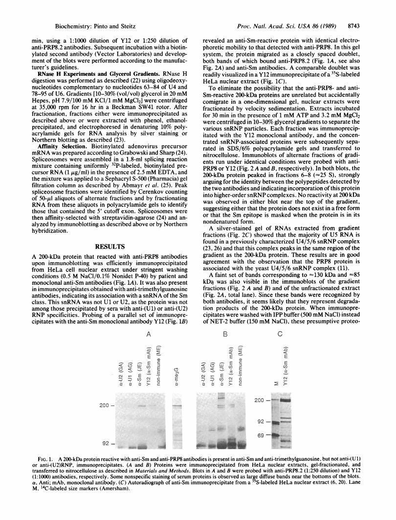
RESULTSA 200-kDa protein that reacted with anti-PRP8 antibodiesupon immunoblotting was efficiently immunoprecipitatedfrom HeLa cell nuclear extract under stringent washingconditions (0.5 M NaCl/0.1% Nonidet P-40) by patient andmonoclonal anti-Sm antibodies (Fig. 1A). It was also presentin immunoprecipitates obtained with anti-trimethylguanosineantibodies, indicating its association with a snRNA of the Smclass. This snRNA was not U1 or U2, as the protein was notamong those precipitated by sera with anti-(U1) or anti-(U2)RNP specificities. Probing of a parallel set of immunopre-cipitates with the anti-Sm monoclonal antibody Y12 (Fig. 1B)
A
U
E C
US E
C- .
revealed an anti-Sm-reactive protein with identical electro-phoretic mobility to that detected with anti-PRP8. In this gelsystem, the protein migrated as a closely spaced doublet,both bands of which bound anti-PRP8.2 (Fig. 1A, see alsoFig. 2A) and anti-Sm antibodies. A comparable doublet wasreadily visualized in a Y12 immunoprecipitate ofa 35S-labeledHeLa nuclear extract (Fig. 1C).To eliminate the possibility that the anti-PRP8- and anti-
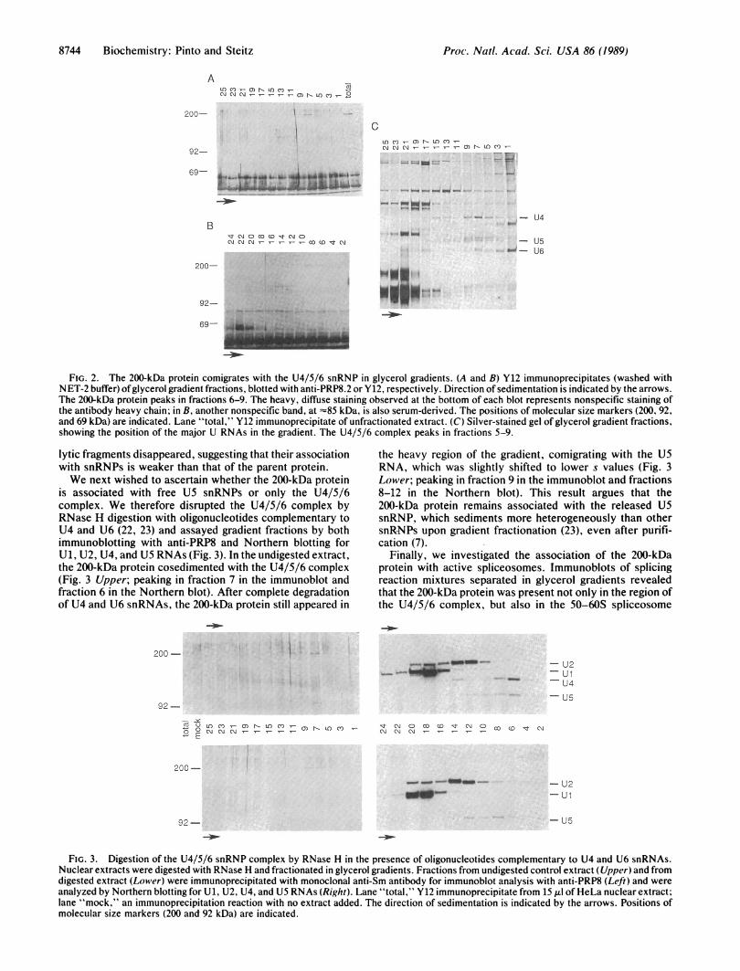
Sm-reactive 200-kDa proteins are unrelated but accidentallycomigrate in a one-dimensional gel, nuclear extracts werefractionated by velocity sedimentation. Extracts incubatedfor 30 min in the presence of 1 mM ATP and 3.2 mM MgCl2were centrifuged in 10-30% glycerol gradients to separate thevarious snRNP particles. Each fraction was immunoprecip-itated with the Y12 monoclonal antibody, and the concen-trated snRNP-associated proteins were subsequently sepa-rated in SDS/6% polyacrylamide gels and transferred tonitrocellulose. Immunoblots of alternate fractions of gradi-ents run under identical conditions were probed with anti-PRP8 or Y12 (Fig. 2 A and B, respectively). In both blots, the200-kDa protein peaked in fractions 6-8 (-25 S), stronglyarguing for the identity between the polypeptides detected bythe two antibodies and indicating incorporation of this proteininto higher-order snRNP complexes. No reactivity at 200 kDawas observed in either blot near the top of the gradient,suggesting either that the protein does not exist in a free formor that the Sm epitope is masked when the protein is in itsnondenatured form.A silver-stained gel of RNAs extracted from gradient
fractions (Fig. 2C) showed that the majority of U5 RNA isfound in a previously characterized U4/5/6 snRNP complex(23, 26) and that this complex peaks in the same region of thegradient as the 200-kDa protein. These results are in goodagreement with the observation that the PRP8 protein isassociated with the yeast U4/5/6 snRNP complex (11).A faint set of bands corresponding to -130 kDa and =85
kDa was also visible in the immunoblots of the gradientfractions (Fig. 2 A and B) and of the unfractionated extract(Fig. 2A, total lane). Since these bands were recognized byboth antibodies, it seems likely that they represent degrada-tion products of the 200-kDa protein. When immunopre-cipitates were washed with IPP buffer (500 mM NaCl) insteadof NET-2 buffer (150 mM NaCl), these presumptive proteo-
< _5OD -rcE- _)_~~~~ B
<: M
Cl)-i C E5E
ECCcn l> c
- o
200 -200
92-|M -
92 - ilgoa_
C
EEcn
'r-Cl)
FIG. 1. A 200-kDa protein reactive with anti-Sm and anti-PRP8 antibodies is present in anti-Sm and anti-trimethylguanosine, but not anti-(U 1)or anti-(U2)RNP, immunoprecipitates. (A and B) Proteins were immunoprecipitated from HeLa nuclear extracts, gel-fractionated, andtransferred to nitrocellulose as described in Materials and Methods. Blots in A and B were probed with anti-PRP8.2 (1:250 dilution) and Y12(1:1000) antibodies, respectively. Some nonspecific staining of serum proteins is observed as large diffuse bands near the bottoms of the blots.a, Anti; mAb, monoclonal antibody. (C) Autoradiograph of anti-Sm immunoprecipitate from a 35S-labeled HeLa nuclear extract (6, 20). LaneM, 'IC-labeled size markers (Amersham).
Biochemistry: Pinto and Steitz
69 -#*-ON"%*
8744 Biochemistry: Pinto and Steitz
A
200-
MMCM - CO N MC) '-
92-
69- ... .. . _-a
Bst N 0 co CD CM OCM CM - - - - - 0C tCM
200-
92-.. ......69p . ,̂
69 A.:
CLA CO) 0 n LACM)CMCMCM - ' O ) L IC')
- U4
- U50- - U6
""....e.I6_
FIG. 2. The 200-kDa protein comigrates with the U4/5/6 snRNP in glycerol gradients. (A and B) Y12 immunoprecipitates (washed withNET-2 buffer) ofglycerol gradient fractions, blotted with anti-PRP8.2 or Y12, respectively. Direction of sedimentation is indicated by the arrows.The 200-kDa protein peaks in fractions 6-9. The heavy, diffuse staining observed at the bottom of each blot represents nonspecific staining ofthe antibody heavy chain; in B, another nonspecific band, at =85 kDa, is also serum-derived. The positions of molecular size markers (200, 92,and 69 kDa) are indicated. Lane "total," Y12 immunoprecipitate of unfractionated extract. (C) Silver-stained gel of glycerol gradient fractions,showing the position of the major U RNAs in the gradient. The U4/5/6 complex peaks in fractions 5-9.
lytic fragments disappeared, suggesting that their associationwith snRNPs is weaker than that of the parent protein.We next wished to ascertain whether the 200-kDa protein
is associated with free U5 snRNPs or only the U4/5/6complex. We therefore disrupted the U4/5/6 complex byRNase H digestion with oligonucleotides complementary toU4 and U6 (22, 23) and assayed gradient fractions by bothimmunoblotting with anti-PRP8 and Northern blotting forU1, U2, U4, and U5 RNAs (Fig. 3). In the undigested extract,the 200-kDa protein cosedimented with the U4/5/6 complex(Fig. 3 Upper; peaking in fraction 7 in the immunoblot andfraction 6 in the Northern blot). After complete degradationof U4 and U6 snRNAs, the 200-kDa protein still appeared in
200-
92-
1' 0LA C') ) N LA C') C)
-E
the heavy region of the gradient, comigrating with the U5RNA, which was slightly shifted to lower s values (Fig. 3Lower; peaking in fraction 9 in the immunoblot and fractions8-12 in the Northern blot). This result argues that the200-kDa protein remains associated with the released U5snRNP, which sediments more heterogeneously than othersnRNPs upon gradient fractionation (23), even after purifi-cation (7).
Finally, we investigated the association of the 200-kDaprotein with active spliceosomes. Immunoblots of splicingreaction mixtures separated in glycerol gradients revealedthat the 200-kDa protein was present not only in the region ofthe U4/5/6 complex, but also in the 50-60S spliceosome
- U2-Ul
- U4
- U5
CU Co Lw ' MN Co D CMCM C\M - - -
20C --_ .-- -
tow
92-
- U2-Ul
- U5
FIG. 3. Digestion of the U4/5/6 snRNP complex by RNase H in the presence of oligonucleotides complementary to U4 and U6 snRNAs.Nuclear extracts were digested with RNase H and fractionated in glycerol gradients. Fractions from undigested control extract (Upper) and fromdigested extract (Lower) were immunoprecipitated with monoclonal anti-Sm antibody for immunoblot analysis with anti-PRP8 (Left) and wereanalyzed by Northern blotting for U1, U2, U4, and U5 RNAs (Right). Lane "total," Y12 immunoprecipitate from 15 Al of HeLa nuclear extract;lane "mock," an immunoprecipitation reaction with no extract added. The direction of sedimentation is indicated by the arrows. Positions ofmolecular size markers (200 and 92 kDa) are indicated.
Proc. Natl. Acad. Sci. USA 86 (1989)

Proc. Natl. Acad. Sci. USA 86 (1989) 8745
region (data not shown). Northern blots of the same gradi-ents, however, showed that although splicing precursor andintermediates peaked in the 50-60S region, all the majorsnRNP particles were distributed throughout the entire gra-dient, so that it was impossible to distinguish between thosein true splicing complexes and those that were simply inaggregates. A two-step procedure utilizing size fractionation(29) followed by affinity selection based on the presence ofpre-mRNA (24) was therefore used to generate pure spliceo-somes to analyze for the presence of the 200-kDa protein.
Splicing complexes (29) were assembled on a biotinylatedadenovirus precursor mRNA (24) in the presence of 2.5 mMEDTA (25). Under these conditions spliceosomes form butdo not promote pre-mRNA cleavage, thereby leading to theiraccumulation. The restriction is somewhat leaky, however,and the first step ofthe splicing reaction often does occur withvariable low efficiency. EDTA also serves to protect theprecursor mRNA from Mg2+-dependent ribonucleases. Re-action mixtures were then size-fractionated on SephacrylS-500 (25, 29) and spliceosomes were affinity-purified frompeak fractions by precipitation with streptavidin-agarose(24). A second reaction mixture, identical except for theomission of precursor RNA, was carried through the sameprocedure for comparison. Northern blotting of fractionsfrom the spliceosome region of the gel filtration columnsshowed that the relative abundances of the splicing snRNAsUl, U2, U4, U5, and U6 were unchanged regardless of thepresence of precursor (Fig. 4A, lanes labeled "unsel").Precipitation with streptavidin-agarose, however, selectedonly those snRNPs that had bound pre-mRNA and were thuspresent in true splicing complexes (Fig. 4A, lanes "sel").Immunoblots of this streptavidin-selected material with theanti-PRP8 antibody showed that the 200-kDa protein isindeed a component of the spliceosome, as it was found inaffinity-selected fractions only from the reaction mixturecontaining precursor RNA (Fig. 4B). Two proteins of lower
A
pre -
a
U2
UlU4
U5
U6 -
sel unsel
a a2 + +
a
-"-0cm
^
Bsel
Qa)a)51
200 -
92 -
69 -5' exon
FIG. 4. The 200-kDa protein is found in affinity-purified spliceo-somes. (A) Northern blot ofRNAs from the spliceosome region ofgelfiltration column runs with or without precursor (+ or - pre). Lanes"sel," RNA that had been affinity-selected (from 0.6 ml of columnfractions) with streptavidin-agarose; lanes "unsel," total RNA from0.1 ml of these fractions. Positions of major U RNAs, pre-mRNA(pre), and 5' cut-offexon (5' exon) are marked. Lane M, a 32P-labeledMsp I digest of pBR322 DNA used as size markers. (B) Immunoblotwith the anti-PRP8.2 antibody of streptavidin-agarose-selected ma-terial from 1.2 ml of fractions from the spliceosome region of gelfiltration columns with or without precursor (+ or - pre). Positionsof molecular size markers (200, 92, and 69 kDa) are indicated.
molecular mass were visible in both lanes of the immunoblot(Fig. 4B). As they appeared in both reaction mixtures, and asthe streptavidin-agarose beads were washed only with low-salt (100 mM KCl) buffer to keep spliceosomes intact, theymost likely represent the nonspecific association of proteo-lytic fragments of the 200-kDa protein, as discussed above.
DISCUSSIONHere we report the identification of the mammalian analogueof the yeast PRP8 protein first characterized by Lossky andcoworkers (11, 12). Like its yeast counterpart, this largepolypeptide (200 kDa in HeLa cells, 260 kDa in yeast) isassociated with the U4/5/6 snRNP complex in HeLa nuclearextracts. The mammalian protein not only reacts with anti-sera against yeast PRP8, but has its own Sm epitope asdetermined by immunoblot experiments. A 200-kDa proteinthat reacts with anti-PRP8 antibodies and weakly with theY12 monoclonal anti-Sm antibody was also reported inpurified preparations of U5 snRNPs by Bach et al. (7). Ourimmunoblots of proteins from size-fractionated, affinity-purified splicing complexes demonstrate that the 200-kDaprotein is an integral part of the mammalian spliceosome,consistent with the observation that anti-PRP8 antibodies canprecipitate intermediates from yeast splicing reactions (11).Also in the HeLa system, M. Garcia-Blanco and P. A. Sharp(personal communication) have observed UV-crosslinking ofan -220-kDa protein to competent splicing substrates in thepresence of ATP and Mg2+, with kinetics that parallel splice-osome formation; this crosslinked protein comigrates with ananti-PRP8-reactive band on immunoblots and is weakly im-munoprecipitable with anti-PRP8 antibodies.Bach et al. (7) reported that purified U5 snRNPs contain,
in addition to the common core polypeptides, several uniqueproteins of 200, 116, 102/100, 52, and 40 kDa. The 100-kDaU5-specific protein reacts strongly with anti-Sm antibodiesand is likely to be the intron-binding protein (IBP) previouslyidentified by Tazi et al. (27) and Gerke and Steitz (28). Thesize of IBP reported by the two groups differed (100 vs. 70kDa), but there are many indications (V. Gerke, personalcommunication) that the 70-kDa molecule may be a productof limited proteolysis of the larger polypeptide. We thereforeexpected that a 100-kDa or 70-kDa Sm-reactive proteinassociated with the U5 and/or the U4/5/6 snRNP would beapparent in our analyses. Yet immunoblots of nuclear ex-tracts fractionated on glycerol gradients (Fig. 2B) did notreveal any bands of these sizes in the U5 or U4/5/6 regions.We have, however, repeatedly observed a strongly Sm-reactive protein of -65 kDa in our top gradient fractions (theregion in which uncomplexed proteins would be expected tosediment). We believe this may be the aforementioned 70-kDa fragment of the U5-specific 100-kDa protein and IBP forseveral reasons. First, both its size and Sm antigenicity areconsistent with previous reports. [It should be noted that theUl-specific 70-kDa protein does not react with the Y12monoclonal anti-Sm antibody (5), nor does U1 RNA comi-grate in glycerol gradients with the -65-kDa polypeptideobserved in our gradients (Fig. 2C).] Second, it has beenreported (7) that the majority of U5 snRNPs (80-85%)purified by anti-trimethylguanosine affinity chromatographyfrom splicing extracts do not contain any U5-specific pro-teins. Thus it might be anticipated that the bulk ofany oftheseproteins would separate from the core U5 snRNP upongradient fractionation. Alternatively, it is possible that deg-radation of the 100-kDa protein to 70 kDa results in loss ofinteraction with the U5 snRNP, while the Sm epitope isretained. Further studies are required to clarify this situation.That we do not observe any 200-kDa protein at the top of ourgradients suggests that it, in contrast, is significantly morestably associated with the U5 particle and its higher aggre-
Biochemistry: Pinto and Steitz

8746 Biochemistry: Pinto and Steitz
gates, although the possibility that free 200-kDa protein is notefficiently immunoprecipitated despite its Sm epitope cannotbe excluded.The observation that the 200-kDa protein is present in the
U4/U5/U6 snRNP complex and the spliceosome raises in-triguing questions regarding its role in splicing. Its essenti-ality in yeast and its strong conservation from yeast tohumans indicate some important function. Since the U4/5/6complex is a precursor to the spliceosome (26), possibilitiesinclude recognition of the 3' splice site in either the first orsecond step of the splicing reaction or mediation of snRNP-snRNP interactions during spliceosome assembly or disas-sembly.
Note Added in Proof. Yeast PRP8 protein has recently been identifiedin affinity-purified yeast spliceosomes (E. Whittaker, M. Lossky,and J. Beggs, personal communication), and its mammalian coun-terpart has been shown to be an RNA-binding protein (G. Anderson,M. Bach, R. Luhrmann, and J. Beggs, personal communication). Adoublet at '205 kDa has also been observed in affinity-purified,1251-labeled spliceosomal proteins from HeLa nuclear extracts (R.Reed, personal communication).
We thank Jean Beggs for the generous gift ofanti-PRP8 antibodies.Mariano Garcia-Blanco for the communication of UV-crosslinkingdata prior to publication, and Volker Gerke, Reinhard Luhrmann,Ursula Bond, Kazimierz Tyc, and Michael Lerner for helpful dis-cussions. We thank Karen Montzka for nuclear extract used in theexperiments in Fig. 4. This work was supported by a Public HealthService grant (GM26154) from the National Institutes of Health toJ.A.S. and by a Damon Runyon-Walter Winchell Cancer ResearchFund fellowship (DRG-944) to A.L.P.
1. Reed, R. & Maniatis, T. (1987) Nature (London) 325, 673-678.2. Steitz, J. A., Black, D. L., Gerke, V., Parker, K. A., Kramer,
A., Frendeway, D. & Keller, W. (1988) in Structure andFunction ofMajor and Minor Small Nuclear RibonucleoproteinParticles, ed. Birnstiel, M. L. (Springer, Berlin), pp. 115-154.
3. Sharp, P. A. (1989) J. Am. Med. Assoc. 260, 3035-3041.4. Luhrmann, R. (1988) in Structure and Function of Major and
Minor Small Nuclear Ribonucleoprotein Particles, ed. Birn-stiel, M. L. (Springer, Berlin), pp. 71-99.
5. Pettersson, I., Hinterberger, M., Mimori, T., Gottlieb, E. &Steitz, J. A. (1984) J. Biol. Chem. 259, 5907-5914.
6. Mimori, T., Hinterberger, M., Pettersson, 1. & Steitz, J. A.(1984) J. Biol. Chem. 259, 560-565.
7. Bach, M., Winkelmann, G. & Luhrmann, R. (1989) Proc. Natl.Acad. Sci. USA 86, 6038-6042.
8. Lamond, A. I., Sproat, B., Ryder, U. & Hamm, J. (1989) Cell58, 383-390.
9. Hartwell, L. H. (1967) J. Bacteriol. 93, 1662-1670.10. Vijayrhagavan, U., Company, M. & Abelson, J. (1989) Genes
Dev. 3, 1206-1216.11. Lossky, M., Anderson, G. J., Jackson, S. P. & Beggs, J. (1987)
Cell 51, 1019-1026.12. Jackson, S. P., Lossky, M. & Beggs, J. D. (1988) Mol. Cell.
Biol. 8, 1067-1075.13. Banroques, J. & Abelson, J. (1989) Mol. Cell. Biol. 9, 3710-
3719.14. Bj0rn, S. P., Soltyk,, A., Beggs, J. D. & Friesen, J. D. (1989)
Mol. Cell. Biol. 9, 3698-3709.15. Couto, J. R., Tamm, Jw Parker, R. & Guthrie, C. (1987) Genes
Dev. 1, 445-456.16. Chang, T.-H., Clark, M. W., Lustig, A. J., Cusick, M. E. &
Abelson, J. (1988) Mol. Cell. Biol. 8, 2379-2393.17. Lin, R.-J., Lustig, A. J. & Abelson, J. (1987) Genes Del'. 1,
7-18.18. Lerner, E. A., Lerner, M. R., Janeway, C. A. & Steitz, J. A.
(1981) Proc. Natl. Acaid. Sci. USA 78, 2737-2741.19. Krainer, A. R. (1988) Nucleic Acids Res. 16, 9415-9429.20. Dignam, J. D., Lebovitz, R. M. & Roeder, R. G. (1983) Nu-
cleic Acids Res. 11, 1475-1489.21. Towbin, H., Staehelin, T. & Gordon, J. (1979) Proc. Natl.
Acad. Sci. USA 79, 5188-5189.22. Black, D. L. & Steitz, J. A. (1986) Cell 46, 697-704.23. Black, D. L. & Pinto, A. L. (1989) Mol. Cell. Biol. 9, 3350-
3359.24. Grabowski, P. J. & Sharp, P. A. (1986) Science 233, 1294-
1299.25. Abmayr, S. M., Reed, R. & Maniatis, T. (1988) Proc. Natl.
Acad. Sci. USA 85, 7216-7220.26. Konarska, M. M. & Sharp, P. A. (1987) Cell 49, 763-774.27. Tazi, J., Alibert, C., Temsamani, J., Reveillaud, I., Cathala, G.,
Brunei, C. & Jeanteur, P. (1986) Cell 47, 755-766.28. Gerke, V. & Steitz, J. A. (1986) Cell 47, 973-984.29. Reed, R., Griffith, J. & Maniatis, T. (1988) Cell 49, 949-961.
Proc. Natl. Acad. Sci. USA 86 (1989)