therapeutic effect of lecithinized superoxide dismutase on ... · therapeutic effect of...
TRANSCRIPT

Therapeutic Effect of Lecithinized Superoxide Dismutase onPulmonary Emphysema□S
Ken-Ichiro Tanaka, Yuta Tanaka, Yuri Miyazaki, Takushi Namba, Keizo Sato,Kazutetsu Aoshiba, Arata Azuma, and Tohru MizushimaGraduate School of Medical and Pharmaceutical Sciences, Kumamoto University, Kumamoto, Japan (K-I.T., Y.T., Y.M., T.N.,K.S., T.M.); First Department of Medicine, Tokyo Women’s Medical University, Tokyo, Japan (K.A.); and Department of InternalMedicine, Division of Respiratory, Infection, and Oncology, Nippon Medical School, Tokyo, Japan (A.A.)
Received January 6, 2011; accepted June 3, 2011
ABSTRACTNo medication exists that clearly improves the mortality ofchronic obstructive pulmonary disease (COPD). Oxidative mol-ecules, in particular superoxide anions, play important roles inthe COPD-associated abnormal inflammatory response andpulmonary emphysema, which arises because of an imbalancein proteases and antiproteases and increased apoptosis. Su-peroxide dismutase (SOD) catalyzes the dismutation of super-oxide anions. Lecithinized human Cu/Zn- SOD (PC-SOD) hasovercome a number of the clinical limitations of SOD, includinglow tissue affinity and low stability in plasma. In this study, weexamine the effect of PC-SOD on elastase-induced pulmonaryemphysema, an animal model of COPD. The severity of thepulmonary inflammatory response and emphysema in micewas assessed by various criteria, such as the number of leu-kocytes in the bronchoalveolar lavage fluid and the enlarge-
ment of airspace. Not only intravenous administration but alsoinhalation of PC-SOD suppressed elastase-induced pulmonaryinflammation, emphysema, and dysfunction. Inhalation of PC-SOD suppressed the elastase-induced increase in the pulmo-nary level of superoxide anions and apoptosis. Inhalation ofPC-SOD also suppressed elastase-induced activation of pro-teases and decreased in the level of antiproteases and expres-sion of proinflammatory cytokines and chemokines. We alsofound that inhalation of PC-SOD suppressed cigarette smoke-induced pulmonary inflammation. The results suggest that PC-SOD protects against pulmonary emphysema by decreasingthe pulmonary level of superoxide anions, resulting in the inhi-bition of inflammation and apoptosis and amelioration of theprotease/antiprotease imbalance. We propose that inhalationof PC-SOD would be therapeutically beneficial for COPD.
IntroductionChronic obstructive pulmonary disease (COPD) is cur-
rently the fourth leading cause of death in the world, and itsprevalence and mortality rates have been increasing (Rabe etal., 2007). COPD is a disease state defined by irreversible andprogressive airflow limitation associated with an abnormal
inflammatory response. The most important etiologic factorfor COPD is cigarette smoking (CS) (Peto et al., 1999; Rabe etal., 2007). Pathologic characteristics of COPD include infil-tration of leukocytes, enhanced mucus secretion, dysfunc-tional airway matrix remodeling, and destruction of paren-chyma (enlargement of airspace) (Barnes and Stockley, 2005;Owen, 2005; Rabe et al., 2007). Protease/antiprotease imbal-ance and apoptosis play important roles in this emphysema-tous lung destruction. Unfortunately, there is no effectivedrug therapy that is able to significantly and clearly modu-late disease progression and mortality (Calverley et al., 2007;Miravitlles and Anzueto, 2009).
It has been suggested that oxidative molecules play animportant role in the pathogenesis of COPD (Pinamonti etal., 1998; Nadeem et al., 2005; Mak, 2008). In addition to
This work was supported by the Ministry of Health, Labor, and Welfare ofJapan [Grant-in-Aid H22-005]; the Japan Science and Technology Agency; andthe Ministry of Education, Culture, Sports, Science and Technology of Japan[Grant-in-Aid 20015037].
Article, publication date, and citation information can be found athttp://jpet.aspetjournals.org.
doi:10.1124/jpet.111.179051.□S The online version of this article (available at http://jpet.aspetjournals.org)
contains supplemental material.
ABBREVIATIONS: COPD, chronic obstructive pulmonary disease; BALF, bronchoalveolar lavage fluid; CS, cigarette smoking; DAPI, 4,6-diamino-2-phenylindole; DPhPMPO, 2-diphenylphosphinoyl-2-methyl-3,4-dihydro-2H-pyrrole N-oxide; ELISA, enzyme-linked immunosorbent assay; ESR,electron spin resonance; GAPDH, glyceraldehyde-3-phosphate dehydrogenase; H and E, hematoxylin and eosin; IL, interleukin; IPF, idiopathicpulmonary fibrosis; KC, keratinocyte-derived chemokine; kU, kilounit; MCP, monocyte chemoattractant protein; MIP, macrophage inflammatoryprotein; MMP, matrix metalloproteinase; 8-OHdG, 8-hydroxy-2�-deoxyguanosine; PC, phosphatidylcholine; RT-PCR, reverse transcription-polymerase chain reaction; SOD, superoxide dismutase; PC-SOD, lecithinized human Cu/Zn-SOD; PPE, porcine pancreatic elastase; QOL, qualityof life; ROS, reactive oxygen species; TNF, tumor necrosis factor; TUNEL, terminal deoxynucleotidyl transferase dUTP nick-end labeling.
0022-3565/11/3383-810–818$25.00THE JOURNAL OF PHARMACOLOGY AND EXPERIMENTAL THERAPEUTICS Vol. 338, No. 3Copyright © 2011 by The American Society for Pharmacology and Experimental Therapeutics 179051/3709971JPET 338:810–818, 2011 Printed in U.S.A.
810
at Univ O
f Georgia Libraries on D
ecember 16, 2013
jpet.aspetjournals.orgD
ownloaded from
http://jpet.aspetjournals.org/content/suppl/2011/06/10/jpet.111.179051.DC1.html Supplemental Material can be found at:
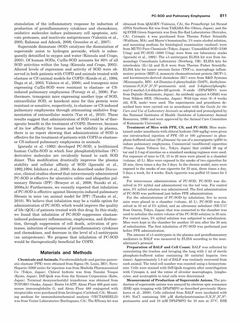
stimulation of the inflammatory response by induction ofproduction of proinflammatory cytokines and chemokines,oxidative molecules induce pulmonary cell apoptosis, acti-vate proteases, and inactivate antiproteases (Valentin et al.,2005; Rahman and Adcock, 2006; Greenlee et al., 2007).
Superoxide dismutase (SOD) catalyzes the dismutation ofsuperoxide anion to hydrogen peroxide, which is subse-quently detoxified to oxygen and water (Kinnula and Crapo,2003). Of human SODs, Cu/Zn-SOD accounts for 80% of allSOD activities within the lung (Kinnula and Crapo, 2003).Altered levels of expression and activity of SOD were ob-served in both patients with COPD and animals treated withelastase or CS (animal models for COPD) (Kondo et al., 1994;Daga et al., 2003; Valenca et al., 2008), and transgenic miceexpressing Cu/Zn-SOD were resistant to elastase- or CS-induced pulmonary emphysema (Foronjy et al., 2006). Fur-thermore, transgenic mice expressing another type of SOD,extracellular SOD, or knockout mice for this protein wereresistant or sensitive, respectively, to elastase- or CS-inducedpulmonary emphysema through attenuating oxidative frag-mentation of extracellular matrix (Yao et al., 2010). Theseresults suggest that administration of SOD could be of ther-apeutic benefit in the treatment of COPD. However, becauseof its low affinity for tissues and low stability in plasma,there is no report showing that administration of SOD iseffective for the treatment of patients with COPD or elastase-or CS-induced pulmonary emphysema in animals.
Igarashi et al. (1992) developed PC-SOD, a lecithinizedhuman Cu/Zn-SOD in which four phosphatidylcholine (PC)derivative molecules are covalently bound to each SODdimer. This modification drastically improves the plasmastability and cellular affinity of SOD (Igarashi et al.,1992,1994; Ishihara et al., 2009). As described under Discus-sion, clinical studies showed that intravenously administeredPC-SOD is effective for ulcerative colitis and idiopathic pul-monary fibrosis (IPF) (Broeyer et al., 2008; Suzuki et al.,2008a,b) Furthermore, we recently reported that inhalationof PC-SOD is effective against bleomycin-induced pulmonaryfibrosis in mice (an animal model for IPF) (Tanaka et al.,2010). We believe that inhalation may be a viable option foradministration of PC-SOD, which would improve the qualityof life (QOL) of patients treated with this drug. In this study,we found that inhalation of PC-SOD suppresses elastase-induced pulmonary inflammation, emphysema, and dysfunc-tion, through suppression of cell death, activation of pro-teases, induction of expression of proinflammatory cytokinesand chemokines, and decrease in the level of �1-antitrypsin(an antiprotease). We propose that inhalation of PC-SODwould be therapeutically beneficial for COPD.
Materials and MethodsChemicals and Animals. Paraformaldehyde and porcine pancre-
atic elastase (PPE) were obtained from Sigma (St. Louis, MO). Novo-Heparin (5000 units) for injection was from Mochida PharmaceuticalCo. (Tokyo, Japan). Chloral hydrate was from Nacalai Tesque(Kyoto, Japan). Diff-Quik was from the Sysmex Corporation (Kobe,Japan). Terminal deoxynucleotidyl transferase was obtained fromTOYOBO (Osaka, Japan). Biotin 14-ATP, Alexa Fluor 488 goat anti-mouse immunoglobulin G, and Alexa Fluor 488 conjugated withstreptavidin were purchased from Invitrogen (Carlsbad, CA). Mount-ing medium for immunohistochemical analysis (VECTASHIELD)was from Vector Laboratories (Burlingame, CA). The RNeasy kit was
obtained from QIAGEN (Valencia, CA), the PrimeScript 1st StrandcDNA Synthesis Kit was from TAKARA Bio (Ohtsu, Japan), and theiQ SYBR Green Supermix was from Bio-Rad Laboratories (Hercules,CA). Cytospin 4 was purchased from Thermo Fisher Scientific(Waltham, MA), and Mayer’s hematoxylin, 1% eosin alcohol solution,and mounting medium for histological examination (malinol) werefrom MUTO Pure Chemicals (Tokyo, Japan). Unmodified SOD (5190U/mg) and PC-SOD (3000 U/mg) were from our laboratory stocks(Igarashi et al., 1992). The �1-antitrypsin ELISA kit was from Im-munology Consultants Laboratory (Newberg, OR). ELISA kits forinterleukin (IL)-1� and IL-6 were from Thermo Fisher Scientific.ELISA kits for tumor necrosis factor (TNF)-�, macrophage inflam-matory protein (MIP)-2, monocyte chemoattractant protein (MCP)-1,and keratinocyte-derived chemokine (KC) were from R&D Systems(Minneapolis, MN). 4,6-Diamino-2-phenylindole (DAPI), diethylene-triamine-N,N,N�,N�,N�-pentaacetic acid, and 2-diphenylphosphi-noyl-2-methyl-3,4-dihydro-2H-pyrrole N-oxide (DPhPMPO) werefrom Dojindo (Kumamoto, Japan). An antibody against 8-OHdG wasfrom Nikken SEIL (Shizuoka, Japan). Wild-type mice (6–8 weeksold, ICR, male) were used. The experiments and procedures de-scribed here were carried out in accordance with the Guide for theCare and Use of Laboratory Animals as adopted and promulgated bythe National Institutes of Health (Institute of Laboratory AnimalResources, 1996) and were approved by the Animal Care Committeeof Kumamoto University.
Treatment of Mice with PPE, CS, and PC-SOD. Mice main-tained under anesthesia with chloral hydrate (500 mg/kg) were givenone intratracheal injection of PPE (50 or 100 �g/mouse) in phos-phate-buffered saline (30 �l/mouse) by use of a micropipette (p200) toinduce pulmonary emphysema. Commercial (nonfiltered) cigarettes(Peace; Japan Tobacco Inc., Tokyo, Japan) that yielded 28 mg oftar and 2.3 mg of nicotine on a standard smoking regimen were used.For exposure of mice to CS, 15 to 20 mice were placed in a chamber(volume, 45 L). Mice were exposed to the smoke of two cigarettes for25 min, three times a day for 3 days. In the chronic model, mice wereexposed to the smoke of one cigarette for 35 min, three times a day,5 days a week, for 4 weeks. Each cigarette was puffed 15 times for 5min.
For intravenous administration of PC-SOD, PC-SOD was dis-solved in 5% xylitol and administered via the tail vein. For controlmice, 5% xylitol solution was administered. The first administrationof PC-SOD was performed just before PPE administration.
For the administration of PC-SOD by inhalation, five to sevenmice were placed in a chamber (volume, 45 L). PC-SOD was dis-solved in 10 ml of 5% xylitol, and an ultrasonic nebulizer (NE-U17from Omron, Tokyo, Japan) that was connected to the chamber wasused to nebulize the entire volume of the PC-SOD solution in 30 min.For control mice, 5% xylitol solution was subjected to nebulization.Mice were kept in the chamber for another 10 min after the 30 minof nebulization. The first inhalation of PC-SOD was performed justbefore PPE administration.
The amount of �1-antitrypsin in the plasma and proinflammatorymediators in BALF was measured by ELISA according to the man-ufacturer’s protocol.
Preparation of BALF and Cell Count. BALF was collected bycannulating the trachea and lavaging the lung with 1 ml of sterilephosphate-buffered saline containing 50 units/ml heparin (twotimes). Approximately 1.8 ml of BALF was routinely recovered fromeach animal. The total cell number was counted using a hemocytom-eter. Cells were stained with Diff-Quik reagents after centrifugationwith Cytospin 4, and the ratios of alveolar macrophages, lympho-cytes, and neutrophils to total cells were determined.
Measurement of Production of Superoxide Anions. The pro-duction of superoxide anions was assayed by electron spin resonance(ESR) spin trapping with DPhPMPO as described previously (Kara-kawa et al., 2008). Cells collected from BALF were incubated with0.9% NaCl containing 500 �M diethylenetriamine-N,N,N�,N�,N�-pentaacetic acid and 10 mM DPhPMPO for 10 min at 37°C. ESR
PC-SOD and Pulmonary Emphysema 811

spectra were recorded at room temperature on a JES-TE200 ESRspectrometer (JEOL, Tokyo, Japan) under the following conditions:modulation frequency, 100 kHz; microwave frequency, 9.43 GHz;microwave power, 40 mW; scanning field, 335.2 � 5 mT; sweep time,2 min; field modulation width, 0.25 mT; receiver gain, 400; and timecount, 0.3 s. Every buffer and solution used in the reaction mixtureused for ESR measurement was treated with Chelex 100 resin(Bio-Rad Laboratories) before use to remove metals.
Histological and Immunohistochemical Analyses and Ter-minal Deoxynucleotidyl Transferase dUTP Nick-End Label-ing Assay. Lung tissue samples were fixed for 24 h at a pressure of25 cm H2O, and then embedded in paraffin before being cut into 4�m-thick sections.
For histological examination, sections were stained first withMayer’s hematoxylin and then with 1% eosin alcohol solution [he-matoxylin and eosin (H and E) staining]. Samples were mountedwith malinol and inspected with the aid of an Olympus (Tokyo,Japan) BX51 microscope. Twenty lines (500 �m) were drawn ran-domly on the image of sections stained with H and E, and theintersection points with the alveolar walls were counted to deter-mine the mean linear intercept. The morphometric analysis at thelight microscopic level was conducted by a blinded investigator.
For immunohistochemical analysis, sections were treated with 20�g/ml protease K for antigen activation. Sections were blocked with2.5% goat serum for 10 min, incubated for 12 h with an antibodyagainst 8-OHdG (1:100 dilution) in the presence of 2.5% bovineserum albumin, and then incubated for 1 h with Alexa Fluor 488 goatanti-mouse IgG in the presence of DAPI (5 �g/ml). Samples weremounted with VECTASHIELD and inspected using fluorescence mi-croscopy (Olympus BX51).
For the TUNEL assay, sections were incubated first with protei-nase K (20 �g/ml) for 15 min at 37°C, then with TdTase and biotin14-ATP for 1 h at 37°C, and finally with Alexa Fluor 488 conjugatedwith streptavidin and DAPI (5 �g/ml) for 2 h. Samples were mountedwith VECTASHIELD and inspected with the aid of a fluorescencemicroscope (Olympus BX51).
Gelatin Zymography. The proteolytic activities of MMP-2 andMMP-9 were assessed by SDS-polyacrylamide gel electrophoresisusing zymogram gels containing 0.1% gelatin as described previously(Namba et al., 2009). The protein concentration was determined bythe Bradford method (Bradford, 1976). After electrophoresis at 4°C(10 �g of protein/lane), the gels were washed with 2.5% Triton X-100for 30 min at room temperature and incubated with zymogramdevelopment buffer for 2 days at 37°C. Bands were visualized bystaining with Coomassie brilliant blue.
Real-Time RT-PCR Analysis. Real-time RT-PCR was per-formed as described previously (Namba et al., 2009) with somemodifications. Total RNA was extracted from pulmonary tissuesusing an RNeasy kit according to the manufacturer’s protocol (QIAGEN).Samples (2.5 �g of RNA) were reverse-transcribed using a Prime-Script first-strand cDNA Synthesis Kit. Synthesized cDNA was usedin real-time RT-PCR (Chromo 4 instrument; Bio-Rad Laboratories)experiments using iQ SYBR GREEN Supermix and analyzed withOpticon Monitor Software (Bio-Rad Laboratories). Specificity wasconfirmed by electrophoretic analysis of the reaction products andinclusion of template- or reverse transcriptase-free controls. To nor-malize the amount of total RNA present in each reaction, GAPDHcDNA was used as an internal standard.
Primers were designed using the Primer3 website (http://frodo.wi.mit.edu/primer3/). The primers used were (forward primer, reverseprimer): TNF-�, 5�-cgtcagccgatttgctatct-3�, 5�-cggactccgcaaagtctaag-3�; IL-1�, 5�-gatcccaagcaatacccaaa-3�, 5�-ggggaactctgcagactcaa-3�;IL-6, 5�-ctggagtcacagaaggagtgg-3�, 5�-ggtttgccgagtagatctcaa-3�; MIP-2�, 5�-accctgccaagggttgacttc-3�, 5�-ggcacatcaggtacgatccag-3�; MCP-1,5�-ctcacctgctgctactcattc-3�, 5�-gcttgaggtggttgtggaaaa-3�; KC, 5�-tg-cacccaaaccgaagtcat-3�, 5�-ttgtcagaagccagcgttcac-3�; and GAPDH, 5�-aactttggcattgtggaagg-3�, 5�-acacattgggggtaggaaca-3�.
Analysis of Lung Function. Analysis of lung function was per-formed with a computer-controlled small-animal ventilator (FlexiVent;SCIREQ, Montreal, QC, Canada), as described previously (Kuraki etal., 2002). Mice were anesthetized with chloral hydrate (500 mg/kg),tracheotomized with an 8-mm section of metallic tubing, and me-chanically ventilated at a rate of 150 breaths/min, using a tidalvolume of 8.7 ml/kg and a positive end-expiratory pressure of 2 to 3cm H2O. The single-compartment model (snap shot) and the con-stant-phase model (forced oscillation technique) were applied to an-alyze lung function. Total respiratory system elastance and tissueelastance were measured by the snap shot and forced oscillationtechniques, respectively. All data were analyzed using FlexiVentsoftware (version 5.3) (SCIREQ).
Statistical Analysis. All values are expressed as the mean �S.E.M. Two-way analysis of variance followed by the Tukey test orthe Student’s t test for unpaired results was used to evaluate differ-ences between three or more groups or between two groups, respec-tively. Differences were considered to be significant for values of P �0.05. We repeated the experiments at least two times as independentexperiments (see figure legends) and selected one set of representa-tive data to show in the figures. The stated number of test sample isnot summation of independent plural experiments but is for only oneindependent experiment.
ResultsEffect of PC-SOD on Elastase-Induced Pulmonary
Emphysema. Pulmonary emphysema was induced in micegiven a single (at day 0) intratracheal administration of PPE.The PPE-induced pulmonary inflammatory response can bemonitored by determining the number of leukocytes (alveolarmacrophages, lymphocytes, and neutrophils) in the BALF 3days after the administration of PPE (50 �g/mouse). Asshown in Fig. 1A, the total number of leukocytes and indi-vidual numbers of alveolar macrophages, lymphocytes, andneutrophils all were increased by the PPE treatment. Thiseffect was suppressed by the simultaneous once-daily intra-venous administration of PC-SOD, suggesting that PC-SODameliorates the PPE-induced inflammatory response. How-ever, a higher dose of PC-SOD (30 kU/kg) did not suppressthe PPE-induced inflammatory response (Fig. 1A), so in thisstudy PC-SOD exhibited a bell-shaped dose-response profile,similar to that observed previously for intravenous adminis-tration of PC-SOD in animal models of other diseases (Ishi-hara et al., 2009; Tanaka et al., 2010). Intravenous adminis-tration of the higher dose (30 kU/kg) of PC-SOD alone(without PPE administration) did not affect the number ofleukocytes in the BALF (data not shown).
PPE-induced pulmonary emphysema can be monitored byhistopathological analysis and measurement of the meanlinear intercept (an indicator of airspace enlargement causedby breakdown of the alveolar walls) 3 days after the admin-istration of PPE. Histopathological analysis of pulmonarytissue using H and E staining revealed that PPE adminis-tration induced severe pulmonary damage (infiltration ofleukocytes and breakdown of the alveolar walls) and thesephenomena were suppressed by the intravenous administra-tion of low doses (1.5 and 3 kU/kg), but not of a high dose (30kU/kg), of PC-SOD (Fig. 1B). The mean linear intercept wasincreased by the administration of PPE; this increase wassuppressed by intravenous administration of low doses (1.5and 3 kU/kg) of PC-SOD but was not significantly suppressedat the higher dose (30 kU/kg) (Fig. 1C). Pulmonary tissuedamage and the increase in the mean linear intercept 14
812 Tanaka et al.

days after PPE administration were also suppressed by theintravenous administration of PC-SOD (Fig. 1, D and E). Weused higher dose of PPE (100 �g/mice) to monitor pulmonaryemphysema 14 days after the administration of PPE.
The alteration in lung mechanics associated with pulmo-nary emphysema is characterized by a decrease in elastance(Kuraki et al., 2002). We thus examined the effect of intra-venous administration of PC-SOD on PPE-induced altera-tions to lung mechanics, using a computer-controlled small-animal ventilator. Total respiratory system elastance(elastance of total lung including bronchi, bronchiole, andalveoli) and tissue elastance (elastance of alveoli) were re-duced by PPE treatment, and intravenous administration ofPC-SOD increased these indexes (Fig. 1F). These resultssuggest that not only PPE-induced pulmonary emphysemabut also PPE-induced pulmonary dysfunction is amelioratedby intravenous administration of PC-SOD.
Effect of Inhalation of PC-SOD on Elastase-InducedPulmonary Emphysema. We recently reported that inha-lation of PC-SOD ameliorates bleomycin-induced pulmonaryfibrosis (Tanaka et al., 2010). This route of administrationdoes not show a bell-shaped dose-response profile (Tanaka etal., 2010) and may result in higher QOL for patients treatedwith PC-SOD. Thus, here we examined the effect of inhala-tion of PC-SOD on PPE-induced pulmonary emphysema.Mice were placed in a chamber connected to an ultrasonicnebulizer, thus exposing them to PC-SOD-containing vapor.We confirmed, by high-performance liquid chromatographyanalysis and measurement of SOD activity, that this treat-ment did not affect the structure and activity of the PC-SOD(data not shown). Inhalation of PC-SOD-containing vaporwas repeated once daily for 3 or 14 days, and the mice wereexamined for PPE-induced pulmonary disorders. As shown inFig. 2A, inhaled PC-SOD ameliorated the PPE-induced in-flammatory response. This ameliorative effect was observedwith not only low doses (30 and 60 kU/chamber) but also ahigh dose (600 kU/chamber) of PC-SOD, suggesting that thedose-response profile for this administration route is notbell-shaped. PPE-induced emphysematous lung damage andthe increase in the mean linear intercept were also sup-pressed by inhalation of PC-SOD (Fig. 2, B–E), suggestingthat inhalation of PC-SOD ameliorates PPE-induced pulmo-nary emphysema. Again, a bell-shaped dose-response profilewas not observed for the ameliorative effect of inhalation ofPC-SOD against PPE-induced pulmonary emphysema (Fig.2, B and C). As shown in Table 1, inhalation of unmodifiedSOD (600 kU/chamber) did not affect the PPE-induced pul-monary inflammatory response and emphysema. This sug-gests that lecithinization of SOD potentiates its ameliorativeeffect against PPE-induced lung disorders, as is the case fordextran sulfate sodium-induced colitis and bleomycin-in-duced pulmonary fibrosis (Ishihara et al., 2009; Tanaka etal., 2010). We also found that inhalation of PC-SOD sup-
Fig. 1. Effect of intravenous administration of PC-SOD on PPE-inducedpulmonary emphysema. Mice treated with (except vehicle) or without(vehicle) PPE (50 or 100 �g/mouse) once at day 0 were intravenouslyadministered the indicated doses of PC-SOD (1.5, 3, or 30 kU/kg) oncedaily for 3 days (days 0–2) (A–C) or 14 days (days 0–13) (D–F). A, thetotal cell number and numbers of alveolar macrophages, lymphocytes, and
neutrophils were determined at day 3 as described under Materials andMethods. B and D, sections of pulmonary tissue were prepared at days 3or 14 and subjected to histopathological examination (H and E staining).C and E, airspace size was estimated by determining the mean linearintercept as described under Materials and Methods. F, at day 14, totalrespiratory system elastance and tissue elastance were determined asdescribed under Materials and Methods. Values are mean � S.E.M. �,P � 0.05; ��, P � 0.01. Data are representative of two independentexperiments.
PC-SOD and Pulmonary Emphysema 813

presses PPE-induced decreases in total respiratory systemelastance and tissue elastance (Fig. 2F), suggesting thatinhalation of PC-SOD ameliorates PPE-induced lung dys-function. We confirmed that inhalation of PC-SOD alonedid not induce pulmonary emphysema and dysfunction(Supplemental Fig. 1).
To consider the clinical relevance, it is important to exam-ine the effect of the drug on predeveloped lesions in ananimal model (Fig. 3). Thus, we examined the effect of inha-lation of PC-SOD on predeveloped pulmonary emphysema.Once-daily inhalation of PC-SOD was started 3 days after theadministration of PPE, and pulmonary emphysema and func-tion were assessed at day 10. Inhalation of PC-SOD causedsuppression of pulmonary emphysema at day 10, suggestingthat the inhalation of PC-SOD is effective for predevelopedlesions.
The inhalation of PC-SOD also suppressed the PPE-in-duced alterations in lung mechanics at day 10 (Fig. 3C),suggesting that inhalation of PC-SOD suppresses the PPE-induced lung dysfunction, even when it is administered afterthe PPE.
Mechanism for the Ameliorative Effects of PC-SODon PPE-Induced Pulmonary Emphysema. To confirmthat inhaled PC-SOD decreases the pulmonary level of su-peroxide anion, we performed an immunohistochemical anal-ysis to monitor the pulmonary level of 8-OHdG, the damagednucleotide produced by various ROS, including the superox-ide anion (Freeman et al., 2009). As shown in Fig. 4A, thepulmonary level of 8-OHdG was significantly increased byPPE administration, and this increase was clearly sup-pressed by inhalation of PC-SOD, suggesting that productionof ROS in the lung was suppressed by inhalation of PC-SOD.We also used ESR analysis to monitor the production ofsuperoxide anion in cells in BALF. The ESR spectrum wasconsistent with a previously reported DPhPMPO-OOH spec-trum (a hyperfine coupling constant of aN � 1.24 mT, aH
� �1.16 mT, aP � 3.95 mT) (Karakawa et al., 2008). As shown inFig. 4, B and C, the peak of a radical spin adduct of the ESRspectrum corresponding to the amount of superoxide anion(DPhPMPO-OOH adduct) was higher for cells prepared fromPPE-administered mice than for cells from control mice. In-halation of PC-SOD lowered this peak, suggesting that in-haled PC-SOD suppresses PPE-induced production of super-oxide anions in the lung.
Fig. 2. Effect of inhalation of PC-SOD on PPE-induced pulmonary em-physema. Mice treated with (except vehicle) or without (vehicle) PPE (50or 100 �g/mouse) once at day 0 inhaled the indicated doses of PC-SOD(30, 60, or 600 kU/chamber) once daily for 3 days (days 0–2) (A–C) or 14days (days 0–13) (D–F). Inflammatory response (A), airspace size (B–E),
and lung mechanics (F) were assessed as described in the legend of Fig.1. Values are mean � S.E.M. �, P � 0.05; ��, P � 0.01. Data arerepresentative of three independent experiments.
TABLE 1Effect of inhalation of unmodified SOD on PPE-induced pulmonaryemphysemaMice were treated with a single dose of PPE (50 �g/mouse) at day 0 and inhaledunmodified SOD (U-SOD; 600 kU/chamber) once daily for 3 days (days 0–2). Inflam-matory response and the mean linear intercept were assessed as described in thelegend of Fig. 1. Values are mean � S.E.M.
PPE (50)(n � 8)
U-SOD(600 kU/chamber)
(n � 4)
Total cells, 105 4.9 � 0.33 4.9 � 0.35Alveolar macrophages, 105 4.7 � 0.36 4.7 � 0.33Lymphocytes, 104 0.40 � 0.07 0.35 � 0.06Neutrophils, 104 1.6 � 0.15 1.3 � 0.19Mean linear intercept, �m 58.2 � 1.30 57.7 � 0.37
814 Tanaka et al.

As described in Introduction, pulmonary cell apoptosisplays an important role in the pathogenesis of COPD andPPE-induced pulmonary emphysema. We examined the ef-fect of inhalation of PC-SOD on PPE-induced pulmonary celldeath by using the TUNEL assay. TUNEL-positive cells (in-dicative of cell death) increased in response to administrationof PPE, and this increase was suppressed by simultaneousinhalation of PC-SOD (Fig. 4, D and E), suggesting thatPC-SOD protects pulmonary cells from PPE-induced celldeath, and this effect is involved in the ameliorative effectsof inhalation of PC-SOD against PPE-induced pulmonaryemphysema.
To examine the effect of inhalation of PC-SOD on thePPE-dependent imbalance in proteases and antiproteases,we first examined the activity of MMPs, MMP-2 and MMP-9,using gelatin zymography. The band intensities of MMP-2and MMP-9, indicative of MMP-2 and MMP-9 activities, werehigher for lung tissues prepared from PPE-administeredmice than for those from control mice, and this increase wassuppressed in mice that had inhaled PC-SOD (Fig. 5, A andB). We also examined the serum level of �1-antitrypsin byELISA and found that the level of �1-antitrypsin was de-creased by PPE administration and partially recovered bysimultaneous inhalation of PC-SOD (Fig. 5C). These resultssuggest that inhalation of PC-SOD improves the PPE-depen-dent protease/antiprotease imbalance and this effect is in-volved in the ameliorative effects of inhalation of PC-SODagainst PPE-induced pulmonary emphysema.
We also examined the effect of inhalation of PC-SOD on themRNA expression of proinflammatory cytokines (TNF-�, IL-1�, and IL-6) and chemokines (MIP-2, MCP-1, and KC) in
Fig. 4. Effect of PC-SOD on the PPE-induced increase in the level of 8-OHdG,production of superoxide anions, and pulmonary cell death. Mice treated withPPE inhaled PC-SOD (kU/chamber) for 3 days (days 0–2) (A, D, and E) or 1 day(day 0) (B and C) as described in the legend of Fig. 2. A, D, and E, sections ofpulmonary tissue were prepared at day 3. A, sections were subjected to immu-nohistochemical analysis with an antibody against 8-OHdG or DAPI staining.B, cells in BALF were collected at day 1, incubated with a spin trap agent(DPhPMPO), and subjected to radical adduct ESR spectrum analysis to deter-mine the amount of superoxide anion present. C, the intensity of the ESR signalof the superoxide anion adduct (DPhPMPO–OOH adduct shown by the bar inB) was determined. D, sections were subjected to TUNEL assay or DAPIstaining. E, the number of TUNEL-positive cells was counted. Values aremean � S.E.M. �, P � 0.05; ��, P � 0.01. Data are representative of twoindependent experiments.
Fig. 3. Effect of PC-SOD on predeveloped pulmonary emphysema. Micetreated with (except vehicle) or without (vehicle) PPE (50 �g/mouse) onceat day 0 inhaled the indicated doses of PC-SOD (kU/chamber) once dailyfrom days 3 to 9. Airspace size (A and B) and lung mechanics (C) wereassessed at day 10 as described in the legend of Fig. 1. Values are mean �S.E.M. �, P � 0.05; ��, P � 0.01. Data are representative of two indepen-dent experiments.
PC-SOD and Pulmonary Emphysema 815
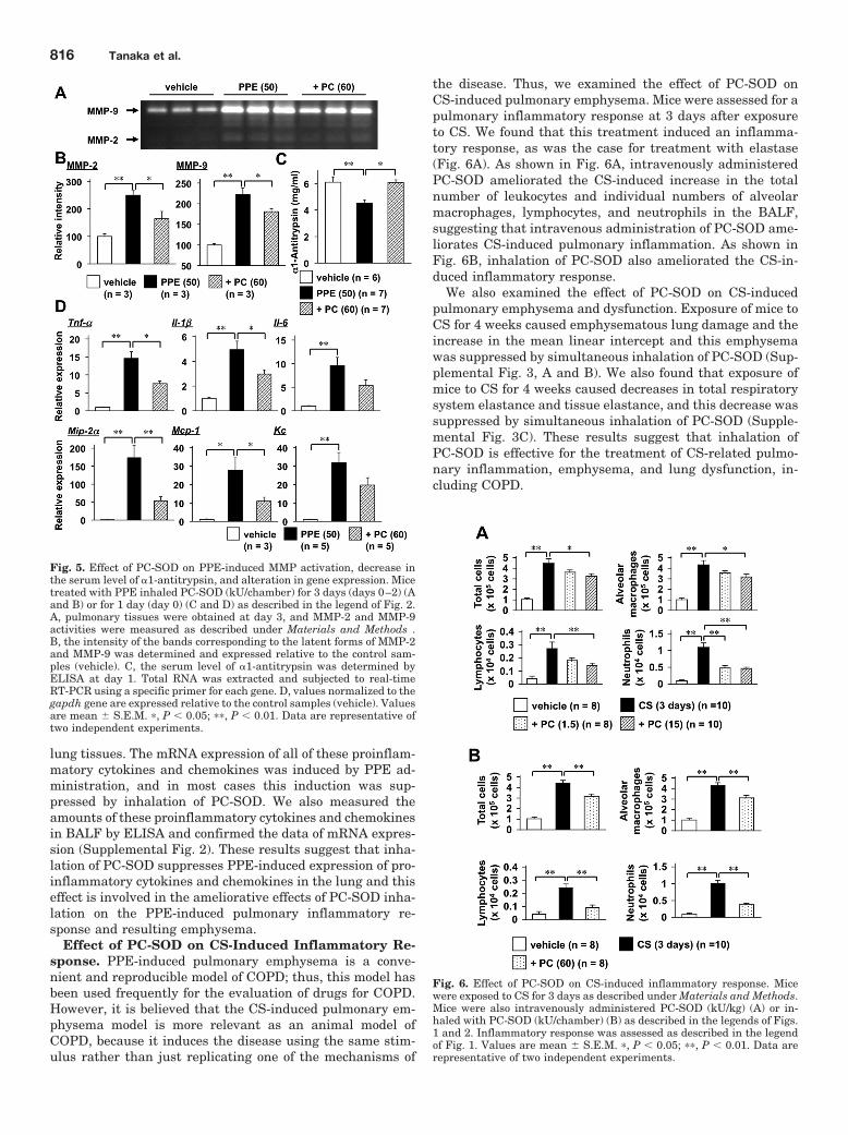
lung tissues. The mRNA expression of all of these proinflam-matory cytokines and chemokines was induced by PPE ad-ministration, and in most cases this induction was sup-pressed by inhalation of PC-SOD. We also measured theamounts of these proinflammatory cytokines and chemokinesin BALF by ELISA and confirmed the data of mRNA expres-sion (Supplemental Fig. 2). These results suggest that inha-lation of PC-SOD suppresses PPE-induced expression of pro-inflammatory cytokines and chemokines in the lung and thiseffect is involved in the ameliorative effects of PC-SOD inha-lation on the PPE-induced pulmonary inflammatory re-sponse and resulting emphysema.
Effect of PC-SOD on CS-Induced Inflammatory Re-sponse. PPE-induced pulmonary emphysema is a conve-nient and reproducible model of COPD; thus, this model hasbeen used frequently for the evaluation of drugs for COPD.However, it is believed that the CS-induced pulmonary em-physema model is more relevant as an animal model ofCOPD, because it induces the disease using the same stim-ulus rather than just replicating one of the mechanisms of
the disease. Thus, we examined the effect of PC-SOD onCS-induced pulmonary emphysema. Mice were assessed for apulmonary inflammatory response at 3 days after exposureto CS. We found that this treatment induced an inflamma-tory response, as was the case for treatment with elastase(Fig. 6A). As shown in Fig. 6A, intravenously administeredPC-SOD ameliorated the CS-induced increase in the totalnumber of leukocytes and individual numbers of alveolarmacrophages, lymphocytes, and neutrophils in the BALF,suggesting that intravenous administration of PC-SOD ame-liorates CS-induced pulmonary inflammation. As shown inFig. 6B, inhalation of PC-SOD also ameliorated the CS-in-duced inflammatory response.
We also examined the effect of PC-SOD on CS-inducedpulmonary emphysema and dysfunction. Exposure of mice toCS for 4 weeks caused emphysematous lung damage and theincrease in the mean linear intercept and this emphysemawas suppressed by simultaneous inhalation of PC-SOD (Sup-plemental Fig. 3, A and B). We also found that exposure ofmice to CS for 4 weeks caused decreases in total respiratorysystem elastance and tissue elastance, and this decrease wassuppressed by simultaneous inhalation of PC-SOD (Supple-mental Fig. 3C). These results suggest that inhalation ofPC-SOD is effective for the treatment of CS-related pulmo-nary inflammation, emphysema, and lung dysfunction, in-cluding COPD.
Fig. 5. Effect of PC-SOD on PPE-induced MMP activation, decrease inthe serum level of �1-antitrypsin, and alteration in gene expression. Micetreated with PPE inhaled PC-SOD (kU/chamber) for 3 days (days 0–2) (Aand B) or for 1 day (day 0) (C and D) as described in the legend of Fig. 2.A, pulmonary tissues were obtained at day 3, and MMP-2 and MMP-9activities were measured as described under Materials and Methods .B, the intensity of the bands corresponding to the latent forms of MMP-2and MMP-9 was determined and expressed relative to the control sam-ples (vehicle). C, the serum level of �1-antitrypsin was determined byELISA at day 1. Total RNA was extracted and subjected to real-timeRT-PCR using a specific primer for each gene. D, values normalized to thegapdh gene are expressed relative to the control samples (vehicle). Valuesare mean � S.E.M. �, P � 0.05; ��, P � 0.01. Data are representative oftwo independent experiments.
Fig. 6. Effect of PC-SOD on CS-induced inflammatory response. Micewere exposed to CS for 3 days as described under Materials and Methods.Mice were also intravenously administered PC-SOD (kU/kg) (A) or in-haled with PC-SOD (kU/chamber) (B) as described in the legends of Figs.1 and 2. Inflammatory response was assessed as described in the legendof Fig. 1. Values are mean � S.E.M. �, P � 0.05; ��, P � 0.01. Data arerepresentative of two independent experiments.
816 Tanaka et al.

DiscussionIn this study, we used PC-SOD, a derivative of SOD with
higher stability in plasma and a higher affinity for tissue,which shows greater therapeutic effects than SOD in animalmodels of various inflammatory diseases, such as IPF, colitis,focal cerebral ischemic injury, and spinal cord injury-inducedmotor dysfunction (Hori et al., 1997; Tamagawa et al., 2000;Ishihara et al., 2009; Tanaka et al., 2010). We have clearlyshown that PC-SOD ameliorates pulmonary emphysema.This result indicates the therapeutic potential of SODagainst COPD-related pulmonary emphysema and is consis-tent with previous results that show transgenic mice express-ing SOD bear a phenotype of resistance to pulmonary em-physema (Foronjy et al., 2006; Petrache et al., 2008). In aphase I clinical study, intravenously administered PC-SOD(40–160 mg) had a terminal half-life of more than 24 h withgood safety and tolerability (Broeyer et al., 2008; Suzuki etal., 2008a). Published results of a phase II clinical study haveshown that intravenously administered PC-SOD (40 or 80mg) significantly improves the symptoms of ulcerative colitispatients, which involves ROS (Suzuki et al., 2008b). A phaseII clinical study has shown that intravenously administeredPC-SOD (40 or 80 mg) is therapeutically effective against IPFas judged by monitoring the serum level of marker proteins(lactate dehydrogenase and surfactant protein-A). Becausethe safety and efficacy of PC-SOD were shown in not only theanimal model but also in clinical studies the application ofPC-SOD for COPD is realistic.
Here, we have shown that not only intravenous adminis-tration but also inhalation of PC-SOD ameliorates pulmo-nary emphysema. We believe that inhalation is a clinicallymore valuable route of administration than the intravenousroute for two reasons. First, PC-SOD administered by inha-lation does not have a bell-shaped dose-response profile. Bell-shaped dose-response curves are of clinical concern becausethey may reflect the presence of side effects. The lack of abell-shaped dose-response profile upon inhalation has alsobeen observed for bleomycin-induced pulmonary fibrosis(Tanaka et al., 2010). Because the efficacy of intravenousadministration of higher doses of PC-SOD on bleomycin-induced pulmonary fibrosis was restored by simultaneousadministration of catalase, which converts hydrogen perox-ide to water and oxygen, the ineffectiveness of high doses ofPC-SOD is probably caused by the accumulation of hydrogenperoxide (Tanaka et al., 2010). However, the reason inhala-tion of PC-SOD does not show the bell-shaped dose-responseprofile remains unknown. Second, patients treated with PC-SOD administered by inhalation would have a higher QOLthan those treated intravenously. Although a phase II clini-cal study has shown that intravenously administered PC-SOD (40 or 80 mg) is effective for both ulcerative colitis(Suzuki et al., 2008b) and IPF, the main obstacle againstproceeding into the next stage of clinical study is the poorQOL for patients undergoing the current clinical protocol ofPC-SOD administration (daily intravenous infusion for 4weeks). Furthermore, in a phase II clinical study for IPF, theplasma levels of markers (lactate dehydrogenase and surfac-tant protein A) but not forced vital capacity were modified byintravenous administration of PC-SOD, suggesting that alonger period of treatment with PC-SOD is required to im-prove forced vital capacity in patients with IPF. However,
daily intravenous infusion for a longer period is not practical.Therefore, we propose that inhalation of PC-SOD for a longerperiod may be effective not only for IPF but also for COPDand would maintain the QOL of patients. The therapeuticpotential of inhalation of PC-SOD for the treatment of COPDis also supported by observations made in this study: inha-lation of PC-SOD ameliorated not only PPE-induced patho-logical alterations but also PPE-induced functional changes,and inhalation of PC-SOD was effective even for predevel-oped pulmonary emphysema (stimulation of spontaneousrestoration from pulmonary emphysema and suppression ofprogression of pulmonary dysfunction). Drugs for COPDshould suppress both the inflammatory response and emphy-sematous lung destruction. Because ROS, especially super-oxide anions, are suggested to induce both an inflammatoryresponse and emphysematous lung destruction (Mak, 2008),PC-SOD was predicted to suppress both of these events. Infact, we showed that inhalation of PC-SOD suppresses aPPE-induced increase in leukocytes in BALF and the expres-sion of proinflammatory cytokines and chemokines. We alsoshowed that inhalation of PC-SOD suppresses PPE-inducedemphysematous lung destruction. Both apoptosis and pro-tease/antiprotease imbalance seem to be involved in emphy-sematous lung destruction associated with COPD (Demedtset al., 2006; Rabe et al., 2007; Petrache et al., 2008). We haveshown that inhalation of PC-SOD suppresses PPE-inducedpulmonary cell death and protease/antiprotease imbalance(activation of MMPs and decrease in the level of �1-antitryp-sin). We recently reported that PC-SOD protects culturedlung epithelial cells from menadione (a superoxide anion-releasing drug)-induced cell death (Tanaka et al., 2010). Ithas also been reported that oxidative molecules activateMMPs and suppress the expression of �1-antitrypsin (Des-rochers and Weiss, 1988; Greenlee et al., 2007; Mak, 2008;Wan et al., 2008). Thus, it seems that a PC-SOD-dependentdecrease in the level of superoxide anions is responsible forthe inhibitory effect of PC-SOD on PPE-induced pulmonarycell death and the protease/antiprotease imbalance.
One of the current standard clinical protocols for treatmentof patients with COPD is administration of a long-acting�2-agonist or anticholinergic along with corticosteroid inha-lation. This combination regime reduces the annual rate ofexacerbation and improves health status and spirometricvalues, although it does not improve the mortality rate withstatistical significance (Calverley et al., 2007). �2-Agonistsand anticholinergics are effective in improving the airflowlimitation associated with COPD (Rabe et al., 2007). On theother hand, some reports have suggested that treatment withcorticosteroids does not clearly modulate the inflammatoryresponse in patients with COPD or in a CS-induced pulmo-nary emphysema animal model (Rabe et al., 2007; Fox andFitzgerald, 2009). Based on these previous observations andthose in this study that inhalation of PC-SOD is effectiveagainst the CS-induced inflammatory response, we considerthat a combination regime of administration of a long-acting�2-agonist (or anticholinergics) along with inhalation of PC-SOD, instead of corticosteroids, may be therapeutically ben-eficial for patients with COPD.
Authorship Contributions
Participated in research design: K.-I. Tanaka and Mizushima.Conducted experiments: K.-I. Tanaka, Y. Tanaka, and Miyazaki.
PC-SOD and Pulmonary Emphysema 817

Contributed new reagents or analytic tools: Namba, Sato, andAoshiba.
Performed data analysis: K.-I. Tanaka and Sato.Wrote or contributed to the writing of the manuscript: K.-I.
Tanaka, Aoshiba, Azuma, and Mizushima.
ReferencesBarnes PJ and Stockley RA (2005) COPD: current therapeutic interventions and
future approaches. Eur Respir J 25:1084–1106.Bradford MM (1976) A rapid and sensitive method for the quantitation of microgram
quantities of protein utilizing the principle of protein-dye binding. Anal Biochem72:248–254.
Broeyer FJ, van Aken BE, Suzuki J, Kemme MJ, Schoemaker HC, Cohen AF,Mizushima Y, and Burggraaf J (2008) The pharmacokinetics and effects of along-acting preparation of superoxide dismutase (PC-SOD) in man. Br J ClinPharmacol 65:22–29.
Calverley PM, Anderson JA, Celli B, Ferguson GT, Jenkins C, Jones PW, Yates JC,Vestbo J, and TORCH Investigators (2007) Salmeterol and fluticasone propionateand survival in chronic obstructive pulmonary disease. N Engl J Med 356:775–789.
Daga MK, Chhabra R, Sharma B, and Mishra TK (2003) Effects of exogenousvitamin E supplementation on the levels of oxidants and antioxidants in chronicobstructive pulmonary disease. J Biosci 28:7–11.
Demedts IK, Demoor T, Bracke KR, Joos GF, and Brusselle GG (2006) Role ofapoptosis in the pathogenesis of COPD and pulmonary emphysema. Respir Res7:53.
Desrochers PE and Weiss SJ (1988) Proteolytic inactivation of �-1-proteinase inhib-itor by a neutrophil metalloproteinase. J Clin Invest 81:1646–1650.
Foronjy RF, Mirochnitchenko O, Propokenko O, Lemaitre V, Jia Y, Inouye M, OkadaY, and D’Armiento JM (2006) Superoxide dismutase expression attenuates ciga-rette smoke- or elastase-generated emphysema in mice. Am J Respir Crit CareMed 173:623–631.
Fox JC and Fitzgerald MF (2009) The role of animal models in the pharmacologicalevaluation of emerging anti-inflammatory agents for the treatment of COPD. CurrOpin Pharmacol 9:231–242.
Freeman TA, Parvizi J, Della Valle CJ, and Steinbeck MJ (2009) Reactive oxygenand nitrogen species induce protein and DNA modifications driving arthrofibrosisfollowing total knee arthroplasty. Fibrogenesis Tissue Repair 2:5.
Greenlee KJ, Werb Z, and Kheradmand F (2007) Matrix metalloproteinases in lung:multiple, multifarious, and multifaceted. Physiol Rev 87:69–98.
Hori Y, Hoshino J, Yamazaki C, Sekiguchi T, Miyauchi S, Mizuno S, and Horie K(1997) Effect of lecithinized-superoxide dismutase on the rat colitis model inducedby dextran sulfate sodium. Jpn J Pharmacol 74:99–103.
Igarashi R, Hoshino J, Ochiai A, Morizawa Y, and Mizushima Y (1994) Lecithinizedsuperoxide dismutase enhances its pharmacologic potency by increasing its cellmembrane affinity. J Pharmacol Exp Ther 271:1672–1677.
Igarashi R, Hoshino J, Takenaga M, Kawai S, Morizawa Y, Yasuda A, Otani M, andMizushima Y (1992) Lecithinization of superoxide dismutase potentiates its pro-tective effect against Forssman antiserum-induced elevation in guinea pig airwayresistance. J Pharmacol Exp Ther 262:1214–1219.
Institute of Laboratory Animal Resources (1996) Guide for the Care and Use ofLaboratory Animals, 7th ed. Institute of Laboratory Animal Resources, Commis-sion on Life Sciences, National Research Council, Washington DC.
Ishihara T, Tanaka K, Tasaka Y, Namba T, Suzuki J, Ishihara T, Okamoto S, HibiT, Takenaga M, Igarashi R, et al. (2009) Therapeutic effect of lecithinized super-oxide dismutase against colitis. J Pharmacol Exp Ther 328:152–164.
Karakawa T, Sato K, Muramoto Y, Mitani Y, Kitamado M, Iwanaga T, NabeshimaT, Maruyama K, Nakagawa K, Ishida K, et al. (2008) Applicability of new spin trapagent, 2-diphenylphosphinoyl-2-methyl-3,4-dihydro-2H-pyrrole N-oxide, in biolog-ical system. Biochem Biophys Res Commun 370:93–97.
Kinnula VL and Crapo JD (2003) Superoxide dismutases in the lung and humanlung diseases. Am J Respir Crit Care Med 167:1600–1619.
Kondo T, Tagami S, Yoshioka A, Nishimura M, and Kawakami Y (1994) Currentsmoking of elderly men reduces antioxidants in alveolar macrophages. Am JRespir Crit Care Med 149:178–182.
Kuraki T, Ishibashi M, Takayama M, Shiraishi M, and Yoshida M (2002) A novel oralneutrophil elastase inhibitor (ONO-6818) inhibits human neutrophil elastase-induced emphysema in rats. Am J Respir Crit Care Med 166:496–500.
Mak JC (2008) Pathogenesis of COPD. Part II. Oxidative-antioxidative imbalance.Int J Tuberc Lung Dis 12:368–374.
Miravitlles M and Anzueto A (2009) Insights into interventions in managing COPDpatients: lessons from the TORCH and UPLIFT studies. Int J Chron ObstructPulmon Dis 4:185–201.
Nadeem A, Raj HG, and Chhabra SK (2005) Increased oxidative stress and alteredlevels of antioxidants in chronic obstructive pulmonary disease. Inflammation29:23–32.
Namba T, Homan T, Nishimura T, Mima S, Hoshino T, and Mizushima T (2009)Up-regulation of S100P expression by non-steroidal anti-inflammatory drugs andits role in anti-tumorigenic effects. J Biol Chem 284:4158–4167.
Owen CA (2005) Proteinases and oxidants as targets in the treatment of chronicobstructive pulmonary disease. Proc Am Thorac Soc 2:373–385; discussion 394–395.
Peto R, Chen ZM, and Boreham J (1999) Tobacco–the growing epidemic. Nat Med5:15–17.
Petrache I, Medler TR, Richter AT, Kamocki K, Chukwueke U, Zhen L, Gu Y,Adamowicz J, Schweitzer KS, Hubbard WC, et al. (2008) Superoxide dismutaseprotects against apoptosis and alveolar enlargement induced by ceramide. Am JPhysiol Lung Cell Mol Physiol 295:L44–L53.
Pinamonti S, Leis M, Barbieri A, Leoni D, Muzzoli M, Sostero S, Chicca MC, CarrieriA, Ravenna F, Fabbri LM, et al. (1998) Detection of xanthine oxidase activityproducts by EPR and HPLC in bronchoalveolar lavage fluid from patients withchronic obstructive pulmonary disease. Free Radic Biol Med 25:771–779.
Rabe KF, Hurd S, Anzueto A, Barnes PJ, Buist SA, Calverley P, Fukuchi Y, JenkinsC, Rodriguez-Roisin R, van Weel C, et al. (2007) Global strategy for the diagnosis,management, and prevention of chronic obstructive pulmonary disease: GOLDexecutive summary. Am J Respir Crit Care Med 176:532–555.
Rahman I and Adcock IM (2006) Oxidative stress and redox regulation of lunginflammation in COPD. Eur Respir J 28:219–242.
Suzuki J, Broeyer F, Cohen A, Takebe M, Burggraaf J, and Mizushima Y (2008a)Pharmacokinetics of PC-SOD, a lecithinized recombinant superoxide dismutase,after single- and multiple-dose administration to healthy Japanese and Caucasianvolunteers. J Clin Pharmacol 48:184–192.
Suzuki Y, Matsumoto T, Okamoto S, and Hibi T (2008b) A lecithinized superoxidedismutase (PC-SOD) improves ulcerative colitis. Colorectal Dis 10:931–934.
Tamagawa K, Taooka Y, Maeda A, Hiyama K, Ishioka S, and Yamakido M (2000)Inhibitory effects of a lecithinized superoxide dismutase on bleomycin-inducedpulmonary fibrosis in mice. Am J Respir Crit Care Med 161:1279–1284.
Tanaka K, Ishihara T, Azuma A, Kudoh S, Ebina M, Nukiwa T, Sugiyama Y, TasakaY, Namba T, Ishihara T, et al. (2010) Therapeutic effect of lecithinized superoxidedismutase on bleomycin-induced pulmonary fibrosis. Am J Physiol Lung Cell MolPhysiol 298:L348–L360.
Valenca SS, Bezerra FS, Romana-Souza B, Paiva RO, Costa AM, and Porto LC (2008)Supplementation with vitamins C and E improves mouse lung repair. J NutrBiochem 19:604–611.
Valentin F, Bueb JL, Kieffer P, Tschirhart E, and Atkinson J (2005) Oxidative stressactivates MMP-2 in cultured human coronary smooth muscle cells. Fundam ClinPharmacol 19:661–667.
Wan R, Mo Y, Zhang X, Chien S, Tollerud DJ, and Zhang Q (2008) Matrix metallo-proteinase-2 and -9 are induced differently by metal nanoparticles in humanmonocytes: The role of oxidative stress and protein tyrosine kinase activation.Toxicol Appl Pharmacol 233:276–285.
Yao H, Arunachalam G, Hwang JW, Chung S, Sundar IK, Kinnula VL, Crapo JD,and Rahman I (2010) Extracellular superoxide dismutase protects against pulmo-nary emphysema by attenuating oxidative fragmentation of ECM. Proc Natl AcadSci USA 107:15571–15576.
Address correspondence to: Dr. Tohru Mizushima, Graduated School ofMedical and Pharmaceutical Sciences, Kumamoto University, 5-1 Oe-honma-chi, Kumamoto 862-0973, Japan. E-mail: [email protected]
818 Tanaka et al.