identification of excreted iron superoxide dismutase for … · · 2006-10-25identification of...
TRANSCRIPT
649649649649649Mem Inst Oswaldo Cruz, Rio de Janeiro, Vol. 101(6): 649-654, September 2006
Identification of excreted iron superoxide dismutase for thediagnosis of Phtytomonas
Clotilde Marín, Isabel Rodríguez-González, Manuel Sánchez- Moreno/+
Instituto de Biotecnología, Departamento de Parasitología, Facultad de Ciencias, Universidad de Granada, C/ Severo Ochoa s/n, 18071 Granada, España
An excreted iron superoxide dismutase (FeSODe) of pI 3.6 with a molecular weight of 28-30 kDa was detected inthe in vitro culture of Phytomonas isolated from Euphorbia characias (SODeCHA) and from Lycopersicon esculentum(SODeTOM), in Grace’s medium without serum. These FeSODe excreted into the medium had immunogenic capac-ity: the positivity of the anti-SODeCHA serum persisted to a dilution of 1/30,000, and for the anti-SODeTOM to 1/10,000 by Western blot. In addition, cross reaction was detected between the anti-SODe serum of Phytomonasisolated from E. characias against SODeTOM, and the anti-SODe serum from L. esculentum with SODeCHA. Thischaracteristic offers the possibility of its use to diagnose plant trypanosomatids. The validation of the test wasconfirmed by experimental inoculation of tomato fruits with Phytomonas isolated from L. esculentum. At 7, 10, 15,and 21 days post infection, it was possible to detect the presence of the parasites with the anti-SODe serum ofPhytomonas isolated from L. esculentum at a dilution of 1/250. These serological results were confirmed by visual-ization of the parasites by optical microscopy. The data of this study confirm that the SOD is sufficient to identify atrypanosomatid isolated from plants as belonging to the genus Phytomonas.
Key words: superoxide dismutase - immunogenicity - molecular tool - Phytomonas spp.
Trypanosomatids belonging to the genus Phytomonasare fairly common in the latex, phloem, fruit sap, seed al-bumen, and even in the nectar of many plant families.Also, their world distribution is widespread. They are etio-logical agents in devastating crop epiphytotics (coconut,oil palm, coffee, and manioc) but they also parasitize manyplants without apparent pathogenicity (Dollet 1984).
Probably more than one genus of Trypanosomatidaeis represented among plant parasites. Therefore, both thestudy of the circulation of parasites as well as the demon-stration of infection in hosts require a precise identifica-tion of trypanosomatids. The distinctions between themis difficult on a morphological basis. Attempts to over-come this difficulty prompted the testing of various sero-logical, biochemical, and molecular methods, but none todate have been very successful for genus distinction(Camargo 1999). In recent years, new polymerase chainreaction method for the amplification of the SL sequencehave been successfully developed, and more recently newmolecular markers have become available to help in theidentification (Serrano et al. 1999, Dollet et al. 2001, Marínet al. 2004a).
Superoxide dismutases (SODs, EC 1.15.1.1) are metal-loproteins that occur ubiquitously in nature and efficientlydismutase the superoxide anion into oxygen and hydro-gen peroxide. SODs are assigned to different families,
Finacial support: ATP 2002/03: Circulation of Trypano-somatidae Project (CIRAD, France), grant BIO-2000-1429 (Uni-versity of Granada, Spain)Corresponding author: [email protected] 16 March 2006Accepted 21 June 2006
based on their metal cofactors – iron (FeSOD), manga-nese (MnSOD) and copper-zinc (Cu/ZnSOD) – which alsodiffer in their location: cytosol, cell organelles, and evencell excretions. Thus, the Cu/ZnSODs appear primarily inthe cytosol of eukaryotic cells; the MnSODS in the mito-chondrial matrix; and FeSODs in the cytosol of plant cellsas well as in some free-living protozoa and protozoan para-sites (Fridovich 1995).
SOD activity has been detected in the main speciesbelonging to the family Trypanosomatidae, in Trypano-soma cruzi (Ismail et al. 1997), in T. brucei brucei (Kabiri& Steverding 2001), in several species of the genus Leish-mania (Ismail et al. 1994), in some lower trypanosomatids,and in plant trypanosomes (Quesada et al. 2001, Marín etal. 2004a,b). SODs from protozoan parasites are consid-ered virulence factors that protect the parasites from theattack of the host cells by the action both of oxidant andanti-inflammatory agents (Paramchuck et al. 1997), andcan even confer immunological capacities (Pérez-Fuenteset al. 2003). In general, all the parasitic protozoa studied todate have only FeSOD (Marín et al. 2004a,b).
Extracellular SODs (EcCu/ZnSODs) have been evi-denced in invertebrates, in several species of nematodes,such as, Brugia pahangi (Tang et al. 1994), Onchocercavolvulus (James et al. 1994), and Caenorhabditis elegans(Fujii et al. 1998) and the crustacean Callinectes sapidus(Brouwer et al. 2003). Recently, Villagrán et al. (2005) haveshown the presence of a iron SODe in T. cruzi. Also, inmammals, several researchers have studied extracellularSOD (EC-SOD), which is the major extracellular antioxi-dant enzyme and plays a critical role in the pathogenesisof a variety of pulmonary, neurological, and cardiovascu-lar diseases (Suliman et al. 2001, Fukai et al. 2002).
In a previous paper, we reported in a digitonin-titra-tion experiment that 20% of the SOD activity of Phy-
650650650650650 Superoxide dismutase in Phytomonas spp. • Clotilde Marín et al.
tomonas sp. was not solubilized even at high digitoninconcentrations, indicating that part of the SOD could beassociated with membranes or even excreted by the para-site (Marín et al. 2004b). Therefore, in the present study,we show the existence of a SOD excreted into the super-natant from the culture of Phytomonas isolated from Eu-phorbia characias, and Phytomonas isolated from Ly-copersicon esculentum, cultured in Grace’s medium with-out serum. Also, we have studied the usefulness of ex-creted SOD in the identification and diagnosis ofPhytomonas infections using Western blot in the identifi-cation
MATERIALS AND METHODS
Parasites and SOD excreted fraction extraction -Phytomonas isolated from latex vessels of E. characias(Dollet 1984) and from L. esculentum (tomato) in Spain(Sánchez-Moreno et al. 1995), were grown in axenic Grace’smedium (Sigma) supplemented with 10% heat inactivatedfoetal bovine serum at 28ºC. Cells at the exponential growthphase were collected by centrifugation (1500 × g for 5 minat room temperature). The pellet of cells was washed twicein Grace’s medium without serum, and cells were counted,distributed into aliquots of 5 × 10⁹ parasites/ml in Grace’smedium without serum, and grown for 24 h. The superna-tant was collected by centrifugation at 1500 × g for 10 min,passed through a 0.45 µm pore size filter, and then precipi-tated with ammonium sulphate at 35-85%. This was cen-trifuged (9000 × g for 20 min at 4 ºC), redissolved in 2.5 mlof 50 mM potassium phosphate buffer (pH 7.8), and after-wards desalted by buffer exchange in a Sephadex G-25column (Pharmacia, PD 10), previously balanced with thepotassium buffer, bringing it to a final volume of 3.5-ml.Thus, we obtained the SODe fraction from Phytomonasisolated from E. characias (SODeCHA) and SODe fromPhytomonas isolated from L. esculentum (SODeTOM).These fractions were finally concentrated by ultrafiltra-tion in Centriprep-10 tubes (Amicon) at 3000 × g to a finalconcentration of 2 mg/ml. The protein content was deter-mined in all fractions using the Bio-Rad test, based on theBradford method (Bradford 1976), with BSA as a stan-dard. For the extraction and purification of the P85 frac-tion, we followed the protocol described by Marín et al.(2004b).
Determination of molecular weights and isoelectricpoint of SODe - Apparent molecular weights of the semi-purified enzymes (SODeCHA and SODeTOM) were de-termined by the separation of 7 µg/lane on 20% homoge-neous Native-PAGE gel in the Phast System (AmershamPharmacia Biotech, Uppsala, Sweden). The isoelectricpoints were determined in the Phast System (7 µg/lane) inpolyacrylamide Phast gel pI 3-9 as described Bécuwe etal. (1996). Proteins markers for molecular weights and pIwere provided by Pharmacia (Uppsala, Sweden). The SODactivity on the gels was visualized by staining, followingBeyer and Fridovich (1976), and for protein markers thelanes were stained with silver nitrate and coomassie blue,as described by Phast System handbook.
Polyclonal serum - To obtain the specific antibodiesagainst the fractions SODeCHA and SODeTOM, we im-
munized two female 4-week-old Balb-C mice (concentra-tion of proteins of 2 mg/ml). These fractions were sepa-rated by electrophoresis of IEF 3-9 in polyacrylamide gelsas described elsewhere (Marín et al. 2004a). In this way,we obtained the anti-SODeCHA and anti-SODeTOM sera.In addition, the serum from the mouse that had not beenimmunised with the antigen fraction (control serum) wascollected.
Experimental infection of tomatoes - Isolated tomatowere infected by needle inoculation in two zones, with 5 ×10⁵ culture forms of Phytomonas isolated from L. es-culentum in 50 µl. As controls, three tomatoes were in-oculated with the same volume of axenic Grace’s mediumsupplemented with 10% heat-inactivated foetal bovineserum. At 7, 10, 15, and 21 days post-inoculation, threetomatoes, controls and infected, were processed to ob-tain the extracts for later studies. After the inoculationzones were collected, smears fixed with methanol andstained by Giemsa were used for light microscopy. After-wards, the material was homogenized and centrifuged at1500 × g for 10 min. The supernatant was filtered, precipi-tated with ammonium sulphate, passed through a Se-phadex G-25 column, and concentrated as describedabove for obtaining the SODe fraction. Also, the zonesopposite the inoculation from infected tomatoes wereprocessed the same way.
Western-blot analysis - For the polyclonal serum titra-tion, we used the SODe fraction (SODeCHA andSODeTOM) as the antigen fraction. These were run onIEF 3-9 gels (protein concentration of proteins of 2 mg/ml), and afterwards transferred to nitrocellulose, for 30min, as prescribed in the Phast-System manual. The mem-brane was blocked for 2 h at room temperature using 0.4%gelatine and 0.2% Tween 20 in PBS, followed by threewashes in 0.1% Tween 20 in PBS (PBS-T). Next, the mem-brane was incubated for 2 h at room temperature, eitherwith negative serum at a 1/100 dilution or with anti-SODeCHA or anti-SODeTOM sera at dilutions of: 1/100,1/500, 1/1000, 1/5000, 1/10,000, 1/20,000, and 1/30,000. Af-ter being washed as above, the membrane was furtherincubated for 2 h at room temperature with the secondantibody, anti-mouse IgG (Fc specific) peroxidase conju-gate (Sigma®) (dilution 1/1000). After washing, the sub-strate diaminobenzidine was added and the reactionstopped with several washes in distilled water (Marín etal. 2004a). For the cross reaction analyses, we followedthe same protocol described above but with a dilution ofthe anti-SODeCHA and anti-SODeTOM sera of 1/5000.
Western-blot was also used to detect the SODeTOMin experimentally inoculated tomato fruits, as describedabove. For this, the extracts from the tomato of 7, 10, 15,and 21 days were used as the antigen fraction, as were theextracts of control tomatoes. The dilution of the anti-SODeTOM serum of 1/250 was used.
RESULTS
Isolation of an excreted FeSOD - When promastigoteforms of Phytomonas isolated from E. characias and L.esculentum (5 × 10⁹ parasites) were cultured for 24 h inGrace’s medium without serum and the cell-free superna-
651651651651651Mem Inst Oswaldo Cruz, Rio de Janeiro, Vol. 101(6), September 2006
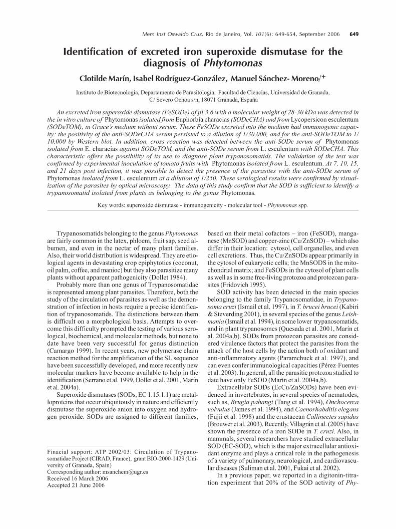
tant was collected, concentrated, separated by isoelectricfocusing (pI 3-9) and 20% homogeneous native-page andfollowed by SOD activity staining. We detected a singleSOD band (Fig. 1A, lanes 2 and 4; Fig.1B, lanes 5 and 6),which we called SODeCHA and SODeTOM, respectively.
The isoelectric point of these bands of excreted SOD(SODeCHA and SODeTOM) was consistent with the iso-electric point (pI 3.6) of the SODII band detected in thepartially purified fraction of Phytomonas promastigotescultured under normal conditions (P85) (Fig. 1, lanes, 1and 3).
The two isoenzymes excreted had very similar molecu-lar weights, of approximately 28-30 kDa (Fig. 1). In bothcases, they were demonstrated to be FeSOD (data notshown).
Marker enzymes (pyruvate kinase and hexokinase)have confirmed that there was no lysis of the parasiteduring this culture period and that the presence of theSOD was due only to excretion by the parasite (data notshown).
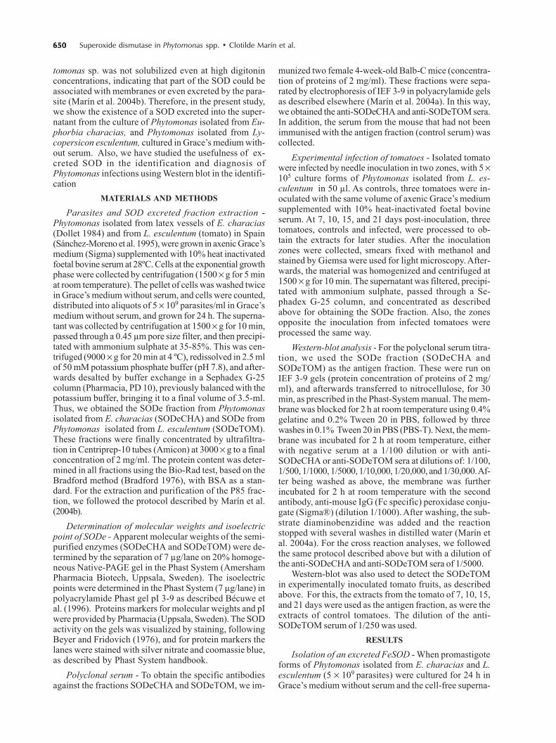
Immunogenicity of the SODe - Polyclonal antibodiesagainst the enzymes SODeCHA and SODeTOM were ob-tained from immunized Balb-C mice, and, by Western blot,
we demonstrated their immunogenic capacity (Fig. 2). Withthe control serum (Fig. 2, lines 1 and 9), the reaction wasnegative at a dilution of 1/100, regardless of the antigenfraction. Meanwhile, in the case of the anti-SODe serum(Fig. 2; lanes 2-8 and lanes 10-16, respectively), the reac-tions proved positive up to a dilution of 1/30,000 for SODe-CHA and 1/10,000 for SODe-TOM.
We determined whether there was any cross reactionbetween the polyclonal sera antiSODeCHA andSODeTOM, by Western blot (Fig. 3). There was crossreactivity between the sera to a dilution of 1/5000.
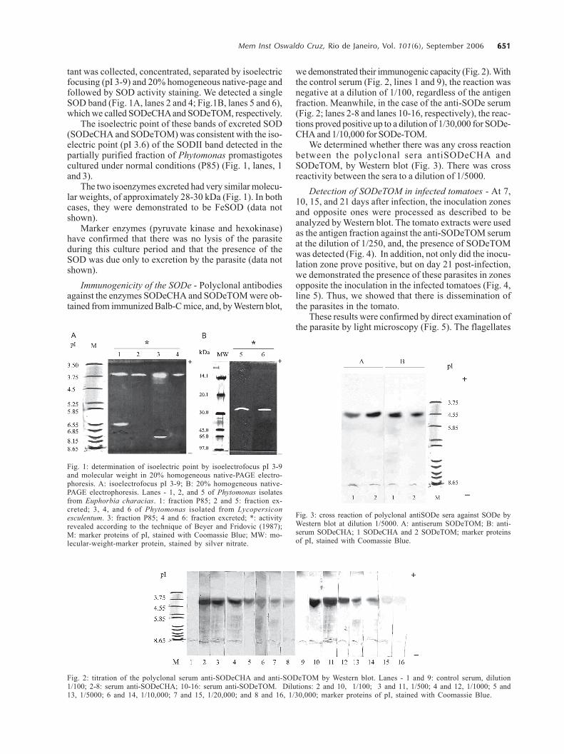
Detection of SODeTOM in infected tomatoes - At 7,10, 15, and 21 days after infection, the inoculation zonesand opposite ones were processed as described to beanalyzed by Western blot. The tomato extracts were usedas the antigen fraction against the anti-SODeTOM serumat the dilution of 1/250, and, the presence of SODeTOMwas detected (Fig. 4). In addition, not only did the inocu-lation zone prove positive, but on day 21 post-infection,we demonstrated the presence of these parasites in zonesopposite the inoculation in the infected tomatoes (Fig. 4,line 5). Thus, we showed that there is dissemination ofthe parasites in the tomato.
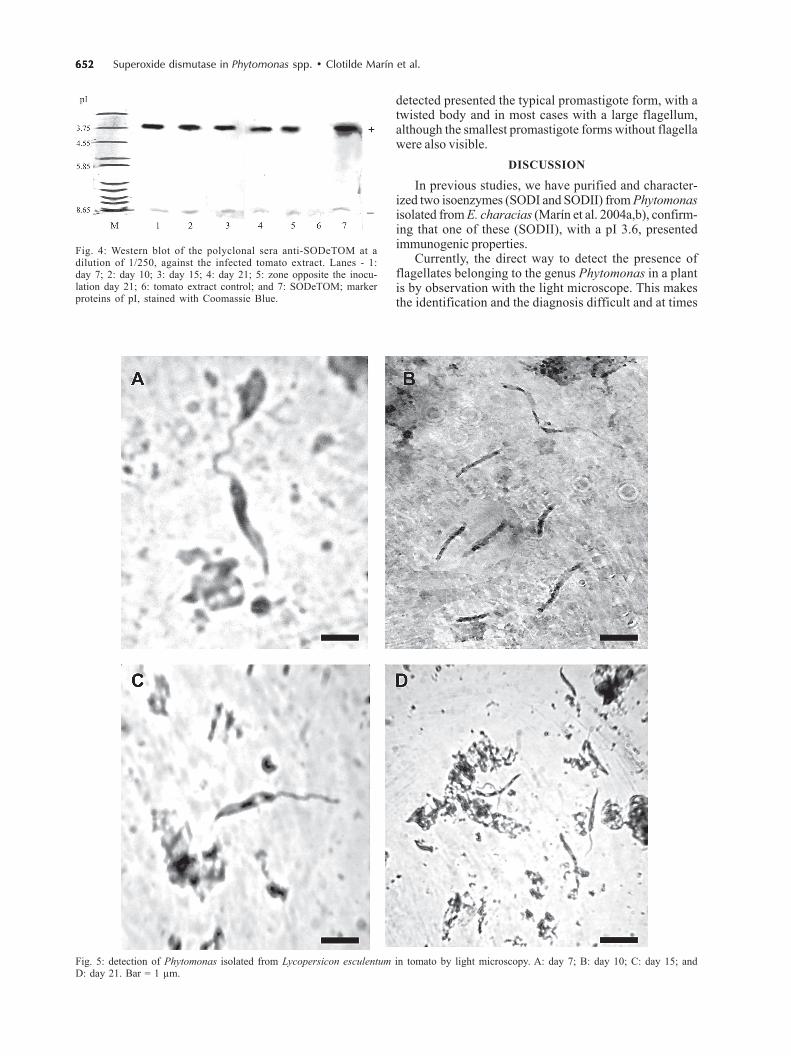
These results were confirmed by direct examination ofthe parasite by light microscopy (Fig. 5). The flagellates
Fig. 1: determination of isoelectric point by isoelectrofocus pI 3-9and molecular weight in 20% homogeneous native-PAGE electro-phoresis. A: isoelectrofocus pI 3-9; B: 20% homogeneous native-PAGE electrophoresis. Lanes - 1, 2, and 5 of Phytomonas isolatesfrom Euphorbia characias. 1: fraction P85; 2 and 5: fraction ex-creted; 3, 4, and 6 of Phytomonas isolated from Lycopersiconesculentum. 3: fraction P85; 4 and 6: fraction excreted; *: activityrevealed according to the technique of Beyer and Fridovic (1987);M: marker proteins of pI, stained with Coomassie Blue; MW: mo-lecular-weight-marker protein, stained by silver nitrate.
Fig. 2: titration of the polyclonal serum anti-SODeCHA and anti-SODeTOM by Western blot. Lanes - 1 and 9: control serum, dilution1/100; 2-8: serum anti-SODeCHA; 10-16: serum anti-SODeTOM. Dilutions: 2 and 10, 1/100; 3 and 11, 1/500; 4 and 12, 1/1000; 5 and13, 1/5000; 6 and 14, 1/10,000; 7 and 15, 1/20,000; and 8 and 16, 1/30,000; marker proteins of pI, stained with Coomassie Blue.
Fig. 3: cross reaction of polyclonal antiSODe sera against SODe byWestern blot at dilution 1/5000. A: antiserum SODeTOM; B: anti-serum SODeCHA; 1 SODeCHA and 2 SODeTOM; marker proteinsof pI, stained with Coomassie Blue.
652652652652652 Superoxide dismutase in Phytomonas spp. • Clotilde Marín et al.
detected presented the typical promastigote form, with atwisted body and in most cases with a large flagellum,although the smallest promastigote forms without flagellawere also visible.
DISCUSSION
In previous studies, we have purified and character-ized two isoenzymes (SODI and SODII) from Phytomonasisolated from E. characias (Marín et al. 2004a,b), confirm-ing that one of these (SODII), with a pI 3.6, presentedimmunogenic properties.
Currently, the direct way to detect the presence offlagellates belonging to the genus Phytomonas in a plantis by observation with the light microscope. This makesthe identification and the diagnosis difficult and at times
Fig. 4: Western blot of the polyclonal sera anti-SODeTOM at adilution of 1/250, against the infected tomato extract. Lanes - 1:day 7; 2: day 10; 3: day 15; 4: day 21; 5: zone opposite the inocu-lation day 21; 6: tomato extract control; and 7: SODeTOM; markerproteins of pI, stained with Coomassie Blue.
Fig. 5: detection of Phytomonas isolated from Lycopersicon esculentum in tomato by light microscopy. A: day 7; B: day 10; C: day 15; andD: day 21. Bar = 1 µm.
653653653653653Mem Inst Oswaldo Cruz, Rio de Janeiro, Vol. 101(6), September 2006
even impossible, because promastigote stages of Lepto-monas, Herpetomonas, Crithidia, and Blastocrithidiaalso occur in Phytomonas vectors (Wallace et al. 1992,Camargo & Wallace 1994, Podlipaev 2000), and have some-times been detected in plant tissues. Thus, it becomesnecessary to seek new methods to detect the parasite.Diverse authors have suggested that Western blot canbe used as a complementary and alternative method forconventional serology tests (Ávila et al. 1993), and there-fore may be useful in detecting and identifying parasiticprotozoa using antigens from epimastigotes forms andSODe (Vissoci et al. 1998, Villagrán et al. 2005). Also, wehave demonstrated that the SODII for its immunogeniccharacteristics is a excellent molecular marker of the ge-nus Phytomonas, which could be applied to the differentimmunological techniques.
To our knowledge, this is the first evidence that mem-bers belonging to the genus Phytomonas excrete a FeSODinto the culture medium. These data confirm the findingsof our earlier study (Marín et al. 2004a), showing thatSODII would be formed in the cytosol and afterwards trans-ported to the glycosomes, as occurs with other glycosomalenzymes (Parsons et al. 2001), and to the membranes, andpart of this enzyme could even be secreted to the exteriorby the parasite, as a defence mechanism of the parasiteagainst toxic radicals generated by the host (Kabiri &Steverding 2001).
One of the aim in the present work was to study theimmunogenic properties of this FeSODe, for which weobtained polyclonal antibodies against SODeCHA andSODeTOM from immunized Balb-C mice. Western blotshowed a positive reaction up to a dilution of 1/30,000.This result indicates that, FeSODe is a highly immuno-genic protein, opening the possibility of its use for thediagnosis of parasitism by species belonging to the ge-nus Phytomonas.
To confirm the usefulness of this excreted protein(SODe) as a marker in Western-blot diagnosis, we per-formed an inoculation experiment on tomato fruits. Thepresence of Phytomonas was detected at 7, 10, 15, and 21days after infection in the inoculation zones with the anti-SODeTOM serum at the dilution of 1/250, and also on day21 in the zones opposite the inoculation. Hence, SODe isa specific and sensitive molecular tool and, used withWestern blot, can be useful to the diagnosis of Phytomonasspp. The flagellates detected in the laboratory-infectedtomatoes by light microscopy presented the typicalpromastigote form, although the smallest promastigoteforms without flagella were also visible, these being verysimilar to others reported elsewhere (Jankevicius et al.1989).
The data of this study together with those gatheredrecently in our laboratory (Marín et al. 2004a,b) confirmthat the SOD (SODII or SODe) is sufficient to identify atrypanosomatid isolated from plants as belonging to thegenus Phytomonas and to distinguish between a truePhytomonas and other trypanosomatids that can provoketransient infections in plants. Also, a certain amount ofSOD is secreted by the parasites, which is highly immu-nogenic and by Western blot assay capable of detectingthe presence of Phytomonas isolates from L. esculentum
in crude preparations from tomato saps. Thus, we wereable to detect this parasitism without the need for isola-tion, culture, or DNA extraction of flagellates. Of course,the validity of the SODe by Phytomonas spp. as a diag-nostic tool needs to be continuously evaluated as newisolates and molecular data became available
REFERENCES
Ávila HA, Pereira JB, Thiermann O, Paiva E, Degrave W, Mo-rel, C M, Simpson, L 1993. Detection of Trypanosomacruzi in blood specimens of chronic chagasic patients bypolymerase chain reaction amplification of kinetoplastminicircle DNA: comparison with serology and xenodiag-nosis. J Clin Microbiol 21: 2421-2426
Bécuwe P, Gratepanche S, Fourmaux MN, Van Beeumen J,Samyn B, Mercereau-Puijalon O, Touzel JP, Slomianny C,Camus D, Dive D 1996. Characterization of iron-depen-dent endogenous superoxide dismutase of Plasmodiumfalciparum. Mol Biochem Parasitol 76: 125-134
Beyer WF, Fridovich I 1987. Assaying for superoxide dismutaseactivity: some large consequences of minor changes in con-ditions. Anal Biochem 161: 559-566.
Bradford MM 1976. A refined and sensitive method for thecuantification of microgram quantities of protein-dye bind-ing. Anal Biochem 72: 248.
Brouwer M, Hoexum Brouwer T, Grater W, Brown-Peterson N2003. Replacement of a cytosolic copper/zinc superoxidedismutase by a novel cytosolic manganese superoxidedismutase in crustaceans that use copper (haemocyanin)for oxygen transport. Biochem J 374: 219-228.
Camargo EP 1999. Phytomonas and other trypanosomatid para-sites of plants and fruit. Adv Parasitol 42: 29-112.
Camargo EP, Wallace G 1994. Vectors of plant parasites of thegenus Phytomonas (Protozoa, Zoomastigophore, Kine-toplastida). In KF Harris, Advances in Disease Vector Re-search, vol. 10, Academic Press, New York, p. 333-359.
Dollet M, Sturm NR, Campbell DA 2001. The spliced leaderRNA gene array in phloem-restricted plant trypanosomatids(Phytomonas) partitions into two major groupings: epide-miological implications. Parasitology 122: 289-297.
Dollet M 1984. Plant trypanosomes case study. In DLHawksworth, Identification and Characterization of PestOrganism, CAB Interantional, Oxford, p. 415-426.
Fridovich I 1995. Superoxide radical and superoxide dismu-tases. Annu Rev Biochem 64: 97-112.
Fujii M, Ishii N, Joguchi A, Yasuda K, Ayusawa D 1998. Anovel superoxide dismutase gene encoding membrane-bound and extracellular isoforms by alternative splicing inCaenorhabditis elegans. DNA Res 5: 25-30.
Fukai T, Folz RJ, Landmesser U, Harrison DG 2002. Extracel-lular superoxide dismutase and cardiovascular disease.Cardiovasc Res 55: 239-249.
Ismail SO, Paramchuk W, Yasir A, Skeiky YA, Reed SG, BathiaA, Gedamu L 1997. Molecular cloning and characterizationof two iron superoxide dismutase cDNAs from Trypano-soma cruzi. Mol Biochem Parasitol 86: 187-197.
Ismail SO, Skeiky YA, Bhatia A, Omara-Opyene LA, GedamuL 1994. Molecular cloning, characterization, and expresiónin E. coli of iron superoxide dismutase cDNA from Leish-
654654654654654 Superoxide dismutase in Phytomonas spp. • Clotilde Marín et al.
mania donovani chagasi. Infect Immun 62: 657-664.
James ER, McLean DC, Perler F 1994. Molecular cloning of anOnchocerca volvulus extracellular Cu-Zn superoxidedismutase. Infect Immun 62: 713-716.
Jankevicius JV, Jankevicius SI, Campaner M, Conchon I, MaedaLA, Teixeira MMG, Freymuller E, Camargo E 1989. Lifecycle and culturing of Phytomonas serpens (Gibbs), atrypanosomatid parasite of tomatoes. J Protozool 36:265-271.
Kabiri M, Steverding D 2001. Identification of a developmen-tally regulated iron superoxide dismutase of Trypanosomebrucei. Biochem J 360: 173-177.
Marín C, Hitos AB, Rodríguez-González I, Dollet M, Sánchez-Moreno M 2004a. Phytomonas Iron Superoxide dismutase:A possible molecular marker. FEMS Microbiol Letters234: 69-74.
Marín C, Rodríguez-González I, Hitos AB, Rosales MJ, DolletM, Sánchez-Moreno M 2004b. Purification and character-ization of two iron superoxide dismutases of Phytomonassp. isolated from Euphorbia characias (plant trypano-somatids). Parasitology 129: 79-86.
Paramchuck WJ, Ismail SO, Bhatia A, Gedamu L 1997. Clon-ing, characterization and overexpression of two iron super-oxide dismutase cDNAs from Leishmania chagasi: role inpathogenesis. Mol. Biochem Parasitol 90: 203-221.
Parsons M, Furuya T, Pal S, Kessler P 2001. Biogenesis andfunction of peroxisomes and glycosomes. Mol BiochemParasitol 115: 19-28.
Pérez-Fuentes R, Guégan JF, Barnabé C, López-Colombo A,Salgado-Rosas H, Torres-Rasgado E, Briones B, Romero-Diaz M, Ramos-Jiménez J, Sánchez-Guillén MC 2003.Severtity of chronic Chagas disease is associated withcitokine/antioxidant imbalance in chronically infected indi-viduals. Int J Parasitol 33: 293-299.
Podlipaev S 2000. Insect trypanosomatids: the need to knowmore. Mem Inst Oswaldo Cruz 95: 517-522.
Quesada JM, Entrala E, Fernandez-Ramos C, Marin C, Sanchez-Moreno M. 2001. Phytomonas spp: superoxide dismutasein plant trypanosomes. Mol Biochem Parasitol 115: 123-127.
Sánchez-Moreno M, Fernández-Becerra C, Mascaró C, RosalesMJ, Dollet M, Osuna A 1995. Isolation, in vitro culture,ultrastructure study and characterization by lectin-aggluti-nation tests of Phytomonas isolated from tomatoes(Lycopersicon esculentum) and cherimoyas (Annonacherimolia) in southeastern Spain. Parasitol Res 81: 575-581.
Serrano MG, Numes LR, Campaner M, Buck GA, Camargo EP,Teixeira MMG 1999. Trypanosomatidae: Phytomonas de-tection in plants phytophagous insects by PCR amplifica-tion of a genus-specific sequence of the Splice Leader gene.Exp Parasitol 91: 268-279.
Suliman HB, Ryan LK, Bioshop L, Folz RJ 2001. Preventionof influenza-induced lung injury in mice overexpressingextracellular superoxide dismutase. Am J Physiol Lung CellMol Physiol 280: 169-178.
Tang L, Ou X, Henkle-Duhrsen K, Selkirk ME 1994. Extracel-lular and cytoplasmic CuZn superoxide dismutases fromBrugia lymphatic filarial nematode parasites, Infect Immun62: 961-967.
Villagrán ME, Marín C, Rodríguez-González I, De Diego JA,Sánchez- Moreno M 2005. An iron superoxide dismutase(Fesode) excreted by Trypanosoma cruzi useful in the diag-nosis of Chagas disease: seroprevalence of this infection inrural zones of the State of Queretaro (Mexico). Am J TropMed Hyg 73: 510-516.
Vissoci Reiche, EM, Cavazzana M, Okamura H, Tagata EL,Jankevicius SI, Jankevicius JV 1998. Evaluation of the West-ern blot in the confirmatory serologic diagnosis of Chagasdisease. Am J Trop Med Hyg 59: 750-756.
Wallace FG, Roitman I, Camargo EP 1992. Trypanosomatids ofplants. In JP Kreir, LR Baker (eds), Parasitic Protozoa,Academic Press, New York, p. 55-85.