extracellular superoxide dismutase ameliorates...
TRANSCRIPT
1
Extracellular Superoxide Dismutase Ameliorates Skeletal Muscle Abnormalities, Cachexia and Exercise Intolerance in Mice with Congestive Heart Failure
Okutsu et al: EcSOD-mediated Protection Against Cardiac Cachexia
Mitsuharu Okutsu, PhD1,4; Jarrod A. Call, PhD1,4; Vitor A. Lira, PhD1,4;
Mei Zhang, B.S.Med.1,4; Jean A. Donet, MD1,4; Brent A. French, PhD5;
Kyle S. Martin, BS5; Shayn M. Peirce-Cottler, PhD5; Christopher M. Rembold, MD1;
Brian H. Annex, MD1; Zhen Yan, PhD1,2,3,4
Departments of Medicine1, Pharmacology2, and Molecular Physiology and Biological
Physics3; Center for Skeletal Muscle Research4 at Robert M. Berne Cardiovascular Research
Center, Department of Biomedical Engineering5, University of Virginia, Charlottesville, VA
Correspondence to Zhen Yan, PhD
409 Lane Road, MR4-6041A, P.O. Box 801394
Charlottesville, VA 22908
Telephone: 434-982-4477
Fax: 434-982-3139
Email: [email protected]
DOI: 10.1161/CIRCHEARTFAILURE.113.000841
Journal Subject Code: [110] Congestive heart failure
g
M
V
9
39
MMMMMRRR4RR -6041AAA, PPP.OO. BBBBBox 888880101139944
VA 22908
998282-44474777
3399
by guest on May 20, 2018
http://circheartfailure.ahajournals.org/D
ownloaded from
by guest on M
ay 20, 2018http://circheartfailure.ahajournals.org/
Dow
nloaded from
by guest on May 20, 2018
http://circheartfailure.ahajournals.org/D
ownloaded from
by guest on M
ay 20, 2018http://circheartfailure.ahajournals.org/
Dow
nloaded from
by guest on May 20, 2018
http://circheartfailure.ahajournals.org/D
ownloaded from
2
Abstract
Background—Congestive heart failure (CHF) is a leading cause of morbidity and mortality,
and oxidative stress has been implicated in the pathogenesis of cachexia (muscle wasting)
and the hallmark symptom, exercise intolerance. We have previously shown that a nitric
oxide (NO)-dependent antioxidant defense renders oxidative skeletal muscle resistant to
catabolic wasting. Here, we aimed to identify and determine the functional role of the
NO-inducible antioxidant enzyme(s) in protection against cardiac cachexia and exercise
intolerance in CHF.
Methods and Results—We demonstrated that systemic administration of endogenous nitric
oxide donor S-Nitrosoglutathione in mice blocked the reduction of extracellular superoxide
dismutase (EcSOD) protein expression, the induction of MAFbx/Atrogin-1 mRNA expression
and muscle atrophy induced by glucocorticoid. We further showed that endogenous EcSOD,
expressed primarily by type IId/x and IIa myofibers and enriched at endothelial cells, is
induced by exercise training. Muscle-specific overexpression of EcSOD by somatic gene
transfer or transgenesis [muscle creatine kinase (MCK)-EcSOD] in mice significantly
attenuated muscle atrophy. Importantly, when crossbred into a mouse genetic model of CHF
[ -myosin heavy chain (MHC)-calsequestrin] MCK-EcSOD transgenic mice had significant
attenuation of cachexia with preserved whole body muscle strength and endurance capacity
in the absence of reduced heart failure. Enhanced EcSOD expression significantly
ameliorated CHF-induced oxidative stress, MAFbx/Atrogin-1 mRNA expression, loss of
mitochondria and vascular rarefaction in skeletal muscle.
Conclusions—EcSOD plays an important antioxidant defense function in skeletal muscle
against cardiac cachexia and exercise intolerance in CHF.
Key Words: oxidative stress, catabolic muscle wasting, exercise intolerance, mitochondrial
dysfunction, vascular rarefaction
wweddddeddd ttttttthahahahahahahatttt t t t enenenenenenendododododododogegegegegegegenonononononono
rily by type IId/x and IIa myofibers and enriched at endothelial cf
c a
g c
le atrophy. Importantly, when crossbred into a mouse genetic o
y chain (MHC) calsequestrin] MCK EcSOD transgenic mice had
rily yyyy bybybybyby ttttypypypyy e ee e e IIIIII d/x and IIa myofibererererers and enrichededededed at endothelial cff
ciiisii eee e trainingngngngng. MuMuMuMuMuscscscscsclelelelele-spepepepepecccic fffic oovereerexexxxxprpppp esesesesessisiiiiononn ooooffff f EcEccccSOSOSOSOSODDDDD bybybybyby sssssomomomomoma
gennnnnesesesesesisisisis [[[[[mmmmmusuusuu clclclcc eeeee crcrcrcc eaaaaatititititinenenenene kkkkinininininasasasasaseee ee (M(M(MMMCKCKCKKCK)-)-)-)-)-EcEcEcEcEcSOSOSOSOSOD]DD]DD iiin nnn mimimimimicecececece sssssigiggggnininiinififififificccmm
le atrophy. Importatatataantntntttlylylylyly, whwhwhwhwheneneneen croooossssssbrbrbrbrbredeed iiiiintntntnn o ooo a aaaa mmmomm use genetic mo
yy chchchaiaiainn (M(M(MHCHCHC))) cacalslslseqequeueststriririn]n]n] MCMCMCKKK EcEcEcSOSOSODDD ttraransnsgegenininicc mimimicece hhhadadad
by guest on May 20, 2018
http://circheartfailure.ahajournals.org/D
ownloaded from
3
Congestive heart failure (CHF) is a leading cause of morbidity and mortality in industrialized
countries. Cachexia, characterized by severe loss of muscle mass and exercise intolerance,
manifested as fatigue and dyspnea during minimal physical activities, are important
predictors of mortality in CHF. The progressive decline of exercise capacity results from
multiple skeletal muscle abnormalities, including atrophy, impaired excitation-contraction
coupling, mitochondrial dysfunction and vascular rarefaction1-3. Oxidative stress due to
excessive production of reactive oxygen species (ROS) and/or reduced antioxidant defenses
has emerged as a central player in cardiac cachexia, and is mechanistically linked to
intracellular signaling, contractile protein function and degradation3-6. Although animal
studies have shown effectiveness of antioxidant supplements in treatment, clinical trials have
limited successes7, 8, possibly due to lack of specificity to the target cells and subcellular
environment leading to either poor efficacy or impairment of physiological signaling. It is
critical to identify a viable approach to augment the endogenous antioxidant defense system
to cope with this complicated clinical syndrome.
Our body has very sophisticated, functional antioxidant defense systems for maintaining a
delicate redox homeostasis. The first line of antioxidant defense is comprised of superoxide
dismutases (SODs), which scavenge superoxide (O2-) to produce the less reactive hydrogen
edededededededucucucucucucucededededededed aaaaaaantntntntntntntioioioioioioioxixixixixixixidadadadadadada
a central player in cardiac cachexia, and is mechanistically link d
n
wn effectiveness of antioxidant supplements in treatment clinica
a ccccceenenenentralllll pppplayeyyyy r in cardiac cachexiiiiiaa, and is memmmm chhhhhanistically yyy linked
naling, contrac iitillle prpppp otttein fuffff nction anddddd dddddegraddaddd iition3-63-63-63-63-6. AAlAAA thhhou hghh
wnwn eeffffffecectititivevenenessss oofff anantititioxoxidididanantt ssupupplplplememenentsts iiinn trtreaeatmtmenentt cclililinininicaca
by guest on May 20, 2018
http://circheartfailure.ahajournals.org/D
ownloaded from
4
peroxide (H2O2). Skeletal muscle expresses all three isoforms of SOD: the cytosolic
copper/zinc-containing SOD (CuZnSOD or SOD1), the mitochondrial manganese-containing
SOD (MnSOD or SOD2) and the extracellular SOD (EcSOD or SOD3). EcSOD is a
glycoprotein that is secreted and has high affinity for sulfated polysaccharides, such as
heparin and heparin sulfate, through its haprin-binding domain on the C-terminus. EcSOD
may also be internalized by the cells that it binds through endocytosis9. Therefore, EcSOD
may exert antioxidant function intracellularly and extracellularly in producing cells and target
cells.
Expression of this antioxidant enzyme appears to be prone to suppression by pathological
conditions, potentially underlying the etiology of the diseases. For example, reduced EcSOD
expression has been reported in many chronic diseases, including CHF10, 11. In terms of
functionality, genetic deletion of the EcSOD gene exacerbates12-14 whereas transgenic
overexpression attenuates the pathologies in mouse models of chronic diseases14, 15. These
findings have clearly established the functional importance of EcSOD in protection against
diseases caused by oxidative stress; however, its importance of skeletal muscle mass and
function is unknown.
We have recently identified a nitric oxide (NO)-dependent antioxidant defense mechanism
y yyyyyy ininininininin ppppppprororororororodududududududucicicicicicicinngnnnngng cceeeeeee
i h
ntially underlying the etiology of the diseases For example redu
is anttttiioxidaddd nt enzymyyyy e appears to bbbe prpppp one tott suppres isiiiion bbby path
ntntiaiaiallllllyy unundedederlrlrlyiyiyingng tthehehe eetititiololologogyy ofofof tthehehe dddisisiseaeasesess FFForor eexaxampmplelele reredududu
by guest on May 20, 2018
http://circheartfailure.ahajournals.org/D
ownloaded from
5
that underlies the resistance of oxidative muscles to catabolic wasting3, 16, 17. To ascertain the
protective function of NO and to identify the antioxidant gene(s) that plays a major role in
this protection, we first administer systemically an endogenous NO donor,
S-nitrosoglutathione (GSNO), in a mouse model of catabolic muscle wasting induced by
glucocorticoid, a hormone that is elevated in CHF and involved in muscle catabolism18. Our
findings underscore the importance of NO-mediated induction of EcSOD expression in
protection against catabolic muscle wasting. We then employed two independent genetic
approaches and showed that enhanced EcSOD expression in skeletal muscle ameliorates
cardiac cachexia and exercise intolerance along with blunted oxidative stress, atrophy gene
expression, mitochondrial loss and microvascular abnormalities in a mouse model of CHF.
We have, therefore, provided convincing evidence to support the effectiveness of enhancing
EcSOD expression and antioxidant activity in protecting skeletal muscle structural and
functional integrity under the condition of CHF.
Methods
Animals
Wild type mice (male, 8 weeks old, C57BL/6J) were obtained commercially (Jackson
twtwtwtwtwtwtwo oo o o o o ininininininindededededededepepepepepepependndndndndndndeneneeeee ttttttt
s e
a and exercise intolerance along with blunted oxidative stress, a r
chondrial loss and microvascular abnormalities in a mouse mode
shohohohohowwwwew d thththtthattt enhanced EcSOD exxppression iiin nn n n skelllleletal muscle ame
a anddddd exerciiise inii tolerance allllong wiii hhthh bbbbblllull ttnteddddd o iixidddad tiiiive stress, atr
chchchonondrdrdriaiaialll lololossss aandndnd mmicicicrorovavascscululularar aabnbnbnorormamalililitititieses iiinn aa momoususee momodedede
by guest on May 20, 2018
http://circheartfailure.ahajournals.org/D
ownloaded from
6
Laboratory, Bar Harbor) and housed in temperature-controlled (22 °C) quarters with a
12:12-h light-dark cycle and free access to water and normal chow (Harlan). A
muscle-specific EcSOD transgenic mouse line was generated at the University of Virginia
Gene Targeting and Transgenic Facility. Briefly, RT-PCR was performed using mouse
skeletal muscle RNA with forward primer
5’-TGGATCCACCATGTTGGCCTTCTTGTTC-3’ and reverse primer
5’-AGGTACCAGTGGTCTTGCACTCGCTCTC-3’ to obtain EcSOD cDNA (NM_011435).
A Bam H1-Kpn I fragment (762 bp) of the coding region was then fused to a FLAG sequence
followed by a stop codon in pTarget (Promega, Madison) to create pEcSOD. A 1.3-kb Hind
III-Eco RI fragment of FLAG-tagged EcSOD was then inserted into pMCKhGHpolyA
between a 4.8-kb mouse muscle creatine kinase (MCK) promoter and the human growth
hormone polyadenylation site19. A linear Not I-Xho I fragment (6.7 kb) was isolated and used
for pro-nuclear injection to generate transgenic mice (C57BL/6.SJL F1 background). We
backcrossed the transgene onto pure C57BL/6J background ( 7 generations). To determine if
enhanced EcSOD expression prevents CHF-induced muscle atrophy, MCK-EcSOD mice
were crossbred with -myosin heavy chain ( -MHC)-calsequestrin mice (CSQ; DBA
background)20 to generate double transgenic MCK-EcSOD: -MHC-CSQ mice (DTG). The
EEEEEEEcScScScScScScSODODODODODODOD cccccccDNDNDNDNDNDNDNAAAAAAA (N(N(N(N(N(N(N
I A
o 1
ment of FLAG tagged EcSOD was then inserted into pMCKhGHp
I fffffrararararagmenenenenent (7(7(7(7(762 bp)ppp of the codinggg rreggion wawawawawas thhhhhen fused to a FLAI
op c ddddodon iiiin TTpTargeggg t (P(P(PPPromegagg , MaMM dididiidison)n)n))) to create pEEEEcSODOD. AA 1
mementnt oofff FLFLFLAGAGAG ttagaggegeddd EcEcEcSOSOSODDD wwasas tthehehenn inininsesertrtededed iiintntoo pMpMpMCKCKCKhGhGhGHpHpHp
by guest on May 20, 2018
http://circheartfailure.ahajournals.org/D
ownloaded from
7
wild type (WT) littermates or littermates with only MCK-EcSOD allele (TG) were used as
controls. Therefore, the mice used were of the same genetic background. To prevent negative
impact of heart failure on embryonic development, the -MHC-CSQ breeder mice were
treated with -blocker metoprolol in drinking water before and during breeding6, and the
-blocker was removed at birth such that none of the mice used in the studies received
-blocker after birth. Voluntary wheel running protocol is as described21. All animal
protocols were approved by the University of Virginia Animal Care and Use Committee.
Genotyping--PCR of tail DNA was performed using MCK transgenic forward primer, 5’-
GAC TGA GGG CAG GCT GTA AC-3’; EcSOD intron forward primer, 5’-GGG AGG
TGG GAA GAG TTA GG-3’; and EcSOD coding reverse primer, 5’-CAC CTC CAT CGG
GTT GTA GT-3’. Exogenous EcSOD expression in adult skeletal muscle was confirmed by
immunoblot as described6.
Dexamethasone and GSNO injections
Mice received daily injection of dexamethasone (25 mg/kg, i.p.) (Sigma, St. Louis) or normal
saline with/without GSNO (2 mg/kg) twice a day for 7 days22. At 12 and 24 hrs after the last
injection of GSNO and dexamethasone, respectively, soleus, plantaris, tibialis anterior,
gastrocnemius, extensor digitorum longus (EDL) and white vastus lateralis muscles were
CCCCCCCararararararareeeeeee ananananananand d d d dd d UsUsUsUsUsUsUse e e eee e CoCoCoCoCoCoCommmmmmm
r
G G
G TTA GG 3’; and EcSOD coding reverse primer 5’ CAC CTC
R RR R R ofofofofo taiiiilll ll DNDNDNNNA was pepp rformed usiniinii g MCK tttttrarrrr nsgegggg nic forward prK
G CAAAAAGGG G GCGCGGCTT GTGGTAAA ACACAAC-3’; EcEEEE SOSOSSOS DDD iii ttnttron ffofff rwarddd primiiii er, 5’5’ G-GGGGG
GGG TTTTTTAAA GGGGGG 333’;;; aandndnd EcEcEcSOSOSODDD ccodododininingg rereveversrsee prprimimimerer 5’55 CCCACACAC CCCTCTCTC
by guest on May 20, 2018
http://circheartfailure.ahajournals.org/D
ownloaded from
8
harvested and processed for further analyses.
Semi-quantitative RT-PCR and real-time PCR
These analyses were performed for MAFbx/Atrogin-1, MuRF1 and Gapdh mRNAs in
plantaris muscles as described23. Real-time PCR was performed using primers for
MAFbox/Atrogin 1 and 18S rRNA from Applied Biosystems (Foster City, CA).
Immunoblot analysis—Immunoblot analysis was performed as described6 with the following
primary antibodies: CuZnSOD (Abcam), MnSOD (Abcam), EcSOD (R&D, Minneapolis),
peroxisome proliferator-activated receptor coactivator 1- (PGC- 1 )
(Chemicon/Millipore), cytochrome oxidase IV (COX IV) (Invitrogen), cytochrome C (Cyt C)
(Cell Signaling, Danvers), malondialdehyde (MDA) (Academy Biomedical Company, Inc.,
Houston) and 4-hydroxynonenal (4-HNE) (ab48506, Abcam). Hybridoma for antibodies
against MHC I (BA-F8), MHC IIa (SC-71) and MHC IIb (BF-F3) were purchased from
German Collection of Microorganisms and Cell Cultures. All the bands for analysis have
been validated previously3, 6, 24. OxyBlot Protein Oxidative Detection Kit (Millipore) was
used for immunblot detection of carbonylated proteins. Immunoblots were analyzed by
Odyssey Infrared Imaging System (LI-COR Biosciences, NE) and quantified by Scion Image
software.
SOSOSOSOSOSOSOD D D D D DD (R(R(R(R(R(R(R&D&D&D&D&D&D&D, , ,,,, MiMiMiMiMiMiMinnnnnnn
i
i m
Danvers) malondialdehyde (MDA) (Academy Biomedical Com
ifefefefeferrrarr tor-acaaaa tiiiiivated receptpppp or coactttivvator 1- (((PGGGGGC- 1 )))))
ipor ))e)), cyto hchhrome o iixidadd se IIIIV VVVV (CCOXXOXX IIIV)V)V)V)V) (InIII ivitrogen)))),) cytochrhh om
DDDananveversrs))) mmalalalonondididialalaldededehyhyhydedede (((MDMDMDA)A)A) (((AcAcAcadadadememyy BiBiBiomomedededicicicalalal CCComom
by guest on May 20, 2018
http://circheartfailure.ahajournals.org/D
ownloaded from
9
Immunohistochemistry
Fresh frozen muscle sections (5 m) were stained with rabbit anti-EcSOD (Sigma), and
images acquisition and analysis of capillary density were performed as described3.
Somatic gene transfer and measurement of myofiber size
We performed electric pulse-mediated gene transfer by injecting DNA (15 g of pGFP3 + 50
g of pEcSOD or pCI-neo) in tibialis anterior muscles as described24. Confocal microscopy
(Fluoview 1000; Olympus) was performed to measure GFP-positive fibers fiber size (>100
fibers for each mouse) using Image J6.
Treadmill running test
The test was performed as described3. Blood lactate level was measured before, at 40 min
and immediately after the test by Lactate Scout (SensLab, Leipzig)
Whole body tension test (WBT)
WBT was performed according to an established method25 in a custom-designed device. Ten
trials were performed with 10-sec intervals for each mouse, and the mean value of the five
peak readings was used for statistical analysis.
Magnetic resonance imaging (MRI)
MRI was used to assess cardiac function as described26.
iiiiiiitititititititiveveveveveveve fffffffibibibibibibibererererererers ss s s s s fififififififibebebebebebeberr
m
formed as described Blood lactate level was measured before a
mooooousususususe) usisisiss nggggg Imagegggg J6.
ing ttettt ttst
fofoformrmededed aass dededescscririribebebeddd3 BlBlBlooooddd lalalactctatatee lelelevevelll wawass memeasasururededed bbbefefefororee aa
by guest on May 20, 2018
http://circheartfailure.ahajournals.org/D
ownloaded from
10
Electrocardiography
Electrocardiography was performed on conscious mice using the ECGenie (Mouse Specifics,
Boston). Mice were placed on the platform for 10 min to acclimate. Data with continuous
recordings of over 40 heart beats were used for analyses by using eMOUSE, a physiologic
waveform analysis platform (Mouse Specifics, Boston).
Whole mount immunofluorescence
Flexor digitorum brevis (FDB) muscles were immunolabeled for smooth muscle -actin
(IA4-Cy3, Sigma) and isolectin (IB4-Alexa647 or Alexa488, Invitrogen) as described27.
Statistics
The results are presented as mean ± SE with each of the individual data points plotted.
Comparisons between two groups were analyzed by the Wilcoxon rank sum test. A two-way
ANOVA was used for comparisons across groups, and when appropriate a post-hoc test was
used for detection of differences. When the data failed to meet the normality assumption,
logarithmic transformation (base 10) was used prior to ANOVA. When the data failed to
meet the equal variance assumption, a non-parametric test (Kruskal-Wallis) was used, and
when appropriate, the Dunn’s post-hoc analysis to detect difference across specific means. A
p-value <0.05 was required to report significance.
ddddddd fffffffororororororor sssssssmomomomomomomoototototototothhhhhh h mumumumumumumu
a r
resented as mean ± SE with each of the individual data points pl
a))))) aaaaannd isososososolectcc in (((((IB4-Alexa647 orrr AAlexa488,8,8,88 Invitrogegggg n) as descr
rresesenenteteddd asas mmeaeann ±± SESESE wwititithhh eaeachchch oofff thththee ininindididivivividududualalal dddatataa popoininintsts pplll
by guest on May 20, 2018
http://circheartfailure.ahajournals.org/D
ownloaded from
11
Results
Systemic administration of GSNO induces EcSOD expression and prevents
dexamethasone-induced muscle wasting
We have recently shown that an NO-dependent antioxidant defense system may underlie the
resistance of oxidative muscles to catabolic muscle wasting3, 6, 16. To determine whether NO
donor is sufficient to provide protection in vivo and to identify the antioxidant gene(s) that is
functionally involved, we performed daily injections of dexamethasone (25 mg/kg, i.p.)
with/without endogenous NO donor GSNO (2 mg/kg, i.p. twice a day) in mice for 7 days.
Dexamethasone injection led to a significant increase in lipid peroxidation in skeletal muscle
as assessed by 4-HNE immunoblot (Supplemental Figure S1). Dexamethasone injection
caused a significant reduction of EcSOD protein expression (-29.7%, p<0.001), but not for
CuZnSOD, MnSOD or catalase (Figure 1A). GSNO alone induced a moderate increase of
EcSOD (p<0.05) and completely prevented the reduction of EcSOD protein expression
induced by dexamethasone (Figure 1A). Dexamethasone induced a significant increase of
ubiquitin E3 ligase MAFbx/Atrogin-1 mRNA (1.9-fold, p<0.05), which was completely
blocked by GSNO (Figure 1B). No significant differences were detected for MuRF1 mRNA.
Importantly, dexamethasone injection induced significant atrophy in fast-twitch, glycolytic
thththththththasasasasasasasonononononononeeeeeee (2(2(2(2(2(2(25555555 mgmgmgmgmgmgmg/k/k/k/k/k/k/k
dogenous NO donor GSNO (2 mg/kg, i.p. twice a day) in mice o
injection led to a significant increase in lipid peroxidation in s e
HNE immunoblot (Supplemental Figure S1) Dexamethasone in
dogogogogogeeeene ousssss NONONONOO donor GSNO (2( mg/gg/kg, i.p.pppp ttwiwwww ce a dayyyyy))))) in mice fo
inje tcttiiioii n lelll ddd to a sigggg iiiniiffficanttttt increase iiin lllliipiii ididididid peroxididddd ttttation iini skkek
HHHNENENE iiimmmmununoboboblololott (S(S(Supupplplplememenentatalll FiFiFigugurere SSS1)1)1) DeDeDexaxamemethththasasononee ininin
by guest on May 20, 2018
http://circheartfailure.ahajournals.org/D
ownloaded from
12
EDL (-14.5%, p<0.001), tibialis anterior (-4.6%, p<0.05), gastrocnemius (-10.8%, p<0.001)
and plantaris muscles (-9.5%, p<0.001), but not in slow-twitch, oxidative soleus muscle.
Therefore, endogenous NO donor administration significantly attenuated muscle atrophy in
all the fast-twitch, glycolytic muscles (Figure 1C).
EcSOD is highly expressed in oxidative muscle, induced by endurance exercise training
and accumulates at capillary endothelial cells
To determine which type(s) of muscle fibers expresses EcSOD, we employed
immunofluorescence. We found that EcSOD was expressed at a moderate level in most of the
fibers in fast-twitch, glycolytic plantaris muscle (Figure 2A). In contrast, EcSOD was highly
expressed in most of the fibers in slow-twitch, oxidative soleus muscle with clear evidence of
enrichment at capillary endothelial cells (Figure 2A, indicated by arrows). The specificity of
the immunofluorescence was confirmed both by negative staining without primary antibody
(not shown) and by the positive staining of aortic smooth muscle and bronchioles of the lung
(Figure 2A). Fiber type analysis revealed that EcSOD was primarily expressed by type IIa
and IId/x myofibers (Figure 2B) while neither type IIb nor type I fibers expressed detectable
levels of EcSOD. Furthermore, immunoblot analysis provided clear evidence that EcSOD is
expressed more abundantly in slow-twitch, oxidative soleus muscle than fast-twitch,
wwwwwwwee eeeee emememememememplplplplplplployoyoyoyoyoyoyededededededed
ence. We found that EcSOD was expressed at a moderate level n
t D
st of the fibers in slow twitch oxidative soleus muscle with clear
ennnnncccecec . WeWeWeWeWe found that EcSOD was eexpressed d d d ataa a moderate level in
tch, glllyll colylll iitic llplanta iiriiis muscllelll ((((FiFiFigure 22222A)A)A)A)). InI contrttt ast, EE ScSSODOD
sstt ofofof tthehehe fffibibibererss ininin sslololoww twtwitititchchch ooxixixidadadatititiveve ssolololeueuss mumuscsclelele wwititithhh clclcleaearr
by guest on May 20, 2018
http://circheartfailure.ahajournals.org/D
ownloaded from
13
glycolytic white vastus lateralis muscle (p<0.05; Figure 2C). Finally, voluntary running (4
wks) induced EcSOD protein expression in plantaris muscle (+79.9%, p<0.01; Figure 2D).
Therefore, EcSOD expression in skeletal muscle is enhanced by contractile activities, which
may underlie the protective effects of exercise training and oxidative phenotype3, 6
Somatic gene transfer-mediated EcSOD overexpression in adult skeletal muscle protects
myofibers from atrophy
Our finding that dexamethasone injection decreases EcSOD expression in atrophying muscle
suggests that reduced EcSOD expression plays a role in catabolic wasting. Blocking the
reduction of EcSOD expression along with the prevention of muscle atrophy by GSNO
administration further supports this notion. To determine if enhanced EcSOD expression is
sufficient to protect myofibers from catabolic wasting, we performed electric pulse-mediated
gene transfer in fast-twitch, glycolytic tibialis anterior muscle. High levels of exogenous
EcSOD expression were confirmed when compared to the contralateral control muscle
(pCI-neo) (Figure 3A). Systemic administration of dexamethasone resulted in a leftward shift
(reduction) of the fiber size histogram with a significant reduction of the mean
cross-sectional area (CSA) in pCI-neo transfected myofibers (-14.5%, p<0.01), whereas
myofibers transfected with EcSOD were protected from atrophy (Figure 3B and 3C).
ppppprererererereressssssssssssssioioioioioioion nnnn nn ininininininin aaaaaaatrtrtrtrtrtrtropopopopopopophhhhhhh
duced EcSOD expression plays a role in catabolic wasting. Block
SOD expression along with the prevention of muscle atrophy by G
urther supports this notion To determine if enhanced EcSOD exp
duuuuucececececeddd d EcccSOSOSOSOSODD DD exprpppp ession pplaysy a rrroole in cattttababababa ollllliiciic wasting. Block
SODDDD expressiiion llallong withhhh ttttthhehhh pppreventtttiiioi n offfff muscle ttttatrophhhy bbyb G
ururthththerer ssupuppoportrtss thththisisis nnototioioionn TTToo dededetetermrminininee ififif eenhnhnhananceceddd EcEcEcSOSOSODDD exexpp
by guest on May 20, 2018
http://circheartfailure.ahajournals.org/D
ownloaded from
14
Muscle-specific EcSOD expression in transgenic mice significantly attenuates muscle
wasting
To further ascertain the protective role of EcSOD in catabolic muscle wasting, we generated
a transgenic mouse line with muscle-specific overexpression of EcSOD under the control of
the MCK promoter (Figure 4A, top left panel). We detected significantly greater EcSOD
protein expression in skeletal muscles in MCK-EcSOD mice than in the wild-type littermates
(Figure 4A, top right panel). The increases appeared to be more profound in plantaris and
white vastus lateralis muscles than in soleus muscle. Immunofluorescence of EcSOD along
with fiber typing analysis showed that exogenous EcSOD was highly expressed in type IIb
fibers (Figure 4A, bottom panel). These fiber type-specific expression patterns are consistent
with the notion that the MCK promoter is more active in fast-twitch, glycolytic fibers.
EcSOD also appeared to be enriched at endothelial cells (Figure 4A, bottom panel by arrows).
Consistent with findings presented in Figure 1, dexamethasone injection induced muscle
mass loss in wild-type mice by -27.5% for EDL (p<0.001), -19.5% for tibialis anterior
(p<0.01), 19.0% for gastrocnemius (p<0.001) and 14.7% for plantaris (p<0.001) muscles, but
not in soleus muscle (Figure 4B). More importantly, dexamethasone injection did not cause
significant reduction in muscle mass in any of these muscles in MCK-EcSOD mice,
prprprprprprprofofofofofofofouououououououndndndndndndnd iiiiiiin nnnnn n plplplplplplplananananananan
e S
g analysis showed that exogenous EcSOD was highly expressed i
A bottom panel) These fiber type specific expression patterns ar
eralalalalalisisisisis musususususclllles than in soleus musclccle. Immunnununnofoo luorescence of EcS
g anallllysiiis shhhoweddd d thattt exogenous EE SScSSSODODODODD was hhhiiighly expressedd d i
AAA bbbotottotomm papanenel)l)l) ThThThesesee fififibebeberr tytypepe sspepecicicififificc exexprpresessisisionon ppaatttterernsns aarr
by guest on May 20, 2018
http://circheartfailure.ahajournals.org/D
ownloaded from
15
suggesting that enhanced EcSOD expression is sufficient to protect fast-twitch, glycolytic
fibers from catabolic wasting (Figure 4B). Protein expression of antioxidant enzymes
(CuZnSOD, MnSOD and Catalase), mitochondrial (COX IV and Cyt C) and contractile
proteins (MHC I, IIa and IIb) were not significantly different between MCK-EcSOD mice
and the wild-type littermates in these muscles (Figure 4C), suggesting that the protection in
MCK-EcSOD mice was most likely due to enhanced antioxidant function, not adaptive
responses induced by the transgene overexpression.
Muscle-specific EcSOD transgenic mice are resistant to cachexia and exercise intolerance
induced by CHF
Our previous studies6, 16 and the current findings (Figure 1, 3 and 4) suggest that enhanced
EcSOD expression is sufficient to preserve skeletal muscle mass and function under
conditions of cachexia. We expected that MCK-EcSOD mice would be resistant to CHF in
developing cachexia and exercise intolerance. To this end, we generated
MCK-EcSOD: -MHC-CSQ double transgenic mice by crossbreeding. Although -MHC-CSQ
and MCK-EcSOD: -MHC-CSQ double transgenic mice had similar mortality probably due to
severe dilated cardiomyopathy (Supplemental Figure S2), a significant attenuation of
cachexia (assessed by body weight) was observed in MCK-EcSOD: -MHC-CSQ double
EcSOD transgenic mice are resistant to cachexia and exerci e
F
udies and the current findings (Figure 1 3 and 4) suggest that
EcEcEcEcEcSSOSSS D DDDD tttransgggenic mice are resiiisstant to ccccacaaaa hexxia and exercise
F
uudididieses6, , 166666 aandndnd tthehehe ccururrerentnt fffininindididingngss (F(F(Figigigururee 111 333 aandndnd 444))) susuggggesestt thththatat
by guest on May 20, 2018
http://circheartfailure.ahajournals.org/D
ownloaded from
16
transgenic mice at 7 wks of age compared with -MHC-CSQ mice (p<0.01; Figure 5A).
Importantly, CHF-induced exercise intolerance as indicated by a dramatic reduction in
treadmill running distance (p<0.001) was blunted in MCK-EcSOD: -MHC-CSQ mice
(p<0.001 vs. CSQ; Figure 5B). This functional protection was confirmed by blood lactate
levels; all four groups of mice displayed increases in blood lactate, but the highest levels
independent of time points were observed in -MHC-CSQ mice (p<0.05 vs. WT, TG, DTG;
Figure 5C). The area under curve of blood lactate for the first 40 min of the running test was
significantly higher in -MHC-CSQ mice compared with wild-type mice (p<0.01), which was
blunted in MCK-EcSOD: -MHC-CSQ mice (p<0.05 vs. CSQ). Non-invasive whole body
tension test showed that -MHC-CSQ mice have a significantly reduced force production
(39.9%, p<0.01) compared with wild-type mice (Figure 5D), which was also blunted in
MCK-EcSOD: -MHC-CSQ mice (p<0.05 vs. CSQ).
Improved skeletal muscle function in MCK-EcSOD: -MHC-CSQ mice could theoretically be
a result of improved cardiac function by protection from overexpressed EcSOD redistributing
to the heart. To test this possibility, we first performed immunoblot and semi-quantitative
RT-PCR and found significantly increased EcSOD protein in the heart without increased
EcSOD mRNA (Supplemental Figure S3) along with elevated EcSOD in the serum in
0000000 mmmmmmmininininininin ooooooof f f f ff f thththththththe eeeee e rrrrururr nnnnnnnnnnnnnn
h 1
- h
wed that MHC CSQ mice have a significantly reduced force pr
hhhhhererererer iiiin -M-M-M-M-MHCHHHH -CSQQQQQ mice compparrreed with wiwiwiwiwildlll -typyyyy e mice (p<pppp 0.01
-EcSOSOSSOSODDDD: M-MHCHHC-CSQSQSSQ mice (p(p(p(( <000 0.0555 vs. CCCSCC QQ)QQQ . NNon-iiinvasiiive whhh
weweddd thththatat MMMHCHCHC CCCSQSQSQ mmicicicee hahahaveve aa ssigigignininififificacantntlylyly rredededucucededed fffororcece pprr
by guest on May 20, 2018
http://circheartfailure.ahajournals.org/D
ownloaded from
17
MCK-EcSOD mice, suggesting that muscle EcSOD redistributed to the heart through the
circulation. MCK-EcSOD: -MHC-CSQ mice, however, developed the same degree of
cardiac hypertrophy (Figure 5E) and reduction of ejection fraction as -MHC-CSQ mice
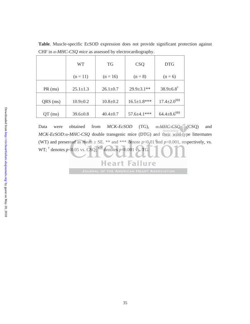
(Figure 5F). Electrocardiography showed significantly increased QRS intervals (Table) and
decreased heart rate (p<0.01) in these mice to the same degree (Figure 5G). Therefore,
reduced cachexia and improved muscle function in MCK-EcSOD: -MHC-CSQ mice did not
appear to be a result of improved cardiac function. We also crossbred MCK-EcSOD mice
with a genetic model of amyotrophic lateral sclerosis (ALS), SOD1-G93A mice. SOD1-G93A
mice harbor transgene expressing autosomal dominant fashion of a mutant form of
CuZnSOD that results in ALS-like motor neuron disease. SOD1-G93A and
SOD1-93A:MCK-EcSOD mice had similar survival curve (Supplemental Figure S5) with
significant reduction of body weight at 14 wks of age and reduced muscle force production at
8 weeks of age (Supplemental Figure S5). Therefore, EcSOD did not provide protection for
skeletal muscle when the insults were of motor neuron origin.
Muscle-specific EcSOD transgenic mice are resistant to CHF in developing cachexia and
multiple abnormalities
To obtain structural and biochemical evidence of EcSOD-mediated protection, we measured
sbsbsbsbsbsbsbrerererererered ddddd d MCMCMCMCMCMCMCK-K-K-K-K-K-K-EcEcEcEcEcEcEcSOSOSOSOSOSOSO
m
sgene expressing autosomal dominant fashion of a mutant form
esults in ALS like motor neuron disease SOD1 G93A and
modododododelelelee of amaamama yoyyyy troppppphic lateral sclerooossis (A(((( LSSSS),),),),), SOOOOODD1DD -G93A mice.
sgene expres iising autttosomalllll dddddomiiinanttt faf shioiiii n ffof a m tutant ffof rm
esesululultsts iiinn ALALALSSS lililikekeke mmototoror nneueuroronn dididiseseasasee SOSOSOD1D1D1 GGG939393AAA aandndnd
by guest on May 20, 2018
http://circheartfailure.ahajournals.org/D
ownloaded from
18
muscle mass and normalized by tibia length to minimize the effect of body weight reduction.
CHF led to a significant loss of muscle mass in EDL (-38.4%; p<0.01), tibialis anterior
(-37.3%; p<0.01), gastrocnemius (-42.3%; p<0.001), plantaris (-43.3%; p<0.01) but to a less
degree in soleus (-28.7%; p<0.001) muscles in -MHC-CSQ mice compared to the wild-type
littermates, which were significantly attenuated in MCK-EcSOD: -MHC-CSQ mice (Figure
6A). Fiber size analyses showed that -MHC-CSQ mice had significantly reduced IId/x/IIb
fiber size (-42.8%; p<0.05) (Figure 6B), which was blunted by EcSOD overexpression
(p<0.05 vs. CSQ). Consistent with our previous finding3, -MHC-CSQ mice had significantly
decreased capillary contacts in glycolytic type IId/x/IIb fibers (p<0.01), but not in oxidative
type IIa fibers. The loss of capillarity was significantly attenuated in
MCK-EcSOD: -MHC-CSQ mice (p<0.05 vs. CSQ; Figure 6C). We also performed whole
mount immunofluorescence staining for CD31 and smooth muscle -actin in FDB muscle
and measured the fractal dimension of microvessels (arterioles and venules), a measure of
number of branches and microvessels. There were no significant differences in fractal
dimension among different groups (Figure 6D), suggesting that vascular rarefaction did not
occur at the arteriole and venule levels in -MHC-CSQ mice. -MHC-CSQ mice are known
to have reduced mitochondrial (Cyt C, COX IV) and PGC-1 protein expression indicative
EcEcEcEcEcEcEcSOSOSOSOSOSOSODDDDDDD ovovovovovovoverererererererexexexexexexexprprprprprprpreeeeee
Q d
a n
The loss of capillarity was significantly attenuated in
Q))))). CCCoCC nsisisisisistentnn with our prppp evious fffffiniinii dinggg3, -M-M-M-M-MHCHCHCHCHC-CSQQQQQ mice had
ary co ttntacts iiin llglycyyy olllll ttytic typppe IIIId/d/d/ /x//IIIIIIIIbbb bb fififififibbebbb rs (((p(( <0.000001)1)1))1), bubb t not in
TTThehehe lllososss ofofof ccapapililillalalaririrityty wwasas ssigigignininififificacantntlylyly aattttenenuauateteddd ininin
by guest on May 20, 2018
http://circheartfailure.ahajournals.org/D
ownloaded from
19
of mitochondrial dysfunction in fast-twitch, glycolytic muscles3, 6. Here, we showed that a
preferentially reduced expression of these proteins in fast-twitch, glycolytic white vastus
lateralis muscle was absent in MCK-EcSOD: -MHC-CSQ mice (Figure 6D), suggesting that
overexpression of EcSOD is sufficient to block CHF-induced mitochondrial dysfunction in
skeletal muscle. Consistently, CHF condition in -MHC-CSQ mice did not cause any
significant changes in myosin heavy chain protein isoforms, and EcSOD overexpression
alone in MCK-EcSOD mice did not cause compensatory changes either. Real-time PCR
analysis showed that -MHC-CSQ mice had significantly elevated MAFbx/Atrogin-1 mRNA
(p<0.001), which was significantly reduced in MCK-EcSOD: -MHC-CSQ mice (p<0.05 vs.
CSQ) (Figure 6F). We did not detect significantly differences of apoptosis assessed by
TUNEL staining (Supplemental Figure S5). Most importantly, we have performed three
different analyses to determine if EcSOD overexpression had any significant impact on
oxidative stress induced by CHF. We found that CHF (in -MHC-CSQ mice) led to
significantly elevated levels of oxidative stress markers, MDA and protein carbonyls, in
fast-twitch, glycolytic white vastus lateralis muscle, which were significantly reduced in
MCK-EcSOD: -MHC-CSQ mice (Figure 6G and Supplemental Figure 6S). A similar trend,
although not statistically significant, was found for another oxidative stress marker of lipid
sssssss eeeeeeeitititititititheheheheheheher.rrr.rrr RRRRRRReaeaeaeaeaeaeal-l-l-l-l-l-l-tititititititimmmmmmm
g
h
F) We did not detect significantly differences of apoptosis assess
thththththatatatata -MMMMMHCHCHCHCHC-CSQQQQQ mice had signg ifiifii icantly yyyy eleleleleleveeee atttede MAFbx/Atrog
h was siiigii niiififificantlylylyly reddddduceddddd iiiiin MCMMCKKK-EcEEEE SOSOSOSOSOD:DDD MM-MHCHCHCCC CCCC-CSQQ miiice
F)F)F) WeWeWe dddididid nnotot dddetetecectt sisisigngnifififiiicacantntlylyly dddifififfefefererencnceses oofff apapopoptotosisisiss asassesessss
by guest on May 20, 2018
http://circheartfailure.ahajournals.org/D
ownloaded from
20
peroxidation, 4-HNE (Supplemental Figure 6S). These findings provide evidence for EcSOD
function in protecting skeletal muscle from cardiac cachexia at molecular, biochemical,
structural and functional levels likely through its ectopic antioxidant function.
Discussion
Studies with genetic and pharmacological interventions in cultured myocytes and intact
skeletal muscles have provided ample evidence that intracellular antioxidant defenses are
critical in maintaining normal redox state, muscle mass and function28, 29. Here, we provide
clear in vivo evidence that EcSOD expression in skeletal muscle is reduced under
glucocorticoidism, a condition of many chronic diseases including CHF that is associated
with increased oxidative stress30, muscle wasting and loss of muscle function18. We showed
that systemic administration of endogenous NO donor could attenuate skeletal muscle
atrophy induced by dexamethasone in mice concurrent with a restoration of EcSOD
expression. These findings suggest that NO-dependent EcSOD expression is important for
muscle maintenance in vivo. We then employed two independent genetic approaches and
showed that myofibers with acquired EcSOD expression do not undergo significant atrophy
upon glucocorticoidism. Muscle-specific EcSOD transgenic mice are resistant to CHF,
rrrrrrr aaaaaaantntntntntntntioioioioioioioxixixixixixixidadadadadadadantntntntntnn dddddddefefefefefefefeeeeeee
a w
dence that EcSOD expression in skeletal muscle is reduced un
m a condition of many chronic diseases including CHF that is as
ainininnininininining nooooorrrmalaa redox state,, muscleee mmass and d d d fufff nction28, 29. Here, w
dence thththththat EE SScSODDOD expressioiiii n iinii skkkeletttalllll muscllle iiis reddudd ceddd undded
mm aa cconondididitititionon oofff mamanyny cchrhrhrononiiicc dididiseseasaseses iiincnclululudididingng CCCHFHFHF tthahahatt isisis aass
by guest on May 20, 2018
http://circheartfailure.ahajournals.org/D
ownloaded from
21
displaying significant protections against cachexia, exercise intolerance, and loss of
mitochondria, rarefaction of microvasculature, and increased atrophy gene expression in
skeletal muscle. Most importantly, we have shown evidence of reduced oxidative stress when
EcSOD is ectopic overexpressed in a mouse model of cardiac cachexia. These findings
provide for the first time unequivocal evidence that induced EcSOD expression protects
myofibers from catabolic muscle wasting in vivo, suggesting that promoting EcSOD
expression by augmenting NO bioavailability in skeletal muscle could be an effective
intervention for CHF patients.
NO plays important signaling role in physiological and pathological processes. We have
reported that injection of lipopolysaccharide (LPS), a major mediator of sepsis, induces
greater NO production in slow-twitch, oxidative muscle than in fast-twitch, glycolytic muscle
in mice3, 16. Since these oxidative muscles are resistant to catabolic muscle wasting under
many disease conditions, the findings suggest that elevated NO production is protective. Our
previous studies in cultured myocytes provided further evidence for a functional link between
NO and antioxidant gene expression6, 16. Consistently, the data presented in this and previous
studies16, 31 showed robust induction of EcSOD expression when there is increased NO
bioavailability, suggesting that NO-induced EcSOD expression in skeletal muscle may
cccccccouououououououldldldldldldld bbbbbbbeeee e e e ananananananan eeffffffffffffffeeeeeee
C
tant signaling role in physiological and pathological processes. W
ection of lipopolysaccharide (LPS) a major mediator of sepsis in
CCCCCHFHFHFHFHF patttttieieieii nttts.
tant siignaliliil ng r llole in phhhyhh siiii llllologic llal anddddd ppathohhhh lloll iigical processes. WW
eeectctioioionn ofofof lllipipipopopolololysysacacchchchararidididee (L(L(LPSPSPS))) aa mmajajajoror mmededediaiaiatotorr ofofof ssepepsisisiss iiinnn
by guest on May 20, 2018
http://circheartfailure.ahajournals.org/D
ownloaded from
22
contribute significantly to the protection associated with oxidative phenotype.
The functional importance of EcSOD has been shown by its reduced expression associated
with various chronic diseases10, 11, and by the fact that genetic deletion and overexpression of
EcSOD leading to exacerbation and protection against pathological changes in animal models
of diseases, respectively12-15. Glucocorticoids are used as therapeutic agents due to their
potent anti- inflammatory and immunosuppressive functions; however, high dose and
long-term use of glucocorticoids causes muscle atrophy22, 32. Previous studies and our current
data support an increased oxidative stress induced by dexamethasone in skeletal muscle with
direct evidence of increased free radicals22, 33. Oxidative stress may promotes catabolic
wasting and loss of muscle function through cell signaling, impairment of contractile protein
and promotion of protein degradation4, 5, 34, 35. Here, we provide new evidence for reduced
EcSOD protein expression in catabolic muscle wasting and the functional importance of the
NO-mediated EcSOD expression in protection against catabolic wasting.
A key question is how EcSOD protects skeletal muscle from atrophy. Since EcSOD is the
only isoform of superoxide dismutase that is secreted to the extracellular space, one
straightforward explanation is that elevated extracellular ROS, which could be scavenged by
EcSOD, plays an importantly role in cachexia and exercise intolerance. Several pathological
evevevevevevevioioioioioioioususususususus ssssssstututututututudididididididieeesesesees aaaaaaandnndndndndnd
ncreased oxidative stress induced by dexamethasone in skeletal
o t
of muscle function through cell signaling impairment of contra
nnnnncrcrcrcrcreased dddd oxiidiii ative stress induceddddd bby dexamemmmm thhhhhasone in skeletal
of increaseddd fffree radiiiiicals22,222 333333333. OOOOOxidididatiiivii e stress may promotes cat
oofff mumuscsclelele fffununctctioioionn thththrorougughhh cecellllll ssigigignanalililingng imimimpapairirirmementnt oofff cocontntrara
by guest on May 20, 2018
http://circheartfailure.ahajournals.org/D
ownloaded from
23
steps might be relevant here. First, extracellular ROS could cause endothelial cell apoptosis36,
leading to vascular rarefaction, an important feature of cardiac cachexia. Vascular rarefaction
not only impairs microcirculation and tissue perfusion but also exacerbates muscle wasting
due to hypoxia. Second, superoxide reacts readily with NO to reduce NO bioavailability37,
impairing exercise-induced increase of blood flow and contributing to exercise intolerance.
Finally, extracellular ROS-mediated intracellular ROS production and signaling38 may play
an important role in catabolic muscle wasting. Enhanced EcSOD expression could provide
protection through one or more of these mechanisms. Studies showing protective effects of
antioxidants when applied directly to cultured cells appear to be consistent with EcSOD
function in scavenging extracellular superoxide; however, in light of the findings that EcSOD
can be internalized by cells that it binds9, elevated intracellular EcSOD in myofibers and/or
other cells in skeletal muscle may also contribute to the protection.
The protective role of EcSOD is likely mediated by its antioxidant properties. Based on the
evidence of reduced oxidative stress markers and atrogene expression in glycolytic muscles
(Figure 6F and 6G), we believe that transgenic overexpression of EcSOD in skeletal muscle
exerts its protective function by counteracting oxidative stress induced by CHF. The findings
of muscle mass, mitochondrial protein and vascular rarefaction are all extremely consistent
DD DDDDD exexexexexexexprprprprprprpresesesesesesessisisisisisisiononononononon ccouououououououllllll
gh one or more of these mechanisms. Studies showing protecti e
en applied directly to cultured cells appear to be consistent with E
enging extracellular superoxide; however in light of the findings
ghhhhh ooooone ooor r r rr more of these mechanisssmms. Studieieieieies shhhhhowing gggg protective
en ap lplllliiieii d dididirectlylyly to cultur dedd celllllls appppppppppear tott bbbbe consiisii tent wiiithhh E
enengigigingng eextxtraracecellllllululularar ssupupereroxoxididide;e; hhhowoweveverer ininin llligigighththt oofff thththee fififindndndininingsgs
by guest on May 20, 2018
http://circheartfailure.ahajournals.org/D
ownloaded from
24
with this notion. Future studies should focus on elucidating the necessity of EcSOD
expression in skeletal muscle in maintain muscle mass and function under catabolic wasting
conditions.
It has been shown that EcSOD expression by smooth muscle can be enhanced by exercise
training in an NO-dependent manner31, and EcSOD mRNA is increased by acute exercise in
skeletal muscle39. It has also been shown recently that exercise training protects oxidative
stress and proteasome-dependent protein degradation in CHF-induced skeletal muscle
atrophy40. An unanswered question is which effecter gene(s) is responsible for the enhanced
antioxidant function induced by exercise training. Our findings suggest EcSOD could be such
an important player. We show here that exercise training promotes EcSOD expression in
skeletal muscle (Figure 2), and that enhanced EcSOD expression is sufficient to provide
protection against cardiac cachexia and exercise intolerance.
Another issue is whether enhanced expression of EcSOD from skeletal muscle redistributes
to the extracellular space of the heart leading to improved cardiac function and preventing
cachexia. EcSOD-mediated protection against cardiac toxicity has been shown by Kliment et
al where EcSOD global knockout mice showed exacerbated cardiac dysfunction, myocardial
apoptosis and fibrosis following doxorubicin injection41. We have previously shown that
ddddddducucucucucucucededededededed ssssssskekekekekekekeleleleleleleletatatatataaal l llll l mmmmmmm
n h
t c
yer We show here that exercise training promotes EcSOD expre
naaaansnsnsnsnswereeeeeddd dd quqq estion is which effecccteer gegggg ne(ss(ss( ) ) ) )) is responsible for th
tion iiii ddnddduceddd bbbby exer iicise traiiniii iiiiingggg. OOOur fififiii dndddings sugge ttttst EE ScSODODO c
yeyerr WWWee shshshowow hhhereree thththatat eexexercrcisisisee trtraiaiainininingng pproromomotetess EcEcEcSOSOSODDD exexprpree
by guest on May 20, 2018
http://circheartfailure.ahajournals.org/D
ownloaded from
25
adenovirus-mediated gene transfer of EcSOD in the heart elicits protective effects on cardiac
function following myocardial infarction in rabbits42. In this study, although forced
expression of EcSOD from skeletal muscle redistributed to the heart (Supplemental Figure
S3), it did not improve cardiac function and prevent premature death. These findings allowed
us to focus on the impact of ectopic EcSOD expression in skeletal muscle on catabolic
muscle wasting without confounding factors of EcSOD overexpression in impeding heart
failure. Sicne abnormal calcium and G-protein coupling signaling is caused by calsequestrin
overexpression from within cardiac myocytes43, the negative findings of EcSOD effects in
the heart suggest that elevated EcSOD in the heart of -MHC-CSQ mouse is not enough to
deal with such a severe CHF condition or with pathogenesis of CHF of an intracellular origin.
Previous findings support the view that exercise intolerance in CHF is a result of skeletal
muscle abnormalities although the failing heart is the primary cause44-46. Consistent with this
scenario, our findings suggest that the improved skeletal muscle outcomes in
MKC-EcSOD: -MHC-CSQ mice were not due to improved cardiac function. Our
measurements of baseline heart function, heart weight, skeletal muscle weight, and survival
curve collectively suggest that EcSOD overexpression had a negligible impact on the
progression of heart failure induced by cardiac overexpression of calsequestrin in our model.
ngngngngngngng iiiiiiiss s s s s s cacacacacacacausususususususededededededed bbbbbbbyy yy y y y cacaaaaaa
f
t
severe CHF condition or with pathogenesis of CHF of an intrace
frooooommmm m withththththin cardiac myoyyyy cyytes43, thhhhhe negagg tiveveveveve finddidd ngggggs of EcSOD
t thattttt elllell vateddd d EEcSSOD DDD iin thehhhh hhhhheart of ffff -MMMMMHCHHCCHC-CCSQQSQ mouse iiis not
ssevevereree CHCHCHFFF cocondndnditititioioionn oror wwititithhh papathththogogenenesesisisis oofff CHCHCHFFF ofofof aann ininintrtracaceee
by guest on May 20, 2018
http://circheartfailure.ahajournals.org/D
ownloaded from
26
On the contrary, skeletal muscle abnormalities are significantly blocked/attenuated along
with improved contractile function and exercise capacity. We, therefore, believe that the
effects of elevated EcSOD in the heart on cardiac function, if present, likely has a minor
impact on exercise capacity. It would be interesting to ascertain whether muscle
overexpression of EcSOD is protective against other types of CHF.
The concept that exercise intolerance in CHF results from multiple skeletal muscle
abnormalities will be instrumental to developing effective interventions. We have previously
shown that CHF leads to preferential vascular rarefaction in fast-twitch, glycolytic muscle
fibers in CHF3, and that skeletal muscles with overexpression of PGC- 1 have significantly
elevated EcSOD expression, less muscle atrophy and vascular rarefaction under the condition
of CHF6. Here, we have again detected vascular rarefaction in fast-twitch, glycolytic fibers in
-MHC-CSQ mice, which was significantly attenuated by EcSOD overexpression. Since
EcSOD accumulates at the endothelium, EcSOD may also protect endothelial cells against
oxidative stress-induced apoptosis. We have also previously shown that skeletal muscles
undergoing catabolic wasting lose mitochondria with reduced expression of PGC-1 and
increased oxidative stress3, 6, which may contribute directly to exercise intolerance. The fact
that EcSOD overexpression prevents the reduction of mitochondrial and PGC-1 proteins
veveveveveveventntntntntntntioioioioioioionsnsnsnsnsnsns...... WeWeWeWeWeWeWe hhhhhhhavavaavaaa eeeeeee
t
a s
expression less muscle atrophy and vascular rarefaction under t
lllleaeaeaeaeadddsdd to oooo ppprefefeferential vascular rareeeffaction innnnn fast-t-t twitch, glycyyyy olyt
and thhththth tat skekk lletall muscllles wiiii hhhthh overexpppppression fof PPPGCCCCC- 1 hhave s
eexpxpreressssioioionn lllesesss mumuscsclelele aatrtroophphphyy ananddd vavascscululularar rrararefefefaactctioioionn unundedederr ttt
by guest on May 20, 2018
http://circheartfailure.ahajournals.org/D
ownloaded from
27
strongly suggests that ROS induced by CHF induces pathological events in muscle. This
could explain why enhanced EcSOD expression in skeletal muscle can provide potent
protection and lead to preserved exercise capacity in MCK-EcSOD: -MHC-CSQ mice.
Previous studies have also linked oxidative stress to catabolic muscle wasting with evidence
for enhanced ubiquitin-proteasome16, 47-49 and the autophagy-lysosome systems22, 50. The
link of oxidative stress to autophagy, particularly mitochondrial autophagy (mitophagy), may
underlie the loss of mitochondria in CHF. A vicious cycle between oxidative stress and
mitochondrial dysfunction could be critical for the development of cardiac cachexia and
exercise intolerance.
Finally, we have previously shown that overexpression of PGC-1 in skeletal muscle leads to
increased iNOS, eNOS, EcSOD, MnSOD, CuZnSOD and CAT expression in skeletal muscle
along with increased FOXO and Akt phosphorylation6. We postulated that PGC-1 enhances
FOXO and Akt phosphorylation in parallel with enhanced NO-antioxidant defense axis. Our
current findings, consistent with this posulation, suggest that increased NO can directly
stimulate EcSOD in skeletal muscle providing protection. It is of note that increased NO
alone without overexpression of PGC-1 only stimulates EcSOD, but not the other
antioxidant enzymes. This NO inducibility of EcSOD expression helped us to focus on the
eeeeeeeeeeeeeennnn nnn oxoxoxoxoxoxoxidididididididatatatatatatativiviviviviviveeee e ee stststststststrereeeeee
y e
n
previously shown that overexpression of PGC 1 in skeletal mu
ysfsfsfsfsfuuuunu ctiooooonnn could be critical for theee developmpmpmpmpment tt of cardiac cache
nce.
pprereviviviououslslslyy shshshowownn thththatat oovevererexpxpreressssioioionn ofofof PPPGCGCGC 111 iiinn skskskeleleletetalalal mmuu
by guest on May 20, 2018
http://circheartfailure.ahajournals.org/D
ownloaded from
28
function of this important, yet understudied, antioxidant enzyme in protection against muscle
wasting.
In summary, we have obtained novel findings to support that NO-mediated protection against
catabolic muscle wasting is associated with enhanced EcSOD protein expression. Genetic
interventions with forced expression of EcSOD in skeletal muscles significantly attenuates
muscle atrophy. Importantly, muscle-specific EcSOD transgenic mice are resistant to CHF in
developing cachexia and exercise intolerance with significant protection against oxidative
stress, atrophic gene expression, and loss of mitochondria and microvasculature. We now
propose a model where enhanced skeletal muscle EcSOD expression prevents endothelial
and muscular abnormalities in cachexia and exercise intolerance likely due to the
maintenance of redox homeostasis (Figure 7). The findings pave the way for targeting
NO-dependent EcSOD expression for the prevention and treatment of cardiac cachexia.
Acknowledgements
We thank Mrs. John Sanders and R. Jack Roy for their excellent technical supports in
immunofluorescence and MRI, respectively.
rrrrrrotototototototececececececectititititititiononononononon aaaaaaagagagagagagagainininininininsssstststst
gene expression, and loss of mitochondria and microvasculature.
n
normalities in cachexia and exercise intolerance likely due to the
geneneneneneeee e exprprprprreese isiisision, ,, and loss of mitochcchondria aaaandnnnn microvasculature.
whhhhhere enhahhh nc dded skellletal muscle EE SScSSSODODODODD expression prevents en
nnnorormamalililitititieses iiinn cacachchchexexiaiaia aandndnd eexexercrcisisisee ininintotolelelerarancncee lililikekekelylyly dddueue ttoo thththee
by guest on May 20, 2018
http://circheartfailure.ahajournals.org/D
ownloaded from
29
Sources of Funding
This project was supported by grant AR050429 (Z.Y.). The research was also supported by
Nakatomi Foundation (M.O.), Postdoctoral Fellowship in Physiological Genomics by the
American Physiological Society (V.A.L.), National Health Institutes T32-HL007284
(J.A.C.), and American Heart Association 12POST12030231 (J.A.C.). This research was
supported in part by grant to S.M.P. (HL082838).
Disclosures
None.
References 1. Sullivan M, Duscha B, Klitgaard H, Kraus W, Cobb F, Saltin B. Altered expression of
myosin heavy chain in human skeletal muscle in chronic heart failure. Med Sci Sports
Exerc. 1997;29:860-866.
2. Schaufelberger M, Eriksson B, Grimby G, Held P, Swedberg K. Skeletal muscle fiber
composition and capillarization in patients with chronic heart failure: Relation to
exercise capacity and central hemodynamics. J Card Fail. 1995;1:267-272.
3. Li P, Waters RE, Redfern SI, Zhang M, Mao L, Annex BH, Yan Z. Oxidative
phenotype protects myofibers from pathological insults induced by chronic heart
failure in mice. Am J Pathol. 2007;170:599-608.
4. Reid MB, Moylan JS. Beyond atrophy: Redox mechanisms of muscle dysfunction in
chronic inflammatory disease. J Physiol. 2011;589:2171-2179.
5. Powers SK, Smuder AJ, Judge AR. Oxidative stress and disuse muscle atrophy:
Cause or consequence? Current opinion in clinical nutrition and metabolic care.
2012;15:240-245.
6. Geng T, Li P, Yin X, Yan Z. Pgc-1alpha promotes nitric oxide antioxidant defenses
and inhibits foxo signaling against cardiac cachexia in mice. Am J Pathol.
2011;178:1738-1748.
M, Duscha B, Klitgaard H, Kraus W, Cobb F, Saltin B. Altered
ReReReReRefefeff rerr ncncnceseseseses MM, Dussssschchchchcha aa a a B,B,B,B,B, KKKKKliiiiitgtgtgtgtgaaaaaaaaaardrdrdrdd HHHHH,,,,, KrKrKrKrKrauauauauausssss W,W,W,WW CCCCCobobobobobb bbbb F,F,F,F,F, SSSSSalalalalaltitititiin n n n n B.B.B.B.B. AAAAAltered
by guest on May 20, 2018
http://circheartfailure.ahajournals.org/D
ownloaded from
30
7. Hauer K, Hildebrandt W, Sehl Y, Edler L, Oster P, Droge W. Improvement in
muscular performance and decrease in tumor necrosis factor level in old age after
antioxidant treatment. J Mol Med (Berl). 2003;81:118-125.
8. Mantovani G, Maccio A, Madeddu C, Gramignano G, Lusso MR, Serpe R, Massa E,
Astara G, Deiana L. A phase ii study with antioxidants, both in the diet and
supplemented, pharmaconutritional support, progestagen, and anti-cyclooxygenase-2
showing efficacy and safety in patients with cancer-related anorexia/cachexia and
oxidative stress. Cancer Epidemiol Biomarkers Prev. 2006;15:1030-1034.
9. Chu Y, Piper R, Richardson S, Watanabe Y, Patel P, Heistad DD. Endocytosis of
extracellular superoxide dismutase into endothelial cells: Role of the heparin-binding
domain. Arterioscler Thromb Vasc Biol. 2006;26:1985-1990.
10. Dahl M, Bowler RP, Juul K, Crapo JD, Levy S, Nordestgaard BG. Superoxide
dismutase 3 polymorphism associated with reduced lung function in two large
populations. American journal of respiratory and critical care medicine.
2008;178:906-912.
11. Chen Y, Hou M, Li Y, Traverse JH, Zhang P, Salvemini D, Fukai T, Bache RJ.
Increased superoxide production causes coronary endothelial dysfunction and
depressed oxygen consumption in the failing heart. Am J Physiol Heart Circ Physiol.
2005;288:H133-141.
12. Lu Z, Xu X, Hu X, Zhu G, Zhang P, van Deel ED, French JP, Fassett JT, Oury TD,
Bache RJ, Chen Y. Extracellular superoxide dismutase deficiency exacerbates
pressure overload-induced left ventricular hypertrophy and dysfunction.
Hypertension. 2008;51:19-25.
13. Carlsson LM, Jonsson J, Edlund T, Marklund SL. Mice lacking extracellular
superoxide dismutase are more sensitive to hyperoxia. Proc Natl Acad Sci U S A.
1995;92:6264-6268.
14. Yao H, Arunachalam G, Hwang JW, Chung S, Sundar IK, Kinnula VL, Crapo JD,
Rahman I. Extracellular superoxide dismutase protects against pulmonary
emphysema by attenuating oxidative fragmentation of ecm. Proc Natl Acad Sci U S
A. 2010;107:15571-15576.
15. Prasad KM, Smith RS, Xu Y, French BA. A single direct injection into the left
ventricular wall of an adeno-associated virus 9 (aav9) vector expressing extracellular
superoxide dismutase from the cardiac troponin-t promoter protects mice against
myocardial infarction. J Gene Med. 2011;13:333-341.
16. Yu Z, Li P, Zhang M, Hannink M, Stamler J, Yan Z. Fiber type-specific nitric oxide
lung gggggg functiononononnnn i
nd crcrcrcrcrcrcritititititititicicicicicicicalalalalalalal cccccccararaaaara
8:906-912.
Hou M, Li Y, Traverse JH, Zhang P, Salvemini D, Fukai T
d f
d
8
8:90606060606-9-9-9-9-91212121212.
HoHHHH u M, LLLLLi i Y,Y,Y,Y,Y, TTTTTrararararaveveveveversrsrsrsrseeee JHHHHH, ZhZhannnng gggg P,P,PP,P SSSSalalalveveveveemimimiininininini DDDDD, FuFuFuFuFukakakakakai i iii T
d sususususupepepepep rororororoxixixixixidededeee ppppprodudududuductctctcttioioioioionn nnn cacacacc uusuuu eseseseses cccccooooorororororonananananaryryryryry eeeeendndndnddotototototheheheeelililililiaaala dddddysysysysy f
d oxygen consumpppptttit ononononon iiiiin n nn n ththhthhee ee failillinininnng gggg hehhehh araraarrt.tt AmAmAmAmAm J Physiol Heart
88:H133-14141414141.11.1.1
by guest on May 20, 2018
http://circheartfailure.ahajournals.org/D
ownloaded from
31
protects oxidative myofibers against cachectic stimuli. PLoS ONE. 2008;3:e2086.
17. Geng T, Li P, Okutsu M, Yin X, Kwek J, Zhang M, Yan Z. Pgc-1alpha plays a
functional role in exercise-induced mitochondrial biogenesis and angiogenesis but not
fiber-type transformation in mouse skeletal muscle. Am J Physiol, Cell Physiol.
2010;298:C572-579.
18. Hasselgren PO. Glucocorticoids and muscle catabolism. Current opinion in clinical
nutrition and metabolic care. 1999;2:201-205.
19. Naya F, Mercer B, Shelton J, Richardson J, Williams R, Olson E. Stimulation of slow
skeletal muscle fiber gene expression by calcineurin in vivo. J Biol Chem.
2000;275:4545-4548.
20. Jones L, Suzuki Y, Wang W, Kobayashi Y, Ramesh V, Franzini-Armstrong C,
Cleemann L, Morad M. Regulation of ca2+ signaling in transgenic mouse cardiac
myocytes overexpressing calsequestrin. J Clin Invest. 1998;101:1385-1393.
21. Akimoto T, Ribar T, Williams R, Yan Z. Skeletal muscle adaptation in response to
voluntary running in ca2+/calmodulin-dependent protein kinase iv-deficient mice. Am
J Physiol Cell Physiol. 2004;287:C1311-1319.
22. McClung JM, Judge AR, Powers SK, Yan Z. P38 mapk links oxidative stress to
autophagy-related gene expression in cachectic muscle wasting. Am J Physiol Cell
Physiol. 2010;298:C542-549.
23. Yan Z, Choi S, Liu X, Zhang M, Schageman JJ, Lee SY, Hart R, Lin L, Thurmond
FA, Williams RS. Highly coordinated gene regulation in mouse skeletal muscle
regeneration. J Biol Chem. 2003;278:8826-8836.
24. Pogozelski AR, Geng T, Li P, Yin X, Lira VA, Zhang M, Chi J-T, Yan Z. P38gamma
mitogen-activated protein kinase is a key regulator in skeletal muscle metabolic
adaptation in mice. PLoS ONE. 2009;4:e7934.
25. Carlson CG, Makiejus RV. A noninvasive procedure to detect muscle weakness in the
mdx mouse. Muscle Nerve. 1990;13:480-484.
26. Berr SS, Roy RJ, French BA, Yang Z, Gilson W, Kramer CM, Epstein FH. Black
blood gradient echo cine magnetic resonance imaging of the mouse heart. Magn
Reson Med. 2005;53:1074-1079.
27. Bailey AM, O'Neill TJt, Morris CE, Peirce SM. Arteriolar remodeling following
ischemic injury extends from capillary to large arteriole in the microcirculation.
Microcirculation. 2008;15:389-404.
28. Muller FL, Song W, Liu Y, Chaudhuri A, Pieke-Dahl S, Strong R, Huang TT, Epstein
CJ, Roberts LJ, 2nd, Csete M, Faulkner JA, Van Remmen H. Absence of cuzn
98;1; 01:1385-5-131313131313139
scle aaaaaaadadadadadadadaptptptptptptptatatatatatatatioioioioioioionnnn n nn ininininininin
y running in ca2+/calmodulin-dependent protein kinase iv-defici
l
g JM, Judge AR, Powers SK, Yan Z. P38 mapk links oxi t
g
2
y ruuuuunnnnnnnnnnininininng gggg innnnn ca2+/calmodulin-depepeppepeneeee dent proteininininin kinase iv-defici
l CCeCCC ll Physisisisisiolo . 20202020200404040404;2;2;2;2;28787878787:CCCCC131313331111111 -1113319.9.9.9.9.
g JMJMJMJMJM, ,,, , JuJuJuJuudgdgdgdgdge e e ARARARARAR, , PoPoPoPoPowewewewewersssss SSSSSK,K,K,K,K, YYYYYananananan ZZZZZ..... P3P3P3P3P38 8888 mamamamamapkpkpkkpk linnnnnkkskkk oooooxiiiiidddad t
gy-related gene expxpxpprerereeessssssssssioioioioion n n nn inininin cacacchehehehehectctctctcticc mmmmususususu clclclclcle eee wasting. Am J
22010;29898989898:C:C:C:C:C54545454542-2-22-2 545454449.99.99
by guest on May 20, 2018
http://circheartfailure.ahajournals.org/D
ownloaded from
32
superoxide dismutase leads to elevated oxidative stress and acceleration of
age-dependent skeletal muscle atrophy. Free radical biology & medicine.
2006;40:1993-2004.
29. Dobrowolny G, Aucello M, Rizzuto E, Beccafico S, Mammucari C, Boncompagni S,
Belia S, Wannenes F, Nicoletti C, Del Prete Z, Rosenthal N, Molinaro M, Protasi F,
Fano G, Sandri M, Musaro A. Skeletal muscle is a primary target of
sod1g93a-mediated toxicity. Cell Metab. 2008;8:425-436.
30. Ohtani T, Ohta M, Yamamoto K, Mano T, Sakata Y, Nishio M, Takeda Y, Yoshida J,
Miwa T, Okamoto M, Masuyama T, Nonaka Y, Hori M. Elevated cardiac tissue level
of aldosterone and mineralocorticoid receptor in diastolic heart failure: Beneficial
effects of mineralocorticoid receptor blocker. Am J Physiol Regul Integr Comp
Physiol. 2007;292:R946-954.
31. Fukai T, Siegfried MR, Ushio-Fukai M, Cheng Y, Kojda G, Harrison DG. Regulation
of the vascular extracellular superoxide dismutase by nitric oxide and exercise
training. J Clin Invest. 2000;105:1631-1639.
32. Waddell DS, Baehr LM, van den Brandt J, Johnsen SA, Reichardt HM, Furlow JD,
Bodine SC. The glucocorticoid receptor and foxo1 synergistically activate the skeletal
muscle atrophy-associated murf1 gene. Am J Physiol Endocrinol Metab.
2008;295:E785-797.
33. Konno S. Hydroxyl radical formation in skeletal muscle of rats with
glucocorticoid-induced myopathy. Neurochem Res. 2005;30:669-675.
34. Smuder AJ, Kavazis AN, Hudson MB, Nelson WB, Powers SK. Oxidation enhances
myofibrillar protein degradation via calpain and caspase-3. Free radical biology &
medicine. 2010;49:1152-1160.
35. Clavel S, Siffroi-Fernandez S, Coldefy AS, Boulukos K, Pisani DF, Derijard B.
Regulation of the intracellular localization of foxo3a by stress-activated protein
kinase signaling pathways in skeletal muscle cells. Mol Cell Biol. 2010;30:470-480.
36. Cho KS, Lee EH, Choi JS, Joo CK. Reactive oxygen species-induced apoptosis and
necrosis in bovine corneal endothelial cells. Invest Ophthalmol Vis Sci.
1999;40:911-919.
37. Jung O, Marklund SL, Geiger H, Pedrazzini T, Busse R, Brandes RP. Extracellular
superoxide dismutase is a major determinant of nitric oxide bioavailability: In vivo
and ex vivo evidence from ecsod-deficient mice. Circ Res. 2003;93:622-629.
38. Daiber A. Redox signaling (cross-talk) from and to mitochondria involves
mitochondrial pores and reactive oxygen species. Biochim Biophys Acta.
a G, , Harrisonon DDDDDDDGG
by nininininininitrtrtrtrtrtrtricicicicicicic oooooooxixixixixixixidedededededede
J Clin Invest 2000;105:1631 1639
DS Baehr LM van den Brandt J Johnsen SA Reichardt M
SC. The glucocorticoid receptor and foxo1 synergistically activ t
r
5
J ClClClClClininininin IIIIInvnvnvnvn esesesesesttttt. 2000;105:1631-166663939393939.
DSDSDSDSDS, Baehehhhhr r rr r MLMLMMM,,, ,, vavavavavan nn nn dededededen n BrBrBranaanaa dtt JJ, JoJoJoJoJohnhnhnhnhnseennn SASASASASA, ReReReReReicicicicichahahahahardrdrdrdrdt tttt HMHMHMHMHM
SC.C.C.C.C. TTTTTheheheheh ggggglululululucococooocooooorrtrr icccoioioioioidddd d rererererececececec ptptptptptororororor aaaandndndndnd fffffoxoxoxoxoxo1o1o1o1o1 ssssynynynynyneeeree ggiggig stststststicicicicicalalalalallylylylyly aaactccc ivivivii ataa
atrophy-associateeed d d d mumumumumurfrfrrfr 11111 geeneneneee..... AmAmAmmAm JJJJJ PPPPhysiol Endocr
55:E785-79797979797.7.7.7.7
by guest on May 20, 2018
http://circheartfailure.ahajournals.org/D
ownloaded from
33
2010;1797:897-906.
39. Hitomi Y, Watanabe S, Kizaki T, Sakurai T, Takemasa T, Haga S, Ookawara T,
Suzuki K, Ohno H. Acute exercise increases expression of extracellular superoxide
dismutase in skeletal muscle and the aorta. Redox Rep. 2008;13:213-216.
40. Cunha TF, Bacurau AV, Moreira JB, Paixao NA, Campos JC, Ferreira JC, Leal ML,
Negrao CE, Moriscot AS, Wisloff U, Brum PC. Exercise training prevents oxidative
stress and ubiquitin-proteasome system overactivity and reverse skeletal muscle
atrophy in heart failure. PLoS One. 2012;7:e41701.
41. Kliment CR, Suliman HB, Tobolewski JM, Reynolds CM, Day BJ, Zhu X,
McTiernan CF, McGaffin KR, Piantadosi CA, Oury TD. Extracellular superoxide
dismutase regulates cardiac function and fibrosis. J Mol Cell Cardiol.
2009;47:730-742.
42. Li Q, Bolli R, Qiu Y, Tang XL, Guo Y, French BA. Gene therapy with extracellular
superoxide dismutase protects conscious rabbits against myocardial infarction.
Circulation. 2001;103:1893-1898.
43. Cho M, Rapacciuolo A, Koch W, Kobayashi Y, Jones L, Rockman H. Defective
beta-adrenergic receptor signaling precedes the development of dilated
cardiomyopathy in transgenic mice with calsequestrin overexpression. J Biol Chem.
1999;274:22251-22256.
44. Sullivan M, Green H, Cobb F. Skeletal muscle biochemistry and histology in
ambulatory patients with long-term heart failure. Circulation. 1990;81:518-527.
45. Massie B. Exercise tolerance in congestive heart failure. Role of cardiac function,
peripheral blood flow, and muscle metabolism and effect of treatment. Am J Med.
1988;84:75-82.
46. Jondeau G, Katz S, Zohman L, Goldberger M, McCarthy M, Bourdarias J, LeJemtel
T. Active skeletal muscle mass and cardiopulmonary reserve. Failure to attain peak
aerobic capacity during maximal bicycle exercise in patients with severe congestive
heart failure. Circulation. 1992;86:1351-1356.
47. Jin B, Li Y-P. Curcumin prevents lipopolysaccharide-induced atrogin-1/mafbx
upregulation and muscle mass loss. J Cell Biochem. 2007;100:960-969.
48. Li Y, Chen Y, John J, Moylan J, Jin B, Mann D, Reid M. Tnf-alpha acts via p38
mapk to stimulate expression of the ubiquitin ligase atrogin1/mafbx in skeletal
muscle. Faseb J. 2005;19:362-370.
49. McClung JM, Kavazis AN, Whidden MA, DeRuisseau KC, Falk DJ, Criswell DS,
Powers SK. Antioxidant administration attenuates mechanical ventilation-induced rat
ene therapppppy y wiwiwiwiiiiththththththth
ainssssssstttttt t mymymymymymymyocococococococarararararrardididididididiaa
on 2001;103:1893 1898
Rapacciuolo A, Koch W, Kobayashi Y, Jones L, Rockman
energic receptor signaling precedes the development
y J
4
on..... 20202020200101010101;1;;; 0303030303:1893-1898.
RRRRRaaapa acciuooooololololol AAAAA,,,,, KoKoKoKoKochchchchch WWWWW,,, KoKoKKK baaayyashshshshshiiii Y,Y,Y,YY JJJJonnnnnes LLLLL, RoRoRoRoRockckckckckmamamamamannn nn
ennnnererererergigigigigic rereererecececec ppptpp ooroo sssssiggigigignananananalililililingngngngng ppppprrerrr cececececedededededessss thththththeee ee dededededevevevevevelololololopmpmpmpmpmennnnnttt t
yopathy in transgenenennicicccc mmmmmicicicicice e ee wiwww thh ccccalalalalalseseseses ququuuesesesestrtrrtrrininininn overexpression. J
44:22251-2-2-2-2-222222222225656565656.....
by guest on May 20, 2018
http://circheartfailure.ahajournals.org/D
ownloaded from
34
diaphragm muscle atrophy independent of protein kinase b (pkb akt) signalling. J
Physiol. 2007;585:203-215.
50. Zhao J, Brault JJ, Schild A, Cao P, Sandri M, Schiaffino S, Lecker SH, Goldberg AL.
Foxo3 coordinately activates protein degradation by the autophagic/lysosomal and
proteasomal pathways in atrophying muscle cells. Cell Metab. 2007;6:472-483.
by guest on May 20, 2018
http://circheartfailure.ahajournals.org/D
ownloaded from
35
Table. Muscle-specific EcSOD expression does not provide significant protection against
CHF in -MHC-CSQ mice as assessed by electrocardiography.
WT
(n = 11)
TG
(n = 16)
CSQ
(n = 8)
DTG
(n = 6)
PR (ms) 25.1±1.3 26.1±0.7 29.9±3.1** 38.9±6.8†
QRS (ms) 10.9±0.2 10.8±0.2 16.5±1.8*** 17.4±2.0§§§
QT (ms) 39.6±0.8 40.4±0.7 57.6±4.1*** 64.4±8.6§§§
Data were obtained from MCK-EcSOD (TG), -MHC-CSQ (CSQ) and
MCK-EcSOD: -MHC-CSQ double transgenic mice (DTG) and their wild-type littermates
(WT) and presented as mean ± SE. ** and *** denote p<0.01 and p<0.001, respectively, vs.
WT; † denotes p<0.05 vs. CSQ; §§§ denotes p<0.001 vs. TG.
-M-M-M-M-M-M-MHCHCHCHCHCHCHC-C-C-C-C-C-C-CSQSQSQSQSQSQSQ (((((((
nd ththhhhhheieieieieieieir r r r r r r wiwiwiwiwiwiwildldldldldldld-t-t-t-ttttyypyyy
n p
<
ntededededed as mmmemm ananananan ± SE. ** and *** deddedd note p<00.0.. 1 aanaaa d p<0.001, resp
<<<0<< .05 vs. CSCCSQ;;; §§§§§§§§§§§§§§ dddddenottttotes p<0<0<0.001 vvvvvs.s.s.s.s. TTTGG.
by guest on May 20, 2018
http://circheartfailure.ahajournals.org/D
ownloaded from
36
Figure Legends
Figure 1. Systemic administration of endogenous NO donor GSNO induces EcSOD
expression and prevents dexamethasone-induced muscle atrophy. Wild type mice were
subjected to daily injections of dexamethasone (Dex) or saline as control (Con) with/without
endogenous NO donor GSNO for 7 days. Various muscles were harvested for muscle weight
measurements, and plantaris muscles were processed for immunoblot and semi-quantitative
PCR analyses. A) Immunoblot images of antioxidant enzymes in plantaris muscle (40 g
total protein) with samples from a MCK-Pgc-1 transgenic mouse (Pgc-1 TG) and a
wild-type littermate (WT) as positive and negative controls (20 g), respectively. -tubulin was probed as loading control. Quantitative and statistical analyses are presented on the right
(n=9-11/group); B) Images of semi-quantitative RT-PCR analysis for MAFbx/Atrogin-1 and
MuRF1 mRNA with quantitative and statistical analyses presented on the right
(n=9-11/group); and C) Quantification of muscle weight of EDL, tibialis anterior (TA),
gastrocnemius (GA), plantaris (PL) and soleus (SO) muscles from mice. Data are presented
as normalized muscle weight by body weight (n=9-11/group). *, ** and *** denote p<0.05,
p<0.01 and p<0.001, respectively.
Figure 2. EcSOD is highly expressed in oxidative muscle and is induced by endurance
exercise training. Various tissues were harvested from wild type mice and from sedentary
mice and exercise trained mice after 4 weeks of voluntary running and processed for
immunoblot analysis and immunohistochemistry. A) Immunofluorescence staining showing
EcSOD expression in plantaris (PL) and soleus (SO) muscle sections in comparison with the
aorta and lung sections. High magnification images were shown in the brackets with arrows
pointing to the capillary endothelial cells. Scale bar = 100 m; B) Immunofluorescence staining of PL and SO muscle sections showing EcSOD (blue) is mainly expressed by type
IIa (pink) and IId/x (fibers with no staining) myofibers; C) Immunoblot images of EcSOD
protein in SO and white vastus lateralis (WV) muscles (40 g total protein) with -tubulin as loading control. Quantitative and statistical analyses are presented on the right (n=6/group);
and D) Immunoblot images of EcSOD protein in plantaris muscle (40 g total protein) from sedentary (Sed) and exercise trained (Ex) mice after 4 weeks of voluntary running with
-tubulin as loading control. Quantitative and statistical analyses are presented on the right (n=10/group). * and ** denote p<0.05 and p<0.01, respectively.
ttededededededed ooooooon n n nn n n ththththththheeeeeee riririririririghghghghghghght tttttt
L, tibbbbbbbiaiaiaiaiaialillilililiiss anannnnnnteteteteteteteriririririririoro
G e
u o
0
D is highly expressed in oxidative muscle and is induced by en u
GA)A)A)A)A), plplplplplananananantaaaaaririririris (PL) and soleus (SSSSSO)OOOO musclesssss frooooomm mmm mice. Data are
uuusccccle weighghghhhtt bybybyb bbbbbododododody weweweweweigigght (nn=9-111111/g/g/g/g/grororoupupp). *****, ****** and d d dd ********* dededededeno
00101010101,,,,, rerererer spspspspspecececcctitiiiivevevevevelylyyyy.
D DD isisis hhhigigighlhlhlh yyyyy exexexprprprpp esesessesesed d d d ininin ooooxixixidadadatititiveveve mmmususussclclclclcle e e ananand d d dd isisis iiindndnducucucucedededed bbbyyy enenendududu
by guest on May 20, 2018
http://circheartfailure.ahajournals.org/D
ownloaded from
37
Figure 3. Somatic gene transfer-mediated EcSOD overexpression in adult skeletal muscle is
sufficient to prevent dexamethasone-induced catabolic muscle wasting. Ten days after
electric pulse-mediated gene transfer in tibialis anterior muscles (see Methods), mice
received daily injections of dexamethasone (Dex) or saline (Sal) as control for 7 days, and the
tibialis anterior muscles were harvested and processed for protein and muscle fiber size
analyses by immunoblot and fluorescent microscopy. A) Immunoblot images of EcSOD
protein expression in left tibialis anterior muscle transfected with pEcSOD and the
contralateral control muscle transfected with empty vector pCI-neo (pCI) with -tubulin as
loading control (40 g total protein); B) Fluorescent microscopy images of muscle sections with co-transfection of pGFP3; and C) Histograms of fiber size distribution according to
cross-sectional area (CSA) (left, top and bottom panels) and comparison of the mean CSA
across treatment conditions (right panel, n=5-9/group). ** and *** denote p<0.01 and
p<0.001, respectively.
Figure 4. Muscle-specific EcSOD transgenic mice are resistant to dexamethasone-induced
catabolic muscle wasting. Skeletal muscles were harvested from MCK-EcSOD mice (TG)
and wild type littermates (WT) and processed for immunofluorescence and immunoblot
analyses. Muscles were also harvested for muscle weight from TG and WT mice following 7
days of dexamethasone injections (Dex) with saline (Sal) injections as control. A) EcSOD
transgene construct is illustrated at the top. Immunoblot images of EcSOD expression in
soleus (SO), plantaris (PL) and white vastus lateralis (WV) muscles from TG and WT mice
(40 g total protein) with -tubulin as loading control are presented on the right. Immunofluorescence images are presented at the bottom showing EcSOD overexpression
mainly in fast-twitch, glycolytic type IIb fibers (green) with enrichment around endothelial
cells (indicated by arrows); B) Quantification of muscle weight in EDL, tibialis anterior (TA),
gastrocnemius (GA), PL and SO muscles as normalized muscle weight by body weight
(n=9-12/group); and C) Immunoblot images of other antioxidant enzymes, mitochondrial and
muscle contractile proteins in SO, PL and WV muscle in TG and WT mice. *, ** and ***
denote p<0.05, p<0.01 and p<0.001, respectively.
Figure 5. Muscle-specific EcSOD transgenic mice are resistant to cardiac cachexia and
exercise intolerance. MCK-EcSOD mice (TG) were crossbred with -MHC-CSQ mice (CSQ)
to generate MCK-EcSOD: -MHC-CSQ double transgenic mice (DTG). These mice were
analyzed for body weight, running capacity, whole body tension, electrocardiography and
MRI with wild-type littermates (WT) as control. A) Body weight at 7 wks of age
to deeeeeeexaxaxaxaxaxaxamememememememethththththththasasasasasasasoooononoone
e m
termates (WT) and processed for immunofluorescence and im u
e e
hasone injections (Dex) with saline (Sal) injections as control. A
ct is ill strated at the top Imm noblot images of EcSOD e pre
e wwwwwaaasaa tinggggg.. SkSkSkSkSkeletetetetetalalalaa musususususclccc eseses wwerere hahharvvvesesesese tettteted d d d d frffrfrf ommmmom MCCCCCK-KKKK EcSOSOSOSOSODDDDD m
terereree mmmamm tes (W(W(W( TT) aandddd ppppproceeeeesssssedd ffor immmmmmmunununuu ofoofluluuoreseesceeeenncnnn e anananananddd immmmmu
es were also harvesesesessteteteteed for muuuscleee wweiiiiighghghgg t from TG and WT mice
hhasasasonononeee ininjejectctctioionsnsns (((DeDeD x)x) wwwitiithhh sasasalilil nenene (((((SaSSal)l)))) iiiiinjnjjececectitionononsss asasas cccononontrtrtrolol.. AA
ctt iiis iiillllll sttr tat dded att thththe ttop IIImm n bboblllott iiimages offf EEEcSOSOSODDD e pre
by guest on May 20, 2018
http://circheartfailure.ahajournals.org/D
ownloaded from
38
(n=8-14/group); B) Treadmill running test at 5 wks of age; C) Blood lactate levels
(n=11-17/group) during treadmill running test (left) and the area under the curve (AUC)
during the first 40 min of the test (right); D) Representative force production records of WBT
at 7 wks of age (left) with quantitative and statistical analyses presented on the right
(n=8-13/group); E) Representative heart images (left; bar = 1 cm) with quantitative and
statistical analyses presented on the right (n=10-12/group); F) Representative MRI (left; scale
bar = 2 cm) of the thoracic chambers with quantification of end-systolic volume (ESV),
end-diastolic volume (EDV) and left ventricle ejection fraction (LVEF) of the heart at 6 wks
of age (n=4-6/group); and G) Representative CKG recordings (left, scale bar = 0.5 sec) and
quantification of the heart rate (right) at 7 wks of age (n=6-16/group). *, ** and *** denote
p<0.05, p<0.01 and p<0.001, respective
Figure 6. Muscle-specific overexpression of EcSOD prevents CHF-induced catabolic muscle
wasting, loss of mitochondria, vascular rarefaction and oxidative stress. Skeletal muscles
were harvested from MCK-EcSOD (TG), -MHC-CSQ (CSQ) and
MCK-EcSOD: -MHC-CSQ double transgenic mice (DTG) and their wild-type littermates
(WT) at 7 wks of age and processed for measurements of muscle weight, muscle fiber size,
contractile and mitochondrial protein expression, atrogene mRNA expression and oxidative
stress. A) Quantification of muscle weight normalized by tibia length in EDL, tibialis anterior
(TA), gastrocnemius (GA), plantaris (PL) and soleus (SO) muscles (n=9-12/group); B)
Immunofluorescence images of type IIa (green) and type IId/x/IIb fibers (negative staining)
with quantification of fiber size in plantaris muscle (n=4-5/group); C) Immunofluorescence
staining for CD31 (red) and MHC IIa (green) with quantification of fiber type specific
capillary contacts in plantaris muscle (n=4-5/group); D) Whole mount immunofluorescence
staining of CD31 (green) and smooth muscle -actin (red) in FDB muscle (left) with quantification of the fractional dimension of microvessels (right) (n=5-12/group); E)
Representative immuoblot images of PGC-1 , COX IV, Cyt c, EcSOD, MHC I, MHC IIa,
and MHC IIb proteins in SO and white vastus lateralis muscles (WV) with -actin as loading control; F) Real-time PCR analysis for MAFbx/Atrogin-1 mRNA expression in PL muscle
(n=7-9/group); and G) Representative immunoblot images of protein carbonylation in WV
muscle with GAPDH as loading control. *, ** and *** denote p<0.05, p<0.01 and p<0.001,
respectively.
Figure 7. Schematic presentation of the working hypothesis for EcSOD-mediated protection
against CHF-induced catabolic muscle wasting and exercise intolerance. CHF and the
eeeeeee ssssssstrtrtrtrtrtrtresesesesesesess.s.ss.s.s.s. SSSSSSSkekekekekekekelelleleleleletatatatatatatal l l l l
nd
- i
f e
mitochondrial protein expression, atrogene mRNA expression a d
ification of muscle weight normalized by tibia length in EDL, tib
mi s (GA) plantaris (PL) and sole s (SO) m scles (n 9 12/gro
-MMMMMHHHCHH -CCCCCSQSQSQSQSQ dddddouuuuublbblbbleeee trrrrananananansgsgsgggenennicic mmice (DD(DD( TGTTTGG) ) ) ) ) anana d thtttt eir r rr wiwiwiwiwild-tttttypypypypype eeee li
ffff aagggegg and prorroceeesssedddd for mmmmeeasuurremmenennnntststststs oooff f mumuusclelle wwwweeieee ghhhht,t,t,t,, mmmusclclle
mitochhhondrial proteieieein nnnn exprpp essiision, ata roooogegggeg ne mRNA expression and
ifificicatatatioionnn ofof mmmusususclcleee weweweigigiii hthhthh nnnororormamamalilizezezeddd byby tttibibibiaia lllllenenengtgtgthh inin EEDLDL,,, titibb
miii s (G(G(GA)A)A) llplantta iiris (P(P(PL)L)L) anddd sollle s (((SOSOSO))) m scllles (((n 999 1112/2/2/gro
by guest on May 20, 2018
http://circheartfailure.ahajournals.org/D
ownloaded from
39
resulting systemic factors exacerbates the production of extracellular free radical superoxide
ion O2.-, which in turn cause abnormalities in muscle vasculature (endothelial dysfunction
and apoptosis) and muscle fibers (atrophy and mitochondrial dysfunction). These
abnormalities in skeletal muscle underscore the development of exercise intolerance
presumably by impairing blood supply and muscle contractile and metabolic functions.
EcSOD in extracellular space in skeletal muscle may provide the first line of defense against
the excessive production of extracellular free radicals under the condition of CHF, ultimately
preserving muscle function.
by guest on May 20, 2018
http://circheartfailure.ahajournals.org/D
ownloaded from
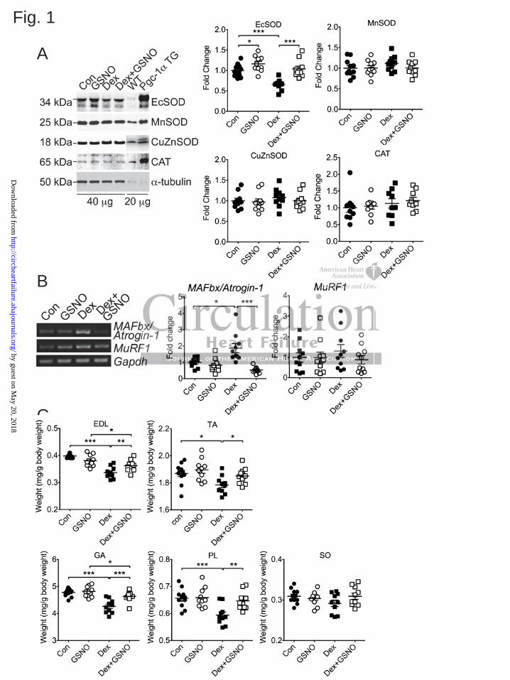
Fig. 1
B
C
A
by guest on May 20, 2018
http://circheartfailure.ahajournals.org/D
ownloaded from
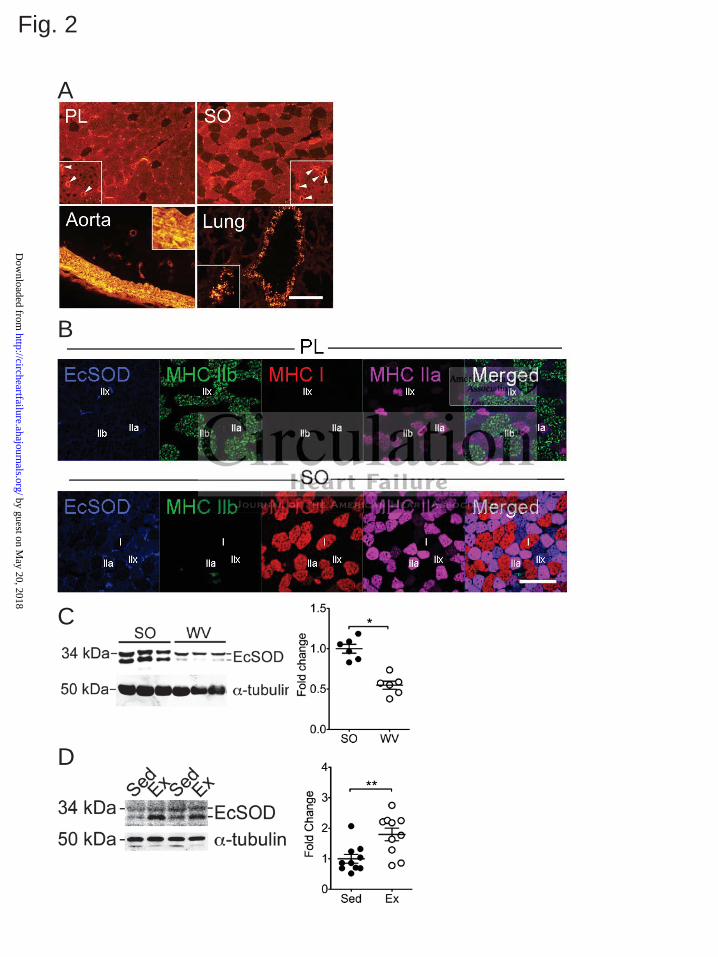
A
Fig. 2
C
B
D
by guest on May 20, 2018
http://circheartfailure.ahajournals.org/D
ownloaded from
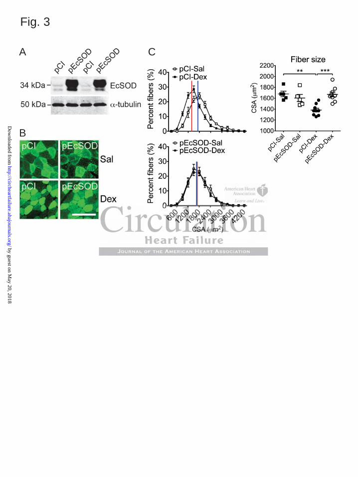
Fig. 3
C
B
A
by guest on May 20, 2018
http://circheartfailure.ahajournals.org/D
ownloaded from
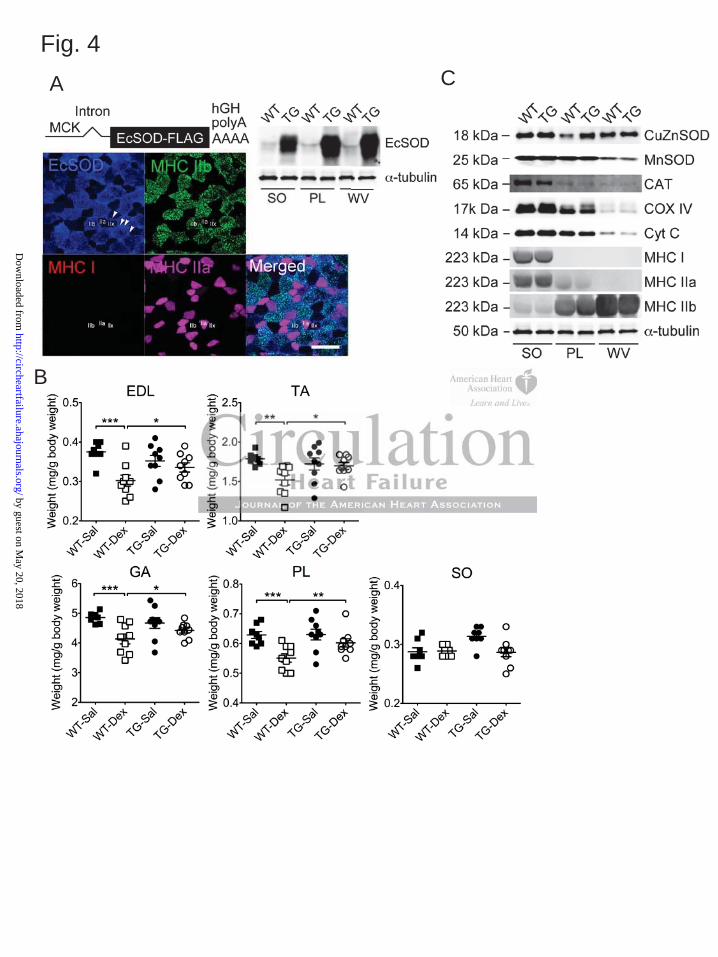
Fig. 4CA
B
by guest on May 20, 2018
http://circheartfailure.ahajournals.org/D
ownloaded from
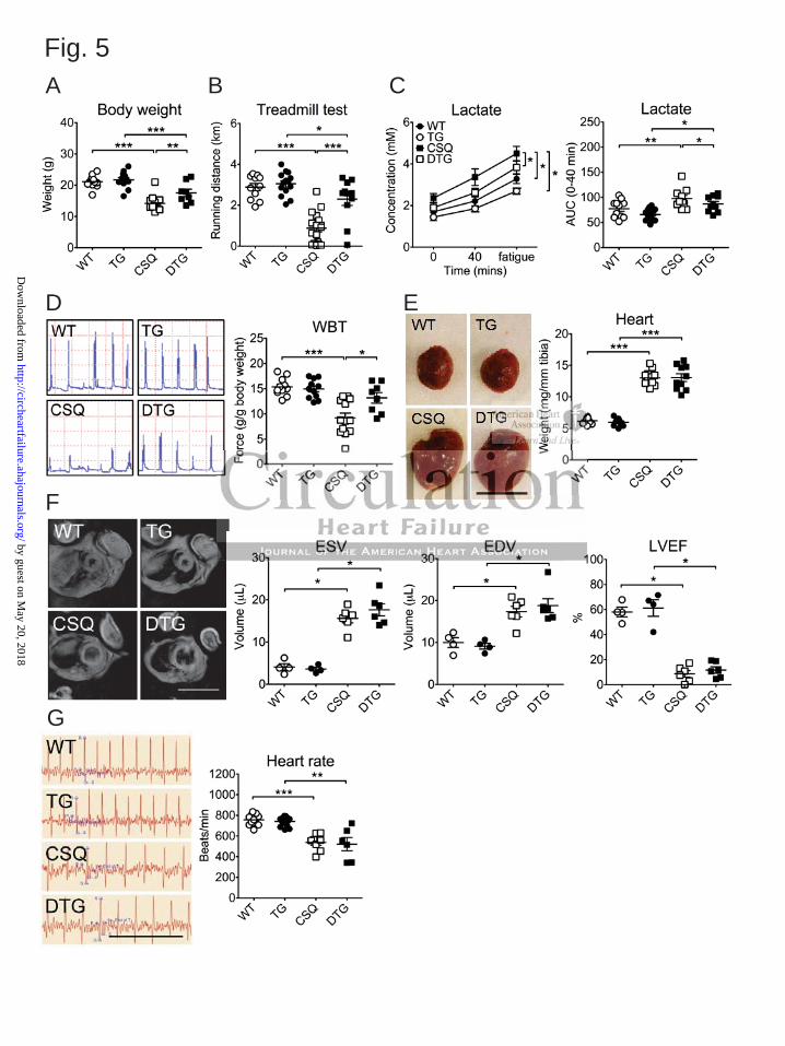
F
G
Fig. 5A B
D E
C
by guest on May 20, 2018
http://circheartfailure.ahajournals.org/D
ownloaded from
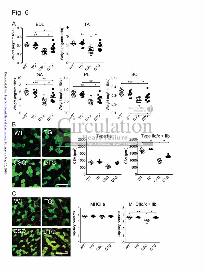
Fig. 6
C
B
A
by guest on May 20, 2018
http://circheartfailure.ahajournals.org/D
ownloaded from
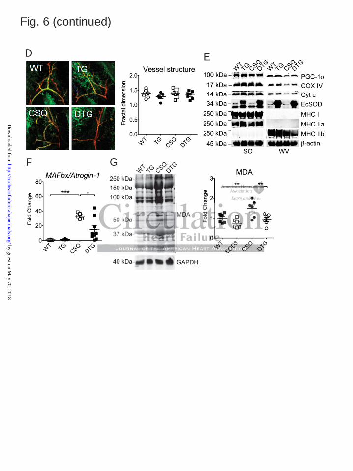
Fig. 6 (continued)
D
F
E
G
by guest on May 20, 2018
http://circheartfailure.ahajournals.org/D
ownloaded from
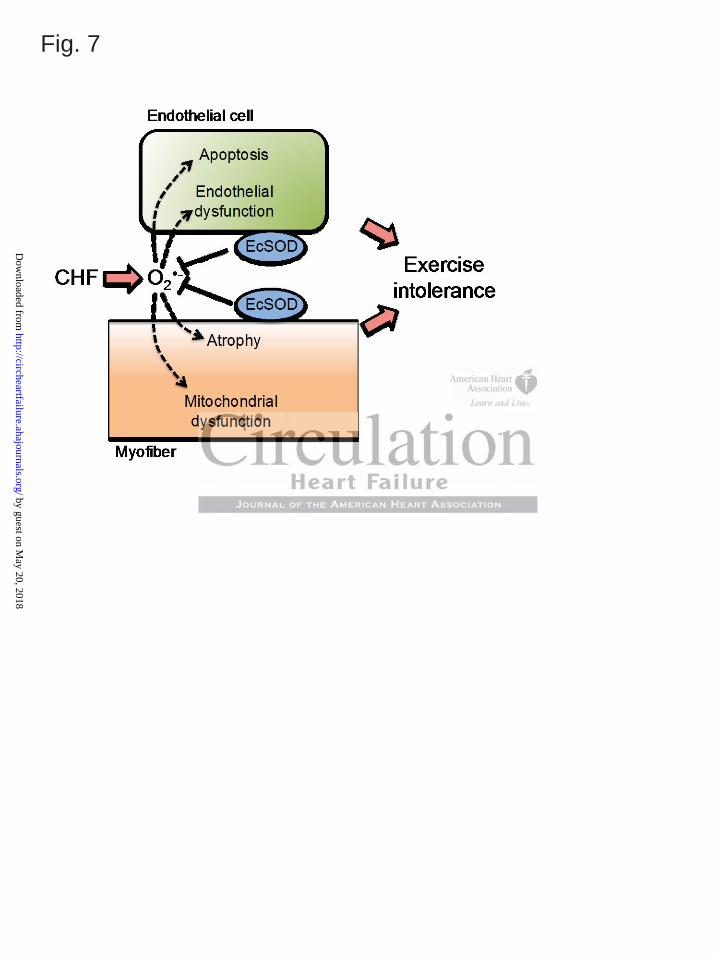
Fig. 7
by guest on May 20, 2018
http://circheartfailure.ahajournals.org/D
ownloaded from
Martin, Shayn M. Peirce-Cottler, Christopher M. Rembold, Brian H. Annex and Zhen YanMitsuharu Okutsu, Jarrod A. Call, Vitor A. Lira, Mei Zhang, Jean A. Donet, Brent A. French, Kyle S.
Exercise Intolerance in Mice with Congestive Heart FailureExtracellular Superoxide Dismutase Ameliorates Skeletal Muscle Abnormalities, Cachexia and
Print ISSN: 1941-3289. Online ISSN: 1941-3297 Copyright © 2014 American Heart Association, Inc. All rights reserved.
is published by the American Heart Association, 7272 Greenville Avenue, Dallas, TX 75231Circulation: Heart Failure published online February 12, 2014;Circ Heart Fail.
http://circheartfailure.ahajournals.org/content/early/2014/02/12/CIRCHEARTFAILURE.113.000841World Wide Web at:
The online version of this article, along with updated information and services, is located on the
http://circheartfailure.ahajournals.org/content/suppl/2014/02/12/CIRCHEARTFAILURE.113.000841.DC1Data Supplement (unedited) at:
http://circheartfailure.ahajournals.org//subscriptions/
is online at: Circulation: Heart Failure Information about subscribing to Subscriptions:
http://www.lww.com/reprints Information about reprints can be found online at: Reprints:
document. Permissions and Rights Question and Answer process is available in the
click Request Permissions in the middle column of the Web page under Services. Further information about thisEditorial Office. Once the online version of the published article for which permission is being requested is located,
can be obtained via RightsLink, a service of the Copyright Clearance Center, not theCirculation: Heart Failure Requests for permissions to reproduce figures, tables, or portions of articles originally published inPermissions:
by guest on May 20, 2018
http://circheartfailure.ahajournals.org/D
ownloaded from
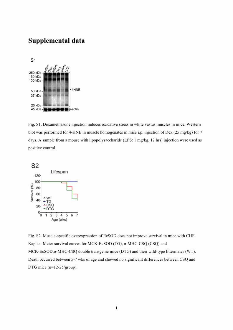
1
Supplemental data
Fig. S1. Dexamethasone injection induces oxidative stress in white vastus muscles in mice. Western
blot was performed for 4-HNE in muscle homogenates in mice i.p. injection of Dex (25 mg/kg) for 7
days. A sample from a mouse with lipopolysaccharide (LPS: 1 mg/kg, 12 hrs) injection were used as
positive control.
Fig. S2. Muscle-specific overexpression of EcSOD does not improve survival in mice with CHF.
Kaplan–Meier survival curves for MCK-EcSOD (TG), α-MHC-CSQ (CSQ) and
MCK-EcSOD:α-MHC-CSQ double transgenic mice (DTG) and their wild-type littermates (WT).
Death occurred between 5-7 wks of age and showed no significant differences between CSQ and
DTG mice (n=12-25/group).
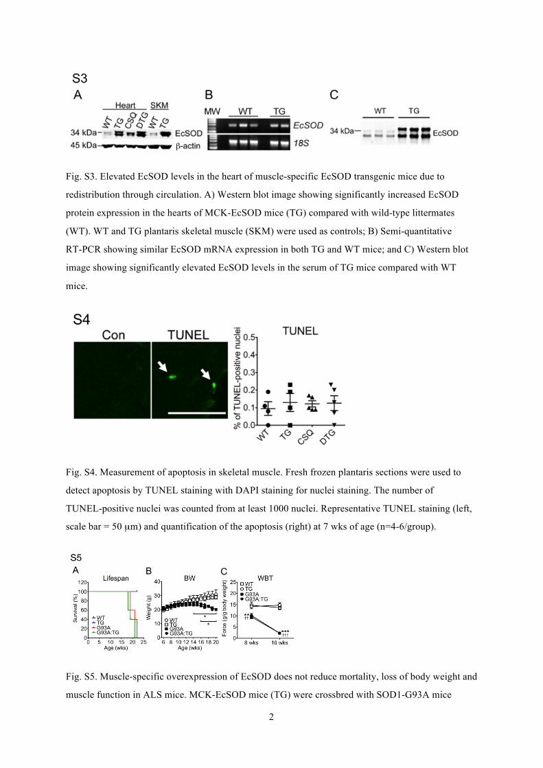
2
Fig. S3. Elevated EcSOD levels in the heart of muscle-specific EcSOD transgenic mice due to
redistribution through circulation. A) Western blot image showing significantly increased EcSOD
protein expression in the hearts of MCK-EcSOD mice (TG) compared with wild-type littermates
(WT). WT and TG plantaris skeletal muscle (SKM) were used as controls; B) Semi-quantitative
RT-PCR showing similar EcSOD mRNA expression in both TG and WT mice; and C) Western blot
image showing significantly elevated EcSOD levels in the serum of TG mice compared with WT
mice.
Fig. S4. Measurement of apoptosis in skeletal muscle. Fresh frozen plantaris sections were used to
detect apoptosis by TUNEL staining with DAPI staining for nuclei staining. The number of
TUNEL-positive nuclei was counted from at least 1000 nuclei. Representative TUNEL staining (left,
scale bar = 50 µm) and quantification of the apoptosis (right) at 7 wks of age (n=4-6/group).
Fig. S5. Muscle-specific overexpression of EcSOD does not reduce mortality, loss of body weight and
muscle function in ALS mice. MCK-EcSOD mice (TG) were crossbred with SOD1-G93A mice
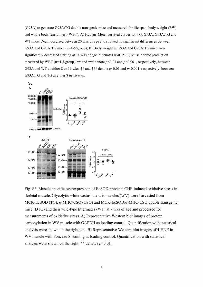
3
(G93A) to generate G93A:TG double transgenic mice and measured for life span, body weight (BW)
and whole body tension test (WBT). A) Kaplan–Meier survival curves for TG, G93A, G93A:TG and
WT mice. Death occurred between 20 wks of age and showed no significant differences between
G93A and G93A:TG mice (n=4-5/group); B) Body weight in G93A and G93A:TG mice were
significantly decreased starting at 14 wks of age. * denotes p<0.05; C) Muscle force production
measured by WBT (n=4-5/group). ** and *** denote p<0.01 and p<0.001, respectively, between
G93A and WT at either 8 or 16 wks. †† and ††† denote p<0.01 and p<0.001, respectively, between
G93A:TG and TG at either 8 or 16 wks.
Fig. S6. Muscle-specific overexpression of EcSOD prevents CHF-induced oxidative stress in
skeletal muscle. Glycolytic white vastus lateralis muscles (WV) were harvested from
MCK-EcSOD (TG), α-MHC-CSQ (CSQ) and MCK-EcSOD:α-MHC-CSQ double transgenic
mice (DTG) and their wild-type littermates (WT) at 7 wks of age and processed for
measurements of oxidative stress. A) Representative Western blot images of protein
carbonylation in WV muscle with GAPDH as loading control. Quantification with statistical
analysis were shown on the right; and B) Representative Western blot images of 4-HNE in
WV muscle with Ponceau S staining as loading control. Quantification with statistical
analysis were shown on the right. ** denotes p<0.01.