superoxide dismutase and oxidative stress in amyotrophic...
TRANSCRIPT
Chapter 5
Superoxide Dismutase and Oxidative Stress inAmyotrophic Lateral Sclerosis
María Clara Franco, Cassandra N. Dennys,Fabian H. Rossi and Alvaro G. Estévez
Additional information is available at the end of the chapter
http://dx.doi.org/10.5772/56488
1. Introduction
Oxidative stress is defined as the imbalance between reactive species such as free radicals andoxidants and the antioxidant defenses. Free radicals are molecules with one or more unpairedelectrons, while oxidants are molecules with a high potential for taking electrons from othermolecules. The more recognized reactive species are the reactive oxygen species (ROS), whichinclude oxygen and its reduction products superoxide, hydrogen peroxide and hydroxylradical, and the reactive nitrogen species (RNS) such as the free radical nitric oxide and its by-products, including the powerful oxidant peroxynitrite and the sub-product of peroxynitritedecomposition nitrogen dioxide.
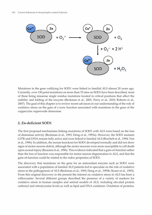
As part of the antioxidant defense system, superoxide dismutase 1 (SOD1) is an abundant andhighly conserved cytosolic enzyme responsible for the disproportionation of superoxide tomolecular oxygen and hydrogen peroxide (McCord and Fridovich, 1969). SOD1 is a relativelysmall protein of 153 amino acids that works as a tight homodimer and requires a high stabilityfor fast catalysis (Perry et al., 2010; Trumbull and Beckman, 2009). The stability is conferred bythe quaternary structure of the protein, an eight-strand beta-barrel, as well as the binding ofCu and Zn, two metal ions with catalytic roles positioned in the active site channel (Perry etal., 2010; Trumbull and Beckman, 2009). The disproportionation of superoxide is a two-stepoxidation-reduction reaction that involves the cycling of the copper atom in SOD1 from Cu2+
to Cu+ and back to Cu+2.
The zinc does not participate in this reaction but is essential for the structure of the active site.In addition, the formation of an intrasubunit disulfide bridge stabilizes the enzyme and playsan important role in preventing aggregation of metal-deficient SOD (Getzoff et al., 1989).
© 2013 Franco et al.; licensee InTech. This is an open access article distributed under the terms of the CreativeCommons Attribution License (http://creativecommons.org/licenses/by/3.0), which permits unrestricted use,distribution, and reproduction in any medium, provided the original work is properly cited.
Mutations in the gene codifying for SOD1 were linked to familial ALS almost 20 years ago.Currently, over 130 point mutations on more than 70 sites on SOD1 have been described, mostof these being missense single residue mutations located in critical positions that affect thestability and folding of the enzyme (Beckman et al., 2001; Perry et al., 2010; Roberts et al.,2007). The goal of this chapter is to review recent advances in our understanding of the role ofoxidative stress on the gain of a toxic function associated with mutations in the gene of thecopper/zinc superoxide dismutase.
2. Zn-deficient SOD1
The first proposed mechanisms linking mutations of SOD1 with ALS were based on the lossof dismutase activity (Beckman et al., 1993; Deng et al., 1993a). However, the SOD1 mutantsG37R and G93A remain fully active and were linked to familial ALS (Borchelt et al., 1994; Yimet al., 1996). In addition, the mouse knockout for SOD1 developed normally and did not showsigns of motor neuron deficit, although the motor neurons were more susceptible to cell deathupon axonal injury (Reaume et al., 1996). This evidence indicated that a gain-of-function ratherthan the loss of function was responsible for motor neuron degeneration in ALS, and that thegain-of-function could be related to the redox properties of SOD1.
The discovery that mutations on the gene for an antioxidant enzyme such as SOD1 wereassociated with a population of familial ALS patients led to speculate on the role of oxidativestress in the pathogenesis of ALS (Beckman et al., 1993; Deng et al., 1993b; Rosen et al., 1993).From this original discovery to the present the interest on oxidative stress in ALS has been arollercoaster. Several different groups described the presence of a variety of markers foroxidative stress in human samples and animal models of ALS, including elevated proteincarbonyl and nitrotyrosine levels as well as lipid and DNA oxidation. Oxidation of proteins,
Current Advances in Amyotrophic Lateral Sclerosis142
lipids, and DNA was also found in transgenic mice and cell culture models (Barber and Shaw,2010). On the other hand, other groups failed to find markers of oxidative damage in animalmodels of ALS, casting doubt on the relevance of oxidative stress in the pathogenesis of thedisease (Barber and Shaw, 2010). Currently, a role for oxidative stress in ALS is generallyaccepted but whether oxidative stress is responsible for the mutant SOD1 gain-of-function isstill controversial.
2.1. Mutant SOD1 aggregation and Zn-deficiency
Mutant SOD1s have a tendency to aggregate when expressed in bacterial systems andtransfected cells, and the presence of mutant and wild type SOD1-containing aggregates hasbeen described in animal models of ALS (Bruijn and Cleveland, 1996; Watanabe et al., 2001).The formation of aggregates clogging the proteasome and containing other relevant proteinsalong with mutant SOD1 is one of the possible explanations for SOD1 toxic gain-of-function.However, in mice expressing the SOD1A4V mutant, the most common mutation linked tofamilial ALS in humans, the mutant is expressed at high levels and forms protein aggregatesbut does not cause disease (Gurney et al., 1994). Alternatively, other groups proposed ahypothesis in which the formation of aggregates is a protective mechanism rather than causeof toxicity. In vitro experiments showed that both wild type SOD1 and SOD1 with mutation ofthe cysteine residues involved in protein aggregation were able to stabilize the mutant SOD1enzymes, increasing their toxicity (Clement et al., 2003; Fukada et al., 2001; Sahawneh et al.,2010; Witan et al., 2009). Additionally, it was recently described that overexpression of thedeubiquitinating enzyme ataxin-3 stimulates the formation of SOD1-containing aggresomesby trimming K63-linked polyubiquitin chains. The knockdown of ataxin-3 decreases theformation of aggresomes and increases cell death induced by mutant SOD1 (Wang et al.,2012). These results suggest a toxic gain-of-function for the stabilized and soluble mutantSOD1, rather than toxicity due to aggregation. Indeed, by removing the toxic soluble mutantSOD1, the formation of aggregates has been proposed to be a protective mechanism (Trumbulland Beckman, 2009). Further support is provided by recent studies of crossbreeding showingan acceleration of the disease in mutant SOD1 transgenic mice overexpressing wild type SOD1,which was linked to the formation of disulfide bridges in the enzyme by oxidation of cysteineresidues, increasing the formation of aggregates (Deng et al., 2006; Furukawa et al., 2006; Wanget al., 2009). Other investigations reproduced the acceleration of the disease in animalsexpressing both wild type and mutant SOD1 but failed to find a correlation between expressionof wild type SOD1 and protein aggregation (Prudencio et al., 2009).
The link between the gain-of-function and the redox activity of soluble mutant SOD1 as asource of oxidative stress is based on the presence of the copper atom in the active site of theenzyme as well as the loss of zinc. The requirement for copper was challenged by geneticexperiments in which the chaperone that delivers the copper metal to SOD1 was deleted. Theablation of the chaperone in the G93A, G85R, or G73R-SOD1 mutant mice decreased theactivity of the enzyme but had no effect on the progression of the disease (Subramaniam et al.,2002), although it may be possible for SOD1 to acquire copper from an alternative source(Beckman et al., 2002). The transgenic expression of a SOD1 with mutations that eliminate the
Superoxide Dismutase and Oxidative Stress in Amyotrophic Lateral Sclerosishttp://dx.doi.org/10.5772/56488
143

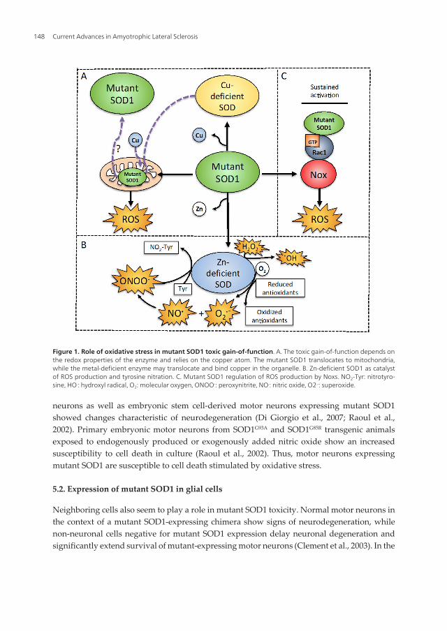
copper-binding site still produced disease (Prudencio et al., 2012; Wang et al., 2003). In contrast,another study showed that the mutant enzymes A4V, G85R, and G93A had a higher affinityfor copper than the wild type protein, and that this aberrant copper binding was mediated bycysteine 111 (Watanabe et al., 2007), implying that the enzyme binds copper in an alternate site(Figure 1A).
Some SOD1 mutants bind copper and zinc and are fully active (Borchelt et al., 1994; Marklundet al., 1997) but many mutations affect the binding of zinc while copper remains tightly bound,thus favoring the formation of Zn-deficient SOD. In the SOD1G93A mouse model of ALS, thedietary depletion of zinc accelerates the progression of the disease while moderate supplementof zinc provides protection (Ermilova et al., 2005). Indeed, a peak corresponding to one-metalSOD1 was detected in vivo in spinal cords from the SOD1G93A rat model using the recentlydeveloped methodology of electrospray mass spectrometry. The one-metal peak was 2-foldlarger in the disease-affected ventral spinal cord compare to that of the dorsal spinal cord(Rhoads et al., 2011), suggesting that Zn-deficient SOD1 may be present in vivo in the affectedtissue.
ALS-linked mutant SOD1s have 5-50 fold less affinity for zinc than the wild type protein (Crowet al., 1997a; Lyons et al., 1996). The loss of zinc disorganizes the structure of the active siteleaving the copper metal more expose and accessible to substrates other than superoxide,decreasing the normal activity of the enzyme. When replete with zinc, SOD1 mutants cangenerally fulfill the antioxidant activity of wild type SOD (Crow et al., 1997a). Early studiesshowed that mutant SOD1 has an aberrant chemistry and is reduced abnormally fast whichallows the reaction with oxidants such as hydrogen peroxide and peroxynitrite (Crow et al.,1997a; Crow et al., 1997b; Lyons et al., 1996; Wiedau-Pazos et al., 1996), thus turning theantioxidant enzyme into a catalyst for oxidation. The conversion of SOD1 from antioxidant topro-oxidant due to the loss of zinc is a simple explanation for the gain-of-function attributedto the ALS-linked SOD mutants, but is still highly controversial.
2.2. Formation of hydroxyl radical from hydrogen peroxide
In normal conditions SOD1 catalyzes the disproportionation of superoxide to hydrogenperoxide, but due to changes in mutant SOD1 conformation, the mutant enzyme can catalyzethe production of hydroxyl radical from hydrogen peroxide in vitro (Yim et al., 1990) (Figure1B). The G93A-SOD1 mutant has enhanced free-radical production compare to the wild typeenzyme due to a more open active site, decreasing the Km for hydrogen peroxide (Yim et al.,1996). Accordingly, an increase in the levels of hydrogen peroxide and hydroxyl radical wasreported in vivo in the spinal cord from mice expressing the G93A mutant (Liu et al., 1999).
The aberrant chemistry of mutant SOD1 was shown to inactivate the glutamate transporterEAAT2 by oxidative reactions catalyzed by the A4V and I113T-SOD1 mutants and triggeredby hydrogen peroxide (Trotti et al., 1999; Trotti et al., 1996). The function of this transporter isdown regulated in human patients and animal models of ALS and its inactivation results inneuronal degeneration (Rothstein et al., 2005; Tanaka et al., 1997). Moreover, the aberrant SOD1chemistry increases the vulnerability of a variety of cells in culture to hydrogen peroxide, withan increased susceptibility to inhibition by copper chelators. The G37R, G41D, and G85R-SOD1
Current Advances in Amyotrophic Lateral Sclerosis144
mutants induce activation of caspase 1 and promoted apoptosis in N2a cells and tissueexpressing mutant SOD1 when exposed to hydrogen peroxide. In NSC34 cells, a motor neuronmodel, mutant SOD1 induces cell death upon exposure of the cells to hydrogen peroxide(Pasinelli et al., 1998; Wiedau-Pazos et al., 1996). These findings suggest that the ALS pheno‐type may require both, the genetic background and an additional oxidative challenge.
2.3. Production of peroxynitrite
Nitric oxide alone is not toxic to normal motor neurons (Estévez et al., 1999), but whensuperoxide is also produced it can react with nitric oxide to form the powerful oxidantperoxynitrite, responsible for the induction of cell death. Overexpression of mutant SOD1makes motor neurons vulnerable to exogenous and endogenous production of nitric oxide.The increased vulnerability is linked to the activation of the Fas death pathways (Raoul et al.,2002). More recently it was shown that motor neurons from mutant SOD1 transgenic animalshave lower levels of a calcium-binding ER chaperone calreticulin. A decrease in the expressionof this protein is necessary and sufficient to activate the Fas/NO pathways in motor neurons.Further evidence in vivo shows that this protein is decreased in the spinal motor neurons ofSOD1G93A transgenic animals prior to muscle denervation (Bernard-Marissal et al., 2012).Therefore, motor neurons expressing mutant SOD1 may produce superoxide making themsusceptible to the formation of peroxynitrite in the presence of nitric oxide. In the presence ofreductants, Zn-deficient SOD1 is able to produce superoxide. For instance, ascorbate reducesthe copper on Zn-deficient SOD1 from Cu2+ to Cu+. In turn, Zn-deficient SOD1 can transferthe electrons from ascorbate to oxygen to produce superoxide slowly but significantly over aperiod of minutes. Indeed, Zn-deficient SOD1 is able to oxidize ascorbate 3000-fold faster thanmutant or wild type Cu,Zn-SOD1 in vitro (Estévez et al., 1999). In the cells, ascorbate and othercellular antioxidants such as glutathione, urate, and cysteine could have a similar effect.Normally, superoxide would be removed by the dismutase activity of the remaining and fullyactive Cu,Zn-SOD1. However, if nitric oxide is also produced it can effectively compete withCu,Zn-SOD1 for superoxide to produce peroxynitrite. Because nitric oxide is a small moleculeable to diffuse 10-fold faster than a small size protein, the reaction of nitric oxide with super‐oxide occurs 10 times faster than that with SOD1 (Beckman et al., 2001; Estévez et al., 1999;Franco and Estévez, 2011; Nauser and Koppenol, 2002) (Figure 1B). Wild type Cu,Zn-SOD1can also produce peroxynitrite by a similar mechanism but requires superoxide in the initialstep to be efficiently reduced (Beckman et al., 2001).
2.4. Catalysis of tyrosine nitration
Cu,Zn-SOD1 is not only responsible for the production of peroxynitrite but it can also catalyzetyrosine nitration in vitro (Beckman et al., 1993; Crow et al., 1997b; Ischiropoulos et al., 1992).The mechanism for tyrosine nitration depends on the copper atom in SOD1 that reacts withperoxynitrite. The loss of zinc from Cu,Zn-SOD1 increases by 2-fold the efficiency of theenzyme to catalyze tyrosine nitration (Crow et al., 1997a) (Figure 1B). Moreover, SOD1 is notinactivated by peroxynitrite and can catalyze tyrosine nitration indefinitely. Indeed, reactivityfor nitrotyrosine was found in vivo in the SOD1G93A mouse model and in patients with ALS
Superoxide Dismutase and Oxidative Stress in Amyotrophic Lateral Sclerosishttp://dx.doi.org/10.5772/56488
145
(Beal et al., 1997; Ferrante et al., 1997). In spite of the indirect evidence of mass spectrometryshowing a peak corresponding to a one-metal SOD1 in a rat model of ALS (Rhoads et al.,2011), whether Zn-deficient SOD1 is present in vivo and catalyzes tyrosine nitration is stillsource of debate and remains to be determined.
3. Regulation of NADPH oxidase activity by mutant SOD1
Several lines of evidence support the role of oxidative stress in mutant SOD1 toxicity, but someevidence suggest that interactions other than the redox properties of the enzyme stimulateoxidative stress by different mechanisms. Mutant SOD1 can induce oxidative stress bydisruption of the redox-sensitive regulation of NADPH oxidase (Nox) in microglial cells. Noxsare transmembrane proteins that catalyze the reduction of oxygen to superoxide usingNADPH as an electron donor (Brown and Griendling, 2009). Superoxide is then converted tohydrogen peroxide by SOD1. Under reducing conditions, SOD1 regulates Nox2 activation bybinding and stabilizing Rac1. The oxidation of Rac1 by hydrogen peroxide disrupts thecomplex with SOD1 and inactivates Nox2. Upon expression of certain ALS SOD1 mutants, thedissociation of Rac1 from SOD1 is impaired and Nox2 remains active (Figure 1C). In addition,the expression of Nox2 is upregulated in the SOD1G93A mouse model and in ALS patients. Infact, gene deletion of Nox1 or Nox2 provides the larger protection to date in animal models ofALS (Harraz et al., 2008; Marden et al., 2007).
4. Mutant SOD1 translocation to mitochondria
Mitochondria are one of the major sources of cellular ROS formed as by-products of oxidativephosphorylation. Abnormalities in the mitochondrial structure, localization and number aswell as altered activity of the electron transport chain have been described in both, sporadicand familial ALS (Manfredi and Xu, 2005). The mitochondrial electron transport chain andATP synthesis are severely impaired at disease onset in spinal cord and brain of SOD1G93A
transgenic mice (Lin and Beal, 2006). Both, wild type and mutant SOD localize in mitochondriain the central nervous system (Higgins et al., 2002). Mutant human SOD1 was found in themitochondrial outer membrane, intermembrane space and matrix in transgenic mice, whileinactive mutant SOD1 accumulates and forms aggregates in the mitochondrial matrix in thebrain (Vijayvergiya et al., 2005). Aggregates of the mutant enzyme are also selectively foundin the mitochondrial outer membrane in spinal cord from mouse models of ALS (Liu et al.,2004). Interestingly, the anti-apoptotic protein Bcl-2 binds to mutant SOD1 and aggregates inspinal cord mitochondria from patients and a mouse model of ALS, suggesting that mutantSOD1 may be toxic by depleting motor neurons of this anti-apoptotic protein (Pasinelli et al.,2004). Mutant SOD1 targeted to the mitochondrial intermembrane space in NSC34 cellsinduces cell death upon exposure of the cells to hydrogen peroxide (Magrane et al., 2009). Inaddition, the increase in carbonylated proteins and lipid hydroperoxides in mitochondria, aswell as the abnormally high rates of production of hydrogen peroxide in SOD1G93A transgenic
Current Advances in Amyotrophic Lateral Sclerosis146
mice (Mattiazzi et al., 2002; Panov et al., 2011) support the mutant SOD1 aberrant catalyticgain-of-function. Indeed, it was shown that metal-deficient SOD1s are prone to mitochondrialtranslocation and are found in the mitochondrial intermembrane space (Okado-Matsumotoand Fridovich, 2002). The mitochondria contain the majority of the cellular copper because isrequired by the oxygen-consuming proteins. The insertion of copper into the translocatedmetal-deficient SOD would result in the formation of Zn-deficient SOD inside the mitochon‐dria (Figure 1A). This could explain why the mitochondria are affected early in the onset ofthe disease (Beckman et al., 2002). The ROS-linked toxic gain-of-function of mutant SOD1would produce hydroxyl radical from H2O2 as well as peroxynitrite in the mitochondria. Themutant enzyme could then catalyze the nitration of mitochondrial proteins such as cyclophilinD and the adenine nucleotide translocator (Martin, 2010). Due to these toxic effects of mutantSOD1 on mitochondria, it has been proposed that the abnormal activity of the mitochondriain ALS may account for the initiation and progression of the disease. However, whether themitochondrial localization of mutant SOD1 is cause or a consequence of pathology needs tobe established.
5. Expression of mutant SOD1 in motor neurons and neighboring cells
A new mechanism integrating the autonomous and non-autonomous induction of motorneuron death in ALS is emerging. In this scenario, the role of motor neurons and surroundingcells in the onset and progression of ALS is temporally determined. Several studies wereconducted where mutant SOD1 was selectively expressed in vivo either in motor neurons ormicroglia of chimeric mice, or in culture in embryonic primary or stem cell-based models,allowing the study of the role of individual population of cells in the onset and progression ofALS. The cell-autonomous degeneration of motor neurons expressing mutant SOD1 seems tobe more relevant for the onset and early progression of the disease, while microglia, peripheralmacrophages, and astrocytes would play a role in the late disease progression.
5.1. Expression of mutant SOD1 in motor neurons
ALS is a motor neuron disease characterized by the gradual and selective loss of both, upperand lower motor neurons. Expression of mutant SOD1 in spinal motor neurons and inter‐neurons of chimeric mice is enough to induce neuronal degeneration (Boillee et al., 2006; Wanget al., 2008). The mice do not develop clinical ALS but the motor neurons expressing mutantSOD1 exhibit pathological and immunohistochemical abnormalities, while motor neuronsnegative for mutant SOD1 expression do not. These observations indicate that in the chimericmice the degeneration of motor neurons can be cell-autonomous. The fact that only some ofthe motor neurons express mutant SOD1 in this model may explain why the animals do notdevelop the disease (Wang et al., 2008). Indeed, normal motor neurons can prevent or delaythe degeneration of mutant SOD1-expressing motor neurons (Clement et al., 2003). In addition,decreased expression of mutant SOD1 in motor neurons has a modest effect on the durationof the disease but significantly delay the onset and early phase of the disease progression(Wang et al., 2008). Similar results were observed in culture, where primary spinal motor
Superoxide Dismutase and Oxidative Stress in Amyotrophic Lateral Sclerosishttp://dx.doi.org/10.5772/56488
147
neurons as well as embryonic stem cell-derived motor neurons expressing mutant SOD1showed changes characteristic of neurodegeneration (Di Giorgio et al., 2007; Raoul et al.,2002). Primary embryonic motor neurons from SOD1G93A and SOD1G85R transgenic animalsexposed to endogenously produced or exogenously added nitric oxide show an increasedsusceptibility to cell death in culture (Raoul et al., 2002). Thus, motor neurons expressingmutant SOD1 are susceptible to cell death stimulated by oxidative stress.
5.2. Expression of mutant SOD1 in glial cells
Neighboring cells also seem to play a role in mutant SOD1 toxicity. Normal motor neurons inthe context of a mutant SOD1-expressing chimera show signs of neurodegeneration, whilenon-neuronal cells negative for mutant SOD1 expression delay neuronal degeneration andsignificantly extend survival of mutant-expressing motor neurons (Clement et al., 2003). In the
Figure 1. Role of oxidative stress in mutant SOD1 toxic gain-of-function. A. The toxic gain-of-function depends onthe redox properties of the enzyme and relies on the copper atom. The mutant SOD1 translocates to mitochondria,while the metal-deficient enzyme may translocate and bind copper in the organelle. B. Zn-deficient SOD1 as catalystof ROS production and tyrosine nitration. C. Mutant SOD1 regulation of ROS production by Noxs. NO2-Tyr: nitrotyro‐sine, HO.: hydroxyl radical, O2: molecular oxygen, ONOO-: peroxynitrite, NO.: nitric oxide, O2.-: superoxide.
Current Advances in Amyotrophic Lateral Sclerosis148
last few years, a role for microglia and astrocytes in the induction of motor neuron death hasbecome evident.
5.2.1. Role of microglia in the induction of motor neuron death
Activated microglia is found in the spinal cord of SOD1G93A transgenic mice, suggesting thatit may play a role in the neurodegeneration of neighboring motor neurons (Beers et al., 2006).Reducing the expression of mutant SOD1 in microglia and peripheral macrophages in chimericmice leads to a delay in the late progression of ALS but has little effect on the onset and earlydisease progression (Boillee et al., 2006). Likewise, in the PU.1(-/-)/SOD1G93A mice unable tosynthesize myeloid cells, the replacement of microglia, monocyte, and macrophage lineageswith genotypically identical wild type cells slows disease progression and extends overallsurvival (Beers et al., 2006), suggesting that non cell-autonomous effects contribute to ALSprogression independently of disease onset. Comparable findings were observed in co-culturestudies where glial cells expressing mutant-SOD1 had a direct adverse effect on motor neuronsurvival (Di Giorgio et al., 2007). Microglia expressing G93A-SOD1 is toxic to primary motorneurons in vitro. In addition, SODG93A microglia show an increase in superoxide and nitric oxideproduction and release respect to wild type microglia. Treatment with lipopolysaccharidefurther increases SODG93A microglia activation and induction of motor neuron death (Beers etal., 2006). Hence, mutant SOD1-expressing microglia is activated, is more susceptible toactivation, and it is capable of inducing motor neuron death in vitro (Beers et al., 2006).Interestingly, PU.1(-/-) mice transplanted with bone marrow from a SOD1G93A donor do notdevelop clinical or pathological evidence of ALS, suggesting that expression of mutant SOD1in microglia is not enough to induce motor neuron disease in vivo (Beers et al., 2006). The factthat expression of mutant SOD1 in microglia alone does not induce motor neuron degenerationsuggests that motor neurons and other glial cells play a role in the pathological process. Indeed,motor neurons expressing mutant SOD1 are more susceptible to cell death induced byexposure to nitric oxide or Fas activation (Raoul et al., 2002).
5.2.2. Role of astrocytes in the induction of motor neuron death
Astrocytes are the most abundant non-neuronal cells in the nervous system. The co-culture ofnormal primary embryonic or stem cell-derived motor neurons with astrocytes expressingmutant SOD1 result in motor neuron death. The death pathway is triggered by a toxic factorreleased by the astrocytes (Aebischer et al., 2011; Nagai et al., 2007). A population of pheno‐typically aberrant astrocytes was recently described in the SOD1G93A mouse model of ALS(Diaz-Amarilla et al., 2011). These astrocytes, referred to as “AbA cells”, have an increasedproliferative capacity and secrete soluble factors that are 10 times more potent than neonatalSOD1G93A astrocytes for the induction of motor neuron death. AbA cells are present indegenerating spinal cord of SODG93A rats surrounding affected motor neurons, and theirnumber increases dramatically after disease onset, highlighting the importance of this finding.Interestingly, the levels of interferon-γ (IFNγ) are significantly increased in mutant SOD1-expressing astrocytes, and IFNγ induces motor neuron death (Aebischer et al., 2011), suggest‐ing that this cytokine may be one of the toxic factors mediating induction of cell death (Figure
Superoxide Dismutase and Oxidative Stress in Amyotrophic Lateral Sclerosishttp://dx.doi.org/10.5772/56488
149
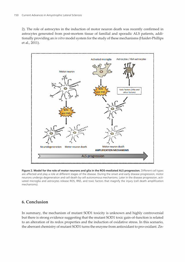
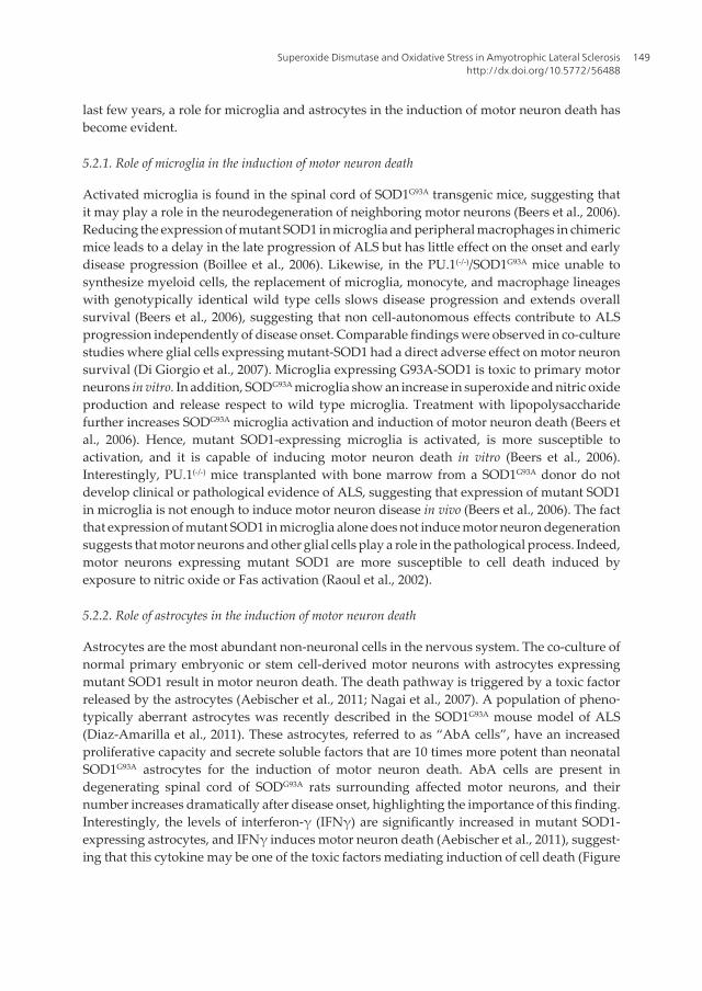
2). The role of astrocytes in the induction of motor neuron death was recently confirmed inastrocytes generated from post-mortem tissue of familial and sporadic ALS patients, addi‐tionally providing an in vitro model system for the study of these mechanisms (Haidet-Phillipset al., 2011).
Figure 2. Model for the role of motor neurons and glia in the ROS-mediated ALS progression. Different cell typesare affected and play a role at different stages of the disease. During the onset and early disease progression, motorneurons undergo degeneration and cell death by cell-autonomous mechanisms. Later in the disease progression, acti‐vated microglia and astrocytes release ROS, RNS, and toxic factors that magnify the injury (cell death amplificationmechanisms).
6. Conclusion
In summary, the mechanism of mutant SOD1 toxicity is unknown and highly controversialbut there is strong evidence suggesting that the mutant SOD1 toxic gain-of-function is relatedto an alteration of its redox properties and the induction of oxidative stress. In this scenario,the aberrant chemistry of mutant SOD1 turns the enzyme from antioxidant to pro-oxidant. Zn-
Current Advances in Amyotrophic Lateral Sclerosis150
deficient SOD1 reacts with hydrogen peroxide, produces superoxide and peroxynitrite, andis able to catalyze tyrosine nitration, altering the cellular redox balance. In addition, althoughnot related to the redox properties of the enzyme, the interaction of mutant SOD1 withmitochondria and Nox, the two major sources of cellular ROS, further support the involvementof oxidative stress in the toxic gain-of-function. The cell type affected by mutant SOD1 is alsocontroversial. A picture in which several cell types are affected and play a role at differentstages of the disease seems to be emerging. In this context, during onset and early stages ofthe disease SOD1-expressing motor neurons undergo neurodegeneration and cell death bycell-autonomous processes. The activation of microglia and astrocytes may work as anamplification mechanism in the induction of motor neuron death in the late progression of thedisease (Figure 2).
Author details
María Clara Franco1,2, Cassandra N. Dennys1,2, Fabian H. Rossi1,2 and Alvaro G. Estévez1,2
1 Burnett School of Biomedical Sciences, College of Medicine, University of Central Florida,Orlando, FL, USA
2 Orlando VA Healthcare System, Orlando, USA
References
[1] Aebischer, J, Cassina, P, Otsmane, B, Moumen, A, Seilhean, D, Meininger, V, Barbei‐to, L, Pettmann, B, & Raoul, C. (2011). IFNgamma triggers a LIGHT-dependent selec‐tive death of motoneurons contributing to the non-cell-autonomous effects of mutantSOD1. Cell death and differentiation , 18, 754-768.
[2] Barber, S. C, & Shaw, P. J. (2010). Oxidative stress in ALS: key role in motor neuroninjury and therapeutic target. Free radical biology & medicine , 48, 629-641.
[3] Beal, M. F, Ferrante, L. J, Browne, S. E, Matthews, R. T, Kowall, N. W, & Brown, R. H.(1997). Increased 3-nitrotyrosine in both sporadic and familial amyotrophic lateralsclerosis. Ann Neurol , 42, 644-654.
[4] Beckman, J. S, Carson, M, Smith, C. D, & Koppenol, W. H. (1993). ALS, SOD and per‐oxynitrite. Nature 364:584.
[5] Beckman, J. S, Estévez, A. G, Barbeito, L, & Crow, J. P. (2002). CCS knockout miceestablish an alternative source of copper for SOD in ALS. Free Rad. Biol. Med. , 33,1433-1435.
Superoxide Dismutase and Oxidative Stress in Amyotrophic Lateral Sclerosishttp://dx.doi.org/10.5772/56488
151
[6] Beckman, J. S, Estevez, A. G, Crow, J. P, & Barbeito, L. (2001). Superoxide dismutaseand the death of motoneurons in ALS. Trends in neurosciences 24:S, 15-20.
[7] Beers, D. R, Henkel, J. S, Xiao, Q, Zhao, W, Wang, J, Yen, A. A, Siklos, L, Mckercher,S. R, & Appel, S. H. (2006). Wild-type microglia extend survival in PU.1 knockoutmice with familial amyotrophic lateral sclerosis. Proceedings of the National Academy ofSciences of the United States of America , 103, 16021-16026.
[8] Bernard-marissal, N, Moumen, A, Sunyach, C, Pellegrino, C, Dudley, K, Henderson,C. E, Raoul, C, & Pettmann, B. (2012). Reduced Calreticulin Levels Link EndoplasmicReticulum Stress and Fas-Triggered Cell Death in Motoneurons Vulnerable to ALS.The Journal of neuroscience : the official journal of the Society for Neuroscience , 32,4901-4912.
[9] Boillee, S, Yamanaka, K, Lobsiger, C. S, Copeland, N. G, Jenkins, N. A, Kassiotis, G,Kollias, G, & Cleveland, D. W. (2006). Onset and progression in inherited ALS deter‐mined by motor neurons and microglia. Science , 312, 1389-1392.
[10] Borchelt, D. R, Lee, M. K, Slunt, H. S, Guarnieri, M, Xu, Z. S, Wong, P. C, Brown, R.H, Jr, D. L, Price, S. S, & Sisodia, D. W. Cleveland. (1994). Superoxide dismutase 1with mutations linked to familial amyotrophic lateral sclerosis possesses significantactivity. Proceedings of the National Academy of Sciences of the United States of America ,91, 8292-8296.
[11] Brown, D. I, & Griendling, K. K. (2009). Nox proteins in signal transduction. Free radi‐cal biology & medicine , 47, 1239-1253.
[12] Bruijn, L. I, & Cleveland, D. W. (1996). Mechanisms of selective motor neuron deathin ALS: insights from transgenic mouse models of motor neuron disease. Neuropa‐thology and applied neurobiology , 22, 373-387.
[13] Clement, A. M, Nguyen, M. D, Roberts, E. A, Garcia, M. L, Boillee, S, Rule, M, Mcma‐hon, A. P, Doucette, W, Siwek, D, Ferrante, R. J, Brown, R. H, Jr, J. P, Julien, L. S, &Goldstein, D. W. Cleveland. (2003). Wild-type nonneuronal cells extend survival ofSOD1 mutant motor neurons in ALS mice. Science , 302, 113-117.
[14] Crow, J. P, Sampson, J. B, Zhuang, Y, Thompson, J. A, & Beckman, J. S. (1997a). De‐creased zinc affinity of amyotrophic lateral sclerosis-associated superoxide dismu‐tase mutants leads to enhanced catalysis of tyrosine nitration by peroxynitrite. J.Neurochem. , 69, 1936-1944.
[15] Crow, J. P, Strong, M. J, Zhuang, Y, Ye, Y, & Beckman, J. S. (1997b). Superoxide di‐mutase catalyzes nitration of tyrosines by peroxinitrite in the rod and head domainsof neurofilament L. J Neurochem , 69, 1945-1953.
[16] Deng, H, Shi, X. , Y, Furukawa, Y, Zhai, H, Fu, R, Liu, E, Gorrie, G. H, Khan, M. S,Hung, W. -Y, Bigio, E. H, & Lukas, T. M.C. Dal Canto, T.V. O’Halloran, and T. Siddi‐que. (2006). Conversion to the amyotrophic lateral sclerosis phenotype is associated
Current Advances in Amyotrophic Lateral Sclerosis152
with intermolecular linked insoluble aggregates of SOD1 in mitochondria. PNAS ,103, 7142-7147.
[17] Deng, H. X, Hentati, A, Tainer, J. A, Iqbal, Z, Cayabyab, A, Hung, W. Y, Getzoff, E. D,Hu, P, Herzfeldt, B, Roos, R. P, et al. (1993a). Amyotrophic lateral sclerosis and struc‐tural defects in Cu,Zn superoxide dismutase. Science , 261, 1047-1051.
[18] Deng, H. X, Hentati, A, Tainer, J. A, Iqbal, Z, Cayabyab, A, Hung, W. Y, Getzoff, E. D,Hu, P, Herzfeldt, B, Roos, R. P, Warner, C, Deng, G, Soriano, E, Smyth, C, Parge, H.E, Ahmed, A, Roses, A. D, Hallewell, R. A, Prericak-vance, M. A, & Siddique, T.(1993b). Amyotrophic lateral sclerosis and structural defects in Cu,Zn superoxidedismutase. Science , 261, 1047-1051.
[19] Di GiorgioF.P., M.A. Carrasco, M.C. Siao, T. Maniatis, and K. Eggan. (2007). Non-cellautonomous effect of glia on motor neurons in an embryonic stem cell-based ALSmodel. Nature neuroscience , 10, 608-614.
[20] Diaz-amarilla, P, Olivera-bravo, S, Trias, E, Cragnolini, A, Martinez-palma, L, Cassi‐na, P, Beckman, J, & Barbeito, L. (2011). Phenotypically aberrant astrocytes that pro‐mote motoneuron damage in a model of inherited amyotrophic lateral sclerosis.Proceedings of the National Academy of Sciences of the United States of America , 108,18126-18131.
[21] Ermilova, I. P, Ermilov, V. B, Levy, M, Ho, E, Pereira, C, & Beckman, J. S. (2005). Pro‐tection by dietary zinc in ALS mutant G93A SOD transgenic mice. Neuroscience let‐ters , 379, 42-46.
[22] Estévez, A. G, Crow, J. P, Sampson, J. B, Reiter, C, Zhuang, Y. -X, Richardson, G. J,Tarpey, M. M, Barbeito, L, & Beckman, J. S. (1999). Induction of nitric oxide-depend‐ent apoptosis in motor neurons by zinc-deficient superoxide dismutase. Science , 286,2498-2500.
[23] Ferrante, R. J, Shinobu, L. A, Schulz, J. B, Mathews, R. T, Thomas, C. E, Kowall, N. W,Gurney, M. E, & Beal, M. F. (1997). Increased 3-nitrotyrosine and oxidative damagein mice with a human copper/zinc superoxide dismutase mutation. Ann Neurol , 42,326-334.
[24] Franco, M. C, & Estévez, A. G. (2011). Reactive Nitrogen Species in Motor NeuronApoptosis. In: Amyotrophic Lateral Sclerosis, edited by Martin H. Mauer. InTech, Ri‐jeka, Croatia. pp., 313-334.
[25] Fukada, K, Nagano, S, Satoh, M, Tohyama, C, Nakanishi, T, Shimizu, A, Yanagihara,T, & Sakoda, S. (2001). Stabilization of mutant Cu/Zn superoxide dismutase (SOD1)protein by coexpressed wild SOD1 protein accelerates the disease progression infamilial amyotrophic lateral sclerosis mice. Eur J Neurosci , 14, 2032-2036.
[26] Furukawa, Y, Fu, R, Deng, H. -X, Siddique, T, & Halloran, T. V. O. (2006). From theCover: Disulfide cross-linked protein represents a significant fraction of ALS-associ‐
Superoxide Dismutase and Oxidative Stress in Amyotrophic Lateral Sclerosishttp://dx.doi.org/10.5772/56488
153
ated Cu, Zn-superoxide dismutase aggregates in spinal cords of model mice. PNAS ,103, 7148-7153.
[27] Getzoff, E. D, Tainer, J. A, Stempien, M. M, Bell, G. I, & Hallewell, R. A. (1989). Evo‐lution of CuZn superoxide dismutase and the Greek key beta-barrel structural motif.Proteins , 5, 322-336.
[28] Gurney, M. E, Pu, H, Chiu, A. Y, Canto, M. C. D, Polchow, C. Y, Alexander, D. D,Caliendo, J, Hentati, A, Kwon, Y. W, Deng, H. -X, Chen, W, Zhai, P, Sufit, R. L, &Siddique, T. (1994). Motor neuron degeneration in mice that express a human Cu,Znsuperoxide dismutase mutation. Science , 264, 1772-1775.
[29] Haidet-phillips, A. M, Hester, M. E, Miranda, C. J, Meyer, K, Braun, L, Frakes, A,Song, S, Likhite, S, Murtha, M. J, Foust, K. D, Rao, M, Eagle, A, Kammesheidt, A,Christensen, A, Mendell, J. R, Burghes, A. H, & Kaspar, B. K. (2011). Astrocytes fromfamilial and sporadic ALS patients are toxic to motor neurons. Nature biotechnology ,29, 824-828.
[30] Harraz, M. M, Marden, J. J, Zhou, W, Zhang, Y, Williams, A, Sharov, V. S, Nelson, K,Luo, M, Paulson, H, Schoneich, C, & Engelhardt, J. F. (2008). SOD1 mutations disruptredox-sensitive Rac regulation of NADPH oxidase in a familial ALS model. J Clin In‐vest , 118, 659-670.
[31] Higgins, C. M, Jung, C, Ding, H, & Xu, Z. (2002). Mutant Cu, Zn superoxide dismu‐tase that causes motoneuron degeneration is present in mitochondria in the CNS. TheJournal of neuroscience : the official journal of the Society for Neuroscience 22:RC215.
[32] Ischiropoulos, H, Zhu, L, Chen, J, Tsai, M, Martin, J. C, Smith, C. D, & Beckman, J. S.(1992). Peroxynitrite-mediated tyrosine nitration catalyzed by superoxide dismutase.Archives of Biochemistry and Biophysics , 298, 431-437.
[33] Lin, M. T, & Beal, M. F. (2006). Mitochondrial dysfunction and oxidative stress inneurodegenerative diseases. Nature , 443, 787-795.
[34] Liu, D, Wen, J, Liu, J, & Li, L. (1999). The roles of free radicals in amyotrophic lateralsclerosis: reactive oxygen species and elevated oxidation of protein, DNA, and mem‐brane phospholipids. FASEB journal : official publication of the Federation of AmericanSocieties for Experimental Biology , 13, 2318-2328.
[35] Liu, J, Lillo, C, Jonsson, P. A, Velde, C. V, Ward, C. M, Miller, T. M, Subramaniam, J.R, Rothstein, J. D, Marklund, S, Andersen, P. M, Brannstrom, T, Gredal, O, Wong, P.C, Williams, D. S, & Cleveland, D. W. (2004). Toxicity of Familial ALS-Linked SOD1Mutants from Selective Recruitment to Spinal Mitochondria. Neuron , 43, 5-17.
[36] Lyons, T. J, Liu, H, Goto, J. J, Nersissian, A, Roe, J. A, Graden, J. A, Cafe, C, Ellerby,L. M, Bredesen, D. E, Gralla, E. B, & Valentine, J. S. (1996). Mutations in copper-zincsuperoxide dismutase that cause amyotrophic lateral sclerosis alter the zinc binding
Current Advances in Amyotrophic Lateral Sclerosis154
site and the redox behavior of the protein. Proceedings of the National Academy of Scien‐ces of the United States of America , 93, 12240-12244.
[37] Magrane, J, Hervias, I, Henning, M. S, Damiano, M, Kawamata, H, & Manfredi, G.(2009). Mutant SOD1 in neuronal mitochondria causes toxicity and mitochondrialdynamics abnormalities. Hum Mol Genet , 18, 4552-4564.
[38] Manfredi, G, & Xu, Z. (2005). Mitochondrial dysfunction and its role in motor neurondegeneration in ALS. Mitochondrion , 5, 77-87.
[39] Marden, J. J, Harraz, M. M, Williams, A. J, Nelson, K, Luo, M, Paulson, H, & Engel‐hardt, J. F. (2007). Redox modifier genes in amyotrophic lateral sclerosis in mice. JClin Invest , 117, 2913-2919.
[40] Marklund, S. L, Andersen, P. M, Forsgren, L, Nilsson, P, Ohlsson, P. I, Wikander, G,& Oberg, A. (1997). Normal binding and reactivity of copper in mutant superoxidedimutase isolated from amyotrophic lateral sclerosis patients. J Neurochem , 69,675-681.
[41] Martin, L. J. (2010). The mitochondrial permeability transition pore: a molecular tar‐get for amyotrophic lateral sclerosis therapy. Biochimica et biophysica acta , 1802,186-197.
[42] Mattiazzi, M, Aurelio, M. D, Gajewski, C. D, Martushova, K, Kiaei, M, Beal, M. F, &Manfredi, G. (2002). Mutated human SOD1 causes dysfunction of oxidative phos‐phorylation in mitochondria of transgenic mice. The Journal of biological chemistry ,277, 29626-29633.
[43] Mccord, J. M, & Fridovich, I. (1969). Superoxide dismutase. An enzymic function forerythrocuprein (hemocuprein). The Journal of biological chemistry , 244, 6049-6055.
[44] Nagai, M, Re, D. B, Nagata, T, Chalazonitis, A, Jessell, T. M, Wichterle, H, & Przed‐borski, S. (2007). Astrocytes expressing ALS-linked mutated SOD1 release factors se‐lectively toxic to motor neurons. Nature neuroscience , 10, 615-622.
[45] Nauser, T, & Koppenol, W. H. (2002). The rate constant of the reaction of superoxidewith nitrogen monoxide: Approaching the diffusion limit. J Phys Chem A , 106,4084-4086.
[46] Okado-matsumoto, A, & Fridovich, I. (2002). Amyotrophic lateral sclerosis: a pro‐posed mechanism. Proceedings of the National Academy of Sciences of the United States ofAmerica , 99, 9010-9014.
[47] Panov, A, Kubalik, N, Zinchenko, N, Hemendinger, R, Dikalov, S, & Bonkovsky, H.L. (2011). Respiration and ROS production in brain and spinal cord mitochondria oftransgenic rats with mutant G93a Cu/Zn-superoxide dismutase gene. Neurobiol Dis ,44, 53-62.
Superoxide Dismutase and Oxidative Stress in Amyotrophic Lateral Sclerosishttp://dx.doi.org/10.5772/56488
155
[48] Pasinelli, P, Belford, M. E, Lennon, N, Bacskai, B. J, Hyman, B. T, Trotti, D, & Brown,R. H. Jr. (2004). Amyotrophic lateral sclerosis-associated SOD1 mutant proteins bindand aggregate with Bcl-2 in spinal cord mitochondria. Neuron , 43, 19-30.
[49] Pasinelli, P, Borchelt, D. R, Houseweart, M. K, & Cleveland, D. W. and R.H. BrownJr. (1998). Caspase-1 is activated in neural cells and tissue with amyotrophic lateralsclerosis-associated mutations in copper-zinc superoxide dismutase. Proc Natl AcadSci USA , 95, 15763-15768.
[50] Perry, J. J, Shin, D. S, Getzoff, E. D, & Tainer, J. A. (2010). The structural biochemistryof the superoxide dismutases. Biochimica et biophysica acta , 1804, 245-262.
[51] Prudencio, M, Durazo, A, Whitelegge, J. P, & Borchelt, D. R. (2009). Modulation ofMutant Superoxide Dismutase 1 Aggregation by Co-Expression of Wild-Type En‐zyme. J Neurochem , 108, 1009-1018.
[52] Prudencio, M, Lelie, H, Brown, H. H, Whitelegge, J. P, Valentine, J. S, & Borchelt, D.R. (2012). A novel variant of human superoxide dismutase 1 harboring amyotrophiclateral sclerosis-associated and experimental mutations in metal-binding residuesand free cysteines lacks toxicity in vivo. Journal of neurochemistry , 121, 475-485.
[53] Raoul, C, Estévez, A. G, Nishimune, H, Cleveland, D. W, Delapeyrière, O, Hender‐son, C. E, Haase, G, & Pettmann, B. (2002). Motoneuron death triggered by a specificpathway downstream of Fas: potentiation by ALS-linked SOD1 mutations. Neuron ,35, 1067-1083.
[54] Reaume, A. G, Elliott, J. L, Hoffman, E. K, Kowall, N. W, Ferrante, R. J, Siwek, D. F,Wilcox, H. M, Flood, D. G, Beal, M. F, Brown, R. H, Jr, R. W, & Scott, W. D. Snider.(1996). Motor neurons in Cu/Zn superoxide dismutase-deficient mice develop nor‐mally but exhibit enhanced cell death after axonal injury. Nature genetics , 13, 43-47.
[55] Rhoads, T. W, Lopez, N. I, Zollinger, D. R, Morre, J. T, Arbogast, B. L, Maier, C. S,Denoyer, L, & Beckman, J. S. (2011). Measuring copper and zinc superoxide dismu‐tase from spinal cord tissue using electrospray mass spectrometry. Analytical biochem‐istry , 415, 52-58.
[56] Roberts, B. R, Tainer, J. A, Getzoff, E. D, Malencik, D. A, Anderson, S. R, Bomben, V.C, Meyers, K. R, Karplus, P. A, & Beckman, J. S. (2007). Structural characterization ofzinc-deficient human superoxide dismutase and implications for ALS. Journal of mo‐lecular biology , 373, 877-890.
[57] Rosen, D. R, Siddique, T, Patterson, D, Figlewicz, D. A, Sapp, P, Hentati, A, Donald‐son, D, Goto, J, Regan, J. P. O, Deng, H. -X, Rahmani, Z, Krizus, A, Mckenna-yasek,D, Cayabyab, A, Gaston, S. M, Berger, R, Tanszi, R. E, Halperin, J. J, Herzfeldt, B,Bergh, R. V. d, Hung, W. -Y, Bird, T, Deng, G, Mulder, D. W, Smyth, C, Lang, N. G,Soriana, E, Pericak-vance, M. A, Haines, J, Rouleau, G. A, Gusella, J. S, Horvitz, H. R,& Brown, R. H. J. (1993). Mutations in Cu/Zn superoxide dimutase gene are associat‐ed with familial amyotrophic lateral sclerosis. Nature , 362, 59-62.
Current Advances in Amyotrophic Lateral Sclerosis156

[58] Rothstein, J. D, Patel, S, Regan, M. R, Haenggeli, C, Huang, Y. H, Bergles, D. E, Jin, L,Dykes, M, Hoberg, S, Vidensky, D. S, Chung, S. V, Toan, L. I, Bruijn, Z. Z, & Su, P.Gupta, and P.B. Fisher. (2005). Beta-lactam antibiotics offer neuroprotection by in‐creasing glutamate transporter expression. Nature , 433, 73-77.
[59] Sahawneh, M. A, Ricart, K. C, Roberts, B. R, Bomben, V. C, Basso, M, Ye, Y, Sahaw‐neh, J, Franco, M. C, Beckman, J. S, & Estevez, A. G. (2010). Cu,Zn superoxide dismu‐tase (SOD) increases toxicity of mutant and Zn-deficient superoxide dismutase byenhancing protein stability. The Journal of biological chemistry , 285, 33885-33897.
[60] Subramaniam, J. R, Lyons, W. E, Liu, J, Bartnikas, T. B, Rothstein, J, Price, D. L,Cleveland, D. W, Gitlin, J. D, & Wong, P. C. (2002). Mutant SOD1 causes motor neu‐ron disease independent of copper chaperone-mediated copper loading. Nature Neu‐rosci , 5, 301-307.
[61] Tanaka, K, Watase, K, Manabe, T, Yamada, K, Watanabe, M, Takahashi, K, Iwama,H, Nishikawa, T, Ichihara, N, Kikuchi, T, Okuyama, S, Kawashima, N, Hori, S, Taki‐moto, M, & Wada, K. (1997). Epilepsy and exacerbation of brain injury in mice lack‐ing the glutamate transporter GLT-1. Science , 276, 1699-1702.
[62] Trotti, D, Rolfs, A, Danbolt, N. C, Brown, R. H. J, & Hediger, M. A. (1999). SOD 1 mu‐tants linked to amyotrophic lateral sclerosis selectivity inactivate a glial glutamatetransporter. Nature Neurosci , 2, 427-433.
[63] Trotti, D, Rossi, D, Gjesdal, O, Levy, L. M, Racagni, G, Danbolt, N. C, & Volterra, A.(1996). Peroxynitrite inhibits glutamate transporter subtypes. J Biol. Chem. , 271,5976-5979.
[64] Trumbull, K. A, & Beckman, J. S. (2009). A role for copper in the toxicity of zinc-defi‐cient superoxide dismutase to motor neurons in amyotrophic lateral sclerosis. Antiox‐idants & redox signaling , 11, 1627-1639.
[65] Vijayvergiya, C, Beal, M. F, Buck, J, & Manfredi, G. (2005). Mutant superoxide dismu‐tase 1 forms aggregates in the brain mitochondrial matrix of amyotrophic lateral scle‐rosis mice. The Journal of neuroscience : the official journal of the Society for Neuroscience ,25, 2463-2470.
[66] Wang, H, Ying, Z, & Wang, G. (2012). Ataxin-3 Regulates Aggresome Formation ofCopper-Zinc Superoxide Dismutase (SOD1) by Editing K63-linked PolyubiquitinChains. The Journal of biological chemistry , 287, 28576-28585.
[67] Wang, J. J, Slunt, H. H, Gonzales, V. V, Fromholt, D. D, Coonfield, M. M, Copeland,N. G. N. G, Jenkins, N. A. N. A, & Borchelt, D. R. D. R. (2003). Copper-binding-site-null SOD1 causes ALS in transgenic mice: aggregates of non-native SOD1 delineate acommon feature. Human molecular genetics 12:2753.
Superoxide Dismutase and Oxidative Stress in Amyotrophic Lateral Sclerosishttp://dx.doi.org/10.5772/56488
157
[68] Wang, L, Deng, H. X, Grisotti, G, Zhai, H, Siddique, T, & Roos, R. P. (2009). Wild-type SOD1 overexpression accelerates disease onset of a G85R SOD1 mouse. HumMol Genet , 18, 1642-1651.
[69] Wang, L, Sharma, K, Deng, H. X, Siddique, T, Grisotti, G, Liu, E, & Roos, R. P. (2008).Restricted expression of mutant SOD1 in spinal motor neurons and interneurons in‐duces motor neuron pathology. Neurobiol Dis , 29, 400-408.
[70] Watanabe, M, Dykes-hoberg, M, Culotta, V. C, Price, D. L, Wong, P. C, & Rothstein, J.D. (2001). Histological evidence of protein aggregation in mutant SOD1 transgenicmice and in amyotrophic lateral sclerosis neural tissues. Neurobiol Dis , 8, 933-941.
[71] Watanabe, S, Nagano, S, Duce, J, Kiaei, M, Li, Q. -X, Tucker, S. M, Tiwari, A, Brown,J. R. H, Beal, M. F, Hayward, L. J, Culotta, V. C, Yoshihara, S, Sakoda, S, & Bush, A. I.(2007). Increased affinity for copper mediated by cysteine 111 in forms of mutant su‐peroxide dismutase 1 linked to amyotrophic lateral sclerosis. Free Radical Biology andMedicine , 42, 1534-1542.
[72] Wiedau-pazos, M, Gato, J. J, Rabizadeh, S, Gralla, E. B, Roe, B, Lee, C. K, Valentine, J.S, & Bredesen, D. E. (1996). Alterd reactivity of superoxide dismutase in familialamyotrophic lateral sclerosis. Science , 271, 515-518.
[73] Witan, H, Gorlovoy, P, Kaya, A. M, Koziollek-drechsler, I, Neumann, H, Behl, C, &Clement, A. M. (2009). Wild-type Cu/Zn superoxide dismutase (SOD1) does not facil‐itate, but impedes the formation of protein aggregates of amyotrophic lateral sclero‐sis causing mutant SOD1. Neurobiol Dis , 36, 331-342.
[74] Yim, M. B, Chock, P. B, & Stadtman, E. R. (1990). Copper, zinc superoxide dismutasecatalyzes hydroxyl radical production from hydrogen peroxide. Proceedings of the Na‐tional Academy of Sciences of the United States of America , 87, 5006-5010.
[75] Yim, M. B, Kang, J. H, Yim, H. S, Kwak, H. S, Chock, P. B, & Stadtman, E. R. functionof an amyotrophic lateral sclerosis-associated Cu,Zn-superoxide dismutase mutant:An enhancement of free radical formation due to a decrease in Km for hydrogen per‐oxide. Proceedings of the National Academy of Sciences of the United States of America , 93,5709-5714.
Current Advances in Amyotrophic Lateral Sclerosis158