the mouse bagpipe gene controls development of axial skeleton, skull, and spleen
TRANSCRIPT
The Mouse Bagpipe Gene Controls Development of Axial Skeleton, Skull, and SpleenAuthor(s): Laura A. Lettice, Lorna A. Purdie, Geoffrey J. Carlson, Fiona Kilanowski, JuliaDorin and Robert E. HillSource: Proceedings of the National Academy of Sciences of the United States of America,Vol. 96, No. 17 (Aug. 17, 1999), pp. 9695-9700Published by: National Academy of SciencesStable URL: http://www.jstor.org/stable/48617 .
Accessed: 07/05/2014 14:23
Your use of the JSTOR archive indicates your acceptance of the Terms & Conditions of Use, available at .http://www.jstor.org/page/info/about/policies/terms.jsp
.JSTOR is a not-for-profit service that helps scholars, researchers, and students discover, use, and build upon a wide range ofcontent in a trusted digital archive. We use information technology and tools to increase productivity and facilitate new formsof scholarship. For more information about JSTOR, please contact [email protected].
.
National Academy of Sciences is collaborating with JSTOR to digitize, preserve and extend access toProceedings of the National Academy of Sciences of the United States of America.
http://www.jstor.org
This content downloaded from 169.229.32.136 on Wed, 7 May 2014 14:23:27 PMAll use subject to JSTOR Terms and Conditions
Proc. Natl. Acad. Sci. USA Vol. 96, pp. 9695-9700, August 1999 Developmental Biology
The mouse bagpipe gene controls development of axial skeleton, skull, and spleen LAURA A. LETTICE*, LORNA A. PURDIE*, GEOFFREY J. CARLSONt, FIONA KILANOWSKI*, JULIA DORIN*, AND ROBERT E. HILL*t
*Medical Research Council-Human Genetics Unit, Western General Hospital, Crewe Road Edinburgh EH4 2XU, United Kingdom; arid tFujisawa Institute of Neuroscience, Department of Neuroscience, University of Edinburgh, George Square, Edinburgh EH8 9JZ, United Kingdom
Edited by Shirley M. Tilghman, Princeton University, Princeton, NJ, and approved June 22, 1999 (received for review December 28, 1998)
ABSTRACT The mouse Bapxl gene is homologous to the Drosophila homeobox containing bagpipe (bap) gene. A shared characteristic of the genes in these two organisms is expres- sion in gut mesoderm. In Drosophila, bap functions to specify the formation of the musculature of the midgut. To determine the function of the mammalian cognate, we targeted a muta- tion into the Bapxl locus. Bapxl, similar to Drosophila, does have a conspicuous role in gut mesoderm; however, this appears to be restricted to development of the spleen. In addition, Bapxl has a major role in the development of the axial skeleton. Loss of Bapxl affects the distribution of sclerotomal cells, markedly reducing the number that appear ventromedially around the notochord. Subsequently, the structures in the midaxial region, the intervertebral discs, and centra of the vertebral bodies, fail to form. Abnormalities are also found in those bones of the basal skull (basioccipital and basisphenoid bones) associated with the notochord. We pos- tulate that Bapxl confers the capacity of cells to interact with the notochord, effecting inductive interactions essential for development of the vertebral column and chondrocranium.
The mouse and human Bapxl genes (1, 2) are homeobox- containing genes that belong to the NK-2 family first described in Drosophila (3). This Drosophila gene family is comprised of three members: the tinman (tin), ventral nervous system defec- tive (vnd), and bagpipe (bap) genes (reviewed in ref. 4). Bapxl is most similar to the Drosophila bap gene and, along with two other closely related vertebrate genes, Xenopus Xbap (5) and the urodele Nkx-3.2 (S. Nicolas and Y. Le Parco, GenBank accession no. 488714), appears to form a distinct subgroup within the NK-2 family (1, 4).
Bapxl expression in the mouse (1) is detected initially at E8.5 in the splanchnic mesoderm adjacent to the prospective gut endoderm and in the sclerotomal portion of the somites. At E10.5, Bapxl is detected in limb mesenchyme and first branchial arch that becomes restricted to the precursor of Meckel's cartilage. In Drosophila, bap is required for the specification of the visceral mesoderm during midgut muscu- lature formation (6). Genetic lesions within this gene show a reduction or deletion in the visceral musculature. The role of bap in gut musculature and the expression of Bapxl in splanch- nic mesoderm surrounding the gut led to the suggestion that the Drosophila and mouse genes may have similar roles during gut development (1). Here, we inactivate the Bapxl gene in mouse and show that this gene does have a role in splanchnic mesoderm; however, this role appears to be restricted to the early development of the spleen. In addition, this gene plays a major role in the development of the axial skeleton and the base of the chondrocranium.
The publication costs of this article were defrayed in part by page charge payment. This article must therefore be hereby marked "advertisement" in accordance with 18 U.S.C. ?1734 solely to indicate this fact. PNAS is available online at www.pnas.org.
MATERIALS AND METHODS Targeting Strategy. To obtain a genomic clone, a Aget
library (7) was screened. A clone containing an insert of approximately 7 kb, stretching 5' from the KpnI site at nucleotide 1,272 to the vector cloning site, was subcloned (in pBluescript II S/K), digested with RsrII at nucleotide 710, and the Xhol-BamHI fragment of pMClneo-polyA (Stratagene) was inserted. The final clone was designated pBapneo45.
Approximately 100 ,ug of the pBapneo45 insert was elec- troporated into 2 x 10⁷ E14 embryonic stem (ES) cells (a kind gift of Austin Smith). Individual neomycin-resistant clones (309) were picked and grown, of which 173 were analyzed (9). Genomic DNA was extracted (10), digested with Sacl, run on a 1% agarose gel, and blotted onto Zeta-Probe GT membranes (Bio-Rad) for Southern analysis. The membranes were hybrid- ized with a probe external to the DNA within the targeting construct; i.e., representing 3' Bapxl nucleotides 1,275 to 1,472 (Fig. 1A). The wild-type allele gives a band of 2.5 kb, whereas the correctly targeted allele is represented by a band 1.1 kb larger. Five clones were identified as correctly targeted and were reanalyzed by using the external 3' probe and an internal neomycin probe. Two of these clones were prepared for karyotyping and then injected into blastocysts (8).
The resulting male chimeras were crossed to C57BL/6 females, and the progeny was analyzed by PCR. Primers used are from the 5' exon of Bapxl (CCGAACCAGAACAGCC- GTGG) (a in Fig. 1A) and the 5' end of the intron (CAGC- CCCCTTCCTGGAGAAC) (b in Fig. 1A). These PCR reac- tions, done as described in the manufacturer's recommenda- tions (Perkin-Elmer) (except the reactions were supplemented with 10% DMSO), give rise to a wildtype product of 280 bp and a targeted product of 1.4 kb (Fig. 1D). The presence of the neomycin-containing allele was initially confirmed by using a primer from within the 5' end of neomycin (CATCGCCTT- CTATCGCCTTC) (c in Fig. 1A) in conjunction with the Bapxl intron primer. Neomycin-positive Fl progeny were then intercrossed and the F2 generation examined at weaning, birth, and as embryos.
To look at expression from the muitant allele, RNA was extracted from E11.5 embryos with RNAzol (Biogenesis, Bournemouth, U.K.) for reverse transcription-PCR. First Strand cDNA Synthesis Kit (Amershani Pharmacia Biotech) was used for cDNA synthesis and PCR according to the manufacturer's instructions. Primer combinations used were either a and d in Fig. 1 (35 cycles) or a primer within exon 1 of BapXl but 3' of the Rsrll site (AGCGAGACGTCAGCC- AGCG) with primer d. As a positive control, primers within the HPRT locus (CACAGGACTAGAACACCTGC and GCTGGTGAAAAGGACCTCT) (30 cycles) were used. In all
This paper was submitted directly (Track II) to the Proceedings office. Abbreviation: ES, embryonic stem. tTo whom reprint requests should be addressed. E-mail: bobh@hgu. mrc.ac.uk.
9695
This content downloaded from 169.229.32.136 on Wed, 7 May 2014 14:23:27 PMAll use subject to JSTOR Terms and Conditions
9696 Developmental Biology: Lettice et al. Proc. Natl. Acad. Sci. USA 96 (1999)
4 - 25M A S R KS
; b d Wild type allele x x
Sau S K
1 Targeting construct
3.6Kb
Sau S K S I ,t I I 7/7/7/70 I I
a c b d Mutated allele
B C D 1 2 3 4 1 2 3 4 M 1 2 3 4 5 6
*o neoo
4- WT
E M 1 2 3 4 5 6 7 S 9 10 11 12 M
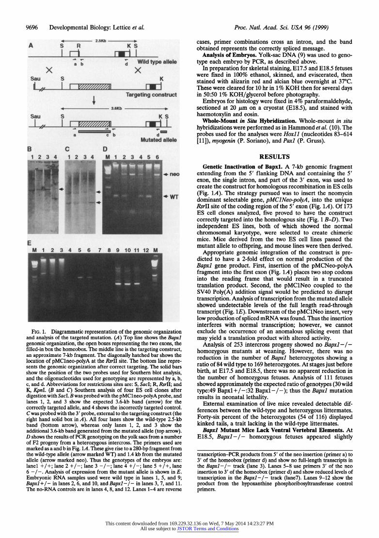
FIG. 1. Diagrammatic representation of the genomic organization and analysis of the targeted mutation. (A) Top line shows the Bapxl genomic organization, the open boxes representing the two exons, the filled-in box the homeobox. The middle line is the targeting construct, an approximate 7-kb fragment. The diagonally hatched bar shows the location of pMClneo-polyA at the RsrII site. The bottom line repre- sents the genomic organization after correct targeting. The solid bars show the position of the two probes used for Southern blot analysis, and the oligonucleotides used for genotyping are represented by a, b, c, and d. Abbreviations for restrictions sites are: 5, SacI; R, RsrII; and K, KpnI. (B and C) Southern analysis of four ES cell clones after digestion with SacL B was probed with the pMClneo-polyA probe, and lanes 1, 2, and 3 show the expected 3.6-kb band (arrow) for the correctly targeted allele, and 4 shows the incorrectly targeted control. C was probed with the 3' probe, external to the targeting construct (the right hand solid box in A). All four lanes show the wild-type 2.5-kb band (bottom arrow), whereas only lanes 1, 2, and 3 show the additional 3.6-kb band generated from the mutated allele (top arrow). D shows the results of PCR genotyping on the yolk sacs from a number of F2 progeny from a heterozygous intercross. The primers used are marked as a and b in Fig. 1A. These give rise to a 280-bp fragment from the wild-type allele (arrow marked WT) and 1.4 kb from the mutated allele (arrow marked neo). Thus the genotypes of the embryos are: lanel-4 +/+; lane, 2 +./-; lane 3A - -In-n; lAn 4 +/-; la-ne-1 5- +/4 lanen
6 _I.Aayi fexrsinfo h m utn leei hw nE
Emroi RN sape sd _eewl tp nlnes ,5 ad9 Baxl/ inlns26_ n_0 n apl/ nlns37 n 1
Th n-RN cnrosaeilae4,8an12Lae1-arrvre
cases, primer combinations cross an intron, and the band obtained represents the correctly spliced message.
Analysis of Embryos. Yolk-sac DNA (9) was used to geno- type each embryo by PCR, as described above.
In preparation for skeletal staining, E17.5 and E18.5 fetuses were fixed in 100% ethanol, skinned, and eviscerated, then stained with alizarin red and alcian blue overnight at 37?C. These were cleared for 10 hr in 1% KOH then for several days in 50:50 1% KOH/glycerol before photography.
Embryos for histology were fixed in 4% paraformaldehyde, sectioned at 20 Am on a cryostat (E18.5), and stained with haemotoxylin and eosin.
Whole-Mount in Situ Hybridization. Whole-mount in situ hybridizations were performed as in Hammond et al. (10). The probes used for the analyses were Hoxll (nucleotides 83-614 [11]), myogenin (P. Soriano), and Paxl (P. Gruss).
RESULTS Genetic Inactivation of Bapxl. A 7-kb genomic fragment
extending from the 5' flanking DNA and containing the 5' exon, the single intron, and part of the 3' exon, was used to create the construct for homologous recombination in ES cells (Fig. 1A). The strategy pursued was to insert the neomycin dominant selectable gene, pMClNeo-polyA, into the unique RsrII site of the coding region of the 5' exon (Fig. 1A). Of 173 ES cell clones analyzed, five proved to have the construct correctly targeted into the homologous site (Fig. 1 B-D). Two independent ES lines, both of which showed the normal chromosomal karyotype, were selected to create chimeric mice. Mice derived from the two ES cell lines passed the mutant allele to offspring, and mouse lines were then derived.
Appropriate genomic integration of the construct is pre- dicted to have a 2-fold effect on normal production of the Bapxl gene product. First, insertion of the pMCNeo-polyA fragment into the first exon (Fig. 1A) places two stop codons into the reading frame that would result in a truncated translation product. Second, the pMClNeo coupled to the SV40 Poly(A) addition signal would be predicted to disrupt transcription. Analysis of transcription from the mutated allele showed undetectable levels of the full length read-through transcript (Fig. 1E). Downstream of the pMClNeo insert, very low production of spliced mRNAwas found. Thus the insertion interferes with normal transcription; however, we cannot exclude the occurrence of an anomalous splicing event that may yield a translation product with altered activity.
Analysis of 253 intercross progeny showed no Bapxl-/- homozygous mutants at weaning. However, there was no reduction in the number of Bapxl heterozygotes showing a ratio of 84 wild type to 169 heterozygotes. At stages just before birth, at E17.5 and E18.5, there was no apparent reduction in the number of homozygous fetuses. Analysis of 111 fetuses showed approximately the expected ratio of genotypes (30 wild type:49 Bapxl+/-:32 Bapxl-/-); thus the Bapxl mutation results in neonatal lethality.
External examination of live mice revealed detectable dif- ferences between the wild-type and heterozygous littermates. Forty-six percent of the heterozygotes (54 of 116) displayed kinked tails, a trait lacking in the wild-type littermates.
Bapxl Mutant Mice Lack Ventral Vertebral Elements. At E18.5, Bapxl-l- homozygous fetuses appeared slightly
transcription-PCR products from 5' of the neo insertion (primer a) to 3' of the homeobox (primer d) and show no full-length transcripts in the Bapxl-/- track (lane 3). Lanes 5-8 use primers 3' of the neo insertion to 3' of the homeobox (primer d) and show reduced levels of transcription in the Bapxl -/- track (lane7). Lanes 9-12 show the product from the hypoxanthine phosphoribosyltransferase control primers.
This content downloaded from 169.229.32.136 on Wed, 7 May 2014 14:23:27 PMAll use subject to JSTOR Terms and Conditions
Developmental Biology: Lettice et al. Proc. Natl. Acad. Sci. USA 96 (1999) 9697
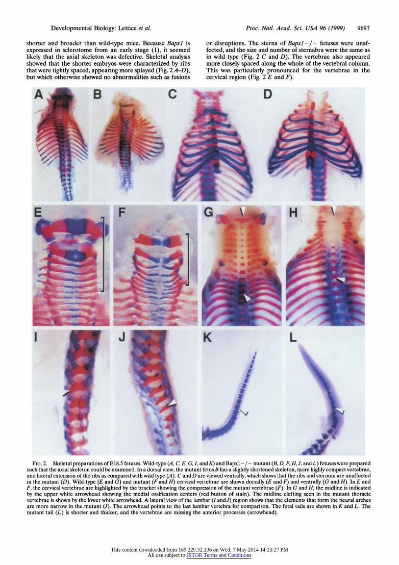
shorter and broader than wild-type mice. Because Bapxl is expressed in sclerotome from an early stage (1), it seemed likely that the axial skeleton was defective. Skeletal analysis showed that the shorter embryos were characterized by ribs that were tightly spaced, appearing more splayed (Fig. 2A-D), but which otherwise showed no abnormalities such as fusions
or disruptions. The sterna of Bapxl - / - fetuses were unaf- fected, and the size and number of sternabra were the same as in wild type (Fig. 2 C and D). The vertebrae also appeared more closely spaced along the whole of the vertebral column. This was particularly pronounced for the vertebrae in the cervical region (Fig. 2 E and F).
Ao B C t D i o~~~~~~~~~~~~~~~~~~~~~~~~~~~~~~~~~~~~~~~~~~~~~~~~~~~~~~~~~~~~~~~~~~~~~~~~~~~~~~~~~~~~~~~~~~~~~~! 1
FIG. 2. Skeletal preparations of El8.5 fetuses. Wild-type (A,C,E,G,I, and K) and Bapxl -/-mutant (B,D,F,H, ,andL) fetuses were prepared such that the axial skeleton could be examined. In a dorsal view, the mutant fetus B has a slightly shortened skeleton, more highly compact vertebrae, and lateral extension of the ribs as compared with wild type (A). C and D are viewed ventrally, which shows that the ribs and sternum are unaffected in the mutant (D). Wild-type (E and G) and mutant (F and H) cervical vertebrae are shown dorsally (E and F) and ventrally (G and H). In E and F, the cervical vertebrae are highlighted by the bracket showing the compression of the mutant vertebrae (F). In G and H, the midline is indicated by the upper white arrowhead showing the medial ossification centers (red button of stain). The midline clefting seen in the mutant thoracic vertebrae is shown by the lower white arrowhead. A lateral view of the lumbar (I and J) region shows that the elements that form the neural arches are more narrow in the mutant (J). The arrowhead points to the last lumbar vertebra for comparison. The fetal tails are shown in K and L. The mutant tail (L) is shorter and thicker, and the vertebrae are missing the anterior processes (arrowhead).
This content downloaded from 169.229.32.136 on Wed, 7 May 2014 14:23:27 PMAll use subject to JSTOR Terms and Conditions
9698 Developmental Biology: Lettice et al. Proc. Natl. Acad. Sci. USA 96 (1999)
, j
K' ;c ? V
~~~ . ~ ~ ~
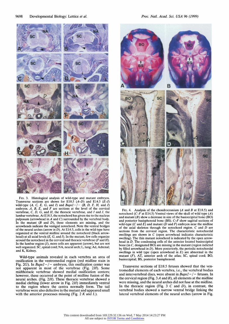
'. '4 ,6J~ FIG. 3. Histological analysis of wild-type and mutant embryos.
Transverse sections are shown for E18.5 (A-D) and E14.5 (E-J) wild-type (A, C, E, G, and I) and Bapxl-/- (B, D, F, H, and J) embryos. A, B, E, and F are sections at the level of the cervical vertebrae, C, D, G, and H, the thoracic vertebrae, and I and J, the lumbar vertebrae. At E18.5, the notochord has given rise to the nucleus pulposum (arrowhead in A and C) surrounded by the vertebral body. In the mutant (B and D), these elements are missing, and the arrowheads indicate the vestigial notochord. Note the ventral bridges of the neural arches (arrow in D). At E14.5, cells in the wild type have organized at the ventral midline around the notochord (black arrow- head) at all axial levels (E, G, and I). In the mutant, few cells organize around the notochord in the cervical and thoracic vertebrae (F and H). In the lumbar region (J), more cells are apparent (arrow), but are not well organized. SC, spinal cord; NA, neural arch; L, lung; Ad, Adrenal; and K, Kidney.
Wild-type animals revealed in each vertebra an area of ossification in the ventromedial region (red midline stain in Fig. 2G). In Bapxl -I- embryos, this ossification center was not apparent in most of the vertebrae (Fig. 2H). Some midthoracic vertebrae showed medial ossification centers; however, these occurred at the point of midline fusion of the neural arches. (Fig. 2H). These thoracic vertebrae showed a medial clefting (lower arrow in Fig. 211) immediately ventral to the region where the centra normally form. The tail vertebrae were also defective in the mutant and appeared small with the anterior processes missing (Fig. 2 K and L).
W I
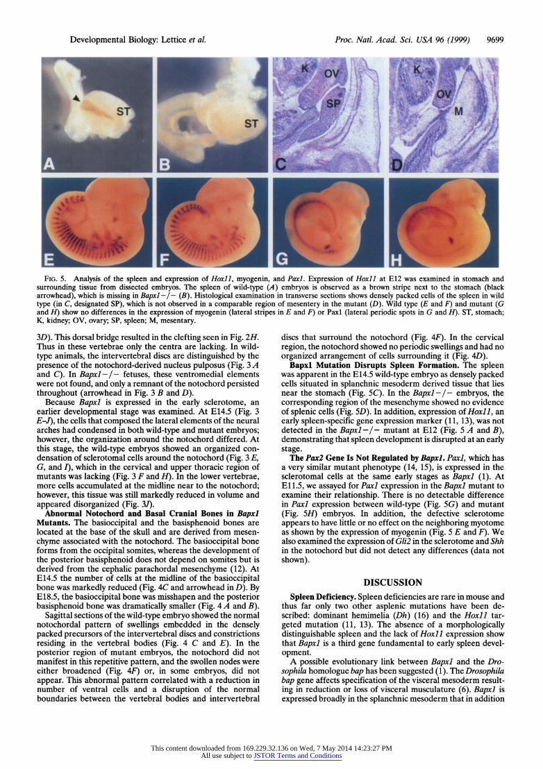
FI.4. Aayi ftecodornu AadBa 1.5an
~. 3:
FIG. 4. Analysis ofgh e chondrocranium (A and B at E18.5) and notochord (C-F at E14.5) Ventral views of the skull of wild type (A) and mutant (B) show a decrease in size of the basioccipital bone (BO) and posterior basisphenoid bone (BS). C-F show sagittal sections of wild-type (C and E) and mutant (D and F) embryos near the midline of the axial skeleton through the notochord region. C and D are sections from the cervical region. The characteristic notochordal swellings are shown in C (open arrowhead indicates characteristic swelling). The thin mutant notochord is indicated by the open arrow- head in D. The condensing cells of the anterior located basioccipital bone (in C, designated BO) are missing in the mutant (region indicted by filled arrowhead in D). More posteriorly, the periodic notochordal swellings in wild type (open arrowhead in E) are abnormal in the mutant (F). AT, anterior arch of the atlas; SC, spinal cord; BO, basioccipital; BS, posterior basisphenoid.
Transverse sections of E18.5 fetuses showed that the ven- tromedial elements of each vertebra, i.e., the vertebral bodies and intervertebral discs, were absent in Bapxl - /- fetuses. In the cervical region (Fig. 3A and B), all elements at the midline were missing, and the neural arches did not fuse at the midline. In the thoracic region (Fig. 3 C and D), in contrast, the vertebral bodies showed a narrow ventral bridge linking the lateral vertebral elements of the neural arches (arrow in Fig.
This content downloaded from 169.229.32.136 on Wed, 7 May 2014 14:23:27 PMAll use subject to JSTOR Terms and Conditions
Developmental Biology: Lettice et al. Proc. Natl. Acad. Sci. USA 96 (1999) 9699
S...~~~~~~~~~~~~~~~~~~~~~~~~~~~~~~~~~~~~~~~~~~R
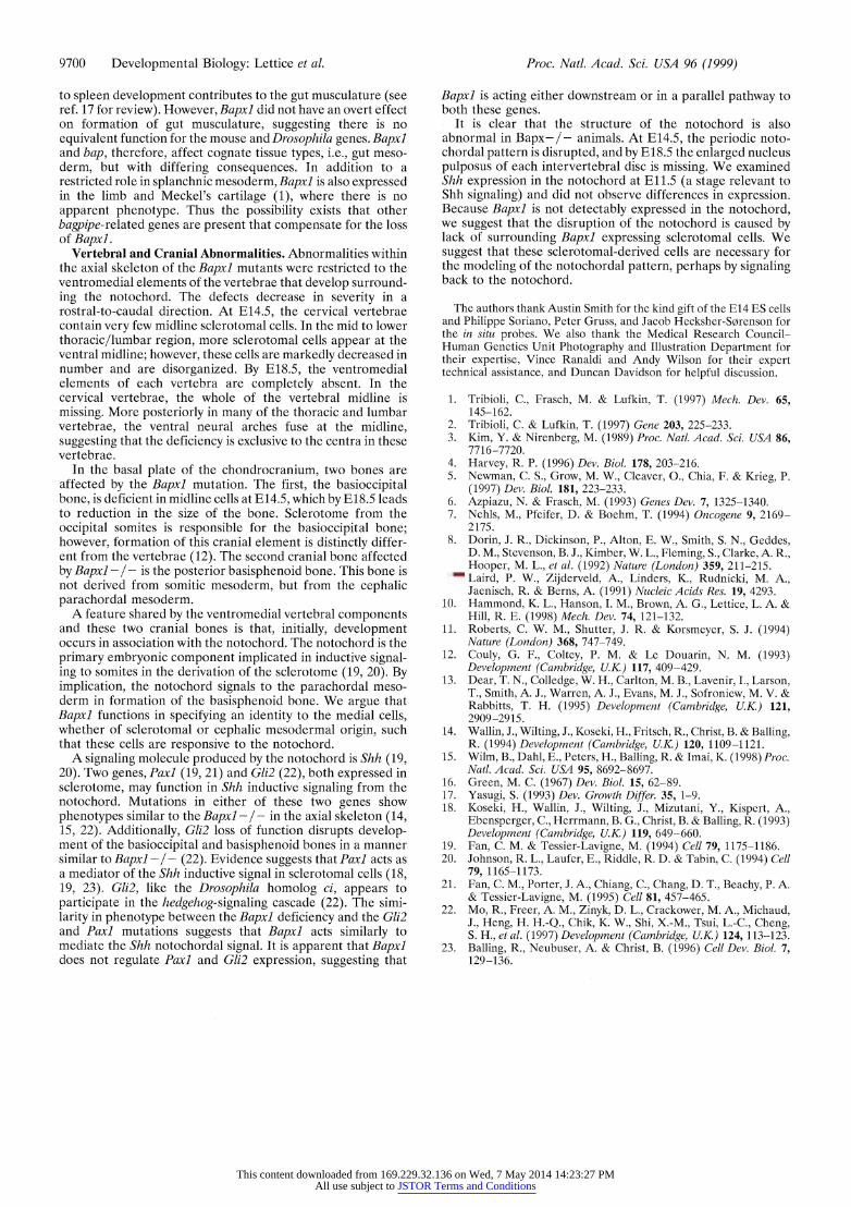
FIG. 5. Analysis of the spleen and expression of Hoxill myogenin, and Paxil Expression of Hoxil at E12 was examined in stomach and surrounding tissue from dissected embryos. The spleen of wild-type (A) embryos is observed as a brown stripe next to the stomach (black arrowhead), which is missing in Bapxl -/- (B). Histological examination in transverse sections shows densely packed cells of the spleen in wild type (in C, designated SP), which is not observed in a comparable region of mesentery in the mutant (D). Wild type (E and F) and mutant (G and H) show no differences in the expression of myogenin (lateral stripes in E and F) or Paxl (lateral periodic spots in G and H). ST, stomach; K, kidney; OV, ovary; SP, spleen; M, mesentary.
3D). This dorsal bridge resulted in the clefting seen in Fig. 2H. Thus in these vertebrae only the centra are lacking. In wild- type animals, the intervertebral discs are distinguished by the presence of the notochord-derived nucleus pulposus (Fig. 3 A and C). In Bapxl- / - fetuses, these ventromedial elements were not found, and only a remnant of the notochord persisted throughout (arrowhead in Fig. 3 B and D).
Because Bapxl is expressed in the early sclerotome, an earlier developmental stage was examined. At E14.5 (Fig. 3 E-J), the cells that composed the lateral elements of the neural arches had condensed in both wild-type and mutant embryos; however, the organization around the notochord differed. At this stage, the wild-type embryos showed an organized con- densation of sclerotomal cells around the notochord (Fig. 3 E, G, and I), which in the cervical and upper thoracic region of mutants was lacking (Fig. 3 F and H). In the lower vertebrae, more cells accumulated at the midline near to the notochord; however, this tissue was still markedly reduced in volume and appeared disorganized (Fig. 3J).
Abnormal Notochord and Basal Cranial Bones in Bapxl Mutants. The basioccipital and the basisphenoid bones are located at the base of the skull and are derived from mesen- chyme associated with the notochord. The basioccipital bone forms from the occipital somites, whereas the development of the posterior basisphenoid does not depend on somites but is derived from the cephalic parachordal mesenchyme (12). At E14.5 the number of cells at the midline of the basioccipital bone was markedly reduced (Fig. 4C and arrowhead in D). By E18.5, the basioccipital bone was misshapen and the posterior basisphenoid bone was dramatically smaller (Fig. 4 A and B).
Sagittal sections of the wild-type embryo showed the normal notochordal pattern of swellings embedded in the densely packed precursors of the intervertebral discs and constrictions residing in the vertebral bodies (Fig. 4 C and E). In the posterior region of mutant embryos, the notochord did not manifest in this repetitive pattern, and the swollen nodes were either broadened (Fig. 4F) or, in some embryos, did not appear. This abnormal pattern correlated with a reduction in number of ventral cells and a disruption of the normal boundaries between the vertebral bodies and intervertebral
discs that surround the notochord (Fig. 4F). In the cervical region, the notochord showed no periodic swellings and had no organized arrangement of cells surrounding it (Fig. 4D).
Bapxl Mutation Disrupts Spleen Formation. The spleen was apparent in the E14.5 wild-type embryo as densely packed cells situated in splanchnic mesoderm derived tissue that lies near the stomach (Fig. SC). In the Bapxl-I- embryos, the corresponding region of the mesenchyme showed no evidence of splenic cells (Fig. SD). In addition, expression of Hoxll, an early spleen-specific gene expression marker (11, 13), was not detected in the Bapxl -/- mutant at E12 (Fig. 5 A and B), demonstrating that spleen development is disrupted at an early stage.
The Pax2 Gene Is Not Regulated by Bapxl. Paxl, which has a very similar mutant phenotype (14, 15), is expressed in the sclerotomal cells at the same early stages as Bapxl (1). At E11.5, we assayed for Paxl expression in the Bapxl mutant to examine their relationship. There is no detectable difference in Paxl expression between wild-type (Fig. 5G) and mutant (Fig. 5H) embryos. In addition, the defective sclerotome appears to have little or no effect on the neighboring myotome as shown by the expression of myogenin (Fig. 5 E and F). We also examined the expression of Gli2 in the sclerotome and Shh in the notochord but did not detect any differences (data not shown).
DISCUSSION Spleen Deficiency. Spleen deficiencies are rare in mouse and
thus far only two other asplenic mutations have been de- scribed: dominant hemimelia (Dh) (16) and the Hoxll tar- geted mutation (11, 13). The absence of a morphologically distinguishable spleen and the lack of Hoxll expression show that Bapxl is a third gene fundamental to early spleen devel- opment.
A possible evolutionary link between Bapxl and the Dro- sophila homologue bap has been suggested (1). The Drosophila bap gene affects specification of the visceral mesoderm result- ing in reduction or loss of visceral musculature (6). Bapxl is expressed broadly in the splanchnic mesoderm that in addition
This content downloaded from 169.229.32.136 on Wed, 7 May 2014 14:23:27 PMAll use subject to JSTOR Terms and Conditions
9700 Developmental Biology: Lettice et al. Proc. Natl. Acad. Sci. USA 96 (1999)
to spleen development contributes to the gut musculature (see ref. 17 for review). However, Bapxl did not have an overt effect on formation of gut musculature, suggesting there is no equivalent function for the mouse and Drosophila genes. Bapxl and bap, therefore, affect cognate tissue types, i.e., gut meso- derm, but with differing consequences. In addition to a restricted role in splanchnic mesoderm, Bapxl is also expressed in the limb and Meckel's cartilage (1), where there is no apparent phenotype. Thus the possibility exists that other bagpipe-related genes are present that compensate for the loss of Bapxl.
Vertebral and Cranial Abnormalities. Abnormalities within the axial skeleton of the Bapxl mutants were restricted to the ventromedial elements of the vertebrae that develop surround- ing the notochord. The defects decrease in severity in a rostral-to-caudal direction. At E14.5, the cervical vertebrae contain very few midline sclerotomal cells. In the mid to lower thoracic/lumbar region, more sclerotomal cells appear at the ventral midline; however, these cells are markedly decreased in number and are disorganized. By E18.5, the ventromedial elements of each vertebra are completely absent. In the cervical vertebrae, the whole of the vertebral midline is missing. More posteriorly in many of the thoracic and lumbar vertebrae, the ventral neural arches fuse at the midline, suggesting that the deficiency is exclusive to the centra in these vertebrae.
In the basal plate of the chondrocranium, two bones are affected by the Bapxl mutation. The first, the basioccipital bone, is deficient in midline cells at E14.5, which by E18.5 leads to reduction in the size of the bone. Sclerotome from the occipital somites is responsible for the basioccipital bone; however, formation of this cranial element is distinctly differ- ent from the vertebrae (12). The second cranial bone affected by Bapxl -/ - is the posterior basisphenoid bone. This bone is not derived from somitic mesoderm, but from the cephalic parachordal mesoderm.
A feature shared by the ventromedial vertebral components and these two cranial bones is that, initially, development occurs in association with the notochord. The notochord is the primary embryonic component implicated in inductive signal- ing to somites in the derivation of the sclerotome (19, 20). By implication, the notochord signals to the parachordal meso- derm in formation of the basisphenoid bone. We argue that Bapxl functions in specifying an identity to the medial cells, whether of sclerotomal or cephalic mesodermal origin, such that these cells are responsive to the notochord.
A signaling molecule produced by the notochord is Shh (19, 20). Two genes, Paxi (19, 21) and Gli2 (22), both expressed in sclerotome, may function in Shh inductive signaling from the notochord. Mutations in either of these two genes show phenotypes similar to the Bapxl -/- in the axial skeleton (14, 15, 22). Additionally, Gli2 loss of function disrupts develop- ment of the basioccipital and basisphenoid bones in a manner similar to Bapxl -/- (22). Evidence suggests that Paxi acts as a mediator of the Shh inductive signal in sclerotomal cells (18, 19, 23). Gli2, like the Drosophila homolog ci, appears to participate in the hedgehog-signaling cascade (22). The simi- larity in phenotype between the Bapxl deficiency and the Gli2 and Paxi mutations suggests that Bapxl acts similarly to mediate the Shh notochordal signal. It is apparent that Bapxl does not regulate Paxi and Gli2 expression, suggesting that
Bapxl is acting either downstream or in a parallel pathway to both these genes.
It is clear that the structure of the notochord is also abnormal in Bapx-/- animals. At E14.5, the periodic noto- chordal pattern is disrupted, and by E18.5 the enlarged nucleus pulposus of each intervertebral disc is missing. We examined Shh expression in the notochord at E11.5 (a stage relevant to Shh signaling) and did not observe differences in expression. Because Bapxl is not detectably expressed in the notochord, we suggest that the disruption of the notochord is caused by lack of surrounding Bapxl expressing sclerotomal cells. We suggest that these sclerotomal-derived cells are necessary for the modeling of the notochordal pattern, perhaps by signaling back to the notochord.
The authors thank Austin Smith for the kind gift of the E14 ES cells and Philippe Soriano, Peter Gruss, and Jacob Hecksher-S0renson for the in situ probes. We also thank the Medical Research Council- Human Genetics Unit Photography and Illustration Department for their expertise, Vince Ranaldi and Andy Wilson for their expert technical assistance, and Duncan Davidson for helpful discussion.
1. Tribioli, C., Frasch, M. & Lufkin, T. (1997) Mech. Dev. 65, 145-162.
2. Tribioli, C. & Lufkin, T. (1997) Gene 203, 225-233. 3. Kim, Y. & Nirenberg, M. (1989) Proc. Natl. Acad. Sci. USA 86,
7716-7720. 4. Harvey, R. P. (1996) Dev. Biol. 178, 203-216. 5. Newman, C. S., Grow, M. W., Cleaver, O., Chia, F. & Krieg, P.
(1997) Dev. Biol. 181, 223-233. 6. Azpiazu, N. & Frasch, M. (1993) Genes Dev. 7, 1325-1340. 7. Nehls, M., Pfeifer, D. & Boehm, T. (1994) Oncogene 9, 2169-
2175. 8. Dorin, J. R., Dickinson, P., Alton, E. W., Smith, S. N., Geddes,
D. M., Stevenson, B. J., Kimber, W. L., Fleming, S., Clarke, A. R., Hooper, M. L., et al. (1992) Nature (London) 359, 211-215.
9. Laird, P. W., Zijderveld, A., Linders, K., Rudnicki, M. A., Jaenisch, R. & Berns, A. (1991) Nucleic Acids Res. 19, 4293.
10. Hammond, K. L., Hanson, I. M., Brown, A. G., Lettice, L. A. & Hill, R. E. (1998) Mech. Dev. 74, 121-132.
11. Roberts, C. W. M., Shutter, J. R. & Korsmeyer, S. J. (1994) Nature (London) 368, 747-749.
12. Couly, G. F., Coltey, P. M. & Le Douarin, N. M. (1993) Development (Cambridge, U.K) 117, 409-429.
13. Dear, T. N., Colledge, W. H., Carlton, M. B., Lavenir, I., Larson, T., Smith, A. J., Warren, A. J., Evans, M. J., Sofroniew, M. V. & Rabbitts, T. H. (1995) Development (Cambridge, U.K) 121, 2909-2915.
14. Wallin, J., Wilting, J., Koseki, H., Fritsch, R., Christ, B. & Balling, R. (1994) Development (Cambridge, UK) 120, 1109-1121.
15. Wilm, B., Dahl, E., Peters, H., Balling, R. & Imai, K. (1998) Proc. Natl. Acad. Sci. USA 95, 8692-8697.
16. Green, M. C. (1967) Dev. Biol. 15, 62-89. 17. Yasugi, S. (1993) Dev. Growth Differ. 35, 1-9. 18. Koseki, H., Wallin, J., Wilting, J., Mizutani, Y., Kispert, A.,
Ebensperger, C., Herrmann, B. G., Christ, B. & Balling, R. (1993) Development (Cambridge, U.K) 119, 649-660.
19. Fan, C. M. & Tessier-Lavigne, M. (1994) Cell 79, 1175-1186. 20. Johnson, R. L., Laufer, E., Riddle, R. D. & Tabin, C. (1994) Cell
79, 1165-1173. 21. Fan, C. M., Porter, J. A., Chiang, C., Chang, D. T., Beachy, P. A.
& Tessier-Lavigne, M. (1995) Cell 81, 457-465. 22. Mo, R., Freer, A. M., Zinyk, D. L., Crackower, M. A., Michaud,
J., Heng, H. H.-Q., Chik, K. W., Shi, X.-M., Tsui, L.-C., Cheng, S. H., et al. (1997) Development (Cambridge, UK) 124, 113-123.
23. Balling, R., Neubuser, A. & Christ, B. (1996) Cell Dev. Biol. 7, 129-136.
This content downloaded from 169.229.32.136 on Wed, 7 May 2014 14:23:27 PMAll use subject to JSTOR Terms and Conditions