tetranor pgdm: an abundant urinary … pgdm: an abundant urinary metabolite reflects biosynthesis of...
TRANSCRIPT

1
TETRANOR PGDM: AN ABUNDANT URINARY METABOLITE REFLECTS BIOSYNTHESIS OF PGD2 IN MICE AND HUMANS
Wen-Liang Song, Miao Wang, Emanuela Ricciotti, Susanne Fries, Ying Yu, Tilo Grosser, Muredach Reilly, John A. Lawson, and Garret A. FitzGerald
Institute for Translational Medicine and Therapeutics, School of Medicine, University of Pennsylvania, Philadelphia, PA 19104
Running Title: a major PGD2 metabolite: tetranor PGDM
Address for correspondence: G. A. FitzGerald M.D., 153 Johnson Pavilion, School of Medicine, Hamilton Walk, Philadelphia, Pa 19104, USA
Tel 215 898 1184; FAX 215 573 9135 E-mail [email protected]
ABSTRACT
PGD2 is a cyclooxygenase (COX) product of arachidonic acid which activates D prostanoid receptors to modulate vascular, platelet and leukocyte function in vitro. However, little is known about its enzymatic origin or its formation in vivo in cardiovascular or inflammatory disease. 11,15-dioxo-9α-hydroxy-2,3,4,5-tetranor-prostan-1,20-dioic acid (tetranor PGDM) was identified by mass spectrometry as a metabolite of infused PGD2 that is detectable in mouse and human urine. Using LC/MS/MS, tetranor PGDM was much more abundant than the PGD2 metabolites, 11β-PGF2α and 2,3-dinor-11β-PGF2α in human urine and was the only endogenous metabolite
detectable in mouse urine. Infusion of PGD2 dose dependently increased urinary tetranor PGDM >2,3-dinor-11β-PGF2α > 11β-PGF2α in mice. Deletion of either lipocalin type – or hemopoietic - PGD synthase enzymes decreased urinary tetranor PGDM. Deletion or knock down of COX-1, but not deletion of COX-2, decreased urinary tetranor PGDM in mice. Correspondingly, both PGDM and 2, 3-dinor-11β-PGF2α were suppressed by inhibition of COX-1 and COX-2, but not by selective inhibition of COX-2 in humans. PGD2 has been implicated in both the development and resolution of inflammation. Administration of bacterial lipopolysaccharide coordinately elevated tetranor PGDM and 2, 3-dinor-11β-PGF2α in volunteers, coincident with a pyrexial and systemic inflammatory response, but both metabolites fell during the resolution phase. Niacin increased tetranor PGDM and 2,3-dinor-11β-PGF2α in humans coincident with facial flushing. Tetranor PGDM is an abundant metabolite in
http://www.jbc.org/cgi/doi/10.1074/jbc.M706839200The latest version is at JBC Papers in Press. Published on November 8, 2007 as Manuscript M706839200
Copyright 2007 by The American Society for Biochemistry and Molecular Biology, Inc.
by guest on June 10, 2018http://w
ww
.jbc.org/D
ownloaded from

2
urine that reflects modulated biosynthesis of PGD2 in humans and mice.
INTRODUCTION:
Prostaglandin (PG) D2 is formed from PGH2, a cyclooxygenase (COX) product of arachidonic acid by the action of either a lipocalin (L)–like or hemopoietic (H) PGD synthase (1). Both enzymes may form PGD2
in vitro, but it is unclear which PGDS enzymes predominate under varied conditions in vivo. Suppression of PGD2 has been implicated in the bronchoconstriction of aspirin evoked asthma (2, 3) and release of PGD2 mediates the facial flushing and vascular instability of systemic mastocytosis (4). PGD2 relaxes vascular smooth muscle cells in vitro and its release by dermal dendritic cells contributes to the facial flushing which complicates administration of the hypolipidemic drug, niacin (5). PGD2
mediates its effects via activation of D prostanoid receptors (DPs). DP1, a member of the prostanoid family of G protein coupled receptors (GPCRs), mediates the vasorelaxant and bronchodilator effects (6, 7), while DP2, a GPCR of the fMLP receptor subfamily regulates Th1 and Th2 switching in lymphocytes (8) and is also expressed on eosinophils and basophils (9).
Recent interest in PGD2 has been prompted by the use of DP1 blockade as an adjunct to niacin therapy (10) and by the potential role of PGD2 and its metabolites in the resolution of inflammation (11). However, DP1 is expressed on human platelets and its activation in vitro results in a cyclic AMP dependent inhibition of platelet function (12,13). Nothing is known about the formation of PGD2 or the consequences of its inhibition in hyperlipidemic patients. Aside from a potential role in cardiovascular disease, PGD2 may be of importance in the
resolution of inflammation. A metabolite of PGD2, 15–deoxy-Δ12,14 PGJ2, has been postulated to activate PPARγ (14) and promote resolution of an inflammatory infiltrate (11). However, it remains to be determined by physicochemical methodology whether formation of 15–deoxy-Δ12,14 PGJ2 is indeed augmented during the resolution of human inflammation and although it can activate PPARγ, the concentrations required are unlikely to be attained in vivo (15).
Attempts to assess the biosynthesis of PGD2
have been constrained by a paucity of commercially available, sensitive and specific methodology. Aside from asthma and mastocytosis (2 - 4), little information on biosynthesis of PGD2 in humans has been acquired. Given the evanescence of primary PGs, biosynthesis is classically estimated by measurement of metabolites (16, 17). However, despite deletion of both DPs and PGDS enzymes, no metabolites of PGD2 have been reported in mouse, preventing assessment of biosynthetic response to experimental manipulation in that species. Initial attempts at commercial assay development in humans have focused on 11β-PGF2α (18) and 2,3-dinor-11β-PGF2α. Both are formed as minor urinary metabolites in monkeys and in a human volunteer following infusion of radiolabelled PGD2 (19). Indeed, paired analysis of 11β-PGF2α by gas chromatography mass spectrometry (GC/MS) and the commercially available immunoassay revealed poor concordance in the urine of patients with asthma (20, 21). Quantitative analysis of the major F-ring metabolite identified in urine after PGD2 infusion in the volunteer, 9α,11β–dihydroxy–15–oxo-2,3,18,19–tetranorprost–5–ene-1,20-dioic acid (19) has been
by guest on June 10, 2018http://w
ww
.jbc.org/D
ownloaded from

3
reported in human plasma and urine (22) and reflects nicely the marked augmentation of PGD2 biosynthesis in systemic mastocytosis (4). Here, we report the identification of a novel D-ring metabolite formed from infused PGD2, 11,15-dioxo-9α-hydroxy-,2,3,4,5- tetranor-prostan-1,20-dioic acid (tetranor PGDM) as an abundant endogenous metabolite of PGD2 in both human and mouse urine. The formation from PGD2 of its F-, J- and D-ring products is depicted in Figure 1. Gene manipulations in mice and pharmacological studies in humans implicate COX-1 as the major source of PGD2 as reflected by this metabolite in urine.
EXPERIMENTAL PROCEDURES:
Reagents: Authentic [2H6]tetranor PGDM was kindly synthesized on request by Cayman Chemical Co. (Ann Arbor, MI). Authentic 2,3-dinor 11β-PGF2α, [2H4]11β-PGF2α and PGD2 were purchased from the same source for use as standards. H2
18O was purchased from Cambridge Isotope Laboratories (Andover, MA). All mobile phases incorporated HPLC-grade solvents from Honeywell Burdick and Jackson. Reagent-grade acetic acid was purchased from Thermo Fisher Scientific. HPLC-grade ammonium hydroxide was purchased from Mallinckrodt Baker Chemicals. [18O2]2,3-dinor-11β-PGF2α was prepared as previously described (23). Methoxyamine (MO) HCl was purchased from Sigma Aldrich, Inc.
Urine analysis: [2H6]tetranor PGDM, [18O2]2,3-dinor-11β-PGF2α and [2H4]11β- PGF2α were added to 1 ml of human urine or 100 µl of mouse urine and were allowed to equilibrate for 15 min. One-half the urine
volume of an aqueous solution of MO HCl (1 g/ml) was added and allowed to stand for 15 min at room temperature. The samples were purified by solid phase extraction (SPE) using StrataX C18 cartridges (Phenomenex, Torrance, CA). The SPE cartridge was conditioned with 1 ml of acetonitrile and equilibrated with 1 ml of water. The sample was applied to the cartridge, which was then washed with 1 ml 5% acetonitrile in water and dried with vacuum for 15 min. The analyte and internal standards were eluted from the cartridge using 1 ml of 5% acetonitrile in ethyl acetate. The eluate was collected and dried under a gentle stream of nitrogen. The resulting residue was then reconstituted in 200 µl of 5% acetonitrile in water and filtered by centrifugation using 0.2-µm Nylon Microspin filters purchased from Alltech Associates (Deerfield, IL). High Performance Liquid Chromatography (HPLC): A Shimadzu Prominence HPLC system (Shimadzu, Columbia, MD) consisting of two LC-20AD-vp pumps, a CBM-20A system controller, and a SIL-5000 autosampler was used for all chromatography. The HPLC column used was a 150 x 2 mm Luna C18(2) with 3 � particles (Phenomenex, Torrance, CA). The mobile phase was generated from water (solvent A) and acetonitrile:methanol, 95:5, (solvent B), both containing 0.005% acetic acid adjusted to pH 5.7 with ammonium hydroxide. The flow rate was 0.2 ml/min. Separations were carried out with various linear solvent gradients.
Mass Spectrometry: A Thermo Finnigan TSQ Quantum Ultra tandem instrument (Thermo Fisher Scientific) equipped with a heated coaxial electrospray source and triple quadrupole analyzer was used in these studies. The ESI source was maintained at
by guest on June 10, 2018http://w
ww
.jbc.org/D
ownloaded from

4
240oC and used nitrogen for both sheath and auxiliary gas at 70 and 5 arbitrary units, respectively. The mass spectrometer was operated in the negative ion mode with a capillary temperature of 350oC and a spray voltage of 2.0 kV. The source collision-induced dissociation (SCID) was maintained at 10 eV. The collision gas was Argon, 1.5 mTorr. The analyzer was operated in the selected reaction monitoring (SRM) mode for the analysis of urinary PGD2 metabolites. For analysis of tetranor PGDM, the transitions monitored were m/z 385→336 for the endogenous material and m/z 391→342 for the deuterated internal standard. The collision energy (CE) was 15 eV. The transitions for [2H4]11β-PGF2α and endogenous 11β-PGF2α were 357→197 and 353→193 respectively, with CE 24 eV. The transition for [18O2]2,3-dinor-11β-PGF2α was m/z 329→145 and m/z 325→145 for the endogenous material, CE 13 eV.
The product ion scan mode was used for spectral analysis of tetranor PGDM. Precursor ions (m/z 385 and 391 for endogenous tetranor PGDM and the [2H6]tetranor PGDM internal standard, respectively) were collisionally activated at 15 eV under 1.5 mTorr argon gas producing the collision-induced dissociation (CID) spectra.
Studies in mice: All studies were performed following protocol review and approval by the Institutional Animal Care and Use Committee (IAUCC) of the University of Pennsylvania.
(i) Infusion studies: vehicle or PGD2 [20,150 and 500 μg] was infused i.p. into twelve week old male C57/BL6 mice (n = 5 per group). Urine was collected for 24 hrs in
metabolic cages for analysis of PGD2 metabolites.
(ii) PGDS knockouts. Urine was collected for 24 hrs from three to four month old male wild type (WT) mice, L-PGDS knockout mice (kindly provided by Dr. Yoshihiro
Urade, Osaka, Japan) and H-PGDS
knockout mice (kindly provided by Dr. Yoshihide Kanaoka, Boston, MA), all on a C57/BL6 background (n=15 for WT and LPGDS knockouts and n=16 for H-PGDS knockouts). (iii) COX knockdown (KD) and knockout (KO) mice. Urine was collected for 24 hrs from two to three month old COX-1 KD mice (24), COX-1 KOs and their WT controls (n=7-9 each) and COX-2 KOs (n=15) and their WT controls (n=7).
Clinical Studies: Five clinical studies were performed. The study protocols were approved by the Institutional Review Board of the University of Pennsylvania and by the Advisory Council of the Clinical and Translational Research Center (CTRC) of the University of Pennsylvania. All volunteers were apparently healthy on physical examination, were non smokers and refrained from all medications for 2 weeks before and then during the course of the studies. Volunteers with a history of coagulation disorders, a bleeding tendency, drug allergy, or gastrointestinal disorders were excluded from participation in the studies.
In the first study, 12 volunteers (6 male and 6 female) received a bolus injection of single dose 3 ng/kg of bacterial lipopolysaccharide (LPS) under controlled conditions as we have previously described (25). Subjects were admitted to the CTRC the evening before the study, and an intravenous infusion of saline was
by guest on June 10, 2018http://w
ww
.jbc.org/D
ownloaded from

5
commenced. The study involved a 60-hour inpatient stay in the CTRC of the University of Pennsylvania (www.itmat.upenn.edu) comprising an overnight acclimatization phase, a 24-hour saline administration control phase, and a 24-hour post-LPS study phase. Urinary PGD2 metabolites, were assessed in urines collected at the following time intervals before (-24 to -18, -18 to -12, -12 to -6, -6 to 0 hrs) and after (0 to 2, 2 to 4, 4 to 6, 6 to 8, 8 to 12, 12 to 18, 18 to 24 hrs) LPS administration. Data were plotted at the midpoint of each corresponding urine collection. Body temperature was recorded at the following time points before (-4, -2hrs) and after (0,2,4,6,8,10,12,14,16 hrs) LPS administration.
In the second study, Niacin (600mg) was administered to two healthy male volunteers. Urinary PGD2 metabolites were assessed in spot urines collected at the following time points before (time 0) and after (1, 2, 3, 4, 5, 6 hrs) niacin administration.
In a third study, 18 healthy volunteers (9 male and 9 female) received, in random order, a single dose of placebo or rofecoxib (25 mg) under double-blind conditions, separated by washout periods of at least 2 weeks. Urinary PGD2 metabolites were assessed at 0 and 4 hrs in spot urine samples that were collected 30 minutes after voiding.
In a fourth study, 8 healthy volunteers were orally administered 200mg celecoxib twice daily for nine days. Measurements of urinary PGD2 metabolites were performed predose and four hrs after drug administration on days 1 and 8.
In a fifth study, a single dose of aspirin (325 mg) was administered to 18 healthy volunteers (9 males and 9 females). Urinary
PGD2 metabolites were assessed in spot urine at time 0 and 4 hrs after dosing.
Data Analysis: Data are expressed after correction for urinary creatinine (Cr) concentrations and are reported as nanograms per milligram Cr. Results are expressed as mean +/- SEM. Statistical comparisons were performed initially using a two way ANOVA, with subsequent two-tailed comparisons as appropriate.
RESULTS:
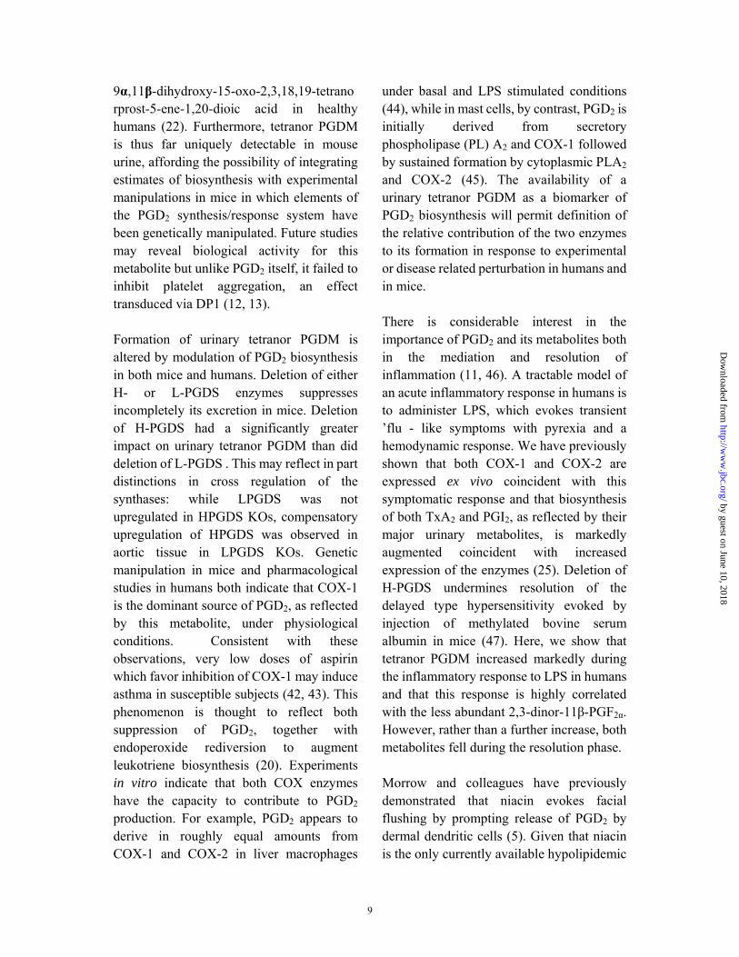
Discovery of tetranor PGDM: While measuring 9,15-dioxo-11α-hydroxy-2,3,4,5- tetranor-prostan-1,20-dioic acid (tetranor PGEM), the major urinary PGE metabolite in mouse urine (26), four other major peaks, apparently tetranor-PGEM isomers, were observed in the mass chromatogram (Fig2-A, lower panel). Because PGD2 and PGE2 are themselves structural isomers (27), it was surmised that some of these peaks might originate from PGD2. When PGD2 was infused into mice, two of these four peaks were increased dramatically (Fig2-B, lower panel), consistent with the hypothesis that they corresponded to the analogous D-ring tetranor metabolites. Following addition of authentic [2H6]tetranor PGDM to mouse urine, the deuterated compound coeluted with the endogenous material (Fig 2-A, B). There are four chromatographic peaks for tetranor PGDM MO derivatives. However, the two smaller peaks were variably detectable. Mass spectral analysis of human urine also revealed the existence of tetranor PGDM, which differed from mouse only in its abundance relative to tetranor PGEM (Fig2-C). Co-injection of extracts of urine from mice infused with PGD2 with extracts of human urine indicated that the endogenous peaks corresponding to tetranor PGDM in human urine corresponded to the
by guest on June 10, 2018http://w
ww
.jbc.org/D
ownloaded from

6
peaks increased dose dependently by PGD2 infusion in mice. (Supplemental Fig1) Product ion analysis of the [2H6]tetranor PGDM at m/z 391 gave rise to a series of major fragment ions with m/z values of 373, 342, 313, 267, 182, 164, and 142 (Fig3-A), virtually identical to the product ion spectrum obtained from endogenous tetranor PGDM (m/z 385); m/z 367, 336, 307, 261, 182, 164, and 142 (Fig3-B). The differences in m/z values between these two groups were either 0 or 6 mass units reflecting fragments with or without deuterium, again consistent with the original hypothesis. The transitions m/z 391>336 and m/z 385>336 are the same as those for tetranor PGEM, facilitating an integrated approach to lipidomic analysis (28). The assay was highly reproducible and the D-ring metabolite was chemically stable at -20°C and -80°C (Supplemental Fig2). A study was performed to determine whether tetranor PGDM might activate the DP1 receptor on human platelets (12, 13). However, unlike PGD2 itself, tetranor PGDM did not inhibit platelet aggregation
(Supplemental. Fig3).
HPLC/MS/MS analysis of urinary 2, 3-dinor-11β-PGF2α and 11β-PGF2α. A representative SRM chromatogram of 11β-PGF2α in human urine is shown in Fig 4-A. Transitions characteristic of [2H4]11β-PGF2α (m/z 357 197) and 11β-PGF2α (m/z 353 193) are shown in the upper and lower panel, respectively. A detectable endogenous chromatographic peak that co-eluted with spike was absent from most human urine samples. Some peaks eluted close to, but not coincident with the internal standard, as in Fig. 4-A. These peaks
were further confirmed to be distinct from endogenous 11β-PGF2α by addition of synthetic exogenous standards at the time of analysis. We surmise that these peaks represent F2-isoprostanes which would have the potential to compete with 11β-PGF2α in an immunoassay and also may not have separated from endogenous 11β-PGF2α under the elution conditions utilized in a GC/MS assay (29). While 2, 3-dinor-11β-PGF2α was readily detectable in human urine, it required a long LC program (28) to achieve separation from interfering compounds. A representative SRM chromatogram of 2,3-dinor-11β-PGF2α in human urine is shown in Fig 4-B. Urinary 2, 3-dinor-11β-PGF2α and 11β-PGF2α were both below the limits of detection (~1ng/mg Cr) in mouse urine (data not shown).
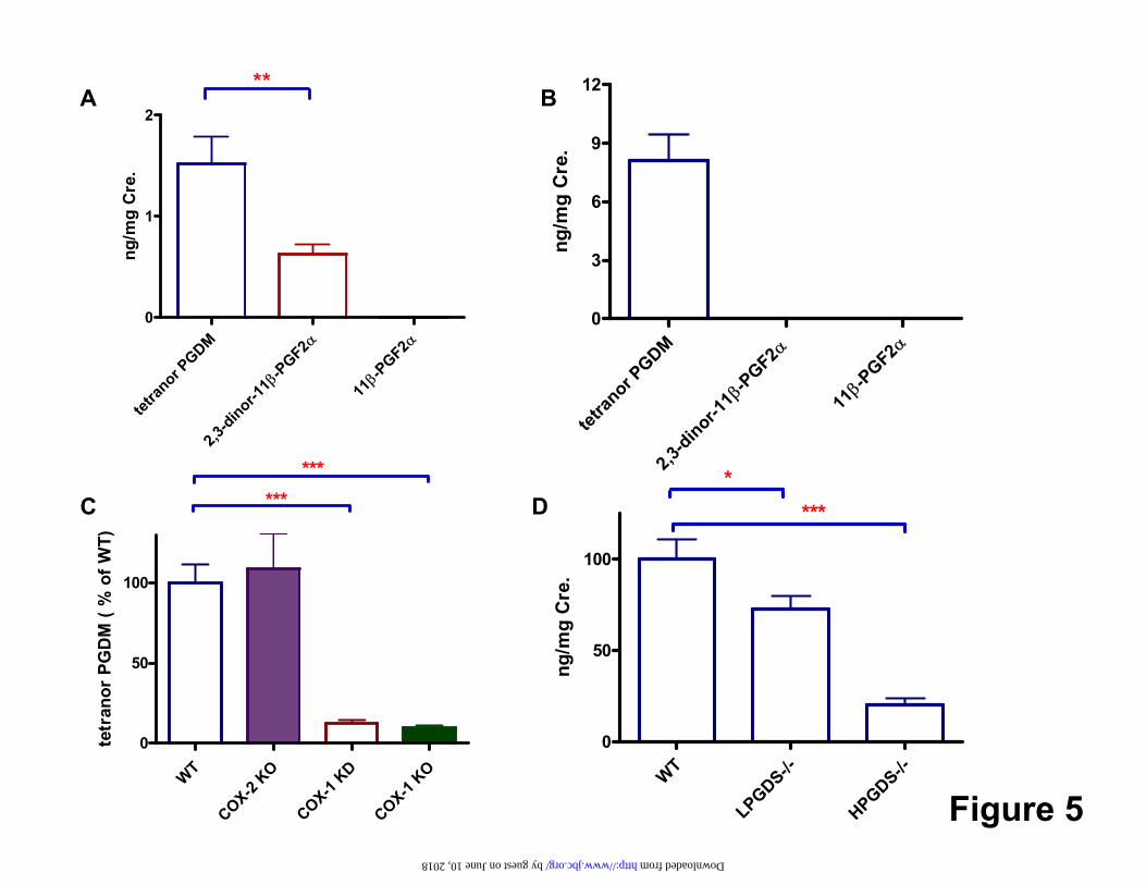
Comparative levels of endogenous metabolites of PGD2 metabolites: In human urine, the comparative levels of tetranor PGDM and 2,3-dinor-11β-PGF2α
were 1.5 ± 0.3 ng/mg Cr and 0.6 ± 0.1 ng/mg Cr (p < 0.01) respectively, while 11β-PGF2α was usually below the limits of detection (Fig 5-A). Tetranor PGDM was the only metabolite detectable in mouse urine at 8.1 ± 1.3 ng/mg Cr (Fig5-B).
Enzymatic contributions to the biosynthesis of PGD2: Both lipocalin (L)–like or hemopoietic (H) PGD synthase may form PGD2 in vitro, but it is unclear which PGDS enzyme predominates under varied conditions in vivo. The enzymatic contributions to the biosynthesis of PGD2 were examined by L-PGDS and H-PGDS mutant mouse models. Deletion of either PGD synthase significantly reduced biosynthesis of PGD2. Tetranor PGDM was suppressed about 30% on average by deletion of L-PGDS (p < 0.05)
by guest on June 10, 2018http://w
ww
.jbc.org/D
ownloaded from

7
and 80 % on average by deletion of H-PGDS (P<0.0001). (Fig5-C) Deletion of H-PGDS had a significantly greater (p < 0.001) impact on urinary tetranor PGDM than did deletion of L-PGDS in mice. Cross regulation of PGDS enzymes differed in response to gene deletion. Expression of H-PGDS was up-regulated in the aorta of L-PGDS KOs. By contrast, compensatory up-regulation of L-PGDS was not observed in H-PGDS KOs (Supplemental Fig4). The comparative contribution to metabolite excretion of the COX enzymes in mice was addressed by comparing the impact of genetic manipulation of COX-1 and COX-2. Both KO and KD of COX-1 suppressed urinary tetranor PGDM by about 90%, whereas deletion of COX-2 failed to alter significantly metabolite excretion (Fig5-D). In humans, aspirin at a dose of 325mg inhibits both COXs (30) while rofecoxib 25mg and celecoxib 200mg inhibit selectively COX-2 (31). Corresponding to the results in mice, inhibition of both COX-1 and COX-2 by aspirin, but not COX-2 alone by rofecoxib, depressed tetranor PGDM (Fig6A). Similar results were obtained with urinary 2, 3-dinor-11β-PGF2α (Fig6B). Both acute and chronic dosing with a second COX-2 inhibitor, celecoxib also failed to depress tetranor PGDM (Fig6C). Aspirin decreased urinary tetranor PGDM from 1.71 ± 0.21 ng/mg Cr to 0.86 ± 0.07 ng/mg Cr ( p < 0.001; Fig7A) and 2,3-dinor-11β-PGF2α decreased from 0.63 ± 0.11 ng/mg Cr to 0.32 ± 0.04 ng/mg Cr ( p < 0.01 ; Fig7B). This dose of aspirin inhibits COX enzymes incompletely; as reflected by comparable suppression of PGEM and PGIM. By contrast, urinary
isoprostanes, as reflected by the major F2 isoprostane in urine, 8,12–iso–iPF2α–VI is unaltered by this regimen (Supplemental Fig5). Excretion of PGD2 metabolites in PGD2
infused mice. The comparative disposition of PGD2 was examined by infusion of exogenous PGD2. Urinary metabolites of PGD2 increased dose dependently in response to the infusion (Fig 8-A). The levels of tetranor PGDM, 2,3-dinor-11β-PGF2α and 11β-PGF2α attained after the maximal dose (500 μg) of PGD2 were 2498±792 ng/mg Cr, 809 ± 346ng/mg Cr and 207 ± 67 ng/mg Cr, respectively (Fig8-B). Roughly 3-10% of infused PGD2 was excreted as the tetranor PGDM metabolite in mouse urine. The fractional conversion to each metabolite appeared to be uninfluenced by dose. Evoked biosynthesis of PGD2: LPS evokes a systemic inflammatory response in humans that is accompanied by regulated expression of both COX isozymes ex vivo and augmented biosynthesis of thromboxane A2 and prostacyclin (25). LPS induced a mean increase in both tetranor PGDM (1.49 ng/mg Cr at baseline to 2.15 ng/mg Cr at 2 hrs and 4.36 ng/mg Cr at 4 hrs after administration, (Fig 9-A) and correspondingly in 2,3-dinor-11β-PGF2α from 0.6 ng/mg Cr to 1.2ng/mg Cr and 2.1 ng/mg Cr respectively (Fig 9-B). The peak pyrexial response to LPS (from an average 97.8oF to 100.1oF) occurred ~4 hrs after administration (Fig 9-C). The alterations in urinary tetranor PGDM and 2,3-dinor-11β-PGF2α in response to LPS occur in a highly coordinated manner (Fig 9-D). Both metabolites fell after the inflammatory response and were not significantly different from basal levels
by guest on June 10, 2018http://w
ww
.jbc.org/D
ownloaded from

8
during the resolution phase (8 to 10 hrs after LPS). Oral administration of niacin, 600mg, evoked an intense flush in both volunteers, primarily involving the face and upper part of the body. Flushing was most pronounced during the first hour after dosing and had dissipated after approximately 2-3 hrs. Both urinary tetranor PGDM and 2, 3-dinor-11β-PGF2α were elevated by niacin (Fig 10-A, B), peaking 2-3 hrs and falling to basal levels 5-6 hrs after dosing. Again, reflecting the distinction from isoprostanes, urinary 8,12–iso–iPF2α–VI was unaltered by niacin administration (Fig 10-C). DISCUSSION: PGD2 is the predominant COX product of mast cells and contributes to the cutaneous flushing and hemodynamic dysregulation that characterizes excessive mast cell activation (2, 4). Mast cells are evident in the potentially vulnerable shoulder region of human atherosclerotic plaques (32) and mast cell depletion retards atherogenesis in mice (33). Mast cells have recently been implicated in aortic aneurysm formation (34). Other cells of relevance to atherogenesis, amongst them macrophages, platelets and leukocytes, may also generate PGD2 (35, 36). Studies of L-PGDS also implicate PGD2 in cardiovascular biology. Levels are induced in endothelial cells by laminar shear (37) and are elevated in the circulation after angioplasty (38) and in angina (39). However, lipocalins, such as L-PGDS, may subserve diverse biological functions (40); thus these observations only indirectly implicate PGD2. Previous attempts to study biosynthesis of PGD2 have relied particularly on commercially available assays of 11β-PGF2α.
This compound can be formed from PGD2 in vitro by bovine PGF synthase (18), and was the earliest metabolite detectable by Liston and Roberts after administration of radiolabelled PGD2 in a human (19) and itself retains biological activity, causing bronchoconstriction when inhaled by humans (41). The hemodynamic instability in patients with systemic mastocytosis is thought to reflect metabolism of PGD2, and other vasodilator D-ring metabolites, to vasoconstrictor F-ring metabolites, such as 11β-PGF2α. However, although endogenous concentrations of 11β-PGF2α have been reported in human urine, there has been poor concordance between estimates based on immunoassay and GC/MS. Here, we failed to detect urinary 11β-PGF2α using a more specific approach, HPLC/MS/MS. This raises the possibility that cross contaminating substances, particularly F2 isoprostanes (29), may have confounded, in some instances, analysis of 11β-PGF2α by GC/MS. However, we did identify endogenous 2, 3-dinor-11β-PGF2α in human urine. Liston and Roberts detected predominantly F-ring metabolites in the 39% of administered radioactivity recovered after administration of radiolabelled PGD2 to a volunteer. The most abundant of these metabolites was 9α,11β-dihydroxy-15- oxo-2,3,18,19-tetranorprost-5-ene-1,20-dioic acid (22). Surprisingly, they failed to detect tetranor PGDM; indeed the detected D-ring metabolites accounted for less than 4% of administered radioactivity. It is unknown whether this reflected a feature of this particular individual’s metabolism or technical factors. In the present studies tetranor PGDM was detected in the urines of all mice and humans studied under basal conditions. Indeed the levels in urine exceeded not only 2,3-dinor-11β-PGF2α, but also the reported levels of
by guest on June 10, 2018http://w
ww
.jbc.org/D
ownloaded from
9
9α,11β-dihydroxy-15-oxo-2,3,18,19-tetranorprost-5-ene-1,20-dioic acid in healthy humans (22). Furthermore, tetranor PGDM is thus far uniquely detectable in mouse urine, affording the possibility of integrating estimates of biosynthesis with experimental manipulations in mice in which elements of the PGD2 synthesis/response system have been genetically manipulated. Future studies may reveal biological activity for this metabolite but unlike PGD2 itself, it failed to inhibit platelet aggregation, an effect transduced via DP1 (12, 13). Formation of urinary tetranor PGDM is altered by modulation of PGD2 biosynthesis in both mice and humans. Deletion of either H- or L-PGDS enzymes suppresses incompletely its excretion in mice. Deletion of H-PGDS had a significantly greater impact on urinary tetranor PGDM than did deletion of L-PGDS . This may reflect in part distinctions in cross regulation of the synthases: while LPGDS was not upregulated in HPGDS KOs, compensatory upregulation of HPGDS was observed in aortic tissue in LPGDS KOs. Genetic manipulation in mice and pharmacological studies in humans both indicate that COX-1 is the dominant source of PGD2, as reflected by this metabolite, under physiological conditions. Consistent with these observations, very low doses of aspirin which favor inhibition of COX-1 may induce asthma in susceptible subjects (42, 43). This phenomenon is thought to reflect both suppression of PGD2, together with endoperoxide rediversion to augment leukotriene biosynthesis (20). Experiments in vitro indicate that both COX enzymes have the capacity to contribute to PGD2
production. For example, PGD2 appears to derive in roughly equal amounts from COX-1 and COX-2 in liver macrophages
under basal and LPS stimulated conditions (44), while in mast cells, by contrast, PGD2 is initially derived from secretory phospholipase (PL) A2 and COX-1 followed by sustained formation by cytoplasmic PLA2 and COX-2 (45). The availability of a urinary tetranor PGDM as a biomarker of PGD2 biosynthesis will permit definition of the relative contribution of the two enzymes to its formation in response to experimental or disease related perturbation in humans and in mice. There is considerable interest in the importance of PGD2 and its metabolites both in the mediation and resolution of inflammation (11, 46). A tractable model of an acute inflammatory response in humans is to administer LPS, which evokes transient ’flu - like symptoms with pyrexia and a hemodynamic response. We have previously shown that both COX-1 and COX-2 are expressed ex vivo coincident with this symptomatic response and that biosynthesis of both TxA2 and PGI2, as reflected by their major urinary metabolites, is markedly augmented coincident with increased expression of the enzymes (25). Deletion of H-PGDS undermines resolution of the delayed type hypersensitivity evoked by injection of methylated bovine serum albumin in mice (47). Here, we show that tetranor PGDM increased markedly during the inflammatory response to LPS in humans and that this response is highly correlated with the less abundant 2,3-dinor-11β-PGF2α. However, rather than a further increase, both metabolites fell during the resolution phase. Morrow and colleagues have previously demonstrated that niacin evokes facial flushing by prompting release of PGD2 by dermal dendritic cells (5). Given that niacin is the only currently available hypolipidemic
by guest on June 10, 2018http://w
ww
.jbc.org/D
ownloaded from
10
drug that elevates high density lipoprotein (48), it is hoped that co-administration of a DP1 antagonist might reduce this complication and enhance compliance. However, DP1 activation elevates cyclic AMP and inhibits aggregation of human platelets (49), raising concern that it might function as an endogenous modulator of platelet activation in vivo, much like PGI2
(50). Urinary tetranor PGDM reflects the increase in biosynthesis evoked by niacin and should facilitate elucidation of the role of PGD2 in cardiovascular disease. In summary, here we report a novel, abundant, D-ring urinary PGD2 metabolite 11,15-dioxo-9α-hydroxy-2,3,4,5-tetranor-prostan-1,20-dioic acid, tetranor PGDM, which is detectable in mouse and human
urine. Analysis of this compound reflects modulated biosynthesis of PGD2 in both species and will complement the use of genetic and pharmacological probes in the further elucidating the biology of PGD2 in vivo. Footnotes: Supported by a Specialized Center in Clinical Research in Vascular Injury (HL 83799) and a Clinical and Translational Science Award (RR-023567), HL 073278 (to M.R.) and Science Development Grants from the American Heart Association to M.W.; Y.Y and T.G. GAF is the McNeil Professor in Translational Medicine and Therapeutics. We appreciate technical help from Wenxuan Li, Helen Zou, Matthew Stetz and Azri Mohd.
References:
1. Urade Y and Hayaishi O. (2000) Vitamins and Hormones, Vol 58, pp. 89-120 2. O'Sullivan S, Dahlen B, Dahlen SE, Kumlin M. (1996) J Allergy Clin Immunol 98:
421-432 3. Bochenek G, Nagraba K, Nizankowska E, Szczeklik A. (2003) J Allergy Clin Immunol
111: 743-749 4. Roberts LJ 2nd, Sweetman BJ, Lewis RA, Austen KF, Oates JA. (1980) N Engl J Med.
303(24):1400-4. 5. Morrow JD, Parsons WG 3rd, Roberts LJ 2nd. (1989) Prostaglandins 38: 263-274 6. Williams, T. J. & Peck, M. J. (1977) Nature 270, 530–532 7. Matsuoka T, Hirata M, Tanaka H, Takahashi Y, Murata T, Kabashima K, Sugimoto Y,
Kobayashi T, Ushikubi F, Aze Y. et al. (2000) Science 287, 2013–2017. 8. Nagata K, Tanaka K, Ogawa K, Kemmotsu K, Imai T, Yoshie O, Abe H, Tada K,
Nakamura M, Sugamura K. et al. (1999) J. Immunol. 162, 1278–1286. 9. Nagata K, Hirai H, Tanaka K, Ogawa K, Aso T, Sugamura K, Nakamura M, Takano S. et
al. (1999) FEBS Lett. 459, 195–199. 10. Cheng K, Wu TJ, Wu KK, Sturino C, Metters K, Gottesdiener K, Wright SD, Wang Z,
O'Neill G, Lai E et al. (2006) Proc. Natl Acad. Sci. USA 103, 6682–6687 11. Gilroy DW, Colville-Nash PR, Willis D, Chivers J, Paul-Clark MJ, Willoughby DA.
(1999) Nat Med 5: 698-701 12. Oelz O, Oelz R, Knapp HR, Sweetman BJ, Oates JA. (1977) Prostaglandins 13: 225-234 13. Bushfield M, McNicol A, MacIntyre DE.(1985) Biochem J 232: 267-271 14. Forman BM, Tontonoz P, Chen J, Brun RP, Spiegelman BM, Evans RM. (1995) Cell 83:
803-812
by guest on June 10, 2018http://w
ww
.jbc.org/D
ownloaded from

11
15. Bell- Parikh L.C., Ide T., Lawson J.A., McNamara P., Reilly M. and FitzGerald G.A. (2003) J Clin Invest.112(6):945-55
16. McAdam BF, Catella-Lawson F, Mardini IA, Kapoor S, Lawson JA, FitzGerald GA. (1999) Proc Natl Acad Sci U S A. 96(1):272-7.
17. Catella F, Healy D, Lawson JA, FitzGerald GA. (1986) Proc Natl Acad Sci U S A. 83(16):5861-5.
18. Watanabe K, Iguchi Y, Iguchi S, Arai Y, Hayaishi O, Roberts LJ 2nd. (1986) Proc Natl Acad Sci U S A. 83(6):1583-7.
19. Liston, T.E., Roberts, L.J. (1985) J Biol Chem 260 13172-13180 20. Misso NL, Aggarwal S, Phelps S, Beard R, Thompson PJ. (2004) Clin Exp Allergy.
34(4):624-31. 21. Bochenek G, Nizankowska E, Gielicz A, Swierczynska M, Szczeklik A. (2004).Thorax.
59(6):459-64 22. Morrow JD, Guzzo C, Lazarus G, Oates JA, Roberts LJ 2nd. (1995) J Invest Dermatol
104: 937-940 23. Pickett WC, Murphy RC. (1981) Anal Biochem. 111(1):115-21. 24. Yu Y, Cheng Y, Fan J, Chen XS, Klein-Szanto A, Fitzgerald GA, Funk CD.(2005) J Clin
Invest. 115(4):986-95. Epub 2005 Mar 17. 25. McAdam BF, Mardini IA, Habib A, Burke A, Lawson JA, Kapoor S, FitzGerald GA.
(2000) J Clin Invest. 105(10):1473-82. 26. Cheng Y, Wang M, Yu Y, Lawson J, Funk CD, FitzGerald GA. (2006) J Clin Invest.
116(5):1391-9. 27. Hamberg M, Fredholm BB. (1976) Biochim Biophys Acta. 431(1):189-83. 28. Song WL, Lawson JA, Wang M, Zou H, and FitzGerald GA.(2007)(in press)
Methods in Enz. Vol (433) 29. O’Sullivan, S., Mueller, M.J., Dahlén, S., et al. (1999) Prostaglandins and Other Lipid
Mediators 57 149-165 30. FitzGerald GA, Oates JA, Hawiger J, Maas RL, Roberts LJ 2nd, Lawson JA, Brash AR.
Endogenous biosynthesis of prostacyclin and thromboxane and platelet function during chronic administration of aspirin in man. J Clin Invest. 1983 Mar;71(3):676-88.
31. Fries S, Grosser T, Price TS, Lawson JA, Kapoor S, DeMarco S, Pletcher MT, Wiltshire T, FitzGerald GA. Marked interindividual variability in the response to selective inhibitors of cyclooxygenase-2. Gastroenterology. 2006 Jan;130(1):55-64.
32. BanderLaan PA and Reardon CA. (2005) J of Lipid Res 46: 829838 33. Sun J, Sukhova GK, Wolters PJ, Yang M, Kitamoto S, Libby P, MacFarlane LA, Clair JM,
Shi GP. (2007) Nat Med. 13(6):719-24. 34. Sun J., Sukhova G.K., Yang M., Wolters P.J., MacFarlane L.A., Libby P., Sun C., Zhang
Y., Liu J., Ennis T.L., Knispel R., Xiong W., Thompson R.W. Baxter B.T. and Shi G.P. (2007) Mast cells modulate the pathogenesis of elastase-induced abdominal aortic aneurysms in mice. J Clin Invest. 2007 Nov 1;117(11):3359-3368
35. J. MacDermot, C.R. Kelsey, K.A. Waddell, R. Richmond, R.K. Knight, P.J. Cole et al., (1984) Prostaglandins 27 2 pp. 163–179.
36. W.G. Parsons, II and L.J. Roberts, II, (1988) J Immunol 141 7, pp. 2413–2419
by guest on June 10, 2018http://w
ww
.jbc.org/D
ownloaded from

12
37. Taba Y, Sagaguri T, Miyagi M, Abumiya T, Miwa Y, Ikeda T, Mitsumata M. (2000) Circ Res 86: 967-973
38. Inoue T, Takayanagi K, Morooka S, Uehara Y, Oda H, Seiki K, Nakajima H, Urade Y. (2001)Thromb Haemost 85: 165-170.
39. Eguchi Y, Eguchi N, Oda H, Seiki K, Kijima Y, Matsu-ura Y, Urade Y, Hayaishi O. (1997) Proc Natl Acad Sci USA 93: 14689-14694
40. Flower DR. (1996) Biochem J. 318 (Pt 1):1-14. Review. 41. C.R. Beasley, C. Robinson, R.L. Featherstone, J.G. Varley, C.C. Hardy and M.K. Church,
(1987)J Clin Invest 79 3 pp. 978–983 42. Barr RG, Kurth T, Stampfer MJ, Buring JE, Hennekens CH, Gaziano JM. Aspirin and
decreased adult-onset asthma: randomized comparisons from the physicians' health study. Am J Respir Crit Care Med. 2007 Jan 15;175(2):120-5.
43. Catella-Lawson F, Reilly MP, Kapoor SC, Cucchiara AJ, DeMarco S, Tournier B, Vyas SN, FitzGerald GA. Cyclooxygenase inhibitors and the antiplatelet effects of aspirin. (2001) N Engl J Med. Dec 20;345(25):1809-17.
44. Dieter P, Scheibe R, Jakobsson PJ, Watanable K, Kolada A, Kamionka S. (2000) Biochem Biophys. Res. Commun 276: 488-492
45. Reddy ST and Hershman HR. (1997) J Biol Chem 272: 3231-3237. 46. Gilroy DW, Colville-Nash PR, McMaster S, Sawatzky DA, Willoughby DA, Lawrence T.
(2003) FASEB J 17: 2269-2271. 47. Trivedi SG, Newson J, Rajakariar R, Jacques TS, Hannon R, Kanaoka Y, Eguchi N,
Colville-Nash P, Gilroy DW. (2006) Proc Natl Acad Sci U S A. 103(13):5179-84. 48. Carlson LA. (2005) J Intern Med. 258(2):94-114. 49. Giles, H., Leff, P., Bolofo, M. L., Kelly, M. G. & Robertson, A. D. (1989) Br. J.
Pharmacol. 96, 291–300 50. FitzGerald GA, Smith B, Pederson AK, Brash AR. (1984) N Engl J Med 310: 1065-1068
Figure Legends:
Fig 1.Biosynthetic pathway of PGD2 and its derivatives.
Fig 2.HPLC/MS/MS of a major urinary PGD2 metabolite. Representative selected reaction monitoring chromatogram of [2H6]tetranor PGDM (Upper), and co-eluted peaks corresponding to the endogenous compound (Lower) in (A) mouse urine and (B) in mouse urine following intraperitoneal administration of 500 μg of PGD2, resulting in marked elevation of the endogenous compound and (C) in human urine. Transitions characteristic of [2H6]tetranor PGDM (m/z 391 342) and tetranor PGDM (m/z 385 336) are shown in the upper and lower panels, respectively; Fig 3 Product ion analysis of tetranor PGDM. (A) Product ion spectrum of [2H6]tetranor PGDM (m/z 391) with m/z values of 373, 342, 313,267,182,164, and 142. (B) Product ion spectrum of the endogenous tetranor PGDM (m/z 385) with m/z values of 367, 336, 307,261,182,164, and 142. The differences in m/z values between these two groups were either 0 or 6 mass units reflecting
by guest on June 10, 2018http://w
ww
.jbc.org/D
ownloaded from

13
fragments with or without deuterium. Fig4 HPLC/MS/MS analysis of urinary 2, 3-dinor-11β-PGF2α and 11β-PGF2α. (A) Representative selected reaction monitoring chromatogram of 11β-PGF2α in human urine. Transitions characteristic of [2H4]11β-PGF2α (m/z 357 197) and 11β-PGF2α (m/z 353 193) are shown in the upper and lower panel, respectively. Note that a peak corresponding to endogenous material co-eluting with the standard is not evident in the lower panel (B) Representative selected reaction monitoring chromatogram of 2, 3-dinor-11β-PGF2α in human urine. Transitions characteristic of [18O2]2, 3-dinor-11β-PGF2α (m/z 329 145) and 2, 3-dinor-11β-PGF2α (m/z 325 145) are shown in the upper and lower panel, respectively. Fig 5 Biosynthesis of PGD2 in humans and mice. (A) Urinary metabolites of PGD2 were examined in human urine. Both tetranor PGDM and 2, 3-dinor-11β-PGF2α, but not 11β-PGF2α were detectable. (B) Urinary metabolites of PGD2 were examined in mouse urine. Tetranor PGDM, but not 2, 3-dinor-11β-PGF2α or 11β-PGF2α was detectable (C) Tetranor PGDM was depressed in urine from L-PGDS and H-PGDS knockout mice compared to wild type controls. (D) Tetranor PGDM was depressed in urine from COX-1 knockdown and COX-1 knockout mice but not COX-2 knockout mice compared to wild type controls. Data shown are the mean ± SEM. * P<0.05, ** P<0.01, ***P<0.001 Fig 6 The effects of selective COX-2 inhibition by rofecoxib and celecoxib on excretion of major urinary PGD2 metabolites. (A) Single dose Rofecoxib failed to decrease significantly excretion of tetranor PGDM (B) Single dose Rofecoxib failed to decrease significantly excretion of 2, 3-dinor-11β-PGF2α (C) Single dose and multiple dose of Celecoxib failed to decrease significantly excretion of tetranor PGDM Fig 7 Nonselective COX inhibition by aspirin325 mg. Inhibition of systemic COX-1 and COX-2 with 325mg aspirin significantly decreased excretion of both tetranor PGDM (A) and 2, 3-dinor-11β-PGF2α (B). Fig 8 Excretion of PGD2 metabolites in PGD2 infused mice. (A) Urinary PGD2 metabolites following intraperitoneal administration of PGD2 (B) Comparison of urinary PGD2 metabolites at 500 μg PGD2 infusion Infusion of PGD2 dose dependently increased urinary tetranor PGDM >2,3-dinor-11β-PGF2α > 11β-PGF2α. Fig 9 LPS induced an increase in excretion of PGD2 metabolites. (A) LPS induced a mean increase in tetranor PGDM from 1.49 ng/mg Cr at baseline to 2.15 ng/mg Cr at 2 hrs and 4.36 ng/mg Cr at 4 hrs after administration;(B) LPS induced a mean increase in 2, 3- dinor -11β-PGF2α from 0.6 ng/mg Cr at baseline to1.2ng/mg Cr at 2 hrs and 2.1ng/mg Cr at 4 hrs after administration; (C) In healthy volunteers, temperature increased transiently during endotoxemia returning to baseline by 24 hrs after LPS. (D) Correlation between log transformed urinary tetranor PGDM and 2, 3-dinor-11β-PGF2α. Data shown are
by guest on June 10, 2018http://w
ww
.jbc.org/D
ownloaded from

14
the mean ± SEM. *P<0.05 ***P<0.001 Fig 10 Niacin-evoked urinary PGD2 metabolite excretion. Sequential measurement of (A) tetranor PGDM , (B) 2, 3-dinor-11β-PGF2α and (C) 8,12 – iso iPF2α – VI at various time points before (time 0) and after 600 mg niacin administration to two healthy volunteers. Both urinary tetranor PGDM and 2, 3-dinor-11β-PGF2α, were elevated by niacin, peaking 2-3 hrs and falling to basal levels 5-6 hrs after dosing. Urinary 8,12 – iso iPF2α – VI is unaltered by niacin.
by guest on June 10, 2018http://w
ww
.jbc.org/D
ownloaded from

Arachidonic Acid
PGH2
PGD2
PGJ2
D12-PGJ2
15-deoxy-Δ12,14-PGJ2
11β-PGF2α
2, 3-dinor-11β-PGF2α
Tetranor PGDM
9α,11β-dihydroxy-15-oxo-2,3,18,19-tetranorprost-
5-ene-1,20-dioic acid
DP1 DP2 PPARγ
?
COX1
COX2
L-PGDS H-PGDS
D-ring
F-ring J-ring
Figure 1
by guest on June 10, 2018 http://www.jbc.org/ Downloaded from
A C
B tetranor PGDM in PGD2 infused mouse urine
tetranor PGDM in mouse urine tetranor PGDM in human urine
Figure 2
by guest on June 10, 2018 http://www.jbc.org/ Downloaded from

A
B spectrum of endogenous material
spectrum of [2H6]tetranor PGDM
Figure 3
m/z
m/z
by guest on June 10, 2018 http://www.jbc.org/ Downloaded from

A B11β-PGF2αin human urine 2,3-dinor-11β-PGF2α in human urine
Figure 4
Time (mins) Time (mins)55 56 57 58 59
40 41 42 43 44 45 46
by guest on June 10, 2018 http://www.jbc.org/ Downloaded from
A B
C
Figure 5
D
tetran
or PGDM α
-PGF2
β
2,3-dinor-1
1
α
-PGF2
β11
0
3
6
9
12
ng/m
g C
re.
WT
COX-2 KO
COX-1 KD
COX-1 KO
0
50
100
******
tetr
anor
PG
DM
( %
of W
T)
WT
LPGDS-/-
HPGDS-/-
0
50
100
****
ng/m
g C
re.
tetran
or PGDM α
-PGF2
β
2,3-dinor-1
1
α
-PGF2
β11
0
1
2
**
ng/m
g C
re.
by guest on June 10, 2018 http://www.jbc.org/ Downloaded from

Figure 6
A B
C
Rofecoxib 25mg
pre post pre post0.0
0.5
1.0
1.5
Placebo Rofecoxib
2,3
-din
or-1
1¦Â-
PGF 2
¦Áng
/mg
Cre
.
Rofecoxib 25 mg
pre post pre post0
1
2
3
4
Placebo Rofecoxib
tetr
anor
PG
DM
ng/m
g C
re.
Celecoxib 200mg
pre post post0
2
4
6
tetr
anor
PG
DM
ng/m
g C
re
by guest on June 10, 2018 http://www.jbc.org/ Downloaded from

0
1
2
3
4
pre post
***
Aspirin 325mg
tetr
anor
PG
DM
ng/m
g C
re.
0
1
2
3
4
pre post
*
Aspirin 325mg
2,3
-din
or-1
1β-P
GF2α
ng/m
g C
re.
Figure 7
A B
by guest on June 10, 2018 http://www.jbc.org/ Downloaded from

2,3-dinor-11β-PGF2αtetranor PGDM
11β-PGF2α
500 ug PGD2 infused mouse urine
0 1 2 3 4 50
500
1000
1500
2000
2500
3000
3500
ng/m
g C
re.
0 20 150 500 ug0 1 2 3 4 50
500
1000
1500
2000
2500
3000
3500
ng/m
g C
re.
0 20 150 500 ug0 1 2 3 4 50
500
1000
1500
2000
2500
3000
3500
ng/m
g C
re.
0 1 2 3 4 50
500
1000
1500
2000
2500
3000
3500
ng/m
g C
re.
0 20 150 500 ug
1
10
100
1000
10000
ng/m
g C
re.
PGD2 - + - + - +1
10
100
1000
10000
ng/m
g C
re.
PGD2 - + - + - +
PGD2 infused mouse urineA B
Figure 8
by guest on June 10, 2018 http://www.jbc.org/ Downloaded from

A B
C
LPSLPS
Figure 9
LPS
2,3-dinor-11¦Â-PGF2¦Á
-24~-1
8-18
~-12
-12~-6 -6~
00~
22~
44~
66~
88~
1010
~12
12~1
818
~24
0
2
4
6
Time (hours)
****
*ng/m
g C
re.
tetranor-PGDM
-24~-1
8-18
~-12
-12~-6 -6~
00~
22~
44~
66~
88~
1010
~12
12~1
818
~24
0
2
4
6
Time (hours)
*
***ng
/mg
Cre
.
-4 -2 0 2 4 6 8 10 12 14 16 18 20 22 24
96.5
99.0
101.5
Time (hours)
tem
pera
ture
(F)
D
by guest on June 10, 2018 http://www.jbc.org/ Downloaded from

0
10
20
30subject 1 (600mg NA)
subject 2 (600mg NA)
hours
ng/m
g C
re.
8,12-iso-iPF2α -VI
0
1
2
3
4
5 2,3-dinor-11β -PGF2α
hours
ng/m
g C
re.
Figure 10
0
1
2
3
4
5tetranor PGDM
hours
ng/m
g C
re.
A B
C
by guest on June 10, 2018 http://www.jbc.org/ Downloaded from

Muredach Reilly, John A. Lawson and Garret A. FitzGeraldWen-Liang Song, Miao Wang, Emanuela Ricciotti, Susanne Fries, Ying Yu, Tilo Grossee,
mice and humansTetranor PGDM: An abundant urinary metabolite reflects biosynthesis of PGD2 in
published online November 8, 2007J. Biol. Chem.
10.1074/jbc.M706839200Access the most updated version of this article at doi:
Alerts:
When a correction for this article is posted•
When this article is cited•
to choose from all of JBC's e-mail alertsClick here
Supplemental material:
http://www.jbc.org/content/suppl/2007/11/09/M706839200.DC1
by guest on June 10, 2018http://w
ww
.jbc.org/D
ownloaded from