structures of replication initiation proteins from staphylococcal
TRANSCRIPT
Published online 20 January 2016 Nucleic Acids Research, 2016, Vol. 44, No. 5 2417–2428doi: 10.1093/nar/gkv1539
Structures of replication initiation proteins fromstaphylococcal antibiotic resistance plasmids revealprotein asymmetry and flexibility are necessary forreplicationStephen B. Carr1,*, Simon E.V. Phillips1 and Christopher D. Thomas2
1Research Complex at Harwell, Rutherford Appleton Laboratory, Harwell Oxford, Didcot, Oxfordshire OX11 0FA, UKand 2Astbury Centre for Structural Molecular Biology, University of Leeds, Leeds LS2 9JT, UK
Received November 04, 2015; Revised December 18, 2015; Accepted December 22, 2015
ABSTRACT
Antibiotic resistance in pathogenic bacteria is a con-tinual threat to human health, often residing in extra-chromosomal plasmid DNA. Plasmids of the pT181family are widespread and confer various antibioticresistances to Staphylococcus aureus. They repli-cate via a rolling circle mechanism that requiresa multi-functional, plasmid-encoded replication pro-tein to initiate replication, recruit a helicase to the siteof initiation and terminate replication after DNA syn-thesis is complete. We present the first atomic res-olution structures of three such replication proteinsthat reveal distinct, functionally relevant conforma-tions. The proteins possess a unique active site andhave been shown to contain a catalytically essentialmetal ion that is bound in a manner distinct from thatof any other rolling circle replication proteins. Thesestructures are the first examples of the Rep transPfam family providing insights into the replication ofnumerous antibiotic resistance plasmids from Gram-positive bacteria, Gram-negative phage and the mo-bilisation of DNA by conjugative transposons.
INTRODUCTION
Resistance to antibiotics in pathogenic organisms such asStaphylococcus aureus often resides in extra-chromosomalplasmid DNA (1). This is a major concern for human healthsince the resistance determinants encoded in these plasmidsnot only render antibiotics ineffective, they are also read-ily transferred between bacteria, exacerbating the spreadof resistance. Such plasmids can be broadly grouped intotwo classes: the first are larger plasmids of 20 kb or greaterthat carry multiple resistance markers, while the second aresmaller, 5 kb or less, carry a single resistance determinantor may even be cryptic (2). In Gram-positive organisms, the
smaller plasmids often replicate via a rolling circle mech-anism (Figure 1A) (2,3), a process mediated by a multi-functional replication initiation protein (Rep) encoded onthat plasmid.
Replication of pT181 family plasmids (2) is initiated whenthe dimeric Rep protein makes a sequence specific nick inthe (+) strand at the double-stranded origin of replicationvia one of its active sites, resulting in a covalent adduct tothe 5′ side of the nick. The nick site is located in the loopregion of a putative stem loop structure, which is followedby a second inverted repeat containing the recognition se-quence of the cognate replication initiation protein (Figure1b). After nicking, the Rep protein assists recruitment ofPcrA helicase, which is responsible for unwinding the plas-mid during replication. The interaction with the Rep pro-tein not only targets the helicase to its substrate, but alsogreatly enhances the processivity of the enzyme enabling itto unwind the entire plasmid (4). DNA polymerase III fromthe host cell commences synthesis of a new (+) strand by ex-tension of the 3′ end, leading to the displacement of the old(+) strand. Once replication of the (+) strand is complete theRep protein cleaves a second time and religates the two endsof the displaced DNA to produce a single stranded DNAmolecule. Synthesis of the (−) strand by host cell factorsfrom a separate single stranded origin completes the repli-cation cycle. While the Rep protein catalyzes the religationof the displaced (+) strand with one active site it simulta-neously nicks the newly synthesized strand with the secondactive site to maintain a covalent link to the DNA. Replica-tion of the (+) strand continues 10–12 nucleotides beyondthe nick site, recreating the stem-loop substrate for the Repprotein to perform another cycle of nicking/religation tojoin the ends of the newly synthesized (+) strand and cre-ate an inactivated Rep* protein with the 10–12 nucleotideadduct covalently linked to the catalytic tyrosine. The Repproteins have also been termed DNA relaxases, since theyare capable of nicking and religating negatively supercoiledplasmid DNA carrying a related origin sequence in vitro, to
*To whom correspondence should be addressed. Tel: +44 1235567717; Fax: +44 1235567799; Email: [email protected]
C© The Author(s) 2016. Published by Oxford University Press on behalf of Nucleic Acids Research.This is an Open Access article distributed under the terms of the Creative Commons Attribution License (http://creativecommons.org/licenses/by/4.0/), whichpermits unrestricted reuse, distribution, and reproduction in any medium, provided the original work is properly cited.
Downloaded from https://academic.oup.com/nar/article-abstract/44/5/2417/2464892by gueston 19 February 2018
2418 Nucleic Acids Research, 2016, Vol. 44, No. 5
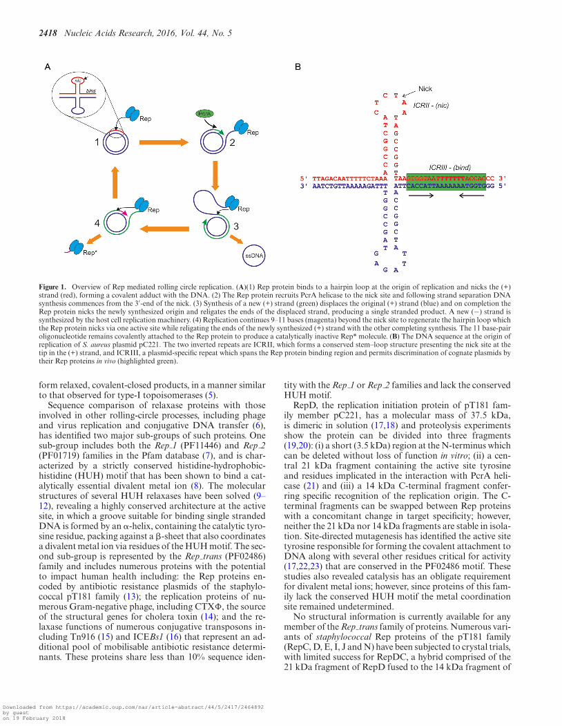
Figure 1. Overview of Rep mediated rolling circle replication. (A)(1) Rep protein binds to a hairpin loop at the origin of replication and nicks the (+)strand (red), forming a covalent adduct with the DNA. (2) The Rep protein recruits PcrA helicase to the nick site and following strand separation DNAsynthesis commences from the 3′-end of the nick. (3) Synthesis of a new (+) strand (green) displaces the original (+) strand (blue) and on completion theRep protein nicks the newly synthesized origin and religates the ends of the displaced strand, producing a single stranded product. A new (−) strand issynthesized by the host cell replication machinery. (4) Replication continues 9–11 bases (magenta) beyond the nick site to regenerate the hairpin loop whichthe Rep protein nicks via one active site while religating the ends of the newly synthesized (+) strand with the other completing synthesis. The 11 base-pairoligonucleotide remains covalently attached to the Rep protein to produce a catalytically inactive Rep* molecule. (B) The DNA sequence at the origin ofreplication of S. aureus plasmid pC221. The two inverted repeats are ICRII, which forms a conserved stem–loop structure presenting the nick site at thetip in the (+) strand, and ICRIII, a plasmid-specific repeat which spans the Rep protein binding region and permits discrimination of cognate plasmids bytheir Rep proteins in vivo (highlighted green).
form relaxed, covalent-closed products, in a manner similarto that observed for type-I topoisomerases (5).
Sequence comparison of relaxase proteins with thoseinvolved in other rolling-circle processes, including phageand virus replication and conjugative DNA transfer (6),has identified two major sub-groups of such proteins. Onesub-group includes both the Rep 1 (PF11446) and Rep 2(PF01719) families in the Pfam database (7), and is char-acterized by a strictly conserved histidine-hydrophobic-histidine (HUH) motif that has been shown to bind a cat-alytically essential divalent metal ion (8). The molecularstructures of several HUH relaxases have been solved (9–12), revealing a highly conserved architecture at the activesite, in which a groove suitable for binding single strandedDNA is formed by an �-helix, containing the catalytic tyro-sine residue, packing against a �-sheet that also coordinatesa divalent metal ion via residues of the HUH motif. The sec-ond sub-group is represented by the Rep trans (PF02486)family and includes numerous proteins with the potentialto impact human health including: the Rep proteins en-coded by antibiotic resistance plasmids of the staphylo-coccal pT181 family (13); the replication proteins of nu-merous Gram-negative phage, including CTX�, the sourceof the structural genes for cholera toxin (14); and the re-laxase functions of numerous conjugative transposons in-cluding Tn916 (15) and ICEBs1 (16) that represent an ad-ditional pool of mobilisable antibiotic resistance determi-nants. These proteins share less than 10% sequence iden-
tity with the Rep 1 or Rep 2 families and lack the conservedHUH motif.
RepD, the replication initiation protein of pT181 fam-ily member pC221, has a molecular mass of 37.5 kDa,is dimeric in solution (17,18) and proteolysis experimentsshow the protein can be divided into three fragments(19,20): (i) a short (3.5 kDa) region at the N-terminus whichcan be deleted without loss of function in vitro; (ii) a cen-tral 21 kDa fragment containing the active site tyrosineand residues implicated in the interaction with PcrA heli-case (21) and (iii) a 14 kDa C-terminal fragment confer-ring specific recognition of the replication origin. The C-terminal fragments can be swapped between Rep proteinswith a concomitant change in target specificity; however,neither the 21 kDa nor 14 kDa fragments are stable in isola-tion. Site-directed mutagenesis has identified the active sitetyrosine responsible for forming the covalent attachment toDNA along with several other residues critical for activity(17,22,23) that are conserved in the PF02486 motif. Thesestudies also revealed catalysis has an obligate requirementfor divalent metal ions; however, since proteins of this fam-ily lack the conserved HUH motif the metal coordinationsite remained undetermined.
No structural information is currently available for anymember of the Rep trans family of proteins. Numerous vari-ants of staphylococcal Rep proteins of the pT181 family(RepC, D, E, I, J and N) have been subjected to crystal trials,with limited success for RepDC, a hybrid comprised of the21 kDa fragment of RepD fused to the 14 kDa fragment of
Downloaded from https://academic.oup.com/nar/article-abstract/44/5/2417/2464892by gueston 19 February 2018
Nucleic Acids Research, 2016, Vol. 44, No. 5 2419
RepC (24). In this report we describe the structure determi-nation of the core domain of replication initiator protein ofcryptic plasmid pSTK1 (25,26) from Geobacillus stearother-mophilus (RepSTK1 residues 1–269 (27), hereafter referredto as RepSTK1). This was then used to aid the structuresolution of two staphylococcal Rep variants, RepDE andRepDN (containing the 21 kDa fragment of RepD fused tothe 14 kDa fragment of RepE or RepN, respectively). TheGeobacillus and staphylococcal proteins share only 13% se-quence identity, yet display remarkable structural conserva-tion. The architecture of the active site and the location ofthe metal ion required for catalysis are revealed. Addition-ally, the structures provide clues to how the proteins bind tothe origin of replication and suggest a mechanism for PcrArecruitment. They also provide opportunities for the devel-opment of novel, potentially broad spectrum antimicrobialagents, since inhibition of such Rep proteins would preventthe replication of numerous plasmids and bacteriophagesor the spread of related conjugative transposons containingdiverse resistance or pathogenic functions.
MATERIALS AND METHODS
Cloning, expression purification of Rep proteins
RepSTK1 was cloned as described previously (27). A fulldescription of the generation of chimeric RepDE andRepDN constructs is provided in the supplementary ma-terial, briefly the 21 kDa N-terminal domain of RepDwas PCR amplified from plasmid pC221 (using primersF35+/S−) and the 14 kDa C-terminal domains of RepEand RepN were amplified from plasmids pS194 and pCW7respectively using primer pairs (ES+/EE−) and (S+/Ter−).All primers sequences are listed in supplementary mate-rial Supplementary Table S1. The N-terminal domain fromRepD was fused with the C-terminal domain of RepE orRepN via SacI sites introduced during the PCR and the re-sulting chimeric constructs ultimately cloned into pET11a-derived expression vectors via NdeI and BamHI restrictionsites.
Following expression, RepDE and RepDN were purifiedas previously described (19) and both variants were shownto bind and nick DNA with similar activity to RepD (Sup-plementary Figure S1). RepSTK1 was expressed and pu-rified using a variation of this method (27). Prior to crys-tallization RepDE and RepDN were dialyzed against 50mM Tris–HCl pH 7.5, 700 mM KCl, 10% (v/v) ethane-diol, and RepSTK1 was dialyzed against 50 mM Tris–HClpH 7.5, 700 mM KCl. All proteins were concentrated to5 mg ml−1 using Amicon Ultra centrifugal concentratorswith a 10 kDa MWCO membrane. All protein concentra-tions were assessed by absorbance at 280 nm and extinctioncoefficients calculated from their primary sequences.
Crystallisation and X-ray Data collection
Crystals of all RepSTK1 were grown as described previ-ously (27). Crystals of RepDE and RepDN were obtainedby mixing 500 nl of protein solution with 500 nl 0.5 M am-monium citrate pH 8.5, 15% PEG 8000 or 0.1 M sodiumcitrate pH 5.5, 2.5 M 1,6-hexane-diol, respectively using an
Oryx 6 crystallization robot (Douglas Instruments, Hunger-ford, UK). Seleno-methionine labelled RepDN required alower pH of 5.2 for crystal growth. All crystallization ex-periments were incubated at 294 K and crystals grew within24 h (RepSTK1) or 5 days (RepDE and RepDN). Crystalswere cryoprotected by the addition of 25% (v/v) glycerol(RepSTK1), 25% (v/v) ethanediol (RepDE) or 20% (v/v)ethanediol (RepDN) to the crystallization solution followedby flash-cooling in liquid nitrogen. X-ray diffraction datawere collected using multiple beam-lines at Diamond LightSource, ESRF and Daresbury laboratory (Table 1). All datacollections were performed at a temperature of 100 K andall data reduction was performed using MOSFLM (28) andeither SCALA (29) or AIMLESS (30).
Structure solution and refinement
Initial phase estimates for RepSTK1 were obtained from aMIRAS phasing experiment with crystals soaked in 1 mMHgCl2 or K2PtCl4 for 10–20 min prior to flash-cooling.Diffraction data were collected at the k-edge of each heavyatom and their positions identified, refined and phases cal-culated using the autoSHARP pipeline (31). The resultantelectron density maps were of sufficient quality to allow au-tomated chain tracing using BUCCANEER (32) to producean initial model, which was refined using REFMAC5 (33).Model phases were combined with experimental phases toimprove the quality of the electron density map. Furtherrounds of manual rebuilding in Coot (34) and refinementwith REFMAC5 completed the model. To locate the metalion in the active site crystals were soaked in reservoir so-lution containing 10 mM MnCl2 prior to cryo-cooling anddiffraction data collected at the Mn K-edge. Anomalous dif-ference maps were calculated by combining model phaseswith manganese anomalous differences using FFT (35).
Attempts to generate phase estimates for RepDE orRepDN by molecular replacement using RepSTK1 as asearch model were unsuccessful, however, SAD phasingwas possible using selenomethionine labelled RepDN. Thetwinned, selenomethionine-labelled data were processed inthe higher apparent symmetry space group I4132, result-ing from a combination of the true space-group and thetwinning operator. Selenium atoms were located and phasescalculated using autoSHARP, producing electron densitymaps in which some secondary structural motifs were vis-ible. Ideal alpha-helices and beta-sheets were manuallydocked into electron density using Coot to produce an ini-tial model. Fragments of the RepSTK1 structure were su-perposed onto the partial model of RepDN and used as aguide to aid further building of the RepDN structure. Itera-tive rounds of refinement (REFMAC), phase combinationand manual rebuilding, using RepSTK1 as a guide, werecontinued until no further electron density could be inter-preted, to produce a model for much of the catalytic domainof RepDN.
The model of the catalytic domain of RepDN was usedas a search model to generate phase estimates for RepDEby molecular replacement using Phaser (36). The resultingelectron density maps showed clear density for the miss-ing regions of the catalytic domain, but also additionallyfor the DNA binding domain. Automatic chain tracing
Downloaded from https://academic.oup.com/nar/article-abstract/44/5/2417/2464892by gueston 19 February 2018
2420 Nucleic Acids Research, 2016, Vol. 44, No. 5
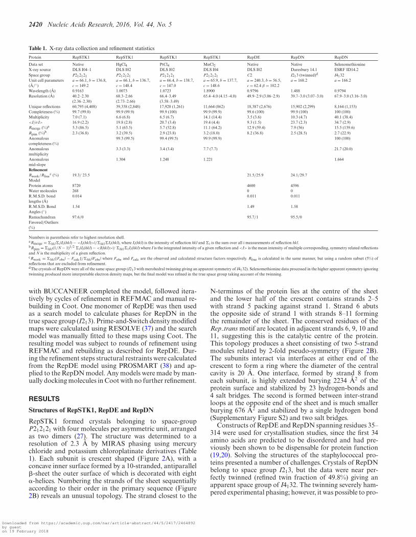
Table 1. X-ray data collection and refinement statistics
Protein RepSTK1 RepSTK1 RepSTK1 RepSTK1 RepDE RepDN RepDN
Data set Native HgCl4 PtCl4 MnCl2 Native Native SelenomethionineX-ray source DLS I04–1 DLS I02 DLS I02 DLS I04 DLS I02 Daresbury 14.1 ESRF ID14.2Space group P212121 P212121 P212121 P212121 C2 I213 (twinned)d I4132Unit cell parameters(A/◦)
a = 66.1, b = 136.8,c = 149.2
a = 66.1, b = 136.7,c = 148.4
a = 66.4, b = 138.7,c = 147.0
a = 65.9, b = 137.7,c = 148.6
a = 240.3, b = 56.5,c = 62.4 β = 102.2
a = 168.2 a = 166.2
Wavelength (A) 0.9163 1.0073 1.0723 1.8900 0.9796 1.488 0.9794Resolution (A) 40.2–2.30
(2.36–2.30)68.3–2.66(2.73–2.66)
66.4–3.49(3.58–3.49)
65.4–4.0 (4.15 -4.0) 49.9–2.9 (3.06–2.9) 39.7–3.0 (3.07–3.0) 67.9–3.0 (3.16–3.0)
Unique reflections 60,795 (4,408) 39,338 (2,848) 17,928 (1,261) 11,664 (862) 18,387 (2,676) 15,902 (2,299) 8,164 (1,153)Completeness (%) 99.7 (99.0) 99.9 (99.9) 99.9 (100) 99.9 (99.9) 99.6 (100) 99.9 (100) 100 (100)Multiplicity 7.0 (7.1) 6.6 (6.8) 6.5 (6.7) 14.1 (14.4) 3.5 (3.6) 10.3 (4.7) 40.1 (38.4)<I/σ I> 16.9 (2.2) 19.8 (2.8) 20.7 (3.4) 19.4 (4.4) 9.3 (1.5) 23.7 (2.3) 34.7 (2.9)Rmerge (%)a 5.3 (86.5) 5.1 (63.5) 5.7 (52.8) 11.1 (64.2) 12.9 (59.4) 7.9 (56) 15.5 (139.6)Rpim (%)b 2.3 (36.8) 3.2 (39.5) 2.9 (23.8) 3.2 (18.0) 8.2 (36.8) 2.5 (28.5) 2.7 (22.9)Anomalouscompleteness (%)
99.3 (99.5) 99.4 (99.5) 99.9 (99.9) 100 (100)
Anomalousmultiplicity
3.3 (3.3) 3.4 (3.4) 7.7 (7.7) 21.7 (20.0)
Anomalousmid-slope
1.304 1.248 1.221 1.664
RefinementRwork/Rfree
c (%) 19.3/ 23.5 21.5/25.9 24.1/29.7ModelProtein atoms 8720 4600 4596Water molecules 268 0 0R.M.S.D. bondlengths (A)
0.014 0.011 0.011
R.M.S.D. BondAngles (◦)
1.54 1.49 1.58
RamachandranFavored/Outliers(%)
97.6/0 95.7/1 95.5/0
Numbers in parenthesis refer to highest resolution shell.aRmerge = �hkl�i |Ii(hkl) − <Ii(hkl)>|/�hkl�Ii(hkl), where Ii(hkl) is the intensity of reflection hkl and �i is the sum over all i measurements of reflection hkl.bRpim = �hkl(1/N − 1)1/2 �i |Ii(hkl) − <I(hkl)>|/ �hkl�iIi(hkl) where I is the integrated intensity of a given reflection and <I> is the mean intensity of multiple corresponding, symmetry related reflectionsand N is the multiplicity of a given reflection.cRwork = �hkl‖Fobs | − Fcalc‖/�hkl |Fobs | where Fobs and Fcalc are the observed and calculated structure factors respectively. Rfree is calculated in the same manner, but using a random subset (5%) ofreflections that are excluded from refinement.dThe crystals of RepDN were all of the same space group (I213 with merohedral twinning giving an apparent symmetry of I4132). Selenomethionine data processed in the higher apparent symmetry ignoringtwinning produced more interpretable electron density maps, but the final model was refined in the true space group taking account of the twinning.
with BUCCANEER completed the model, followed itera-tively by cycles of refinement in REFMAC and manual re-building in Coot. One monomer of RepDE was then usedas a search model to calculate phases for RepDN in thetrue space group (I213). Prime-and-Switch density modifiedmaps were calculated using RESOLVE (37) and the searchmodel was manually fitted to these maps using Coot. Theresulting model was subject to rounds of refinement usingREFMAC and rebuilding as described for RepDE. Dur-ing the refinement steps structural restraints were calculatedfrom the RepDE model using PROSMART (38) and ap-plied to the RepDN model. Any models were made by man-ually docking molecules in Coot with no further refinement.
RESULTS
Structures of RepSTK1, RepDE and RepDN
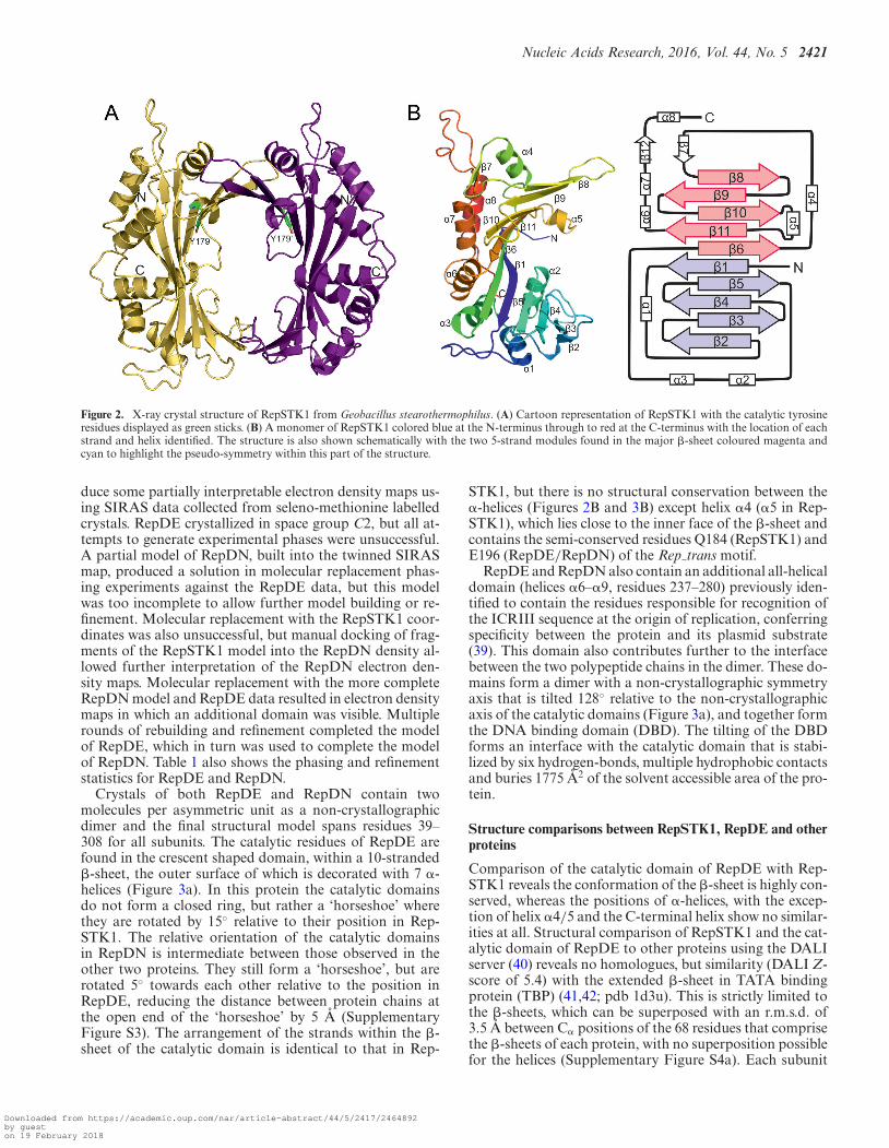
RepSTK1 formed crystals belonging to space-groupP212121 with four molecules per asymmetric unit, arrangedas two dimers (27). The structure was determined to aresolution of 2.3 A by MIRAS phasing using mercurychloride and potassium chloroplatinate derivatives (Table1). Each subunit is crescent shaped (Figure 2A), with aconcave inner surface formed by a 10-stranded, antiparallel�-sheet the outer surface of which is decorated with eight�-helices. Numbering the strands of the sheet sequentiallyaccording to their order in the primary sequence (Figure2B) reveals an unusual topology. The strand closest to the
N-terminus of the protein lies at the centre of the sheetand the lower half of the crescent contains strands 2–5with strand 5 packing against strand 1. Strand 6 abutsthe opposite side of strand 1 with strands 8–11 formingthe remainder of the sheet. The conserved residues of theRep trans motif are located in adjacent strands 6, 9, 10 and11, suggesting this is the catalytic centre of the protein.This topology produces a sheet consisting of two 5-strandmodules related by 2-fold pseudo-symmetry (Figure 2B).The subunits interact via interfaces at either end of thecrescent to form a ring where the diameter of the centralcavity is 20 A. One interface, formed by strand 8 fromeach subunit, is highly extended burying 2234 A2 of theprotein surface and stabilized by 23 hydrogen-bonds and4 salt bridges. The second is formed between inter-strandloops at the opposite end of the sheet and is much smallerburying 676 A2 and stabilized by a single hydrogen bond(Supplementary Figure S2) and two salt bridges.
Constructs of RepDE and RepDN spanning residues 35–314 were used for crystallisation studies, since the first 34amino acids are predicted to be disordered and had pre-viously been shown to be dispensable for protein function(19,20). Solving the structures of the staphylococcal pro-teins presented a number of challenges. Crystals of RepDNbelong to space group I213, but the data were near per-fectly twinned (refined twin fraction of 49.8%) giving anapparent space group of I4132. The twinning severely ham-pered experimental phasing; however, it was possible to pro-
Downloaded from https://academic.oup.com/nar/article-abstract/44/5/2417/2464892by gueston 19 February 2018
Nucleic Acids Research, 2016, Vol. 44, No. 5 2421
Figure 2. X-ray crystal structure of RepSTK1 from Geobacillus stearothermophilus. (A) Cartoon representation of RepSTK1 with the catalytic tyrosineresidues displayed as green sticks. (B) A monomer of RepSTK1 colored blue at the N-terminus through to red at the C-terminus with the location of eachstrand and helix identified. The structure is also shown schematically with the two 5-strand modules found in the major �-sheet coloured magenta andcyan to highlight the pseudo-symmetry within this part of the structure.
duce some partially interpretable electron density maps us-ing SIRAS data collected from seleno-methionine labelledcrystals. RepDE crystallized in space group C2, but all at-tempts to generate experimental phases were unsuccessful.A partial model of RepDN, built into the twinned SIRASmap, produced a solution in molecular replacement phas-ing experiments against the RepDE data, but this modelwas too incomplete to allow further model building or re-finement. Molecular replacement with the RepSTK1 coor-dinates was also unsuccessful, but manual docking of frag-ments of the RepSTK1 model into the RepDN density al-lowed further interpretation of the RepDN electron den-sity maps. Molecular replacement with the more completeRepDN model and RepDE data resulted in electron densitymaps in which an additional domain was visible. Multiplerounds of rebuilding and refinement completed the modelof RepDE, which in turn was used to complete the modelof RepDN. Table 1 also shows the phasing and refinementstatistics for RepDE and RepDN.
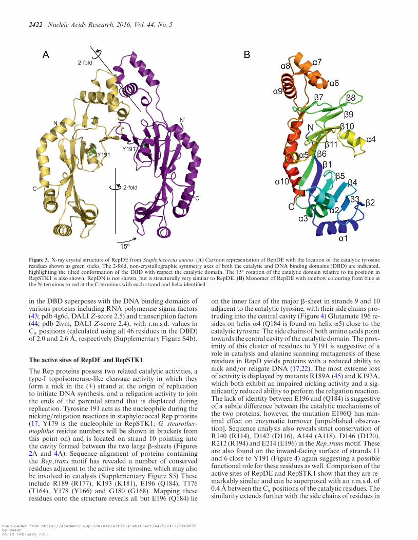
Crystals of both RepDE and RepDN contain twomolecules per asymmetric unit as a non-crystallographicdimer and the final structural model spans residues 39–308 for all subunits. The catalytic residues of RepDE arefound in the crescent shaped domain, within a 10-stranded�-sheet, the outer surface of which is decorated with 7 �-helices (Figure 3a). In this protein the catalytic domainsdo not form a closed ring, but rather a ‘horseshoe’ wherethey are rotated by 15◦ relative to their position in Rep-STK1. The relative orientation of the catalytic domainsin RepDN is intermediate between those observed in theother two proteins. They still form a ‘horseshoe’, but arerotated 5◦ towards each other relative to the position inRepDE, reducing the distance between protein chains atthe open end of the ‘horseshoe’ by 5 A (SupplementaryFigure S3). The arrangement of the strands within the �-sheet of the catalytic domain is identical to that in Rep-
STK1, but there is no structural conservation between the�-helices (Figures 2B and 3B) except helix �4 (�5 in Rep-STK1), which lies close to the inner face of the �-sheet andcontains the semi-conserved residues Q184 (RepSTK1) andE196 (RepDE/RepDN) of the Rep trans motif.
RepDE and RepDN also contain an additional all-helicaldomain (helices �6–�9, residues 237–280) previously iden-tified to contain the residues responsible for recognition ofthe ICRIII sequence at the origin of replication, conferringspecificity between the protein and its plasmid substrate(39). This domain also contributes further to the interfacebetween the two polypeptide chains in the dimer. These do-mains form a dimer with a non-crystallographic symmetryaxis that is tilted 128◦ relative to the non-crystallographicaxis of the catalytic domains (Figure 3a), and together formthe DNA binding domain (DBD). The tilting of the DBDforms an interface with the catalytic domain that is stabi-lized by six hydrogen-bonds, multiple hydrophobic contactsand buries 1775 A2 of the solvent accessible area of the pro-tein.
Structure comparisons between RepSTK1, RepDE and otherproteins
Comparison of the catalytic domain of RepDE with Rep-STK1 reveals the conformation of the �-sheet is highly con-served, whereas the positions of �-helices, with the excep-tion of helix �4/5 and the C-terminal helix show no similar-ities at all. Structural comparison of RepSTK1 and the cat-alytic domain of RepDE to other proteins using the DALIserver (40) reveals no homologues, but similarity (DALI Z-score of 5.4) with the extended �-sheet in TATA bindingprotein (TBP) (41,42; pdb 1d3u). This is strictly limited tothe �-sheets, which can be superposed with an r.m.s.d. of3.5 A between C� positions of the 68 residues that comprisethe �-sheets of each protein, with no superposition possiblefor the helices (Supplementary Figure S4a). Each subunit
Downloaded from https://academic.oup.com/nar/article-abstract/44/5/2417/2464892by gueston 19 February 2018
2422 Nucleic Acids Research, 2016, Vol. 44, No. 5
Figure 3. X-ray crystal structure of RepDE from Staphylococcus aureus. (A) Cartoon representation of RepDE with the location of the catalytic tyrosineresidues shown as green sticks. The 2-fold, non-crystallographic symmetry axes of both the catalytic and DNA binding domains (DBD) are indicated,highlighting the tilted conformation of the DBD with respect the catalytic domain. The 15◦ rotation of the catalytic domain relative to its position inRepSTK1 is also shown. RepDN is not shown, but is structurally very similar to RepDE. (B) Monomer of RepDE with rainbow colouring from blue atthe N-terminus to red at the C-terminus with each strand and helix identified.
in the DBD superposes with the DNA binding domains ofvarious proteins including RNA polymerase sigma factors(43; pdb 4g6d, DALI Z-score 2.5) and transcription factors(44; pdb 2ivm, DALI Z-score 2.4), with r.m.s.d. values inC� positions (calculated using all 46 residues in the DBD)of 2.0 and 2.6 A, respectively (Supplementary Figure S4b).
The active sites of RepDE and RepSTK1
The Rep proteins possess two related catalytic activities, atype-I topoisomerase-like cleavage activity in which theyform a nick in the (+) strand at the origin of replicationto initiate DNA synthesis, and a religation activity to jointhe ends of the parental strand that is displaced duringreplication. Tyrosine 191 acts as the nucleophile during thenicking/religation reactions in staphylococcal Rep proteins(17, Y179 is the nucleophile in RepSTK1; G. stearother-mophilus residue numbers will be shown in brackets fromthis point on) and is located on strand 10 pointing intothe cavity formed between the two large �-sheets (Figures2A and 4A). Sequence alignment of proteins containingthe Rep trans motif has revealed a number of conservedresidues adjacent to the active site tyrosine, which may alsobe involved in catalysis (Supplementary Figure S5) Theseinclude R189 (R177), K193 (K181), E196 (Q184), T176(T164), Y178 (Y166) and G180 (G168). Mapping theseresidues onto the structure reveals all but E196 (Q184) lie
on the inner face of the major �-sheet in strands 9 and 10adjacent to the catalytic tyrosine, with their side chains pro-truding into the central cavity (Figure 4) Glutamate 196 re-sides on helix �4 (Q184 is found on helix �5) close to thecatalytic tyrosine. The side chains of both amino acids pointtowards the central cavity of the catalytic domain. The prox-imity of this cluster of residues to Y191 is suggestive of arole in catalysis and alanine scanning mutagenesis of theseresidues in RepD yields proteins with a reduced ability tonick and/or religate DNA (17,22). The most extreme lossof activity is displayed by mutants R189A (45) and K193A,which both exhibit an impaired nicking activity and a sig-nificantly reduced ability to perform the religation reaction.The lack of identity between E196 and (Q184) is suggestiveof a subtle difference between the catalytic mechanisms ofthe two proteins; however, the mutation E196Q has min-imal effect on enzymatic turnover [unpublished observa-tion]. Sequence analysis also reveals strict conservation ofR140 (R114), D142 (D116), A144 (A118), D146 (D120),R212 (R194) and E214 (E196) in the Rep trans motif. Theseare also found on the inward-facing surface of strands 11and 6 close to Y191 (Figure 4) again suggesting a possiblefunctional role for these residues as well. Comparison of theactive sites of RepDE and RepSTK1 show that they are re-markably similar and can be superposed with an r.m.s.d. of0.4 A between the C� positions of the catalytic residues. Thesimilarity extends further with the side chains of residues in
Downloaded from https://academic.oup.com/nar/article-abstract/44/5/2417/2464892by gueston 19 February 2018
Nucleic Acids Research, 2016, Vol. 44, No. 5 2423
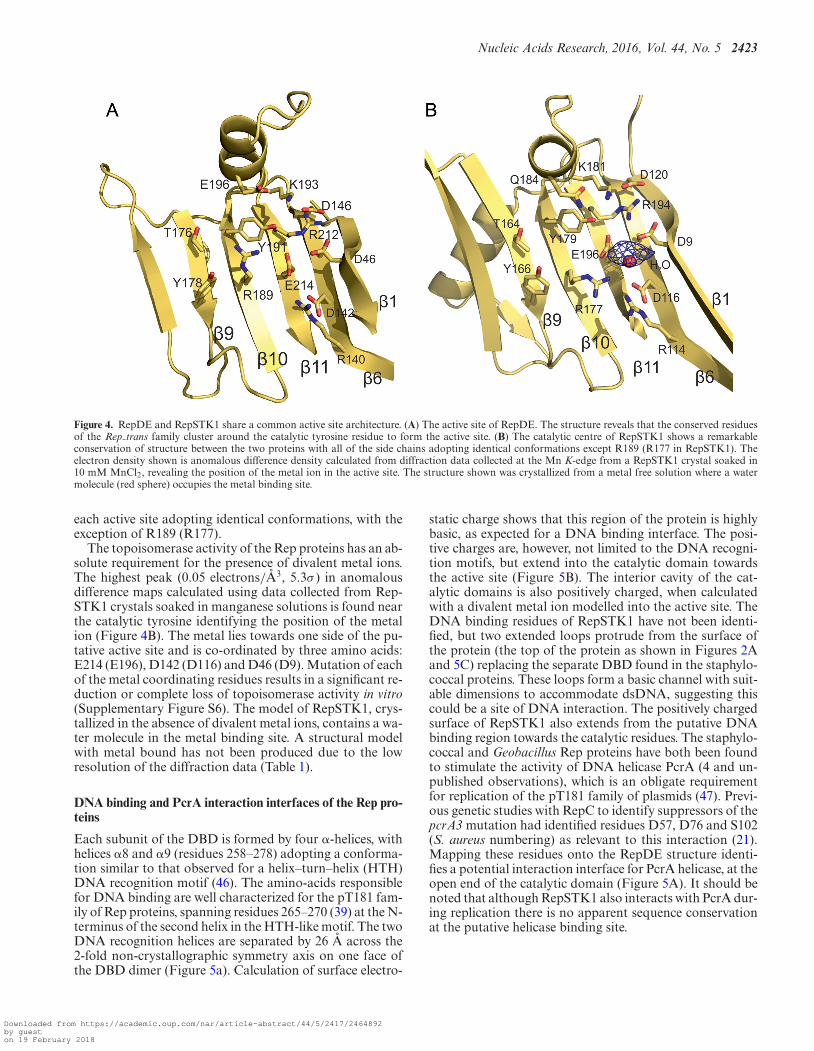
Figure 4. RepDE and RepSTK1 share a common active site architecture. (A) The active site of RepDE. The structure reveals that the conserved residuesof the Rep trans family cluster around the catalytic tyrosine residue to form the active site. (B) The catalytic centre of RepSTK1 shows a remarkableconservation of structure between the two proteins with all of the side chains adopting identical conformations except R189 (R177 in RepSTK1). Theelectron density shown is anomalous difference density calculated from diffraction data collected at the Mn K-edge from a RepSTK1 crystal soaked in10 mM MnCl2, revealing the position of the metal ion in the active site. The structure shown was crystallized from a metal free solution where a watermolecule (red sphere) occupies the metal binding site.
each active site adopting identical conformations, with theexception of R189 (R177).
The topoisomerase activity of the Rep proteins has an ab-solute requirement for the presence of divalent metal ions.The highest peak (0.05 electrons/A3, 5.3σ ) in anomalousdifference maps calculated using data collected from Rep-STK1 crystals soaked in manganese solutions is found nearthe catalytic tyrosine identifying the position of the metalion (Figure 4B). The metal lies towards one side of the pu-tative active site and is co-ordinated by three amino acids:E214 (E196), D142 (D116) and D46 (D9). Mutation of eachof the metal coordinating residues results in a significant re-duction or complete loss of topoisomerase activity in vitro(Supplementary Figure S6). The model of RepSTK1, crys-tallized in the absence of divalent metal ions, contains a wa-ter molecule in the metal binding site. A structural modelwith metal bound has not been produced due to the lowresolution of the diffraction data (Table 1).
DNA binding and PcrA interaction interfaces of the Rep pro-teins
Each subunit of the DBD is formed by four �-helices, withhelices �8 and �9 (residues 258–278) adopting a conforma-tion similar to that observed for a helix–turn–helix (HTH)DNA recognition motif (46). The amino-acids responsiblefor DNA binding are well characterized for the pT181 fam-ily of Rep proteins, spanning residues 265–270 (39) at the N-terminus of the second helix in the HTH-like motif. The twoDNA recognition helices are separated by 26 A across the2-fold non-crystallographic symmetry axis on one face ofthe DBD dimer (Figure 5a). Calculation of surface electro-
static charge shows that this region of the protein is highlybasic, as expected for a DNA binding interface. The posi-tive charges are, however, not limited to the DNA recogni-tion motifs, but extend into the catalytic domain towardsthe active site (Figure 5B). The interior cavity of the cat-alytic domains is also positively charged, when calculatedwith a divalent metal ion modelled into the active site. TheDNA binding residues of RepSTK1 have not been identi-fied, but two extended loops protrude from the surface ofthe protein (the top of the protein as shown in Figures 2Aand 5C) replacing the separate DBD found in the staphylo-coccal proteins. These loops form a basic channel with suit-able dimensions to accommodate dsDNA, suggesting thiscould be a site of DNA interaction. The positively chargedsurface of RepSTK1 also extends from the putative DNAbinding region towards the catalytic residues. The staphylo-coccal and Geobacillus Rep proteins have both been foundto stimulate the activity of DNA helicase PcrA (4 and un-published observations), which is an obligate requirementfor replication of the pT181 family of plasmids (47). Previ-ous genetic studies with RepC to identify suppressors of thepcrA3 mutation had identified residues D57, D76 and S102(S. aureus numbering) as relevant to this interaction (21).Mapping these residues onto the RepDE structure identi-fies a potential interaction interface for PcrA helicase, at theopen end of the catalytic domain (Figure 5A). It should benoted that although RepSTK1 also interacts with PcrA dur-ing replication there is no apparent sequence conservationat the putative helicase binding site.
Downloaded from https://academic.oup.com/nar/article-abstract/44/5/2417/2464892by gueston 19 February 2018

2424 Nucleic Acids Research, 2016, Vol. 44, No. 5
Figure 5. The location of DNA and PcrA binding interfaces. (A) The structure of RepDE with the DNA binding interface shown as red sticks, and thelocation of pcrA3 suppressor mutations shown as blue sticks. The catalytic tyrosine is also indicated. (B) Electrostatic surface representation of RepDEwith the location of the protease K sensitive loop indicated by an arrow. (C) Electrostatic surface representation of RepSTK1, with the two basic loopswhich take the place of the DBD indicated with arrows.
DISCUSSION
Despite low overall sequence identity, there is a high de-gree of structural conservation between the catalytic do-mains of the Rep proteins from these Geobacillus stearother-mophilus and Staphylococcus aureus plasmids. Structure-based sequence alignment reveals the conserved residuesare almost exclusively located in the �-strands contain-ing the Rep trans motifs, and it is likely the crescent-shaped catalytic domain represents the canonical fold forthe Rep trans protein family. The most strictly conservedresidues in the Phage Cri family of replication proteinsare arranged in motifs similar to those observed for theRep trans family (Supplementary Figure S5), suggesting acomparable arrangement of residues at the catalytic centrecould be achieved if members of the Phage Cri family alsoadopt a similar crescent-shaped fold. The topology of the �-sheet is uncommon, but not unique in the PDB, with TBPalso containing an extended, highly curved sheet where thestrands show the same connectivity and internal symmetry.In TBP, this symmetry is reflected in the primary sequenceand has been proposed to have arisen from a gene duplica-tion event (48), however, there is no evidence of symmetryin the primary sequence of either Rep protein.
The differing quaternary structures of the catalytic do-mains of the Rep proteins may represent functionally rel-evant conformations, since the active site lies on the in-ner surface of the �-sheet and the diameter of the centralcavity of RepSTK1 would severely hinder access of the ds-DNA substrate. Any steric hindrance to DNA access wouldbe greatly reduced by the catalytic domains swinging apartto open the ring and adopting the ‘horseshoe’ conforma-tion observed for both staphylococcal proteins. The differ-ent conformations adopted by RepDE and RepDN demon-strate the catalytic domains can move relative to one an-other to modify the size of the cavity containing the activesites. Enabling RepSTK1 to undergo such a conformational
change would require disruption of one of the interfacesstabilising the ring, with the smaller of the two interfaces(Supplementary Figure S2b) needing significantly less en-ergy to disrupt. Since Geobacillus stearothermophilus typi-cally grows at 65◦C the kinetic energy of the protein at thistemperature could be sufficient to enable ring opening, andin vitro nicking assays with RepSTK1 show significantly re-duced DNA cleavage at temperatures <65◦C. The remain-der of the discussion will focus primarily the staphylococcalRep proteins, since all the biochemical data we will discussin relation to the structures were obtained using membersof the pT181 family of replication initiation proteins.
The fold of the catalytic domain is significantly differ-ent from that observed for the HUH relaxases (9–12),which consist of an �-helix containing the catalytic tyrosineresidue packing against a �-sheet containing the metal co-ordinating histidine residues. Both of which are adjacent toa narrow groove capable of binding single stranded DNA(49). In contrast, all of the residues necessary for catalysis inthe Rep trans family of relaxases lie on the inner face of the�-sheet in the catalytic domain, with those co-ordinatingthe metal ion each on a separate strand. Despite the dif-ferences in protein architecture there are some similaritiesbetween the active sites of these distinct families. The sidechains of the catalytic tyrosine residues adopt a similar ori-entation with respect to the metal ions and are separatedfrom them by ∼5 A (Supplementary Figure S7a). Such simi-larities might be expected since relaxases from both familiescatalyze the same nicking reaction and may represent a caseof convergent evolution. The arrangement of catalytic tyro-sine and metal binding site formed from three carboxylategroups is also similar to that observed in type II topoiso-merases. The active site of topoisomerase II, however, is as-sembled by bringing together a tyrosine from the gyrase do-main of the topoisomerase A subunit with the metal ion co-ordinating residues from the TOPRIM domain in the topoi-somerase B subunit in a multi-protein complex (50) (Sup-
Downloaded from https://academic.oup.com/nar/article-abstract/44/5/2417/2464892by gueston 19 February 2018
Nucleic Acids Research, 2016, Vol. 44, No. 5 2425
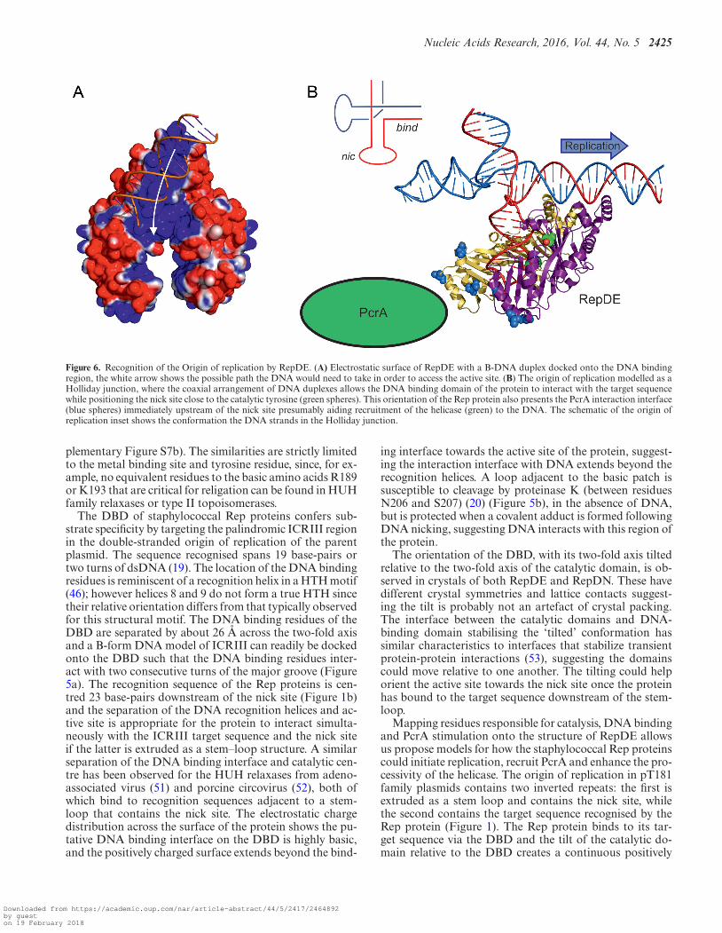
Figure 6. Recognition of the Origin of replication by RepDE. (A) Electrostatic surface of RepDE with a B-DNA duplex docked onto the DNA bindingregion, the white arrow shows the possible path the DNA would need to take in order to access the active site. (B) The origin of replication modelled as aHolliday junction, where the coaxial arrangement of DNA duplexes allows the DNA binding domain of the protein to interact with the target sequencewhile positioning the nick site close to the catalytic tyrosine (green spheres). This orientation of the Rep protein also presents the PcrA interaction interface(blue spheres) immediately upstream of the nick site presumably aiding recruitment of the helicase (green) to the DNA. The schematic of the origin ofreplication inset shows the conformation the DNA strands in the Holliday junction.
plementary Figure S7b). The similarities are strictly limitedto the metal binding site and tyrosine residue, since, for ex-ample, no equivalent residues to the basic amino acids R189or K193 that are critical for religation can be found in HUHfamily relaxases or type II topoisomerases.
The DBD of staphylococcal Rep proteins confers sub-strate specificity by targeting the palindromic ICRIII regionin the double-stranded origin of replication of the parentplasmid. The sequence recognised spans 19 base-pairs ortwo turns of dsDNA (19). The location of the DNA bindingresidues is reminiscent of a recognition helix in a HTH motif(46); however helices 8 and 9 do not form a true HTH sincetheir relative orientation differs from that typically observedfor this structural motif. The DNA binding residues of theDBD are separated by about 26 A across the two-fold axisand a B-form DNA model of ICRIII can readily be dockedonto the DBD such that the DNA binding residues inter-act with two consecutive turns of the major groove (Figure5a). The recognition sequence of the Rep proteins is cen-tred 23 base-pairs downstream of the nick site (Figure 1b)and the separation of the DNA recognition helices and ac-tive site is appropriate for the protein to interact simulta-neously with the ICRIII target sequence and the nick siteif the latter is extruded as a stem–loop structure. A similarseparation of the DNA binding interface and catalytic cen-tre has been observed for the HUH relaxases from adeno-associated virus (51) and porcine circovirus (52), both ofwhich bind to recognition sequences adjacent to a stem-loop that contains the nick site. The electrostatic chargedistribution across the surface of the protein shows the pu-tative DNA binding interface on the DBD is highly basic,and the positively charged surface extends beyond the bind-
ing interface towards the active site of the protein, suggest-ing the interaction interface with DNA extends beyond therecognition helices. A loop adjacent to the basic patch issusceptible to cleavage by proteinase K (between residuesN206 and S207) (20) (Figure 5b), in the absence of DNA,but is protected when a covalent adduct is formed followingDNA nicking, suggesting DNA interacts with this region ofthe protein.
The orientation of the DBD, with its two-fold axis tiltedrelative to the two-fold axis of the catalytic domain, is ob-served in crystals of both RepDE and RepDN. These havedifferent crystal symmetries and lattice contacts suggest-ing the tilt is probably not an artefact of crystal packing.The interface between the catalytic domains and DNA-binding domain stabilising the ‘tilted’ conformation hassimilar characteristics to interfaces that stabilize transientprotein-protein interactions (53), suggesting the domainscould move relative to one another. The tilting could helporient the active site towards the nick site once the proteinhas bound to the target sequence downstream of the stem-loop.
Mapping residues responsible for catalysis, DNA bindingand PcrA stimulation onto the structure of RepDE allowsus propose models for how the staphylococcal Rep proteinscould initiate replication, recruit PcrA and enhance the pro-cessivity of the helicase. The origin of replication in pT181family plasmids contains two inverted repeats: the first isextruded as a stem loop and contains the nick site, whilethe second contains the target sequence recognised by theRep protein (Figure 1). The Rep protein binds to its tar-get sequence via the DBD and the tilt of the catalytic do-main relative to the DBD creates a continuous positively
Downloaded from https://academic.oup.com/nar/article-abstract/44/5/2417/2464892by gueston 19 February 2018
2426 Nucleic Acids Research, 2016, Vol. 44, No. 5
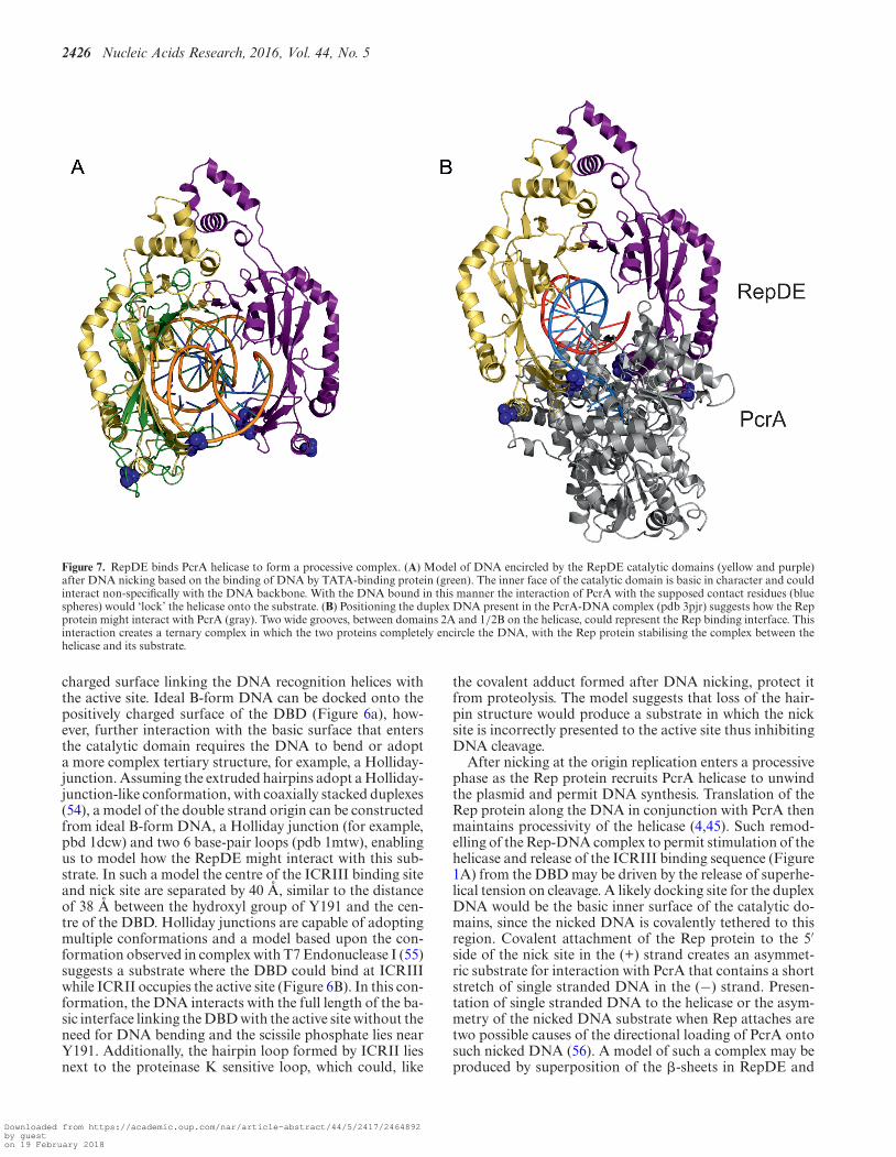
Figure 7. RepDE binds PcrA helicase to form a processive complex. (A) Model of DNA encircled by the RepDE catalytic domains (yellow and purple)after DNA nicking based on the binding of DNA by TATA-binding protein (green). The inner face of the catalytic domain is basic in character and couldinteract non-specifically with the DNA backbone. With the DNA bound in this manner the interaction of PcrA with the supposed contact residues (bluespheres) would ‘lock’ the helicase onto the substrate. (B) Positioning the duplex DNA present in the PcrA-DNA complex (pdb 3pjr) suggests how the Repprotein might interact with PcrA (gray). Two wide grooves, between domains 2A and 1/2B on the helicase, could represent the Rep binding interface. Thisinteraction creates a ternary complex in which the two proteins completely encircle the DNA, with the Rep protein stabilising the complex between thehelicase and its substrate.
charged surface linking the DNA recognition helices withthe active site. Ideal B-form DNA can be docked onto thepositively charged surface of the DBD (Figure 6a), how-ever, further interaction with the basic surface that entersthe catalytic domain requires the DNA to bend or adopta more complex tertiary structure, for example, a Holliday-junction. Assuming the extruded hairpins adopt a Holliday-junction-like conformation, with coaxially stacked duplexes(54), a model of the double strand origin can be constructedfrom ideal B-form DNA, a Holliday junction (for example,pbd 1dcw) and two 6 base-pair loops (pdb 1mtw), enablingus to model how the RepDE might interact with this sub-strate. In such a model the centre of the ICRIII binding siteand nick site are separated by 40 A, similar to the distanceof 38 A between the hydroxyl group of Y191 and the cen-tre of the DBD. Holliday junctions are capable of adoptingmultiple conformations and a model based upon the con-formation observed in complex with T7 Endonuclease I (55)suggests a substrate where the DBD could bind at ICRIIIwhile ICRII occupies the active site (Figure 6B). In this con-formation, the DNA interacts with the full length of the ba-sic interface linking the DBD with the active site without theneed for DNA bending and the scissile phosphate lies nearY191. Additionally, the hairpin loop formed by ICRII liesnext to the proteinase K sensitive loop, which could, like
the covalent adduct formed after DNA nicking, protect itfrom proteolysis. The model suggests that loss of the hair-pin structure would produce a substrate in which the nicksite is incorrectly presented to the active site thus inhibitingDNA cleavage.
After nicking at the origin replication enters a processivephase as the Rep protein recruits PcrA helicase to unwindthe plasmid and permit DNA synthesis. Translation of theRep protein along the DNA in conjunction with PcrA thenmaintains processivity of the helicase (4,45). Such remod-elling of the Rep-DNA complex to permit stimulation of thehelicase and release of the ICRIII binding sequence (Figure1A) from the DBD may be driven by the release of superhe-lical tension on cleavage. A likely docking site for the duplexDNA would be the basic inner surface of the catalytic do-mains, since the nicked DNA is covalently tethered to thisregion. Covalent attachment of the Rep protein to the 5′side of the nick site in the (+) strand creates an asymmet-ric substrate for interaction with PcrA that contains a shortstretch of single stranded DNA in the (−) strand. Presen-tation of single stranded DNA to the helicase or the asym-metry of the nicked DNA substrate when Rep attaches aretwo possible causes of the directional loading of PcrA ontosuch nicked DNA (56). A model of such a complex may beproduced by superposition of the �-sheets in RepDE and
Downloaded from https://academic.oup.com/nar/article-abstract/44/5/2417/2464892by gueston 19 February 2018

Nucleic Acids Research, 2016, Vol. 44, No. 5 2427
the TBP–DNA complex (42), where the DNA interacts withthe inner surface of an almost identical �-sheet. This su-perposition places a DNA duplex between the two catalyticsubunits of RepDE such that it would be almost completelyencircled (Figure 7a), with RepDE residues identified as im-portant to the interaction with PcrA exposed to solvent.With the DNA in this position, interaction with PcrA wouldeffectively lock the helicase onto the substrate next to the ex-posed (−) strand, enhancing the processivity of the unwind-ing reaction. The structure of PcrA in complex with DNAhas been solved (pdb 3pjr), and in this structure the DNAhas both double and single stranded regions. Assuming theduplex DNA lies in a similar position to that observed in3pjr, superposition of the duplex into the DNA in the Rep-DNA model shown in Figure 7a leads to a possible approx-imate model for the RepDE-PcrA-DNA ternary complex(Figure 7B). In this model the two basic PcrA interactioninterfaces at the open end of the RepDE catalytic horse-shoe occupy two acidic grooves on the surface of PcrA (be-tween domains 2A and 1B/2B). Opening of the ring struc-ture of RepSTK1 exposes two basic patches positioned todock into the acidic grooves on PcrA, indicating that theoverall charge distribution of the PcrA binding site is con-served between the two homologues even in the absence ofany clear sequence conservation. The positioning of eitherRep protein would not impede any conformational changesin PcrA during DNA unwinding. As well as enhancing pro-cessivity, encircling the DNA would also provide a mecha-nism for the Rep protein to monitor the displaced (+) strandfor the next encounter with the origin of replication to en-sure correct termination of DNA synthesis.
ACCESSION NUMBERS
Coordinates and structure factors have been deposited inthe protein data bank with accession codes 4cij (RepSTK1),4cwc (RepDE) and 4cwe (RepDN).
SUPPLEMENTARY DATA
Supplementary Data are available at NAR Online.
ACKNOWLEDGEMENT
We thank R. Zhang for technical support and beam-line sci-entists from Diamond Light Source beam-lines I02, I04 andI04-1, ESRF beam-line ID14.2 and Daresbury Laboratorystation 14-1 for their assistance during data collection.
FUNDING
Wellcome Trust [062973]; Medical Research Council[92141]. Funding for open access charge: Medical Re-search Council grant––Structural Studiesof Protein–DNAComplexes in recombination and repair [92141].Conflict of interest statement. None declared.
REFERENCES1. Lyon,B.R. and Skurray,R. (1987) Antimicrobial resistance of
Staphylococcus aureus: genetic basis. Microbiol. Rev., 51, 88–134.
2. Novick,R.P. (1989) Staphylococcal plasmids and their replication.Annu. Rev. Microbiol., 43, 537–565.
3. del Solar,G., Giraldo,R., Ruiz-Echevarria,M.J., Espinosa,M. andDiaz-Orejas,R. (1998) Replication and control of circular bacterialplasmids. Microbiol. Mol. Biol. Rev., 62, 434–464.
4. Slatter,A.F., Thomas,C.D. and Webb,M.R. (2009) PcrA helicasetightly couples ATP hydrolysis to unwinding double-stranded DNA,modulated by the initiator protein for plasmid replication, RepD.Biochemistry, 48, 6326–6334.
5. Koepsel,R.R., Murray,R.W., Rosenblum,W.D. and Khan,S.A. (1985)The replication initiator protein of plasmid pT181 hassequence-specific endonuclease and topoisomerase-like activities.Proc. Natl. Acad. Sci. U.S.A., 82, 6845–6849.
6. Koonin,E.V. and Ilyina,T.V. (1993) Computer-assisted dissection ofrolling circle DNA replication. BioSystems, 30, 241–268.
7. Punta,M., Coggill,P.C., Eberhardt,R.Y., Mistry,J., Tate,J.,Boursnell,C., Pang,N., Forslund,K., Ceric,G., Clements,J. et al.(2012) The Pfam protein families database. Nucleic Acids Res., 40,D290–D301.
8. Chandler,M., de la Cruz,F., Dyda,F., Hickman,A.B., Moncalian,G.and Ton-Hoang,B. (2013) Breaking and joining single-strandedDNA: the HUH endonuclease superfamily. Nat. Rev. Microbiol., 11,525–538.
9. Hickman,A.B., Ronning,D.R., Kotin,R.M. and Dyda,F. (2002)Structural unity among viral origin binding proteins: crystal structureof the nuclease domain of adeno-associated virus Rep. Mol. Cell, 10,327–337.
10. Campos-Olivas,R., Louis,J.M., Clerot,D., Gronenborn,B. andGronenborn,A.M. (2002) The structure of a replication initiatorunites diverse aspects of nucleic acid metabolism. Proc. Natl. Acad.Sci. U.S.A., 99, 10310–10315.
11. Boer,D.R., Ruiz-Maso,J.A., Lopez-Blanco,J.R., Blanco,A.G.,Vives-Llacer,M., Chacon,P., Uson,I., Gomis-Ruth,F.X.,Espinosa,M., Llorca,O. et al. (2009) Plasmid replication initiatorRepB forms a hexamer reminiscent of ring helicases and has mobilenuclease domains. EMBO J., 28, 1666–1678.
12. Datta,S., Larkin,C. and Schildbach,J.F. (2003) Structural insightsinto single-stranded DNA binding and cleavage by F factor TraI.Structure, 11, 1369–1379.
13. Projan,S.J. and Novick,R. (1988) Comparative analysis of five relatedstaphylococcal plasmids. Plasmid, 19, 203–221.
14. Waldor,M.K. and Mekalanos,J.J. (1996) Lysogenic conversion by afilamentous phage encoding cholera toxin. Science, 272, 1910–1914.
15. Rocco,J.M. and Churchward,G. (2006) The integrase of theconjugative transposon Tn916 directs strand- and sequence-specificcleavage of the origin of conjugal transfer, oriT, by the endonucleaseOrf20. J. Bacteriol., 188, 2207–2213.
16. Lee,C.A. and Grossman,A.D. (2007) Identification of the origin oftransfer (oriT) and DNA relaxase required for conjugation of theintegrative and conjugative element ICEBs1 of Bacillus subtilis. J.Bacteriol., 189, 7254–7261.
17. Thomas,C.D., Balson,D.F. and Shaw,W.V. (1990) In vitro studies ofthe initiation of staphylococcal plasmid replication. Specificity ofRepD for its origin (oriD) and characterization of the Rep-ori tyrosylester intermediate. J. Biol. Chem., 265, 5519–5530.
18. Rasooly,A., Wang,P.Z. and Novick,R.P. (1994) Replication-specificconversion of the Staphylococcus aureus pT181 initiator protein froman active homodimer to an inactive heterodimer. EMBO J., 13,5245–5251.
19. Thomas,C.D., Nikiforov,T.T., Connolly,B.A. and Shaw,W.V. (1995)Determination of sequence specificity between a plasmid replicationinitiator protein and the origin of replication. J. Mol. Biol., 254,381–391.
20. Thomas,C.D., Balson,D.F., Wigley,D.B. and Shaw,W.V. (1990)Chapter 15: Structure and function of the RepD protein of plasmidpC221. In: Novick,RP (ed), Molecular Biology of the Staphylococci.VCH, New York, pp. 211–218.
21. Iordanescu,S. (1993) Plasmid pT181-linked suppressors of theStaphylococcus aureus pcrA3 chromosomal mutation. J. Bacteriol.,175, 3916–3917.
22. Thomas,C.D., Davy,C.E., Jennings,L.J. and Papadopoulos,F. (1999)Replication of staphylococcal plasmid pC221: Molecular mechanismof initiator protein activity. Plasmid, 41, 156.
Downloaded from https://academic.oup.com/nar/article-abstract/44/5/2417/2464892by gueston 19 February 2018
2428 Nucleic Acids Research, 2016, Vol. 44, No. 5
23. Thomas,C.D., Balson,D.F. and Shaw,W.V. (1988) Identification of thetyrosine residue involved in bond formation between replicationorigin and initiator protein of plasmid pC221. Biochem. Soc. Trans.,16, 758–759.
24. Klimenko,D.E., Convery,M.A., Rowsell,S., Thomas,C.D. andPhillips,S.E. (1999) Crystallization and preliminary X-raycrystallographic studies of RepDC, a hybrid rolling-circle plasmidreplication initiator protein. Acta Crystallogr. D Biol. Crystallogr., 55,1076–1078.
25. Nakayama,N., Narumi,I., Nakamoto,S. and Kihara,H. (1993)Complete nucleotide sequence of pSTK1, a cryptic plasmid fromBacillus stearothermophilus TK015. Biotechnol. Lett., 15, 1013–1016.
26. Narumi,I., Nakayama,N., Nakamoto,S., Kimura,T., Yanagisawa,T.and Kihara,H. (1993) Construction of a new shuttle vector pSTE33and its stabilities in Bacillus stearothermophilus, Bacillus subtilis, andEscherichia coli. Biotechnol. Lett., 15, 815–820.
27. Carr,S.B., Mecia,L.B., Phillips,S.E.V. and Thomas,C.D. (2013)Identification, characterization and preliminary X-ray diffractionanalysis of the rolling-circle replication initiator protein from plasmidpSTK1. Acta Crystallogr. Sect. F Struct. Biol. Cryst. Commun., 69,1123–1126.
28. Leslie,A.G.W. and Powell,H.R. (2007) Processing diffraction datawith MOSFLM. Evolving Methods Macromol. Crystallogr., 245,41–51.
29. Evans,P. (2006) Scaling and assessment of data quality. ActaCrystallogr. D Biol. Crystallogr., 62, 72–82.
30. Evans,P.R. (2011) An introduction to data reduction: space-groupdetermination, scaling and intensity statistics. Acta Crystallogr. DBiol. Crystallogr., 67, 282–292.
31. Vonrhein,C., Blanc,E., Roversi,P. and Bricogne,G. (2007) Automatedstructure solution with autoSHARP. Methods Mol. Biol., 364,215–230.
32. Cowtan,K. (2006) The Buccaneer software for automated modelbuilding. 1. Tracing protein chains. Acta Crystallogr. D Biol.Crystallogr., 62, 1002–1011.
33. Murshudov,G.N., Skubak,P., Lebedev,A.A., Pannu,N.S.,Steiner,R.A., Nicholls,R.A., Winn,M.D., Long,F. and Vagin,A.A.(2011) REFMAC5 for the refinement of macromolecular crystalstructures. Acta Crystallogr. D Biol. Crystallogr., 67, 355–367.
34. Emsley,P., Lohkamp,B., Scott,W.G. and Cowtan,K. (2010) Featuresand development of Coot. Acta Crystallogr. D Biol. Crystallogr., 66,486–501.
35. Ten Eyck,L.F. (1973) Crystallographic fast Fourier transforms. ActaCrystallogr. A, 29, 183–191.
36. McCoy,A.J., Grosse-Kunstleve,R.W., Adams,P.D., Winn,M.D.,Storoni,L.C. and Read,R.J. (2007) Phaser crystallographic software.J. Appl. Crystallogr., 40, 658–674.
37. Terwilliger,T.C. (2000) Maximum-likelihood density modification.Acta Crystallogr. D Biol. Crystallogr., 56, 965–972.
38. Nicholls,R.A., Long,F. and Murshudov,G.N. (2012) Low-resolutionrefinement tools in REFMAC5. Acta Crystallogr. D Biol. Crystallogr.,68, 404–417.
39. Wang,P.Z., Projan,S.J., Henriquez,V. and Novick,R.P. (1992)Specificity of origin recognition by replication initiator protein inplasmids of the pT181 family is determined by a six amino acidresidue element. J. Mol. Biol., 223, 145–158.
40. Holm,L. and Rosenstrom,P. (2010) Dali server: conservationmapping in 3D. Nucleic Acids Res., 38, W545–W549.
41. Kim,Y., Geiger,J.H., Hahn,S. and Sigler,P.B. (1993) Crystal structureof a yeast TBP/TATA-box complex. Nature, 365, 512–520.
42. Littlefield,O., Korkhin,Y. and Sigler,P.B. (1999) The structural basisfor the oriented assembly of a TBP/TFB/promotor complex. Proc.Nat. Acad. Sci U.S.A., 86, 13668–13673.
43. Osmundson,J., Montero-Diez,C., Westblade,L.F., Hochschild,A. andDarst,S.A. (2012) Promoter-specific transcription inhibition inStaphylococcus aureus by a phage protein. Cell, 151, 1005–1016.
44. Shrivastava,T. and Ramachandran,R. (2007) Mechanistic insightsfrom the crystal structures of a feast/famine regulatory protein fromMycobacterium tuberculosis H37Rv. Nucleic Acids Res., 35,7324–7335.
45. Soultanas,P., Dillingham,M.S., Papadopoulos,F., Phillips,S.E.,Thomas,C.D. and Wigley,D.B. (1999) Plasmid replication initiatorprotein RepD increases the processivity of PcrA DNA helicase.Nucleic Acids Res., 27, 1421–1428.
46. Wintjens,R. and Rooman,M. (1996) Structural classification of HTHDNA-binding domains and protein-DNA interaction modes. J. Mol.Biol., 262, 294–313.
47. Iordanescu,S. and Bargonetti,J. (1989) Staphylococcus aureuschromosomal mutations that decrease efficiency of Rep utilization inreplication of pT181 and related plasmids. J. Bacteriol., 171,4501–4503.
48. Hancock,J.M. (1993) Evolution of sequence repetition and geneduplications in the TATA-binding protein TBP (TFIID). NucleicAcids Res., 21, 2823–2830.
49. Barabas,O., Ronning,D.R., Guynet,C., Hickman,A.B.,Ton-Hoang,B., Chandler,M. and Dyda,F. (2008) Mechanism ofIS200/IS605 family DNA transposases: activation andtransposon-directed target site selection. Cell, 132, 208–220.
50. Laponogov,I., Pan,X.S., Veselkov,D.A., McAuley,K.E., Fisher,L.M.and Sanderson,M.R. (2010) Structural basis of gate-DNA breakageand resealing by type II topoisomerases. PLoS One, 5, e11338.
51. Hickman,A.B., Ronning,D.R., Perez,Z.N., Kotin,R.M. and Dyda,F.(2004) The nuclease domain of adeno-associated virus repcoordinates replication initiation using two distinct DNA recognitioninterfaces. Mol. Cell, 13, 403–414.
52. Vega-Rocha,S., Byeon,I.J., Gronenborn,B., Gronenborn,A.M. andCampos-Olivas,R. (2007) Solution structure, divalent metal andDNA binding of the endonuclease domain from the replicationinitiation protein from porcine circovirus 2. J. Mol. Biol., 367,473–487.
53. Janin,J., Bahadur,R.P. and Chakrabarti,P. (2008) Protein-proteininteraction and quaternary structure. Q. Rev. Biophys., 41, 133–180.
54. Lilley,D.M. (2000) Structures of helical junctions in nucleic acids. Q.Rev. Biophys., 33, 109–159.
55. Hadden,J.M., Declais,A.-C., Carr,S.B., Lilley,D.M. and Phillips,S.E.(2007) The structural basis of Holliday junction resolution by T7endonuclease I. Nature, 449, 621–624.
56. Zhang,W., Dillingham,M.S., Thomas,C.D., Allen,S., Roberts,C.J. andSoultanas,P. (2007) Directional loading and stimulation of PcrAhelicase by the replication initiator protein RepD. J. Mol. Biol., 371,336–348.
Downloaded from https://academic.oup.com/nar/article-abstract/44/5/2417/2464892by gueston 19 February 2018