immunoglobulin chainenhancer initiation dna replication · initiation...
TRANSCRIPT
Proc. Natl. Acad. Sci. USAVol. 90, pp. 3695-3699, April 1993Immunology
Immunoglobulin heavy chain enhancer is located near or in aninitiation zone of chromosomal DNA replicationKIYOSHI ARIIZUMI*, ZHIYONG WANG, AND PHILIP W. TUCKERtDepartment of Microbiology, The University of Texas, Southwestern Medical Center, 5323 Harry Hines Boulevard, Dallas, TX 75235
Communicated by Jonathan W. Uhr, December 17, 1992
ABSTRACT In several animal viruses, enhancers havebeen implicated in both DNA replication and transcriptionalactivation. The linkage of the two mechanisms appears inti-mate, in that common DNA binding factors can be shared. Theimmunoglobulin heavy chain (Igh) intronic [heavy chain join-ing region (JH-A chain constant region (C,u)] enhancer (E,u)is required for tissue-specific transcription of Igh genes and isessential for somatic recombination of diversity (D) and Jsegments. We show here that E,u is located at or near an originof chromosomal DNA replication, which is more active in Blymphocytes than fibroblasts. EM does not fulfill two criteriademonstrated for some ceflular origins. E,u can initiate but notmaintain autonomous replicating activity in B cells. EMA isunable to impart early replication timing to a transfectedVDJ-C,L Igh locus in B cells. Instead we propose that E,M-associated ori activity contributes to tissue-specific Igh expres-sion through local effects on chromatin structure leading tosubsequent accessibility of transcription and/or recombina-tion factors for the enhancer.
Gene expression in eukaryotic cells is controlled by trans-acting factors and cis-acting transcriptional elements. Sometranscription factors have been shown to activate DNAreplication when the factor-binding sequences are located inthe vicinity of a minimal origin (1, 2). Activation of DNAreplication by transcription factors is likely to be evokedthrough a change in chromatin structure (1, 3). The initiationevents of transcription and DNA replication may be closelyrelated in that they both necessitate the transient breakdownof higher-ordered chromatin.
All detailed analyses of the relationship between transcrip-tion and DNA replication have been performed in viralsystems (for review, see ref. 4). The immunoglobulin heavychain (Igh) locus provides an excellent system to address thisissue for a cellular gene because its mechanism of expressionhas been studied extensively (for review, see ref. 5). Tran-scription of Igh genes is regulated by elements within theheavy chain variable region (VH) promoter and the Ighenhancer (EMu). Some cis-acting sites, such as the octamer,are shared between EM and promoter and play a key role inlymphoid-restricted expression (6, 7). Oct-i, a ubiquitousoctamer-binding factor, can activate adenovirus DNA repli-cation (8), suggesting that EM-associated motifs may beinvolved in more than transcriptional enhancement.S phase of the cell cycle can be divided into stages, with
actively expressed genes often replicated in early S phase andrepressed genes replicated during late S phase (for review,see ref. 9). Studies ofDNA replication timing of the Igh locusrevealed that, in nonlymphoid cells, DNA replication ini-tiates far downstream [3' to the a chain constant region (Ca)]and VH segments replicate late in S phase (10). In lymphoidcells, however, productively rearranged VH genes and moreEM-proximal C regions replicate as early as or earlier than Ca
replicates in nonlymphoid cells (11). These data suggest thatthe Igh locus has another origin(s) that could be develop-mentally regulated.
E, is active throughout B-cell differentiation, even prior tocomplete (V to DJ, where D is diversity and J is joining)assembly of immunoglobulin VH genes (12, 13). Consistentwith its JH-proximal location, E,M is essential for activatingthe locus for recombination. Transgenic studies (14) havedemonstrated that EMA can act dominantly in cis to initiate theD-to-J rearrangement in lymphoid tissues. Transfection of arecombination test gene containing EM and the metallothio-nein promoter into plasmacytoma cells showed that thepromoter, but not the enhancer, was dispensable for efficientV-J joining (15). These studies suggest that what is requiredfor rearrangement is not EM-mediated transcriptionper se butan open chromatin state.The above considerations prompted us to search for an
origin of chromosomal DNA replication in the EM region ofthe Igh locus. Our results revealed that EMu is at or within thevicinity of an initiation zone of chromosomal DNA replica-tion that is more active in B cells than in fibroblasts. Wepropose a model for a functional relationship between EM-associated DNA replication and EM-dependent transcrip-tional and recombinational activities.
MATERIALS AND METHODSSemiquantitative PCR. Primer pairs and hybridization
probes are 24 bases long and composed of 13 G-C pairs.Positions of each primer pair and probe are as follows:J-specific, 5' primer (nt 913-936), 3' primer (nt 1168-1191),and probe (nt 1017-1040); E-specific, 5' primer (nt 2553-2576), 3' primer (nt 2822-2845), and probe (nt 2710-2733);switch (S)-specific, 5' primer (nt 5501-5524), 3' primer (nt5779-5802), and probe (nt 5542-5565). These positions areaccording to our numbering system (GenBank accession no.X57331). Thirty cycles of PCR were performed (16). PCRproducts were alkaline-denatured, slot-blotted, and hybrid-ized with each oligonucleotide probe at 10°C below thetheoretical melting temperature. The radioactivity from eachhybridized slot was quantified by ion emission (Betagen,Waltham, MA).
Purification of Nascent DNA. Purification of nascent-strandDNA was performed essentially as described (17). Approx-imately 1 x 10⁸ cells were labeled with [3H]deoxycytidine (2MuCi/ml; 1 Ci = 37 GBq) and 5-bromodeoxyuridine (BrdUrd;20 MM) for 10 min. The resulting high molecular weight DNAwas size-fractionated on 5-20% (wt/vol) linear alkaline su-crose gradients. Radioactivity in a 4% volume of each frac-tion was measured by liquid scintilation. BrdUrd and 3H-
Abbreviations: Igh, immunoglobulin heavy chain; J, joining; C,constant; EA, Igh enhancer; D, diversity; V, variable; H, heavy; L,light; CIL, VH, etc., ,A chain C region, H chain V region, etc.; Fr,fraction(s); S, switch; ARS, autonomously replicating sequence.*Present address: Department ofDermatology, University ofTexas,Southwestern Medical Center, Dallas, TX 75235.tTo whom reprint requests should be addressed.
3695
The publication costs of this article were defrayed in part by page chargepayment. This article must therefore be hereby marked "advertisement"in accordance with 18 U.S.C. §1734 solely to indicate this fact.

Proc. Natl. Acad. Sci. USA 90 (1993)
labeled DNA was isolated by two rounds of immunoprecip-itation with anti-BrdUrd monoclonal antibodies. The specificactivity of the purified DNA fraction was increased by 400-to 800-fold over the initial specific activity. Size distributionof nascent DNA was determined by alkaline agarose gelelectrophoresis and Southern blot analysis with 32P-labeledchromosomal DNA. The concentration of purified nascentDNA was measured by fluorometry (18).
Transient Transfections and Dpn I Assays. Transfection oflymphoid cells and nonlymphoid cells was carried out byelectroporation (19) and by calcium phosphate methods (20),respectively. At 48 h after transfection, low molecular weightDNAs were extracted (21) and linearized by digestion withBamHI (22). Fragments were separated on an 0.8% agarosegel and hybridized with control DNA probe whose sequenceis identical in both test and control plasmids.
Semiconservative Replication Assay. At 2 days after trans-fection, cells were washed with fresh medium and labeledwith BrdUrd (12.5 ,g/ml) for 24 h. Plasmid DNA waspurified and BrdUrd-substituted and unsubstituted DNAswere resolved on native CsCl gradients (23). The DNAs wereelectrophoresed and blot-hybridized to vector DNA. Thebanding densities of light-light (LL) and heavy-light (HL)DNAs are determined by electrophoresing marker DNAs inparallel.
Replication Timing Assay. M12.4 B lymphoma cells werestably transfected with a 15-kbp VDJ-C,u construct as de-scribed (24). Cells were labeled with BrdUrd as describedabove, then stained with chromomycin (CA3, 20 ,ug/ml), andsorted on a FACStar III (Becton Dickinson) adjusted todetect nuclei excited at 457 nm at a flow of 2000 cells per sec(25). Based on DNA content, -0.5-2 x 10⁶ cells werecollected in six windows. Isolation, gradient separation, andslot hybridization of replicated and unreplicated DNA fromthe cell-cycle fractions was performed as above. A 1.2-kbBamHI fragment from plasmid pLLn2R (24) was used as aVDJ-specific probe (S107) and a 3.2-kb Sac I fragment wasused as a-globin genomic probe.
RESULTSStrategy for Mapping an Origin of Chromosomal DNA
Replication. Several general approaches have been devel-oped to map chromosomal origins (16, 17, 26-31). The PCRmethod (16, 17) was chosen in this study because the entireDNA sequence from VDJ1 to Cu is known. In principle, thismethod determines the distance of specific DNA sequencemarkers to an origin by fractionating nascent DNA chainsaccording to size and identifying the shortest nascent DNAchains (i.e., most origin-proximal sequences) by PCR ampli-fication and hybridization with 32P-labeled probes directedagainst the specific sequence markers. We chose the B-cellleukemia line BCL1 for our analysis because it is represen-tative in many respects (32) of a murine B lymphocyte andoffers the operational advantage of containing a single pro-ductively rearranged VDJ-Cu allele locus; the other appar-ently was deleted on adaption to culture (33).
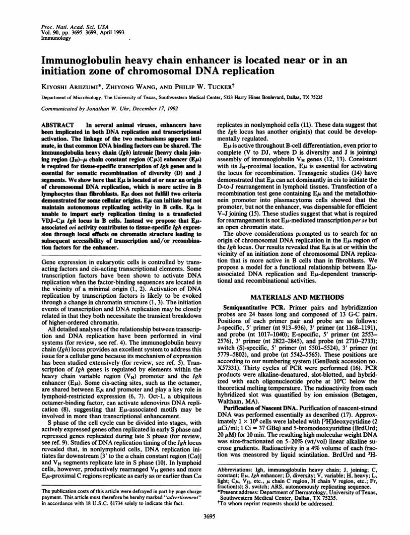
Purification of Nascent DNA. Nascent DNA from asyn-chronously growing cells was labeled with [3H]deoxycytidineand BrdUrd, size-fractionated on an alkaline sucrose gradi-ent, and purified by anti-BrdUrd antibodies (17) (Fig. 1). Toavoid contamination of Okazaki fragments and unreplicatedhigh molecular weight DNA, fractions (Fr) 1, 2, 12, and 13were discarded, and Fr 3-11 were relabeled Fr 1-9 in Fig. 1B.Fr 9 does not contain nascent DNA fragments >100 kb.Incorporation of [3H]deoxycytidine into nascent DNA de-pends on the size of the DNA in a fraction (Fig. 1A), exceptfor Fr 1, in which Okazaki fragments were presumablypresent. The intensities of hybridized bands increased pro-portionally with DNA length (Fig. 1B). This suggested that
A
0
xEa
f
m
B
15-
10o
Fr. 1 2 3 4 5 6 7 8 9 10 11 12 13Top Bottom
1 2 3 4 5 6 7 8 9kbp
-10
-5-4-3
- 1.6* w. .wt.,. -1
0.5
FIG. 1. Size separation and size determination of pulse-labeledDNA. (A) BrdUrd- and 3H-labeled DNA was size-fractionated in analkaline sucrose gradient. The gradients were collected in 13 frac-tions from the top to the bottom, measured for 3H incorporation, andplotted. (B) Fr 1, 2, 12, and 13 were discarded and the remainingfractions were renamed to Fr 1-9. The renamed fractions werepurified by immunoprecipitation with anti-BrdUrd, separated onalkaline 1.0%o agarose gel, and hybridized with 32P-labeled BCL1chromosomal DNA. Marker DNA is the 1-kb DNA ladder (BRL).The average DNA size was determined by comparison of themigration of the hybridized bands with marker DNA.
the number of nascent DNA molecules were approximatelythe same in each fraction. Since these results were verysimilar to those reported (17), we concluded that the condi-tions shown were optimal to quantitate the relative amount ofspecific sequence in a given sample and to accurately size-fractionate nascent DNA.
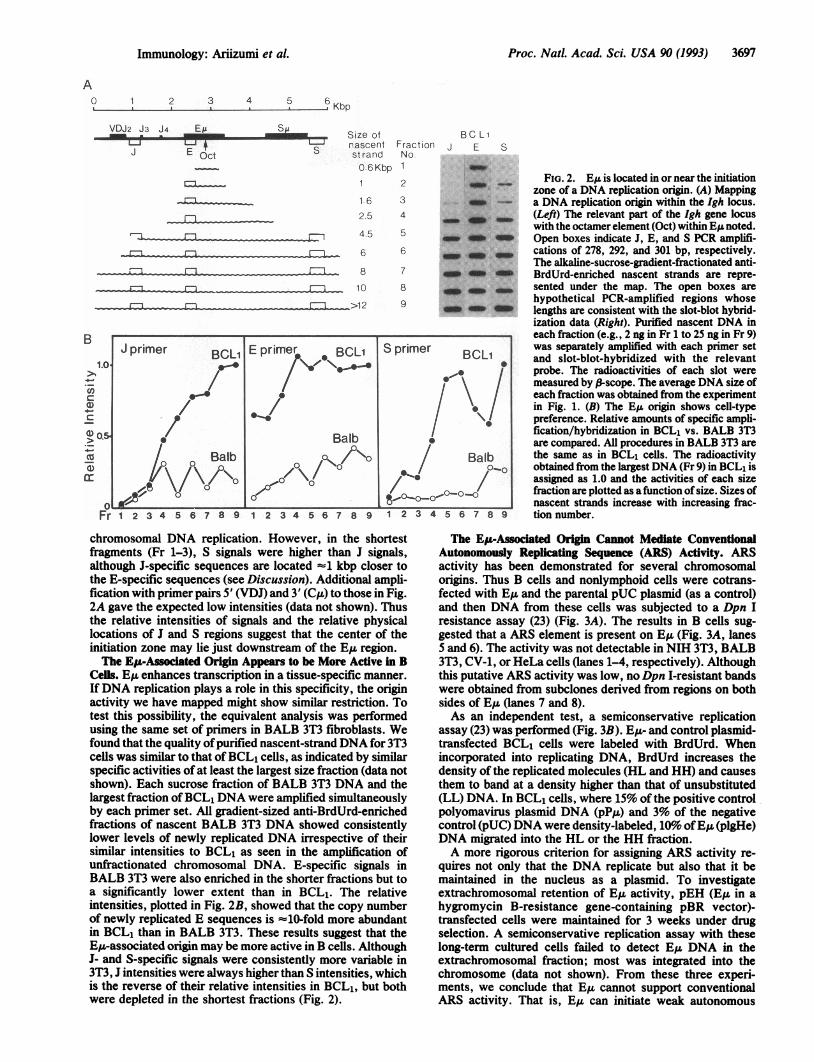
EIA Is Located Near or in the Initiation Zone of Chromoso-mal DNA Replication. To map a chromosomal origin, weestablished semiquantitative PCR conditions within the rangeof at least 0.5-32 ng (data not shown). Each primer set hada similar efficiency and was designed to avoid binding simul-taneously to Okazaki fragments. The purified nascent DNAwithin each fraction was separately amplified at the sametime with primer-pair J, located between JH2 and JH3; primer-pair E, just 5' to E,; and primer-pair S, 3' to the ,u chainswitch region (Su). Hybridization signals of E-specific se-quences were high in every size fraction of anti-BrdUrd-enriched nascent DNA, whereas signals of J- and S-specificsequences were significantly decreased in the shortest frac-tions (Fig. 2A). This indicated that the copy number ofE-specific sequences was high in the smaller-size fractionsrelative to those of S- and J-specific sequences. In addition,the presence of E-specific sequences in the shortest nascentfragments indicated that E,u is in the initiation zone of
3696 Immunology: Ariizumi et al.
Proc. Natl. Acad. Sci. USA 90 (1993) 3697
A0 1 2 3
a A4 5 6Kbp
VDJ2 J3 J4 ESu S~~~~~~~~~~~~size c-i4 I-ISnasce
J E Oct strar0 61
1 6
2.5
4.5
----- 6_1 - X1 10L._ 1 28Fl-~~~~~~T73~~~ 10
1 2 3 4 5 6 7 8 9 1 2 3 4 5 6 7 8 9
ifLntndKbp
BC LiFraction J E SNo.
2 :U _
3 _*4 -am
7 a. a~~. m5 __
6_:
7 Am Am8 __
9 a. a
FIG. 2. EA is located in or near the initiationzone of a DNA replication origin. (A) Mappinga DNA replication origin within the Igh locus.(Left) The relevant part of the Igh gene locuswith the octamer element (Oct) within EA noted.Open boxes indicate J, E, and S PCR amplifi-cations of 278, 292, and 301 bp, respectively.The alkaline-sucrose-gradient-fractionated anti-BrdUrd-enriched nascent strands are repre-sented under the map. The open boxes arehypothetical PCR-amplified regions whoselengths are consistent with the slot-blot hybrid-ization data (Right). Purified nascent DNA ineach fraction (e.g., 2 ng in Fr 1 to 25 ng in Fr 9)was separately amplified with each primer setand slot-blot-hybridized with the relevantprobe. The radioactivities of each slot weremeasured by f-scope. The average DNA size ofeach fraction was obtained from the experimentin Fig. 1. (B) The E,I origin shows cell-typepreference. Relative amounts of specific ampli-fication/hybridization in BCL1 vs. BALB 3T3are compared. All procedures in BALB 3T3 arethe same as in BCL1 cells. The radioactivityobtained from the largest DNA (Fr 9) in BCL1 isassigned as 1.0 and the activities of each sizefraction are plotted as afunction of size. Sizes ofnascent strands increase with increasing frac-tion number.
chromosomal DNA replication. However, in the shortestfragments (Fr 1-3), S signals were higher than J signals,although J-specific sequences are located -1 kbp closer tothe E-specific sequences (see Discussion). Additional ampli-fication with primer pairs 5' (VDJ) and 3' (Cu) to those in Fig.2A gave the expected low intensities (data not shown). Thusthe relative intensities of signals and the relative physicallocations of J and S regions suggest that the center of theinitiation zone may lie just downstream of the E,u region.The Ep-Associated Origin Appears to be More Active in B
Cells. E,u enhances transcription in a tissue-specific manner.IfDNA replication plays a role in this specificity, the originactivity we have mapped might show similar restriction. Totest this possibility, the equivalent analysis was performedusing the same set of primers in BALB 3T3 fibroblasts. Wefound that the quality ofpurified nascent-strand DNA for 3T3cells was similar to that ofBCL, cells, as indicated by similarspecific activities of at least the largest size fraction (data notshown). Each sucrose fraction of BALB 3T3 DNA and thelargest fraction ofBCL1 DNA were amplified simultaneouslyby each primer set. All gradient-sized anti-BrdUrd-enrichedfractions of nascent BALB 3T3 DNA showed consistentlylower levels of newly replicated DNA irrespective of theirsimilar intensities to BCL1 as seen in the amplification ofunfractionated chromosomal DNA. E-specific signals inBALB 3T3 were also enriched in the shorter fractions but toa significantly lower extent than in BCL1. The relativeintensities, plotted in Fig. 2B, showed that the copy numberof newly replicated E sequences is -10-fold more abundantin BCL1 than in BALB 3T3. These results suggest that theE,u-associated origin may be more active in B cells. AlthoughJ- and S-specific signals were consistently more variable in3T3, J intensities were always higher than S intensities, whichis the reverse of their relative intensities in BCL1, but bothwere depleted in the shortest fractions (Fig. 2).
The EIA-Associated Origin Cannot Mediate ConventionalAutonomously Replicating Sequence (ARS) Activty. ARSactivity has been demonstrated for several chromosomalorigins. Thus B cells and nonlymphoid cells were cotrans-fected with E,u and the parental pUC plasmid (as a control)and then DNA from these cells was subjected to a Dpn Iresistance assay (23) (Fig. 3A). The results in B cells sug-gested that a ARS element is present on Eu (Fig. 3A, lanes5 and 6). The activity was not detectable in NIH 3T3, BALB3T3, CV-1, or HeLa cells (lanes 1-4, respectively). Althoughthis putative ARS activity was low, no Dpn I-resistant bandswere obtained from subclones derived from regions on bothsides of E,u (lanes 7 and 8).As an independent test, a semiconservative replication
assay (23) was performed (Fig. 3B). E,u- and control plasmid-transfected BCL1 cells were labeled with BrdUrd. Whenincorporated into replicating DNA, BrdUrd increases thedensity of the replicated molecules (HL and HH) and causesthem to band at a density higher than that of unsubstituted(LL) DNA. In BCL1 cells, where 15% of the positive controlpolyomavirus plasmid DNA (pP,u) and 3% of the negativecontrol (pUC) DNA were density-labeled, 10%'o ofE,u (plgHe)DNA migrated into the HL or the HH fraction.A more rigorous criterion for assigning ARS activity re-
quires not only that the DNA replicate but also that it bemaintained in the nucleus as a plasmid. To investigateextrachromosomal retention of E,L activity, pEH (E,u in ahygromycin B-resistance gene-containing pBR vector)-transfected cells were maintained for 3 weeks under drugselection. A semiconservative replication assay with theselong-term cultured cells failed to detect E,u DNA in theextrachromosomal fraction; most was integrated into thechromosome (data not shown). From these three experi-ments, we conclude that E, cannot support conventionalARS activity. That is, E,u can initiate weak autonomous
Immunology: Ariizumi et al.

3698 Immunology: Ariizumi et al.
A Non-Bcells B cellsC')
I D-_ Cl> ')z m 01
A
(I) CC)W C)m a) a)
00 0C
test- 1l
control
1 2 3 4 5 6
HH HLB 1.420 -
1.410
1.40
pp;,..._. .
pigHe
pUC
4
7 8
LL
BE G1 [ S3A
Si k- S4
1 11 w11 1F....I11
G1 Si S2 S3 S4 G2/MC ,
HL
LL -
-0
(D;'t;-m
-11
,*ffi.
.s ....s_ Om%%:_:...... G,....3__.
_:_11_:-111.,.
_
:. ..
: " Om:. .:
:..:.., :..
_:_ _II_-111
:w.w.... -I
FIG. 3. Measurement ofARS activity ofthe E,u in B cell lines. (A)Dpn I-resistance assay. Solid and open arrowheads indicate linear-ized newly replicated test and parental vectors, respectively. pIgHe(0.9-kb E,-containing pUC vector) and pUC DNAs were cotrans-fected into NIH 3T3 (lane 1), BALB 3T3 (lane 2), CV-1 (lane 3), andHeLa (lane 4) cells by the calcium phosphate transfection method. Ahuman pre-B-cell line, SB (lane 5), and a human Epstein-Barrvirus-transformed lymphoblastoid line, Cess (lane 6), were trans-fected by electroporation. Two cotransfections into Cess cells wereperformed: pB2 (pBR322 containing the 7.6-kb Su and CA fragments)with pBR322 (lane 7) and pV,C(CAT) vector containing bases -550to 0 of the VH1 promoter region] with chloramphenicol acetyltrans-ferase (CAT) vector (lane 8). Parental vector DNA was used as aprobe for Southern blot analyses. (B) BrdUrd density labeling ofE.-containing plasmid and banding on neutral CsCl. Plasmid DNAwas purified from transfected cells and density-fractionated. Thedensity and DNA concentration in each fraction were obtained byrefractive index (R.I.) and Southern blot analysis, respectively. Thepositive control vector pP,t contains the early region ofpolyomavirusand a functional C,u gene (34) and is capable of autonomous repli-cation in murine cells. Om, oligomers; I, II, and III, forms I, II, andIII of the plasmid.
zS2 JG2/M
FIG. 4. Analysis of replication timing of transfected VDJ-CJL inM12.4 cells. (A) Flow histogram of transfectants. Relative DNAcontent, measured by CA3 flourescence, is shown in a linear scale onthe x axis and relative numbers of cells are shown on the y axis. (B)Resorting of cells, as indicated by brackets in A, revealed the purityof each fraction. (C) Replication times of VDJ-CI (S107 probe)(Upper) and mouse a-globin (Lower) determined by slot-blot hy-bridization. DNA was isolated from BrdUrd-labeled cells sorted intothe six windows indicated in A and B, fractionated on a CsClgradient, and blotted. Positions of HL and LL DNAs are indicated.
productively rearranged 15-kbp VDJ-C,u construct (24) wasstably transfected into a B-cell lymphoma, M12.4, that isdevoid of an endogenous Igh locus. Transcriptionally activetransformants (data not shown) were analyzed by the retro-active synchrony method (36).Approximately 107 cells labeled with BrdUrd were sorted
into six fractions by using equally spaced windows over therange of CA3 fluorescence from G1 (2N) to G2 + M (4N)(where N is the haploid number of chromosomes) (Fig. 4A).The purity of cells was checked by resorting (Fig. 4B). Equalquantities of fractionated DNA from each sort window wereblotted and hybridized to a probe specific for the rearrangedVDJ (S107, Fig. 4C). The S107 probe detected HL DNAacross all S-phase fractions. Rehybridization of the blot to ana-globin probe, previously shown in B cells to replicate early(37), showed that the vast majority of a-globin replication isconfined to the first half of S phase. Therefore, the sortingand gradient fractionation procedures were adequate to con-clude that, unlike the endogenous rearranged locus, replica-tion of the integrated transcriptionally active VDJ-C,g "mini-locus" was not confined to early S phase. This implies thatE,u along with the 17 kbp of colinear 5' and 3' sequences areinsufficient to control replication timing.
replication in B cells but is insufficient for maintaining aplasmid in an extrachromosomal state.
E,L and Associated VDJ-CIA Sequences Cannot ControlReplication Timing. Constant regions downstream of rear-ranged VDJ segments replicate earlier in S phase than theirunrearranged counterparts (11, 35). It was feasible that theorigin activity we measured for E,u might be activated afterDNA rearrangement. Therefore, we tested whether E,and/or associated sequences that form a ,u transcription unitare sufficient to control replication timing in a B-cell line. A
DISCUSSIONWe have demonstrated that E,u is located near or in theinitiation zone of chromosomal DNA replication, which maybe more active in B cells. This is consistent with its require-ment for transcriptional activation. A functional relationshipbetween enhancers and DNA replication has been estab-lished in several viral systems, including polyoma (38). Thepolyoma enhancer has been reported to activate DNA rep-lication to levels dependent on its distance from the origin;
Proc. Natl. Acad. Sci. USA 90 (1993)
11
n

Proc. Natl. Acad. Sci. USA 90 (1993) 3699
the enhancer effect is eliminated if the distance is too great(39). We cannot formally conclude that E,u is equivalent to achromosomal origin. We can, however, minimally infer thatE,u is located very close to the initiation zone of a DNAreplication origin. As in the above case, E,u may activateorigin activity.A relationship between replication and transcription could
account for the differing intensities of E,u flanking regions (Jand S in Fig. 2) in B cells and fibroblasts. Transcribed c-myc(40) and globin (41) genes are replicated in their transcrip-tional direction, whereas their quiescent germ-line counter-parts are not. This implies that some origins are not bidirec-tional and their polarities are affected by transcription. TheIgh locus is transcribed from J through S in B cells. Consis-tent with this, we observed higher replication signals in S thanin J, even though J sequences are closer to the apparent originregion (E). There is no transcription of the locus in BALB3T3. Accordingly, the more E-proximal J signal was higherthan the more S-distal signal. Thus relative J and S intensitiesseem to be dependent on the presence of transcription,whereas the E signals were always highest irrespective oftranscription. We suggest that E, is in the initiation zone ofDNA replication in both cell types and that replication fromit is not always driven bidirectionally.There is a controversy as to whether an ARS element can
serve as a chromosomal origin (42). Some chromosomallymapped origins function as ARS elements (29), although notall ARS elements serve as chromosomal replication origins(43). As with E,u, some yeast ARS sequences contain matrix-associated regions (MARs) (for review, see ref. 44). We foundthat a 0.9-kb fragment containing EZ and its associatedMARs expressed a weak ARS activity in a cell-type-preferential manner. It was detectable by transient but notlong-term replication assays. Perhaps E,u can convey weakARS activity in B cells but requires additional cis-actingsequences for maintenance of the plasmid in the replicatingpool. However, immediate flanking sequences, at least, thosecontained within the =15 kbp spanning E,u in a rearrangedtranscription unit, were insufficient to control replicationtiming in stable B-cell transfectants. In the extensively stud-ied CAD and ADA system in CHO cells, timing control couldonly be localized only to within 30 kbp of a probe used todetect an -260-kbp replicon (45). Therefore, if ARS and/ortiming control sequences are linked to EA&, larger constructswill be required to detect them.How might ori activity of E,u manifest an effect on immu-
noglobulin gene expression? Several examples of stimulationofreplication by transcription factors have been described (1,46-48). A mechanism often proposed is change in chromatinstructure, leading to an increased accessibility ofthe origin toreplication proteins. That EA contains a VDJ recombinationenhancing activity (14, 15) is consistent with its pattern oftranscriptional enhancement in B and T cells. However, inmost (15, 49, 50) but not all (51) experiments employingrearrangement substrates, the rate of transcription is notcorrelated with the frequency of recombination. CpG meth-ylation was implicated in VDJ accessibility, but only if thetest plasmids were replicating (52). A direct role for tran-scription seemed unlikely because there is little or no geneexpression from fully CpG methylated plasmids (52, 53). Wesuggest a model in which E,u-associated replication leads toa nuclease-sensitive VDJ chromatin structure (including tar-geted demethylation), which then allows recombination tooccur.
We thank B. Garrard and B. Gerard for helpful discussions, J.Kettman for help with the flow cytometry analysis, and H. Crawford,U. Das, and M. Gardner-Attah for help in preparing this manuscript.This work was supported by National Institutes of Health GrantA118016.
1. Cheng, L. & Kelly, T. J. (1989) Cell 59, 541-559.2. Yang, L., Li, R., Mohr, I. J., Clark, R. & Botchan, M. R. (1991) Nature
(London) 353, 628-632.3. Kelly, T. J. (1988) J. Biol. Chem. 263, 17889-17892.4. Villarreal, L. P. (1991) Microbiol. Rev. 55, 512-542.5. Staut, L. M. & Lenardo, M. J. (1991) Annu. Rev. Immunol. 9, 373-398.6. Lenardo, M., Pierce, J. W. & Baltimore, D. (1987) Science 236, 1573-
1577.7. Jenuwein, T. & Grosschedl, R. (1991) Genes Dev. 5, 932-943.8. Verrijzer, C. P., Kal, A. J. & Van der Vliet, P. C. (1990) EMBO J. 9,
1883-1888.9. Villareal, L. P. (1991) Microbiol. Rev. 55, 512-542.
10. Braunstein, J. D., Schulze, D., Delgiudice, T., Furst, A. & Schildkraut,C. L. (1982) Nucleic Acids Res. 10, 6887-6902.
11. Brown, E. H., Iqbal, M. A., Stuart, S., Hatton, K. S., Valinsky, J. &Schildkraut, C. L. (1987) Mol. Cell. Biol. 7, 450-457.
12. Lenon, G. G. & Perry, R. P. (1985) Nature (London) 318, 475-478.13. Nelsen, B., Helman, L. & Sen, R. (1988) Mol. Cell. Biol. 8, 3521-3531.14. Ferrier, P., Krippl, B., Blackwell, T. K., Furley, A. J. W., Suh, H.,
Winoto, A., Cook, W., Hood, L., Constantini, F. & Alt, F. W. (1990)EMBO J. 9, 117-125.
15. Engler, P., Roth, P., Kim, J. H. & Storb, U. (1991) J. Immunol. 146,2826-2835.
16. Vassilev, L. & Johnson, E. M. (1989) Nucleic Acids Res. 17, 7693-7705.17. Vassilev, L. T., Burhans, W. C. & DePamphilis, M. L. (1990) Mol. Cell.
Biol. 10, 4685-4689.18. Latarca, C. & Paigen, K. (1980) Anal. Biochem. 102, 344-352.19. Chu, G., Hayakawa, H. & Herzenberg, H. (1987) Nucleic Acids Res. 15,
1311-1326.20. Busslinger, M., Moschonas, N. & Flavell, R. A. (1981) CeU 27, 289-298.21. Hirt, B. (1967) J. Mol. Biol. 26, 265-269.22. Latarca, C. & Paigen, K. (1980) Anal. Biochem. 102, 344-352.23. McWhinney, C. & Leffak, M. (1990) Nucleic Acids Res. 18, 1233-
1242.24.24. Guise, J. W., Lim, P. L. & Tucker, P. W. (1988) J. Immunol. 140,
3988-3994.25. Carroll, S. M., Trotter, J. & Wahl, G. M. (1991) Mol. Cell. Biol. 11,
4779-4785.26. Brewer, B. J. & Fragman, W. L. (1987) Cell 51, 463-471.27. Huberman, J. A., Spotila, L. D., Nawotka, K. A., El-Assouli, S. M. &
Davis, L. R. (1987) Cell 51, 473-481.28. Nawotka, K. A. & Huberman, J. A. (1988) Mol. Cell. Biol. 8, 1408-1413.29. Vassilev, L. & Johnson, E. M. (1990) Mol. Cell. Biol. 10, 4899-4904.30. Burhans, W. C., Vassilev, L. T., Wu, J., Sogo, J. M., Nallaseth, F. S.
& DePamphilis, M. L. (1991) EMBO J. 10, 4351-4360.31. Wessel, R., Muller, H. & Hoffmann-Berling, H. (1990) Eur. J. Biochem.
192, 695-701.32. Vitetta, E. S., Brooks, K., Chen, Y.-W., Isakson, P., Jones, S., Layton,
J., Mishra, G. C., Pure, E., Weiss, E., Word, C., Yuan, D., Tucker,P. W., Uhr, J. W. & Krammer, P. H. (1984) Immunol. Rev. 78,137-157.
33. Chen, Y.-W., Word, C., Dev, V., Uhr, J. W., Vitetta, E. S. & Tucker,P. W. (1986) J. Exp. Med. 164, 562-579.
34. Grosschedl, R. & Baltimore, D. (1985) Cell 41, 885-897.35. Calza, R. E., Eckhardt, L. A., Delgiudice, T. & Schildkraut, C. L. (1984)
Cell 36, 689-696.36. Gilbert, D. M. (1986) Proc. Natl. Acad. Sci. USA 83, 2924-2928.37. Hatton, K. S., Dhar, V., Brown, E. H., Iqbal, M. A., Stuart, S.,
Didamo, V. T. & Schildkraut, C. L. (1988) Mol. Cell. Biol. 8, 2149-2158.38. DePamphilis, M. L. (1988) Cell 52, 635-638.39. Murakami, Y., Satake, M., Yamaguchi-Iwai, Y., Sakai, M., Muramatsu,
M. & Ito, Y. (1991) Proc. Natl. Acad. Sci. USA 88, 3947-3951.40. Leffak, M. & James, C. D. (1989) Mol. Cell. Biol. 9, 586-593.41. James, C. D. & Leffak, M. (1986) Mol. Cell. Biol. 6, 976-984.42. Umek, R. M., Linskens, M. H. K., Kowalski, D. & Huberman, J. A.
(1989) Biochim. Biophys. Acta 1007, 1-14.43. Dubey, D. D., Davis, L. R., Greenfeder, S. A., Ong, L. Y., Zhu, J.,
Broach, J. R., Newlon, C. S. & Huberman, J. A. (1991) Mol. Cell. Biol.11, 5346-5355.
44. Garrard, W. T. (1990) Nucleic Acids Research and Molecular Biologyeds. in Eckstein, F. & Lilley, D. M. J. (Springer, Berlin), Vol. 4, pp.163-175.
45. Carroll, S. M., Gaudray, P., DeRose, M., Emery, J., Meinkoth, J.,Nakkim, E., Subler, M., Van Hoff, D. & Wahl, G. (1987) Mol. Cell. Biol.7, 1740-1750.
46. Mul, Y. M. & VanderVliet, P. C. (1992) EMBO J. 11, 751-760.47. Christ, C. & Tye, B. K. (1991) Genes Dev. 5, 751-764.48. Bennett-Cook, E. R. & Hassell, J. A. (1991) EMBO J. 10, 959-969.49. Lieber, M. R., Hesse, J. E., Mizuuchi, K. & Gellert, M. (1987) Genes
Dev. 1, 751-760.50. Bucchini, D. C., Reynaud, C.-A., Ripoche, M.-A., Grimal, H., Jami, J.
& Weill, J.-C. (1987) Nature (London) 326, 409-414.51. Blackwell, T. K., Moore, M. W., Yancopoulos, G. D., Suh, H., Lutz-
ker, S., Selsing, E. & Alt, F. W. (1986) Nature (London) 324, 585-588.52. Hsieh, C.-L. & Lieber, M. R. (1992) EMBO J. 11, 315-325.53. Boyes, J. & Bird, A. (1991) Cell 64, 1123-1134.
Immunology: Ariizumi et al.