straightjacket is required for the synaptic cacophony, a
TRANSCRIPT

TH
EJ
OU
RN
AL
OF
CE
LL
BIO
LO
GY
JCB: ARTICLE
© The Rockefeller University Press $30.00The Journal of Cell Biology, Vol. 181, No. 1, April 7, 2008 157–170http://www.jcb.org/cgi/doi/
JCB 157 10.1083/jcb.200712152
Correspondence to H. Bellen: [email protected]
P. Verstreken ’ s present address is VIB Department of Developmental Genetics, K.U. Leuven Center for Human Genetics, 3000 Leuven, Belgium.
Abbreviations used in this paper: Brp, Bruchpilot; Cac, Cacophony; CPG, cen-tral pattern generator; EJP, excitatory junctional potential; ERG, electroretino-gram; mEJP, miniature EJP; NMJ, neuromuscular junction; PR, photoreceptor; SSR, subsynaptic reticulum; stj , straightjacket ; TEM, transmission electron micros-copy; TTX, tetrodotoxin; VGCC, voltage-gated calcium channel; VNC, ventral nerve cord; VWA, von Willebrand factor A.
The online version of this article contains supplemental material.
Introduction Neuronal voltage-gated calcium channels (VGCCs) mediate
neuronal migration ( Komuro and Rakic, 1998 ), neurite out-
growth ( Rieckhof et al., 2003 ), synaptogenesis ( Bahls et al.,
1998 ), neuronal excitability ( Pietrobon, 2005 ), and neuro-
transmission ( Smith and Augustine, 1988 ; Robitaille et al.,
1990 ). VGCCs are comprised of a pore-forming � 1 subunit as-
sociated with accessory subunits � 2 � , � , and � ( Takahashi
et al., 1987 ; Tanabe et al., 1987 ). � 2 � consists of two disulfi de-
linked subunits, � 2 and � , derived from posttranslational cleav-
age of a single gene product ( Ellis et al., 1988 ; De Jongh et al.,
1990 ). Although � is a minimal transmembrane domain that
anchors the subunit to the plasma membrane, � 2 is extracellu-
lar and heavily glycosylated, a modifi cation important for
regulating � 1 activity ( Jay et al., 1991 ; Gurnett et al., 1996 ;
Sandoval et al., 2004 ).
Our understanding of how � 2 � affects � 1 pore subunits
mostly derives from work in heterologous expression systems in
which these subunits were coexpressed and biophysical parameters
assessed by whole cell recording. Four � 2 � homologues exist in
vertebrates. Although several studies describe a role for � 2 � 1 – 3 in
modulating the kinetics and voltage-dependence of channel gating
( Singer et al., 1991 ; Felix et al., 1997 ; Klugbauer et al., 1999 ;
Herlitze et al., 2003 ), others found no effect for � 2 � in regulat-
ing these properties ( Mikami et al., 1989 ; Gao et al., 2000 ).
Research also suggests that � 2 � 1 – 2 increases Ca 2+ currents ( Singer
et al., 1991 ; Felix et al., 1997 ; Klugbauer et al., 1999 ; Gao et al.,
2000 ; Canti et al., 2005 ), and � 2 � overexpression in nonneuronal
cells enriches N-, P/Q-, and L-type channels at the plasma mem-
brane ( Felix et al., 1997 ; Canti et al., 2005 ). However, no current
enhancement is observed when � 2 � 1 is coexpressed with R-type
channels ( Qin et al., 1998 ). Though these studies highlight the po-
tential effects of � 2 � on VGCCs, in vivo studies based on loss-of-
function data should reveal the contribution of � 2 � to regulation
of native channels.
ducky mice that harbor mutations in � 2 � 2 have spike-wave
seizures and are ataxic ( Barclay et al., 2001 ; Brill et al., 2004 ;
Donato et al., 2006 ). Also, dissociated ducky mutant Purkinje cells
exhibit reduced Ca 2+ currents ( Barclay et al., 2001 ; Donato et al.,
2006 ). Notably, gabapentin, an antiepileptic drug also used to
In a screen to identify genes involved in synaptic func-
tion, we isolated mutations in Drosophila melanogaster
straightjacket ( stj ), an � 2 � subunit of the voltage-gated
calcium channel. stj mutant photoreceptors develop nor-
mal synaptic connections but display reduced “ on – off ”
transients in electroretinogram recordings, indicating a
failure to evoke postsynaptic responses and, thus, a defect
in neurotransmission. stj is expressed in neurons but ex-
cluded from glia. Mutants exhibit endogenous seizure-like
activity, indicating altered neuronal excitability. However,
at the synaptic level, stj larval neuromuscular junctions ex-
hibit approximately fourfold reduction in synaptic release
compared with controls stemming from a reduced release
probability at these synapses. These defects likely stem
from destabilization of Cacophony (Cac), the primary
presynaptic � 1 subunit in D. melanogaster . Interestingly,
neuronal overexpression of cac partially rescues the via-
bility and physiological defects in stj mutants, indicating a
role for the � 2 � Ca 2+ channel subunit in mediating the
proper localization of an � 1 subunit at synapses.
straightjacket is required for the synaptic stabilization of cacophony , a voltage-gated calcium channel � 1 subunit
Cindy V. Ly , 1 Chi-Kuang Yao , 2,3 Patrik Verstreken , 2,3 Tomoko Ohyama , 2 and Hugo J. Bellen 1,2,3,4
1 Department of Neuroscience, 2 Department of Molecular and Human Genetics, 3 Howard Hughes Medical Institute, and 4 Program in Developmental Biology, Baylor College of Medicine, Houston, TX 77030
Dow
nloaded from http://rupress.org/jcb/article-pdf/181/1/157/1336920/jcb_200712152.pdf by guest on 25 January 2022

JCB • VOLUME 181 • NUMBER 1 • 2008 158
mentation group. Using the eyFLP system ( Stowers and
Schwarz, 1999 ; Newsome et al., 2000 ), we made fl ies homozy-
gous for randomly induced ethyl methanesulfonate mutations in
the visual system that were otherwise heterozygous in the body,
thus circumventing the lethality associated with many muta-
tions affecting synaptic transmission. Mutant fl ies were behav-
iorally screened for a response to light in a phototaxis assay
( Benzer, 1967 ). Flies with reduced phototaxis responses were
crossed, and progeny with homozygous mutant eyes were sub-
jected to ERG recordings ( Pak et al., 1969 ), extracellular fi eld
recordings of the photoreceptor (PR) response to light. Although
all mutants initially failed to phototax, this defect failed to per-
sist in later generations (unpublished data), as observed for
other mutants isolated in similar screens ( Verstreken et al.,
2003, 2005 ). However, in response to a light stimulus, stj mu-
tants exhibit a reduced depolarization in the ERG, which sug-
gests a defect in phototransduction, as well as a lack of “ on – off ”
transients ( Fig. 1 A , arrowheads), indicating a failure to induce
a postsynaptic response ( Pak et al., 1969 ). To determine whether
the ERG defect stems from a pre- or postsynaptic requirement for
the disrupted gene (the eyFLP system creates homozygous mutant
PRs as well as mutant postsynaptic cells; Hiesinger et al., 2006 ),
treat neuropathic pain, binds specifi cally to � 2 � ( Gee et al., 1996 ),
an interaction thought to reduce neurotransmission in these patho-
logical conditions. Therefore, a better understanding of how � 2 �
subunits affect neurotransmission may shed insight into the mode
of action of gabapentinoid drugs as well as VGCC function.
In a screen for genes affecting synaptic function, we identi-
fi ed straightjacket ( stj ), which encodes a Drosophila melanogas-ter � 2 � similar to vertebrate � 2 � 3 . stj mutants exhibit a severe
reduction in Ca 2+ -dependent evoked neurotransmitter release that
stems from a presynaptic role for stj based on in situ hybridization
studies, enhancer trap expression, and analysis of spontaneous re-
lease at mutant synapses. Furthermore, we observe a reduction of
the primary presynaptic D. melanogaster � 1 subunit, Cacophony
(Cac), at mutant synapses, indicating that the synaptic defects re-
sult from a failure to properly localize synaptic Ca 2+ channels.
Results stj mutants display electroretinogram (ERG) defects In a forward genetic screen designed to isolate genes involved
in synaptic function, we isolated three alleles in one comple-
Figure 1. stj mutant PRs have synaptic defects. (A) ERGs from control (y w eyFLP GMR-lacZ; FRT42D/FRT42D cl2R w + ), eyFLPstj 1 , and eyFLPstj 2 (y w eyFLP GMR-lacZ; FRT42D stj 1 or 2 /FRT42D cl2R w + ). On – off transients are indi-cated by arrowheads. (B) ERGs from control (y w ey3.5FLP; FRT42D/FRT42D cl2R w + ), ey3.5FLPstj 1 , and ey3.5FLPstj 2 (y w ey3.5FLP; FRT42D stj 1 or 2 /FRT42D cl2R w + ), which ren-der only PRs homozygous mutant. Arrowheads denote on – off transients. (C – E) TEM of laminar cartridges of control (C), eyFLPstj 1 (D), and eyFLPstj 2 (E). PR terminals are pseudo-colored green. Bars, 1 μ m. (F – H) Histograms of PR sort-ing within cartridges for control (F), eyFLPstj 1 (G), and eyFLPstj 2 (H). 30 cartridges from three fl ies were quantifi ed for each genotype.
Dow
nloaded from http://rupress.org/jcb/article-pdf/181/1/157/1336920/jcb_200712152.pdf by guest on 25 January 2022

159 DROSOPHILA � 2 � AFFECTS VGCCS AND NEUROTRANSMISSION • LY ET AL.
map stj to the 50C cytological interval of chromosome 2R.
Defi ciencies within the interval were then used to confi rm and re-
fi ne the mapping position. All three stj alleles failed to complement
Df(2R)CX1 [49C1-4;50C23-D2] and Df(2R)Exel7128 [50C5;50C9],
which spans a region < 100 kbp ( Fig. 2 A ). Using P elements
within the 50C5 – 50C9 interval, we performed fi ne mapping
and found that a P element inserted in CG12295 , P { KG06941 }
yielded zero recombinants out of � 10,000 fl ies ( Fig. 2 A ), which
suggests that CG12295 corresponds to stj . Sequencing of
CG12295 revealed a point mutation in stj 1 , a Glu133Ala transi-
tion, altering a residue conserved in human and mouse homo-
logues, and a nonsense mutation in stj 2 , a Gln488STOP mutation
( Figs. 2 C and S1, available at http://www.jcb.org/cgi/content/full/
jcb.200712152/DC1). The stj 3 allele fails to complement the other
two alleles and independently maps to the same locus as stj 1 and stj 2 but we were unable to defi ne the molecular lesion. These
data indicate that stj is CG12295 .
CG12295 encodes an � 2 � subunit of VGCCs. It contains a
signal peptide, von Willebrand factor A (VWA) domain, Cache
domain, and minimal transmembrane domain ( Fig. 2 C ). VWA
domains are protein interaction domains common to inte-
grins and other cell adhesion molecules ( Whittaker and Hynes,
2002 ), whereas Cache domains, originally found in prokaryotic
we used the ey3.5FLP system, which only drives FLP recombi-
nase in presynaptic PRs ( Chotard et al., 2005 ; Mehta et al., 2005 ).
As shown in Fig. 1 , the “ on ” transients remain absent, suggest-
ing that the affected gene is required presynaptically ( Fig. 1 B ).
Because failure to evoke a postsynaptic response may derive
from impaired synaptic development or synaptic function, we
performed electron microscopy to examine the lamina where
R1-R6 PRs synapse with the postsynaptic monopolar cells to
form cartridges with stereotyped organization ( Kirschfeld, 1967 ;
Clandinin and Zipursky, 2000 ). Similar to controls ( Fig. 1, C and F ),
stj 1 ( Fig. 1, D and G ) and stj 2 ( Fig. 1, E and H ) eyFLP mutant
cartridges predominantly possess six PR terminals per cartridge
( Kirschfeld, 1967 ; Clandinin and Zipursky, 2000 ). We observe
no other obvious morphological defects at these synapses.
These data suggest that mutants have defective postsynaptic re-
sponses because of aberrant synaptic communication, not aber-
rant neuronal development.
stj encodes a D. melanogaster � 2 � subunit stj mutant third instar larvae are immobile with the exception of
some head movements. Given the paralytic and ERG pheno-
types, we sought to identify the gene affected in stj mutants.
We used molecularly defi ned P elements ( Zhai et al., 2003 ) to
Figure 2. stj encodes an accessory subunit of VGCCs. (A) Mapping of stj . Recombination mapping using 4 P ele-ments within an � 98-kbp region covered by a defi ciency, Df(2R)Exel7128 (gray bar), that fails to complement all stj al-leles. Df(2R)CX1 also fails to complement stj mutants (gray bar with arrows). The number of recombinants per fl ies scored is indicated below each P element (inverted triangles). Nearby genes are indicated (black bars). The region covered by a genomic rescue construct is indicated (green bar). (B) Exonic – intronic structure of CG12295 ( stj ). The insertion position of NP1574 ( stj-GAL4 ; green inverted triangle) in the 5 � non-coding exon is designated. (C) Protein structure of Stj. Stj is 1,218 amino acids in length and contains a signal sequence (SS, green), a VWA domain (red), a Cache domain (blue), and a short C-terminal transmembrane region (TM, line). Mutations associated with the two characterized stj alleles are indicated. (D) � 2 � phylogeny relating fl y � 2 � s to nematode, mouse, and human homologues. (E) Lethal phase analysis of stj alleles. Extent of survival: L1, fi rst instar; L2, second instar; P, pupae; UA, uncoordinated adult; � , not determined. Cir-cles denote rescue to healthy adults by genomic construct. (F) Rescue of ERG defects by genomic stj construct. Arrow-heads indicate the position of on – offs.
Dow
nloaded from http://rupress.org/jcb/article-pdf/181/1/157/1336920/jcb_200712152.pdf by guest on 25 January 2022

JCB • VOLUME 181 • NUMBER 1 • 2008 160
Given the paralysis in late larval stages, we turned to the
third instar nervous system. We attempted to generate anti-
bodies but were unsuccessful. However, we obtained an enhancer
trap line ( NP1574 ) that contains a GAL4 driver (Kyoto Institute
of Technology Drosophila Genetic Resource Center; Hayashi
et al., 2002 ) within the 5 � untranslated region of stj ( Fig. 2 B ).
Hence, in combination with a UAS reporter, GAL4 expression of
this enhancer trap may reveal the expression pattern of stj . In agreement with our in situ hybridization data, the GAL4 driver
is expressed in the brain and VNC in adults ( Fig. 3 C ) and larvae
( Fig. 3, D – I � � ), but is not present during early embryogenesis
(not depicted). Therefore, we refer to NP1574-GAL4 as stj-GAL4 . Given the visual processing defects observed in stj eyFLP mutants ( Fig. 1 A ), we assessed stj-GAL4 – driven GFP
expression in the visual system. Cytoplasmic GFP is evident in
PR axons and terminals and in the optic lobes of the adult brain,
which is consistent with a role for stj in visual processing ( Fig.
3 C ). In third instar larvae, we detected stj-GAL4 – driven GFP
signal predominantly in a subset of cells in the VNC ( Fig. 3 D )
and salivary glands (not depicted). Here, GFP-positive cells are
colabeled with the panneuronal marker Elav ( Fig. 3 E ; O ’ Neill
et al., 1994 ) but not Repo ( Fig. 3 F ; Muhlig-Versen et al., 2005 ),
a glial marker. However, stj-GAL4 is expressed only in a subset
of neurons within the VNC, some of which colocalize with
Even-skipped ( Fig. 3 G ; Patel et al., 1994 ), a motor neuron
marker. These motor neurons send axonal projections that form
synapses outlined by the pre- and postsynaptic marker Dlg
( Parnas et al., 2001 ) on body wall muscles 6/7 ( Fig. 3, H and H � ).
Also, some GFP-positive neurons coincide with cell bodies la-
beled with anti-GABA ( Fig. 3, I – I � � ). GABA is synthesized by
glutamate decarboxylase (GAD) found exclusively in inhibitory
neurons, and, notably, GABA and GAD are present in cell bod-
ies of the D. melanogaster nervous system ( Buchner et al.,
1988 ). Thus, stj is also present in a subpopulation of inhibitory
interneurons. Therefore, stj is expressed in a discrete subset of
neurons in the third instar larva, including motor neurons and
inhibitory interneurons.
To examine the subcellular localization of Stj, we drove
UAS-FLAG- stj -HA panneuronally using C155-GAL4 in stj 1 /Df mutants and labeled using antibodies to Syb ( Fig. 3 J � ), a synap-
tic marker, and HA ( Fig. 3 J ) to detect Stj. Unlike Cac, a pre-
synaptic VGCC subunit that localizes to puncta corresponding
to active zones ( Kawasaki et al., 2004 ), Stj shows extensive
colocalization with Syb and is distributed throughout the syn-
apse ( Fig. 3 J � � ).
stj mutants are hyperexcitable Disruption of vertebrate and invertebrate VGCCs have been
shown to predispose organisms to epileptic events. Mouse mu-
tants that affect various VGCC subunits, including � 1 A ( totter-ing ), � 4 ( lethargic ), and � 2 � 2 ( ducky ), exhibit epileptic phenotypes
( Burgess and Noebels, 1999 ). Furthermore, hypomorphic muta-
tions in cac display seizure-like activity at elevated temperatures
( Rieckhof et al., 2003 ), and stj ( CG12295 ) expression is dynami-
cally regulated in D. melanogaster seizure mutants ( Guan et al.,
2005 ). We therefore explored whether mutations in stj might
also affect neuronal excitability by recording the endogenous
chemotaxis receptors, are thought to mediate binding to small
molecules such as amino acids ( Anantharaman and Aravind,
2000 ). BLAST searches revealed three putative homologues of
� 2 � in the D. melanogaster genome compared with four in
mammalian species. CG12295 most closely resembles human
� 2 � 3 (33% identical and 60% similar) and � 2 � 4 (31% identical
and 59% similar; Figs. 2 D and S1). The VWA domain is partic-
ularly conserved, with 44% identity to � 2 � 3 and 48% identity to
� 2 � 4 . In addition, the Cache domain is 49 – 50% and 45% identi-
cal to vertebrate � 2 � 3 and � 2 � 4 , respectively. � 2 � 3 and � 2 � 4 are
less extensively characterized relative to other isoforms. Though
no mutants currently exist for � 2 � 3 , mutations in � 2 � 4 in mice
and humans underlie PR dysfunction and progressive blindness
( Wycisk et al., 2006a , b ).
As homozygotes, the three stj alleles failed to survive
beyond the early larval stages. However, when placed over
Df(2R)Exel7128 or in trans to one another, these larvae arrest as
pupae and some emerge as uncoordinated adults, suggesting
that these alleles may contain extraneous second site mutations
that contribute to the homozygous lethality ( Fig. 2 E ). Of note,
when over defi ciency, both stj 1 and stj 3 alleles have similar le-
thal phases compared with the truncation mutant stj 2 , indicating
that both may constitute null or severe hypomorphic alleles.
To ascertain that the defects stem from loss of � 2 � , we intro-
duced a 28.6-kbp genomic transgene in P[acman] ( Venken et al.,
2006 ) and neuronally expressed a full-length UAS-FLAG- stj -HA
cDNA transgene in the mutants. The genomic construct rescued
stj / Df and transheterozygote mutant combinations to adulthood
( Fig. 2 E , circles). Note that some stj 1 /Df and stj 2 /Df animals
eclose occasionally as adults but are severely uncoordinated (un-
published data) and unable to fl y, whereas rescued adults walk and
fl y normally (unpublished data). In addition, the genomic trans-
gene restored the physiological defects observed by ERG. Similar
to eyFLP mutants ( Fig. 1 A ), stj 1 /Df and stj 2 /Df adult escapers
also had reduced depolarization and loss of on – off transients.
However, these ERG anomalies were corrected in the rescued
adults ( Fig. 2 F ). The genomic stj transgene also restored on – off
transients in eyFLPstj 1 and eyFLPstj 2 mutants (not depicted).
Furthermore, when we used C155-GAL4 to drive expression of
UAS-FLAG- stj -HA panneuronally in stj 1 /Df and stj 2 /Df mutants,
we also recovered viable adults. Thus, stj is required in the ner-
vous system. Together, these fi ndings show that stj is a crucial
neuronal gene necessary for proper synaptic communication.
stj is expressed in neurons To determine where STJ mRNA is expressed, we performed
in situ hybridization on whole mount embryos. As shown in Fig.
3 A , the STJ message is expressed in the embryonic nervous
system starting at stages 11 and 12 and is highly enriched in
the brain and ventral nerve cord (VNC) in late stage embryos.
A sense probe fails to label the embryonic brain ( Fig. 3 B ), in-
dicating that the signal is specifi c to STJ . This is consistent with
data showing that STJ mRNA is abundant in the brain and
thoracico-abdominal ganglion of adult fl ies but is not detected
in other adult tissues (www.FlyAtlas.org; Chintapalli et al.,
2007 ). Thus, the STJ message is highly expressed in the D. mela-nogaster nervous system.
Dow
nloaded from http://rupress.org/jcb/article-pdf/181/1/157/1336920/jcb_200712152.pdf by guest on 25 January 2022

161 DROSOPHILA � 2 � AFFECTS VGCCS AND NEUROTRANSMISSION • LY ET AL.
pressed in motor neurons. Intriguingly, the loss of stj in a dis-
crete subset of neurons, particularly GABA-ergic neurons, may
contribute to neuronal hyperexcitability in these mutants by al-
tering the balance of excitation and inhibition in the neuronal
circuit subserving locomotion. Of note, GABA blockade has
been shown to lead to seizure-like activity in fl ies ( Stilwell et al.,
2006 ). Together, this suggests that stj mutants show defects at
both the network and synaptic levels.
stj mutants have defects in evoked neurotransmission To assess neurotransmitter release at stj synapses, we performed ad-
ditional electrophysiological recordings at the larval neuromuscular
activity of the central pattern generator (CPG) for locomotion.
We recorded from muscles 6/7 of dissected third instar larvae
with intact VNCs at elevated temperature, a common paradigm
for assessing seizure-like activity in D. melanogaster ( Budnik
et al., 1990 ; Rieckhof et al., 2003 ). Controls often exhibit rhythmic
activity ( Fig. 4, A and E ). However, though burst events are rela-
tively rare in stj 1 /Df and stj 2 /Df mutants, activity trains often last
30 s or longer ( Fig. 4, B, C, and E ). In addition, a genomic � 2 �
transgene restores rhythmic CPG activity in stj 1 /Df and stj 2 /Df mutants, indicating that these defects are specifi c to loss of stj ( Fig. 4, D – E; and not depicted). Notably, mutant bursts are also
lower in amplitude compared with the control ( Fig. 4, B and C ).
The reduced amplitude of events is consistent with stj being ex-
Figure 3. Spatial expression of stj In situ hybridization on whole mount embryos. (A) STJ message is enriched in the embryonic nervous system in stage 11 and 12 and stage 17 embryos. (B) A sense probe fails to label embryonic brain. Bars, 50 μ m. (C – I � � ) stj-GAL4 driven expression of UAS-GFP in adult (C) and larval brain (D – I � � ). (C) Adult brain with stj-GAL4 > UAS -GFP enhanced using GFP antibody. la, lamina; md, medulla; cb, central brain. (D) Third instar larval brain with stj-GAL4 > UAS -GFP enhanced using GFP antibody. (E and F) In larval brain, GFP expression is present in cells express-ing the neuronal marker Elav (E, magenta) but is excluded from cells expressing the glial marker Repo (F, magenta). (G) stj-GAL4 > UAS -GFP expression coincides with neurons expressing motorneuron marker Even-skipped (magenta). (H and H � ) GFP-positive motor neurons send projections that synapse upon muscles 6/7. Dlg demarcates the NMJ syn-apse and the synapse on muscles 6/7 is shown (H � ). The green channel is shown separately in H. (I – I � � ) stj-GAL4 drives GFP expression in neurons that costain with GABA antibody (I). Separated channels are shown in I � (GFP, green) and I � � (GABA, magenta). (J – J � � ) A tagged stj transgene driven in neurons ( C155-GAL4 ; stj 1 /Df ; UAS-FLAG- stj -HA) labeled by an HA antibody (J) colocalizes with synaptic marker Syb (J � ). A merged image is shown in J � � . Bars: (A and B) 50 μ m; (C) 30 μ m; (D) 50 μ m; (E – G) 4 μ m; (H and H � ) 25 μ m; (I – I � � ) 4 μ m; (J – J � � ) 2 μ m.
Dow
nloaded from http://rupress.org/jcb/article-pdf/181/1/157/1336920/jcb_200712152.pdf by guest on 25 January 2022

JCB • VOLUME 181 • NUMBER 1 • 2008 162
between control and mutants are not different ( Fig. 5, C and D ),
nor are the distributions of mEJP event amplitudes ( Fig. 5, F – H ),
which suggests that vesicle loading and postsynaptic receptor
function are intact. In addition, we did not observe a difference
in mEJP frequency between controls and stj mutants ( Fig. 5 E ).
These results suggest that stj loss of function has predominant
deleterious effects on exocytosis.
At synapses, Ca 2+ is a key regulator of vesicle fusion and
the amount of neurotransmitter released ( Katz and Miledi, 1969 ).
To explore the relationship between Ca 2+ entry and evoked
release at stj synapses, we measured EJPs, counting failures, at
different extracellular Ca 2+ concentrations from 0.1 to 1 mM Ca 2+
([Ca 2+ ] ext ). At every [Ca 2+ ] ext studied, EJP amplitudes in the mutant
were reduced compared with controls ( Fig. 5 I ). To examine
[Ca 2+ ] sensitivity, we corrected for nonlinear summation of EJPs
( Martin, 1955 ), determined quantal content, and generated a
logarithmic plot of quantal content versus low [Ca 2+ ] ext ( Fig. 5 J ).
Triggering of exocytosis relies upon cooperative binding of
approximately three to four Ca 2+ ions, which is refl ected in the
slope of the logarithmic plot ( Dodge and Rahamimoff, 1967 ).
We fi nd that this slope is similar for both control ( n = 3.1) and
stj 1 /Df ( n = 3.0), which suggests that cooperativity is not
affected. However, the plot is right-shifted in stj 1 /Df ( Fig. 5 J )
and stj 2 /Df (not depicted), indicating a reduction in synaptic
Ca 2+ sensitivity.
We also examined control and stj mutant synapses for
paired pulse facilitation, an enhancement of neurotransmitter re-
lease caused by elevation of residual Ca 2+ in the nerve terminal
( Zucker and Regehr, 2002 ). We applied two stimuli spaced 20,
50, and 100 ms apart and recorded EJPs in 1 mM Ca 2+ . The ex-
tent of facilitation was expressed as the paired pulse ratio (PPR),
EJP 2 / EJP 1 . When the pulse interval is 100 ms, there is no signifi -
cant difference between the PPR in control and mutant animals.
However, at pulse intervals of 50 and 20 ms ( Fig. 5 K ), stj 1 /Df synapses exhibit increased facilitation compared with controls.
Thus, the release probability at stj mutant synapses is reduced.
stj mutants exhibit a mild NMJ overgrowth but normal synaptic bouton ultrastructure Synapse growth is regulated both by synaptic Ca 2+ entry and
activity. For instance, hypomorphic mutations in cac , the
D. melanogaster � 1 subunit with similarity to N-, P/Q-, and
R-type channels, have underdeveloped synapses ( Rieckhof
et al., 2003 ), whereas hyperexcitable D. melanogaster seizure
mutants display synaptic overgrowth ( Budnik et al., 1990 ).
Because stj is a putative VGCC subunit and mutants display
neuronal hyperexcitability, we assessed whether NMJ mor-
phology might be altered by labeling control and mutant lar-
vae with the pre- and postsynaptic marker Dlg ( Parnas et al.,
2001 ) and the presynaptic membrane marker Hrp ( O ’ Neill
et al., 1994 ). Relative to controls ( Fig. 6, A and C ), stj mutants
( Fig. 6, B and C ) exhibit a signifi cant but mild synaptic over-
growth of the NMJ on muscles 6/7 in proportion to muscle
size, refl ected by a proportional increase in bouton number per
muscle area ( Fig. 6 D ). This may be caused by the effect of
hyperexcitability on synapse growth or a compensatory re-
sponse to reduced synaptic transmission.
junction (NMJ). Motor neurons were severed to prevent endog-
enous stimulation. We then stimulated control and mutant motor
axons at 1 Hz in 1 mM Ca 2+ and measured excitatory junctional
potentials (EJPs) from the muscle. Compared with controls, all stj mutations over defi ciency and in trans show a marked reduction
in EJP amplitude ( Fig. 5, A and B ), revealing a severe defect in
Ca 2+ -regulated exocytosis. Notably, stj heterozygotes ( stj/+ ) have
EJP amplitudes similar to controls, indicating that there are no
prominent dominant-negative effects associated with these muta-
tions with respect to synaptic function ( Fig. 5 B ). Furthermore,
when we introduce a genomic rescue construct in mutants over
defi ciency and in trans to one another, the reduced EJP ampli-
tudes are restored ( Fig. 5 B ), which indicates that the loss of stj is
solely responsible for the exocytic defect.
In addition to exocytic defects, a reduction in EJP amp-
litude may also refl ect impairments in vesicular neurotrans-
mitter loading or postsynaptic receptor function. To examine
these possibilities, we measured spontaneous miniature EJPs
(mEJPs) in stj mutants in 0.5 mM Ca 2+ and 10 μ M tetrodotoxin
(TTX) to suppress evoked release. The mean mEJP amplitudes
Figure 4. stj mutants exhibit seizure-like activity. (A – D) Muscle recordings of endogenous CPG activity performed at elevated temperature (36 ° C) with 1.5 mM Ca 2+ for control (A; y w; FRT42D iso ), stj 1 /Df (B; y w eyFLP GMR-lacZ; FRT42D iso stj 1 /Df(2R)Exel7128 ), stj 2 /Df (C; y w eyFLP GMR-lacZ; FRT42D iso stj 2 /Df(2R)Exel7128 ), and rescued stj 1 /Df (D; y w eyFLP GMR-lacZ; FRT42D iso stj 1 /Df(2R)Exel7128 , stj + ) third instar larvae. (E) Histo-gram of burst duration. Events were analyzed from control ( n = 274 from 8 larvae), stj 1 /Df ( n = 38 from six larvae), and rescued stj 1 /Df ( n = 135 from 10 larvae) by student ’ s t test ( stj 1 /Df , P < 0.01).
Dow
nloaded from http://rupress.org/jcb/article-pdf/181/1/157/1336920/jcb_200712152.pdf by guest on 25 January 2022

163 DROSOPHILA � 2 � AFFECTS VGCCS AND NEUROTRANSMISSION • LY ET AL.
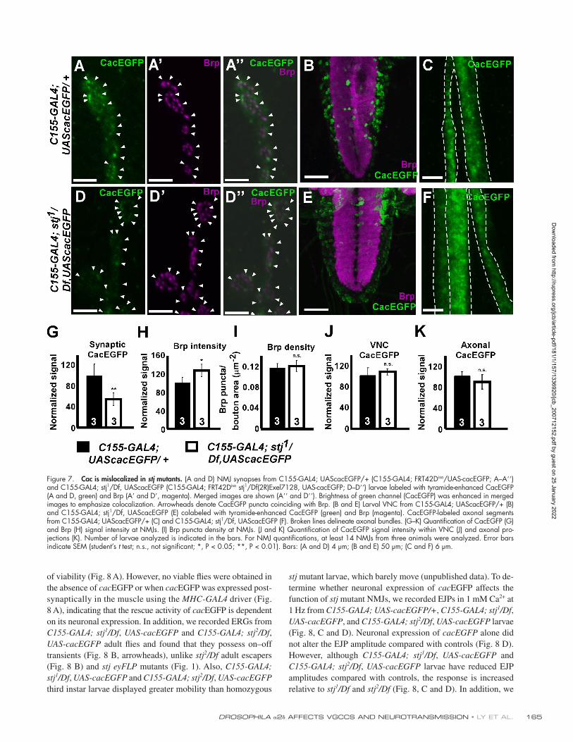
Cac is reduced at stj synapses Given the similarity of stj and cac phenotypes ( Smith et al.,
1998 ; Rieckhof et al., 2003 ), we determined whether Cac is
properly localized in stj mutants. When expressed solely in neu-
rons, cacEGFP ( Kawasaki et al., 2004 ) rescues the embryonic
lethality as well as synaptic function of cac null mutants and
localizes to synaptic active zones. Therefore, we expressed cacE-GFP in control and mutant neurons using C155-GAL4 . We visu-
alized C155-GAL4 – driven expression of CacEGFP (green) at
control and stj mutant NMJs ( Fig. 7, A and D ) and costained
with Bruchpilot (Brp; Fig. 7, A � and D � , magenta), an active
zone marker ( Wucherpfennig et al., 2003 ), and Dlg to outline
synapses (not depicted). Because native CacEGFP fl uorescence
is weak, we amplifi ed the signal using tyramide enhancement
To more closely examine single boutons, ultrastructural
analyses were performed on control ( Fig. 6, E and G ) and mu-
tant ( Fig. 6, F and H ) boutons. However, these studies revealed
no differences in vesicle density ( Fig. 6 I ), synaptic length ( Fig.
6 J ), active zone density ( Fig. 6 K ), number of active zone –
associated vesicles ( Fig. 6 L ), or vesicle size ( Fig. 6, M and N ).
Notably, the subsynaptic reticulum (SSR) surrounding the bou-
tons appears more disordered in mutant boutons. However, even
when SSR is almost entirely absent, as in dpix mutants ( Parnas
et al., 2001 ), synaptic function is only mildly affected. Thus, our
SSR defect is not likely to contribute to the impaired synaptic
release in stj mutants. These data indicate that though NMJ syn-
apses are slightly overgrown in stj mutants, most aspects of
bouton architecture are intact.
Figure 5. stj mutants have severe defects in evoked neurotransmitter release. (A) Sample EJPs recorded in 1 mM Ca 2+ at 1 Hz in con-trol, stj 1 /Df , and stj 2 /Df third instar NMJs. (B) Quantifi cation of EJP amplitudes recorded at 1 Hz in 1 mM Ca 2+ . Genotypes: control, 39 ± 5 mV; stj 1 /+ ( y w; FRT42D iso stj 1 /FRT42D iso ), 43 ± 4 mV; stj 2 /+ ( y w; FRT42D iso stj 2 /FRT42D iso ), 39 ± 3 mV; stj 1 /Df , 8 ± 2 mV; stj 2 /Df , 11 ± 5 mV; stj 3 /Df ( y w eyFLP GMR-lacZ; FRT42D iso stj 3 /Df(2R)Exel7128 ), 8 ± 1 mV; stj 1 /stj 2 ( y w eyFLP GMR-lacZ; FRT42D iso stj 1 /FRT42D iso stj 2 ), 13 ± 1 mV; stj 1 /stj 3 ( y w eyFLP GMR-lacZ; FRT-42D iso stj 1 /FRT42D iso stj 3 ), 13 ± 2 mV; stj 1 /Df rescue, 45 ± 4 mV; stj 2 /Df rescue ( y w eyFLP GMR-lacZ; FRT42D iso stj 2 /Df(2R)Exel7128 ), stj + ), 41 ± 5 mV; stj 3 /Df rescue ( y w eyFLP GMR-lacZ; FRT42D iso stj 3 /Df(2R)Exel7128 ), stj + ), 44 ± 3 mV; stj 1 /stj 2 rescue ( y w eyFLP GMR-lacZ; FRT-42D iso stj 1 /FRT42D iso stj 2 ; stj + /+ ), 42 ± 4 mV. 60 EJP amplitudes were averaged per record-ing and the number of larvae tested is indicated in the bars. Error bars indicate SEM (student ’ s t test; ** , P < 0.01). (C) Sample mEJPs were re-corded in 0.5 mM Ca 2+ with 10 μ M TTX to sup-press evoked neuronal activity. Traces shown for control, stj 1 /Df , and stj 2 /Df . The mEJP amp-litude (D), frequency (E), and mEJP amplitude distribution (F – H) of events larger than 0.4 mV are shown. The number of larvae analyzed is indicated in the bars (D and E). mEJP events were analyzed for control ( n = 1,365), stj 1 /Df ( n = 1,109), and stj 2 /Df ( n = 1,215). The amp-litude bin size was 0.2 mV. Error bars indicate SEM (student ’ s t test; n.s., not signifi cant). (I) EJPs amplitudes were measured at different [Ca 2+ ] ext (0.1, 0.2, 0.4, 0.6, 0.8, and 1 mM). Failures were included in the determination of EJP amplitudes to more accurately refl ect syn-aptic release probabilities. Genotypes were control and stj 1 /Df . Error bars indicate SEM; error bars smaller than data markers are not shown. (student ’ s t test; * * , P < 0.01). (J) Loga-rithmic plot of quantal content versus [Ca 2+ ] ext . The slope of the line ( n ), indicated for control ( n = 3.1) and stj 1 /Df ( n = 3.0), represents cal-cium cooperativity at these respective synapses. (K) Quantifi cation of paired pulse ratio (PPR), EJP 2 /EJP 1 for time intervals of 20 ms (control, PPR = 1.11 ± 0.03; stj 1 /Df , PPR = 1.5 ± 0.1), 50 ms (control, PPR = 1.06 ± 0.02; stj 1 /Df , PPR = 1.36 ± 0.07), and 100 ms (control, PPR = 1.018 ± 0.007; stj 1 /Df , PPR = 1.21 ± 0.1). Error bars indicate SEM (student ’ s t test; n.s., not signifi cant; *, P < 0.05; **, P < 0.01).
Dow
nloaded from http://rupress.org/jcb/article-pdf/181/1/157/1336920/jcb_200712152.pdf by guest on 25 January 2022

JCB • VOLUME 181 • NUMBER 1 • 2008 164
this suggests that CacEGFP can properly traffi c to active zones
independently of stj but requires stj to ensure appropriate levels
of Cac at the synapse.
To determine whether the reduction in synaptic CacEGFP
might stem from global mistraffi cking or reduced stability of
Cac in stj mutants, we also looked at the VNC ( Fig. 7, B and E )
and axonal projections ( Fig. 7, C and F ) of control and mutant
larvae overexpressing cacEGFP . We found that CacEGFP was
distributed similarly within cell bodies throughout the VNC in
control ( Fig. 7 B ) and mutant ( Fig. 7 E ) animals. Furthermore,
the signal intensities of CacEGFP within the VNC of control
and mutant animals are not different ( Fig. 7 J ). In addition, lev-
els of CacEGFP are similar within axonal projections ( Fig. 7 K ).
Hence, the data indicate that stj is not crucial for the global
stabilization or axonal transport of Cac but rather plays a more
discrete role in stabilizing Cac locally at synapses.
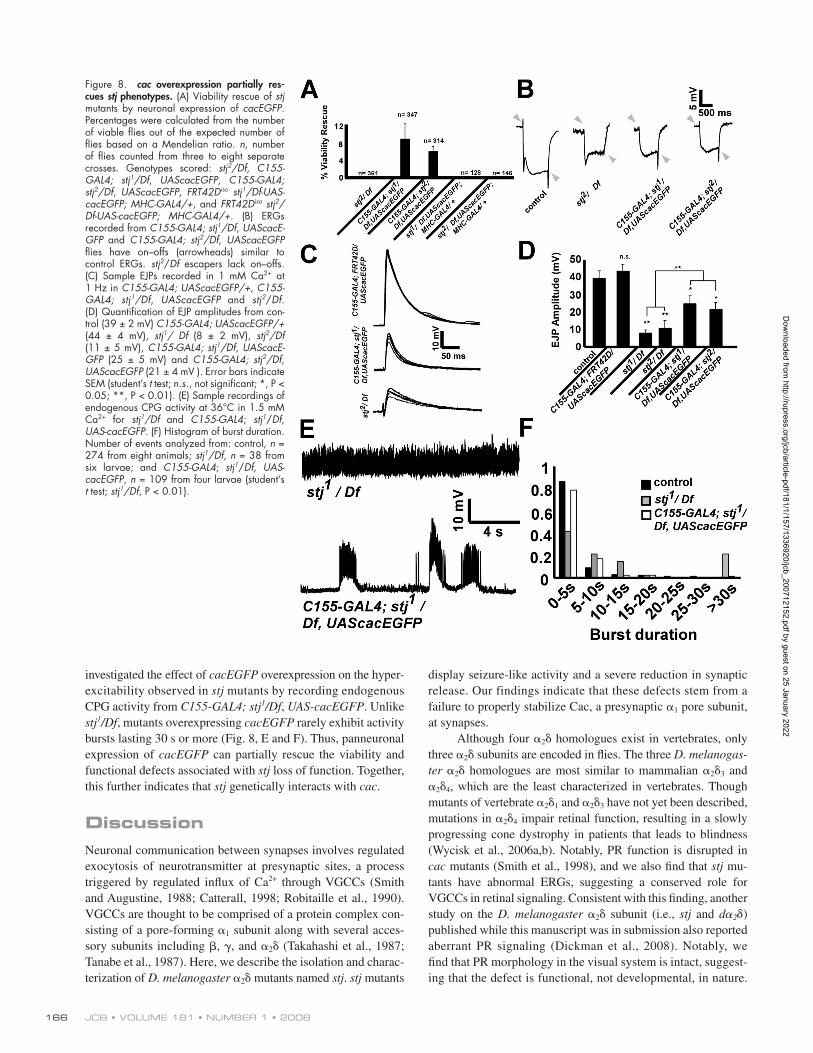
Neuronal overexpression of cac partially rescues stj phenotypes Interestingly, when we expressed cac EGFP panneuronally in
stj 1 /Df and stj 2 /Df mutant backgrounds, we observed a partial rescue
(see Materials and methods). At C155-GAL4/ + ; UAS-cacE-GFP/+ NMJs, we often observed CacEGFP puncta that are
adjacent to Brp ( Fig. 7, A – A � � ) despite some diffusion of the
enhanced CacEGFP signal. However, in C155-GAL4/ + ; stj 1 /Df , UAS-cacEGFP synapses, although we also observed co-
localization between CacEGFP and Brp ( Fig. 7, D – D � � ), the
CacEGFP signal intensity was signifi cantly reduced compared
with controls ( Fig. 7 G ). To determine the background signal
associated with tyramide enhancement, we simultaneously la-
beled Canton-S larvae. We observed a weak, nonspecifi c synaptic
signal associated with the tyramide reaction (Fig. S2, avail-
able at http://www.jcb.org/cgi/content/full/jcb.200712152/DC1)
but never observed punctate signal in Canton-S larvae, which
suggests that the CacEGFP signal we detected in C155-GAL4/ + ; UAS-cacEGFP/+ and C155-GAL4/ + ; stj 1 /Df , UAS-cacEGFP
larvae is specifi c. Moreover, we quantifi ed the density and
signal intensity of Brp at control and mutant synapses over-
expressing CacEGFP and found that although signal intensity is
mildly increased in the mutants ( Fig. 7 H ), Brp puncta density
( Fig. 7 I ) is not different. Thus, active zones remain stable even
when synaptic Ca 2+ channels are markedly reduced. Together,
Figure 6. stj mutant synapses exhibit NMJ overgrowth but have normal bouton ultrastruc-ture. Third instar NMJ synapses of muscles 6/7 in control (A) and stj 2 /Df (B) stained for Dlg (ma-genta) and Hrp (green). Bar, 25 μ m. (C and D) Quantifi cation of NMJ synapse length (C; con-trols, 0.0036 ± 0.0002 μ m � 1 ; stj 1 /Df , 0.0044 ± 0.0003 μ m � 1 ; stj 2 /Df , 0.0053 ± 0.0005 μ m � 1 ; stj 2 /Df , stj + , 0.0038 ± 0.0004 μ m � 1 ) and bouton number (D; controls, 0.00045 ± 0.00003 μ m � 2 ; stj 1 /Df , 0.00064 ± 0.00005 μ m � 2 ; stj 2 /Df , 0.0008 ± 0.0001 μ m � 2 ; stj 2 /Df , stj + , 0.0004 ± 0.00003 μ m � 2 ) normalized to muscle area. Number of animals analyzed: controls, n = 21; stj 1 /Df , n = 13; stj 2 /Df , n = 10; stj 2 /Df , stj + , n = 7. Error bars indicate SEM (student ’ s t test; * , P < 0.05, **, P < 0.01). (E – H) TEM of NMJ boutons in control (E and G) and stj 2 /Df (F and H). Synaptic features such as vesicles, active zones (AZ), and the SSR (ar-rowheads) are readily observed. Quantifi ca-tion of bouton parameters in control, stj 1 /Df , and stj 2 /Df animals including vesicle density (I), synaptic length (J), AZ density (K), AZ-associated vesicles (within a 100-nm perimeter of the AZ; L), and vesicle size (M and N) re-vealed no signifi cant differences. Number of vesicles analyzed from: control, n = 425; and stj 1 /Df , n = 475. However, the SSR appears disordered in stj 1 /Df (not depicted) and stj 2 /Df (F, arrowhead) boutons. Cross sections from at least 10 boutons from three animals were ex-amined for each genotype. Error bars indicate SEM (student ’ s t test; n.s., not signifi cant). Bars: (A and B) 25 μ m; (E) 0.5 μ m; (F) 0.2 μ m; (G and H) 100 nm.
Dow
nloaded from http://rupress.org/jcb/article-pdf/181/1/157/1336920/jcb_200712152.pdf by guest on 25 January 2022
165 DROSOPHILA � 2 � AFFECTS VGCCS AND NEUROTRANSMISSION • LY ET AL.
stj mutant larvae, which barely move (unpublished data). To de-
termine whether neuronal expression of cac EGFP affects the
function of stj mutant NMJs, we recorded EJPs in 1 mM Ca 2+ at
1 Hz from C155-GAL4; UAS-cacEGFP/+ , C155-GAL4; stj 1 /Df , UAS-cacEGFP , and C155-GAL4; stj 2 /Df , UAS-cacEGFP larvae
( Fig. 8, C and D ). Neuronal expression of cacEGFP alone did
not alter the EJP amplitude compared with controls ( Fig. 8 D ).
However, although C155-GAL4; stj 1 /Df , UAS-cacEGFP and
C155-GAL4; stj 2 /Df , UAS-cacEGFP larvae have reduced EJP
amplitudes compared with controls, the response is increased
relative to stj 1 /Df and stj 2 /Df ( Fig. 8, C and D ). In addition, we
of viability ( Fig. 8 A ). However, no viable fl ies were obtained in
the absence of cac EGFP or when cac EGFP was expressed post-
synaptically in the muscle using the MHC-GAL4 driver ( Fig.
8 A ), indicating that the rescue activity of cac EGFP is dependent
on its neuronal expression. In addition, we recorded ERGs from
C155-GAL4; stj 1 /Df , UAS-cacEGFP and C155-GAL4; stj 2 /Df , UAS-cacEGFP adult fl ies and found that they possess on – off
transients ( Fig. 8 B , arrowheads), unlike stj 2 /Df adult escapers
( Fig. 8 B ) and stj eyFLP mutants ( Fig. 1 ). Also, C155-GAL4; stj 1 /Df , UAS-cacEGFP and C155-GAL4; stj 2 /Df , UAS-cacEGFP
third instar larvae displayed greater mobility than homozygous
Figure 7. Cac is mislocalized in stj mutants. (A and D) NMJ synapses from C155-GAL4; UAScacEGFP/+ (C155-GAL4; FRT42D iso /UAS-cacEGFP; A – A � � ) and C155-GAL4; stj 1 /Df, UAScacEGFP (C155-GAL4; FRT42D iso stj 1 /Df(2R)Exel7128, UAS-cacEGFP; D – D � � ) larvae labeled with tyramide-enhanced CacEGFP (A and D, green) and Brp (A � and D � , magenta). Merged images are shown (A � � and D � � ). Brightness of green channel (CacEGFP) was enhanced in merged images to emphasize colocalization. Arrowheads denote CacEGFP puncta coinciding with Brp. (B and E) Larval VNC from C155-GAL4; UAScacEGFP/+ (B) and C155-GAL4; stj 1 /Df, UAScacEGFP (E) colabeled with tyramide-enhanced CacEGFP (green) and Brp (magenta). CacEGFP-labeled axonal segments from C155-GAL4; UAScacEGFP/+ (C) and C155-GAL4; stj 1 /Df, UAScacEGFP (F). Broken lines delineate axonal bundles. (G – K) Quantifi cation of CacEGFP (G) and Brp (H) signal intensity at NMJs. (I) Brp puncta density at NMJs. (J and K) Quantifi cation of CacEGFP signal intensity within VNC (J) and axonal pro-jections (K). Number of larvae analyzed is indicated in the bars. For NMJ quantifi cations, at least 14 NMJs from three animals were analyzed. Error bars indicate SEM (student ’ s t test; n.s., not signifi cant; *, P < 0.05; **, P < 0.01). Bars: (A and D) 4 μ m; (B and E) 50 μ m; (C and F) 6 μ m.
Dow
nloaded from http://rupress.org/jcb/article-pdf/181/1/157/1336920/jcb_200712152.pdf by guest on 25 January 2022
JCB • VOLUME 181 • NUMBER 1 • 2008 166
display seizure-like activity and a severe reduction in synaptic
release. Our findings indicate that these defects stem from a
failure to properly stabilize Cac, a presynaptic � 1 pore subunit,
at synapses.
Although four � 2 � homologues exist in vertebrates, only
three � 2 � subunits are encoded in fl ies. The three D. melanogas-ter � 2 � homologues are most similar to mammalian � 2 � 3 and
� 2 � 4 , which are the least characterized in vertebrates. Though
mutants of vertebrate � 2 � 1 and � 2 � 3 have not yet been described,
mutations in � 2 � 4 impair retinal function, resulting in a slowly
progressing cone dystrophy in patients that leads to blindness
( Wycisk et al., 2006a , b ). Notably, PR function is disrupted in
cac mutants ( Smith et al., 1998 ), and we also fi nd that stj mu-
tants have abnormal ERGs, suggesting a conserved role for
VGCCs in retinal signaling. Consistent with this fi nding, another
study on the D. melanogaster � 2 � subunit (i.e., stj and d � 2 � ) published while this manuscript was in submission also reported
aberrant PR signaling ( Dickman et al., 2008 ). Notably, we
fi nd that PR morphology in the visual system is intact, suggest-
ing that the defect is functional, not developmental, in nature.
investigated the effect of cacEGFP overexpression on the hyper-
excitability observed in stj mutants by recording endogenous
CPG activity from C155-GAL4; stj 1 /Df , UAS-cacEGFP . Unlike
stj 1 /Df , mutants overexpressing cacEGFP rarely exhibit activity
bursts lasting 30 s or more ( Fig. 8, E and F ). Thus, panneuronal
expression of cacEGFP can partially rescue the viability and
functional defects associated with stj loss of function. Together,
this further indicates that stj genetically interacts with cac .
Discussion Neuronal communication between synapses involves regulated
exocytosis of neurotransmitter at presynaptic sites, a process
triggered by regulated infl ux of Ca 2+ through VGCCs ( Smith
and Augustine, 1988 ; Catterall, 1998 ; Robitaille et al., 1990 ).
VGCCs are thought to be comprised of a protein complex con-
sisting of a pore-forming � 1 subunit along with several acces-
sory subunits including � , � , and � 2 � ( Takahashi et al., 1987 ;
Tanabe et al., 1987 ). Here, we describe the isolation and charac-
terization of D. melanogaster � 2 � mutants named stj . stj mutants
Figure 8. cac overexpression partially res-cues stj phenotypes. (A) Viability rescue of stj mutants by neuronal expression of cacEGFP . Percentages were calculated from the number of viable fl ies out of the expected number of fl ies based on a Mendelian ratio. n , number of fl ies counted from three to eight separate crosses. Genotypes scored: stj 2 /Df , C155-GAL4; stj 1 /Df , UAScacEGFP , C155-GAL4; stj 2 /Df , UAScacEGFP , FRT42D iso stj 1 /Df-UAS-cacEGFP; MHC-GAL4/+ , and FRT42D iso stj 2 /Df-UAS-cacEGFP; MHC-GAL4/+ . (B) ERGs recorded from C155-GAL4; stj 1 /Df , UAScacE-GFP and C155-GAL4; stj 2 /Df , UAScacEGFP fl ies have on – offs (arrowheads) similar to control ERGs. stj 2 / Df escapers lack on – offs. (C) Sample EJPs recorded in 1 mM Ca 2+ at 1 Hz in C155-GAL4; UAScacEGFP/+ , C155-GAL4; stj 1 /Df , UAScacEGFP and stj 2 / Df . (D) Quantifi cation of EJP amplitudes from con-trol (39 ± 2 mV) C155-GAL4; UAScacEGFP/+ (44 ± 4 mV), stj 1 / Df (8 ± 2 mV), stj 2 /Df (11 ± 5 mV), C155-GAL4; stj 1 /Df , UAScacE-GFP (25 ± 5 mV) and C155-GAL4; stj 2 /Df , UAScacEGFP (21 ± 4 mV ). Error bars indicate SEM (student ’ s t test; n.s., not signifi cant; *, P < 0.05; **, P < 0.01). (E) Sample recordings of endogenous CPG activity at 36 ° C in 1.5 mM Ca 2+ for stj 1 /Df and C155-GAL4 ; stj 1 / Df , UAS - cacEGFP . (F) Histogram of burst duration. Number of events analyzed from: control, n = 274 from eight animals; stj 1 /Df , n = 38 from six larvae; and C155-GAL4 ; stj 1 / Df , UAS - cacEGFP , n = 109 from four larvae (student ’ s t test; stj 1 /Df , P < 0.01).
Dow
nloaded from http://rupress.org/jcb/article-pdf/181/1/157/1336920/jcb_200712152.pdf by guest on 25 January 2022
167 DROSOPHILA � 2 � AFFECTS VGCCS AND NEUROTRANSMISSION • LY ET AL.
Ca 2+ -dependent release probability because we observed in-
creased facilitation at stj synapses and a rightward shift in the
Ca 2+ dependence of neurotransmitter release. The alternate
study partially attributes the reduction in synaptic release to a
reduced number of active zones, suggested by a decreased mEJP
frequency and a reduction in Brp labeling ( Dickman et al.,
2008 ). However, we do not observe a loss of active zones in stj mutants. Notably, they compared their mutants to Canton-S to
assess Brp labeling, whereas a different control ( w 118 ) was used
for electrophysiological analyses. Because it was not reported
whether these defects could be corrected by d � 2 � transgene ex-
pression, we cannot exclude that some of the defects are due to
genetic background.
However, we do fi nd that CacEGFP is dramatically re-
duced at mutant synapses when expressed panneuronally, demon-
strating a direct role for an � 2 � subunit in regulating the synaptic
levels of an � 1 subunit in vivo, a fi nding also corroborated
by Dickman et al. ( 2008 ). Interestingly, although CacEGFP
is reduced at stj mutant synapses, it properly localizes to active
zones, suggesting that the synaptic targeting of Cac does not
depend solely on stj and requires other factors. In addition, we
do not observe differences in CacEGFP distribution or signal in
the VNC or axonal projections, indicating that the global stabil-
ity and axonal transport of Cac are not affected by stj loss of
function. Hence, stj most likely plays a discrete role in the syn-
aptic stabilization of Cac.
Interestingly, we also fi nd that neuronal overexpression of
cac can partially rescue the viability and electrophysiological
phenotypes observed in stj mutants, providing further evidence
that they interact. In contrast, although Dickman et al. ( 2008 )
found that cacEGFP overexpression improves the survival of
their mutants, they did not observe an improvement in EJP amp-
litude when cacEGFP is overexpressed in the mutants. To mea-
sure cacEGFP rescue activity at the NMJ, Dickman et al. ( 2008 )
performed electrophysiological recordings in 0.3 mM Ca 2+ ,
whereas our studies were done in 1 mM Ca 2+ . It is possible that
at 0.3 mM Ca 2+ , the mutants, which display a right-shift in Ca 2+
sensitivity, are operating in a subcooperative regimen where the
ameliorative effects of cacEGFP overexpression are masked.
Notably, the ERG, hyperexcitability, and NMJ synaptic release
phenotypes are improved in our mutants with neuronal cac
overexpression. Hence, stj is required for the proper function
and localization of Cac but this defect can be partially overcome
with cac overexpression.
In summary, we have isolated novel mutations in stj , a
neuronal D. melanogaster � 2 � subunit. Our studies defi ne a pre-
dominant role for a D. melanogaster � 2 � in regulating neuronal
excitability and neurotransmitter release by specifi cally stabi-
lizing cac , a presynaptic VGCC � 1 subunit, at synapses. This
work should facilitate further studies to illuminate the role of
� 2 � as a therapeutic target and modulator of VGCC function.
Materials and methods D. melanogaster strains and genetics The isolation of 118 mutants on chromosome 2R, including stj 1 , stj 2 , and stj 3 , has been described previously ( Verstreken et al., 2002 ). The control genotype was y w; P { ry + neoFRT } 42D isogenized (FRT42D iso ). Flies with visual
Further insight into the in vivo role of � 2 � has been garnered
through work on a spontaneous mouse mutant of � 2 � 2 known as
ducky . The three alleles of ducky , ducky ( Barclay et al., 2001 ),
ducky 2J ( Donato et al., 2006 ), and Cacna2d2 entla ( Brill et al.,
2004 ), exhibit spike-wave seizures reminiscent of absence epi-
lepsy and are ataxic. Similarly, we also fi nd that stj mutants have
altered neuronal excitability and surmise that this may be caused
by a selective loss of stj in a subset of neurons, possibly inhibi-
tory interneurons. Consistent with this possibility, blockade of
GABA receptors in D. melanogaster larvae has been shown to
predispose to neuronal hyperactivity ( Stilwell et al., 2006 ).
Five functionally distinct VGCC � 1 subtypes encoded by
10 different genes are expressed in mammalian excitable tissues,
L-, N-, P/Q-, R-, and T-types ( Catterall, 2000 ). However, within
the D. melanogaster genome, there are only four genes that en-
code � 1 subunits representing homologues of vertebrate N-,
P/Q-, and R-type ( cac , also known as dmca1A ; Smith et al., 1996 ),
L-type ( dmca1D ; Zheng et al., 1995 ), and T-type ( dmca1T )
channels ( CG15899 ; http://fl ybase.bio.indiana.edu/) and an
invertebrate-specifi c � 1 subunit ( dmalpha1U , also known as
narrow abdomen and halothane resistance ; Nash et al., 2002 ).
However, we focused on cac for the following reasons. First,
mutations in dmalpha1U are viable and predominantly affect di-
urnal locomotor activity patterns but exhibit no other obvious
neurological defi cits ( Nash et al., 2002 ). Second, dmca1T has
not been characterized in the fl y and no phenotypic analysis is
available. Third, dmca1D is thought to primarily underlie mus-
cle Ca 2+ currents ( Zheng et al., 1995 ; Ren et al., 1998 ). Although
dmca1D transcripts are expressed in the brain, the function of
dmca1D in the nervous system has not been established ( Zheng
et al., 1995 ). However, cac mutants display a marked reduction
in synaptic release, similar to what is observed when stj is lost,
and is likely the primary � 1 subunit involved in neurotransmis-
sion in fl ies ( Smith et al., 1996 ; Kawasaki et al., 2000 ). In addi-
tion, cac mutants have ERG ( Smith et al., 1998 ) and seizure
( Rieckhof et al., 2003 ) phenotypes similar to stj mutants. The
synaptic defects in cac mutants can be corrected by neuronal ex-
pression of cac cDNA, which suggests a requirement for this
gene in the nervous system ( Kawasaki et al., 2004 ). Consistent
with this data, stj is predominantly expressed and required in the
nervous system, as demonstrated by our ability to rescue the mu-
tants with a neuronally driven stj transgene and in situ hybridiza-
tion studies. Together, the neuronal localization and phenotypic
similarities suggest that cac is a likely target for stj function.
Work in heterologous expression systems has demon-
strated that � 2 � subunits can increase Ca 2+ current amplitudes
approximately threefold by increasing the expression of � 1 on
the membrane ( Singer et al., 1991 ; Felix et al., 1997 ; Klugbauer
et al., 1999 ; Gao et al., 2000 ; Canti et al., 2005 ). In addition,
ducky mutant Purkinje cells exhibit a � 35% reduction in P-type
Ca 2+ currents despite no changes in unitary Ca 2+ currents
( Barclay et al., 2001 ). However, evidence of channel mislocaliza-
tion in ducky mice has not been demonstrated and the mechanism
by which � 2 � 2 loss leads to the defects in ducky mice remains
unclear. Similar to Dickman et al. ( 2008 ), we observe a severe
reduction in EJP amplitude at stj mutant NMJ synapses. We fi nd
that this impairment is likely caused by a reduction in the highly
Dow
nloaded from http://rupress.org/jcb/article-pdf/181/1/157/1336920/jcb_200712152.pdf by guest on 25 January 2022
JCB • VOLUME 181 • NUMBER 1 • 2008 168
several washes, samples were incubated with tyramide in amplifi cation buffer (PerkinElmer) for 30 min. Samples were then washed in PBT, blocked in 10% NGS, and incubated with additional primary antibodies for 2 h. After several washes and a blocking treatment, secondary antibodies were applied for 2 h. After a fi nal wash, preparations were mounted in Vecta-shield and imaged.
Electrophysiology ERGs were recorded as described previously ( Fabian-Fine et al., 2003 ; Verstreken et al., 2003 ). Third instar larval electrophysiological recordings were performed as described previously ( Verstreken et al., 2002 ). Larvae were maintained in modifi ed HL3 (110 mM NaCl, 5 mM KCl, 10 mM NaHCO 3 , 5 mM Hepes, 30 mM sucrose, 5 mM trehalose, and 10 mM MgCl 2 , pH 7.2) and recordings were performed in various extracellular [Ca 2+ ] as indicated. In CPG recordings, larval motor axons were left intact and endogenous neural activity was recorded from muscles 6/7 at 36 ° C with the temperature controlled as described previously ( Koh et al., 2004 ). To measure EJPs, we stimulated cut motor neurons and recorded from mus-cles 6/7/12/13. Quantal content was estimated by including failures and correcting for nonlinear summation of EJPs ( Martin, 1955 ). Cooperativity coeffi cients were then assessed by determining the slope of log-transformed measurements for quantal content for Ca 2+ concentrations of 0.1 – 0.8 mM. mEJPs were recorded in the presence of 0.5 mM extracellular Ca 2+ and 10 μ M TTX (Sigma-Aldrich). EJPs and mEJPs were analyzed using pClamp6 and Mini Analysis Program (Synaptosoft) software, respectively.
Transmission electron microscopy (TEM) TEM of PRs was performed as described previously ( Hiesinger et al., 2006 ). At least 15 cartridges from three animals were examined. TEM of NMJ boutons was performed as described previously ( Verstreken et al., 2002 ). Analysis was performed on � 15 boutons from at least three lar-vae. Images were analyzed using ImageJ.
Online supplemental material Fig. S1 shows a protein sequence alignment comparing stj with murine and human � 2 � 1 – 4 proteins and indicates the residues affected in stj 1 and stj 2 al-leles. Fig. S2 shows that tyramide enhancement in non- cacEGFP – expressing controls leads to a nonspecifi c synaptic signal that is diffuse, not punctate, as is the case when cacEGFP is present. Online supplemental material is avail-able at http://www.jcb.org/cgi/content/full/jcb.200712152/DC1.
We thank Koen Venken for sharing the P[acman] vector before publication. We thank Richard Ordway, the Bloomington Drosophila Stock Center, and Kyoto Drosophila Genetic Resource Center for reagents. We thank Amir Fay-azzudin, Yong Qi Lin, other members of the Bellen laboratory, and Christian Rosenmund for discussions. We thank Karen Schulze, Bryan Michael Thomas, and Kyong-Mi Um for their help. We are grateful to Richard Atkinson and Claire Haueter for microscopy expertise and Yuchun He for injections to make transgenic lines.
Confocal microscopy was supported by the Baylor College of Medicine Mental Retardation and Developmental Disabilities Research Center. C.V. Ly is supported by an Ruth L. Kirschstein National Research Service Award from the National Institute of Neurological Disorders and Stroke (grant F30NS056520). H.J. Bellen is an Howard Hughes Medical Institute investigator.
Submitted: 26 December 2007 Accepted: 10 March 2008
References Anantharaman , V. , and L. Aravind . 2000 . Cache - a signaling domain common
to animal Ca(2+)-channel subunits and a class of prokaryotic chemotaxis receptors. Trends Biochem. Sci. 25 : 535 – 537 .
Bahls , F.H. , R. Lartius , L.E. Trudeau , R.T. Doyle , Y. Fang , D. Witcher , K. Campbell , and P.G. Haydon . 1998 . Contact-dependent regulation of N-type calcium channel subunits during synaptogenesis. J. Neurobiol. 35 : 198 – 208 .
Barclay , J. , N. Balaguero , M. Mione , S.L. Ackerman , V.A. Letts , J. Brodbeck , C. Canti , A. Meir , K.M. Page , K. Kusumi , et al . 2001 . Ducky mouse pheno-type of epilepsy and ataxia is associated with mutations in the Cacna2d2 gene and decreased calcium channel current in cerebellar Purkinje cells. J. Neurosci. 21 : 6095 – 6104 .
Bellen , H.J. , R.W. Levis , G. Liao , Y. He , J.W. Carlson , G. Tsang , M. Evans-Holm , P.R. Hiesinger , K.L. Schulze , G.M. Rubin , et al . 2004 . The BDGP
systems homozygous for stj were y w eyFLP GMR-lacZ; FRT42D iso stj/FRT-42D iso , P { w + ry + } 47A l(2)cl-R11 1 . Mapping of stj is described in Fig. 2 A . P element stocks and Df(2R)CX1 (indicated in Fig. 2 A ) were obtained from the Bloomington Drosophila Stock Center ( Brizuela et al., 1994 ; Bellen et al., 2004 ; Parks et al., 2004 ). Df(2R)Exel7128 was obtained from the Exelixis stock collection ( Thibault et al., 2004 ). stj 1 , stj 2 , stj 3 , and Df(2R)Exel7128 were maintained over CyO; Kr-GAL4 UAS-GFP and mu-tant animals were cultured on grape juice plates with yeast paste. NP1574-GAL4 was obtained from the Kyoto Institute of Technology Drosophila Genetic Resource Center ( Hayashi et al., 2002 ) and was crossed to UAS-GFP , UAS-GFP nls , or UAS-mCD8GFP as indicated ( Fig. 3 ). UAS-cac EGFP fl ies were obtained from R. Ordway (Pennsylvania State University, Univer-sity Park, PA; Kawasaki et al., 2004 ). We analyzed C155-GAL4 /+; FRT42D iso / UAS - cac EGFP, C155-GAL4 /+; stj 1 / Df(2R)Exel7128 , UAS - cac EGFP, and C155-GAL4 /+; stj 2 / Df(2R)Exel7128 , UAS - cacEGFP .
Molecular biology 28 kb of genomic DNA harboring CG12295 ( stj ) was recovered from BAC16G12 (BACPAC Resources Center) by gap repair in P[acman] ApR F-2-5 (provided by K. Venken, Baylor College of Medicine, Houston, TX; Venken et al., 2006 ). We cloned a 501-bp left and 448-bp right homology arm separated by a BamHI site in P[acman]. The vector was digested with BamHI and transformed into recombination competent mini- � -Tet DH10B bacteria containing BAC16G12. Gap-repaired vectors containing the ge-nomic stj sequence were selected by AMP and verifi ed by sequencing and restriction analysis. To generate the UAS-FLAG-stj-HA transgene, we PCR amplifi ed the coding region of stj from cDNA clone SD07723 (Dro-sophila Gene Collection 1; Rubin, 2000 ) by primers CG12295–BglII–F (GAAGATCTAATGGCCTGGTCCCGTCTCCTG) and stj -HA–XbaI–R (TTTC-TAGATCGCGTAGTCGGGGACGTCGTAGGGGTATCTAGACAGCCAAC-GACTCAGTATGTG), which includes the corresponding sequence of the HA tag, and subcloned it into the BglII and XbaI sites of the pUAST-Flag vector ( Yao and Sun, 2005 ).
In situ hybridization A 1,489-bp sequence corresponding to nucleotides 1 – 1,489 of stj was amplifi ed from cDNA clone SD07723 (Drosophila Gene Collection 1; Rubin, 2000 ) and cloned into the pBluescript KS (+) vector containing fl anking T7 and T3 promoter elements using BglII and XhoI restriction sites. Probe synthesis, embryo fi xation, and in situ hybridization was performed as described previously ( Lecuyer et al., 2007 ).
Immunohistochemistry Larvae and adult fl ies raised at 22 ° C were dissected in modifi ed HL3 solu-tion (110 mM NaCl, 5 mM KCl, 10 mM NaHCO 3 , 5 mM Hepes, 30 mM sucrose, 5 mM trehalose, and 10 mM MgCl 2 , pH 7.2) and fi xed in 3.7% formaldehyde. Stainings were performed using standard protocols ( Bellen et al., 2004 ). Samples were mounted in Vectashield (Vector Laboratories). Images were captured at room temperature using a confocal microscope (LSM 510; Carl Zeiss, Inc.) with LSM5 software (Carl Zeiss, Inc.) using the following objectives: Plan Apochromat 63 × 1.4 NA, Plan Neofl uar 40 × 1.3 NA, and Plan Neofl uar 16 × 0.5 NA (all from Carl Zeiss, Inc.). Images were processed with Amira 2.2 (TGS), Photoshop 7.0 (Adobe), and Im-ageJ. Primary antibodies specifi c to these antigens were used at the follow-ing dilutions: Brp, 1:20 (mouse, nc82; Developmental Studies Hybridoma Bank; Wucherpfennig et al., 2003 ); Dlg, 1:50 (mouse, 4F3; Developmen-tal Studies Hybridoma Bank; Parnas et al., 2001 ); Dlg, 1:200 (rabbit, pro-vided by K. Choi, Baylor College of Medicine, Houston, TX); Elav, 1:50 (mouse, 9F8A9; Developmental Studies Hybridoma Bank; O ’ Neill et al., 1994 ); Even-skipped, 1:10 (mouse, 2B8; Developmental Studies Hybrid-oma Bank; Patel et al., 1994 ); HA, 1: 500 (mouse, 16B12; Covance); Hrp, 1:200 (rabbit; Jackson ImmunoResearch Laboratories); GFP, 1:200 (rabbit; Invitrogen); GFP, 1:10,000 (chicken; Abcam); Repo, 1:10 (mouse, 8D12; Developmental Studies Hybridoma Bank; Muhlig-Versen et al., 2005 ); and Syb, 1: 200 (rat; Wu et al., 1999 ). Secondary antibodies tagged with Alexa 488 (Invitrogen), Cy3, Cy5, or Hrp (Jackson Immuno-Research Laboratories) were used at 1:200.
We detected CacEGFP signals by performing tyramide signal ampli-fi cation (PerkinElmer). In brief, after a 10-min fi xation with 3.7% formalde-hyde, we washed dissected third instar larvae three times with PBT (PBS with 0.2% Triton X-100) and incubated preparations in 1% hydrogen per-oxide for 1 h. After several washes, samples were blocked in 10% normal goat serum (NGS) for 1 h. Samples were incubated overnight with chicken anti-GFP. The preparations were washed with PBT, incubated in 10% NGS for 1 h, and allowed to incubate with Hrp-conjugated anti – chicken. After
Dow
nloaded from http://rupress.org/jcb/article-pdf/181/1/157/1336920/jcb_200712152.pdf by guest on 25 January 2022
169 DROSOPHILA � 2 � AFFECTS VGCCS AND NEUROTRANSMISSION • LY ET AL.
Hayashi , S. , K. Ito , Y. Sado , M. Taniguchi , A. Akimoto , H. Takeuchi , T. Aigaki , F. Matsuzaki , H. Nakagoshi , T. Tanimura , et al . 2002 . GETDB, a database compiling expression patterns and molecular locations of a collection of Gal4 enhancer traps. Genesis . 34 : 58 – 61 .
Herlitze , S. , M. Xie , J. Han , A. Hummer , K.V. Melnik-Martinez , R.L. Moreno , and M.D. Mark . 2003 . Targeting mechanisms of high voltage-activated Ca2+ channels. J. Bioenerg. Biomembr. 35 : 621 – 637 .
Hiesinger , P.R. , R.G. Zhai , Y. Zhou , T.W. Koh , S.Q. Mehta , K.L. Schulze , Y. Cao , P. Verstreken , T.R. Clandinin , K.F. Fischbach , et al . 2006 . Activity-independent prespecifi cation of synaptic partners in the visual map of Drosophila . Curr. Biol. 16 : 1835 – 1843 .
Jay , S.D. , A.H. Sharp , S.D. Kahl , T.S. Vedvick , M.M. Harpold , and K.P. Campbell . 1991 . Structural characterization of the dihydropyridine-sensitive calcium channel alpha 2-subunit and the associated delta pep-tides. J. Biol. Chem. 266 : 3287 – 3293 .
Katz , B. , and R. Miledi . 1969 . Spontaneous and evoked activity of motor nerve endings in calcium Ringer. J. Physiol. 203 : 689 – 706 .
Kawasaki , F. , R. Felling , and R.W. Ordway . 2000 . A temperature-sensitive para-lytic mutant defi nes a primary synaptic calcium channel in Drosophila . J. Neurosci. 20 : 4885 – 4889 .
Kawasaki , F. , B. Zou , X. Xu , and R.W. Ordway . 2004 . Active zone localiza-tion of presynaptic calcium channels encoded by the cacophony locus of Drosophila . J. Neurosci. 24 : 282 – 285 .
Kirschfeld , K. 1967 . The projection of the optical environment on the screen of the rhabdomere in the compound eye of the Musca . [In German.] Exp. Brain Res. 3 : 248 – 270
Klugbauer , N. , L. Lacinova , E. Marais , M. Hobom , and F. Hofmann . 1999 . Molecular diversity of the calcium channel alpha2delta subunit. J. Neurosci. 19 : 684 – 691 .
Koh , T.W. , P. Verstreken , and H.J. Bellen . 2004 . Dap160/intersectin acts as a stabilizing scaffold required for synaptic development and vesicle endo-cytosis. Neuron . 43 : 193 – 205 .
Komuro , H. , and P. Rakic . 1998 . Orchestration of neuronal migration by activity of ion channels, neurotransmitter receptors, and intracellular Ca2+ fl uc-tuations. J. Neurobiol. 37 : 110 – 130 .
Lecuyer , E. , H. Yoshida , N. Parthasarathy , C. Alm , T. Babak , T. Cerovina , T.R. Hughes , P. Tomancak , and H.M. Krause . 2007 . Global analysis of mRNA localization reveals a prominent role in organizing cellular architecture and function. Cell . 131 : 174 – 187 .
Martin , A.R. 1955 . A further study of the statistical composition on the end-plate potential. J. Physiol. 130 : 114 – 122 .
Mehta , S.Q. , P.R. Hiesinger , S. Beronja , R.G. Zhai , K.L. Schulze , P. Verstreken , Y. Cao , Y. Zhou , U. Tepass , M.C. Crair , and H.J. Bellen . 2005 . Mutations in Drosophila sec15 reveal a function in neuronal targeting for a subset of exocyst components. Neuron . 46 : 219 – 232 .
Mikami , A. , K. Imoto , T. Tanabe , T. Niidome , Y. Mori , H. Takeshima , S. Narumiya , and S. Numa . 1989 . Primary structure and functional expression of the cardiac dihydropyridine-sensitive calcium channel. Nature . 340 : 230 – 233 .
Muhlig-Versen , M. , A.B. da Cruz , J.A. Tschape , M. Moser , R. Buttner , K. Athenstaedt , P. Glynn , and D. Kretzschmar . 2005 . Loss of Swiss cheese/neuropathy target esterase activity causes disruption of phosphatidyl-choline homeostasis and neuronal and glial death in adult Drosophila . J. Neurosci. 25 : 2865 – 2873 .
Nash , H.A. , R.L. Scott , B.C. Lear , and R. Allada . 2002 . An unusual cation chan-nel mediates photic control of locomotion in Drosophila . Curr. Biol. 12 : 2152 – 2158 .
Newsome , T.P. , B. Asling , and B.J. Dickson . 2000 . Analysis of Drosophila photoreceptor axon guidance in eye-specifi c mosaics. Development . 127 : 851 – 860 .
O ’ Neill , E.M. , I. Rebay , R. Tjian , and G.M. Rubin . 1994 . The activities of two Ets-related transcription factors required for Drosophila eye development are modulated by the Ras/MAPK pathway. Cell . 78 : 137 – 147 .
Pak , W.L. , J. Grossfi eld , and N.V. White . 1969 . Nonphototactic mutants in a study of vision of Drosophila . Nature . 222 : 351 – 354 .
Parks , A.L. , K.R. Cook , M. Belvin , N.A. Dompe , R. Fawcett , K. Huppert , L.R. Tan , C.G. Winter , K.P. Bogart , J.E. Deal , et al . 2004 . Systematic genera-tion of high-resolution deletion coverage of the Drosophila melanogaster genome. Nat. Genet. 36 : 288 – 292 .
Parnas , D. , A.P. Haghighi , R.D. Fetter , S.W. Kim , and C.S. Goodman . 2001 . Regulation of postsynaptic structure and protein localization by the Rho-type guanine nucleotide exchange factor dPix. Neuron . 32 : 415 – 424 .
Patel , N.H. , B.G. Condron , and K. Zinn . 1994 . Pair-rule expression patterns of even-skipped are found in both short- and long-germ beetles. Nature . 367 : 429 – 434 .
Pietrobon , D. 2005 . Function and dysfunction of synaptic calcium channels: in-sights from mouse models. Curr. Opin. Neurobiol. 15 : 257 – 265 .
gene disruption project: single transposon insertions associated with 40% of Drosophila genes. Genetics . 167 : 761 – 781 .
Benzer , S. 1967 . Behavioral nutants of Drosophila isolated by countercurrent distribution. Proc. Natl. Acad. Sci. USA . 58 : 1112 – 1119 .
Brill , J. , R. Klocke , D. Paul , D. Boison , N. Gouder , N. Klugbauer , F. Hofmann , C.M. Becker , and K. Becker . 2004 . entla, a novel epileptic and ataxic Cacna2d2 mutant of the mouse. J. Biol. Chem. 279 : 7322 – 7330 .
Brizuela , B.J. , L. Elfring , J. Ballard , J.W. Tamkun , and J.A. Kennison . 1994 . Genetic analysis of the brahma gene of Drosophila melanogaster and polytene chromosome subdivisions 72AB. Genetics . 137 : 803 – 813 .
Buchner , E. , R. Bader , S. Buchner , J. Cox , P.C. Emson , E. Flory , C.W. Heizmann , S. Hemm , A. Hofbauer , and W.H. Oertel . 1988 . Cell-specifi c immuno-probes for the brain of normal and mutant Drosophila melanogaster . I. Wildtype visual system. Cell Tissue Res. 253 : 357 – 370 .
Budnik , V. , Y. Zhong , and C.F. Wu . 1990 . Morphological plasticity of mo-tor axons in Drosophila mutants with altered excitability. J. Neurosci. 10 : 3754 – 3768 .
Burgess , D.L. , and J.L. Noebels . 1999 . Single gene defects in mice: the role of voltage-dependent calcium channels in absence models. Epilepsy Res. 36 : 111 – 122 .
Canti , C. , M. Nieto-Rostro , I. Foucault , F. Heblich , J. Wratten , M.W. Richards , J. Hendrich , L. Douglas , K.M. Page , A. Davies , and A.C. Dolphin . 2005 . The metal-ion-dependent adhesion site in the Von Willebrand factor-A domain of alpha2delta subunits is key to traffi cking voltage-gated Ca2+ channels. Proc. Natl. Acad. Sci. USA . 102 : 11230 – 11235 .
Catterall , W.A. 1998 . Structure and function of neuronal Ca2+ channels and their role in neurotransmitter release. Cell Calcium . 24 : 307 – 323 .
Catterall , W.A. 2000 . Structure and regulation of voltage-gated Ca2+ channels. Annu. Rev. Cell Dev. Biol. 16 : 521 – 555 .
Chintapalli , V.R. , J. Wang , and J.A. Dow . 2007 . Using FlyAtlas to identify better Drosophila melanogaster models of human disease. Nat. Genet. 39 : 715 – 720 .
Chotard , C. , W. Leung , and I. Salecker . 2005 . glial cells missing and gcm2 cell autonomously regulate both glial and neuronal development in the visual system of Drosophila . Neuron . 48 : 237 – 251 .
Clandinin , T.R. , and S.L. Zipursky . 2000 . Afferent growth cone interactions control synaptic specifi city in the Drosophila visual system. Neuron . 28 : 427 – 436 .
De Jongh , K.S. , C. Warner , and W.A. Catterall . 1990 . Subunits of purifi ed cal-cium channels. Alpha 2 and delta are encoded by the same gene. J. Biol. Chem. 265 : 14738 – 14741 .
Dickman , D.K. , P.T. Kurshan , and T.L. Schwarz . 2008 . Mutations in a Drosophila alpha2delta voltage-gated calcium channel subunit reveal a crucial synap-tic function. J. Neurosci. 28 : 31 – 38 .
Dodge , F.A. Jr ., and R. Rahamimoff . 1967 . On the relationship between calcium concentration and the amplitude of the end-plate potential. J. Physiol. 189 : 90P – 92P .
Donato , R. , K.M. Page , D. Koch , M. Nieto-Rostro , I. Foucault , A. Davies , T. Wilkinson , M. Rees , F.A. Edwards , and A.C. Dolphin . 2006 . The ducky(2J) mutation in Cacna2d2 results in reduced spontaneous Purkinje cell activity and altered gene expression. J. Neurosci. 26 : 12576 – 12586 .
Ellis , S.B. , M.E. Williams , N.R. Ways , R. Brenner , A.H. Sharp , A.T. Leung , K.P. Campbell , E. McKenna , W.J. Koch , A. Hui , et al . 1988 . Sequence and expression of mRNAs encoding the alpha 1 and alpha 2 subunits of a DHP-sensitive calcium channel. Science . 241 : 1661 – 1664 .
Fabian-Fine , R. , P. Verstreken , P.R. Hiesinger , J.A. Horne , R. Kostyleva , Y. Zhou , H.J. Bellen , and I.A. Meinertzhagen . 2003 . Endophilin promotes a late step in endocytosis at glial invaginations in Drosophila photoreceptor ter-minals. J. Neurosci. 23 : 10732 – 10744 .
Felix , R. , C.A. Gurnett , M. De Waard , and K.P. Campbell . 1997 . Dissection of functional domains of the voltage-dependent Ca2+ channel alpha2delta subunit. J. Neurosci. 17 : 6884 – 6891 .
Gao , B. , Y. Sekido , A. Maximov , M. Saad , E. Forgacs , F. Latif , M.H. Wei , M. Lerman , J.H. Lee , E. Perez-Reyes , et al . 2000 . Functional properties of a new voltage-dependent calcium channel alpha(2)delta auxiliary subunit gene (CACNA2D2). J. Biol. Chem. 275 : 12237 – 12242 .
Gee , N.S. , J.P. Brown , V.U. Dissanayake , J. Offord , R. Thurlow , and G.N. Woodruff . 1996 . The novel anticonvulsant drug, gabapentin (Neurontin), binds to the alpha2delta subunit of a calcium channel. J. Biol. Chem. 271 : 5768 – 5776 .
Guan , Z. , S. Saraswati , B. Adolfsen , and J.T. Littleton . 2005 . Genome-wide tran-scriptional changes associated with enhanced activity in the Drosophila nervous system. Neuron . 48 : 91 – 107 .
Gurnett , C.A. , M. De Waard , and K.P. Campbell . 1996 . Dual function of the volt-age-dependent Ca2+ channel alpha 2 delta subunit in current stimulation and subunit interaction. Neuron . 16 : 431 – 440 .
Dow
nloaded from http://rupress.org/jcb/article-pdf/181/1/157/1336920/jcb_200712152.pdf by guest on 25 January 2022
JCB • VOLUME 181 • NUMBER 1 • 2008 170
in the auxiliary calcium-channel subunit CACNA2D4 causes autosomal recessive cone dystrophy. Am. J. Hum. Genet. 79 : 973 – 977 .
Yao , J.G. , and Y.H. Sun . 2005 . Eyg and Ey Pax proteins act by distinct transcrip-tional mechanisms in Drosophila development . EMBO J. 24 : 2602 – 2612 .
Zhai , R.G. , P.R. Hiesinger , T.W. Koh , P. Verstreken , K.L. Schulze , Y. Cao , H. Jafar-Nejad , K.K. Norga , H. Pan , V. Bayat , et al . 2003 . Mapping Drosophila mutations with molecularly defi ned P element insertions. Proc. Natl. Acad. Sci. USA . 100 : 10860 – 10865 .
Zheng , W. , G. Feng , D. Ren , D.F. Eberl , F. Hannan , M. Dubald , and L.M. Hall . 1995 . Cloning and characterization of a calcium channel alpha 1 subunit from Drosophila melanogaster with similarity to the rat brain type D iso-form. J. Neurosci. 15 : 1132 – 1143 .
Zucker , R.S. , and W.G. Regehr . 2002 . Short-term synaptic plasticity. Annu. Rev. Physiol. 64 : 355 – 405 .
Qin , N. , R. Olcese , E. Stefani , and L. Birnbaumer . 1998 . Modulation of human neuronal alpha 1E-type calcium channel by alpha 2 delta-subunit. Am. J. Physiol. 274 : C1324 – C1331 .
Ren , D. , H. Xu , D.F. Eberl , M. Chopra , and L.M. Hall . 1998 . A mutation af-fecting dihydropyridine-sensitive current levels and activation kinetics in Drosophila muscle and mammalian heart calcium channels. J. Neurosci. 18 : 2335 – 2341 .
Rieckhof , G.E. , M. Yoshihara , Z. Guan , and J.T. Littleton . 2003 . Presynaptic N-type calcium channels regulate synaptic growth. J. Biol. Chem. 278 : 41099 – 41108 .
Robitaille , R. , E.M. Adler , and M.P. Charlton . 1990 . Strategic location of cal-cium channels at transmitter release sites of frog neuromuscular syn-apses. Neuron . 5 : 773 – 779 .
Rubin , G.M. 2000 . Biological annotation of the Drosophila genome sequence. Novartis Found. Symp. 229 : 79 – 82 (discussion 82 – 3) .
Sandoval , A. , N. Oviedo , A. Andrade , and R. Felix . 2004 . Glycosylation of as-paragines 136 and 184 is necessary for the alpha2delta subunit-mediated regulation of voltage-gated Ca2+ channels. FEBS Lett. 576 : 21 – 26 .
Singer , D. , M. Biel , I. Lotan , V. Flockerzi , F. Hofmann , and N. Dascal . 1991 . The roles of the subunits in the function of the calcium channel. Science . 253 : 1553 – 1557 .
Smith , S.J. , and G.J. Augustine . 1988 . Calcium ions, active zones and synaptic transmitter release. Trends Neurosci. 11 : 458 – 464 .
Smith , L.A. , X. Wang , A.A. Peixoto , E.K. Neumann , L.M. Hall , and J.C. Hall . 1996 . A Drosophila calcium channel alpha1 subunit gene maps to a ge-netic locus associated with behavioral and visual defects. J. Neurosci. 16 : 7868 – 7879 .
Smith , L.A. , A.A. Peixoto , E.M. Kramer , A. Villella , and J.C. Hall . 1998 . Courtship and visual defects of cacophony mutants reveal functional complexity of a calcium-channel alpha1 subunit in Drosophila . Genetics . 149 : 1407 – 1426 .
Stilwell , G.E. , S. Saraswati , J.T. Littleton , and S.W. Chouinard . 2006 . Development of a Drosophila seizure model for in vivo high-throughput drug screening. Eur. J. Neurosci. 24 : 2211 – 2222 .
Stowers , R.S. , and T.L. Schwarz . 1999 . A genetic method for generating Drosophila eyes composed exclusively of mitotic clones of a single geno-type. Genetics . 152 : 1631 – 1639 .
Takahashi , M. , M.J. Seagar , J.F. Jones , B.F. Reber , and W.A. Catterall . 1987 . Subunit structure of dihydropyridine-sensitive calcium channels from skeletal muscle. Proc. Natl. Acad. Sci. USA . 84 : 5478 – 5482 .
Tanabe , T. , H. Takeshima , A. Mikami , V. Flockerzi , H. Takahashi , K. Kangawa , M. Kojima , H. Matsuo , T. Hirose , and S. Numa . 1987 . Primary struc-ture of the receptor for calcium channel blockers from skeletal muscle. Nature . 328 : 313 – 318 .
Thibault , S.T. , M.A. Singer , W.Y. Miyazaki , B. Milash , N.A. Dompe , C.M. Singh , R. Buchholz , M. Demsky , R. Fawcett , H.L. Francis-Lang , et al . 2004 . A complementary transposon tool kit for Drosophila melanogaster using P and piggyBac. Nat. Genet. 36 : 283 – 287 .
Venken , K.J. , Y. He , R.A. Hoskins , and H.J. Bellen . 2006 . P[acman]: a BAC transgenic platform for targeted insertion of large DNA fragments in D. melanogaster . Science . 314 : 1747 – 1751 .
Verstreken , P. , O. Kjaerulff , T.E. Lloyd , R. Atkinson , Y. Zhou , I.A. Meinertzhagen , and H.J. Bellen . 2002 . Endophilin mutations block clathrin-mediated endocytosis but not neurotransmitter release. Cell . 109 : 101 – 112 .
Verstreken , P. , T.W. Koh , K.L. Schulze , R.G. Zhai , P.R. Hiesinger , Y. Zhou , S.Q. Mehta , Y. Cao , J. Roos , and H.J. Bellen . 2003 . Synaptojanin is re-cruited by endophilin to promote synaptic vesicle uncoating. Neuron . 40 : 733 – 748 .
Verstreken , P. , C.V. Ly , K.J. Venken , T.W. Koh , Y. Zhou , and H.J. Bellen . 2005 . Synaptic mitochondria are critical for mobilization of reserve pool vesi-cles at Drosophila neuromuscular junctions. Neuron . 47 : 365 – 378 .
Whittaker , C.A. , and R.O. Hynes . 2002 . Distribution and evolution of von Willebrand/integrin A domains: widely dispersed domains with roles in cell adhesion and elsewhere. Mol. Biol. Cell . 13 : 3369 – 3387 .
Wu , M.N. , T. Fergestad , T.E. Lloyd , Y. He , K. Broadie , and H.J. Bellen . 1999 . Syntaxin 1A interacts with multiple exocytic proteins to regulate neu-rotransmitter release in vivo. Neuron . 23 : 593 – 605 .
Wucherpfennig , T. , M. Wilsch-Brauninger , and M. Gonzalez-Gaitan . 2003 . Role of Drosophila Rab5 during endosomal traffi cking at the synapse and evoked neurotransmitter release. J. Cell Biol. 161 : 609 – 624 .
Wycisk , K.A. , B. Budde , S. Feil , S. Skosyrski , F. Buzzi , J. Neidhardt , E. Glaus , P. Nurnberg , K. Ruether , and W. Berger . 2006a . Structural and functional abnormalities of retinal ribbon synapses due to Cacna2d4 mutation. Invest. Ophthalmol. Vis. Sci. 47 : 3523 – 3530 .
Wycisk , K.A. , C. Zeitz , S. Feil , M. Wittmer , U. Forster , J. Neidhardt , B. Wissinger , E. Zrenner , R. Wilke , S. Kohl , and W. Berger . 2006b . Mutation
Dow
nloaded from http://rupress.org/jcb/article-pdf/181/1/157/1336920/jcb_200712152.pdf by guest on 25 January 2022