some observations on the corneal endothelium
TRANSCRIPT
ACTA OPHTHALMOLOGICA VOL. 4 1 1963
Department of Anatomy, and Yerkes Regional Primate Research Center, Emory University, Atlanta. Georgia, U. S. A.
SOME OBSERVATIONS ON THE CORNEAL ENDOTHELIUM")
BY
T. R. Shanthaveerappa and G. H . Bourne
INTRODUCTION
The corneal endothelium along with the trabecular structure has been the subject of extensive studies both by light as well as electron microscopy. Light microscopical studies of the cornea1 endothelium have described it as being composed of hexagonal shaped cells each with a pale staining nucleus. The cells are arranged in a single layer or sheet covering the entire surface of the Descemet's membrane. In recent years extensive studies have bee; made by various electron microscopists (3, 5 , 6, 7) with respect to the function in uptake and transport of colloidal particles of these cells. The results of our work which are described here, have been presented in the light of the above studies since they may have an important bearing on them. These findings were ob- served while we were doing histological and histochemical studies of the choroid, and its relationship to the pia-arachnoid mater covering the optic nerve, the perineural epithelium of peripheral nerves, the coverings of the autonomic nervous system, and the lamellae of the Pacinian Corpuscle (9, 10, 11, 12, 13, 14, 15, 16, 17, 18). This work will be described in detail elsewhere.
MATERIALS AND METHOD
Rat and rabbit eyes were used in our studies. The animals were anaesthetized by means of general ether administration or by appropriate doses of intra- peritoneal Nembutal (Abbot) anaesthesia. The eyes were carefully enucleated by using eye enucleation instruments. Immediately after enucleation they were plunged into a dark brown bottle with wide mouth containing 1 Q / o aqueous silver nitrate solution. After this, each eye was taken out and a small incision was made at the corneoscleral junction and the aqueous humour was removed
')) Received July 30th 1963.
683
with the help of a syringe, replacing it with 1 O/O aqueous solution of silver nitrate. Then it was once again plunged into the silver nitrate solution thus allowing the silver nitrate into the anterior chamber to act on endothelial cell cement substance to demonstrate their cell borders. These eyes were incubated for five minutes to one hour in the dark. After the incubation they were re- moved from the silver nitrate solution and cut into two halves meridianally. The anterior half had the cornea and iris and was washed briefly in distilled water and then cut into four equal parts and exposed to sunlight in glycerine to reduce the silver. Each of the quarters was taken separately and the corneal endothelium was isolated along with the endothelium covering the trabecular meshwork in one continuous sheet with the help of fine forceps and scissors under the dissection microscope. This isolated endothelium was mounted on a pre-cleaned glass slide, dehydrated in ascending grades of alcohol, cleared in xylol and mounted in D. P. X.
The same method was also used to isolate the pigmented epithelial cell layer of the retina, and staining to demonstrate the cellular borders was carried out in the same fashion. The nuclei of these pigmented epithelial cells were demon- strated by counter staining with 1 O / o aqueous cresyl violet solution.
RESULTS
Corneal endothelial cell borders were very well demonstrated with the help of 1 O/O aqueous silver nitrate. As described by various workers the cells have a hexagonal shape, and almost all the cells are of a uniform size. Near the trabecular meshwork, the corneal endothelium becomes continuous with the endothelial cells covering this meshwork, and the shape of the cells suddenly changes and gives rise to more irregular, oblong shaped trabecular endothelial cells. These resemble very much the cells of the choroidal epithelium (details to be published later).
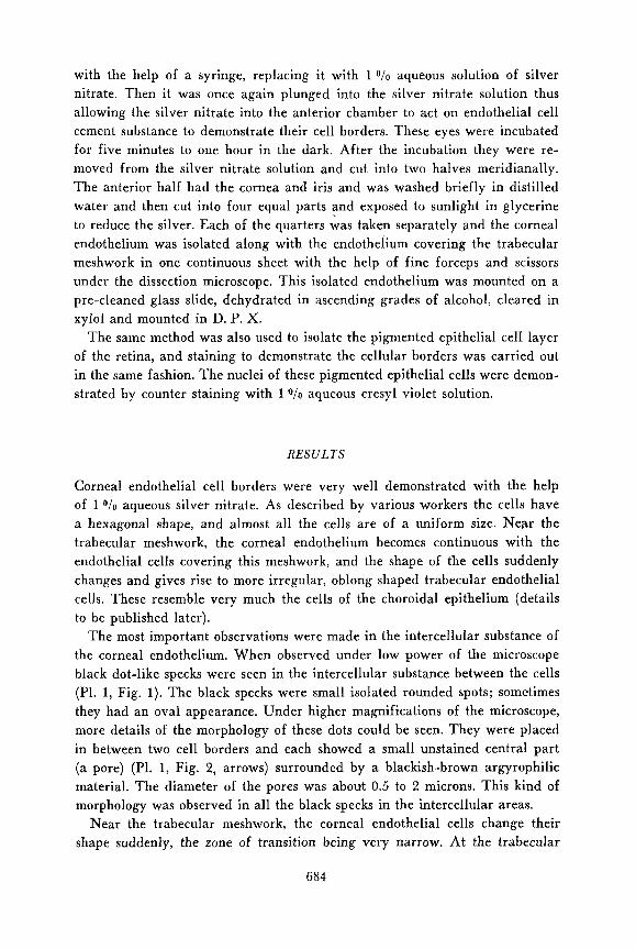
The most important observations were made in the intercellular substance of the corneal endothelium. When observed under low power of the microscope black dot-like specks were seen in the intercellular substance between the cells (Pl. 1, Fig. 1) . The black specks were small isolated rounded spots; sometimes they had an oval appearance. Under higher magnifications of the microscope, more details of the morphology of these dots could be seen. They were placed in between two cell borders and each showed a small unstained central part (a pore) (Pl. 1, Fig. 2, arrows) surrounded by a blackish-brown argyrophilic material. The diameter of the pores was about 0.5 to 2 microns. This kind of morphology was observed in all the black specks in the intercellular areas.
Near the trabecular meshwork, the corneal endothelial cells change their shape suddenly, the zone of transition being very narrow. At the trabecular
684
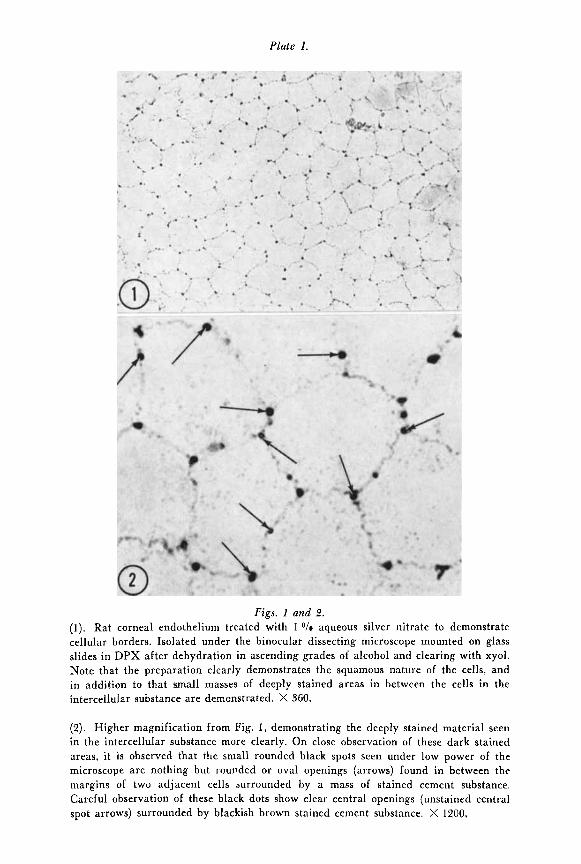
Plate 1.
Figs. 1 and 2. (1). Rat corneal endothelium treated with 1 O/o aqueous silver nitrate to demonstrate cellular borders. Isolated under the binocular dissecting microscope mounted on glass slides in DPX after dehydration in ascending grades of alcohol and clearing with xyol. Note that the preparation clearly demonstrates the squamous nature of the cells, and in addition to that small masses of deeply stained areas in between the cells in the intercellular substance are demonstrated. X 560.
(2). Higher magnification from Fig. 1 , demonstrating the deeply stained material seen in the intercellular substance more clearly. On close observation of these dark stained areas, it is observed that the small rounded black spots seen under low power of the microscope are nothing but rounded or oval openings (arrows) found in between the margins of two adjacent cells surrounded by a mass of stained cement substance. Careful observation of these black dots show clear central openings (unstained central spot arrows) surrounded by blackish brown stained cement substance. X 1200,
Plate II.
Figs . 3 and 4. (3). Endothelium covering the trahecular meshwork in rat, isolated along with the corneal endothelium in continuity under the dissection microscope. Stained with 1 O/n
aqueous silver nitrate. Note that at the zone of transition the shape of the cell changes and large masses of argyrophilic material are seen surrounding minute pores in between the cells (arrows). Some of the pores (arrow = p) surrounded by cement substance can be seen even in this low power photomicrograph. This fact is very well seen under high magnification. X 360.
(4). White rabbit eye. Pigment epithelium from the retina, isolated under the dissection microscope. Stained with 1 O i o aqueous silver nitrate and cresyl violet to demonstrate cellular borders and nuclei. Note that this does not demonstrate any areas of pores in between the cells as seen in the corneal endothelium. Compare this picture with Figs. 1 and 2 of Plate I. X 1200.

meshwork the cells become very irregular in shape and we find large irregular- ly shaped black spots in the intercellular substance between the cells (Pl. 11, Fig. 3, arrows). Here too on close observation small rounded pores are seen in the center of each high area of black-stained material (Plate 11, Fig. 3, arrow P). These pores were very well seen under the high power of the microscope.
The pigmented epithelium of the retina did not demonstrate any such ar- gyrophilic pores (Plate 11, Fig. 4), but the intercellular substance was very well stained with the silver nitrate technique, and the nuclei of the pigment cells were also clearly shown. I t was not uncommon to see more than one nucleus in these pigment cells:
DISCUSSION
The description of the size and shape of the corneal endothelium is in agree- ment with the findings of other histologists. Vrabec (20, 21), in his studies on cornea and trabecular meshwork said that the corneal endothelium intercellular substance appeared as coarse granular lines, which broaden as they approach the periphery of the cornea. H e also described the presence of pores sur- rounded by masses of cement substance in the trabecular meshwork. Our findings in the trabecular meshwork coincide with that of Vrabec but in addition densely stained spots of varying size were seen disposed irregularly between the cells throughout the whole endothelial areas in the mounts of cornea.
The presence of definite spaces in between the stromal end of the endo- thelial cells is noted in electron microscopic studies of the corneal endothelium by various authors (3, 5 , 6, 7 , 19), but they have found the penetration of this space through to the aqueous to be blocked by terminal bars. Our study sug- gests that the terminal bars do not extend completely across the apex of the intercellular junctions, but that they are interrupted from time to time by pores which give continuity between the intercellular spaces, described elec- tronmicroscopically, and aqueous humour.
The only possible way to demonstrate these pores electronmicroscopically would be to study serial sections of the corneal endothelium. Our results clearly indicate that there is a direct communication from the anterior chamber through these intercellular pores into the intercellular spaces, thus bringing the aqueous humour along with its contained nutrients in contact with the Descemet’s membrane, corneal stroma and corneal epithelium.
There is no direct experimental evidence as to the specific function of these intercellular pores, although the suggestion made above appears to be a logical deduction especially in view of the size (0.5 to 2 microns in diameter) of the pores. Extensive work on the function of the epithelium and endothelium of the cornea has been carried out with respect to the transfer of ions and water
685
across these membranes. The presence of these pores in the endothelium sug- gests a route for free passage of water and dissolved solutes across this par- ticular membrane.
It is important to remember that the cornea is shown to depend on aqueous humour for its nutrition, and excretes most of the waste into it (Pirie and Van Heyningen, 8). I t has also been shown that corneal grafts remain viable with half of the cornea replaced in which the donor cornea is connected with the recipients by only a clot of fibrinous material for two to three days after operation, indicating that aqueous humour nourishes the transplanted cornea.
Gunderson (4) has shown by autotransplantation of the cornea into the anterior chamber that the corneal tissue, other than the corneal epithelium can live almost unaltered in aqueous humour for a long time. The stroma did not show any change a t all. Descemet’s membrane remained as it was. The endothelium was living and healthy and in addition proliferated and grew around the implant. The results of this paper support the conception that nutrients from the aqueous humour reach and nourish the corneal stroma probably through the intracellular pores described in this paper.
Cogan and Kinsey (I , 2) demonstrated that a net transfer of water through the excised cornea by >>osmosis<< can be demonstrated in the posterior to the anterior direction, whereas this was not possible in a reverse direction (anterior to posterior) under similar conditions. They also showed that heavy water was transferred through the excised cornea in both directions by diffusion but the epithelium somewhat decreased the rate of diffusion, these results might be expected in view of the pores in the endothelium.
Recently much light has been thrown on the functional aspects of the structure of the corneal endothelium largely due to the work of Donn et al. (3) and Kaye (7). Donn et al. (3), in their electron microscope studies on pino- cytosis in the corneal endothelium by using thorium dioxide as marker sub- stance, has shown the marker substance in the intercellular space. They also pointed out that the thorium dioxide containing vesicles which were pinocytosed by the corneal endothelium carry the marker around the terminal bar and empty into the intercellular space.
They concluded that the movement of the ions across this membrane is pre- dominantly by diffusion through the intercellular space. Kaye ( 7 ) , in his elec- tron microscope studies on the rabbit and frog corneal endothelium by using colloidal marker substances showed that there are two separate pathways across the cornea. Transport into the cornea across the endothelium occurs via the intercellular spaces, whereas the marker substance placed intrastromally goes out of the cornea by means of intracellular mechanisms, and very rarely the tracer material is found in the intercellular space. On this basis he supposed that, as the cornea mostly derives its nutrients from the aqueous humour and excretes to the aqueous humour, the inward fluid movement would be non-
686
selective and hydrostatic pressure of the aqueous humour containing nutrients would be sufficient to ndrive it across the endothelium<<; in actual fact the inward fluid movement probably takes place through the pores we have described, whereas removal of waste being a more selective phenomenon pre- sumably is an active process by the endothelial cells.
These findings of various workers as well as our light microscope findings suggest very strongly that the intercellular pores, convey nutritive material and fluid from the aqueous to the cornea. The pigment epithelium of the retina does not show pores and presumably therefore such a mechanism for transfer of solutes does not occur in the retina.
S U M M A R Y
Light microscopically the existence of pores has been demonstrated in corneal endothelium. They are found in the intercellular space in between two lateral cellular borders. Their shape and size (0.5 to 2 microns in diameter) are variable. W e think that these pores are very important in supplying nutrition to the cornea from aqueous humour without which proper nutrition might not reach the cornea. Such pores are also shown in the endothelial cell layer covering the trabecular meshwork. They probably play a role in regulating the aqueous flow.
Pigmented epithelial cells do not demonstrate any such pores indicating the importance of pores in the endothelial cells of the cornea.
A C K N O W L E D G E M E N T S
This work was supported by grant NB-01914 from the National Institute of Neuro- logical Diseases and Blindness.
REFERENCES
1. Cogan, D. A. and Kinsey, V . E.: A.M. A. Arch. Ophthal. 27: 466, 1942. 2. Cogan, D. A. and Kinsey, V . E.: A.M. A. Arch. Ophthal. 28: 661, 1942. 3. Donn, A., Kaye, G. I . , Mallett, N . M. and Pappas, G. D.: A.M. A. Arch. Ophthal.
4. Gunderson, T.: A.M. A. Arch. Ophthal. 20: 645, 1938. 5. Kaye, G. I. and Palpas, G. D.: J. Cell B i d . 12: 457, 1962. 6. Kaye, G. I., Pappas, G. D., Donn, A . and Mallett, N.: J. Cell Biol. 12: 481, 1962. 7. Kaye, G. I.: J. Cell Biol. 15: 241, 1962. 8. Pirie, A. and van den Heyningen, R.: Biochemistry of the Eye, Blackwell, Oxford,
9. Shanthaveerappa, T . R. and Bourne, G. H.: J. Cell Biol. 14: 343, 1962. 10. Shanthaveerappa, T. R. and Boiirne, G . H.: J. Anat. Lond. 96: 527, 1962.
66: 835, 1961.
1956.
68 7
11. Shanthaveerafifia, T. R. and Bourne, G. H.: Amer. J. Anat. 112: 97, 1963. 12. Shanthaveerappa, T. R. and Bourne, G. H.: Nature 197: 702, 1963. IS. Shanthaveerappa, T . R. and Bourne, G. H.: Nature 198: 607, 1963. 14. Shanthaveerappa, T. R. and Bourne, G. H.: Acta Anat. in press, 1963. 15. Shanthaveerappa, T. R. and Bourne, G. H.: Z . Zellforsch., in press, 1963. 16. Shanthaveerappa, T. R. and Bourne, G. H.: Nature 199, 577, 1963. 17. Shanthaveerappa, T. R. and Bourne, G. H.: In preparation, 1963. 18. Shanthaveerappa, T . R., Hope, J . and Bourne, G. H.: Acta Anat. 52: 193, 1963. 19. Speakman, /. S.: A.M. A. Arch. Ophthalm. 62: 748, 1959. 20. Vrabec, F.: Brit. J. Ophthal. 42: 529, 1958. 21. Vrabec, F.: Brit. J. Ophthal. 42: 667, 1958.
688